anterior supine intermuscular tha surgical technique · 2020-01-10 · anterior supine...
TRANSCRIPT

Surgical Technique
Anterior Supine Intermuscular THA
Over 1 million times per year, Biomet helps one surgeon
provide personalized care to one patient.
The science and art of medical care is to provide the right
solution for each individual patient. This requires clinical
mastery, a human connection between the surgeon and the
patient, and the right tools for each situation.
At Biomet, we strive to view our work through the eyes of
one surgeon and one patient. We treat every solution we
provide as if it’s meant for a family member.
Our approach to innovation creates real solutions that assist
each surgeon in the delivery of durable personalized care
to each patient, whether that solution requires a minimally
invasive surgical technique, advanced biomaterials or a
patient-matched implant.
When one surgeon connects with one patient to provide
personalized care, the promise of medicine is fulfilled.
One Surgeon. One Patient.®

1
Anterior Supine Intermuscular THA
This brochure describes the surgical technique and postoperative care protocol used by Erik De Witte, M.D.; Roger H. Emerson, Jr., M.D.; Edward J. Stolarski, M.D.; Michael Pretterklieber, M.D.; W. Vincent Burke, M.D.; Hari P. Bezwada, M.D. and Michael A. Wilmink, M.D. Biomet does not practice medicine and does not recommend this or any other surgical technique for use on a specific patient. The surgeon who performs any implant procedure is responsible for determining and using the appropriate implants and techniques for implanting the prosthesis in each individual patient. This technique can be conducted with various femoral stems.
Figure 1
Surgical PlanningThe patient selected for anterior surgery should be evaluated to ascertain that the hip can be adequately reconstructed anteriorly and that there will be no need to augment the posterior acetabulum. The skin on the front of the hip must be normal in appearance without any maceration.
The radiographs should be templated to suggest the likely implant size and orientation. The level of the hip center and position of the femoral osteotomy from the tip of the greater trochanter or lesser trochanter should be determined.
Note: The Anterior Supine Intermuscular (ASI) surgical technique may be performed on a standard operating room (OR) table or special fracture table. The following technique is for use with a standard OR table.
Patient PositioningPosition the patient supine (Figure 1) on a fluoroscopy capable table, with the fluoroscopy machine in the room.
•Position the Anterior Superior Illiac Spine (ASIS) atthe level of the break in the table. This will permit appropriate motion of the femur as the table is extended.
•TheOR tableneeds tobeable toextendat thehipand the hip must be positioned to permit fluoroscopy views of both hips and the obturator foramen.
•Checkleglengthsinthesupinepositionandcorrelatewith the hip radiographs for later reference.
Anterior Supine Intermuscular THA
2
Patient PreparationPrepare both legs and drape each free to permit crossing the operative leg underneath the nonoperative side. The entire iliac crest should be included in the operative field to permit an extensile exposure if needed. The nonoperative leg should be prepped from the toes to the groin, and then draped to permit full movement of the leg. The draping could be either with stockingette material alone or in combination with adhesive plastic draping for a more secure seal. Ipsilateral side is prepped from the midline above the ASIS to mid-thigh.
•Alsopreparetheentirebuttockontheoperativesidein case of the need to use a counter incision in the buttock. This is rarely required but may be needed to pass a reamer into the femoral canal.
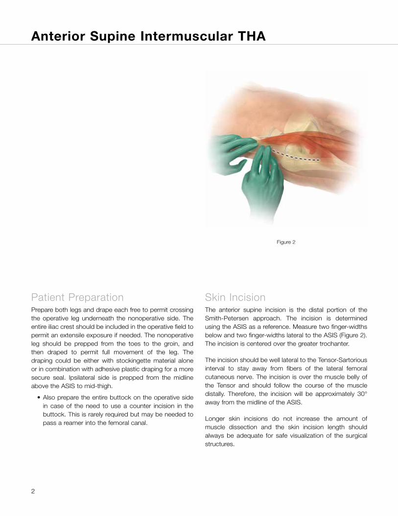
Figure 2
Skin IncisionThe anterior supine incision is the distal portion of the Smith-Petersen approach. The incision is determined using the ASIS as a reference. Measure two finger-widths below and two finger-widths lateral to the ASIS (Figure 2). The incision is centered over the greater trochanter.
The incision should be well lateral to the Tensor-Sartorious interval to stay away from fibers of the lateral femoral cutaneous nerve. The incision is over the muscle belly of the Tensor and should follow the course of the muscle distally. Therefore, the incision will be approximately 30° away from the midline of the ASIS.
Longer skin incisions do not increase the amount of muscle dissection and the skin incision length should always be adequate for safe visualization of the surgical structures.
3
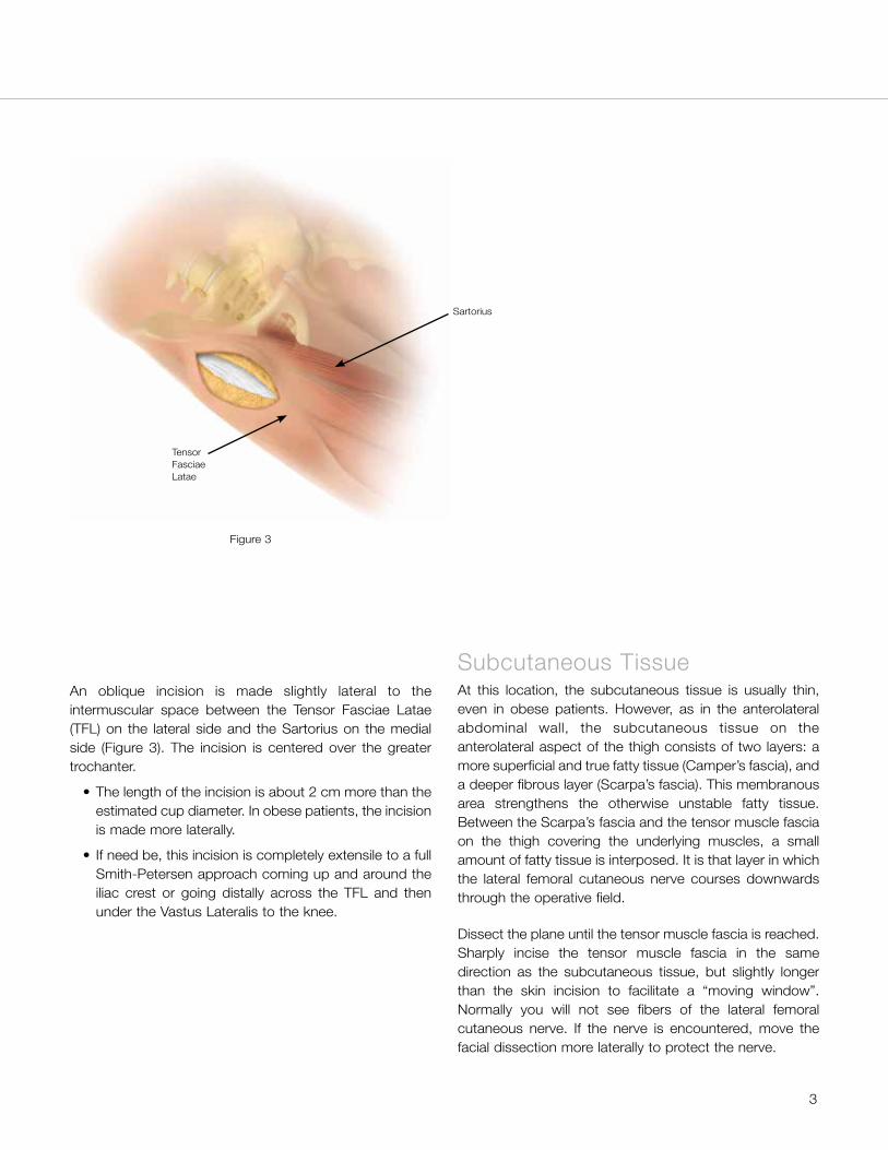
Figure 3
An oblique incision is made slightly lateral to the intermuscular space between the Tensor Fasciae Latae (TFL) on the lateral side and the Sartorius on the medial side (Figure 3). The incision is centered over the greater trochanter.
•Thelengthoftheincisionisabout2cmmorethantheestimated cup diameter. In obese patients, the incision is made more laterally.
• Ifneedbe,thisincisioniscompletelyextensiletoafullSmith-Petersen approach coming up and around the iliac crest or going distally across the TFL and then under the Vastus Lateralis to the knee.
Subcutaneous TissueAt this location, the subcutaneous tissue is usually thin, even in obese patients. However, as in the anterolateral abdominal wall, the subcutaneous tissue on the anterolateral aspect of the thigh consists of two layers: a moresuperficialandtruefattytissue(Camper’sfascia),anda deeper fibrous layer (Scarpa’s fascia). This membranous area strengthens the otherwise unstable fatty tissue. Between the Scarpa’s fascia and the tensor muscle fascia on the thigh covering the underlying muscles, a small amount of fatty tissue is interposed. It is that layer in which the lateral femoral cutaneous nerve courses downwards through the operative field.
Dissect the plane until the tensor muscle fascia is reached. Sharply incise the tensor muscle fascia in the same direction as the subcutaneous tissue, but slightly longer than the skin incision to facilitate a “moving window”. Normally you will not see fibers of the lateral femoral cutaneous nerve. If the nerve is encountered, move the facial dissection more laterally to protect the nerve.
Sartorius
Tensor Fasciae Latae
Anterior Supine Intermuscular THA
4
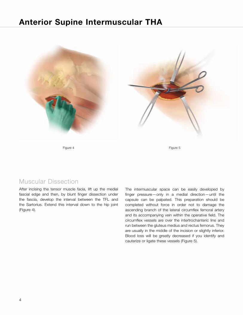
Muscular DissectionAfter incising the tensor muscle facia, lift up the medial fascial edge and then, by blunt finger dissection under the fascia, develop the interval between the TFL and the Sartorius. Extend this interval down to the hip joint (Figure 4).
The intermuscular space can be easily developed by finger pressure—only in a medial direction—until the capsule can be palpated. This preparation should be completed without force in order not to damage the ascending branch of the lateral circumflex femoral artery and its accompanying vein within the operative field. The circumflex vessels are over the intertrochanteric line and run between the gluteus medius and rectus femorus. They are usually in the middle of the incision or slightly inferior. Blood loss will be greatly decreased if you identify and cauterize or ligate these vessels (Figure 5).
Figure 4 Figure 5

5
Figure 7
Utilize a Cobb elevator to dissect the fibers of therectus from the underlying anterior hip capsule (Figure 6).
Figure 6
9
7
6

Anterior Supine Intermuscular THA
6
Figure 8
ExposureThroughCapsuleTo prepare for exposing the anterior capsule, place a Cobra retractor 6 superior to the lateral capsule or against the ilium to retract the abductors. Place a second retractor, a large sharp Hohmann 7 inferior to the femoral neck.
Place third retractor 9 under the rectus tendon, but on top of the anterior acetabular rim in the upper cranial quarter directed to the opposite shoulder to avoid injury to the femoral nerve and vessels (Figure 8). Identify the reflected head of the rectus and release to allow the long head of the rectus to retract medially. Beware of capsular bleeding at the inferior-medial capsule.
Carry out an anterior-superior capsulectomy. Thecapsulectomy results in excellent visualization and aids in femoral mobilization (Figure 9).
Make sure to release the anterior-superior capsule from its insertion in the piriformis fossa in order to facilitate lifting of the femur. Place the first two retractors 6 and
7 inside the capsule for protection when the osteotomy is performed (Figure 9).
Figure 9
9 9
7 7
6 6

7
Figure 11
After removing the head and neck piece, the assistant can externally rotate the leg and the surgeon can palpate the lesser trochanter to further guide the level of the final neck resection. Some anterior capsule may need to be released from the femur to facilitate this maneuver. Beware of damaging the TFL muscle when removing the neck and head.
Note: When removing the head, it may be easier to place the corkscrew prior to cutting the neck. Additionally, retraction of the leg at the ankle will aid in the removal of the femoral head.
Note: If necessary, an additional and final neck cut should be carried out at this time in order to provide as large an opening as possible for reaming the acetabulum. If there is anydoubtabouttheresidualfemoralneck,theC-armcanbe utilized to help determine the exact level of osteotomy.
Osteotomy of the FemurPerform an osteotomy at the cartilage/neck junction (Figure 10).
It may be necessary to perform two separate parallel cuts to facilitate extraction of the femoral head by first removing the wafer of cut bone. The initial osteotomy should be at the head/neck junction. The second should be 5 mm to 1cm distal to the initial osteotomy. The segment of bone can be removed with a tenaculum or threaded Steinman pinchucked in adrill handle. TheC-armcanbeutilizedto help determine the exact level of the osteotomy as planned from preoperative templating.
Place a corkscrew through the cortical side of the femoral head and spin the head to rupture the ligamentum. This will aid with dislocation (Figure 11). The ligamentum cutter (Figure 11) may be used to aid in the cutting of the ligamentum.
Figure 10
9
7
6
9
7
6
Anterior Supine Intermuscular THA
8
Figure 13
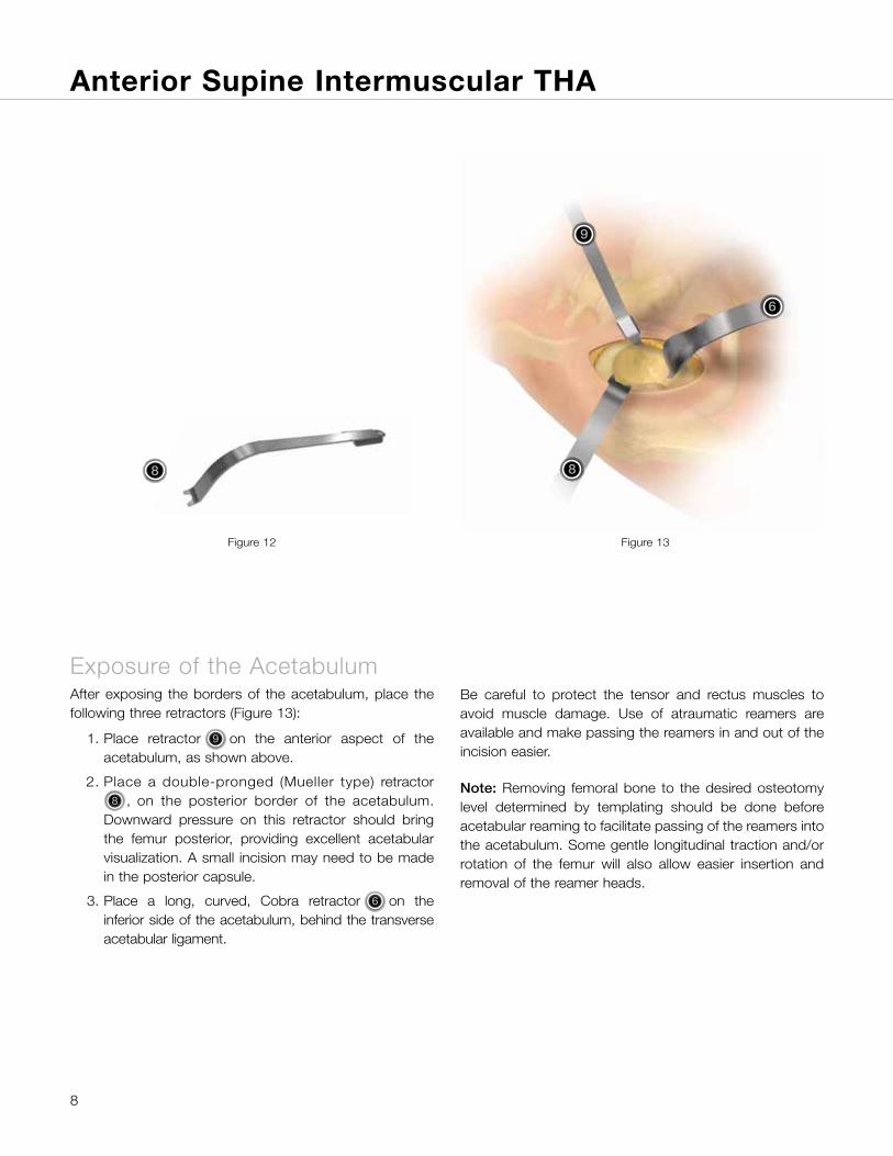
Exposure of the AcetabulumAfter exposing the borders of the acetabulum, place the following three retractors (Figure 13):
1. Place retractor 9 on the anterior aspect of the acetabulum, as shown above.
2. Place a double-pronged (Mueller type) retractor 8 , on the posterior border of the acetabulum.
Downward pressure on this retractor should bring the femur posterior, providing excellent acetabular visualization. A small incision may need to be made in the posterior capsule.
3.Place a long, curved, Cobra retractor 6 on the inferior side of the acetabulum, behind the transverse acetabular ligament.
Figure 12
Be careful to protect the tensor and rectus muscles to avoid muscle damage. Use of atraumatic reamers are available and make passing the reamers in and out of the incision easier.
Note: Removing femoral bone to the desired osteotomy level determined by templating should be done before acetabular reaming to facilitate passing of the reamers into the acetabulum. Some gentle longitudinal traction and/or rotation of the femur will also allow easier insertion and removal of the reamer heads.
9
6
88
9
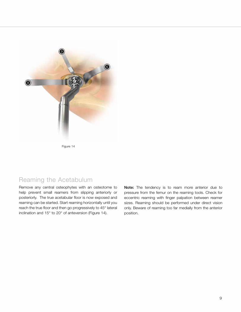
Reaming the AcetabulumRemove any central osteophytes with an osteotome to help prevent small reamers from slipping anteriorly or posteriorly. The true acetabular floor is now exposed and reaming can be started. Start reaming horizontally until you reach the true floor and then go progressively to 45° lateral inclination and 15° to 20° of anteversion (Figure 14).
Figure 14
Note: The tendency is to ream more anterior due to pressurefromthefemuronthereamingtools.Checkforeccentric reaming with finger palpation between reamer sizes. Reaming should be performed under direct vision only. Beware of reaming too far medially from the anterior position.
9
6
8
Anterior Supine Intermuscular THA
10
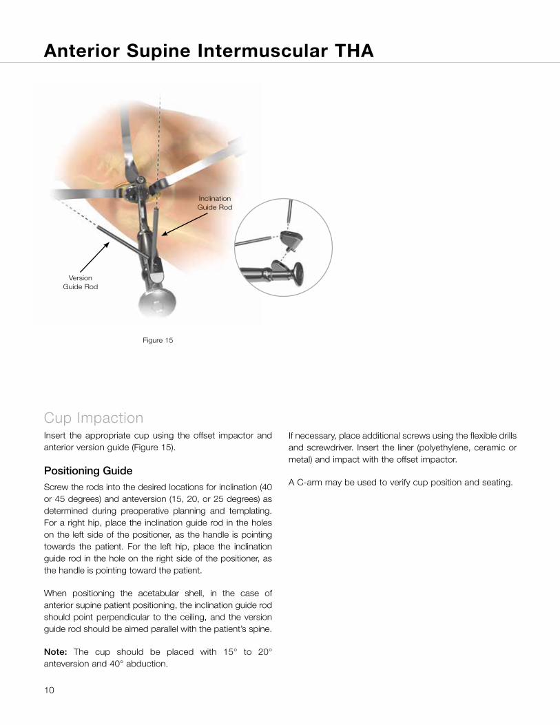
CupImpactionInsert the appropriate cup using the offset impactor and anterior version guide (Figure 15).
Positioning GuideScrew the rods into the desired locations for inclination (40 or 45 degrees) and anteversion (15, 20, or 25 degrees) as determined during preoperative planning and templating. For a right hip, place the inclination guide rod in the holes on the left side of the positioner, as the handle is pointing towards the patient. For the left hip, place the inclination guide rod in the hole on the right side of the positioner, as the handle is pointing toward the patient.
When positioning the acetabular shell, in the case of anterior supine patient positioning, the inclination guide rod should point perpendicular to the ceiling, and the version guide rod should be aimed parallel with the patient’s spine.
Note: The cup should be placed with 15° to 20° anteversion and 40° abduction.
Figure 15
If necessary, place additional screws using the flexible drills and screwdriver. Insert the liner (polyethylene, ceramic or metal) and impact with the offset impactor.
AC-armmaybeusedtoverifycuppositionandseating.
Inclination Guide Rod
Version Guide Rod
11
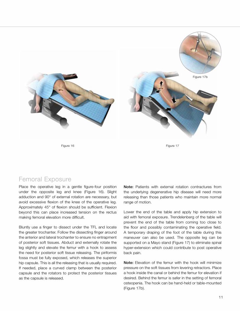
Femoral ExposurePlace the operative leg in a gentle figure-four position under the opposite leg and knee (Figure 16). Slight adduction and 90° of external rotation are necessary, but avoid excessive flexion of the knee of the operative leg. Approximately 45° of flexion should be sufficient. Flexion beyond this can place increased tension on the rectus making femoral elevation more difficult.
Bluntly use a finger to dissect under the TFL and locate the greater trochanter. Follow the dissecting finger around the anterior and lateral trochanter to ensure no entrapment of posterior soft tissues. Abduct and externally rotate the leg slightly and elevate the femur with a hook to assess the need for posterior soft tissue releasing. The piriformis fossa must be fully exposed, which releases the superior hip capsule. This is all the releasing that is usually required. If needed, place a curved clamp between the posterior capsule and the rotators to protect the posterior tissues as the capsule is released.
Note: Patients with external rotation contractures from the underlying degenerative hip disease will need more releasing than those patients who maintain more normal range of motion.
Lower the end of the table and apply hip extension to aid with femoral exposure. Trendelenberg of the table will prevent the end of the table from coming too close to the floor and possibly contaminating the operative field. A temporary draping of the foot of the table during this maneuver can also be used. The opposite leg can be supported on a Mayo stand (Figure 17) to eliminate spinal hyper-extension which could contribute to post operative back pain.
Note: Elevation of the femur with the hook will minimize pressure on the soft tissues from levering retractors. Place a hook inside the canal or behind the femur for elevation if desired. Behind the femur is safer in the setting of femoral osteopenia. The hook can be hand-held or table-mounted (Figure 17b).
Figure 16 Figure 17
Figure 17b
Anterior Supine Intermuscular THA
12
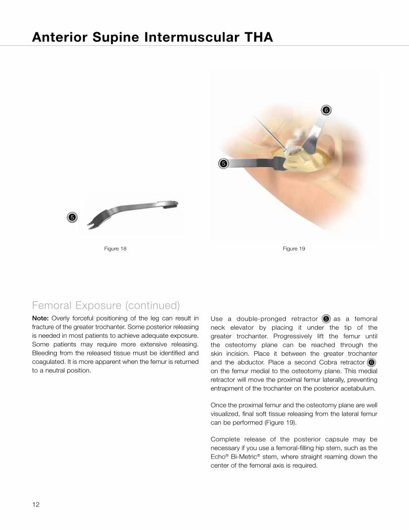
Femoral Exposure (continued)Note: Overly forceful positioning of the leg can result in fracture of the greater trochanter. Some posterior releasing is needed in most patients to achieve adequate exposure. Some patients may require more extensive releasing. Bleeding from the released tissue must be identified and coagulated. It is more apparent when the femur is returned to a neutral position.
Figure 18 Figure 19
Use a double-pronged retractor 5 as a femoral neck elevator by placing it under the tip of the greater trochanter. Progressively lift the femur until the osteotomy plane can be reached through the skin incision. Place it between the greater trochanter and the abductor. Place a second Cobra retractor 6
on the femur medial to the osteotomy plane. This medial retractor will move the proximal femur laterally, preventing entrapment of the trochanter on the posterior acetabulum.
Once the proximal femur and the osteotomy plane are well visualized, final soft tissue releasing from the lateral femur can be performed (Figure 19).
Complete release of the posterior capsule may benecessary if you use a femoral-filling hip stem, such as the Echo® Bi-Metric® stem, where straight reaming down the center of the femoral axis is required.
5
5
6

13
Figure 20 Figure 21
FemoralCanalPreparationOpen the femoral canal with an offset box osteotome, angled gouge or high speed burr (Figure 20).
Note: Femoral bone preparation should not begin until exposure is adequate. Bleeding may be produced by extensive soft tissue releasing. Pre-treatment of the posterior capsule with cautery can aid in hemostasis. If excessive bleeding is encountered from the released capsule, packing off the area and waiting a few minutes will usually produce hemostasis. The addition of hemostatic agents will aid in this technique.
Use an offset canal finder before using a starter reamer or initial broach (Figure 21). Take care to ensure appropriate alignment within the femoral canal. Impaction of a vertical broach handle (excessive varus stem position) can lead to perforation of the lateral cortex.
6 6
5 5
Anterior Supine Intermuscular THA
14
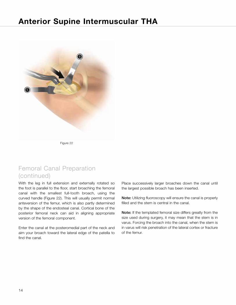
FemoralCanalPreparation (continued)With the leg in full extension and externally rotated so the foot is parallel to the floor, start broaching the femoral canal with the smallest full-tooth broach, using the curved handle (Figure 22). This will usually permit normal anteversion of the femur, which is also partly determined bytheshapeoftheendostealcanal.Corticalboneoftheposterior femoral neck can aid in aligning appropriate version of the femoral component.
Enter the canal at the posteromedial part of the neck and aim your broach toward the lateral edge of the patella to find the canal.
Figure 22
Place successively larger broaches down the canal until the largest possible broach has been inserted.
Note: Utilizing fluoroscopy will ensure the canal is properly filled and the stem is central in the canal.
Note: If the templated femoral size differs greatly from the size used during surgery, it may mean that the stem is in varus. Forcing the broach into the canal, when the stem is in varus will risk penetration of the lateral cortex or fracture of the femur.
6
5
15
Figure 24
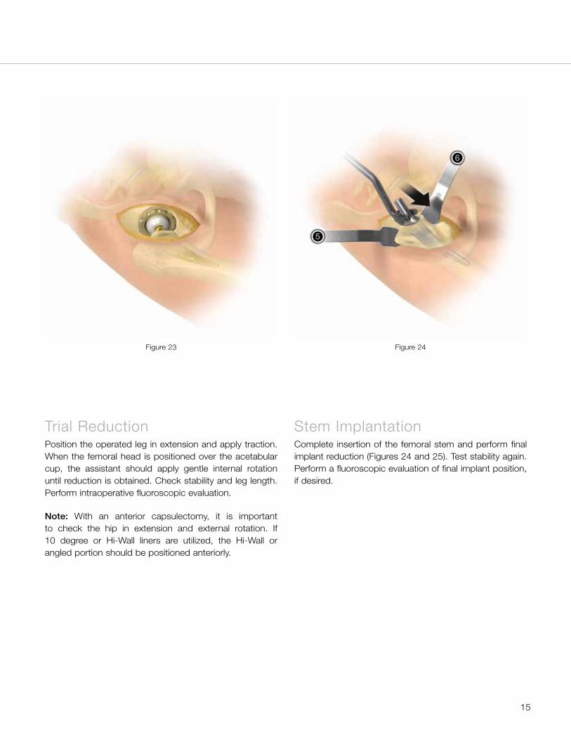
Stem Implantation Complete insertionofthefemoralstemandperformfinalimplant reduction (Figures 24 and 25). Test stability again. Perform a fluoroscopic evaluation of final implant position, if desired.
Trial ReductionPosition the operated leg in extension and apply traction. When the femoral head is positioned over the acetabular cup, the assistant should apply gentle internal rotation untilreductionisobtained.Checkstabilityandleglength.Perform intraoperative fluoroscopic evaluation.
Note: With an anterior capsulectomy, it is important to check the hip in extension and external rotation. If 10 degree or Hi-Wall liners are utilized, the Hi-Wall or angled portion should be positioned anteriorly.
Figure 23
6
5
Anterior Supine Intermuscular THA
16
Figure 25
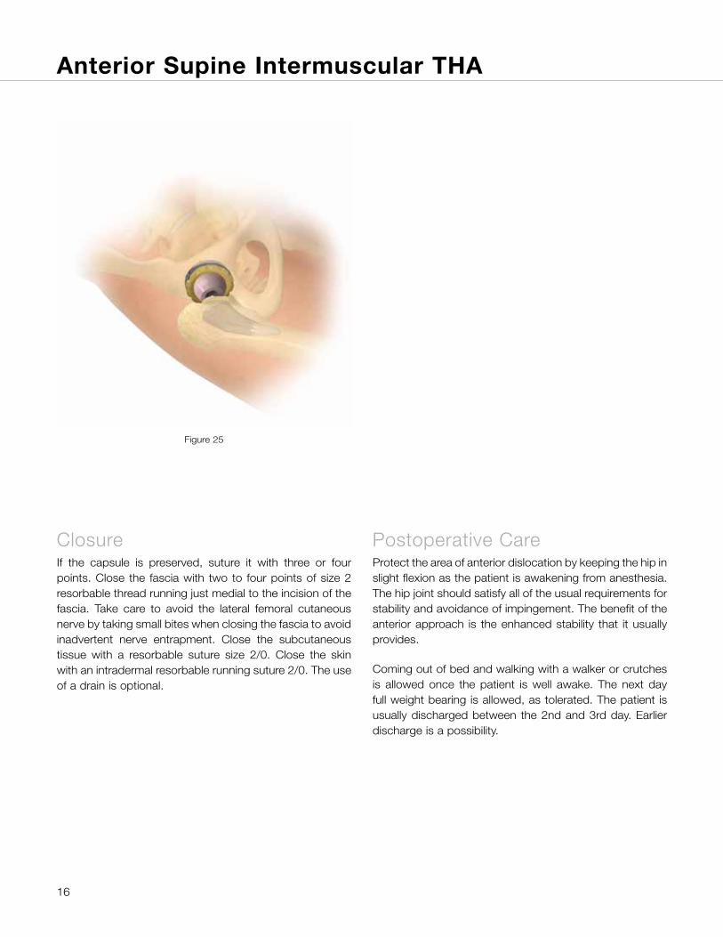
ClosureIf the capsule is preserved, suture it with three or four points.Close the fasciawith twoto fourpointsofsize2resorbable thread running just medial to the incision of the fascia. Take care to avoid the lateral femoral cutaneous nerve by taking small bites when closing the fascia to avoid inadvertent nerve entrapment. Close the subcutaneoustissue with a resorbable suture size 2/0. Close the skinwith an intradermal resorbable running suture 2/0. The use of a drain is optional.
PostoperativeCareProtect the area of anterior dislocation by keeping the hip in slight flexion as the patient is awakening from anesthesia. The hip joint should satisfy all of the usual requirements for stability and avoidance of impingement. The benefit of the anterior approach is the enhanced stability that it usually provides.
Comingoutofbedandwalkingwithawalkerorcrutchesis allowed once the patient is well awake. The next day full weight bearing is allowed, as tolerated. The patient is usually discharged between the 2nd and 3rd day. Earlier discharge is a possibility.
17
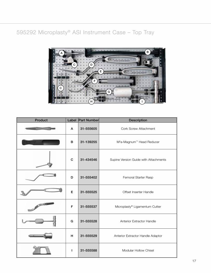
595292 Microplasty®ASIInstrumentCase–TopTray
Product Label Part Number Description
A 31-555605 CorkScrewAttachment
B 31-139255 M2a-Magnum™ Head Reducer
C 31-434546 Supine Version Guide with Attachments
D 31-555402 Femoral Starter Rasp
E 31-555525 Offset Inserter Handle
F 31-555537 Microplasty®LigamentumCutter
G 31-555528 Anterior Extractor Handle
H 31-555529 Anterior Extractor Handle Adaptor
I 31-555588 ModularHollowChisel
B
G
A
C
C D
H
E
F
I
Anterior Supine Intermuscular THA
18
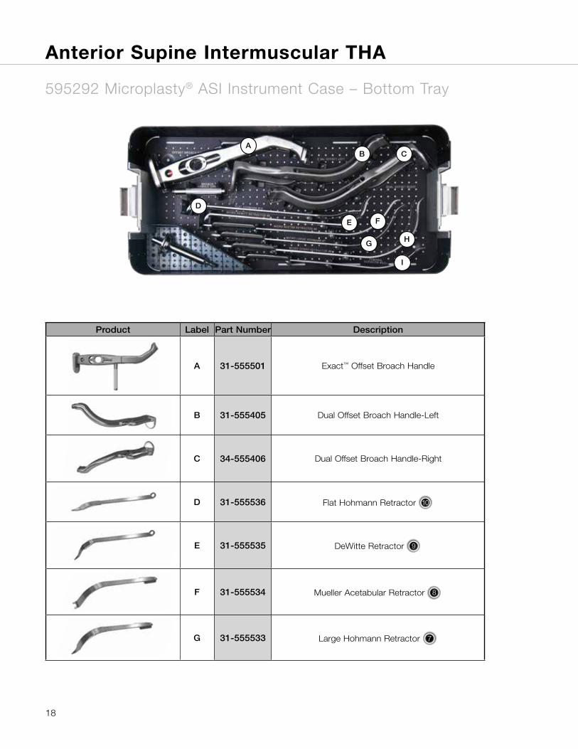
595292 Microplasty®ASIInstrumentCase–BottomTray
Product Label Part Number Description
A 31-555501 Exact™ Offset Broach Handle
B 31-555405 Dual Offset Broach Handle-Left
C 34-555406 Dual Offset Broach Handle-Right
D 31-555536 Flat Hohmann Retractor 10
E 31-555535 DeWitte Retractor 9
F 31-555534 Mueller Acetabular Retractor 8
G 31-555533 Large Hohmann Retractor 7
B
G
AC
D
H
E F
I

19
Product Label Part Number Description
H 31-555532 CobraRetractor 6
I 31-555531 Femoral Elevator 5
Additional Instrumentation Options (Not Shown)
31-555594 Modified #5 ASI Retractor
20
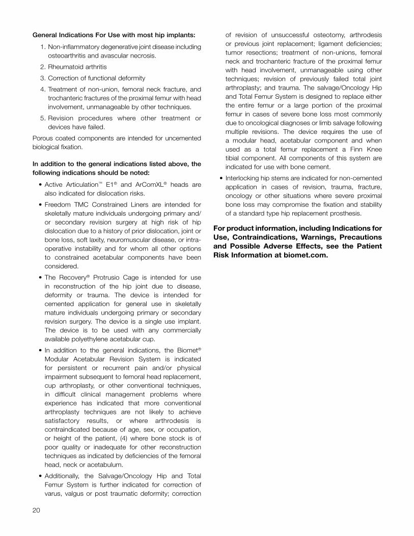
General Indications For Use with most hip implants:
1. Non-inflammatory degenerative joint disease including osteoarthritis and avascular necrosis.
2. Rheumatoid arthritis
3.Correctionoffunctionaldeformity
4. Treatment of non-union, femoral neck fracture, and trochanteric fractures of the proximal femur with head involvement, unmanageable by other techniques.
5. Revision procedures where other treatment or devices have failed.
Porous coated components are intended for uncemented biological fixation.
In addition to the general indications listed above, the following indications should be noted:
•Active Articulation™ E1® and ArComXL® heads are also indicated for dislocation risks.
•Freedom TMC Constrained Liners are intended forskeletally mature individuals undergoing primary and/or secondary revision surgery at high risk of hip dislocation due to a history of prior dislocation, joint or bone loss, soft laxity, neuromuscular disease, or intra-operative instability and for whom all other options to constrained acetabular components have been considered.
•The Recovery® Protrusio Cage is intended for usein reconstruction of the hip joint due to disease, deformity or trauma. The device is intended for cemented application for general use in skeletally mature individuals undergoing primary or secondary revision surgery. The device is a single use implant. The device is to be used with any commercially available polyethylene acetabular cup.
• In addition to the general indications, the Biomet® Modular Acetabular Revision System is indicated for persistent or recurrent pain and/or physical impairment subsequent to femoral head replacement, cup arthroplasty, or other conventional techniques, in difficult clinical management problems where experience has indicated that more conventional arthroplasty techniques are not likely to achieve satisfactory results, or where arthrodesis is contraindicated because of age, sex, or occupation, or height of the patient, (4) where bone stock is of poor quality or inadequate for other reconstruction techniques as indicated by deficiencies of the femoral head, neck or acetabulum.
•Additionally, the Salvage/Oncology Hip and TotalFemur System is further indicated for correction of varus, valgus or post traumatic deformity; correction
of revision of unsuccessful osteotomy, arthrodesis or previous joint replacement; ligament deficiencies; tumor resections; treatment of non-unions, femoral neck and trochanteric fracture of the proximal femur with head involvement, unmanageable using other techniques; revision of previously failed total joint arthroplasty; and trauma. The salvage/Oncology Hip and Total Femur System is designed to replace either the entire femur or a large portion of the proximal femur in cases of severe bone loss most commonly due to oncological diagnoses or limb salvage following multiple revisions. The device requires the use of a modular head, acetabular component and when used as a total femur replacement a Finn Knee tibial component. All components of this system are indicated for use with bone cement.
• Interlockinghipstemsareindicatedfornon-cementedapplication in cases of revision, trauma, fracture, oncology or other situations where severe proximal bone loss may compromise the fixation and stability of a standard type hip replacement prosthesis.
For product information, including Indications for Use, Contraindications, Warnings, Precautions and Possible Adverse Effects, see the Patient Risk Information at biomet.com.

Notes
All trademarks herein are the property of Biomet, Inc. or its subsidiaries unless otherwise indicated.
This material is intended for the sole use and benefit of the Biomet sales force and physicians. It is not to be redistributed, duplicated or disclosed without the express written consent of Biomet.
For product information, including Indications for Use, Contraindications, Warnings, Precautions and Possible Adverse Effects, see the Patient Risk Information at biomet.com.
P.O.Box587,Warsaw,IN46581-0587•800.348.9500x1501 ©2011,2012BiometOrthopedics•biomet.com
FormNo.BMET0499.0•REV0112