anosognosia for hemiplegia - diva portal428376/fulltext01.pdf · the confabulation theory 14 the...
TRANSCRIPT

AHP: THEORETICAL, CLINICAL, AND NEURAL ASPECTS 1
ANOSOGNOSIA FOR HEMIPLEGIA Theoretical, Clinical, and Neural Aspects
Bachelor Degree Project in Cognitive Neuroscience
15 ECTS
Spring term 2011
Joel Gerafi
Supervisor: Judith Annett
Examiner: Pilleriin Sikka

AHP: THEORETICAL, CLINICAL, AND NEURAL ASPECTS 2
Anosognosia for Hemiplegia: Theoretical, Clinical, and Neural Aspects
Submitted by Joel Gerafi to the University of Skövde as a final year project towards the
degree of B.Sc. in the School of Humanities and Informatics. The project has been supervised
by Judith Annett.
10/6/2011
I hereby certify that all material in this final year project which is not my own work has been
identified and that no work is included for which a degree has already been conferred on me.
Signature: ___________________________________________
AHP: THEORETICAL, CLINICAL, AND NEURAL ASPECTS 3
Abstract
Anosognosia for hemiplegia (AHP) is relatively common among patients who suffer from a
stroke. It is characterized as a denial of bodily paralysis and the complexity of studying it is
evident. Anosognosia is a neuropsychological deficit of self-awareness and most frequently
associated with both cortical and subcortical lesions distributed within the right hemisphere,
resulting in a left hemiplegia. The purpose of this review is to provide a comprehensive
overview of AHP by presenting theoretical, clinical, and neural aspects. Different diagnostic
procedures have attempted to clinically evaluate patients with AHP. The timing of assessment
and the characteristic differences between these procedures are crucial factors to consider.
Various theories regarding the underlying mechanisms of AHP are also discussed in this
review, suggesting the cause of AHP from different perspectives. In order to confirm or
disconfirm these theories, several studies are presented concerning the neural aspects, such as
the frequency, related disorders, and anatomical correlates of AHP.
Keywords: AHP, anosognosia, hemiplegia, self-awareness, stroke
AHP: THEORETICAL, CLINICAL, AND NEURAL ASPECTS 4
Table of Contents
Abstract 3
Introduction 6
Clinical Evaluation of Anosognosia 7
Diagnostic Procedures 8
Anosognosia Questionnaires 8
Diagnostic Confounders 9
Theories about the Underlying Mechanisms of Anosognosia 10
Motivational Theories 11
The Psychodynamic Theory 11
Cognitive Theories 12
The Disconnection Theory 12
The Discovery Theory 13
The Feed-Forward Theory 13
The Confabulation Theory 14
The Feedback Theory 15
The Deficient Affective Drive Theory 16
The Two-Factor Theory of Delusions 17
Frequency of Anosognosia for Hemiplegia 18
Related to the Timing of Assessment 19
Related Neurological and Neuropsychological Disorders 20
Anosognosia and Unilateral Neglect 21
Related Disorders of Bodily Awareness 22
Neural Aspects of Anosognosia for Hemiplegia 23
The Role of Insula Cortex 25

AHP: THEORETICAL, CLINICAL, AND NEURAL ASPECTS 5
Motor Awareness 26
Discussion 27
Conclusion 31
References 32

AHP: THEORETICAL, CLINICAL, AND NEURAL ASPECTS 6
Introduction
Anosognosia is generally known as a neuropsychological deficit of self-awareness. The
term anosognosia was first introduced by Babinski (1914) when his patients indicated an
apparent loss of awareness for their hemiplegic disorder (as cited in Heilman, Barret, &
Adair, 1998). Several definitions of anosognosia have been presented in which they all share a
relatively equivalent meaning, even though it is described slightly differently in most
literature. A description that is particularly prominent is when “anosognosia is generally and
comprehensively defined as a disorder in which a patient, affected by a brain dysfunction,
does not recognize the presence or appreciate the severity of deficits in sensory, perceptual,
motor, affective or cognitive functioning” (Orfei et al., 2007, pp. 3075-3076).
Bisiach and Geminiani (1991) argued for the importance of being careful about obtaining a
scientific definition of anosognosia. They stated the following:
A satisfactory definition of anosognosia per se is perhaps impossible, attributable to the
fact that the term anosognosia, rather than referring to a truly distinct symptom, may be
(and has indeed been) used to denote aspects of patients’ behavior in relationship to their
illness that are heterogeneous in appearance and unlikely to depend on a specific set of
causes exclusively related to them. (p. 19)
Anosognosia has been studied primarily in stroke patients who consequentially suffer from
hemiplegic disorder. Hemiplegia is known as a patient’s inability to move his or her paretic
limb, this being due to paralysis caused by stroke. Patients with anosognosia for hemiplegia
(AHP) deny or express an unawareness of their hemiplegic disorder (Orfei et al., 2007).
Neuropsychology is a brain-related discipline which studies the structure and function of the
brain and its relation to specific psychological functions. Experimental neuropsychology is
particularly concerned with disturbances of our self-awareness as a result from brain damage
in patients. Clinical neuropsychology uses the knowledge from experimental
AHP: THEORETICAL, CLINICAL, AND NEURAL ASPECTS 7
neuropsychology in order to develop diagnostic procedures and rehabilitation programs for
these brain damaged patients (Prigatano, 2010). The study of brain-damaged patients with
anosognosia has progressed into an important field of research, since it is capable of
discovering neural components for our self-awareness. This can provide us with a deeper
understanding for and new insights about the neuropsychological mechanisms, while
simultaneously improving post-acute neuropsychological rehabilitation (Heilman et al., 1998;
Prigatano, 2010).
This review will be divided into three sections, initially describing the clinical evaluation
of AHP and different diagnostic procedures that have been developed throughout the years.
Secondly, theories regarding the underlying mechanisms of AHP will be mentioned. Thirdly,
important findings about AHP are going to be presented and discussed for an attempt to draw
inferences about different neural aspects and anatomical structures that seem to be involved. It
is therefore essential to identify a pattern of cortical, subcortical, and association areas that
have been damaged in these patients in order to attain a satisfactory account for this
phenomenon. All in all, the aim of this review is to provide an overview of AHP by
presenting theoretical, clinical, and neural aspects.
Clinical Evaluation of Anosognosia
The diagnostic procedures used to assess anosognosia in stroke patients are relatively
straightforward and simple to use within clinical practice. Instruments and questionnaires are
two other terms that will be used interchangeably, although both refer to diagnostic
procedures as well. These procedures aim primarily towards assessing the presence of the
deficit and measuring the degree of severity by comparing the objective findings from the
evaluation with the patient’s verbal responses (Starkstein, Jorge, & Robinson, 2010). This is
regularly based on meta-cognitive tasks or questionnaires of various kinds, where the patient
is required to provide a self-evaluation by reflecting upon his or her own situation (Prigatano,

AHP: THEORETICAL, CLINICAL, AND NEURAL ASPECTS 8
2010). However, the limitations of these different procedures and methodological approaches
used to diagnose this phenomenon are evident. Furthermore, the complexity of anosognosia
make it difficult to establish consistent evaluations (Starkstein et al., 2010).
Prigatano (2010) argued that structured interviews and questionnaires might not be a
comprehensive approach for representing a patient’s subjective experience, mainly due to
possible inconsistencies between what is verbally recognized and what is less consciously
believed and responsible for the patient’s behavior. Jehkonen, Laihosalo, and Kettunen (2006)
pointed out that future research should consider the importance for developing unified and
systematic methods to assess anosognosia for both verbal and non-verbal forms of the deficit.
Diagnostic Procedures
As mentioned previously, a number of instruments have been developed throughout the
years in order to find a substantial criterion for diagnosing anosognosia in stroke patients
(Orfei, Caltagirone, & Spaletta, 2009). These questionnaires require an examiner who initially
proposes general questions to the patient, concerning his or her own experience of the deficit.
Additionally, more specific questions are asked, such as concerning the patient’s motor
abilities and sense of touch. This is important in order to directly assess subjective knowledge
for the patient and the particular deficit (Vallar & Ronchi, 2006). Although there is a wide
variety of established questionnaires, there are especially some that have been frequently used
to a greater extent than others (Orfei et al., 2009).
Anosognosia questionnaires. With consideration for stroke patients, Cutting (1978)
developed one of the first instruments to diagnose anosognosia for sensory-motor deficits,
including AHP and other related anosognostic phenomena, known as abnormal attitudes
towards a weak limb (Orfei et al., 2007). This instrument is known as Cutting’s questionnaire
and includes general questions regarding the patients’ awareness of the sensory-motor deficit
and specific questions if denial was elicited on the general questions (Cutting, 1978).

AHP: THEORETICAL, CLINICAL, AND NEURAL ASPECTS 9
Approximately a decade later, another instrument was developed by Bisiach, Vallar,
Perani, Papagno, and Berti (1986), known as the Bisiach’s questionnaire. This 4-point scale
evaluates the degree of anosognosia for sensory-motor deficits by rating the severity, ranging
from mild, moderate, to severe. It assesses motor impairments, somatosensory deficits, and
visual field deficits. This questionnaire provided a better description for the presence and
severity of anosognosia in stroke patients, in contrast to Cutting’s (1978) questionnaire, which
did not manage to offer a satisfactory understanding of this phenomenon (Orfei et al., 2007).
Starkstein, Fedoroff, Price, Leiguarda, and Robinson (1992) developed the Anosognosia
Questionnaire, also including a 4-point scale with similar ratings as the one suggested by
Bisiach et al. (1986). However, it additionally required the patient to perform several motor
activities besides answering general and specific questions (Orfei et al., 2007).
Marcel, Tegnér, and Nimmo-Smith (2004) designed the Structured Awareness Interview. It
is comparable to the previous one by Starkstein et al. (1992), except that it entails a more
complex and detailed assessment procedure for the patient. As in previous questionnaires
mentioned above, this procedure by Marcel et al. (2004) also includes both general and more
specific questions about the patient’s awareness of the deficit. However, these questions are
divided into two separate stages. First, a 3-point scale measures the patient’s personal report
about the severity of his or her deficit. Then, these scores are re-classified in order to assess
the reliability of the patient’s self-perception in answering the questions. Finally, unimanual,
bimanual, and bipedal tasks are performed after the patient has given an estimation about his
or her ability to perform these specific tasks, such as jumping, tying a knot, or other activities
involving movement (Orfei et al., 2007; Starkstein et al., 2010).
Diagnostic confounders. Although these diagnostic instruments among many others
provide different techniques to assess anosognosia in stroke patients, they have intrinsic
differences regarding their complexity of assessment. This is clearly shown among the

AHP: THEORETICAL, CLINICAL, AND NEURAL ASPECTS 10
questionnaires mentioned above. In addition, there are potential confounding aspects
considered to be relevant (Starkstein et al., 2010). Pia, Neppi-Modona, Raffaella, and Berti
(2004) pointed out that the frequency of identification of anosognosia seems to depend partly
on the timing of the assessment. They indicated that the occurrence of anosognosia ranged
between 20-44%, depending on the time elapsed since sustaining the brain damage. In support
of this study, Jehkonen et al. (2006) showed that the recovery from anosognosia seems to
progress with time and is most likely to occur in the acute phase and during the first three
months after stroke. Therefore, the condition is rarely chronic or becomes permanent.
According to Pia et al. (2004), the existence of anosognosia is usually documented in
patients with right hemispheric lesions, compared to patients with lesions in the left
hemisphere. Jehkonen et al. (2006) also noted that the occurrence of anosognosia is associated
to which hemisphere has been damaged. Since the occurrence of anosognosia in patients with
damage to the right hemisphere ranged from 11-60%, while occurrence in patients with
damage to the left hemisphere ranged from 6-24%, there have been discussions about another
possible confounder that also tends to influence the frequency of anosognosia in stroke
patients (i.e., excluding patients suffering from aphasia). These patients are not capable of
providing a fully comprehensive verbal report of their deficits because of the damage in their
left hemisphere and its impact on the speech areas. Other confounders may be due to the
extent patients with dementia have been recruited to studies and the extensive preference for
only including patients with right-hemispheric lesions compared to patients with lesions to the
left hemisphere (Starkstein et al., 2010).
Theories about the Underlying Mechanisms of Anosognosia
Generally, there have been two different approaches regarding how to interpret
anosognosia and its underlying neural mechanisms. On one hand, there are motivational
theories trying to provide a reasonable explanation about anosognosia from a psychological
AHP: THEORETICAL, CLINICAL, AND NEURAL ASPECTS 11
perspective. On the other hand, there are cognitive theories that additionally emphasize the
biological component and attempt to approach the study of anosognosia by referring to
damage in specific brain areas. These cognitive theories have proposed various models that
mainly separate them into three categories; neuropsychological models, hemispheric damage
models, and intra-hemispheric localization models (Orfei et al., 2007; Prigatano, 2010). Even
though the motivational theories have given rise to intriguing proposals, they have been
gradually abandoned as the cognitive theories gathered considerably more scientific evidence
for their approach (Prigatano, 2010).
Motivational Theories
The psychodynamic theory. Motivational theories, particularly the psychodynamic
theory developed by Weinstein and Kahn (1955), have one approach concerning the
interpretation of anosognosia. This psychological approach considered anosognosia to be an
immediate defense mechanism when suffering from a serious motor impairment or disease,
thus protecting the self from overwhelming distress (Prigatano, 2010). According to this
theory, all human beings have this capability of denying disorders or illnesses and this
mechanism operates unconsciously. In providing evidence for their hypothesis, Weinstein and
Kahn (1955) conducted a study where they compared stroke patients in relation to their
personality characteristics before their stroke as possibly causing anosognosia for their
disorder or illness. The results revealed that patients who used denial as a strategy more often
in everyday life prior to their stroke were more likely to suffer from AHP as a consequence of
the stroke, compared to patients who did not use denial as frequently in their everyday life
before the stroke occurred.
Bisiach and Geminiani (1991) argued against this motivational approach and presented
several critiques against it, such as that anosognosia has frequently been associated with
damage to the right hemisphere and seems to occur during the acute phase of the disorder.

AHP: THEORETICAL, CLINICAL, AND NEURAL ASPECTS 12
Even though patients use this defense mechanism, this denial strategy should not be
considered as an influential factor related to which side of the brain becomes damaged by
stroke (Heilman et al., 1998). Additionally, there seems to be selectivity involved, where
some patients show anosognosia for some impairments while being aware of others.
Vestibular stimulation, where cold water is directly poured into the patients ear has also
shown a temporary remission of AHP, which contradicts the theory of motivated denial since
this should not affect the patients strategies and ability to deny the disorder. Altogether, these
results suggest that the motivational theories cannot provide sufficient evidence, making them
rather doubtful and unconvincing. This motivates another approach for explaining the
mechanisms of anosognosia (Bisiach & Geminiani 1991; Prigatano, 2010).
Cognitive Theories
The disconnection theory. Geschwind (1965a, 1965b) developed a disconnection
hypothesis that tried to explain anosognosia by approaching it from a more neurological
perspective. It was proposed that the reason why anosognosia appears in relation to
hemiplegia is due to a disconnection between specific areas in the two hemispheres. This
inter-hemispheric disconnection would separate speech areas located in the left hemisphere
from sensory monitors and proprioceptive information (sense of body parts in space) deriving
from the right hemisphere. Consequentially, this would produce a verbal confabulation when
the patient is asked about the hemiplegia. Since approximately 90% of the population have
their left hemisphere dominant for language, this theory might explain right left asymmetries
of the brain, except giving further knowledge about the neural mechanisms underlying
anosognosia (Heilman et al., 1998). However, Bisiach et al. (1986) pointed out that patients
with anosognosia are still able to express their awareness of hemiplegia non-verbally. In
addition, by verbally informing the patient about the deficits, it would seem peculiar to
suggest that the left hemisphere would not recognize the presence of the deficits.
AHP: THEORETICAL, CLINICAL, AND NEURAL ASPECTS 13
The discovery theory. In order to explain the pathogenesis (underlying mechanisms) of
AHP, the discovery theory offered by Levine (1990) advocated for the importance of
considering both proprioceptive loss (no sense of body part positions in space) and mental
impairment. The fundamental idea of this theory is that in contrast to sensory experience
itself, the sensory loss is not immediately phenomenal but must be discovered by the patient
suffering from AHP. This entails self-observation and personal recognition of the failure to
perform tasks involving movement of the paretic limb. If the proprioceptive input is absent,
the patient will not be able to comprehend whether the paralyzed limb has moved or not. The
actual discovery is comparatively easy for cognitively intact patients compared to patients
with cognitive or intellectual impairments after the stroke (Levine, Calvanio, & Rinn, 1991).
The result from a study by Levine et al. (1991) is consistent with the discovery theory. It
indicated that somatosensory loss and mental impairment are necessary components of AHP
but not sufficient, if unaccompanied. AHP is a failure to discover hemiplegia due to a
cognitive problem and because of the damage to the sensorimotor system of the frontal and
parietal lobes, principally of the right hemisphere. This study does not explain why AHP is
more common after lesions to the right hemisphere causing a left hemiplegia, than lesions to
the left hemisphere resulting in a right hemiplegia. However, in contrast to other theories, the
discovery theory allows the existence of AHP after lesions to the spinal cord or brachial
plexus if they are associated with somatosensory loss and if a cognitive impairment can be
identified, such as a confusional state in the patient (Levine, 1990).
The feed-forward theory. Several hypotheses that have been developed emphasized
components such as confusion, confabulation, disconnection, psychological denial and
defective feedback as possible underlying mechanisms of anosognosia. However, they have
not managed to give a comprehensive explanation of anosognosia in all patients. Heilman
(1991) proposed a feed-forward theory of anosognosia. This theory essentially implies that

AHP: THEORETICAL, CLINICAL, AND NEURAL ASPECTS 14
anosognosia is a deficit of the intentional system in formulating expectations of movement.
To clarify this, the model explains that our intentional system is activated simultaneously with
our motor system to perform a movement. A body representation about the specific
movement is continuously compared with afferent information. In patients aware of their
hemiplegia, the comparator system and body representation identify a mismatch between
expectations of movement and the actual failed performance of movement. However, in
patients with anosognosia for their hemiplegia, the damage to the intentional-preparatory
system leads to an absence of their expectations to move their paretic limb. Hence, the
comparator system fails to recognize any mismatch, resulting in the unawareness to detect
their motor or movement failure.
Heilman et al. (1998) proposed that a deficit in this intentional system occurs as a result of
lesions in dorsolateral (Brodmann’s areas (BA) 6 and 8) and medial frontal lesions
(supplementary motor area and cingulate gyrus), and with damage to the inferior parietal lobe,
the thalamus (ventrolateral, anterior lateral and medial) and the basal ganglia (striatum and
substantia nigra). Even though this theory is considerable, it does not explain why
anosognostic patients during a movement task, such as clapping their hands, could have lost
their intention to move, when in fact the unimpaired limb does move (Prigatano, 2010).
The confabulation theory. A confabulation is when a person creates spontaneous false
responses that does not relate to reality. In the context of AHP, patients produce these
confabulations or delusional false claims, caused by the neurological impairment following
the stroke. Thus, anosognosia can be regarded as a form of confabulation (Heilman et al.,
1998; Ramachandran, 1996). According to Ramachandran (1996), the primary reason why
confabulations exist from an evolutionary perspective is to create a coherent belief system and
consequentially maintain control and balance to one’s behavior. In addition, the hemispheric
specialization is essential to consider in order to understand anosognosia. The theory states
AHP: THEORETICAL, CLINICAL, AND NEURAL ASPECTS 15
that the coping strategies of left and right hemispheres are profoundly different. The role of
the left hemisphere is to produce a model and constantly attempt to maintain it. If new
information produces a threat to this maintained model, it will result in denial or
confabulation, hence stabilizing the model. The right hemisphere has another strategy of
dealing with information. It detects anomalous information if it exceeds a certain threshold
and therefore forces the left hemisphere to reconsider the maintained model. This generates a
conflict between both hemispheres due to the attempt of the left hemisphere to maintain the
original model, while the right hemisphere is simultaneously forced to modify it.
Patients suffering from anosognosia because of their damage to the right hemisphere have
lost this mechanism and fail to detect anomalous information. Thus, the left hemisphere
constantly confabulates and denies the hemiplegia without any reconsideration of the model
that would otherwise be reconstructed into a new model using the same data (Ramachandran,
1996). Even if this theory is appealing, it faces several criticisms worth mentioning.
Anosognosia has been found in patients with left hemisphere lesions, which contradicts the
proposed role of the left and right hemispheres in dealing with information. The
confabulations seem to be limited to loss of awareness of deficits or to other anosognostic
phenomena. It is also uncertain if they are produced in a tonic fashion. Additionally, the
theory does not explain the short duration of anosognosia in the majority of stroke patients
(Starkstein et al., 2010).
The feedback theory. Frith, Blakemore, and Wolpert (2000) argued that the model
developed earlier by Heilman (1991; Heilman et al., 1998) did not place any emphasis on
providing an explanation which considered the delusional aspect of anosognosia, since
patients claim to perform a movement although no movement actually occurred. Frith et al.
(2000) proposed a model describing mainly two different types of internal models, more
specifically called predictors and controllers. When a movement is performed, it is generated

AHP: THEORETICAL, CLINICAL, AND NEURAL ASPECTS 16
in the central nervous system (CNS) and a predictor evaluates the sensory consequences of
that movement. The controller estimates the relationship between the specific motor
command and the desired state required to achieve the final position. Additionally, there are
three levels of motor representations also necessary in order for the predictors and controllers
to function properly. These are the actual state, representing the actual motor commands and
the sensory feedback. The desired state refers to the instantaneous goal of the system, and the
predicted state represent the future state of the system as a result from the predictors.
A difference in any combination between the actual, desired, and predicted states will
signal an error that in turn will be adjusted by the controllers and the predictors in the system.
According to Frith et al. (2000), patients with AHP have correct representations of desired
and predicted positions of the limb. However, these patients fail to register the inconsistency
between actual and predicted sensory consequences of the movement. Thus, the predictors fail
to update their operations sufficiently. Consequentially, information derived from the sensory
feedback regarding the actual limb position of the system is not available because of the
hemiplegia caused by lesions to the right parietal lobe. In contrast to Heilman (1991; Heilman
et al., 1998) arguing for a global damage of the motor planning system with his disconnection
theory, the feedback theory by Frith et al. (2000) presented a rather distributed system of
specific components concerning our motor control (Prigatano, 2010).
The deficient affective drive theory. The majority of studies have tried to determine the
underlying neurological, cognitive and motivational aspects of anosognosia primarily by
describing correlations between this deficit and clinical elements. However, the identification
of a consistent pattern regarding brain lesion or dysfunction has not yet entirely been
presented (Vuilleumier, 2004). In order to more specifically explain anosognosia, Vuilleumier
(2004) suggested a framework that proposed various problems concerning appreciation,
belief, and check (ABC) operations that may influence this disturbed cognitive and affective

AHP: THEORETICAL, CLINICAL, AND NEURAL ASPECTS 17
judgment in anosognosia. Different changes in the ABC operations and their functions might
explain the denial for a given situation. Therefore, Vuilleumier (2000) proposed that
anosognosia might reflect a deficient affective drive to appropriately respond to uncertainties
about current cognitive abilities or bodily conditions (as cited in Vuilleumier (2004). In
support of this proposal, a study conducted by Tiedens and Linton (2001) showed that
affective signals may in fact influence cognitive processing and reasoning, thus adjusting both
behavior and beliefs according to new contingencies. Another factor that might prevent the
patient from becoming aware of the deficit is the reduced emotional impact of the perceived
failure (Vuilleumier, 2004). However, in contrast to this factor, many patients with
anosognosia show normal emotional reactions. The limitations of this theory are evident and
the meaning of affective signals and deficient affective drive requires further explanation.
Nevertheless, Vuilleumier (2004) argued for the importance of investigating specific
cognitive and affective components that can be experimentally confirmed. It is therefore
necessary to test hypotheses about the neuropsychological mechanisms crucial in normal
awareness of performance and failure.
The two-factor theory of delusions. Previous theories have not been able to explain the
complexity of the underlying mechanism for AHP since it is unlikely to depend on any one
particular factor. The lesion location primarily within the right hemisphere has varied
significantly in most studies and made it problematic for any of these theories to adequately
pinpoint the specific components and factors responsible for this deficit (Davies, Davies, &
Coltheart, 2005; Starkstein et al., 2010). In regard to AHP, a theory proposed by Davies et al.
(2005) suggested approaching it in the way of a two-factor theory of delusions, where focus
lies in the neuropsychological anomalies and not in the peculiar experiences to which they
may give rise or not. The first factor is the neuropsychological anomaly that impairs the
patient’s awareness of the hemiplegia. The second factor is the inability for the patient to
AHP: THEORETICAL, CLINICAL, AND NEURAL ASPECTS 18
recognize the hemiplegia although sufficient evidence and new information are available.
Accordingly, the second factor prevents the patient from updating his or her old beliefs and
accepting the impairment.
Within this two-factor framework, previous theories have presented possible explanations
for the first factor. For instance, Levine’s (1991) discovery theory stated that a proprioceptive
loss is a factor for AHP. This theory is also an example of a two-factor account for
anosognosia. Other theories pointed out that the hemiplegia is not recognized due to the
impairment of the intentional-preparatory systems involved in the motor control (Frith et al.,
2000; Heilman, 1991; Heilman et al., 1998). However, the first factor is not satisfactory for
explaining anosognosia. The second factor has been assumed to be an impairment regarding
the ability for the patient to revise and evaluate his or her belief system, thus detecting the
impairment. Although the neural and functional natures of the second factor are yet to be
investigated, the theory suggests that it might depend on impairments in working memory and
executive processing, involving brain regions in the right frontal lobe. However, additional
research is required to examine various first factor candidates, cognitive factors, motivational
factors as well as affective and personality aspects in patients suffering from anosognosia,
both in the acute and the chronic phase of the deficit (Davies, Davies, Ogden, Smithson, &
White, 2008).
Frequency of Anosognosia for Hemiplegia
Regarding the occurrence of AHP in hemiplegic stroke patients in general, most empirical
studies have indicated a difference in prevalence rates (Orfei et al., 2007). An early study
reported by Weinstein and Kahn (1955) showed that 32% of their 285 hemiplegic stroke
patients had AHP. A similar result was found by Bisiach et al. (1986) showing a frequency of
33% of their hemiplegic stroke patients having AHP. In contrast to these two studies, Cutting
(1978) reported that 58% of the hemiplegic stroke patients in his study suffered from AHP.

AHP: THEORETICAL, CLINICAL, AND NEURAL ASPECTS 19
More recently, Pia et al. (2004) found that anosognosia could range from 20-44% primarily
due to the time elapsed since stroke. It has been recognized that AHP usually occurs most
frequently during the acute phase within hours or days after the stroke, although it rarely
becomes chronic or persists after three months (Jehkonen et al., 2006; Vocat, Staub,
Stroppini, & Vuilleumier, 2010).
Related to the Timing of Assessment
In a study recently presented by Vocat et al. (2010), the frequency of AHP was
investigated in patients with right hemisphere lesions and left hemiplegia. They examined
patients during the hyperacute phase (within 3 days), in the subacute phase (between 7-10
days), and at the point where the AHP is considered to be chronic (after 6 months). This
allowed them to determine the evolution of AHP from stroke onset to the chronic stage. The
results of the 58 patients participating in the study revealed that the frequency of AHP in the
hyperacute phase was 32%. This percentage decreased at the subacute phase, showing 18%
frequency among the patients. Furthermore, it was documented that AHP is rarely found in
the chronic phase, with a frequency of only 5%. According to retrospective interviews, the
prevalence of AHP in the patients reached 60% within the first hours after the stroke onset.
The analysis of the neural correlates in these different stages showed that AHP was
associated with damage to a selectively distributed set of brain areas. In the hyperacute phase,
lesions were located in the anterior and inferior portions of the insula, the anterior internal
capsula, the rostral caudate nucleus, and the paraventricular white matter of the right
hemisphere. The persistence of AHP in the subacute phase was associated with additional
lesions in the premotor cortex, the dorsal cingulate, the parietotemporal junction, the
hippocampus, and the amygdala (see Figure 1) (Vocat et al., 2010). Altogether, these results
among others suggest that no single brain structure is sufficient to produce AHP, it rather
depends on a distributed complex neural network of both cortical and subcortical regions

AHP: THEORETICAL, CLINICAL, AND NEURAL ASPECTS 20
(Orfei et al., 2007; Pia et al., 2004; Vocat et al., 2010). In addition, AHP gradually decreases
or disappears with time, which once again signifies how crucial the timing of the clinical
evaluation is for investigating this phenomenon (Vocat et al., 2010).
Figure 1. Voxel-based lesion mapping of anosognosia for left hemiplegia. Brain regions where damage was
significantly related to the severity of anosognosia for hemiplegia in the hyperacute phase (A) and in the
subacute phase (B). The voxels highlighted are those that show a significant difference (t > 2.7, P < 0.01, false
discovery rate corrected) in the composite scores of anosognosia for hemiplegia between patients with or without
a lesion in these voxels. L = left; R = right. Reprinted from “Anosognosia for hemiplegia: a clinical-anatomical
prospective study,” by R. Vocat, F. Staub, T. Stroppini, and P. Vuilleumier, 2010, Brain, 133, p. 3590.
Copyright 2010 by Vocat et al.
Related Neurological and Neuropsychological Disorders
In the study by Vocat et al. (2010), they also examined other neurological and
neuropsychological disorders possibly related to AHP among the patients with right
hemisphere damage, either aware or unaware of their hemiplegia. Several different tests were
administrated to examine the patients in the hyperacute, subacute, and chronic stages. They
found that neurological disorders such as anaesthesia (loss of sensation), proprioceptive loss
(no sense of body part positions in space), and visual extinction (difficulty to perceive
contralesional stimuli when presented together with an ipsilesional stimulus) were particularly
associated with AHP. Concerning the neuropsychological disorders, the results indicated a
correlation between AHP, disorientation (inability to orient appropriately in the
surroundings), and visuospatial neglect (inability to perceive stimuli presented in the
contralesional visual field). In the hyperacute phase, the most determinant factor was
AHP: THEORETICAL, CLINICAL, AND NEURAL ASPECTS 21
proprioceptive loss, while showing that visuospatial neglect and disorientation were more
determinant factors for the subacute phase. However, patients with both proprioceptive loss
and neglect were more frequently associated with AHP, compared to patients with one of
these deficits or none, even though a few double dissociations were detected.
Vocat et al. (2010) pointed out that the co-occurrences between these disorders and AHP
do not necessarily suggest a causal role, since double dissociations were found. Additionally,
the quantity, rather than the quality, concerning these disorders seems to be associated with
the prevalence of AHP. This indicates that no single deficit is sufficient for AHP, thus
suggesting a multi-componential model of anosognosia. Therefore, these results support the
two-factor theory developed by Davies et al. (2005), and the deficient affective drive theory
(ABC) by Vuilleumier (2004).
Anosognosia and unilateral neglect. As shown in the study by Vocat et al. (2010), a
correlation between anosognosia and neglect seems to exist. One study by Appelros,
Karlsson, and Hennerdal (2007), particularly aimed to investigate this proposed coexistence
between anosognosia and unilateral neglect (UN) with special emphasis on their relation to
age, severity of the stroke, lesion localization, and pre-stroke dementia, in order to confirm or
disconfirm this suggested association. UN is a failure to attend and perceive stimuli on one
side of the visual field. Right after the stroke event, assessments of both anosognosia and UN
were performed in all 377 patients participating in the study. These patients were then
monitored during a period of one year. The presence of anosognosia and UN was determined
accordingly, showing that 62 (23%) of the patients had UN and 46 (17%) patients
anosognosia. Anosognosia and UN were both present in 26 (9.6%) of the patients while at
least one of these disorders existed in 82 (30%) patients. CT scans identified that 47 (57%) of
the 82 patients with anosognosia, UN, or both had a right hemisphere lesion. Among the 46

AHP: THEORETICAL, CLINICAL, AND NEURAL ASPECTS 22
patients with anosognosia, 25 (54%) had a right hemisphere lesion, while 39 (63%) of the 62
patients with UN had a right hemisphere lesion.
No significant difference was shown for the nature of the stroke among these patients but it
was confirmed that strokes in anosognostic patients were more severe than in patients with
UN. Furthermore, anosognostic patients tend to be older and more often demented prior to the
stroke. Regarding the localization of lesions, UN was more associated with previously defined
lesions validated by Vallar (1993) (as cited in Appelros et al., (2007), including lesions to
frontal and parietal areas, posterior and medial thalamic areas, as well as putaminal lesions,
and lesions to the posterior limb of the internal capsule, while anosognosia seems to be
associated with a more wide range of lesion sites. These results confirmed an association
between anosognosia and UN even if the two conditions differ fundamentally in the aspects
examined in this study (Appelros et al., 2007).
Related Disorders of Bodily Awareness
In the study by Vocat et al. (2010), distortions in body or limb perception reached 62% of
the stroke patients in the hyperacute stage. Common phenomena and their frequency
discovered among the patients included anosodiaphoria (a feeling of irrelevance towards a
disorder 44%), kinaesthetic illusions (illusions of perceiving sensory experience 26%),
strange feelings towards limb (14%), non-belonging of limb (12%), misoplegia (anger
towards limb 10%), personification (body part is something else entirely 8%), and
supernumerary phantoms (the experience of sensory information from limbs that do not
actually exist 8%). However, AHP was not significantly correlated to any of these disorders
except to anosodiaphoria. In the subacute stage, 58% of the patients still showed additional
disorders. The frequency among these disorders now appeared slightly different, showing
anosodiaphoria (36%), kinaesthetic illusions (27%), strange feelings towards limb (13%),
misoplegia (13%, personification (13%), non-belonging of limb (8%), and supernumerary

AHP: THEORETICAL, CLINICAL, AND NEURAL ASPECTS 23
phantoms (8%). AHP correlated more strongly now with kinaesthetic illusions and non-
belonging of limb than with anosodiaphoria, and not correlated at all with the other disorders.
In the chronic stage, the order and frequency among the related disorders changed. 42% of the
patients still showed related disorders, such as kinaesthetic illusions (31%), anosodiaphoria
(21%), strange feelings towards limb (16%), misoplegia (10%), and non-belonging of limb
(5%). Personification or supernumerary phantoms were not included in this phase.
Neural Aspects of Anosognosia for Hemiplegia
There have been several studies that mainly tried to identify specific brain regions
correlating with AHP. These studies investigated the characteristics of various lesions in order
to provide data about the severity of the strokes, the cortical, and the subcortical areas
involved in anosognosia. It has been generally agreed that AHP is a result of lesions to several
brain regions most frequently in the right hemisphere (Orfei et al., 2007; Pia et al., 2004). An
extensive meta-analytic study by Pia et al. (2004) included 85 studies from year 1938 to 2001.
Pia et al. (2004) suggested that anosognosia is equally common after lesions to single cortical
areas in frontal, parietal, or temporal lobes, but also if specific subcortical areas are damaged.
It was noted that anosognosia is more frequent when lesions involve frontal and parietal areas
without subcortical damage, compared to lesions involving the temporal lobe without
subcortical damage. The occurrence of anosognosia is mostly related to a combination
between fronto-parietal lesions and it also appears to be a complex cortico-subcortical
network responsible for the awareness of motor acts. Pia et al. (2004) pointed out that a
limitation to the meta-analysis is that most studies involved did not give a detailed account for
the specific anatomical structures involved even if they provide suggestions for further
research on AHP.
Berti et al. (2005) conducted a study in which they compared 30 patients with right-
hemisphere lesions and left hemiplegia. Accordingly, they were divided into three groups; one
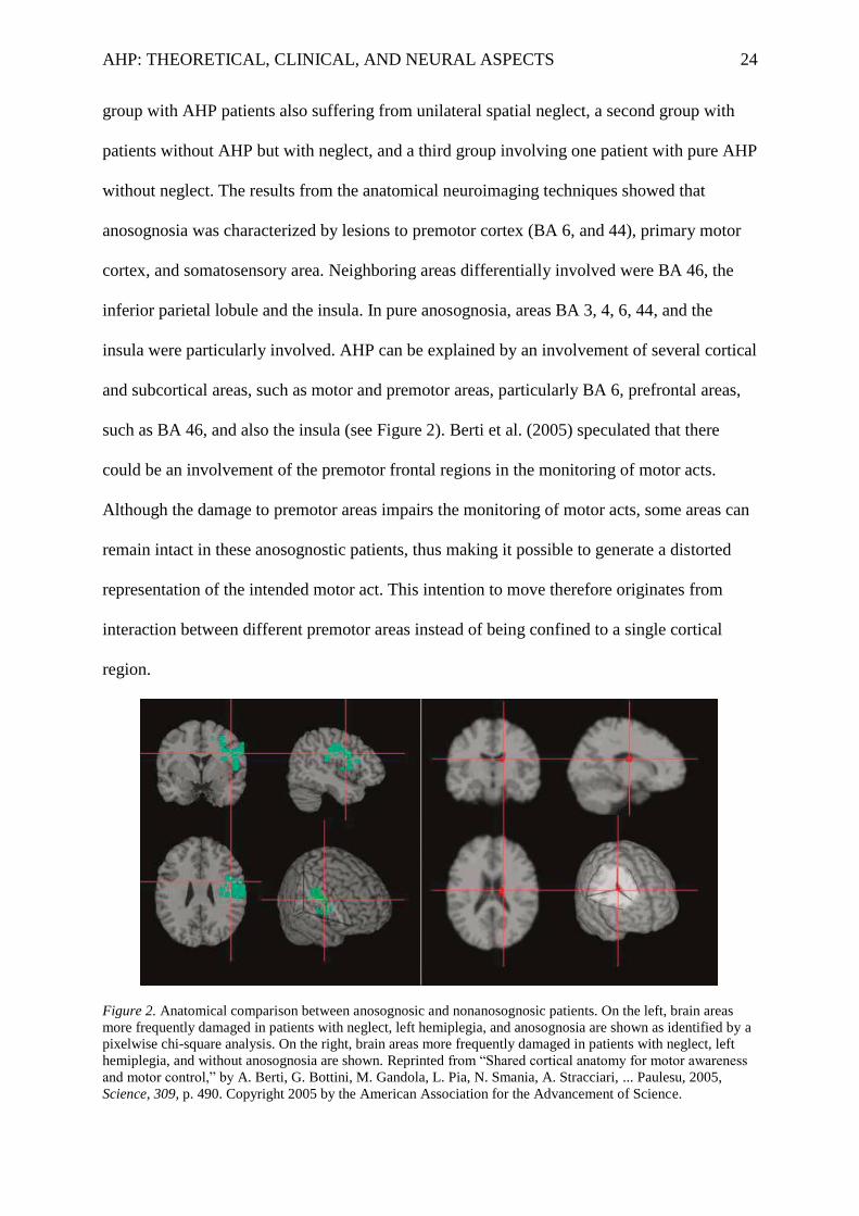
AHP: THEORETICAL, CLINICAL, AND NEURAL ASPECTS 24
group with AHP patients also suffering from unilateral spatial neglect, a second group with
patients without AHP but with neglect, and a third group involving one patient with pure AHP
without neglect. The results from the anatomical neuroimaging techniques showed that
anosognosia was characterized by lesions to premotor cortex (BA 6, and 44), primary motor
cortex, and somatosensory area. Neighboring areas differentially involved were BA 46, the
inferior parietal lobule and the insula. In pure anosognosia, areas BA 3, 4, 6, 44, and the
insula were particularly involved. AHP can be explained by an involvement of several cortical
and subcortical areas, such as motor and premotor areas, particularly BA 6, prefrontal areas,
such as BA 46, and also the insula (see Figure 2). Berti et al. (2005) speculated that there
could be an involvement of the premotor frontal regions in the monitoring of motor acts.
Although the damage to premotor areas impairs the monitoring of motor acts, some areas can
remain intact in these anosognostic patients, thus making it possible to generate a distorted
representation of the intended motor act. This intention to move therefore originates from
interaction between different premotor areas instead of being confined to a single cortical
region.
Figure 2. Anatomical comparison between anosognosic and nonanosognosic patients. On the left, brain areas
more frequently damaged in patients with neglect, left hemiplegia, and anosognosia are shown as identified by a
pixelwise chi-square analysis. On the right, brain areas more frequently damaged in patients with neglect, left
hemiplegia, and without anosognosia are shown. Reprinted from “Shared cortical anatomy for motor awareness
and motor control,” by A. Berti, G. Bottini, M. Gandola, L. Pia, N. Smania, A. Stracciari, ... Paulesu, 2005,
Science, 309, p. 490. Copyright 2005 by the American Association for the Advancement of Science.

AHP: THEORETICAL, CLINICAL, AND NEURAL ASPECTS 25
The Role of Insula Cortex
Similar to the study conducted by Berti et al. (2005), Karnath, Baier, and Nägele (2005)
also used neuroimaging techniques such as magnetic resonance imaging (MRI) or computed
tomography (CT) to discover differences in lesion locations in left hemiplegic patients. These
patients had right hemisphere damage and were divided into two groups; one with AHP and
one without anosognosia for their hemiplegia. The results indicated that lesions in both groups
included cortical areas of the parietal and temporal cortex, the insula, and the deep white
matter. In order to identify the vital anatomical structures involved in AHP, a lesion
subtraction analysis between these two groups was used. Besides the differences detected in
parts of the right hemisphere’s white matter, they found that the right posterior insula was
usually damaged in the group of patients with AHP, compared to the other group (see Figure
3). Vocat et al. (2010) pointed out that their results for the hyperacute phase are in line with
the results presented by Karnath et al. (2005), showing that damage to the insula is common
in patients with AHP.
Orfei et al. (2007) pointed out that these results are not unexpected since the posterior
insula has been considered to be connected with the primary and secondary somatosensory
cortex, the premotor cortex, the prefrontal cortex, and superior and inferior temporal cortex.
An important question to ask is whether a small lesion restricted to the posterior insular cortex
would be sufficient to cause anosognosia. However, even if the posterior insular cortex is
significantly involved in the network of integrating self-awareness, other structures were also
involved in each participant of this study, such as temporal and/or parietal cortex, basal
ganglia, and/or deep white matter. This can be counted as a potential limitation of the results
in this study (Karnath et al., 2005). Thus, Appelros et al. (2007) argued that no particular
lesion localization seems to be sufficient in association to anosognosia. Another limitation is
that the MRI and CT scans might not give adequate descriptions of the functional aspects of

AHP: THEORETICAL, CLINICAL, AND NEURAL ASPECTS 26
the lesions because areas that seem spared in anatomical scans do not necessarily function
normally (Karnath et al., 2005).
Figure 3. A, Overlay lesion plots of the patients with anosognosia for hemiplegia/hemiparesis (n = 14) and of the
patients with right brain damage without anosognosia (controls; n = 13). The number of overlapping lesions is
illustrated by different colors coding increasing frequencies from violet (n = 1) to red (n = maximum number of
subjects in the respective group). Talairach Z-coordinates (Talairach and Tournoux, 1988) of each transverse
section are given. B, Overlay plot of the subtracted superimposed lesions of the patients with anosognosia for
hemiplegia/hemiparesis minus the control group. The percentage of overlapping lesions of the anosognosia
patients after subtraction of controls is illustrated by five different colors coding increasing frequencies from
dark red (difference, 1-20%) to white-yellow (difference, 81-100%). Each color represents 20% increment. The
colors from dark blue (difference, -1 to -20%) to light blue (difference, -81 to -100%) indicate regions damaged
more frequently in control patients than in patients with anosognosia. Wh.mat., White matter. Reprinted from
“Awareness of the functioning of one’s own limbs mediated by the insular cortex?,” by H. O. Karnath, B. Baier,
and T. Nägele, 2005, The Journal of Neuroscience, 25, p. 7136. Copyright 2005 by Society for Neuroscience.
Motor Awareness
Movements are produced by a sequence of neurobiological events usually not available to
consciousness, even though people undoubtedly are aware of performing motor actions or not.
The conscious knowledge and the intentional attitudes represent the motor awareness of a
performed action, which enables us to integrate it into a sense of self. The disturbance of
motor control and self-awareness can particularly be detected in patients with AHP (Berti &
AHP: THEORETICAL, CLINICAL, AND NEURAL ASPECTS 27
Pia, 2006). Since this essay will not provide an extensive discussion concerning motor
awareness, one recent study in particular will briefly be described in order to put the idea
suggested by Berti and Pia (2006) into perspective.
Fotopoulou et al. (2008) investigated the role of motor intention in patients with AHP by
comparing patients with only hemiplegia, to patients with AHP. MRI and CT scans revealed
that all patients had lesions in their right hemisphere, involving frontal, temporal, and parietal
lobes, and lesions to the basal ganglia, the insula, and the internal capsule. The patients in
both groups had a left hemiplegic arm and were exposed to false visual feedback of
movements during the experiment. A realistic prosthetic rubber hand was therefore presented
instead of the patient’s real hand during several trials. It was hypothesized that if motor
intention dominates over sensory feedback in AHP, this would result in an inability to
perceive the lack of movement of the prosthetic hand for patients with AHP. The main finding
of this study was that the patients with AHP actually ignored visual information when they
had an intention to move, in absence of the rubber hand movement. Fotopoulou et al. (2008)
suggested that the results confirmed that AHP is affected by the motor intentions and this is
consistent with the theory proposed by Frith et al. (2000) and contradicts the proposals by
Heilman (1991; Heilman et al., 1998) stating that the lack of motor intention results in AHP.
Discussion
Patients suffering from AHP after stroke have recurrently been recruited as subjects within
neuropsychological research. The main purpose of this discipline is to investigate the
consequences of brain damage in different anatomical structures and its functional impact on
self-awareness, while also developing diagnostic procedures and improving rehabilitation.
This motivated a constant progression of the study about anosognosia because it has the
potential to unravel crucial neural components underlying these psychological functions
(Heilman et al., 1998; Prigatano, 2010).

AHP: THEORETICAL, CLINICAL, AND NEURAL ASPECTS 28
There is a wide range of techniques used to assess anosognosia in stroke patients, and they
all measure the severity and presence of the disorder. It can be concluded that the
development of the questionnaires outlined in this review has improved throughout the years.
By comparing one of the first questionnaires by Cutting (1978), to more recent established
ones by Bisiach et al. (1986), Starkstein et al. (1992), and Marcel et al. (2004), a considerable
increase regarding both complexity and detail can be recognized, which in turn provides a
more efficient evaluation of the patients. However, these different questionnaires vary in their
assessment process, and since anosognosia is a complex phenomenon to investigate, this can
easily generate inconsistencies between the questionnaires. Therefore, possible confounders
in the clinical evaluation of anosognosia have been considered to be important. It primarily
concerns the fundamental differences among these questionnaires in their assessment
technique and degree of complexity (Starkstein et al., 2010). As stated by both Pia et al.
(2004) and Jehkonen et al. (2006), another possible confounder of great importance is the
timing of the actual assessment, since it has been shown to significantly influence the
frequency of AHP in patients (Vocat et al., 2010). Anosognostic patients in most studies have
lesions in the right hemisphere, compared to the left hemisphere. An additional confounder
has therefore been suggested, such as the exclusion of patients with aphasia due to damage to
important speech areas located in their left hemisphere (Jehkonen et al., 2006; Pia et al.,
2004). According to Starkstein et al. (2010), it is also important to consider the extent to
which patients with dementia have been recruited to studies as an additional possible
confounder. Finally, it is important to point out the relevance for developing more consistent
diagnostic procedures concerning the degree of complexity and type of assessment method
among commonly used questionnaires. Future research should attempt to incorporate more
flexible methods in their evaluations, in order to allow assessment in a broader extent among
patients and not only to those who are capable of giving a verbal description of their deficit.

AHP: THEORETICAL, CLINICAL, AND NEURAL ASPECTS 29
The theoretical section in this review presented a number of theories that have given
proposals about the underlying mechanisms of anosognosia. Earlier in history, motivational
theories were considered relevant and emphasized the psychological perspective for
explaining anosognosia. At present, the cognitive theories have more relevance because it also
add the biological component and more adequately provides suggestions for specific
anatomical structures involved in anosognosia. These theories also developed different kinds
of models relating to their distinct explanation of the phenomena, namely; neuropsychological
models, hemispheric damage models, and intra-hemispheric localization models. Since the
cognitive theories gathered more scientific evidence for their approach, the motivational
theories nowadays lack support despite their previous attempts to explain anosognosia (Orfei
et al., 2007; Prigatano, 2010). It should also be pointed out that the cognitive theories also
face serious critiques and there is at present no theory that manages to give a complete
explanation about the underlying mechanisms of anosognosia.
It has been agreed upon that the frequency of AHP among patients has significantly
differed in most studies that have been conducted (Bisiach et al., 1986; Cutting 1978; Orfei et
al., 2007; Weinstein & Kahn, 1955). Other authors revealed that this frequency is correlated
with the timing of the assessment, since the frequency of anosognosia in patients’ decreases
or eventually disappears with time, and rarely becomes chronic (Jehkonen et al., 2006; Pia et
al., 2004; Vocat et al., 2010). A distributed set of cortical and subcortical brain regions could
be identified as damaged and related to the time period within which patients were brain
scanned. This showed that no single brain region is sufficient to produce anosognosia. Areas
in the right hemisphere, including anterior and inferior parts of the insula, anterior internal
capsula, rostral caudate nucleus, and paraventricular white matter were damaged in the early
stage of the disorder. Patients with persistent AHP had additional damage to premotor cortex,
dorsal cingulate, parietotemporal junction, hippocampus, and amygdala (Orfei et al., 2007;

AHP: THEORETICAL, CLINICAL, AND NEURAL ASPECTS 30
Pia et al., 2004; Vocat et al., 2010).
Vocat et al. (2010) found that AHP often coexist together with other neurological
disorders, such as anaesthesia, proprioceptive loss, and visual extinction. It also relates to
neuropsychological disorders, particularly disorientation and visuospatial neglect. The
existence of these related disorders is in turn dependent on the time since the stroke, because
the neurological disorders occur during an earlier stage, while the neuropsychological
disorders occur during persistent AHP. Altogether, this indicates that AHP can be described
with a multi-componential model since no single deficit is sufficient for AHP. This supports
the two-factor theory by Davies et al. (2005) and the deficient affective drive theory (ABC)
by Vuilleumier (2004). The relation between anosognosia and unilateral neglect was
particularly investigated by Appelros et al. (2007) and resulted in support for the idea of their
association, despite identifying fundamental differences in relation to age, severity of stroke,
lesion localization, and pre-stroke dementia. Related disorders of bodily awareness have also
shown that AHP is particularly associated with anosodiaphoria, kinaesthetic illusions, and
non-belonging of limb. As for the neurological and neuropsychological disorders, the
existence of these distortions of body or limb perception also depends on the elapsed time
since the stroke (Vocat et al., 2010).
According to the recent findings of neuroimaging methods on AHP, it can be agreed that
lesions to several cortical and subcortical areas in the right hemisphere are generally damaged
in these patients (Orfei et al., 2007; Pia et al., 2004). These areas involve specific structures in
the frontal lobe, such as the primary and premotor cortices, and prefrontal regions. Other areas
involved are located in the parietal lobe, such as the somatosensory area and the inferior
parietal lobule (Berti et al., 2005). Although AHP is mostly associated with fronto-parietal
lesions, it has also been detected in association with lesions in the temporal lobe (Pia et al.,
2004). A cortical structure often related to AHP is the insula, particularly the right posterior

AHP: THEORETICAL, CLINICAL, AND NEURAL ASPECTS 31
insula (Berti et al., 2005; Karnath et al., 2005; Vocat et al., 2010). MRI and CT scans are
usually used to investigate AHP but it might not give a correct description of the functional
perspective, since anatomical structures observed as intact might still not function properly
(Karnath et al., 2005). A disturbance of self-awareness and motor control are particularly
detected in patients with AHP (Berti & Pia, 2006). This is the reason why the last topic of this
review was dedicated to briefly explain motor awareness and its relation to our intentional
attitudes and motor actions. Fotopoulou et al. (2008) showed that the AHP is affected by our
motor intentions, thus giving support to the theory proposed by Frith et al. (2000), and
contradicts the view by Heilman (1991; Heilman et al., 1998). Nevertheless, the neuroimaging
results in this study by Fotopoulou et al. (2008) support the previously suggested lesion sites,
showing a distributed cortical and subcortical involvement of several specific structures (Berti
et al., 2005; Karnath et al., 2005; Orfei et al., 2007; Pia et al., 2004; Vocat et al., 2010).
Conclusion
It still remains a great challenge to unravel all the complex questions about AHP.
Researchers have indeed uncovered many uncertainties about this disorder, and no entire
explanation has yet been agreed upon. However, the developments of future research should
attempt to further improve the clinical and theoretical aspects, as well as gathering additional
evidence of the neural correlates involved. Improved and new clinical assessment techniques
have to be proposed in order to examine patients more sufficiently. Proposals of theories
about the underlying mechanisms are also crucial in order to provide additional theoretical
suggestions that represent potential models associating with AHP. Accordingly, neuroimaging
studies on the identification of involved anatomical structures are also essential, since AHP
relates to a distributed complex neural network. If this can be achieved and all the important
pieces can be brought together, I am confident that the mysteries behind this puzzling and
fascinating deficit of self-awareness can be discovered.

AHP: THEORETICAL, CLINICAL, AND NEURAL ASPECTS 32
References
Appelros, P., Karlsson, G. M., & Hennerdal, S. (2007). Anosognosia versus unilateral neglect.
Coexistence and their relations to age, stroke severity, lesion site and cognition. European
Journal of Neurology, 14, 54-59. doi:10.1111/j.1468-1331.2006.01544.x
Berti, A., Bottini, G., Gandola, M., Pia, L., Smania, N., Stracciari, A, … Paulesu, E. (2005).
Shared cortical anatomy for motor awareness and motor control. Science, 309, 488-491.
doi:10.1126/science.1110625
Berti, A., & Pia, L. (2006). Understanding motor awareness through normal and pathological
behavior. Current Directions in Psychological Science, 15, 245-250. doi:10.1111/j.1467-
8721.2006.00445.x
Bisiach, E., & Geminiani, G. (1991). Anosognosia related to hemiplegia and hemianopia. In:
G. P. Prigatano & R. L. Schacter (Eds.), Awareness of deficit after brain injury: Clinical
and theoretical issues (pp. 17-39). New York, NY: Oxford University Press.
Bisiach, E., Vallar, G., Perani, D., Papagno, C., & Berti, A. (1986). Unawareness of disease
following lesions to the right hemisphere: Anosognosia for hemiplegia and anosognosia for
hemianopia. Neuropsychologia, 24, 471-482. doi:10.1016/0028-3932(86)90092-8
Cutting, J. (1978). Study of anosognosia. Journal of Neurology, Neurosurgery, and
Psychiatry, 41, 548-555. doi:10.1136/jnnp.41.6.548
Davies, A. M. A., Davies, M., Ogden, J. A., Smithson, M., & White, R. C. (2008). Cognitive
and motivational factors in anosognosia. In T. Bayne and J Fernandez (Eds), Delusions and
self-deception: Affective influences on belief-formation. Hove, ESX: Psychology Press.
Davies, M., Davies, A. M. A., & Coltheart, M. (2005). Anosognosia and the two-factor
theory of delusions. Mind & Language, 20, 209-236. doi:10.1111/j.0268-
1064.2005.00283.x
AHP: THEORETICAL, CLINICAL, AND NEURAL ASPECTS 33
Fotopoulou, A., Tsakiris, M., Haggard, P., Vagopoulou, A., Rudd, A., & Kopelman, M.
(2008). The role of motor intention in motor awareness: An experimental study on
anosognosia for hemiplegia. Brain, 131, 3432-3442. doi:10.1093/brain/awn225
Frith, C. D., Blakemore, S. J., & Wolpert, D. M. (2000). Abnormalities in the awareness and
control of action. Phil. Trans. R. Soc. Lond. B, 355, 1771-1788.
doi:10.1098/rstb.2000.0734
Geschwind, N. (1965a). Disconnexion syndromes in animals and man. I. Brain, 88, 237-294.
doi:10.1093/brain/88.2.237
Geschwind, N. (1965b). Disconnexion syndromes in animals and man. II. Brain, 88, 585-644.
doi:10.1093/brain/88.3.585
Heilman, K. M. (1991). Anosognosia: Possible neurological mechanisms. In: Prigatano GP,
editor. Awareness of deficits after brain injury (pp. 53-62). Oxford, UK: Oxford University
Press.
Heilman, K. M., Barret, A. M., & Adair, J. C. (1998). Possible mechanisms of anosognosia: A
defect in self-awareness. Phil. Trans. R. Soc. Lond. B, 353, 1903-1909.
doi:10.1098/rstb.1998.0342
Jehkonen, M., Laihosalo, M., & Kettunen, J. (2006). Anosognosia after stroke: Assessment,
occurrence, subtypes and impact on functional outcome reviewed. Acta Neurologica
Scandinavica, 114, 293-306. doi:10.1111/j.1600-0404.2006.00723.x
Karnath, H. O., Baier, B., & Nägele, T. (2005). Awareness of the functioning of one’s own
limbs mediated by the insular cortex? The Journal of Neuroscience, 25, 7134-7138.
doi:10.1523/JNEUROSCI.1590-05.2005
Levine, D. N. (1990). Unawareness of visual and sensorimotor defects: A hypothesis. Brain
and Cognition, 13, 233-281. doi:10.1016/0278-2626(90)90052-P

AHP: THEORETICAL, CLINICAL, AND NEURAL ASPECTS 34
Levine, D. N., Calvanio, R., & Rinn, W. E. (1991). The pathogenesis of anosognosia for
hemiplegia. Neurology, 41, 1770-1781.
Marcel, A. J., Tegnér, R., & Nimmo-Smith, I. (2004). Anosognosia for plegia: Specificity,
extension, partially and disunity of bodily unawareness. Cortex, 40, 19-40.
Orfei, M. D., Caltagirone, C., & Spaletta, G. (2009). The evaluation of anosognosia in stroke
patients. Cerebrovascular Diseases, 27, 280-289. doi:10.1159/000199466
Orfei, M. D., Robinson, R. G., Prigatano, G. P., Starkstein, S., Rüsch, N., Bria, P, ... Spaletta,
G. (2007). Anosognosia for hemiplegia after stroke is a multifaceted phenomenon: A
systematic review of the literature. Brain, 130, 3075-3090. doi:10.1093/brain/awm106
Pia, L., Neppi-Modona, M., Raffaella, R., & Berti, A. (2004). The anatomy of anosognosia
for hemiplegia: A meta-analysis. Cortex, 40, 367-377. doi:10.1016 /S0010-
9452(08)70131-X
Prigatano, G. P. (2010). The study of anosognosia. New York, NY: Oxford University Press.
Ramachandran, V. S. (1996). The evolutionary biology of self-deception, laughter, dreaming
and depression: Some clues from anosognosia. Medical Hypotheses, 47, 347-362.
doi:10.1016/S0306-9877(96)90215-7
Starkstein, S. E., Fedoroff, J. P., Price, T. R., Leiguarda, R., & Robinson, R. G. (1992).
Anosognosia in patients with cerebrovascular lesions. A study of causative factors. Stroke,
23, 1446-1453.
Starkstein, S. E., Jorge, R. E., & Robinson, R. G. (2010). The frequency, clinical correlates,
and mechanisms of anosognosia after stroke. The Canadian Journal of Psychiatry, 55,
355-361.
Tiedens, L. Z., & Linton, S. (2001). Judgment under emotional certainty and uncertainty: The
effects of specific emotions on information processing. Journal of Personality and Social
Psychology, 81, 973-988. doi:10.1037/0022-3514.81.6.973
AHP: THEORETICAL, CLINICAL, AND NEURAL ASPECTS 35
Vallar, G., & Ronchi, R. (2006). Anosognosia for motor and sensory deficits after unilateral
brain damage: A review. Restorative Neurology and Neuroscience, 24, 247-257.
Vocat, R., Staub, F., Stroppini, T., & Vuilleumier, P. (2010). Anosognosia for hemiplegia: A
clinical-anatomical prospective study. Brain, 133, 3578-3597. doi:10.1093/brain/awq297
Vuilleumier, P. (2004). Anosognosia: The neurology of beliefs and uncertainties. Cortex, 40,
9-17. doi:10.1016/S0010-9452(08)70918-3
Weinstein, E. A., & Kahn, R. L. (1955). Denial of illness: Symbolic and physiological
aspects. Springfield, IL: Charles C. Thomas.