anomalies. restorative management r of the adult patient · amelogenesis imperfecta - lifelong...
TRANSCRIPT

Amelogenesis imperfecta - lifelong management. Restorative management of the adult patientM. Patel,*1 S. T. McDonnell,2 S. Iram1 and M. F. W-Y. Chan1
inheritance patterns and its prevalence varies from 1:700 to 1:16,000 depending on the population studied.3
The most common classification used for AI is based primarily on phenotype alone.4 Four major categories have been described which include: hypoplastic, hypomatured, hypocalcified and hypomatured – hypo-plastic with taurodontism. Table 1 shows the typical characteristics of each of the dif-ferent AI phenotypes however, it is important to note that any of the phenotypes may coex-ist in the same patient or on the same tooth. These four major phenotypes have been further divided into 15 subtypes based on mode of inheritance (Table 2).
A clinical diagnosis of AI can be aided by asking the patient four questions as shown in Table 3.3 This will help differen-tiate AI from other enamel defects such as fluorosis, which is known to be the com-monest differential diagnosis and can be difficult to distinguish from AI clinically. The severity of AI can vary significantly between patients and often it is difficult to make a diagnosis of the phenotype from clinical examination alone. In some cases the different phenotypes described may coexist in the same patient and on the same tooth. Clinical presentation can range from mild discolouration, slight pitting and minimal post eruptive breakdown of enamel to severe discolouration, pitting or significant tooth
INTRODUCTIONAmelogenesis is a two-staged process where a protein rich matrix is initially laid down during the secretary phase, followed by the mineralisation phase where the pro-teins are replaced by hydroxyapatite crys-tals. This results in the highly mineralised enamel structure. Amelogenesis imperfecta (AI) is a hereditary condition that affects the formation of the enamel matrix or the enamel mineralisation process of both the primary and secondary dentition. It is a clinically and genetically heterogeneous group of conditions that affects both the quantity and quality of the enamel struc-ture and the overall appearance of all or nearly all the teeth in more or less an equal manner, without reference to chronology.1,2 More recently it has been suggested that AI may have a syndromic association due to changes noted in other parts of the body.2 AI has either autosomal dominant, auto-somal recessive, sex-linked or sporadic
The biggest challenge restorative dentists face in rehabilitating patients with amelogenesis imperfecta (AI) is trying to restore aesthetics, function and occlusal stability while keeping the treatment as conservative as possible. The goals of treatment should be to prolong the life of the patient’s own teeth and avoid or delay the need for extractions and subsequent replacement with conventional fixed, removable or implant retained prostheses. In order to achieve these goals a stepwise approach to treatment planning is required starting with the most conservative but aesthetically acceptable treatment. This article discusses the management of AI and presents the various treatment options available for restoring the adult patient who presents to the dentist with AI.
surface loss due to rapid post eruptive break-down of hypomineralised enamel. Figures 1b and 3d highlight the variation in clinical pre-sentation and the difficulty clinicians face in making a clinical diagnosis of the phenotype present. From a practical perspective it may not be absolutely necessary to reach a defini-tive diagnosis of the phenotype as in most cases the management and the treatment options available are often the same.
Most patients with AI will first present to a general dental practitioner whose role in the management may involve a timely referral to the paediatric or restorative specialist, depending on the patient’s age. This may be for treatment of com-plex cases or for treatment planning and advice in management of simpler cases. The paediatric specialist’s role in the management of AI is to provide support and reassurance to the child and parents, motivate the child to maintain good oral hygiene and diet, preserve tooth structure and aesthetics and prevent pain, pathol-ogy and early tooth loss. The treatment provided by the paediatric specialist can be referred to as a transitional phase. Once the patient reaches late adolescence or early adulthood they are often referred to restorative specialist for life long man-agement of their dentition in conjunc-tion with the patient’s general dental practitioner via a shared care approach.
1Barts Helth NHS Trust, Dental Institute, New Road, London, E1 1BB; 2Department of Paediatric Den-tistry, Edinburgh Dental Institute, Lauriston Building, Lauriston Place, Edinburgh, EH3 9HA; 3Department of Restorative Clinical Services, Leeds Dental Institute, Clarendon Way, Leeds, LS2 9LU *Correspondence to: Dr Mital Patel Email: [email protected]
Refereed Paper Accepted 10 July 2013 DOI: 10.1038/sj.bdj.2013.1045 ©British Dental Journal 2013; 215: 449-457
Outlines the clinical presentation of amelogenesis imperfecta (AI) and how to differentiate it from other developmental anomalies.Highlights the challenges faced in the rehabilitation of patients presenting with AI.Discusses the advantages and disadvantages of various restorative treatment modalities available.
I N B R I E F
PRACTICE
BRITISH DENTAL JOURNAL VOLUME 215 NO. 9 NOV 9 2013 449
© 2013 Macmillan Publishers Limited. All rights reserved

PRACTICE
RESTORATIVE CHALLENGESThere are many challenges AI patients present with which need to be carefully managed as part of the overall rehabilita-tion for these patients. Table 4 summarises some of the common challenges and their causes that patients often present with. It is important that the restorative den-tist takes these factors into account dur-ing treatment planning, if rehabilitation and life long management of the patient’s dentition is to be successful.
RESTORATIVE TREATMENT OPTIONSTreatment options available to restore patients with AI vary considerably depend-ing on several factors such as age of the patient, patient motivation, periodontal condition, endodontic status, loss of tooth structure, severity of disorder, socioeco-nomic status and most importantly the patient’s availability for treatment and cooperation.5,6 Often these patients pre-sent young and want a quick result which will improve the appearance of their teeth allowing them to be accepted by their peers and society in general. However, adopting a stepwise approach is essential to help preserve and retain the patient’s own teeth for as long as possible and avoid or delay the need for prosthetic replacement.
ORAL HYGIENE, DIETARY ADVICE, DESENSITISATION AND STABILISATIONIt is crucial that prevention should be included in the initial stages of all treat-ment plans with a particular focus on pro-viding effective oral hygiene instruction and patient motivation. Treatment of den-tine hypersensitivity using either desensi-tising agents, topical fluoride preparations and/or CCP-ACP (casein phosphopeptide-amorphous calcium phosphate) contain- contain-ing products that promote remineralisation should also be introduced at this stage as this will help with the maintenance of good oral hygiene. If the patient has any periodontal problems these should be addressed with non-surgical and/or surgical periodontal therapy as appropri-ate. Comprehensive dietary analysis and advice is also essential. It is important to highlight to patients that AI carries a higher caries risk and therefore poor diet control can have a devastating effect on
Table 1 Clinical and radiographic appearance of the major phenotypes of AI
Hypoplastic form Reduction in the quantity of the enamel matrix usually with normal mineralisation
Hypomaturation form Defect in the quality of mineralisation process with normal quantity of matrix formation
Hypocalcified form Defect in the quality of the mineralisation process with normal quantity of matrix formation
Clinical appearance
Reduced thickness of enamel Normal thickness of enamel Normal thickness of enamel with loss of translucency
Enamel is usually well mineralised and is therefore less prone to attrition than the other forms of AI
Enamel is hypomineralised and prone to post eruptive breakdown and attrition
Enamel is very hypomineralised and often of a soft cheesy consistency. Prone to early rapid post eruptive breakdown and can easily be worn away
The colour can vary from normal colour and translucency to a yellow to dark brown colour depending on how thin the enamel is and the degree of shine through of the underlying dentine
Colour may be affected by post eruptive uptake of staining from the oral environment and the degree of post-eruptive breakdown. It can vary broadly from mottled opaque white to Yellow-brown or red-brown discolouration
Colour may be affected by post eruptive uptake of staining from the oral environment and the degree of post eruptive breakdown and exposure of underlying dentine. Teeth tend to be darker in colour than other types of AI
Spacing between teeth as thinner enamel often reduces tooth size
Rough, irregular or pitted enamel with or without vertical ridges or grooves
Radiographic appearance
Enamel contrasts normally from dentine
Enamel has similar radiodensity as dentine
Enamel is less radiopaque than the dentine
Table 2 Classification of AI based on phenotype and mode of inheritance
Subtype Phenoype Phenotype and mode of inheritance
Type I Hypoplastic
Type IA Hypoplastic, pitted autosomal dominant
Type IB Hypoplastic, local autosomal dominant
Type IC Hypoplastic, local autosomal recessive
Type ID Hypoplastic, smooth autosomal dominant
Type IE Hypoplastic, smooth X-linked dominant
Type IF Hypoplastic, rough autosomal dominant
Type IG Enamel agenesis, autosomal recessive
Type II Hypomaturation
Type IIA Hypomaturation, pigmented autosomal recessive
Type IIB Hypomaturation, X-linked recessive
Type IIC Hypomaturation, snow-capped teeth, X-linked
Type IID Hypomaturation, snow-capped teeth, autosomal dominant?
Type IIi Hypocalcified
Type IIIA Autosomal dominant
Type IIIB Autosomal recessive
Type IV Hypomaturation-hypoplastic with taurodontism
Type IVA Hypomaturation-hypoplastic with taurodontism, autosomal dominant
Type IVB Hypoplastic-hypomaturation with taurodontism, autosomal dominant
450 BRITISH DENTAL JOURNAL VOLUME 215 NO. 9 NOV 9 2013
© 2013 Macmillan Publishers Limited. All rights reserved

PRACTICE
both unrestored and restored teeth. If indi-cated any carious lesions present should be restored without delay; alternatively teeth with poor prognosis or those that are deemed unrestorable should be con-sidered for extraction. Often motivating patients with AI to improve and maintain good oral hygiene can be difficult when they are unhappy about the appearance of their teeth. Oral hygiene usually improves once the patient has been rehabilitated often resulting in recession around the
restoration margins, which can be diffi-cult to manage depending on the choice of restoration.
BLEACHING AND MICROABRASIONIn patients with AI, preservation of tooth structure is vital and minimally invasive treatment options must be considered where possible. Microabrasion using an acidic slurry containing 18% hydro-chloric acid and pumice is often effec-tive in removing superficial stains and
improving the appearance of the teeth.7 Ashkinazi et al. demonstrated the use of this technique in patients with enamel hypomaturation. At four-year follow up they showed that the improvements in aes-thetics were maintained.8 An alternative conservative approach is the use of long-term bleaching or tooth whitening. Up until recently the use of bleaching agents for dental treatment was considered to be illegal practice. However, a recent position statement from the General Dental Council indicated that the use of 0.1-6% hydrogen peroxide in patients over 18 years as part of their dental treatment is now acceptable provided the patient has had an appropri-ate assessment by a dentist.9 Satisfactory aesthetic improvement has been reported10 following six weeks of external vital night guard bleaching, using 10% carbamide peroxide (approximately 3% hydrogen per-oxide). While this conservative treatment option may be effective, it can give rise to sensitivity. Alternating the bleaching agent with the use of a desensitising agent or fluoride containing toothpastes can help in managing the sensitivity.10 The process of bleaching can significantly decrease the bond strength of resin-based materi-als to the bleached tooth surface compared to the unbleached surface. Delaying the final restoration for two weeks and leav-ing the enamel surface exposed to saliva has been shown to eradicate the adverse effect of bleaching on bond strength.11 In many cases these treatment modalities alone are not enough to restore aesthetics and may need to be combined with other treatments (Fig. 1). The use of microabra-sion or bleaching initially can help reduce some of the discolouration making it easier to conservatively mask the teeth with other treatment modalities.
CROWN LENGTHENING SURGERYOften patients with AI have reduced clinical crown height due to loss of tooth structure resulting from enamel chipping away and tooth wear. This tooth surface loss may be compensated for by dento-alveolar compensation leaving a ‘gummy’ appearance to the patient’s smile.12 Prior to the teeth being restored it is therefore important to determine whether or not there has been any dento-alveolar com-pensation and the position of the incisal edge and gingival margin in relation to
Table 3 Questions to aid diagnosis of AI as described by Crawford et al.3
1. Has anyone else in the family had anything like this?
2. Has there been anything in the patient’s medical history which might have caused sufficient metabolic disturbance to affect enamel formation?
3. Are all the teeth affected in a similar manner?
4. Is there a chronological distribution to the appearance to the defect?
Table 4 Restorative challenges faced
Restorative challenges Causes
Psychosocial problemsLow self esteemReclusive and withdrawn
Often due to being bullied at school as a child
Poor oral hygieneChronic gingivitis
Patients avoid cleaning due to sensitivitySome avoid cleaning due to poor motivation as teeth are of a poor appearance
SensitivityDifficult to etch or clean teeth without LA
Thin enamelExposed dentine
Caries Poor oral hygiene combined with thin enamel or hypomineralised enamel makes AI affected teeth more prone to rapid caries progression
Discolouration Yellow dentine shining through thin enamel or may be complete lack of enamelCan be difficult to mask with conservative techniques
Loss of occlusal vertical dimension or alveolar spaceLoss of interocclusal space
Due to rapid tooth surface loss which may be compensated for by down growth of the maxillary complexTeeth trying to maintain opposing contactsOften require complex rehabilitation involving a reorganised approach and an increase in the occlusal vertical dimension
Reduced inter root space Thin enamel or rapid loss of enamel post eruption results in teeth drifting closer togetherRisk of damage to adjacent teethDifficult to prepare teeth for crowns and take impressions
Large pulp to crown ratio Young teeth with large pulps. Lack of secondary dentineIncreased risk of tooth losing vitality
Gingival maturation resulting in exposure of restoration margin
Occurs over a few months post full eruption of toothIf restoration placed too early then margin may become visible after maturation.If lab made restoration then it may need replacing
Decreased bond strength of resin to enamel
Higher protein content in AI affected enamelResults in abnormal etch patternEtch pattern varies between phenotypesDifferent phenotypes can therefore give different bond strengths
Bonding to dentine Due to rapid loss of enamel in some AI patients bonding to dentine is required
BRITISH DENTAL JOURNAL VOLUME 215 NO. 9 NOV 9 2013 451
© 2013 Macmillan Publishers Limited. All rights reserved
PRACTICE
the upper lip when it is at rest and more importantly when the patient is smiling. It is also important to assess the amount of tooth display visible. Ideally in a young female patient there should be 3-4 mm of incisal display of the upper incisors and approximately 1-2 mm less in a young male patient when the upper lip is at rest.13 When smiling the upper lip should be close to the cervical margin of the teeth with no more than 1.5-2 mm gingival display.14 If the teeth were to be restored to ideal size and shape in the presence of signifi-cant alveolar compensation, it can leave the patient with too much tooth display when the lip is at rest or when smiling and a ‘gummy’ appearance to the smile. The teeth should also be assessed to see if there is sufficient clinical crown height avail-able to provide adequate retention and resistance form for the planned restora-tions. Crown lengthening surgery can be used to increase the clinical crown height available, reduce the ‘gummy’ appearance and restore the ideal aesthetic relation-ship between teeth and gingival tissues within the soft tissue frame of the upper lip (Fig. 2). However, care must be taken to assess important factors such as root length, bone support and taper of the root before carrying out this invasive proce-dure. In poorly assessed cases crown-lengthening surgery can result in mobility of the teeth and unpredictable gingival recession. Due to the tapered nature of the root, crown lengthened teeth have reduced thickness of dentine between the external root surface and the pulp chamber towards the new gingival margin. Crown prepara-tions on these teeth are likely to have an increased risk of the teeth loosing vitality due to pulpal trauma.
COMPOSITE RESINS
Direct composite
There is some evidence to suggest that teeth affected by AI do not show a typi-cal etch pattern and this can potentially reduce the bond strength of enamel to the composite resin.15 Despite this, the contin-ued development of adhesive bonding sys-tems has increased the popularity of direct composite restorations to restore both aesthetics and function in patients with AI.16 These restorations are appropriate to restore aesthetics and eliminate sensitivity,
particularly in a young adult patient where definitive restorations are contraindicated until eruption of the clinical crown is com-plete and the soft tissue has matured. As the gingival tissue matures and recedes to the cemento-enamel junction (CEJ), the margin of the restoration will become exposed along with further exposure of the discoloured cervical tooth structure. Similarly, often the lack of motivation to maintain good oral hygiene until the patient has been rehabilitated can result in a similar appearance of recession and exposure of the restoration margin as the gingival health improves following restor-ative treatment (Fig. 4b and Fig. 5f). Unlike with porcelain restorations, recession can be easily masked with composite by refur-bishing the restoration to the new gingi-val margin without the need to replace the whole restoration.
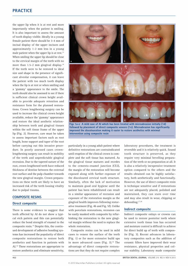
Composite resins can be used in mild cases to veneer the surface of the teeth (Fig. 1) or for more extensive build ups in more advanced cases (Fig. 3).16 The advantage of direct composite restora-tions are that they do not require complex
laboratory procedures, the treatment is reversible and it is relatively quick. Sound tooth structure is preserved, as they require very minimal bevelling prepara-tion of the teeth or no preparation at all. It is also a relatively inexpensive treatment option compared to the others and the results obtained can be highly satisfac-tory, both aesthetically and functionally. However, the use of direct composite resin is technique sensitive and if restorations are not adequately placed, polished and maintained, they can stain over time and may also result in wear, chipping or debonding.17,18
Indirect compositeIndirect composite onlays or crowns can be used to restore posterior teeth where extensive tooth tissue loss has occurred and moisture control is difficult to achieve for direct build up of teeth with compos-ite (Fig. 3). Recent advances in labora-tory composites such as the addition of ceramic fillers have improved their wear resistance, physical properties and col-our stability. Laboratory made composite
a b
c
d e
Figs 1a-e A mild case of AI which has been treated with microabrasion initially (1d) followed by placement of direct composite veneers (1e). Microabrasion has significantly improved the discolouration making it easier to restore aesthetics with minimal intervention using composite resin
452 BRITISH DENTAL JOURNAL VOLUME 215 NO. 9 NOV 9 2013
© 2013 Macmillan Publishers Limited. All rights reserved

PRACTICE
restorations also exhibit improved mar-ginal fit, anatomic contour and reduced shrinkage.19 Indirect resin composite res-torations have shown promising success rates. One study showed a 93% success rate of indirect composite restorations on premolars and molars over a three-year period.20 A seven-year follow up study by Donly et al.19 showed indirect composites placed on premolar teeth had increased longevity than those on molars. This may be due to the fact it is easier to maintain moisture control more anteriorly during cementation and that maintenance is also easier around premolars than molars.
PORCELAIN VENEERSThese restorations are popular in the anterior region because they can achieve excellent aesthetic results with a rela-tively conservative tooth preparation when compared to a full coverage crown. Patient acceptance of porcelain veneers is also high and is reported at 80-100% in patient satisfaction surveys.21,22 In vitro studies have identified some disadvan-tages, such as marginal adaptation and bonding problems;23 however, clinical case reports continue to show success of these restorations.1
When restoring teeth affected by AI with either composite or porcelain veneers, some of the underlying tooth structure may be relatively dark and the translucent nature of these restorations is often unable to adequately mask the discolouration. This can result in poor aesthetics of the restored teeth. Intrinsic opaque porcelain layers incorporated into the restoration or opaque resins used during cementation can help to disguise this although this often results in a loss of translucency, which also detracts from the final appearance.
Most authors advocate some but varia-ble tooth preparation for porcelain veneers (Fig. 4). The teeth can be very sensitive in AI patients and often requires the use local anaesthetic, which is often not necessary when placing direct composite veneers. The use of porcelain veneers in young patients may be associated with early repeat treat-ments due to gingival maturation resulting in exposure of the veneer margin and fur-ther exposure of the discoloured cervical tooth structure. This often requires further destructive preparation to the tooth.
METAL ONLAYSThe use of precious or non-precious metal onlays to restore and protect the occlusal surface of worn posterior teeth can be an effective treatment option. This type of restoration relies more on adhesion of the restoration to the tooth and less on the mechanical retentive features of tooth preparation often needed with conven-tional onlays or crowns.24 Often the prepa-ration for these restorations is minimal and involves a chamfer margin of 0.5-1 mm which wraps over the cusps, removal of any sharp edges and approximately 1.0- 1.5 mm occlusal clearance. It is therefore particularly useful where there is lack of
clinical crown height and where conser-vation of remaining tooth tissue is essen-tial. Metal onlays can control sensitivity and compensate for the loss in occlusal vertical dimension.25 While these restora-tions are ideal for posterior teeth, there is an increasing trend towards patients requesting tooth coloured restorations, particularly in this group of patients.
CROWNSOf the many options available for restoring teeth affected by AI, conventional crowns have been the most predictable and durable aesthetic restoration to date.26 The obvious disadvantage of this approach is that it is very tooth destructive. It is a highly inva-sive procedure for an already compromised and worn dentition in patients that are often young with immature dental pulps. Complications associated with crowned teeth in AI patients have been reported and include: loss of cementation, material fracture, caries and the need for endodon-tic treatment.1,27 Figure 5 gives an example of a AI patient who has been rehabilitated using conventional crowns.
From the various types of crowns avail-able to restore posterior teeth, gold crowns require the most conservative preparations followed by metal ceramic crowns and the most invasive being high strength all ceramic crowns. However, the use of gold may not be acceptable to patients due to aesthetics. Patients should be informed of the advantages and disadvantages of the various materials so that an informed deci-sion can be made. Some patients may be happy to have gold restorations in the pos-terior part of the mouth once the risks and benefits of the alternative crown options are discussed.
In the anterior region and posteriorly for patients where aesthetics is a concern, metal ceramic crowns can be consid-ered. To preserve tooth tissue the crowns should be carefully designed to restrict the porcelain to areas that are of aesthetic importance, such as the buccal and labial surfaces and perhaps only the mesio-buc-cal aspect of molar teeth. Wherever pos-sible a minimal preparation metal margin should be made to preserve tooth tissue, such as the lingual/palatal and mesial/distal aspects of teeth. The literature is abundant with case reports using metal ceramic crowns in patients with AI.6,28,29
a
b
c
d
e
Figs 2a-e Crown lengthening surgery to increase clinical crown height, reduce gingival show and improve overall aesthetic proportions
BRITISH DENTAL JOURNAL VOLUME 215 NO. 9 NOV 9 2013 453
© 2013 Macmillan Publishers Limited. All rights reserved

PRACTICE
A retrospective study assessing the num-ber and type of restorations present in a sample of 15 patients showed that from a total of 213 restorations 57% were metal ceramic crowns which showed good survival at five years.1
Over recent years the use of glass based all ceramic dentine bonded crowns made from feldspathic porcelain has increased because of their inherent aes-thetics, excellent biocompatibility, good marginal fit and improved physical properties.30 These restorations require minimal preparation of 0.5-0.7 mm cir-cumferentially and often no or minimal
preparation occlusally where tooth tissue has already been lost. Essentially these restorations are a 360 degree veneer and can be a conservative aesthetic treatment option for the anterior region, extend-ing to the premolars on carefully selected cases. However, similar to veneer resto-rations a significant thickness of opaque porcelain layer needs to be incorporated into the crown to mask discolouration. This will reduce the translucent appear-ance to the crown and therefore compro-mise the aesthetics.
More recently high strength all ceramic restorations with alumina or zirconia cores
have also been used in the rehabilitation of teeth affected by AI.30,31 These restora-tions require heavier tooth preparation of 1.5-2 mm and therefore increase the risk of the teeth losing vitality.27 There is also a risk of microleakage with zirconia restorations due to the inability to bond zirconia restora-tions to the underlying tooth structure.30 Some reports have suggested that using these restora-tions in AI patients where tooth tissue has been lost, allows for minimal occlusal tooth prepa-ration to restore the patient at an increased OVD. However, they still require the heavier axial and marginal finishing line preparations, which could traumatise the dental pulp.
Figs 3a-n A 29-year-old gentleman who most likely has hypoplastic AI with significant post eruptive tooth surface loss. Rehabilitation involved crown lengthening surgery of the upper anterior teeth (Fig. 2) followed by minimally invasive direct composite restorations on the anterior and premolar teeth and indirect composite onlays on the posterior molars. The case shows restoration of aesthetics, function and occlusal stability with minimal damage to the remaining dentition
a b c
d
e f
g h i
j k l
m n
454 BRITISH DENTAL JOURNAL VOLUME 215 NO. 9 NOV 9 2013
© 2013 Macmillan Publishers Limited. All rights reserved

PRACTICE
REMOVABLE DENTURESHistorically, treatment of patients with AI has included extractions and the fab-rication of complete or partial dentures. These options are detrimental psycho-logically, irreversible and invasive,5,6 and have become unacceptable, in light of the advances made in the field of aesthetic dentistry and adhesive techniques. Even when teeth are deemed unrestorable, they can be retained and the dentition restored with an over denture or onlay denture. The retention of teeth preserves alveolar bone32 which in this group of patients may be important if implant treatment is to be considered at some point through their life.
Over dentures or onlay dentures are the least expensive form of treatment both economically and biologically due to the minimally invasive nature of treatment. They can prove useful in restoring aes-thetics and providing a psychological benefit at a critical stage in the patient’s development or where alternative invasive
treatment is not a suitable treatment option for the patient.33 The use of over dentures also requires careful assessment and planning because of the associated hazards, particularly increased plaque accumulation and susceptibility to caries, periodontal disease and variable patient acceptance.33
IMPLANTSIn advanced cases of AI where the teeth are unrestorable and the patient is seek-ing a fixed option, dental implants can be considered. Careful planning is essential and timing of extractions with respect to implant placement is very important to preserve bone, which will resorb away relatively quickly following tooth extrac-tion.32,34 In cases where there is insufficient
bone width, it may be possible to graft the bone with guided bone regeneration, if the deficiency is minor or block onlay grafts in more severe cases. A maxillary sinus graft-ing procedure may be carried out which can give extra height of bone for implant placement in the posterior maxilla. In other areas of the mouth increasing bone height is extremely difficult and unpredictable. A retrospective study assessing cost implica-tions for rehabilitation of AI patients using implants showed that the long-term cumu-lative treatment costs for implant cases were not statistically significantly different when compared with cases reconstructed with tooth-supported fixed prostheses.35
While dental implant treatment may pro-vide a predictable outcome, many young adult patients have educational, social
Figs 4a-c The use of porcelain veneers to restore anterior teeth affected by AI. Figure 4a highlights the relatively destructive tooth preparation required for these restorations. Figure 4b shows recession around the cervical margin of 32. This is a common problem seen in these patients due to veneers being placed too early before gingival maturation or an improvement in oral hygiene measures following the positive impact of oral rehabilitation
a
b
c
Fig. 5a-h A young adult patient who has had extensive treatment through their teenage years to help preserve the underlying tooth structure. This patient has subsequently been rehabilitated with dentine bonded crowns on the anterior teeth and porcelain bonded and gold crowns on the posterior teeth. Overall there is a vast improvement in the aesthetics, function and occlusal stability, however, again due to an improvement in oral hygiene and/or gingival maturation, recession can develop around these teeth as shown in Figure 5f at 41/42. This is difficult to manage on teeth restored with porcelain restorations. The authors would like to acknowledge Mr S Robinson, Consultant in Restorative Dentistry for the clinical work carried out in this case
a b
c d
e f
g h
BRITISH DENTAL JOURNAL VOLUME 215 NO. 9 NOV 9 2013 455
© 2013 Macmillan Publishers Limited. All rights reserved

PRACTICE
or work commitments, which may make it difficult for them to attend numerous appointments and/or undergo surgery. It is also important to wait until the patient has stopped growing before implant treat-ment is completed.36 Studies have shown that implants placed in a growing patient do not behave like normal teeth. They become ankylosed in the bone resulting in infra occlusion of the implant restoration as the jaw bone continues to grow around it.37 Taking these factors into account, implant treatment may not be the ideal treat-ment choice in the first instance for these young adult patients. It is also important to remember that implant treatment carried out when patients are in their twenties or thirties is likely to require revision treat-ment in the future. In view of the fact that medical technology is constantly improv-ing, delaying implant treatment until later in life may be advantageous to the patient as newly developed materials and clinical techniques are likely to give more predictable long term outcomes.
MULTIDISCIPLINARY TEAM APPROACHMany patients with AI can present with a gross malocclusion and an anterior open bite as well as poorly formed teeth. These patients will require a multidisciplinary team approach to rehabilitation, which may include orthodontic treatment, pos-sible orthognathic surgery followed by specialist restorative treatment (Fig. 6). Following definitive treatment the patient will require a multidisciplinary shared care approach to maintenance between their general dental practitioner, den-tal hygienist and the specialists. This is essential to ensure good longevity to the restorations provided.
DISCUSSIONRehabilitation of patients with AI requires careful planning with the most impor-tant factor to consider being the age and cooperation of the patient. Management of these patients through childhood and the early teens is mainly focused around counselling, prevention and preservation of the deciduous, mixed and adult denti-tion. The restorative treatment prescribed from the late teens onwards should aim to establish health, function and aesthetics of the patient’s own teeth and prevent or
delay the need for extraction and pros-thetic replacement. This life long man-agement requires a stepwise approach to treatment planning starting with the most conservative treatment option first.
Treating this group of patients using an evidence-based approach is difficult as the quality of the evidence is gener-ally poor with most of the evidence being case reports. Most of these predominantly describe the use of a removable prosthesis and conventional crown and bridgework. Very few studies present long-term fol-low up of patients treated for AI using the different treatment options available.
Reversible and non-invasive treatment with composite resin (with or without the
use of microabrasion and bleaching) should be considered before the more destructive treatment options. The use of compos-ite resins allows restoration of aesthetics, which is most important to the patient while preserving tooth tissue. Clinicians often avoid using composite resins, as they are susceptible to staining and technique sensi-tive. Staining can be effectively managed by regular polishing of the restorations. If necessary the surface layer can be removed and the restoration refurbished with a new surface layer without causing further dam-age to the underlying tooth structure. If the composites fracture or chip they can also be repaired easily without the need for remov-ing the whole restoration38 and similarly as
Fig. 6a-h Another example of what looks like hypoplastic AI with post eruptive tooth surface loss. As well as the poorly formed tooth structure there is an associated malocclusion and an anterior open bite (6a). This patient underwent extensive orthodontic treatment (6b) and orthognathic surgery to help improve the malocclusion and reduce the anterior open bite (6c). Subsequent restorative treatment involved minimally invasive direct composite bonding to the anterior and premolar teeth and gold crowns/onlays and a porcelain bonded crown on the posterior teeth to close the anterior open bite (6f), restore aesthetics, function and occlusal stability
a
b
c d
e
f
g h
456 BRITISH DENTAL JOURNAL VOLUME 215 NO. 9 NOV 9 2013
© 2013 Macmillan Publishers Limited. All rights reserved

PRACTICE
the gingival margin continues to mature to the CEJ in young adult patients the exposed tooth structure at the gingival margin can easily be covered by refurbishing the resto-ration. These advantages of using compos-ites make them a cost effective restoration both biologically and economically com-pared to other more invasive and expensive restorations. Composite resins should there-fore be considered as the initial restorative material of choice for all patients, especially when the patient is in their late teens and early twenties, as a medium term option. When they start to repeatedly fail or the maintenance burden becomes too great the treatment could progress to more inva-sive techniques. This may involve adhesive porcelain veneers, dentine bonded crowns and eventually full coverage gold, porcelain bonded to metal or all ceramic crowns. If such treatment can be delayed until later in the patient’s life when the pulps have receded and gingival levels have stabilised, this will be beneficial to the patient as it will limit the biological insult of treatment on the dentition. Whilst initial results with composite resins are promising further research is required to assess the longev-ity of composite restorations in AI affected teeth. Despite the lack of good evidence, due to its reversible and minimally inva-sive nature, rehabilitation with composite resins should be considered as the first line of treatment for patients with AI.
The cases presented in Figures 1 and 3 were awarded the 2010 clinical case award by the British Society for Restorative Dentistry.
1. Lindunger A, Smedberg J I. A retrospective study of the prosthodontic management of patients with amelogenesis imperfecta. Int J Prosthodont 2005; 18: 189–194.
2. Aldred M J, Savarirayan R, Crawford P J. Amelogenesis imperfecta: a classification and catalogue for the 21st century. Oral Dis 2003; 9: 19–23.
3. Crawford P J, Aldred M, Bloch-Zupan A. Amelogenesis imperfecta. Orphanet J Rare Dis 2007; 2: 17.
4. Witkop C J, Jr. Amelogenesis imperfecta, dentinogenesis imperfecta and dentin dysplasia
revisited: problems in classification. J Oral Pathol 1988; 17: 547–553.
5. Sholapurkar A A, Joseph R M, Varghese J M et al. Clinical diagnosis and oral rehabilitation of a patient with amelogenesis imperfecta: a case report. J Contemp Dent Pract 2008; 9: 92–98.
6. Sari T, Usumez A. Restoring function and esthetics in a patient with amelogenesis imperfecta: a clinical report. J Prosthet Dent 2003; 90: 522–525.
7. Price R B, Loney R W, Doyle M G, Moulding M B. An evaluation of a technique to remove stains from teeth using microabrasion. J Am Dent Assoc 2003; 134: 1066–1071.
8. Ashkenazi M, Sarnat H. Microabrasion of teeth with discoloration resembling hypomaturation enamel defects: four-year follow up. J Clin Pediatr Dent 2000; 25: 29–34.
9. General Dental Council. Tooth whitening Q&As. www.gdc-uk.org/dentalprofessionals/standards/pages/tooth-whitening.aspx (accessed 16 December 2012).
10. Nathwani N S, Kelleher M. Minimally destructive management of amelogenesis imperfecta and hypodontia with bleaching and bonding. Dent Update 2010; 37: 170–172, 175–176, 179.
11. Basting R T, Rodrigues J A, Serra M C, Pimenta L A. Shear bond strength of enamel treated with seven carbamide peroxide bleaching agents. J Esthet Restor Dent 2004; 16: 250–260.
12. Berry D C, Poole D F. Attrition: possible mechanisms of compensation. J Oral Rehabil 1976; 3: 201–206.
13. Vig R G, Brundo G C. The kinetics of anterior tooth display. J Prosthet Dent 1978; 39: 502–504.
14. Robbins J W. Differential diagnosis and treatment of excess gingival display. Pract Periodontics Aesthet Dent 1999; 11: 265–272.
15. Venezie R D, Vadiakas G, Christensen J R, Wright J T. Enamel pretreatment with sodium hypochlorite to enhance bonding in hypocalcified amelogenesis imperfecta: case report and SEM analysis. Pediatr Dent 1994; 16: 433–436.
16. Sabatini C, Guzman-Armstrong S. A conservative treatment for amelogenesis imperfecta with direct resin composite restorations: a case report. J Esthet Restor Dent 2009; 21: 161–169, 170.
17. Millar B J, Robinson P B, Inglis A T. Clinical evaluation of an anterior hybrid composite resin over 8 years. Br Dent J 1997; 182: 26–30.
18. Peumans M, Van Meerbeek B, Lambrechts P, Vanherle G. The 5-year clinical performance of direct composite additions to correct tooth form and position. I. Esthetic qualities. Clin Oral Investig 1997; 1: 12–18.
19. Donly K J, Jensen M E, Triolo P, Chan D. A clinical comparison of resin composite inlay and onlay posterior restorations and cast-gold restorations at 7 years. Quintessence Int 1999; 30: 163–168.
20. Manhart J, Neuerer P, Scheibenbogen-Fuchsbrunner A, Hickel R. Three-year clinical evaluation of direct and indirect composite restorations in posterior teeth. J Prosthet Dent 2000; 84: 289–296.
21. Meijering A C, Creugers N H, Roeters F J, Mulder J. Survival of three types of veneer restorations in a clinical trial: a 2.5-year interim evaluation. J Dent 1998; 26: 563–568.
22. Rucker L M, Richter W, MacEntee M, Richardson A. Porcelain and resin veneers clinically evaluated:
2-year results. J Am Dent Assoc 1990; 121: 594–596.23. Karlsson S, Landahl I, Stegersjo G, Milleding P. A
clinical evaluation of ceramic laminate veneers. Int J Prosthodont 1992; 5: 447–451.
24. Walls A W, Nohl F S, Wassell R W. Crowns and other extra-coronal restorations: resin-bonded metal restorations. Br Dent J 2002; 193: 135-138, 141–142.
25. Harley K E, Ibbetson R J. Dental anomalies - are adhesive castings the solution? Br Dent J 1993; 174: 15–22.
26. Yip H K, Smales R J. Oral rehabilitation of young adults with amelogenesis imperfecta. Int J Prosthodont 2003; 16: 345–349.
27. Valderhaug J, Jokstad A, Ambjornsen E, Norheim P W. Assessment of the periapical and clinical status of crowned teeth over 25 years. J Dent 1997; 25: 97–105.
28. Ozturk N, Sari Z, Ozturk B. An interdisciplinary approach for restoring function and esthetics in a patient with amelogenesis imperfecta and malocclusion: a clinical report. J Prosthet Dent 2004; 92: 112–115.
29. Robinson F G, Haubenreich J E. Oral rehabilitation of a young adult with hypoplastic amelogenesis imperfecta: a clinical report. J Prosthet Dent 2006; 95: 10–13.
30. Siadat H, Alikhasi M, Mirfazaelian A. Rehabilitation of a patient with amelogenesis imperfecta using all-ceramic crowns: a clinical report. J Prosthet Dent 2007; 98: 85–88.
31. Oliveira I K, Fonseca Jde F, do Amaral F L, Pecorari V G, Basting R T, Franca F M. Diagnosis and esthetic functional rehabilitation of a patient with amelogenesis imperfecta. Quintessence Int 2011; 42: 463–469.
32. van Waas M A, Jonkman R E, Kalk W, van ‘t Hof M A, Plooij J, Van Os J H. Differences two years after tooth extraction in mandibular bone reduction in patients treated with immediate overdentures or immediate complete dentures. J Dent Res 1993; 72: 1001–1004.
33. Johnson A, Winstanley R B. Use of simple overdentures in the treatment of young patients with developmental anomalies. Quintessence Dent Technol 1987; 11: 27–33.
34. Araujo M G, Lindhe J. Dimensional ridge alterations following tooth extraction. An experimental study in the dog. J Clin Periodontol 2005; 32: 212–218.
35. Incici E, Matuliene G, Husler J, Salvi G E, Pjetursson B, Bragger U. Cumulative costs for the prosthetic reconstructions and maintenance in young adult patients with birth defects affecting the formation of teeth. Clin Oral Implants Res 2009; 20: 715–721.
36. Heij D G, Opdebeeck H, van Steenberghe D, Kokich V G, Belser U, Quirynen M. Facial development, continuous tooth eruption, and mesial drift as compromising factors for implant placement. Int J Oral Maxillofac Implants 2006; 21: 867–878.
37. Thilander B, Odman J, Jemt T. Single implants in the upper incisor region and their relationship to the adjacent teeth. An 8-year follow-up study. Clin Oral Implants Res 1999; 10: 346–355.
38. Brignall I, Mehta S, Banerji S, Millar B. Aesthetic Composite Veneersfor an Adult Patient with Amelogenesis Imperfecta: A Case Report. Dental update 2011; 38: 594–603.
BRITISH DENTAL JOURNAL VOLUME 215 NO. 9 NOV 9 2013 457
© 2013 Macmillan Publishers Limited. All rights reserved