animals containing human material
DESCRIPTION
This report identifies the scope of and regulatory issues of medical research using animals containing human material.\TRANSCRIPT

Animals containing human material
July 2011

The Academy of Medical SciencesThe Academy of Medical Sciences promotes advances in medical science and campaigns to ensure
these are converted into healthcare benefits for society. Our Fellows are the UK’s leading medical
scientists from hospitals and general practice, academia, industry and the public service. The
Academy seeks to play a pivotal role in determining the future of medical science in the UK, and
the benefits that society will enjoy in years to come. We champion the UK’s strengths in medical
science, promote careers and capacity building, encourage the implementation of new ideas and
solutions – often through novel partnerships – and help to remove barriers to progress.
Image credit: Neil Leslie
ISBN No: 978-1-903401-32-3
Animals containing human material
July 2011
Acknowledgements and disclaimerThe Academy of Medical Sciences is most grateful to Professor Martin Bobrow CBE FRS FMedSci
and to the members of the working group for undertaking this study. We thank the Academy’s
Officers, Council members and staff, the external review group, study observers, as well as our
Fellows and all those who have contributed through the call for evidence or by correspondence.
We are grateful to the consortium led by Ipsos MORI for conducting the public dialogue
programme, to the participants for their contributions and to Laura Grant Associates for evaluating
the programme. We thank the study secretariat led by Dr Laura Boothman.
The Academy is grateful to the Department for Business, Innovation and Skills’ Sciencewise Expert
Resource Centre, the Department of Health, Medical Research Council, and Wellcome Trust for their
financial contribution to the study.
This report is published by the Academy of Medical Sciences and has been endorsed by its Officers
and Council. Contributions by the working group were made purely in an advisory capacity.
The members of the working group participated in an individual capacity and not as representatives
of, or on behalf of, their affiliated hospitals, universities, organisations or associations.
Their participation should not be taken as endorsement by these bodies.
All web references were accessed in July 2011.
© The Academy of Medical Sciences 2011
ANIMALS CONTAINING HUMAN MATERIAL
3
CONTENTS
Contents
Summary 5
1 Introduction 11
2 Research involving inter-species mixtures 15
3 Future science and implications 31
4 Welfare and safety aspects of ACHM 59
5 Ethical and social concerns 69
6 Legal and regulatory considerations 83
7 International perspective 99
8 Conclusions and recommendations 109
Annex I Report preparation 115
Annex II Consultation and evidence gathering 121
Annex III Overview of dialogue methodology and evaluation 125
Annex IV Glossary of terms and abbreviations 129

44
ANIMALS CONTAINING HUMAN MATERIAL
5
SUMMARY
This report considers research that involves
the introduction of human DNA sequence into
animals, or the mixing of human and animal
cells or tissues, to create entities we refer
to as ‘animals containing human material’
(ACHM). Such approaches are long-established,
and thousands of different ACHM have been
used in biomedical research, yet they have
received relatively little public discussion.
Technical and scientific advances (such as those
in stem cell science) are rapidly increasing
the sophistication of ACHM and expanding
their utility. This report considers the new
opportunities in research and medicine, and the
ethical and regulatory issues that emerge.
ACHM are used to study human biological
functions or disease that cannot be accurately
modelled in cell cultures or through computer
simulation; where experiments using humans
are infeasible or considered unethical; and where
modification of an animal’s body makes it more
closely represent that of the human. Their use
enables more accurate conclusions to be reached
about the functions of DNA sequence, aspects
of biology and the nature of disease. ACHM are
widely used in finding new ways of diagnosing
and treating disease, and in the development
and even production of therapeutics.
We describe many examples, including: mice
genetically altered to acquire susceptibility to
diseases which do not normally affect them such
as human immunodeficiency virus (HIV) and
hepatitis; chimæric mice, engrafted with pieces
of human tumour, which have for several decades
been an invaluable system in cancer research,
and in which radiotherapy and anti-cancer drugs
have been tested; monoclonal antibody anti-
cancer therapies which have been developed
using mice with their immune system ‘humanised’
by replacement of mouse by human genes; and
goats which produce a human substance used to
treat a blood clotting disorder. Across the spectrum
of ACHM use, the modification of animals to make
them more similar to humans, in specific biological
or disease characteristics, may improve the utility
of the research results and outcomes.
The use of animals in research generally has
received intense public discussion, and remains
unacceptable in principle to some people. We
did not revisit that wider discussion, but started
from the current legislative position that animal
research is permissible (and acceptable to the
majority of the UK population) provided that
it is carried out for good reason, where there
are no feasible alternatives, and under strict
regulation. We then considered what new ethical
and regulatory issues might arise that would be
specific to the creation and use of ACHM.
At the outset of our study, we commissioned
a consortium led by Ipsos MORI to facilitate a
public dialogue on ACHM. The findings showed
a high degree of public acceptance of ACHM
research provided it is well regulated, and
justified by the potential gain in understanding
or treating medical conditions. Areas of
particular sensitivity were identified; however,
in general, the dialogue participants did not
regard ACHM research as being significantly
different from other research involving animals.
Many ACHM models, such as transgenic rodents
each containing one (or a few) human genes,
and animals with human tissue grafts, have a
long history of research use without major ethical
or regulatory difficulties. However, technologies
are advancing rapidly; more extensive sections
of DNA can be manipulated, and methods using
human stem cells to replace parts of tissue, or
even whole organs, are becoming increasingly
refined. By enabling progressively more
extensive, and precise, substitution of human
material in animals, these approaches may soon
enable us to modify animals to an extent that
might challenge social, ethical, or regulatory
boundaries. Based on the evidence we received,
the published literature, our public dialogue, and
our own discussions, we identified areas which
might merit special consideration, including:
Summary
66
ANIMALS CONTAINING HUMAN MATERIAL
• Extensivemodificationofthebrainof
an animal, by implantation of human-
derived cells, which might result in altered
cognitive capacity approaching human
‘consciousness’ or ‘sentience’ or ‘human-
like’ behavioural capabilities.
• Situationswherefunctionalhuman
gametes (eggs, sperm) might develop
from precursor cell-types in an animal; and
where fertilisation between either human
(or human-derived) gametes and animal
gametes might then occur.
• Cellularorgeneticmodificationswhichcould
result in animals with aspects of human-
like appearance (skin type, limb or facial
structure) or characteristics, such as speech.
Current scientific knowledge often does not
permit precise prediction of the effects that
modification of an animal’s organs might
produce. However, we anticipate some
important reasons for possibly undertaking such
research in the future. We therefore recommend
additional expert scrutiny and regulation of
experiments in these sensitive areas.
As researchers seek to create more effective
research models and to evaluate potentially
important medical interventions, there is a
need to ensure a comprehensive system for
the regulation of ACHM that protects animal
welfare, maintains the highest standards of
safety and ethics, and keeps the issues of public
acceptability of research to the forefront. Before
making recommendations on the regulatory
system itself, we considered how each of these
aspects applies specifically to ACHM.
We concluded that research involving ACHM
does not have a generally increased potential
for causing animal suffering, in comparison to
other licensed research involving animals, and
that the development and use of ACHM could
indeed contribute to refining and improving the
effectiveness of experiments involving animals.
Research involving ACHM should be subject
to scrutiny, licensing and advancement from
an animal welfare perspective, in the same
manner as other animal studies.
We considered whether the creation of ACHM
might pose particular safety issues, for example
through the close combination of human
and animal tissue allowing opportunities for
viral reactivation, as well as the potential
consequences of accidental or deliberate
release of ACHM from containment. We
concluded that risks are very low, but not zero,
and that scientists, research institutions and
regulators should remain alert to these risks
and take appropriate precautions.
To consider the distinctive ethical issues
raised by ACHM, we drew from broader ethical
perspectives: concerns about animal welfare
and human dignity, and considerations arising
from our stewardship responsibility towards
animals. We considered how the portrayal of
animal–human entities in literature and culture
influences societal values.
While recognising that, as with any research,
positive outcomes cannot be predicted, and
timescales from research to application may be
long, we concluded that, in our view,
research involving ACHM can in general be
justified by the prospect of facilitating novel
insights into human biology, and treatments for
serious human disorders.
The principal legislation relevant to the
research use of ACHM in the UK is the Animals
(Scientific Procedures) Act (1986) (ASPA),
which is enforced by the Home Office through
a system of licensing and inspection. The
Department of Health, Human Fertilisation and
Embryology Authority, Human Tissue Authority,
the UK Stem Cell Bank and other bodies also
regulate aspects of the use of ACHM. In all, the
regulatory framework is complex, it involves
several different Government departments and
agencies, it was not developed specifically in
reference to ACHM, and the interface between
the different regulators has received little
consideration.

7
SUMMARY
The recommendations of this report should
ensure that valuable and justifiable research
involving ACHM can proceed within a robust,
proportionate regulatory system, which is
capable of responding to developing scientific
knowledge and social attitudes, and which
avoids undue bureaucracy and duplication
of regulation.
We recommend that ACHM research should
be classified in three categories, which would
determine the level of regulatory scrutiny
required prior to authorisation:
1. The great majority of ACHM experiments
pose no novel issues and should continue to
be regulated through the same procedures
as other research involving animals.
2. A limited number of types of ACHM
research should be permitted subject to
additional specialist scrutiny by a national
expert body. We outline a graded approach
that should be considered for research in
this category.
3. A very narrow range of ACHM experiments
should not currently be undertaken, because
they raise very strong ethical concerns and
lack sufficient scientific justification.
While indicating the types of experiment
that we would currently place within these
categories, we emphasise that this classification
would necessarily change over time, in
response to new scientific understanding, and
evolving social attitudes. The regulatory system
should be capable of adapting to such changes.
Assessment of research in the second
and third categories will require specialist
knowledge, and decisions to license such
research may be socially sensitive; moreover
the number of experiments is likely to be
relatively small. Consequently we recommend
that the Home Office put in place a single,
national expert body with a duty to advise
on the use of ACHM, taking social, ethical
and scientific considerations into account.
This body would regularly review the system
of categorisation; advise on the licensing
approach to be taken for experiments in the
second category; maintain consideration of
areas where concerns may arise; and develop
guidance for Government and for researchers.
We recommend that the national expert body
should be multidisciplinary, transparent, and
open to public scrutiny. It should engage
actively and regularly with the public, the
scientific community and with other regulators
to maintain a broad coordinated framework for
regulating research involving ACHM.
There are clear advantages; in terms of
consistency of practice, operational efficiency,
and the best use of specialist expertise; that
research involving ACHM is considered by the
same body that advises Government on other
aspects of animal research. Therefore, the
national expert body we recommend should be
integral to the wider system for the regulation
of animal research.
In implementing the European Directive
2010/63/EU by 2012, the Home Office will
consult on the requirement to establish a
UK ‘national committee for the protection of
animals use for scientific purposes’. We have
placed emphasis on the value of ACHM being
considered alongside other animal research,
and suggest that every effort is made to ensure
that the ‘national committee’ mandated by the
Directive has within its remit and competence,
the function of the ‘national expert body for
ACHM’ that we recommend.
We have described the complexity of the
current regulatory system as it relates
to ACHM, and the involvement of several
Government departments and regulatory
agencies. There are areas in which the close
alignment of various regulators will be essential
in securing comprehensive and functionally
efficient governance of ACHM. The most
striking example is research involving human
admixed embryos, which is tightly regulated
by the Human Fertilisation and Embryology
Authority (HFEA) under the Human Fertilisation
and Embryology Act (HFE Act). It is a matter
88
ANIMALS CONTAINING HUMAN MATERIAL
of expert judgement to distinguish between
embryos that are ‘predominantly human’ and
so come under the HFE Act, and embryos that
are considered to be narrowly on the other
side of the boundary and so ‘predominantly
animal’, and outwith the terms of the HFE
Act. These latter embryos are not currently
regulated during early gestation (although
their mothers are regulated under ASPA).
Since such cases will fall at the boundaries of
the two regulators, we recommended that the
Department of Health and Home Office (and
their expert advisory bodies) work closely
together to ensure that there are no regulatory
gaps, overlaps, or inconsistencies, between
their respective regulatory systems. It is
essential that a smooth operational interface
be established to ensure the timely and
appropriate assessment of such research.
As with much biomedical research, ACHM
research frequently involves international
collaboration. We have noted a paucity of
international guidance relating specifically to
ACHM. We recommend raising international
awareness of ACHM, promoting international
consistency in research practice, and the
development of international standards and
guidance. This is an area in which the UK
can lead.
Public dialogue findings
A majority of participants in the public dialogue accepted and were ultimately supportive of
research using ACHM, on the condition that such research is conducted to improve human
health or to combat disease. Three areas of particular sensitivity to participants were identified:
ACHM research involving the brain, reproductive tissues or aspects of human-like appearance.
Participants also expressed broader concerns, including those relating to the welfare of the
animals involved, safety aspects of research involving ACHM and its regulation.
9
Categorisation of ACHM
We propose that experiments involving ACHM could be usefully classified into three categories:
Category 1The great majority of ACHM experiments, which do not present issues beyond those of the
general use of animals in research, should be subject to the same oversight and regulation
under ASPA as other animal research.
Category 2A limited number of types of ACHM research (outlined below) should be permissible, subject
to additional specialist scrutiny by the national expert body we propose1. Although we would
expect this list to evolve over time as knowledge advances, the major types of research that we
would currently include in this category are:
• Substantialmodificationofananimal’sbrainthatmaymakethebrainfunctionpotentially
more ‘human-like’, particularly in large animals.
• Experimentsthatmayleadtothegenerationorpropagationoffunctionalhumangermcells
in animals.
• Experimentsthatcouldbeexpectedtosignificantlyaltertheappearanceorbehaviour
of animals, affecting those characteristics that are perceived to contribute most to
distinguishing our species from our close evolutionary relatives.
• Experimentsinvolvingtheadditionofhumangenesorcellstonon-humanprimates(NHPs).
We recognise that research on NHPs is appropriate, and in some types of research probably
essential if it is to lead to clinical benefit, but such research should remain under a high
degree of regulatory scrutiny.
Category 3A very narrow range of experiments should not, for now, be licensed because they either
lack compelling scientific justification or raise very strong ethical concerns. The list of such
experiments should be kept under regular review by the proposed national expert body, but
should at present include:
• Allowingthedevelopmentofanembryo,formedbypre-implantationmixingofNHPand
human embryonic or pluripotent stem cells, beyond 14 days of development or the first
signs of primitive streak development (whichever occurs first); unless there is persuasive
evidence that the fate of the implanted (human) cells will not lead to ‘sensitive’ phenotypic
changes in the developing fetus.1,2,3
• Transplantation of sufficient human-derived neural cells into an NHP as to make it possible,
in the judgement of the national expert body, that there could be substantial functional
modification of the NHP brain, such as to engender ‘human-like’ behaviour. Assessing the
likely phenotypic effect of such experiments will be informed by prior work on other species
(possibly including stem cell transfer between NHPs) or by data on the effects of ‘graded’
transplantation of human cells into NHPs.
• Breeding of animals that have, or may develop, human derived germ cells in their gonads, where
this could lead to the production of human embryos or true hybrid embryos within an animal.4
SUMMARY
1 Such experiments should be approached with caution. Strong scientific justification should be provided to the national expert body, who should closely consider the ethical and any safety issues in addition to the potential value of the research. Authorisation may require studies to adopt an incremental (graduated) approach. Proposed studies should be assessed on a case-by-case basis, at least until experience allows the formulation of guidelines
2 This applies whether the embryo is implanted within an animal uterus or maintained as an intact embryo in vitro. Equivalent statutory restrictions are applicable to human and human admixed embryos under the HFE Act (see 6.2.2).
3 This supplements the 14 day provision applied to human admixed embryos under the HFE Act, so that mixed embryos, which are judged to not quite meet the criteria for being ‘predominantly human’, should nevertheless be regulated on the basis of the likely phenotypic effect on the embryos created. Currently, any mixed origin embryo judged to be ‘predominantly human’ is regulated by HFEA and cannot be kept beyond the 14 day stage, whereas an embryo judged to be predominantly animal is unregulated until the mid-point of gestation (likely to be increased to two-thirds on implementation of the European Directive 2010/63/EU) and can in principle be kept indefinitely. As to whether or not an admixed embryo is predominantly ‘human’ is an expert judgement, including an assessment of likely phenotype, but neither the precise eventual composition of an individual embryo nor the phenotypic effect of the admixture will be easily predictable in the current state of knowledge.
4 Placement of human embryos into animals is prohibited by the HFE Act, which seems likely to be interpreted to include placement of human embryos into animals modified to contain human uterine tissue.
1010
ANIMALS CONTAINING HUMAN MATERIAL
Recommendations
1. We recommend that the Home Office ensures that a national expert body with a duty to
advise on the use of ACHM in research is put in place.
2. We recommend that this national expert body should:
2.1 Be multidisciplinary, involving people with knowledge of ethics, the humanities, social
sciences, law and the biological sciences as well as people without specific expertise in
these fields, and be able to co-opt additional expertise when relevant.5
2.2 Be transparent, making its proceedings, deliberations, reasoning, conclusions and
recommendations available for public scrutiny.
2.3 Be outward facing so that interested persons are aware of its function and feel able to
input into its work programme.
2.4 Be actively involved in public engagement and consultation; and maintain regular
forward-looking dialogue with the scientific community.
2.5 Have the power to develop guidelines to promote consistency and transparency in the
regulatory process.
3. We recommend that the Home Office ensures that the body that meets the requirement of
the ‘national committee for the protection of animals used for scientific purposes’ in the UK
has within its remit and competence the function of the national expert body for ACHM.
4. We recommend that, for those classes of ACHM where it is relevant, a risk assessment
should be undertaken and appropriate containment levels specified. The risk assessment is
the responsibility of investigators, research institutions and regulators, and should where
relevant take the advice of an independent virologist.
5. We recommend that the Home Office and the Department of Health work closely together
to ensure that there are no regulatory gaps, overlaps or inconsistencies, between the
two regulatory systems. We consider it essential that the Home Office and the Human
Fertilisation and Embryology Authority (HFEA) (or, as appropriate, the Department
of Health) work together to develop and maintain a smooth, functionally integrated
operational interface, at the boundaries of their areas of responsibility. This should
be supported by clear guidance to the research community, to ensure the timely and
appropriate adjudication of innovative scientific projects without undue bureaucracy. Such
an interface may well involve the expert advisory bodies in the two systems, as well as
officials acting for the agencies concerned.
6. We recommend raising international awareness of ACHM, promoting international
consistency in research practice involving their use, and exploring the development
of international standards or guidance. This might be achieved through international
collaboration among regulators, policy-makers, national and international bioethics bodies
and medical research councils, or initiatives within the research community. This is an area
in which the UK should provide leadership.
5 Given the special issues associated with experiments on NHPs, we recommend that the national expert body should include either in its membership or as an advisor, an independent scientist with experience in NHP research who should be present to advise the group when such issues are discussed.

11
1 INTRODUCTION
Animals containing human genetic or cellular
material are widely used in laboratories
worldwide. There is a long and successful
history of their role in advancing our
understanding of human and animal
physiology and disease, and increasingly in
the development of new treatments. Of the
thousands of examples of animals containing
human material (which we refer to as ‘animals
containing human material’ (ACHM)) developed
since the 1960s, the great majority are mice
each containing a single human gene, used to
study gene function and disease.
The scientific techniques used to transfer
genetic or cellular material from one entity
to another are becoming increasingly
sophisticated. Far greater quantities of genetic
sequence can be manipulated, and stem
cell technologies have enabled significant
percentages of an animal’s tissues or organs
to be replaced with equivalents derived from
human tissues. These techniques are applicable
to fields of research as diverse as neuroscience,
reproductive biology, cancer research,
immunology and many more.
In 2007, the Academy convened a working
group to examine the use of embryos combining
human and animal material in medical research.
To support the revision of UK legislation that
was underway at that time, the study was
concerned with human embryos incorporating
animal material, and focused on one type of
these now known as ‘human admixed embryos’.6
However, the study’s report, ‘Inter-species
embryos’, also mentioned research involving the
converse situation i.e. the use of embryonic or
adult animals containing human material.7 The
report drew attention to the need to review the
regulatory environment in this area in light of
the rapidly developing science, and to engage
the public in discussion of these issues.
Whilst the UK Human Fertilisation and
Embryology Act (2008) (the HFE Act) provided
a contemporary legislative framework for
research involving human embryos, it was
noted that the ‘animal end of the spectrum of
human–animal mixture’ had received relatively
little consideration. Having recognised the
possibility that this area of science could
present future regulatory and ethical challenges
in the UK and beyond, and the relatively
little public attention that it had received, the
Academy committed to undertake further work
in this area to inform future public debate.
1.1 Scope and terms of reference
The Academy’s study on the use of ACHM in
biomedical research was launched in Autumn
2009. The scope of the study was to: examine
the scientific, social, ethical, safety and
regulatory aspects of research involving non-
human embryos and animals containing human
material. The study’s terms of reference were to:
• Agree definitions for animals, and animal
embryos, containing human genetic or
cellular material.
• Describe the current use of animals
containing human material in medical
research, and to anticipate future research
directions and challenges for this work.
• Assess future applications of research
involving animals containing human
material – including potential requirements
for preclinical (animal) studies of candidate
human stem cell therapies.
• Address safety concerns surrounding the
generation and use of animals containing
human material in research, and to consider
welfare issues which apply specifically to
animals containing human material.
1 Introduction
6 Academy of Medical Sciences (2007). Inter-species embryos. http://www.acmedsci.ac.uk/p48prid51.html 7 Academy of Medcal Sciences (2007). Non-human embryos and animals incorporating human material. In Inter-species embryos.
http://www.acmedsci.ac.uk/p48prid51.html
1212
ANIMALS CONTAINING HUMAN MATERIAL
• Explore societal and ethical aspects of
medical research involving the creation of
animals that include significant amounts
of human material, and to develop a
constructive public dialogue in this area.
• Explore the current and future regulation of
the use of animals and embryos containing
human material for research purposes,
including primary legislation, regulations
and guidelines.
• Draw conclusions and make
recommendations for action.
To avoid replication of previous work and
debates, several wider areas were excluded
from the study scope. These are not addressed
in any depth:
• Scientificorethicalissuesrelatingtothe
general use of animals in research. While
recognising the debate in this area, and
the need to be constantly aware of the
importance of minimising the impact
of research on experimental animals,
this report concerns ACHM, which are
a small proportion of animals used in
medical research. We therefore start by
accepting as given, all legislative and
other controls that currently regulate
animal experimentation in the UK, and
restrict our consideration to specific issues
of animal welfare arising from the inter-
species nature of ACHM research.
• Theuseofhumanadmixedembryosin
research. These and other closely
related issues were subject to full public
debate throughout the passage of the
HFE Act (2008).
• Broaderissuesrelatingtogenetic
modification in a wider sense and not
involving human material, such as the
genetic modification of animals, or plants,
for agricultural purposes.
1.2 Conduct of the study
The study was conducted by a working group
chaired by Professor Martin Bobrow CBE FRS
FMedSci, which included expertise in biomedical
science, philosophy, ethics, social science and
law. Observers from Government and research
funding bodies joined working group meetings
but not discussion of the study’s conclusions
and recommendations. (See Annex I for a list of
working group members and observers.)
The Academy issued an open call for evidence
in November 2009 to which submissions were
received from a wide range of organisations
and individuals. Additional consultation was
achieved through oral evidence sessions and
correspondence between the working group and
additional experts (Annex II details contributors
to the study).
The strength of public opinion around the
creation of mixed human–animal entities was
evident throughout parliamentary debates
around the HFE Act (2008), and in associated
media coverage. The Academy’s ‘Inter-species
embryos’ report recognised the importance of
public values and judgements in informing the
development of law and policy in these areas,
but also warned of a gulf between current and
future scientific practices, and public awareness
of them. A programme of public dialogue
was therefore commissioned to inform the
current study (see Annex III for the dialogue
methodology). Its findings were published
in full in 2010 and are also incorporated into
this report (see blue boxes).8 An independent
evaluation of the dialogue process has also
been published.9
8 Ipsos MORI (2010). Exploring the boundaries: report on a public dialogue into animals containing human material. http://www.acmedsci.ac.uk/index.php?pid=209
9 Laura Grant Associates (2010). Exploring the boundaries: a dialogue on animals containing human material. Evaluation report. http://www.acmedsci.ac.uk/index.php?pid=240

13
1 INTRODUCTION
The report was reviewed by a group appointed
by the Academy’s Council (see Annex I) and
has been approved by the Academy’s Council.
We thank all those who contributed to this
study. We are grateful the Department for
Business, Innovation and Skills’ Sciencewise
Expert Resource Centre, the Department
of Health, Medical Research Council, and
Wellcome Trust for their financial contribution
to the study.
1.3 Overview and terminology
Chapter 2 describes the types of ACHM and
briefly illustrates how and why they are
used in biomedical research. In Chapter 3
we consider methodological areas in which
developments relevant to the creation of
ACHM are apparent, and areas in which future
research may approach social, ethical or
regulatory boundaries. Specific welfare and
safety considerations related to ACHM use
are discussed in Chapter 4. Social and ethical
considerations are described in Chapter 5.
Chapter 6 provides an overview of the
regulatory framework governing ACHM use in
the UK; a wider international perspective is
then outlined in Chapter 7. Chapter 8 sets out
our conclusions and recommendations.
Common terminology has as far as possible
been used for simplicity, and a glossary of
terms is given in Annex IV. Though in correct
scientific taxonomy, humans are both primates
and animals, in this text ‘animal’ (rather than
‘non-human animal’) is used to refer to animals
of all species in the animal kingdom except
humans, whereas humans are referred to
as either ‘human’, or ‘man’. Primate species
except humans are referred to as ‘non-human
primates’, abbreviated as ‘NHPs’.
A lay summary of this report is available
separately.10
10 The lay summary is available at www.acmedsci.ac.uk/publications

1414
ANIMALS CONTAINING HUMAN MATERIAL
15
2 RESEARCH INVOLVING INTER-SPECIES MIxTURES
2.1 Overview
A broad range of inter-species entities,
including both animal–animal and animal–
human mixtures, are created and used in
biomedical research. This report focuses on
animal–human mixtures which involve the
incorporation of human genetic or cellular
material into animals. We refer to these as
‘animals containing human material’ (ACHM).
2.1.1 Why are ACHM used in medical
research?
Experiments involving ACHM are undertaken for
several overlapping reasons:
• Understanding human body function, or
malfunction in disease, often requires in
vivo study carried out in humans or, where
that is morally or practically infeasible, in
animals. This is because substitutes such as
cell culture or computer simulation often do
not satisfactorily mimic the complex three-
dimensional structures that typify human
tissues and organs, or their change over time.
• DNAsequencedatafrommanyspeciesis
increasingly available, but often the only
way to determine the function of a specific
piece of DNA is to observe its effect in a
living animal. For example, this can reveal
whether the function of the DNA in man is
the same as in other species, or if it affects
development, or causes disease.
• Inmanycasesresearchisdrivenbya
desire to improve our ability to diagnose
and treat disease. Animals containing
human DNA or cells provide important
methods to study human disease more
effectively, to test potential solutions
and sometimes to develop or produce
therapeutics.11
Of course, scientists like everyone else,
are also motivated by wider factors (e.g. a
desire to understand how things work, career
advancement) and this applies to ACHM research
in the same way as it does to other areas of
science. The outcome of their work may be just
as important, irrespective of their motives.
Animals used in the laboratory are sufficiently
good models of aspects of human biology that
their use can often generate useful information.
However, the differences between species mean
that experimental findings in animals always
need careful consideration before extrapolation
to man. Modifying animals to make them more
similar to humans, in specific biological or disease
characteristics, may improve the utility of results
from such experiments. We recognise that, as
for other types of animal research, the creation
and use of ACHM has the potential to cause
pain, suffering or harm to the animals involved.
Consideration of these matters is the basis of UK
regulation of animal research, which serves to
minimise these concerns (see 4.1 and 5.5).
2.1.2 What species of animals are used?
A wide range of animals are used as recipients
of human material in research. Mice are the
most frequently used due to their small size,
short generation time and well-understood
biology and genetics; the development of
rodents with biology more like that of humans
is an important aspect of inter-species
research. Some species are used because of
their inherent similarity to humans (e.g. the
size and physiology of organs such as the
heart in pigs; the organisation of the NHP
brain), others because aspects of their biology
facilitate the techniques used (e.g. human DNA
can be easily inserted into the eggs of frogs).12
It is difficult to estimate the number of ACHM
used in UK research as these data are not
systematically collected. But, although ACHM
are only a proportion of the animals used
in research, their development and use can
support animal research welfare principles by
contributing to the improvement of research
approaches (see 4.1).
2 Research involving inter-species mixtures
11 It is usually a regulatory requirement to test drugs and other therapies in animals before they can be used in humans, to assess both safety and efficacy. Because ACHM are likely to provide more relevant data than normal animals, it is possible that in future fewer animals may need to be used. The use of ACHM may also in some situations replace the use of NHPs.
12 For a broader discussion of the use of animals in research see Nuffield Council on Bioethics (2005). The ethics of research involving animals. Section 2, 83–184.
1616
ANIMALS CONTAINING HUMAN MATERIAL
2.1.3 Types of research involving ACHM
ACHM are used in both investigational research
(to understand underlying biology) and
translational research (to find treatments and
diagnostics), although the distinction between
these is not clear cut. We consider the research
uses of ACHM in two broad groups:
• Investigating health and disease. By
substituting part of an animal’s genetic
material or tissues with a human equivalent,
animals can be made to more closely
replicate aspects of human biology, or to
become susceptible to human diseases.
These ‘animal models’ are used in
investigational studies to understand human
biological processes in health and in disease.
• Developing and testing therapeutic products. Animals are increasingly used
both to produce humanised substances
(e.g. proteins and antibodies) for use as
therapeutic agents, and to test drugs and
other therapies (including human-derived
products such as stem cell therapies).
There are many different research avenues,
and thousands of studies, in these overlapping
fields. In section 2.3 we give illustrative
examples of work across these areas, to
give a flavour of the research which is being
undertaken. These examples are intended to
inform readers about the range and nature of
work we are discussing, and not to imply that
ACHM research is uniformly successful or that
other research avenues are less valuable.
17
2 RESEARCH INVOLVING INTER-SPECIES MIxTURES
Box 2.1 What do we mean by a ‘species’?
To discuss inter-species (between different species) mixtures it is helpful to consider the
meaning of the term ‘species’. At a simple level, the distinction between animals of different
species is intuitively obvious; a cat is easily recognised as different from a dog, and we
instinctively think of animals from separate species as different ‘kinds’. However, all animals are
evolutionarily related, with a clear gradient of relationships from distant (e.g. beetles and fish)
to close relations (e.g. gorillas and chimpanzees). Some species are so closely related that they
can interbreed, although the resulting offspring are generally sterile: for example a horse and
donkey can breed to produce a mule.
A common biological definition of ‘species’ is ‘a group of organisms capable of interbreeding
and producing fertile offspring’. However, this definition has some limitations, e.g. where
breeding is not attempted owing to geographical separation, we do not know whether mating
would produce fertile offspring.
Since the late 1980s scientists have explored species differences by comparing DNA sequence
similarity – which can be quantified at a molecular level. DNA sequences of closely related
species are more similar than those of distantly related species, and this principle has enabled
the evolutionary relationships between different species to be clarified (an approach known as
molecular phylogenetics). Studies are also now underway to identify regions of DNA that are
species-specific, including those unique to humans and our ancestors (human-lineage specific
sequences: see 3.2).
There must be sequences of DNA that contain the critical variations which set different
species apart by determining their unique spectra of physical characteristics and their ability
to interbreed, but most of these are still unknown. Species boundaries cannot be adequately
defined as percentage variation between DNA sequences, or by the inclusion of currently known
specific DNA sequences, and therefore currently continue to depend on distinctions between
visible characteristics and the ability to interbreed. Indeed, DNA of closely related species is
very similar – and much research involving inter-species mixtures is only possible because
sections of DNA moved between even distantly related species can remain functional.
1818
ANIMALS CONTAINING HUMAN MATERIAL
2.2 Types of ACHM
ACHM are a range of ‘inter-species’ entities in
which the animal component predominates
over the human (for definitions see Box 2.1
and Annex IV).13 We consider three types of
ACHM: genetically altered animals (including
transgenics), chimæras and hybrids.
2.2.1 Genetically altered animals
There are two principal ways in which human
DNA sequence can be incorporated into an
animal’s genome:
1. A section of human DNA sequence can
be inserted into the genome of an animal
cell. Cells carrying the inserted (human)
gene sequence, or animals developed from
them, are often referred to as ‘transgenic’.
This approach is possible in several animal
species, using a range of techniques
(see Box 2.3).
2. The genome of an animal can be modified
so that it has, in part, the same DNA
sequence as that found in the human. This
can be achieved using ‘gene-targeting’
techniques, which are well-established in
mice and in development for use in other
species (including rat and some NHPs)
(see Box 2.3). Specific DNA sequences
can also be deleted to mimic aspects of
the human genome, such as when genes
or regulatory regions have been lost
during human evolution (see Box 2.2).
In such cases the animal’s genome can
be considered to have been humanised
because it is altered to resemble the
human, even though no human DNA
sequence has been added. The use of such
animals in research should therefore be
governed by the same principles as ACHM.
These approaches create an animal with a
genetic sequence that, in a specific part,
resembles that of the human (the animal’s
DNA is humanised or made ‘human-like’). For
simplicity we refer to animals created by these
methods as ‘genetically altered’.
Genetic alterations can range from changes
to one or two DNA base pairs (see FOXP2,
3.6.2), up to the exchange of extensive regions
of animal DNA for human equivalents (see
a-globin locus, 3.2), or the addition of an entire
human chromosome (see Down’s mouse, 3.2).
Where ‘human’ DNA is used to create ACHM,
it is very rarely taken directly from a person.
DNA may be derived from cultured human cell
lines, grown as recombinant DNA in bacteria,
or artificially synthesised to produce the exact
sequence found in humans.
Usually, almost every cell of a genetically
altered animal contains the same DNA.14
Where genetic alterations are present in the
reproductive (germ) cells of the animal, they
can be transmitted to offspring. Methods have
also been developed to introduce genes into
particular somatic tissues (e.g. the lung or eye)
of animals. In this case, modifications are not
introduced into animals’ reproductive cells, and
would not be transmitted. These techniques
are the basis of ‘gene therapy’ approaches to
treating disease (see 2.3.2).
Sections 2.3.1 and 2.3.2 illustrate research uses
of animals humanised by genetic alteration.
2.2.2 Chimæras
Chimæras are formed by mixing together whole
cells originating from different organisms. The
new organism that results is made up of a
‘patchwork’ of cells from the two different sources.
Each cell of a chimæra contains genes from only
one of the organisms from which it is made.15,16,17
In contrast to transgenics, DNA from different
origins is not mixed within individual cells. The
‘mixture’ of cells found in tissues of a chimæra is
not transmitted to future generations.
13 For a discussion of entities in which the human element is predominant see Academy of Medical Sciences (2007). Inter-species embryos. http://www.acmedsci.ac.uk/p48prid51.html
14 With the exception of some unusual cell types e.g. red blood cells that lack DNA, and germ cells after they have undergone meiotic recombination, where the DNA sequence is shuffled.
15 With the exception of certain cell types that naturally undergo cell fusion such as specific cells in the placenta (syncytial trophoblast), and skeletal and cardiac muscle cells.
16 For an example of inter-species fusion involving muscle cells see Gentile A, et al. (2011). Human epicardium-derived cells fuse with high efficiency with skeletal myotubes and differentiate toward the skeletal muscle phenotype: a comparison study with stromal and endothelial cells. Mol Biol Cell 22, 581–92.
17 There are also reports of rare cell fusion events, which complicate the interpretation of results of investigation of stem cell potential in chimæras, see Ying QL, et al. (2002). Changing potency by spontaneous fusion. Nature 416, 545–8.
19
Chimæras can occur naturally, including in man.
For example, cells from a developing fetus can
colonise the mother, maternal cells can colonise
a developing fetus, two pre-implantation
embryos can combine, and in rare instances,
cells can be transferred between siblings during
twin pregnancy.18
The extent to which cells from different origins
become integrated into the body of a chimæra
depends on several factors including:
• Thekindofcellsinvolved(e.g.cellsfrom
the early embryo with broad developmental
potential (the potential to develop into
many kinds of tissue) may integrate widely;
stem cells derived from an adult tissue such
as liver or brain with narrower potential
may integrate less widely).
• Therelativenumbersofcellsofthetwo
species.
• Thedevelopmentalstageoftherecipient
animal (e.g. embryonic, fetal, newborn
or adult). Earlier mixing is more likely
to lead to widespread integration of the
different species’ cells, in many organs and
tissues (although this also depends on the
potential of the donor cells and on species
compatibility: for example, slowly dividing
human cells may not contribute widely to a
rapidly growing animal embryo).
For the purposes of our discussions, we
consider two types of chimæra:
• Primary chimæras are formed by mixing
together two early embryos, or an early
embryo with isolated embryonic cell
types obtained from a different embryo
or cultured stem cell line. The resulting
chimæra has cells of different origins, in
many tissues.
• Secondary chimæras are formed
experimentally by transplanting (or grafting)
cells or tissues into animals at later stages
of development, including late fetal stages,
post-natal or even adult animals.19 The
donor cells are only present in a few
tissues.20 The recipient animal is often
chosen to be immune-deficient, or immune-
suppressed.21 However, especially with
recent developments in imaging techniques,
it is possible to introduce cells into an
embryo in utero (or in ovo) and to study
the results in live-born animals. This can be
done before the development of the host’s
immune system, such that the grafted cells
are recognised as ‘self’ and not rejected.
In making primary chimæras, various methods
can be used to bias the contribution of ‘donor’
versus ‘host’ embryo cells. For example, if one
pre-implantation embryo is more advanced
than the other, the smaller cells of the former
preferentially contribute to the inner cell
mass (ICM; developing embryo proper) of the
resulting blastocyst, whereas the larger cells of
the latter tend to give rise to extra-embryonic
tissues of the placenta. If chimæras are being
made with pluripotent stem cells (embryonic
stem (ES) or induced pluripotent stem (iPS)
cells; for further information on stem cells see
3.3) combined with cleavage stage embryos,
the former will preferentially end up in the ICM.
A more rigorous way to alter the contribution
of cells from two different sources (‘donor’ and
‘host’) to an embryo is to use a method termed
‘tetraploid complementation’ (see 6.2.2).
Some stem cell types, including ES or iPS cells,
(at least of the mouse) readily contribute to
the embryo proper (the developing body of
the organism) but not to extra-embryonic
tissues (e.g. placental tissues). In contrast,
embryo cells made to have double the normal
number of chromosomes (‘tetraploid cells’)
are able to produce extra-embryonic tissues,
but contribute poorly to the embryo proper,
especially in a chimæra where they are in
competition with normal cells. By combining
tetraploid host embryos with pluripotent stem
cells, the latter can give rise to the entire
fetus and thus to the live-born animal while
the host embryo cells become confined to the
placental tissues. This is an example of cell
selection. More sophisticated examples of such
approaches using genetic methods can replace
a whole organ with cells from another species
(see examples in 2.2.3).
2 RESEARCH INVOLVING INTER-SPECIES MIxTURES
18 Boklage CE (2006).Embryogenesis of chimæras, twins and anterior midline asymmetries. Hum Reprod 21, 79–91.19 There is no distinct boundary between primary and secondary chimæras.20 The mixture of tissues in a secondary chimæra cannot be transmitted to its offspring.21 The term ‘xenotransplantation’ is commonly used to refer to animal-to-human xenotransplantation.

2020
ANIMALS CONTAINING HUMAN MATERIAL
Human cells used to create chimæras can
be taken with appropriate consent directly
from early embryos (e.g. surplus from IVF
treatments), aborted fetuses, or a live-born
person (e.g. human liver cells, or a cancer
biopsy) or from cultured human cell lines.
Sections 2.3.3 and 2.3.4 illustrate the uses of
animal–human chimæras in research.
2.2.3 Hybrids
Animals formed by the fertilisation of an egg of
one species by the sperm of a different species
are called ‘true hybrids’.22 Each cell of the hybrid
embryo, and the resulting animal if development
occurs, has a complete set of genes from each
parent. A small number of true hybrid animals
occur in nature, as a consequence of mating
between closely related animal species. The
offspring are usually infertile (e.g. a mule is
the sterile hybrid of horse and donkey). It is
now possible to attempt techniques of assisted
reproduction, such as intra-cytoplasmic sperm
injection (ICSI), using eggs from one species
and sperm from another. However, we are not
aware of the production of viable offspring
between animal species, other than those that
are very closely related, in this way.23
The use of true hybrid animals formed from the
combination of human and animal gametes is
not currently envisaged in medical research.
The fertilisation of animal eggs (hamster or
mouse) by human sperm was previously used
in sperm fertility testing.24 It continues to be
used in studies of reproductive biology, and has
enabled, for example, identification of the roles
of ion channels and enzymes found in human
sperm in the process of egg activation, and the
relationship between factors such as the sperm
head shape and successful egg activation.25,26
This information has been claimed to improve
the selection of sperm for clinical use in
assisted reproductive techniques.27
Although the creation of true hybrids using
human cells is permitted in the UK, it is illegal
to keep or use the hybrid embryos in vitro
beyond very early developmental stages, or
to implant them into a uterus (of a woman or
animal) (see Box 6.5). Such entities would
in any case be very unlikely to survive into
later stages of development (except perhaps
between very closely related species) because
of the multiple biochemical and molecular
incompatibilities between different species.
In contrast to hybrid animals, inter-specific
cell hybrids, created by the fusion of cells
from two different species (e.g. human cells
fused with mouse cells) are widely used in
research. Fusions are usually made between
somatic cells rather than germ cells, and
the cell hybrids do not develop into animals.
They can, however, be made to grow for long
periods of time in cell culture. On fusion, each
hybrid cell contains a full set of chromosomes
from each species; however, chromosomes
are shed during cell culture, resulting in cell
lines in which chromosomes from one species
often predominate. Thousands of hybrid cell
lines have been used over the past 30 years to
explore fundamental issues in biology. Many
human genes were mapped in the 1970s using
this kind of cell hybrid, as a prelude to the
human genome project.28
22 True hybrids are one of five types of human admixed embryos described in the UK’s Human Fertilisation and Embryology Act (see Box 6.4). For further discussion of their use in research see Academy of Medical Sciences (2007). Inter-species embryos. http://www.acmedsci.ac.uk/p48prid51.html
23 Cross-species reproductive cloning methods involve the production of ‘cytoplasmic hybrids’, with nuclear DNA from one species and cytoplasm (containing mitochondrial DNA) from another. Such techniques have been investigated as a method of ‘preserving’ endangered species. For example, successful cloning of closely related sub-species has been achieved in the cat and wolf. However, a recent attempt to clone the panda using rabbit eggs was unsuccessful. See Lanza RP, et al. (2000). Cloning of an endangered species (Bos gaurus) using interspecies nuclear transfer. Cloning 2, 79–90; Gomez MC, et al. (2008). Nuclear transfer of sand cat cells into enucleated domestic cat oocytes is affected by cryopreservation of donor cells. Cloning Stem Cells 10, 469–83; Oh HJ, et al. (2008). Cloning endangered gray wolves (Canis lupus) from somatic cells collected postmortem. Theriogenology 70, 638–47; Chen DY, et al. (2002). Interspecies implantation and mitochondria fate of panda–rabbit cloned embryos. Biol Reprod 67, 637–42.
24 The ‘hamster zona-free ovum test’ initially proved to be a promising new test of fertilisation potential but was not found to be of significant clinical use compared with routine semen analysis. See Yanagimachi H, et al. (1976). The use of zona-free animal ova as a test-system for the assessment of the fertilizing capacity of human spermatozoa. Biology of Reproduction 15 (4), 471–76; Aitken RJ (1985). Diagnostic value of the zona-free hamster oocyte penetration test and sperm movement characteristics in oligozoospermia. Int J Androl 8, 348–56.
25 Li CY, et al. (2010). CFTR is essential for sperm fertilizing capacity and is correlated with sperm quality in humans. Hum Reprod 25, 317–27.26 Heytens E, et al. (2009). Reduced amounts and abnormal forms of phospholipase C zeta (PLCzeta) in spermatozoa from infertile men.
Hum Reprod 24, 2417–28.27 Ito C, et al. (2009). Oocyte activation ability correlates with head flatness and presence of perinuclear theca substance in human and mouse
sperm. Hum Reprod 24, 2588–95.28 By creating a range of cell lines with differing human chromosome content, and comparing the chromosome content with the gene expression
and function of different cell lines, specific genes could be mapped to specific chromosomes. See Griffiths AJ, et al. (2000). Mapping human genes by using human–rodent somatic cell hybrids. In: An Introduction to Genetic Analysis. Freeman WH, New York.

21
Box 2.2 Genes and their function
What is a gene?Most genes encode proteins that are the molecules that comprise much of our cells and tissues.
DNA coding for one protein is seldom found in a single stretch of DNA sequence, but is split into
sections (exons) along the DNA molecule. By splicing different parts a single gene together, cells
can sometimes make several related proteins from a single section of genetic template. The
regulatory elements that function as switches to control gene expression are located adjacent
to the protein coding region, or sometimes at considerable distances ‘upstream’ or ‘downstream’
and/or within the intervals (called ‘introns’) between the protein coding parts of genes.
How do genes ‘work’?In simple terms, a length of DNA known as a gene is ‘read’ (transcribed), by an enzyme in the
cell nucleus, creating a matching chemical message (messenger RNA (mRNA)) which passes
into the cell body and is translated into the protein encoded by the gene. DNA in many different
organisms is remarkably similar, so that some genes can be made to ‘work’ in this way even
when moved between very different organisms. For example, human gene sequences (such as
the cdc2 gene, see 2.3.1) can be read by yeast cells, producing human gene products that can
function in conjunction with other yeast cell components. Some genes do not code for proteins,
but for active RNA molecules, many of which are involved in regulating genes.
What is a ‘disease gene’?While people often speak loosely of a ‘gene for’ a disease, genes actually code for functional
proteins, and disease is a consequence of an error (‘mutation’) within the gene or its regulating
regions, which means the corresponding protein does not function properly. For example, a ‘gene for
haemophilia’ actually codes for a protein that is needed in blood clotting; patients with the damaged
gene lack the functional protein, and the resultant failure of normal clotting is called haemophilia.
What is a ‘human’ gene?What do we mean when we use the terms ‘human gene’ or ‘mouse gene’? We are referring to
the DNA sequence of a gene found in humans or mice. However, DNA can be made synthetically
from its chemical parts, and it is possible to create pieces of DNA identical to the genes found in
a human or mouse, that have never been part of a living animal. The ‘artificial life form’ created
in 2010 is an extreme illustration of this; a copy of the full genome of the bacterium Mycoplasma
mycoides was artificially synthesised and inserted into a cell of another bacterium, producing an
organism able to grow and self-replicate under the direction of artificial DNA alone.29
The DNA sequence of a particular gene is often very similar in different species. For example
the DNA sequence of the PAX6 gene, which codes for a protein in eye and brain development,
is almost identical in human and mouse; the protein coded by the gene has the same amino-
acid sequence in both species. There are also large regions, up to 1000 nucleotides long, of
PAX6 regulatory DNA that are completely identical in humans and mice.30
What we really mean by a ‘human’ gene is a section of DNA performing a particular function,
which carries the few distinctive bits of sequence (which may only be a few percent of its total
length) and which differ between humans and other species. However, there are probably some
genes (and perhaps more regulatory regions) that are unique to humans. We can determine their
importance and relevance to human evolution by asking how they work in transgenic animals.
2 RESEARCH INVOLVING INTER-SPECIES MIxTURES
29 Gibson DG, et al. (2010). Creation of a bacterial cell controlled by a chemically synthesized genome. Science 329, 52-6.30 van Heyningen V & Williamson KA (2002). PAX6 in sensory development. Hum Mol Genet 11, 1161-7.
2222
ANIMALS CONTAINING HUMAN MATERIAL
2.3 How are ACHM used in research?
2.3.1 Genetically altered animals in
investigating health and disease
The DNA sequence of many species is
sufficiently similar for sections from one species
to retain their function when incorporated
into cells of a different species. In a classic
experiment, human DNA was inserted into
mutant yeast cells defective in a gene (cdc2)
known to be crucially important in regulating
yeast cell division. Remarkably, some pieces of
human DNA were able to compensate for the
defective yeast gene, allowing the mutant cells
to divide normally. Researchers thus identified
the human cdc2 gene, which is so similar that
it could compensate for the defective yeast
gene.31 These experiments were important in
demonstrating that some genes responsible
for controlling basic cell functions like cell
division are highly conserved (meaning they
have retained the same structure and function
throughout evolution). The process of cell
division is fundamental to understanding
cancer, and variants of the cdc2 gene are
associated with some forms of human cancer.
(See Box 2.4 for uses of genetically
altered cells.)
It is now almost routine to incorporate
human DNA into animal eggs or embryos; the
resulting genetically altered animals are used
ubiquitously in research to investigate the
function of human genes and the proteins they
encode. For example, the melanocortin receptor
(MC1R) regulates pigmentation in mammals
and is necessary for the production of dark
melanin pigment in skin and hair. Humans
with certain MC1R variants have red hair, pale
ultraviolet-sensitive skin and are at increased
risk of skin cancer. Mice expressing these
human MC1R variants have yellow coats, and
have been used to study the activation of MC1R
receptors, and to identify the cell signalling
pathways through which they work.32
Where the genetic basis of a disease in
humans is known or suspected, the particular
variant of the human gene associated with the
disease can be incorporated into an animal to
study the disease (see Box 2.3). We received
many submissions describing the use of mice
expressing human genes to study conditions
as varied as migraine, anxiety disorders,
osteoporosis, diabetes, heart disease and
cancer.33 However, the use of a wider range
of species was also evident, including fruit
flies expressing human ion channels used to
study neurodegenerative disorders, and pigs
expressing human polypeptide receptors in
diabetes research.34,35
31 Lee MG, et al. (1987). Complementation used to clone a human homologue of the fission yeast cell cycle control gene cdc2. Nature 327 (6117), 31–5.
32 Jackson IJ, et al. (2007). Humanized MC1R transgenic mice reveal human specific receptor function. Hum Mol Genet 16, 2341–8.33 Eikermann-Haerter K, et al. (2009). Androgenic suppression of spreading depression in familial hemiplegic migraine type 1 mutant mice.
Ann Neurol 66, 564–8; Jennings KA, et al. (2006). Increased expression of the 5-HT transporter confers a low-anxiety phenotype linked to decreased 5-HT transmission. J Neurosci 26, 8955–64; Daley E, et al. (2010). Variable bone fragility associated with an Amish COL1A2 variant and a knock-in mouse model. J Bone Miner Res 25, 247–61; King M, et al. (2008). Humanized mice for the study of type 1 diabetes and beta cell function. Ann N Y Acad Sci 1150, 46–53; Su Q, et al. (2008). A DNA transposon-based approach to validate oncogenic mutations in the mouse. Proc Natl Acad Sci USA 105, 19904–9.
34 Moffat KG (2008). Drosophila genetics for the analysis of neurobiological disease. SEB Exp Biol Ser 60, 9–24.35 Renner S, et al. (2010). Glucose intolerance and reduced proliferation of pancreatic ß-cells in transgenic pigs with impaired glucose-dependent
insulinotropic polypeptide function. Diabetes 59, 1228–38.
23
Box 2.3 Examples of research methods used to make genetically altered animals
1. Transgenesis can be achieved in a wide range of species, using methods including:• DNA microinjection. Copies of a segment of (e.g. human) DNA are directly injected
into the nucleus of a fertilised animal egg, which is gestated in a surrogate female.36 The genomes of the offspring are analysed, and animals in which the injected DNA has integrated are bred for use. DNA insertion occurs at random, and often in multiple copies. Genes within the introduced DNA can be expressed in a manner that is expected, or they can show ectopic (out of place) expression depending on the site of integration. In a minority of cases the integration event can disrupt the activity of an endogenous gene.37
• Retrovirus-mediated gene transfer. A modified carrier virus (or ‘vector’) is used to insert a transgene into the cells of a developing embryo, which is gestated in a surrogate female. The resulting offspring are often genetic ‘mosaics’, developed from a mixture of cells with one or more copies of the inserted sequence at different places in their genomes. Animals where the germ cells have the required integrated DNA are bred to create transgenic animal strains. Recent studies indicate that it may be possible to generate transgenic NHPs in this way.38
2. Gene-targeting methods include: • Homologous recombination in embryonic stem (ES) cells is used to engineer
precise changes in the mouse genome.39 ES cells are genetically modified in vitro, e.g. to add, remove or exchange a specific genetic sequence at a specific location in the genome. Individual cells can be selected that following rare DNA recombination events, have the intended changes to their DNA.40,41 These cells are injected into early stage mouse embryos to make chimæras. Mice with germ cells developed from the altered ES cells are bred, to create a line of genetically altered mice.
These methods in the mouse have become very sophisticated. Similar techniques are being developed in other species (see 3.2). In theory it ought to be possible make chimæras with NHP ES cells (which have very similar properties to human ES cells, distinct from those of the mouse) and NHP embryos, though this has not yet been attempted to our knowledge.42 It is not clear whether human pluripotent cells can contribute to pre-implantation human embryos to make chimæras.43 (Additional methods of transgenesis and gene targeting see 44)
3. Somatic cell ‘gene therapy’.Techniques have been developed to integrate transgenes into particular somatic tissues (such as immune cells, the lung or retina). These methods often use modified viruses as ‘vectors’ to carry sections of DNA into the cells of adult animals or humans, rather than embryos. These methods generally involve gene addition rather than replacement,
with the purpose of restoring the function of an abnormal gene.
36 Gestation in a surrogate is used for research involving mammals; the embryos of other genetically altered species, including chick, frog and fish can develop by themselves.
37 For an overview see Gama Sosa MA, et al. (2010). Animal transgenesis: an overview. Brain Struct Funct 214, 91–109.38 Niu Y, et al. (2010). Transgenic rhesus monkeys produced by gene transfer into early-cleavage-stage embryos using a simian
immunodeficiency virus-based vector. Proc Natl Acad Sci USA 107, 17663–7.39 The types of change can include deletions, insertions, or replacement of one DNA sequence with another. These methods rely on the use of
DNA sequences, at the ends of the donor DNA that are homologous to (match) the target site in the ES cell genome.40 While DNA usually integrates at random in mammalian cells, even rare homologous recombination events can be found by screening large
numbers of ES cells.41 Gordon JW, et al. (1980). Genetic transformation of mouse embryos by microinjection of purified DNA. Proc Natl Acad Sci USA 77, 7380–4.42 Wianny F, et al. (2011). Embryonic stem cells in non-human primates: An overview of neural differentiation potential. Differentiation
81, 142–52.43 Although the HFE Act (2008) would allow these experiments to be initiated, it would be illegal to keep such entities intact in vitro for more
than 14 days or to implant them (see Box 6.5).44 a. Sperm-mediated gene transfer. Can also be used to create transgenics. A sequence of DNA is introduced into the head of a sperm, which
is then used for fertilisation. This approach has been used in species including frog, mouse, rat and pig. b. Genetic alteration of somatic cells combined with nuclear transfer. In species for which ES cells are unavailable (e.g. sheep) gene
targeting can be conducted by combining the use of somatic cells (e.g. fibroblasts) genetically modified in culture, with nuclear transfer cloning techniques. See Denning C, et al. (2001). Gene targeting in primary fetal fibroblasts from sheep and pig. Cloning Stem Cells 3, 221–31.
c. Zinc-finger nuclease (ZFN) methods. These methods can be used on cells in culture, or after DNA microinjection into fertilised eggs. In principle this method can be used to introduce human DNA into any animal species and in a targeted fashion. See Whyte JJ, et al. (2011). Gene targeting with zinc finger nucleases to produce cloned eGFP knockout pigs. Mol Reprod Dev 78, 2.
d. Genetic modification of spermatogonial stem cells. Male germ-line (spermatogonial) stem cells can be genetically modified and transplanted into the testicular tissue of an infertile male animal where they give rise to modified sperm cells. This approach has been developed in the mouse. See Takehashi M, et al. (2010). Generation of genetically modified animals using spermatogonial stem cells. Dev Growth Differ 52, 303–10.
2 RESEARCH INVOLVING INTER-SPECIES MIxTURES
2424
ANIMALS CONTAINING HUMAN MATERIAL
Huntington’s disease (HD) is a genetic
neurodegenerative condition, in which nerve
cells in some parts of the brain accumulate
granular protein and subsequently die. Animal
models of HD have been created in flies,
zebrafish, mice and sheep by incorporating the
mutant form of the human Huntingtin gene,
which causes HD in man, into the animals’
genomes.48,49 A rhesus macaque transgenic
model of the disease was also reported in 2008,
although the mutant human Huntingtin gene
did not transmit to offspring.50
Studies using cell cultures and these animal
models indicated that the abnormal granular
protein product of the mutant Huntingtin gene,
which is toxic to brain cells, could be cleared
by a process called autophagy. Drugs that
induce autophagy were identified, and found
to enhance the removal of the protein and
thus decrease its toxicity. The consistent effect
of this strategy in the animal models of HD
suggested that a drug might similarly modify
the accumulation of the toxic protein granules
in human brain cells. Safety testing of one
these drugs is now underway, as a precursor
to clinical trials in patients.51 Autophagy has
also been implicated in other diseases including
Parkinson’s, Alzheimer’s, and forms of cancer
– some of the evidence for this association
comes from comparable studies in transgenic
mice expressing the human proteins mutated in
these diseases.
The study of Duchenne muscular dystrophy
(DMD), a condition that causes progressive
muscle wasting in boys leading to death in early
adulthood, has been facilitated by genetically
altered animals expressing human gene
variants. A mouse was first discovered that
carried a dystrophin gene mutation similar to
that causing DMD in humans.52 Although the
mouse had some biochemical and physical
features of DMD, it lacked the characteristic
45 Bear CE, et al. (1991). Cl- channel activity in Xenopus oocytes expressing the cystic fibrosis gene. J Biol Chem 266, 19142-5.46 Vitart V, et al. (2008). SLC2A9 is a newly identified urate transporter influencing serum urate concentration, urate excretion and gout.
Nat Genet 40, 437-42.47 See the European Medicines Agency http://www.ema.europa.eu/; EMEA/H/C/000726 epoetin alfa for the treatment of anaemia; EU/3/09/655:
Human recombinant octocog alfa for the treatment of haemophilia A.48 Williams A, et al. (2008). Novel targets for Huntington‘s disease in an mTOR-independent autophagy pathway. Nat Chem Biol 4, 295–305.49 Jacobsen JC, et al. (2010). An ovine transgenic Huntington‘s disease model. Hum Mol Genet 19, 1873–82.50 Yang SH, et al. (2008). Towards a transgenic model of Huntington‘s disease in a non-human primate. Nature 453, 921–4.51 Rose C, et al. (2010). Rilmenidine attenuates toxicity of polyglutamine expansions in a mouse model of Huntington‘s disease. Hum Mol Genet
19, 2144–53.52 Bulfield G, et al. (1984). X chromosome-linked muscular dystrophy (mdx) in the mouse. Proc Natl Acad Sci USA 81, 1189–92.
Box 2.4 Transgenic and genetically altered cells
Individual animal cells, or cell lines, into which human genes are inserted (or ‘transfected’) are
widely used in investigational research and drug development.
Expression of human DNA in frog eggs has been used to understand the function of some
human transporter proteins (molecules that move substances into and out of cells). One of the
first demonstrations of the chloride channel function of the cystic fibrosis gene was achieved
using this approach.45 More recently, suggestions arose of an association between variants
of the human gene SLC2A9 with high uric acid levels in gout. Human SLC2A9 was initially
thought to encode a protein used only to transport sugars; however, its expression in frog eggs
revealed a new role for the transporter in carrying uric acid, and suggested a rationale for the
links between human SLC2A9 gene variants and gout.46
Transfected cells lines expressing human genes are also used in the pharmaceutical industry
in screening to identify novel drug molecules, and to express human proteins (marketed
products include human erthyropoetin for use in anaemia, and blood clotting factors for use in
haemophilia, produced in Chinese hamster ovary cells).47
(See also 2.2.3 for the uses of inter-specific cell hybrids.)
25
severe early onset, and so did not fully mimic
human DMD. A second gene, utrophin, was
later identified and found to have a very similar
function to the dystrophin gene. Although the
utrophin gene is inactivated early in embryonic
life in humans, mice can partially re-activate
this gene in adulthood, compensate for an
absence of dystrophin, and ameliorate the
effects of DMD. Mice genetically altered to
lack the function of both genes show severe
disease and more closely model human DMD.
Research using mouse models has since led
to the development of several putative DMD
treatments, including an approach which
partially corrects the genetic defect in many
cases of DMD, now in clinical trial.53
A strain of dog with a spontaneous dystrophin
mutation has also been used in DMD
research.54 Large animal models are not
always needed in disease research, and pre-
clinical research in such species including
dogs is not necessarily a pre-requisite for
drug development. However, conditions such
as heart disease and cognitive dysfunction
may require large animal models because of
the significant biological differences between
man and mouse; humanised animal models
may in future be of use in the development of
therapies for such diseases.
While many human diseases (e.g. HD, DMD)
are caused by mutations in protein coding
regions of DNA, disease-causing mutations also
occur in DNA regulatory regions (which do not
encode protein but regulate gene expression).
Regulatory regions are often located at a
considerable distance from the genes they
control, and the creation of accurate animal
disease models involving mutations in these
regions therefore requires the transfer of
extensive sections of DNA (see the modification
of α-globin gene locus used to model the blood
disorder α-thalassaemia in the mouse in 3.2).
The study of human gene regulatory regions
in transgenic animals (mice, chick, embryos,
frogs and fish), combined with detailed
sequence comparisons, has also led to basic
understanding of how these function normally,
or are defective in genetic disease, and how
they and the gene regulatory mechanisms
have evolved.55,56,57 We anticipate that it will
become increasingly possible to accurately
manipulate large sections of human DNA.
2.3.2 Genetically altered animals used in
developing and testing therapeutics
Animals containing human genetic sequence
can be developed to produce humanised
substances (e.g. proteins and antibodies) for
use as ‘biological therapeutics’ in people with
deficiency of a particular substance, or in other
forms of novel treatment.58
In an approach sometimes called ‘pharming’,
transgenic animals have been created which
carry a human gene, and secrete the associated
human protein e.g. as a component of their
milk. The protein is extracted, purified and used
for treatment. Such ‘therapeutic proteins’ have
been produced in sheep, goats, cattle, and
rabbits; chickens have been developed which
produce human proteins in their egg white.59
In 2009, ATryn, a human anti-thrombin protein
made by transgenic goats was licensed for use
during surgery in patients with a congenital
blood clotting disorder.60 Similar products in
development include human α-1 antitrypsin
for emphysema treatment, and blood clotting
factors for haemophilia treatment. In these
approaches the genetically altered animals
are, in effect, used to manufacture often large
amounts of fully functional proteins, which
cannot be produced in cell lines.
53 Kinali M, et al. (2009). Local restoration of dystrophin expression with the morpholino oligomer AVI-4658 in Duchenne muscular dystrophy: a single-blind, placebo-controlled, dose-escalation, proof-of-concept study. Lancet Neurol 8, 918–28.
54 To date these have not been used extensively in therapeutic drug development.55 For an example of an early transgenic experiment see Koopman P, et al. (1989). Widespread expression of human alpha 1-antitrypsin in
transgenic mice revealed by in situ hybridization. Genes Dev 3, 16–25.56 For an example of a recent paper involving a systematic study of regulatory sequences see Schmidt D, et al. (2010). Five-vertebrate ChIP-seq
reveals the evolutionary dynamics of transcription factor binding. Science 328, 1036–40.57 For an example of a recent work considering loss of regulatory sequences in human evolution see McLean CY, et al. (2011). Human-specific
loss of regulatory DNA and the evolution of human-specific traits. Nature 471, 216–9.58 Biological therapies are treatments for diseases that involve the use of biological materials or biological response modifiers, such as genes,
cells, tissues, organs, serum, vaccines, antibodies or humoral agents. In contrast, pharmacological or chemical therapies are those which use small drug molecules.
59 Written evidence from the Biotechnology and Biological Sciences Research Council (BBSRC), and see for example Lillico SG, et al. (2007). Oviduct-specific expression of two therapeutic proteins in transgenic hens. Proc Natl Acad Sci USA 104, 1771–6.
60 The European Public Assessment Report (EPAR), produced by the European Medicines Agency for ATryn is available at http://www.ema.europa.eu/humandocs/Humans/EPAR/atryn/atryn.htm
2 RESEARCH INVOLVING INTER-SPECIES MIxTURES
2626
ANIMALS CONTAINING HUMAN MATERIAL
Humanised antibodies produced in animals are
increasingly used as biological therapeutics.
Animals produce a huge range of different
antibodies which underpin the immune
recognition and rejection of ‘foreign’ proteins
(‘the adaptive immune response’). Each
antibody interacts highly specifically with a
particular protein. This ability has been used
to develop ‘therapeutic antibodies’ in which an
antibody can act directly as a ‘biological drug’
by blocking some cellular function or killing
the cell type targeted (e.g. cancer cells); or
can be coupled to a drug which the antibody
delivers to a specific target. This field is fast-
growing; in mid-2009, there were close to 50
approved therapeutic antibodies on the market,
and over 150 applications for new antibody
products under consideration in the USA.61
Antibodies are large, complex proteins, which
are difficult to produce synthetically, but they
can be obtained from animals or certain cell
lines. However, animal antibodies injected
into humans would be recognised as ‘foreign
protein’ and eliminated by the human immune
system. Recently, mice with ‘humanised
immune systems’ have been engineered to
produce antibodies that are not rejected by the
human body, and so can be used in therapy.62
This has been achieved using mice with
antibody genes replaced by human equivalents
(e.g. xenoMouse, see also 3.2).63 In response
to immunisation the mouse humanised immune
systems respond by producing humanised
antibodies, which can be selected and
manufactured in cell lines. The human antibody
Panitumumab, licensed for colorectal cancer
treatment, was developed in this way. It targets
a growth factor receptor, and inhibits tumour
growth and vascularisation.64
The concept of ‘gene replacement therapy’ was
first discussed in the early 1970s, but safe,
effective procedures have proved difficult to
develop. Gene therapy is based on the concept
of inserting a functional copy of a gene into
tissues where the gene is dysfunctional or
absent (see Box 2.2). The aim is to perform
human–human gene transfer; however, animal
models are necessary to develop and refine the
required reagents and techniques.
Leber congenital amaurosis (LCA) is a set
of genetic eye diseases which often lead to
complete blindness. One form of LCA is caused
by a mutation in the RPE65 gene, which
encodes a protein needed for the recycling
of visual pigment in the eye’s light-sensing
cells. Gene therapy aims to carry functional
copies of the RPE65 gene into the retina using
a modified viral carrier introduced into the
eye.65 These methods have been developed in
transgenic mice with a defective RPE65 gene
and in the Briard dog which naturally lacks the
RPE65 gene.66 Both the mouse and dog models
have early, severe visual impairment similar
to that in human LCA; however, the dog eye
is more similar to the human eye in size and
structure. The effectiveness of this therapy in
these animals enabled the approach to be taken
forward into clinical trials; initial results suggest
that it can be effective in humans, though
further refinement will be required to produce
a licensed treatment.67,68 This approach may
in future also turn out to be applicable to other
eye diseases. There are particular sensitivities
in using ‘companion’ animals such as dogs and
cats for experimental purposes, but there are
some unusual situations where they have clear
advantages (either because of some aspect of
61 Nelson AL, et al. (2010). Development trends for human monoclonal antibody therapeutics. Nat Rev Drug Discov 9, 767–74.62 Kyowa Hakko Kirin California, Inc. have developed the TransChromo Mouse (TC Mouse™) that is capable of producing a variety of fully human
monoclonal antibodies. They are also developing the TransChromo Cow (TC Cow™) for the production of polyclonal antibodies. See: http://kyowa-kirin-ca.com/tc_pubs.cfm
63 Jakobovits A, et al. (2007). From XenoMouse technology to panitumumab, the first fully human antibody product from transgenic mice. Nat Biotechnol 25, 1134–43; Written evidence from the NC3Rs.
64 Giusti RM, et al. (2007). FDA drug approval summary: panitumumab (Vectibix). Oncologist 12, 577–83.65 Acland GM, et al. (2001). Gene therapy restores vision in a canine model of childhood blindness. Nat Genet 28, 92–5.
See also http://www.ucl.ac.uk/ioo/research/patients/clinical_trials.html66 Bemelmans AP, et al. (2006). Lentiviral gene transfer of RPE65 rescues survival and function of cones in a mouse model of Leber congenital
amaurosis. PLoS Med 3, e347.67 Bainbridge JW, et al. (2008). Effect of gene therapy on visual function in Leber‘s congenital amaurosis. N Engl J Med 358, 2231–9.68 Maguire AM, et al. (2008). Safety and efficacy of gene transfer for Leber‘s congenital amaurosis. N Engl J Med 358, 2240–8.
27
69 Chiara F, et al. (2010). Systemic Delivery of scAAV9 Expressing SMN Prolongs Survival in a Model of Spinal Muscular Atrophy. Science Translational Medicine 2, 35ra42.
70 Sadelain M (2006). Recent advances in globin gene transfer for the treatment of beta-thalassemia and sickle cell anemia. Curr Opin Hematol 13, 142–8.
71 Cooper DK, et al. (2007). Alpha1,3-galactosyltransferase gene-knockout pigs for xenotransplantation: where do we go from here? Transplantation 84, 1–7.
72 Ekser B, et al. (2009). Xenotransplantation of solid organs in the pig-to-primate model. Transpl Immunol 21, 87–92. See also 4.2.4.73 Since 2004, the European Medicines Agency have recognised a role for a some specific transgenic mice carrying human genes in the
carcinogenicity testing of new drugs. See Addendum to ICH S6: preclinical safety evaluation of biotechnology-derived pharmaceuticals at http://www.ema.europa.eu/
74 These are sometimes known as cell ‘potential’ and ‘lineage’ experiments.75 Le Douarin N & McLaren A (1984). Chimæras in Developmental Biology. Harcourt Brace Jovanovich, London.
2 RESEARCH INVOLVING INTER-SPECIES MIxTURES
their normal function or, as here, because of
the presence of a naturally occurring disease
which closely resembles a human disorder)
as to outweigh this aversion. Animal models
are also contributing to attempts to develop
gene therapies for conditions including spinal
muscular atrophy and β-thalassemia.69,70
Owing to a shortage of human donor organs,
tissue from animals, particularly pigs, has for
many years been investigated as a source of
tissue for transplant, although safety concerns
hampered the development of the field. Another
major barrier to the xenotransplantation of
organs from pigs to humans is the ‘hyperacute
immune response’ in which the recipient’s
immune system destroys the lining of blood
vessels in the engrafted tissue. Such rejection
occurs in part because an antigen (alpha-Gal),
which is not made by humans, is expressed
on the surface of pig cells. Attempts are under
way to develop pigs which do not express
alpha-Gal.71 An alternative approach is the
development of transgenic pigs expressing
critical human proteins which inhibit the human
immune response, and whose organs are
therefore less likely to be rejected. Evidence
from pre-clinical studies has indicated the
potential of this approach, for example hearts
from transgenic pigs have been found to
function following transplant into NHPs treated
with immunosuppressive drugs.72
Transgenic mice may, in future, be used in
drug-toxicity testing and in testing biological
products such as live vaccines. These are
avenues in which the use of humanised
animals may reduce, or ultimately replace,
the use of larger animal species. However, the
development of such methods can take several
decades, not only for the necessary scientific
development, but in subsequently gaining
acceptance from regulatory agencies.73
2.3.3 Chimæras in investigating health
and disease
Primary chimæras
Chimæras are formed by combining whole
cells from different origins (see 2.2.2). Primary
chimæras, created by mixing together early
embryos, or embryos and cells, have been
used in the study of developmental biology for
several decades. Embryonic cells (including ES
cells, see Box 3.3) that are identifiably marked,
are isolated from specific regions or at different
embryonic stages, combined with normal
embryos, and traced throughout subsequent
development, revealing the origins of the
different types of cells, organs and tissues in
the developing animal.74 Such research was
fundamental to understanding early vertebrate
development.75 Usually such chimæras are
constructed using embryonic cells from the
same species, although a variety of inter-specific
combinations have been tried. The latter usually
only work at early embryonic stages when
the two species are very close in evolutionary
terms, otherwise incompatibilities, for example
in growth rates or cell adhesion, lead to
abnormalities and to early embryo failure.
The recent availability of human ES and
iPS cells (see Box 3.3) opens the way for
an expanding amount of work of this sort,
though we are aware of relatively few studies
involving the introduction of human ES
pluripotent cells into animal embryos. In 3.2 we
consider situations in which the introduction of
human stem cells into animals might require
particularly careful consideration.
Secondary chimæras
Although human biology and disease pathology
can often be studied directly in volunteers or
patients, this approach is sometimes infeasible
or unethical. Secondary chimæras, made by
transplanting human cells or tissues into adult
animals (see 2.2.2) are therefore used to:
2828
ANIMALS CONTAINING HUMAN MATERIAL
• Maintain human cells and tissues, enabling
their study in vivo (e.g. cancer biopsies).
• Model human organs or organ systems,
by substituting an animal’s cells or tissues
with human equivalents. These approaches
use human cell types which replicate and
colonise in the recipient (e.g. human blood
stem cells used to humanise the immune
system of mice).
• Study infectious diseases which are
normally human-specific (e.g. HIV) by
introducing human cells which confer
disease-susceptibility to the animal.
Engraftment of human cells into animals is
complicated by the recipient’s immune system,
which often rejects foreign tissue. Immuno-
compromised mice, which lack the ability to
mount an adaptive immune response, and
can therefore accept xenografts, have greatly
facilitated such research.76
Studies, particularly in mice, have played a
fundamental role in research over the past
50 years to understand the complex processes
underpinning cancer. In these studies, excised
pieces of human cancers, cancer cells or human
cancer cell lines, are grafted into immune-
deficient animals. These models have enabled
investigation of the mechanisms of cancer
tumour initiation and spread and facilitated the
development of therapies including chemo-
and radiotherapy.
For example, a recent use of cancer xenograft
models has been to investigate the roles of
certain cancer cell types in leukaemia (blood
cancer). Studies in mice engrafted with
human blood stem cells or leukaemic cells
led to the identification of ‘self-renewing’
or ‘cancer stem’ cells. Evidence indicates
that these cells can be responsible for the
creation or relapse of tumours, and that they
are resistant to chemotherapy and radiation
therapy. The significance of these cells was a
major conceptual change in the field, which is
now being investigated in carcinomas (solid
tumour types). Primary xenograft models
(using tissue taken directly from patients) are
becomingly increasingly used in preclinical
drug development as they can show closer
similarity to human cancer, including a
better representation of cancer pathways
and variation in therapeutic response, than
earlier cell culture methods. Biopsied human
cells can also be genetically modified before
implantation, to investigate the specific
mechanisms involved in particular cancers.
These same models can be used to test
therapeutics in vivo.77
Type 1 diabetes results from destruction of
the insulin-producing β-cells in parts of the
pancreas called islets, by the person’s own
immune system. Mice implanted with human
islets have been used to study this condition.
Recently, combined models have been made
by engrafting human blood stem cells into
immune-deficient mice (these cells colonise
and humanise the mouse immune system) and
subsequently implanting human islets. This
approach is being used to refine techniques
for transplanting islets between humans in the
clinic. A long-term research goal is to develop
treatments to restore human β-cells in diabetics
(e.g. using stem cell therapy). The combined
mouse model can be used in the development
of these treatments, to study how human
β-cells, derived from stem cells, colonise and
function in human islets in the presence of a
humanised immune system.78
A humanised mouse model has been used to
study Salmonella enterica serovar Typhi, the
bacterium that causes typhoid and usually
only infects humans. Mice lacking their own
lymphatic system, but engrafted with human
leukocytes (a form of white blood cell), were
found to be susceptible to the bacterial infection
and after inoculation displayed symptoms
76 Immune-deficient mice are widely used in medical research. Their lack of immune response means that they do not reject foreign tissue and can be used to ‘incubate’ cells or tissue from mice or other species, typically as grafts under the skin on the back. The first mice to be used in this way were the ‘nude mice’ in which a mutation in the FOXN1 gene results in the lack of the thymus organ (and so immune deficiency) together with a hairless appearance.
77 Dick JE (2008). Stem cell concepts renew cancer research. Blood 112, 4793–807.78 Brehm MA, et al. (2010). Human immune system development and rejection of human islet allografts in spontaneously diabetic NOD-Rag1null
IL2rgammanull Ins2Akita mice. Diabetes 59, 2265–70.
29
similar to the human disease. The mice have
been used to study the mechanisms of typhoid
disease progression (and to correlate these to
the four stages of untreated typhoid in humans),
and to investigate therapeutic strategies.79
The Epstein-Barr virus (EBV) is associated
with lymphatic system cancers (lymphomas);
the same virus, in adolescence, causes
glandular fever. EBV is a human-specific
pathogen; however, ‘BLT’ mice, humanised by
transplantation of human fetal blood stem cells,
liver and thymus tissues are susceptible to the
virus.80 Studies in these mice using modified
viruses have clarified the way that EBV
establishes lytic (cell killing) rather than latent
(delayed) infection. Findings indicate that the
outcome of EBV infection can be moderated by
immune system responses, and that the
lytic functions of EBV are important in
lymphoma formation.81
Mammalian liver is capable of restoring its own
damaged cells because liver cells (hepatocytes)
have the ability re-enter the cell cycle and
replicate. Isolated human hepatocytes can
be introduced into surgically reduced, or
genetically compromised livers of immune-
deficient mice which they colonise, resulting
in organs made up of cells of both species,
which partially resemble human liver. Up to
95% of mouse liver cells can be replaced by
human hepatocytes in this way.82 Mice with
such humanised livers are used to study
liver diseases including hepatitis B and C
(viruses that usually only infect humans and
chimpanzees), and to test antiviral drugs.83
Mice with humanised livers of this kind should
also be useful for drug toxicity testing, as they
should predict the metabolism of drugs by the
human liver more effectively than tests on
‘ordinary’ mice.
Chimæric mice with humanised immune
systems have been important in studying many
aspects of HIV infection. For example ‘BLT
mice’84 have been used to investigate how HIV
infection causes depletion of a form of white
blood cell important in the immune response
(‘T cells’, which express a protein called CD4),
leaving patients vulnerable to other infections.
Studies in these mice have provided evidence
that HIV causes this effect by directly infecting
CD4-expressing cells, rather than by acting
on other cell types. Humanised mouse models
have also been used in studies to determine
the mechanism of viral spread within the
female reproductive tract, and to investigate
putative prophylactics.85
2.3.4 Chimæras in developing and
testing therapeutics
Stem cell treatments are a form of biological
therapy (see footnote 55) ultimately intended
to treat human patients with human stem
cells. However, chimæric animal models
are used to develop and to establish the
methodologies involved.
Parkinson’s disease (PD) is a degenerative
disorder of the central nervous system, which
involves the loss of multiple populations of
nerve cells. Since the early 1980s human fetal
tissues have been experimentally transplanted
into the brain of patients to replace these lost
neurons. These clinical studies have been
supported by research in an NHP model, in
which grafts of human fetal cells were shown
to reverse Parkinsonian-like movement deficits
induced by treatment with a neurotoxin.
Although early clinical studies showed benefit,
subsequent studies indicated a more variable
outcome with some patients also experiencing
adverse effects caused possibly by the abnormal
innervation of the brain by different populations
79 Firoz Mian M, et al. (2010). Humanized mice are susceptible to Salmonella typhi infection. Cell Mol Immunol 8, 83–7.80 ‘BLT’ is an abbreviation for blood, liver and thymus.81 Ma SD, et al. (2011). A new model of Epstein-Barr virus infection reveals an important role for early lytic viral protein expression in the
development of lymphomas. J Virol 85, 165–77.82 Bissig KD, et al. (2010). Human liver chimeric mice provide a model for hepatitis B and C virus infection and treatment. J Clin Invest 120,
924–30.83 Lupberger J, et al. (2011). EGFR and EphA2 are host factors for hepatitis C virus entry and possible targets for antiviral therapy. Nat Med 17,
589–95.84 Immune-deficient mice in which the immune system is humanised through implantation of human bone marrow stem cells, and the tissues of
the fetal thymus and liver: see footnote 76.85 Olesen R, et al. (2011). Immune reconstitution of the female reproductive tract of humanized BLT mice and their susceptibility to human
immunodeficiency virus infection. J Reprod Immunol 88, 195–203.
2 RESEARCH INVOLVING INTER-SPECIES MIxTURES
3030
ANIMALS CONTAINING HUMAN MATERIAL
of cells within the graft (see 3.3.5). However,
the preclinical studies in NHP plus the clinical
studies have provided important proof-of-
concept for the stem cell therapy and clinical
trials that are currently envisaged, and so
have been pivotal in the development of this
field. Studies aimed at refining treatments
involving both NHP and rodent models of PD
are underway, and include the improvement of
the preparation of the tissue and the way it is
implanted in the brain.86
Rats engrafted with stem cells have been used
to study the potential for repairing damage
to the brain caused by stroke. A rat model of
stroke has been developed in which the middle
cerebral (brain) artery is transiently blocked,
causing a loss of blood supply to brain tissue,
as occurs in the commonest form of human
stroke. The rats subsequently have human
stem cells engrafted into the brain. Human
neural stem cells derived from a human fetal
tissue sample and grown in vitro, mesodermal
or haematopoietic stem cells derived from bone
marrow, or cord blood have been tested.
Typically, a few hundred thousand cells are
injected, so that less than 0.001% of the rat’s
cells are replaced by the human cells. Evidence
suggests that some stem cells become
integrated in the rat brain, but this may not
be necessary to achieve therapeutic effect.
The effect of the stem cell treatment is usually
evaluated by assessing the rats’ behaviour,
in tests of sensory or motor performance.87
Following the evidence gathered in preclinical
studies of this kind, stem cell therapies are
now being clinically trialled in stroke patients.
Approval for the first trial of a human neural
stem-cell-based product in the UK was granted
in 2009. A trial in Glasgow is continuing
following a positive review of the first patient’s
progress in December 2010.
In these animal studies, a relatively small
proportion of the rat or NHP brain cells are
replaced with human-derived cells. Extensions
of these methods might involve a greater
proportion of cells. We consider the implications
of these approaches in Chapter 3.
2.4 Summary
A wide range of genetically altered and
chimæric ACHM are in current use in
investigational research, as models of disease
and in the development, production and
testing of therapeutic products. Although there
is little public awareness of ACHM (see Box
2.5) their use is long-standing and has made
significant contributions across many fields of
research. However, the development of animal
models of human function and disease is
often a gradual process, with models requiring
refinement for particular purposes. This can
involve iterative research processes spanning
several decades. The likelihood of success, and
timescales, are difficult to predict. For example
the development of humanised monoclonal
antibody therapies is one result of over 30
years of intensive research; the development
of animals to provide tissue for transplants
has not yet yielded clinical benefits after some
decades of work.
86 Redmond DE, Jr., et al. (2010). Cellular repair in the parkinsonian nonhuman primate brain. Rejuvenation Res 13, 188–94.87 Pollock K, et al. (2006). A conditionally immortal clonal stem cell line from human cortical neuroepithelium for the treatment of ischemic
stroke. Exp Neurol 199, 143–55.
Box 2.5 Public awareness of research involving ACHM
At the outset of the public dialogue (see Annex III), most participants had little specific
knowledge of research involving ACHM, or of the kinds of research that might be possible in the
future. However, many participants related such research to other, more familiar approaches
(for example the use of animal heart valves transplanted into humans) and were not greatly
surprised to learn that such research is taking place.
31
3 FUTURE SCIENCE AND IMPLICATIONS
3.1 Introduction
The previous chapter described animals
containing human genetic or cellular material
(ACHM) and illustrated their use in biomedical
research. Techniques that enable the transfer
of human DNA sequence and the engraftment
of human cells into animals or animal cells are
well-established. However, continuing advances
in the power of the techniques involved are
rapidly extending the range and complexity
of animal models that can be created. We
anticipate that the use of ACHM will continue
to expand, as more sophisticated models of
human health and disease are developed.
In this section, we consider selected examples
to illustrate possible future research directions.
We describe two methodological areas in
which developments relevant to the creation of
animal–human models are apparent.
1. Genetic engineering methods.
2. Stem cell methods.
We also consider three areas in which future
research may be particularly sensitive or
approach current social, ethical or regulatory
boundaries.
1. Research involving the brain.
2. Research involving the reproductive
system.
3. Research involving aspects of human
appearance or behavioural traits.
These reflect areas highlighted in the public
dialogue (Box 3.1).
3 Future science and implications
Box 3.1 Areas of public interest and concern
Overall, a majority of participants in the public dialogue accepted and were ultimately
supportive of research using animals containing human material, on the condition that such
research is conducted to improve human health or to combat disease. The considerations taken
into account by the public when giving their conditional support will be discussed in more detail
in Chapter 5 (see Box 5.1).
For the majority of public dialogue participants, in vitro experiments such as the creation
of animal–human hybrid cells did not cause concern. However, a very small minority of
participants objected to this type of in vitro research on animal welfare or religious grounds.
Some participants raised concerns around the source and disposal of the human tissues, and
the risk of unintended release of material, in in vitro experiments.
Participants showed greater concern for in vivo experiments, and some found such research
unacceptable (see Box 5.2). Participants tended to focus on the overall outcome for the
research animal involved, in terms of the animal’s welfare, capability, and physical appearance,
rather than either the proportion of human and animal cells in the resulting animal or its
genetic make-up. Internal manipulations, such as the addition of human liver cells to animals,
or the development of humanised organs in animals, were generally accepted. However, three
areas of particular sensitivity to participants were identified. These were research involving the
brain, reproductive tissues or external features (see Boxes 3.9–3.11).

3232
ANIMALS CONTAINING HUMAN MATERIAL
3.2 Genetic alteration of animals
It is now commonplace to genetically alter
animals so that their genomes contain up
to a few thousand bases of human DNA
sequence (see Box 2.2). As genetic technology
advances it is becoming possible to manipulate
increasingly large sections of DNA, and to
modify DNA sequences with greater accuracy.
This ability is markedly increasing the range of
transgenic models that can be created.88
The development of mice generating
humanised monoclonal antibodies (see 2.3.2)
is underpinned by the ability to transfer
extensive sections of DNA which encode the
antibody-producing components of the human
immune system. In the Kymouse™ model
around 3 million base pairs of human sequence
(approximately 0.1% of the human genome)
including coding regions and other DNA
sequences essential for B-lymphocyte (antibody
producing white blood cell) function will be
transferred.89 The extent of this substitution
means that the Kymouse™ more closely models
the human immune system than previous
models, increasing the diversity of human
antibodies which the mouse can produce, from
which the most specific can be selected for
therapeutic development.90
A mouse model of Down’s syndrome was
developed using a chromosome engineering
approach and has the largest addition of
human DNA of which we are aware.91 Cells
within these mice contain almost all of human
chromosome 21 (around 42 million bases of
DNA) replicating the ‘trisomy’ (additional copy)
of this chromosome found in human Down’s
syndrome. The mouse has been developed
to study aspects of Down’s syndrome which
may be treatable (e.g. early-onset Alzheimer’s
disease). The abnormal development of
the mouse’s heart (its ‘cardiac phenotype’)
resembles that of humans with Down’s
syndrome.92 The mice have been found to
have defective blood vessel growth, which
is thought to be important in explaining why
both the mouse model and people with Down’s
syndrome have a low risk of some cancers.93
These phenotypes are probably caused by the
imbalance of multiple genes, and may not have
been discovered without the transfer of a very
large amount of genetic material.
DNA ‘regulatory sequences’ control the
activity of the protein-coding parts of genes,
and influence key aspects of gene function,
including when and in which tissues a gene
is activated, and how much of its product is
made (see Box 2.3). Many diseases are caused
by mutations in these sequences. There is
substantial evidence that changes in regulatory
sequences during evolution can underlie
species divergence (see 2.3.1). The study
of regulatory function will be a major focus
over the next decade. Regulatory sequences
are often located at a long distance (tens or
hundreds of kilobases away) from the protein-
coding part of the gene. However, the ability to
move extensive stretches of DNA means that
the coding sections of human genes can now be
transferred together with their corresponding
regulatory sequences. This can result in an
animal model in which the human gene under
investigation is expressed in a human-specific
way (only in relevant tissues and at specific
times) meaning that the biological function
88 A range of different techniques have been established to transport DNA from one cell into another. For larger amounts of DNA these include the use of vectors, such as bacterial artificial chromosomes (BACs) (which usually carry DNA constructs of 150–350 kilobases (kb)); yeast artificial chromosomes (YACs) (used to clone DNA fragments of 100–3000 kb, and to express proteins that require post-translational modification); mammalian artificial chromosomes (MACSs) (which can carry tens of megabases of DNA). ‘Chromosome engineering’ includes a range of techniques used to create modifications of DNA at a whole chromosome level including chromosomal duplications, deletions, inversions, or translocations. These rearrangements can span many megabases of DNA and hundreds of genes.
89 Oral evidence from Bradley, A. For information on the Kymouse™ see http://www.kymab.com/index.php?option=com_content&view=article&id=52&Itemid=54.
90 Therapeutic products, such as human antibodies or proteins (see 2.3.2) developed in transgenic animals and intended for human application would be subject to pre-clinical safety testing as ‘Biotechnology-derived therapeutic products’. See Guidance from the European Medicines Agency http://www.ema.europa.eu/docs/en_GB/document_library/Scientific_guideline/2009/09/WC500002828.pdf
91 O’Doherty A, et al. (2005). An aneuploid mouse strain carrying human chromosome 21 with Down’s syndrome phenotypes. Science 309, 2033–7.
92 Dunlevy L, et al. (2010). Down’s syndrome-like cardiac developmental defects in embryos of the transchromosomic Tc1 mouse. Cardiovasc Res 88, 287–95.
93 Reynolds LE, et al. (2010). Tumour angiogenesis is reduced in the Tc1 mouse model of Down’s syndrome. Nature 465, 813–7.
33
of the gene is more accurately modelled. The
haemoglobin genes and their corresponding
regulatory sequences have been intensively
studied over recent decades. In one model a
120-kilobase fragment corresponding to the
human α-globin region and all its regulators
replaced the homologous mouse region so
that the mice expressed human α-globin. A
regulatory mutation that causes the human
blood disorder α-thalassaemia was then
recreated in these mice and shown to model
the severe human disease accurately.94,95
We anticipate that methodological
developments will continue to extend the
quantity of DNA that can be manipulated.96
Ultimately, studies may be limited, not by
technical challenges, but by the effect of the
genetic modifications on the animals involved.
For some genes, too much protein product (or
its activity) can cause severe defects.97,98 At
a cellular level, when genes from two different
species are made within the same cell, as
occurs in transgenic animals with human genes,
the proteins produced by the different genes
need to work together. At very high degrees of
transgenesis it seems likely that certain critical
human and animal proteins would not interact
properly and so compromise the animal’s
viability.99 It is known that chromosomes
need to ‘pair’ during meiosis (the special
cell divisions that occur in reproductive cell
precursors). The presence of a large amount
of unpaired DNA, such as a whole extra
chromosome, can lead to the failure of meiosis
and thus infertility; in the Down’s syndrome
mouse model the added human chromosome
is only transmitted along the female germ line
and the male mice are infertile, as are most
men with Down’s syndrome.100
New techniques are enabling models to be
created in which the human DNA functions
in a more biologically accurate manner.
Future developments, for which the α-globin
experiment is a forerunner, might include new
approaches to:
• Replace (rather than add) genetic material
in an animal’s genome.
• Control the location in the genome at which
copies of transgenes are integrated.
• Precisely control gene expression levels.
• Understand and modify regulatory regions
to allow control of temporal and spatial
expression of transgenes.
• Translocate sections of human
chromosomes onto animal
chromosomes.101
• Enable germ-line transmission of
transgenes (this is currently difficult in
some species).102
Most transgenic animals carrying human genes
are mice; however, gene-targeting methods
are now being developed in additional species
including the rat and some NHPs, and can in
principle be used to introduce human DNA
sequence into any animal species (see Box
2.2). There are very few published studies
involving transgenic NHPs to date. Early studies
reported the creation of a rhesus macaque
monkey which expressed a mutant form of
the human gene responsible for Huntington’s
disease, and a marmoset which over-expressed
3 FUTURE SCIENCE AND IMPLICATIONS
94 Wallace HA, et al. (2007). Manipulating the mouse genome to engineer precise functional syntenic replacements with human sequence. Cell 128, 197–209.
95 Vernimmen D, et al. (2009). Chromosome looping at the human alpha-globin locus is mediated via the major upstream regulatory element (HS-40). Blood 114, 4253–60.
96 Written evidence from Wellcome Trust Sanger Institute.97 Woods KS, et al. (2005). Over- and underdosage of SOX3 is associated with infundibular hypoplasia and hypopituitarism.
Am J Hum Genet 76, 833–49.98 Alatzoglou KS, et al. (2011). Increased transactivation associated with SOX3 polyalanine tract deletion in a patient with hypopituitarism.
J Clin Endocrinol Metab 96, E685–90.99 If the proteins produced are very similar then too much protein could lead to abnormal phenotypes. Alternatively, even subtle species
differences could result in one protein interfering with the function of the other. Gene products, whether proteins or RNA, also function via interactions with other molecules, which can be different proteins, RNA or DNA sequences. A human protein may fail to interact properly with its mouse partner protein or target DNA sequence. It is therefore likely that, if very large amounts of human DNA are incorporated into an animal’s genome, one or more of the many human gene products may lead to a deleterious or even lethal phenotype, preventing the establishment of viable transgenic animals.
100 Correspondence from Fisher, E.101 This approach might enable the development of mouse models containing large sections of human chromosomes with greater viability and
stability (e.g. avoiding factors such as the loss of the added chromosome in some tissues over time – creating mosaics). Cell death due to the triggering of an unpaired chromosome in cell division (meiosis) might also be avoided, permitting male germ line transmission of the manipulation.
102 Coors ME, et al. (2010). The ethics of using transgenic non-human primates to study what makes us human. Nat Rev Genet 11, 658–62.
3434
ANIMALS CONTAINING HUMAN MATERIAL
(made too much of) the protein ‘α-synuclein’
to model human Parkinson’s disease, although
in these models the transgenes did not
transmit between NHP generations.103,104 The
introduction of the gene for a protein derived
from jellyfish called ‘green fluorescent protein’
(GFP) into a common marmoset, with germ-
line transmission was reported in 2009, and
a study in 2010 used viral transfer methods
to produce two rhesus monkeys expressing
GFP.105,106,107,108,109 These few reports
indicate the imminent possibility of developing
transgenic NHP models of human disease,
which together with NHP chimæras (see 2.3.4),
might be particularly important in studying
neurological disorders.
Following recent elucidation of the full genome sequences of many animal species, research is underway to identify sections of the genome that are unique to humans or to our near ancestors. When compared between humans and NHPs, these sections (sometimes called ‘human-lineage-specific’ sequences) show increases or decreases in the number of copies of a gene, changes in gene sequence (ranging from one or two base pairs, to much larger differences), or altered gene expression patterns. They include genes important in brain development which have been suggested to have a role in the evolutionary enlargement of the human brain.110,111,112,113
To fully understand the function of some of these sequences, it is likely to be necessary to insert them into (or delete them from) animals during development, while recognising that this may pose some difficult societal questions. We suggest that manipulation of ‘human-lineage-specific’ sequences in animals to increase resemblance to the human form, particularly in NHPs, would require particularly careful consideration (see 8.2.2).
3.3 Stem cell research
3.3.1 ACHM and stem cells
The previous section describes modification
of animals’ genomes to resemble the human,
usually by addition of human gene sequence.
Creation of chimæras, by mixing human and
animal cells, is the second approach that can
be used to make ACHM. Many chimæric ACHM
are developing using the unique properties of
stem cells. These cells can produce specialised
(or ‘differentiated’) cells as well as renewing the
stem cell population. These properties enable
stem cells to ‘colonise’ or reconstitute a tissue
or organ in a recipient animal.114 For example,
human haematopoietic (blood) stem cells can
be grafted into mice, where they replace the
mouse immune system with a human–derived
(humanised) equivalent (see 2.3.3).115,116 The
rapid recent growth of knowledge about human
stem cells is opening many new research
103 Yang SH, et al. (2008). Towards a transgenic model of Huntington’s disease in a non-human primate. Nature 453, 921–4.104 Kirik D, et al. (2003). Nigrostriatal α-synucleinopathy induced by viral vector-mediated overexpression of human α-synuclein: a new primate
model of Parkinson’s disease. Proc Natl Acad Sci USA 100, 2884–9.105 Under certain light, GFP glows and so can be used to ‘mark’ the cells into which it is integrated, without affecting their function.106 Chan AW (2004). Transgenic nonhuman primates for neurodegenerative diseases. Reprod Biol Endocrinol 2, 39.107 Wolfgang MJ, et al. (2001). Rhesus monkey placental transgene expression after lentiviral gene transfer into preimplantation embryos.
Proc Natl Acad Sci USA 98, 10728–32.108 Sasaki E, et al. (2009). Generation of transgenic non-human primates with germline transmission. Nature 459, 523–7.109 Niu Y, et al. (2010). Transgenic rhesus monkeys produced by gene transfer into early-cleavage-stage embryos using a simian
immunodeficiency virus-based vector. Proc Natl Acad Sci USA 107, 17663–7.110 Evans PD, et al. (2004). Adaptive evolution of ASPM, a major determinant of cerebral cortical size in humans. Hum Mol Genet 13, 489–94.111 Coors ME, et al. (2010). The ethics of using transgenic non-human primates to study what makes us human. Nat Rev Genet 11, 658–62.112 Sikela JM (2006). The jewels of our genome: the search for the genomic changes underlying the evolutionarily unique capacities of the
human brain. PLoS Genet 2, e80.113 Evans PD, et al. (2006). Evidence that the adaptive allele of the brain size gene microcephalin introgressed into Homo sapiens from an
archaic Homo lineage. Proc Natl Acad Sci USA 103, 18178–83.114 Unlike many differentiated cell types, such as nerve cells, stem cells retain the ability to divide, producing further stem cells (a process
known as self-renewal) as well as cells that go on to specialise.115 Much of our discussion is focused on stem cells, but we are often using this term loosely also to encompass other progenitor cell types,
notably those present in the embryo or fetus, that do not strictly self-renew under normal circumstances. However, their capacity for proliferation and the generation of many differentiated cell types means that they are very similar to stem cells. In addition, their role in promoting growth and development of the embryo can be harnessed in chimæras to substitute tissues in the same way as with stem cells.
116 Haematopoietic (blood) stem cells (HSCs) are found in bone marrow. They are able to self-renew, and to give rise to cells that differentiate into the different forms of blood cells; these include erythroid (red blood) cells and myeloid (white blood) cells such as lymphocytes, which are the key cellular components of the adaptive immune system. Engraftment of human HSCs can therefore be used to reconstitute the immune system of an immune-deficient mouse; these cells colonise the animal giving rise to a ‘humanised’ immune system.
35
avenues, based on chimæric animals containing
human stem cells.
The same essential properties of stem cells
underpin their roles in ‘regenerative medicine’:
the use of cellular therapies to replace damaged
or dysfunctional cells in humans (e.g. bone
marrow transplants to treat leukaemias or the
use of human neural stem cells to repair brain
tissue after stroke). The rapidly increasing
understanding of stem cell biology is opening
up many potential avenues for their use.
However, advancement of stem-cell-based
treatments is dependent on knowledge of
human stem cell biology, and refinement of
techniques, which often require prior
animal studies.
Stem cell potential
Although much has been learned of the
conditions required for the differentiation of
several cell types in vitro, to fully understand
stem cell potential it is still necessary to study
them in vivo (for further detail on stem cell
potential see Box 3.2). Stem cell potential can
be assessed to determine either the range of
cells a stem cell normally gives rise to, or those
that it can give rise to. The former requires
marking a stem cell in its normal location in vivo
(its ‘niche’) and following the fate of its progeny
over time. The latter can often be explored in
vitro by varying culture conditions, or in vivo,
for example by grafting marked stem cells into
ectopic sites in an embryo or animal.117
It is clearly difficult to conduct such in vivo
experiments in humans although some
information is available, for example after
therapeutic grafts of bone marrow cells from
a male (xY) donor into a female (xx) host and
using Y chromosome DNA as a marker. An
alternative is to use animal hosts, although
care has to be taken when interpreting results
as species differences could affect cell survival
or differentiation.
For the mouse (and recently for other animals)
three techniques have been adopted for
testing the potency of stem cell lines such
as embryonic stem (ES) cells and induced
pluripotent stem (iPS) cells in order to classify
them as pluripotent:
1. Growth in vitro: by changing the culture
conditions ES and iPS cells can give rise to
a wide range of cell types.
2. Growth in vivo in ectopic sites: for example
when implanted under the skin, the kidney
capsule or into the testis of genetically
matched or immuno-compromised
mice implants grow into tumours called
‘teratomas’ or ‘teratocarcinomas’ which
can contain a wide range of cell types, and
can include some organisation into discrete
tissue types (see 3.6.1).
3. Growth and ability to contribute to
normal embryonic development after
reintroduction into an early stage embryo,
which is implanted into the uterus of a
surrogate mother. This method provides
a much stricter test of potential, as it is
possible to determine whether the cells
contribute to all the tissues of the resulting
animal, including the germ line. The
ultimate test (which is not used routinely)
is tetraploid complementation (see 2.2.2),
as this shows whether the stem cells are
able to give rise to an entire animal.
Stem cell lines
Embryonic stem cells (ES cells, obtained from
an animal or human embryo) can be grown in
culture and induced to proliferate indefinitely.
It is also possible to derive cell lines from
certain tissue specific stem cells (e.g. neural
stem cells), although this can be difficult. Unlike
ES or iPS cells, differentiated cells and tissue-
specific stem cells are usually non-tumorigenic.
With prolonged culture, cells (whether stem
cells or specialised cells) can pick up mutations,
which can make them tumorigenic. For clinical
purposes, it is important to avoid tumour
formation, so the majority of stem-cell-based
treatments use either primary cells (e.g. bone
marrow, fetal midbrain cells, limbic cells, skin
grafts), or cell lines that have been rigorously
tested and shown not to lead to tumours in
3 FUTURE SCIENCE AND IMPLICATIONS
117 For stem cells that may be in sites that are difficult to access physically, a range of genetic tools exist, especially in the mouse (which rely on cell-type-specific and conditionally activated reporter transgenes), which can be used in the intact animal.

3636
ANIMALS CONTAINING HUMAN MATERIAL
animals.118 Development of stem cell therapies
is thus dependent on ACHM experiments.
Human stem cells
Human stem cells used in research or for cell-
based treatments can be obtained from early-
stage human embryos, aborted fetuses, cord
blood and some adult tissues.119 Many cell lines
have been derived from human tissue-specific
stem cells, and around 400 human ES cell lines
have also been derived, mainly from embryos
donated by IVF patients. There are now also
many human iPS cell lines (see Box 3.3) from
both normal individuals and patients carrying
a genetic disease. It is illegal to perform the
strictest test of pluripotentiality using human
embryos (the third test of pluripotency,
see above) so the term ‘pluripotent’, when
associated with human ES and human iPS cell
lines, should be used with the caveat that it is
currently only possible to test this by in vitro
differentiation (the first test of pluripotency,
see above) and/or by the ability to make many
tissues in teratomas after grafting the human
cells into mice (the second test of pluripotency,
see above). However, human ES and human iPS
cells are thought to have the potential to give
rise to all the tissues of a human embryo, and
their creation and use are therefore carefully
regulated (see 6.2.7). Realising the potential
of human stem cells will thus require ACHM
experiments.
118 To facilitate the growth of certain stem cells, scientists often ‘conditionally-immortalise’ them using an oncogene (a growth-promoting gene). Such cells could conceivably be tumorigenic unless the activity of the oncogene were turned off. Thus in these lines the gene activity is tightly controlled by a molecular switch. Some such lines are being approved for clinical trials.
119 With appropriate consent.120 The additional term ‘totipotent’ is sometimes used in relation to stem cells. For further discussion see Academy of Medical Sciences (2007).
Inter-species embryos. http://www.acmedsci.ac.uk/p48prid51.html121 Or marking it and then transplanting a marked cell back to its niche. The ‘niche’ for adult stem cells is often complex, requiring several cell
types, secreted molecules and perhaps even three-dimensional organisation; consequently we do not yet know how to culture many adult stem cell types in a manner that supports their ‘normal’ differentiation. Nevertheless, several adult stem cell types are successfully grown in vitro, and these are already the basis for several human therapies, such as skin cells for burns victims and limbic stem cells to repair corneal damage.
Box 3.2 Stem cell potential
Different types of stem cell are found in particular tissues and at different stages of
development, and these vary in the range of specialised cells they produce. This property (stem
cell ‘potential’ or ‘potency’), is often used to group stem cells, as:
• Unipotent: able to give rise to a single specialised cell type.
• Multipotent: able to give rise to more than one, or many, specialised cell types.
• Pluripotent: able to give rise to all cell types of the developing embryo (e.g. ES and iPS
cells, see Box 3.3).120
However, assessment of stem cell potential is complicated by several factors, including that:
• Stem cells in adult tissues can be largely ‘quiescent’ (non-dividing).
• The normal potential of a stem cell type in its ‘niche’ can differ substantially from its
behaviour in vitro or in an ectopic location.121
• Rather than giving rise immediately to specialised cell types, several stem cell types give
rise to ‘transit amplifying cells’ which divide rapidly and often still have several possible
fates. These are not ‘true’ stem cells because they are set on a path to differentiate and
therefore do not strictly self-renew. However, the distinction is often blurred and such
transit amplifying cells may revert to quiescence and/or a true stem cell state in vitro and
in some circumstances in vivo. Indeed, current thinking is that it may be a question of
probability – the further the cell is from its native tissue environment in vivo (its ‘niche’),
the less likely it is to self-renew and the more committed it becomes.

37
3.3.2 Stem cells in pre-clinical research,
and in the development of therapeutics
In previous sections (see 2.2.3) we have
outlined how stem cell methodology has
contributed to the development of chimæric
humanised animals, which are used for a range
of research purposes, for example:
• Engraftment of human haematopoietic cells
into immune-deficient mice, used to produce
mice with humanised immune systems,
susceptible to human-specific diseases
including HIV and hepatitis (see 2.3.3).
• To test possible treatments for Parkinson’s
disease (PD), for example, showing that
neurons derived from human iPS cells can
reverse symptoms in a rat model of PD.122
Stem cell technology is opening up new
avenues in regenerative medicine. For several
decades, bone marrow (and more recently
human cord blood) stem cells have been
successfully used to replace the bone marrow
after treatment for leukaemia, and skin stem
cells grown in vitro are used to treat burns
victims. Limbic stem cells are being used to
treat corneal damage, while the replacement
or restoration of damaged tissue using human
stem cell lines is now being tested for a
much wider range of conditions (e.g. stroke).
Both human tissue-specific and human ES
cells are current candidates for cell-based
clinical therapies. Clinical trials using cells
derived from human ES cells are currently
underway for spinal cord repair and for macular
degeneration.123 Ultimately, it may prove
possible to derive iPS cell treatments from a
patient’s own somatic cells, so avoiding the
problems of immune rejection.
Although the eventual aim of such techniques
is to introduce human stem cells into human
tissues, animal models will increasingly be
required to develop the relevant methodologies
(potential, dosage, stem cell handling techniques)
and to test human stem cell therapies for their
efficacy and safety. For example:
• Human neural stem cells, human mesodermal
stem cells, or human haematopoietic stem
cells have been investigated for efficacy in rat
models of stroke (see 2.3.4).
• Human neural stem cells have been
investigated in the NHP brain as a prelude to
attempts to correct human developmental
disorders such as Batten disease.124
• Human enteric nervous system stem cells
have been investigated in the fetal gut for
Hirschprungs disease.125,126
• Studies in NHPs have investigated the
potential of human neural stem cells in
Parkinson’s disease.127,128
3.3.3 Current boundaries of research
involving human–animal stem cell
chimæras
ACHM involving human tissue-specific stem
cells
Proper understanding of human stem cell biology,
especially stem cell potency, can only be obtained
through studying human stem cell types in vivo.
Whilst the majority of this research has
involved adult animals, there are limited reports
in which human tissue-specific stem cells have
been introduced into animals at early stages of
gestation. For example, human haematopoietic
stem cells were introduced into fetal goats,
and human mesenchymal stem cells (see Box
3.3) into fetal sheep.129 The outcomes of such
122 Hargus G, et al. (2010). Differentiated Parkinson patient-derived induced pluripotent stem cells grow in the adult rodent brain and reduce motor asymmetry in Parkinsonian rats. Proc Natl Acad Sci USA 107, 15921–6.
123 Carr AJ, et al. (2009). Protective effects of human iPS-derived retinal pigment epithelium cell transplantation in the retinal dystrophic rat. PLoS One 4, e8152.
124 See Hayden EC (2008). California institute to help stem-cell biotechs. Nature 455, 436–7.125 Heanue TA & Pachnis V (2011). Prospective identification and isolation of enteric nervous system progenitors using Sox2. Stem Cells 29,
128–40.126 Schafer KH, et al. (2009). Neural stem cell transplantation in the enteric nervous system: roadmaps and roadblocks.
Neurogastroenterol Motil 21, 103–12.127 Muramatsu S, et al. (2009). Multitracer assessment of dopamine function after transplantation of embryonic stem cell-derived neural stem
cells in a primate model of Parkinson’s disease. Synapse 63, 541–8.128 Emborg ME, et al. (2008). GDNF-secreting human neural progenitor cells increase tyrosine hydroxylase and VMAT2 expression in MPTP-
treated cynomolgus monkeys. Cell Transplant 17, 383–95.129 In 2006, Chinese researchers transferred human haematopoietic (blood) stem cells, extracted from cord blood, into fetal goats during
gestation. Analysis at 2 years showed that the stem cells were integrated into the goats’ tissues (including blood, bone marrow, spleen, liver, kidney, muscle, lung) and were expressing human genes and proteins. The chimæric goats provide an in vivo model to study human blood stem cell differentiation. Similar research has been conducted using another form of human stem cells injected into foetal sheep. See Zeng F, et al. (2006). Multiorgan engraftment and differentiation of human cord blood CD34+ Lin– cells in goats assessed by gene expression profiling. Proc Natl Acad Sci USA 103, 7801–6.
3 FUTURE SCIENCE AND IMPLICATIONS
3838
ANIMALS CONTAINING HUMAN MATERIAL
Box 3.3 Stem cell types
• Tissue-specific (or adult) stem cells. Most adult tissues need a supply of new cells to
replace those damaged through normal processes of wear. These new cells are derived
from ‘tissue-specific’ stem cells, which usually contribute only to cells of one tissue type
(e.g. blood cells, or skin cells, not both). Some are unipotent (e.g. spermatogonial stem
cells usually give rise only to sperm), whereas others are multipotent (e.g. haematopoietic
(blood) stem cells in the bone marrow give rise to all the cell types of the blood including
red and white cells).
• Mesenchymal stem cells. MSCs; sometimes called ‘marrow stromal cells’ are multipotent
stem cells that can differentiate into a variety of cell types, including bone, cartilage, and
fat. They can be isolated from several tissues, including fat, and bone marrow. They are
the most widely used stem cell types in clinical trials.130
• Fetal stem cells. The developing embryo contains ‘fetal stem cells’, which can produce
specialised cell types during fetal development. Fetal stem cells tend to have broad
potential which becomes reduced (‘restricted’) as development proceeds, and to change
their potential over time. The conditions for culturing some fetal stem cells (e.g. neural
stem cells from the developing brain) in vitro have been determined. Under these artificial
conditions, the fetal stem cells can grow essentially indefinitely (for far longer than they
exist in vivo) while retaining the ability to differentiate.
• Embryonic stem (ES) cells. ES cells correspond to cells in the very early embryo,
before any restriction has been made to tissue type within the embryo proper. Research,
originally in the mouse, demonstrated that ES cells can give rise to all the cell types of the
developing embryo and adult mouse; they are therefore considered ‘pluripotent’. They can
be maintained essentially indefinitely as a self-renewing cell line in vitro; however, any
such cell type in the embryo must be very short-lived (if they exist there at all), as this
corresponds to a period of very rapid development and ES cells cannot be isolated from an
embryo once it begins the process of gastrulation.
• Extra-embryonic stem cell types. In the mouse, it is possible to derive stem cells that
correspond to the two extra-embryonic stem cell types of the late blastocyst, trophoblast
stem cells and extra-embryonic endoderm stem cells. These are able to differentiate into
cell types of the placenta and yolk sac respectively, but not to cells of the embryo proper.
Other stem cell types with broad potential.
• Embryonic germ (EG) cells can be derived from primordial germ cells (which are
normally fated to give eggs or sperm) isolated from embryonic gonadal precursors. EG
cells are very similar to ES cells in their potential. Those from the mouse can contribute to
normal development after injection into host embryos, and give rise to teratocarcinomas
after injection into ectopic sites.131
• Spermatogonial stem cells (male germline stem cells) are tissue-specific stem cells
present from early postnatal stages in the testis. Their self-renewal and differentiation
in adulthood enable continuous production of sperm. When grown in specific culture
conditions, a minority of spermatogonial stem cells transform into ES-like cells (in a
process that may mimic the origin of spontaneous testicular teratocarcinomas).
• Amniotic stem cells, obtained by amniocentesis, have a broad potential, variously
described as multipotential or pluripotential (although they do not fulfil all the criteria for
this as outlined above). They are being investigated as a source of cells for therapies.
130 For examples of clinical trials involving mesenchymal stem cells see: http://www.osiris.com/clinical.php and http://www.nature.com/stemcells/2008/0804/080410/full/stemcells.2008.55.html
131 Because genomic imprinting is erased in the germ line, both of these germ cell-derived stem cell types may not be useful for obtaining certain functional specialised cell types.
39
• Cord blood stem cells are found in umbilical cord blood. Their potency is not yet fully
understood. Although they are similar to haematopoietic (blood) stem cells (HSCs), several
reports suggest they may be able to give rise to a wider range of cell types, and they
probably include a population of MSC-like cells. They have even been reported to give rise
to some neurons in vitro, although claims that they can do so in vivo are controversial.132
They have been used in treatment to replace bone marrow and blood cells in conditions
such as leukaemia since the 1990s.
• Induced pluripotent stem cells (iPS cells), do not occur naturally, but are created
artificially by ‘reprogramming’ other cell types, such as adult body somatic cells (e.g. skin
cells). For example, iPS cells have been derived by transfection (adding in) of certain genes
into adult fibroblasts.133 iPS cells were first produced in 2006 from mouse cells and from
human cells in 2007. Their properties are broadly similar to ES cells; however, individual
lines vary in their properties, which may reflect incomplete reprogramming, and some
genetic or chromosomal damage. It is not known how relevant these differences will be to
their clinical use. For the time being, ES cells are viewed as the ‘gold standard’ to which iPS
cells should be compared. However, iPS cells are very important for research into genetic
diseases, in cell culture or after introduction into animals, because they can be derived
from specific patients. They are already being used in screens for drugs.
132 Bicknese AR, et al. (2002). Human umbilical cord blood cells can be induced to express markers for neurons and glia. Cell Transplant 11, 261–4; Lim JY, et al. (2011). Neural differentiation of brain-derived neurotrophic factor-expressing human umbilical cord blood-derived mesenchymal stem cells in culture via TrkB-mediated ERK and β-catenin phosphorylation and following transplantation into the developing brain. Cell Transplant. In press.
133 Notably Oct4, SOX2, K1f4 and cMyc, all transcription factors characteristic of pluripotent cells.134 James D, et al. (2006). Contribution of human embryonic stem cells to mouse blastocysts. Dev Biol 295, 90–102.135 Gastrulation is a phase early in the embryonic development of most animals, during which the single layer of cells called the blastula (or in
higher vertebrates the epiblast), is reorganised into a three-layered patterned structure that will go on to form the three primary tissues of the embryo proper (ectoderm, mesoderm, endoderm). In human embryonic development it begins at around 14 days after fertilisation, in the mouse at about 7 days.
136 NHP ES cells have recently been shown also to contribute poorly to early mouse embryos; see Simerly C, et al. (2011). Interspecies chimæra between primate embryonic stem cells and mouse embryos: Monkey ESCs engraft into mouse embryos, but not post-implantation fetuses. Stem Cell Res 7(1), 28-40.
137 See discussion on mice with human immune system or liver 2.3.3.
experiments are currently unpredictable. As the
human stem cells are merged into the animals
at an early stage, there is greater potential for
the stem cells to contribute to a wider range of
tissues, and there is little control over the types
of tissue likely to incorporate the human stem
cells. Although the stem cell types involved
(haematopoeitic, mesenchymal) were thought
to be tissue specific, the actual potential of the
stem cells could not be taken for granted before
these studies were undertaken.
ACHM involving human embryonic stem cells
It is now technically possible to make animal–
human chimæras involving the engraftment of
human ES cells into animal embryos. We are
aware of only a small amount of such research
to date (and this is largely unpublished);
however, the development of human ES cell
lines, and new approaches to create human
pluripotential stem cells, open the way for more
work of this kind.
In 2006, researchers claimed that human ES
cells could engraft into mouse blastocysts,
where they proliferated and differentiated for a
few days when these embryos were maintained
in vitro.134 However, very few human cells
were found within post-gastrulation stage
embryos after transfer into surrogate mice,
suggesting that the human cells were at a
disadvantage compared with the surrounding
mouse cells.135,136 If such chimæras were
allowed to be born, it is highly likely that they
would have very few or no surviving human
cells in most of their tissues. However, because
the earlier in development human cells are
introduced, the less predictable is the outcome,
it remains possible that human cells may not
be at a disadvantage in all tissues, so human
cells could make a significant contribution to
a few cell types in a live-born animal.137 This
might be even more likely if the specific mouse
cell types were themselves compromised or
eliminated (e.g. similar to the way that mice
3 FUTURE SCIENCE AND IMPLICATIONS
4040
ANIMALS CONTAINING HUMAN MATERIAL
with human livers or a human immune system
are made, or as demonstrated in mice carrying
ES-cell-derived rat pancreas (see 3.3.4)).
One concern associated with such studies is
that human ES cells may contribute to the
germ-line cells in the chimæric mouse, resulting
in a mouse with human-derived reproductive
cells (see 3.4). In theory, if such an animal
were to be bred its offspring could be ‘true
hybrids’; or if two such animals were to breed,
this could result in the fertilisation of a human
egg with human sperm. Specific regulation of
such experiments is recommended (see 8.2.3).
The evolutionary distance between mouse
and humans, and the significant difference in
rates of cell division between most human and
mouse cell types (human cells are generally
significantly slower, which puts them at a
competitive disadvantage in a rapidly growing
embryo) reduces the chance of human cells
surviving in the chimæras. However, if the
animal component is one where human cells
are less disadvantaged (e.g. as perhaps
evidenced by the experiments involving human
stem cells introduced into fetal goats), and
particularly if NHPs are used, then the concern
may increase significantly (see 8.2.2).
3.3.4 Future directions in stem cell research
Several new sources of stem cells are being
investigated. iPS cells can be derived and
grown essentially indefinitely, from any
individual. They provide a novel way to study
human genetic disease, where the iPS cells are
directed (in vitro or in animals) to differentiate
into the affected cell type. These can then
be used to study the detailed pathology of
the disease and to search for treatments.
Extending the idea behind iPS cells, several
groups are exploring the possibility of direct cell
reprogramming, to go from one adult cell type
to another.138,139,140,141
New imaging techniques are being applied to
stem cell biology that will allow increasingly
sophisticated observation of cell behaviour
in vivo. Ultrasound imaging can guide
instruments to introduce cells (or DNA) into
precise locations within embryos developing
in utero.142,143,144,145 This can be done, for
example, at early stages of mouse embryos
when the developing organs are first seen.146
Cells to be introduced can be labelled such
that that their fate can be followed in vivo,
using MRI, bioluminescence, fluorescence,
positron-emission tomography (PET) scans
and x-rays.147,148,149,150,151,152 Fluorescence
imaging allows single cells (or subcellular
components, such as nuclei, chromosomes, cell
membranes), to be followed after labelling with
variously coloured fluorescent proteins.153,154
Gene activity can now be manipulated, even
within single cells (transplanted or host)
within an animal. This can be achieved by,
for example, using gene-targeting methods
(see Box 2.2) to allow genes to be switched
on or off by, for example, a drug, temperature
138 Zhou Q, et al. (2008). In vivo reprogramming of adult pancreatic exocrine cells to β-cells. Nature 455, 627–32.139 Ieda M, et al. (2010). Direct reprogramming of fibroblasts into functional cardiomyocytes by defined factors. Cell 142, 375–86.140 Efe JA, et al. (2011). Conversion of mouse fibroblasts into cardiomyocytes using a direct reprogramming strategy. Nat Cell Biol 13, 215–22.141 Kim J, et al. (2011). Direct reprogramming of mouse fibroblasts to neural progenitors. Proc Natl Acad Sci USA 108, 7838–43.142 Nieman BJ & Turnbull DH (2010). Ultrasound and magnetic resonance microimaging of mouse development.
Methods Enzymol 476, 379–400.143 Pierfelice TJ & Gaiano N (2010). Ultrasound-guided microinjection into the mouse forebrain in utero at E9.5. J Vis Exp 13, 45.144 Olsson M, et al. (1997). Specification of mouse telencephalic and mid–hindbrain progenitors following heterotopic ultrasound-guided
embryonic transplantation. Neuron 19, 761–72. 145 Wichterle H, et al. (2001). In utero fate mapping reveals distinct migratory pathways and fates of neurons born in the mammalian basal
forebrain. Development 128, 3759–71.146 These are known as organ primordia.147 Modo M (2008). Noninvasive imaging of transplanted cells. Curr Opin Organ Transplant 13, 654–8.148 Daadi MM, et al. (2009). Molecular and magnetic resonance imaging of human embryonic stem cell-derived neural stem cell grafts in
ischemic rat brain. Mol Ther 17, 1282–91.149 Bible E, et al. (2009). The support of neural stem cells transplanted into stroke-induced brain cavities by PLGA particles.
Biomaterials 30, 2985–94.150 Srinivas M, et al. (2010). (19)F MRI for quantitative in vivo cell tracking. Trends Biotechnol 28, 363–70.151 Daadi MM, et al. (2010). Human neural stem cell grafts modify microglial response and enhance axonal sprouting in neonatal hypoxic–
ischemic brain injury. Stroke 41, 516–23.152 Seiler MJ, et al. (2010). Three-dimensional optical coherence tomography imaging of retinal sheet implants in live rats.
J Neurosci Methods 188, 250–7.153 Udan RS & Dickinson ME (2010). Imaging mouse embryonic development. Methods Enzymol 476, 329–49.154 Vermot J, et al. (2008). Fast fluorescence microscopy for imaging the dynamics of embryonic development. HFSP J 2, 143–55.

41
155 Recently developed ‘optogenetic’ techniques use light to trigger genetic or molecular changes, and are increasingly being applied to study neural function and connectivity because they can be used not only to mark cells, but also to induce activity or inactivity of ion channels, nerve conductance and synaptic function. See Kravitz AV & Kreitzer AC (2011). Optogenetic manipulation of neural circuitry in vivo. Curr Opin Neurobiol; Tonnesen J, et al. (2011). Functional integration of grafted neural stem cell-derived dopaminergic neurons monitored by optogenetics in an in vitro Parkinson model. PLoS One 6, e17560; Carter ME & de Lecea L (2011). Optogenetic investigation of neural circuits in vivo. Trends Mol Med 17, 197–206.
156 Moon JJ, et al. (2010). Biomimetic hydrogels with pro-angiogenic properties. Biomaterials 31, 3840–7.157 Bible E, et al. (2009). The support of neural stem cells transplanted into stroke-induced brain cavities by PLGA particles.
Biomaterials 30, 2985–94.158 Macchiarini P, et al. (2008). Clinical transplantation of a tissue-engineered airway. Lancet 372, 2023–30.159 Orlando G, et al. (2010). Regenerative medicine applied to solid organ transplantation: where do we stand? Transplant Proc 42, 1011–3.160 Tian H, et al. (2010). Differentiation of human bone marrow mesenchymal stem cells into bladder cells: potential for urological tissue
engineering. Tissue Eng Part A 16, 1769–79.161 Badylak SF, et al. (2010). Whole-Organ Tissue Engineering: Decellularization and Recellularization of Three-Dimensional Matrix Scaffolds.
Annu Rev Biomed Eng. In press.162 Iyer RK, et al. (2011). Engineered cardiac tissues. Curr Opin Biotechnol. In press.
3 FUTURE SCIENCE AND IMPLICATIONS
or even light.155 These methods allow the
function of endogenous genes to be assessed,
but they can also be used to manipulate cell
behaviour, including migration, proliferation and
cell death. Many of these techniques are still
technically challenging, and most have been
applied only in the mouse, but the availability
of stem cells will allow much of this to be
applied to other species. Indeed this is already
occurring with NHP and human stem cells and
their differentiated derivatives, and their use in
ACHM is likely to increase rapidly.
A recent study showed that rat iPS cells
injected into mouse blastocysts lacking the
Pdx1 gene required for pancreas formation,
were able to form a fully functional (rat)
pancreas in the resulting mice. This is similar
in concept to the methods used to derive mice
with a human immune system or liver (see
2.3.3), but shows that it can be done with
tissues that do not normally regenerate, if the
donor cells are introduced at a sufficiently early
stage. The availability of human stem cells and
sophisticated ways to genetically manipulate
host embryos and animals may eventually
make it possible to humanise any specific
tissue or body system. This could even include
parts of the brain, although the challenge of
generating functional circuits in rodents from
human cells is formidable.
Tissue engineering is also a rapidly expanding
discipline, where artificial material or tissue-
derived matrices are used to support cells in
vitro or in vivo. Sophisticated chemistry and
optical ‘etching’ techniques can be used to
pattern artificial matrices, such as ‘Matrigel™’,
to create three-dimensional substrates that
can then be seeded with cells, including stem
cells. These can be made to form tissue-
like structures, with cells in the correct
arrangement, including blood vessels or other
structures.156 These entirely artificial structures
could perhaps in future be used to replace
lost or damaged tissue or perhaps to decrease
dependence on animal models for research.157
Decellularised matrix (the extracellular protein
and other molecules that comprise the support
for cells within a tissue) has been found to
have patterning information, such that when
re-seeded with a mixture of the appropriate
cells (or stem cells) for the tissue from which
they were obtained, they can reconstitute a
functional tissue or organ. These are already
being used clinically to replace small sections
of tissue lost through trauma or cancer, e.g.
of bladder, ureter and trachea.158,159,160,161 It
may become possible to use such techniques
to rebuild more complex organs and tissues,
such as the heart, or parts of the brain.162 To
show that these engineered human structures
are functional and safe will require testing in
animals.
3.3.5 Current boundaries and controversies
in stem cell research and application
We discuss below several areas currently under
intense investigation in the development of
potential therapies based on stem cells. The
resolution of almost all of these issues will most
likely involve testing of human cells in animals.
Sources of stem cells
There is considerable debate about how ‘good’
each stem cell type (e.g. ES, fetal, cord blood,
adult, iPS) is with respect to research and
therapeutic potential (including safety). It
4242
ANIMALS CONTAINING HUMAN MATERIAL
seems likely that each source of cells will find
specific applications depending on the tissue
and the problem to be solved.
In developing cell based therapies it is
important to consider whether it is more
effective to transplant stem cells, or a
form of cell derived from them (e.g. transit
amplifying cells, committed progenitors or
fully differentiated cells). The most effective
approach is likely to be specific to the tissue
requiring repair. For example, tissue-specific
stem cells are likely to be better where
production of many new cells is required over
an extended period (e.g. to make new skin),
while in other cases, such as the retina, there
is evidence that post-mitotic (i.e. no longer
dividing) cells are necessary.163,164
Selection and derivation of cells
It can be difficult to obtain specific cell types
from some stem cells. Certain differentiation
protocols (e.g. specific growth factors or
inhibitors added to the cultures) can be used
that favour the production or survival of one cell
type over another. Antibodies that recognise
molecules on the cell surface can be used to
select for or against specific cell types.165
With the mouse (and increasingly for other
animals) it is often possible to genetically
engineer ES cells (or the animal from which
the stem cells are to be derived) to introduce
a marker gene (e.g. encoding a protein that
is fluorescent or confers drug-resistance), to
allow purification of the relevant cell type in
vitro. Clearly it is not an option to genetically
engineer humans for this purpose, and
introducing marker genes directly into stem cell
types including human ES and iPS cells, is often
difficult. In addition, regulatory authorities are
concerned about the use of modified cells as
each alteration carries a risk of damaging an
endogenous gene, perhaps promoting cancer.
Demonstrating the safety of a cell line is costly
and time-consuming, and though this might be
justifiable where a single cell line could treat
many patients, in other cases, especially for
‘personalised’ treatment, it may prove a barrier.
Compared with other stem cell types, pluripotent
stem cells grow well in culture and have greater
potential, allowing many different cell types to be
derived from a single source. While an advantage
in many respects, and essential if the cell type in
question is specified relatively early in the embryo
(e.g. motor neurons), this can cause difficulty
in separating out the required cell type. It has
been difficult to use in vitro differentiation of
pluripotent cells to obtain fully mature functional
cells, even if these are grafted into an appropriate
in vivo site, but because we know that mouse
pluripotent stem cells can form functional tissue
in chimæras or even give rise to entire adult
mice, any inability to obtain mature cell types
possibly reflects our current lack of knowledge,
rather than an intrinsic problem of the pluripotent
stem cells.166,167,168 In contrast, adult stem cells
are thought to be better able to give mature cell
types, but such stem cells are often difficult to
isolate and grow in vitro. This may again reflect
limitations in our understanding, but in some
cases it could be due to an intrinsic property
of the adult stem cells, which are often largely
quiescent in their niche in vivo.
Risks of therapeutic uses of stem cells
The risk of having abnormal cell types (especially
cancer-causing cells) present within a stem
cell line, varies according to stem cell type.
Even a single ES cell is able to give rise to a
teratocarcinoma, so any protocol to derive cells
for transplant has to be very efficient at removing
these.169,170 Various protocols have been
established for trials based on ES cell-derived
cell types (notably oligodendrocyte precursors
163 Lapouge G & Blanpain C (2008). Medical applications of epidermal stem cells.164 West EL, et al. (2009). Cell transplantation strategies for retinal repair. Prog Brain Res 175, 3–21.165 This can be done with techniques such as fluorescence-activated cell sorting, magnetic bead separation or complement-mediated
cell killing.166 Mignone JL, et al. (2010). Cardiogenesis from human embryonic stem cells. Circ J 74, 2517–26.167 Vidarsson H, et al. (2010). Differentiation of human embryonic stem cells to cardiomyocytes for in vitro and in vivo applications.
Stem Cell Rev 6, 108–20.168 We know that certain cell types have fetal and adult forms, where the latter only arise postnatally from an undifferentiated precursor
(e.g. blood stem cells and Leydig cells), and protocols developed to date may favour isolation of the fetal rather than the adult cell type.169 Blum B & Benvenisty N (2009). The tumorigenicity of diploid and aneuploid human pluripotent stem cells. Cell Cycle 8, 3822–30.170 Lindgren AG, et al. (2011). Loss of Pten causes tumor initiation following differentiation of murine pluripotent stem cells due to failed
repression of Nanog. PLoS One 6, e16478.
43
for acute spinal cord repair and pigmented
retina cells for macular degeneration). With the
notable exception of spermatogonial stem cells,
there is little or no risk of teratocarcinomas from
tissue-specific stem cells, especially when these
are obtained from adults; however, these may
show signs of ageing (such as short telomeres
and somatic mutations), and therefore have an
increased risk of carrying tumour-promoting
genetic abnormalities compared with an
embryonic cell type.
Potential risks associated with iPS cells
are even greater. If derived from an adult
cell, they could carry mutations. The iPS
cells are as efficient as ES cells at making
teratocarcinomas. Moreover, there are some
concerns about incorrect reprogramming and
genetic damage in iPS cells.171,172 Incomplete
reprogramming appears to be common,
and can result in the iPS cells retaining a
‘memory’ of the starting cell type, which might
compromise their ability to differentiate into
the desired cells.173 The factors added to
reprogramme the cells are often oncogenic
(tumour-promoting); moreover, they turn cells
that may be relatively quiescent into ones that
divide rapidly, which can lead to ‘replicative
stress’ and to chromosome abnormalities and
other mutations. The original methods to obtain
iPS cells relied on the integration of retroviral
vectors carrying the four reprogramming
genes174, and this could also lead to mutation
of endogenous genes. New methods to induce
reprogramming without integration of vectors
will overcome this problem, but not necessarily
others associated with the process.175,176,177
Regardless of the source of stem cells, there are
issues related to the quantities of cells required
for therapies. Problems of ‘scale-up’ include
risks of contamination and the appearance of
mutations giving a replicative advantage, where
the latter may be associated with a loss of
function and increased cancer risk.178
Another important issue with respect to source
of stem cells to be used for transplants is how
to avoid immune rejection. Some organs such
as the central nervous system are thought
to be sufficiently hidden from the immune
system (‘privileged’ sites) that tissue (human
leukocyte antigen (HLA)) matching is not
essential. However, this may not be entirely
true and immune damage may confuse the
interpretation of results.179 For other organs
and tissue types, immune rejection is a
clear problem. To overcome this a variety of
options is being explored including immune
suppressants, inducing tolerance or use of
closely HLA-matched or even autologous
cell sources (derived from the patient to be
treated).180,181 The last option can include
tissue-specific stem cells (assuming that there
are sufficient remaining in the patient to be
useful), direct reprogramming or iPS cells, even
though personalised treatments are costly and
a regulatory challenge. There are efforts to
derive a minimal set of ES and iPS cells that
would allow at least majority of patients to be
treated with closely matched cells; however,
many hundreds of lines would still not cover
more than 90% of people.
How should potential stem cell therapies be
tested?
Rigorous assays of quality, safety and efficacy
are a regulatory requirement for all treatments,
including those based on cell lines. Until there
are validated in vitro surrogates, it is likely
that human stem cells and their differentiated
derivatives will need to be tested in appropriate
171 Hussein SM, et al. (2011). Copy number variation and selection during reprogramming to pluripotency. Nature 471, 58–62.172 Howden SE, et al. (2011). Genetic correction and analysis of induced pluripotent stem cells from a patient with gyrate atrophy.
Proc Natl Acad Sci USA 108, 6537–42.173 Barrero MJ & Izpisua Belmonte JC (2011). iPS cells forgive but do not forget. Nat Cell Biol 13, 523–5.174 See Footnote 125175 Chen G, et al. (2011). Chemically defined conditions for human iPSC derivation and culture. Nat Methods 8, 424–9.176 Okita K, et al. (2011). A more efficient method to generate integration-free human iPS cells. Nat Methods 8, 409–12.177 Anokye-Danso F, et al. (2011). Highly efficient miRNA-mediated reprogramming of mouse and human somatic cells to pluripotency.
Cell Stem Cell 8, 376–88.178 Olariu V, et al. (2010). Modeling the evolution of culture-adapted human embryonic stem cells. Stem Cell Res 4, 50–6.179 Chen Z, et al. (2011). MHC mismatch inhibits neurogenesis and neuron maturation in stem cell allografts. PLoS One 6, e14787.180 Lui KO, et al. (2009). Embryonic stem cells: overcoming the immunological barriers to cell replacement therapy.
Curr Stem Cell Res Ther 4, 70–80.181 Autologous transfer refers to the movement of cells or tissue from one part of the body to another in the same individual.
3 FUTURE SCIENCE AND IMPLICATIONS
4444
ANIMALS CONTAINING HUMAN MATERIAL
animal models. As outlined in 3.3.4, there are
now many ways to follow transplanted cells
in live animals. Consideration of the type of
animal model is appropriate: rodents, large
animals, such as pigs or sheep, or NHPs? This
may depend on the body system to be treated,
where the animal model is chosen according
to the similarity in physiology and/or size and
complexity of the relevant organ to that of
humans. However, this might seem to imply
that NHPs should always be the model of choice
for assessing treatments for brain disease or
trauma. Most studies so far have made use of
rodent models, and these seem appropriate to
give at least general answers – such as can the
cells engraft, do they promote any functional
repair? However, they may not predict what will
happen in a more complex brain.
How should clinical trials be conducted?
There are still relatively few clinical applications
for stem cell transplants. Protocols have been
established for conducting trials of applications
relying on bone marrow or cord blood stem
cells, which can be introduced into the
circulatory system, as well as some that involve
grafts to surface epithelia (skin and cornea).
For many other tissues it is less clear how to
introduce cells, and how to design clinical trials.
The problem is perhaps most acute for the
central nervous system. Grafts of fetal brain
cells to people with Parkinson’s disease provide
an interesting case history, where extensive
preclinical data in animal models led to some
promising first-in-man experiments, but then
larger trials gave results that were conflicting
and hard to interpret, in part because of
significant variation in the protocols used (see
also 2.3.4).182,183,184
Some of the questions that need to be
considered are:
• Should the trials be double-blind? If so, what
treatment should control patients receive?
• How should patients be chosen: likelihood
of benefit, age or whether terminally ill?
• How will transplanted cells be followed in the
patients: through short term labels, through
genetic engineering to introduce markers for
in vivo imaging, or post-mortem?
• Should the stem cells be engineered to
enable them to be destroyed in some way,
in case something goes wrong?
Ethical and societal problems
There are several ethical and social issues that
affect work in this area, such as the question of
the ownership of stem cells and patent rights
to procedures involving them; questions about
the proper use of these cells, not merely to
cure disease or trauma but also to extend life
span and as a route to genetic enhancement;
and, fundamentally, questions about the
acceptability of research that uses human
embryonic stem cells. Because these issues are
not specific to work involving ACHM and have
been much debated elsewhere, we have chosen
not to pursue them here. For further discussion
of these issues, see reports from the Hinxton
Group (see 7.4.2).185
3.4 Research involving the brain
Many animal models of human diseases
involving the brain have been developed. These
include transgenic mice used to study prion
diseases and dementias.186 A few transgenic
NHP models have also been developed (see
3.2). Attitudes to research involving the
brain expressed in the public dialogue are
summarised in Box 3.9.
Chimæric models that involve the implantation
of human neural stem cells into an animal’s
brain are already used in research. For example,
rats engrafted with human neural stem cells
are used to study the potential of these cells for
repairing damage caused by stroke (2.3.4).187
In research to develop treatments for
182 Brundin P, et al. (2010). Neural grafting in Parkinson’s disease. Problems and possibilities. Prog Brain Res 184, 265–94.183 Dunnett SB, (2010). Neural transplantation. Handbook of Clinical Neurology 95, 885–912.184 Loewenbruck K & Storch A (2011). Stem cell-based therapies in Parkinson’s disease: future hope or current treatment option?
J Neurol 258, S346–53.185 See www.hinxton group.org and Caulfield T, et al. (2010). Stem cell research policy and iPS cells.
Nat Methods 7, 28–33.186 Prion diseases include conditions such as variant Creutzfeldt–Jakob disease (vCJD), a human neurodegenerative condition.187 Pollock K, et al. (2006). A conditionally immortal clonal stem cell line from human cortical neuroepithelium for the treatment of ischemic
stroke. Exp Neurol 199, 143–55.
45
Parkinson’s disease, NHPs with Parkinsonian-like
brain lesions have had human neural stem cells
implanted in their brains. These studies have
provided understanding of the ways in which
stem cells migrate towards the sites of damage
in the primate brain.188 We are not aware of
evidence that the addition of human-derived
cells into an animal’s brain in studies of this
kind has resulted in any obvious changes in the
cognitive abilities of the animals involved.
We have described methods whereby cells
within an animal’s organ, such as the liver,
can be replaced by human cells (see 2.3.3).
Equivalent studies involving the brain have
been the subject of considerable ethical
discussion. The predominant question is
whether populating an animal’s brain with
human-derived cells could result in the
production of an animal with human ‘cognitive
capacity’ (i.e. some aspect of ‘consciousness’,
‘awareness’ or ‘sentience’) or ‘human-like’
behavioural capabilities.189
In 2000, Dr Irving Weissman (at Stanford
University, USA) proposed an experiment to
create what has become known as the ‘human
neuron mouse’, which would involve a far
greater degree of substitution of the mouse
brain with human-derived cells. The proposal
was to use mice with a condition causing death
several days before birth owing to the loss
of most or all of the developing neurons in
the fetal mouse brain. Weissman suggested
transplanting human brain stem cells into the
fetal mice, just as their own neurons were
dying, with the intention of producing a mouse
with a functional brain made up of mouse
glial (supporting) cells and human neurons,
to enable the study of human neurons in vivo.
The proposed experiment was voluntarily
subjected to ethical analysis by an independent
study group, (led by Professor Greely, Stanford
University, USA) which recommended that the
experiment could be performed ethically.190
However, the experiment has not as yet been
performed.191
The balance of opinion on the working
group is that, even if an experiment of this
type produced a functional brain, it would
be very unlikely to result in a mouse with
human cognitive characteristics, as a mouse
brain is much smaller and could not develop
the complex interconnections that occur
in human brains. It would lack much of
the sensory input (e.g. through the visual
system) received by the human brain and the
distinctive motor outputs that characterise
human motor behaviour. As one submission
to our study indicated, ‘If these cells do make
effective connections then the signals that
pass through them will be the signals of the
host. Thus human nerve cells within a mouse
would receive signals from the mouse‘s
sensory organs (e.g. auditory signals about
high frequency sound, vision adapted to
dim illumination but not colour, touch from
whiskers and olfactory input from the mouse‘s
sensory world). Conversely, these cells would
link to cells controlling the movement of
four legs, and not to human hands or facial
movement (speech).’ 192 The extent to which
mouse glial cells could support normal human
neural function is also undetermined. The
development of human capacities of sentience
and cognition are also crucially dependent on
developmental pathways, from conception
to adulthood, which would obviously be
fundamentally different in a rodent model.
However, the precise effects of this modification
on the animal’s phenotype cannot be fully
188 Bjugstad KB, et al. (2008). Human neural stem cells migrate along the nigrostriatal pathway in a primate model of Parkinson’s disease. Exp Neurol 211, 362–9.
189 In its broadest sense, human ‘cognition’ can be defined as the ‘faculty of knowing’, to include aspects such as knowledge, reason, intelligence, understanding, sensation, perception and conception (as distinguished from feeling and volition). In 3.4.1 we describe how experimental measures could act as a proxy for assessment of human ‘cognition’ in animals.
190 The original ethical analysis is unpublished; however, its findings were summarised in Greely HT, et al. (2007). Thinking about the human neuron mouse. Am J Bioeth 7, 27–40. The group examined the potential costs or risks of the experiment, considered factors to mitigate these, and weighed risks against the possible benefits. Risks included the source of the human brain stem cells from aborted human fetuses; potential for pain and suffering to the mice; propriety of this use of human tissues; risks of conferring some degree of humanity on another species; risks to public support of science. Benefits focused on the potential uses of animal containing human neurons for basic science and for clinical applications. The group concluded that the experiments could proceed ethically, subject to careful staging and monitoring. Recommendations included that human brain stem cells only be used with appropriate consent; the experiments should be performed in stages and should be carefully monitored; the experiments should be done in an open manner with appropriate information conveyed to the press; the mice should be disposed of appropriately and should not be allowed to breed.
191 Greely HT, et al. (2007). Response to open peer commentaries on “Thinking about the human neuron mouse”. Am J Bioeth 7, W4–6.192 Written evidence from Parker, A.
3 FUTURE SCIENCE AND IMPLICATIONS
4646
ANIMALS CONTAINING HUMAN MATERIAL
predicted without more experimental evidence.
The potential consequences of a similar
experiment conducted in a larger animal, for
example a sheep or pig are more debatable;
even more so in an NHP, which has sensory and
motor capabilities more similar to the human.
If an NHP modified in some such way came to
approximate the cognitive capacity of a Great
Ape (common chimpanzee, bonobo, orang-
utan and gorilla), would it no longer be deemed
appropriate for use in experimentation, given
that research on Great Apes is not currently
permitted in the UK (see 5.6 and Box 6.1)?
In 2005, a multi-disciplinary working group
considered ethical issues arising from the
transplantation of human neural stem cells into
the brains of NHPs.193 This group concluded
that such research should minimise the risk
that an animal would develop human-like
cognitive capacities, and it set out a series of
factors that should be considered in reviewing
proposals for such research (Box 3.4).
Analogous questions could be asked about
transplantation of NHP neural stem cells into
other animals.
3.4.1 Approaches to assessing alteration in
cognition
It is difficult to predict confidently the outcomes
of experiments such as those described above,
until further evidence is available. However,
we can begin to consider which aspects of
brain function might be considered particularly
‘human’, and how these could be monitored.
Measures of this kind could perhaps act as
a proxy for human ‘sentience’ and provide a
practical basis for assessing change within such
chimæric brains.
Neuroscience has made important advances
in defining aspects of brain function and in
developing methods to assess these functions
in humans and other species. Although we
intuitively think of human brain function and
‘thought’ as unique to humans, studies indicate
that human and animal brain function have
much in common. Some relatively sophisticated
aspects of brain function are evident in a range
of mammalian species (see Box 3.5).
193 Greene M, et al. (2005). Ethics: moral issues of human–non-human primate neural grafting. Science 309, 385–6.194 Ibid.195 Higher proportions of engrafted cells were considered likely to be achieved by implantation early in neural development; such cells were also
considered likely to have greater functional influence.196 The condition of the recipient brain might affect the influence of the graft – for example damage to neural structures in adult animals,
intended to model neurological disease, might give greater scope for engrafted human cells to colonise and in turn effect cognitive capacities. However, such models would also be impaired, and so perhaps less likely to acquire human-like function.
Box 3.4 ‘Moral issues of human-non-human primate neural grafting’ 194
In relation to the introduction/integration of human neural stem cells into NHP brain, Greene
et al. concluded ‘we support the National Academy’s recommendation that human-NHP neural
grafting experiments be subject to special review’ and recommended that ‘experiments
involving human-NHP neural grafting be required, wherever possible, to look for and report
changes in cognitive function. Explicit data collection on cognition and behavior will help to
ensure that ethical guidelines can be developed appropriately as the field advances.’
The group proposed ‘six factors that research oversight committees and other review groups
should use as a starting framework’. These were:
1. The proportion of engrafted human cells.
2. The stage of neural development.195
3. NHP species.
4. Brain size.
5. Site of integration.
6. Brain pathology.196
47
In limited areas, cognitive abilities of some
animals approach, or arguably even exceed,
those of humans. Macaque monkeys are the
most commonly used experimental NHPs.
Visual memory (identification of objects that
they have, or have not, seen previously)
is highly developed in macaques, and they
can out-perform people with Alzheimer’s
disease and even many healthy adults
in some tests.197 This kind of memory in
macaques can also be enhanced, for example
by cognitive-enhancing drugs such as AMPA-
kines. Enhancement of other functions can
be achieved through behavioural approaches
(e.g. Japanese monkeys have been shown to
acquire the ability to use sensory tools such
as endoscopes, through training).198,199 We
distinguish between this type of quantitative
shift in existing animal cognitive capacities and
qualitative change towards ‘uniquely human’
capacities. Merely demonstrating quantitative
enhancement of one aspect of an animal’s
cognitive function does not imply its cognitive
capacity is approaching that of the human.
Conferring an increase in cognitive capacity on
an animal through the addition of human cells
or DNA would not necessarily hold any greater
significance than equivalent effects obtained
through drug or behavioural manipulation.
Certain aspects of brain function are, however,
only evident in humans and others are mainly
present in humans and marginally in the Great
Apes. In these areas, we can begin to identify
the types of brain function that may distinguish
humans from other species (see Box 3.6).
197 Basile BM & Hampton RR (2011). Monkeys recall and reproduce simple shapes from memory. Curr Biol 21, 774–8.198 Yamazaki Y, et al. (2009). Acquisition of an externalized eye by Japanese monkeys. Exp Brain Res 194, 131–42.199 Sensory tools are those used to acquire sensory information or to augment sensory function, including tools such as endoscopes.200 Weed MR, et al. (1999). Performance norms for a rhesus monkey neuropsychological testing battery: acquisition and long-term performance.
Brain Res Cogn Brain Res 8, 185–201.
3 FUTURE SCIENCE AND IMPLICATIONS
Box 3.5 Set shifting in humans, primates and rodents
The Wisconsin card-sorting test is used in human neuropsychology. It measures the subject’s
ability to sort cards according to given rules (e.g. by the colour, shape or number of objects on
the card) on the basis of feedback – and importantly to adapt as the rules are changed. The
test has been described as an assessment of ‘set-shifting’ ability, which may be considered
a form of ‘executive function’ (higher brain processes associated with planning and abstract
thinking). Normal human subjects adapt quickly, but people with brain disorders are slower to
identify and adapt to new rules.
The CANTAB ID-ED test has been developed as an equivalent test for monkeys, based on a
screen touch system. When presented with a series of paired shapes and lines, marmoset
monkeys show the ability to learn to respond to particular shapes, as well as the ability to
shift from responding to shapes, to lines (i.e. they have the ability to learn the concept of
‘classes’ of shape and the capacity to set-shift). Rhesus monkeys are superior to marmosets in
performance on this task.200
Studies using olfactory or textual cues have demonstrated that mice and rats can also set-shift.
The brain regions that underpin this ability in rodents may be equivalent to those used in humans.
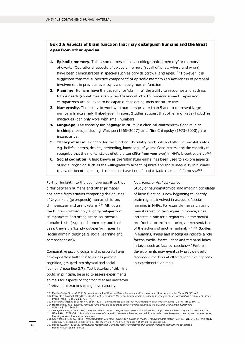
4848
ANIMALS CONTAINING HUMAN MATERIAL
Further insight into the cognitive qualities that
differ between humans and other primates
has come from studies comparing the abilities
of 2-year-old (pre-speech) human children,
chimpanzees and orang-utans.204 Although
the human children only slightly out-perform
chimpanzees and orang-utans on ‘physical
domain’ tests (e.g. spatial memory and tool
use), they significantly out-perform apes in
‘social domain tests’ (e.g. social learning and
comprehension).
Comparative psychologists and ethologists have
developed ‘test batteries’ to assess primate
cognition, grouped into physical and social
‘domains’ (see Box 3.7). Test batteries of this kind
could, in principle, be used to assess experimental
animals for aspects of cognition that are indicators
of relevant alterations in cognitive capacity.
Neuroanatomical correlates
Study of neuroanatomical and imaging correlates
of brain function is now beginning to identify
brain regions involved in aspects of social
learning in NHPs. For example, research using
neural recording techniques in monkeys has
indicated a role for a region called the medial
pre-frontal cortex in capturing a representation
of the actions of another animal.205,206 Studies
in humans, sheep and macaques indicate a role
for the medial frontal lobes and temporal lobes
in tasks such as face perception.207 Further
developments may eventually provide useful
diagnostic markers of altered cognitive capacity
in experimental animals.
201 Martin-Ordas G, et al. (2010). Keeping track of time: evidence for episodic-like memory in Great Apes. Anim Cogn 13, 331–40.202 Penn DC & Povinelli DJ (2007). On the lack of evidence that non-human animals possess anything remotely resembling a ‘theory of mind‘.
Philos Trans R Soc B 362, 731–44.203 For further detail see Jensen K, et al. (2007). Chimpanzees are rational maximizers in an ultimatum game. Science 318, 107–9.204 Herrmann E, et al. (2007). Humans have evolved specialised skills of social cognition: the cultural intelligence hypothesis.
Science 317, 1360–6.205 See Quallo MM, et al. (2009). Gray and white matter changes associated with tool-use learning in macaque monkeys. Proc Natl Acad Sci
USA 106, 18379–84; this study shows use of magnetic resonance imaging and additional techniques to reveal brain region changes during learning of rake tool use in macaques.
206 See Yoshida K, et al. (2011). Representation of others‘ action by neurons in monkey medial frontal cortex. Curr Biol 21, 249-53; this study uses neural recording in monkeys to identify where in the brain the action of others is represented.
207 Peirce JW, et al. (2001). Human face recognition in sheep: lack of configurational coding and right hemisphere advantage. Behav Processes 55, 13–26.
Box 3.6 Aspects of brain function that may distinguish humans and the Great Apes from other species
1. Episodic memory. This is sometimes called ‘autobiographical memory’ or memory
of events. Operational aspects of episodic memory (recall of what, where and when)
have been demonstrated in species such as corvids (crows) and apes.201 However, it is
suggested that the ‘subjective component’ of episodic memory (an awareness of personal
involvement in previous events) is a uniquely human function.
2. Planning. Humans have the capacity for ‘planning’, the ability to recognise and address
future needs (sometimes even when these conflict with immediate need). Apes and
chimpanzees are believed to be capable of selecting tools for future use.
3. Numerosity. The ability to work with numbers greater than 5 and to represent large
numbers is extremely limited even in apes. Studies suggest that other monkeys (including
macaques) can only work with small numbers.
4. Language. The capacity for language in NHPs is a classical controversy. Case studies
in chimpanzees, including ‘Washoe (1965–2007)’ and ‘Nim Chimpsky (1973–2000)’, are
inconclusive.
5. Theory of mind. Evidence for this function (the ability to identify and attribute mental states,
e.g. beliefs, intents, desires, pretending, knowledge of yourself and others, and the capacity to
recognise that the mental states of others can differ from your own) in NHPs is controversial.202
6. Social cognition. A task known as the ‘ultimatum game’ has been used to explore aspects
of social cognition such as the willingness to accept injustice and social inequality in humans.
In a variation of this task, chimpanzees have been found to lack a sense of ‘fairness’.203
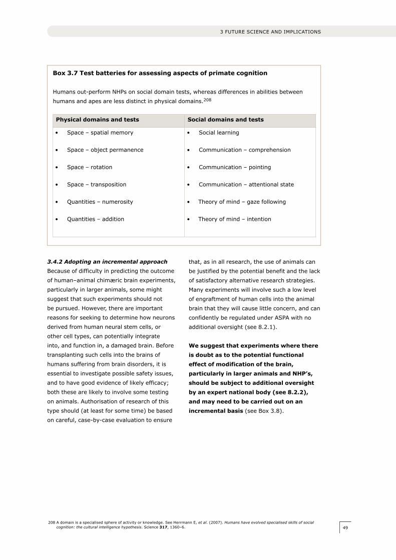
49
3.4.2 Adopting an incremental approach
Because of difficulty in predicting the outcome
of human–animal chimæric brain experiments,
particularly in larger animals, some might
suggest that such experiments should not
be pursued. However, there are important
reasons for seeking to determine how neurons
derived from human neural stem cells, or
other cell types, can potentially integrate
into, and function in, a damaged brain. Before
transplanting such cells into the brains of
humans suffering from brain disorders, it is
essential to investigate possible safety issues,
and to have good evidence of likely efficacy;
both these are likely to involve some testing
on animals. Authorisation of research of this
type should (at least for some time) be based
on careful, case-by-case evaluation to ensure
that, as in all research, the use of animals can
be justified by the potential benefit and the lack
of satisfactory alternative research strategies.
Many experiments will involve such a low level
of engraftment of human cells into the animal
brain that they will cause little concern, and can
confidently be regulated under ASPA with no
additional oversight (see 8.2.1).
We suggest that experiments where there is doubt as to the potential functional effect of modification of the brain, particularly in larger animals and NHP’s, should be subject to additional oversight by an expert national body (see 8.2.2), and may need to be carried out on an incremental basis (see Box 3.8).
208 A domain is a specialised sphere of activity or knowledge. See Herrmann E, et al. (2007). Humans have evolved specialised skills of social cognition: the cultural intelligence hypothesis. Science 317, 1360–6.
3 FUTURE SCIENCE AND IMPLICATIONS
Box 3.7 Test batteries for assessing aspects of primate cognition
Humans out-perform NHPs on social domain tests, whereas differences in abilities between
humans and apes are less distinct in physical domains.208
Physical domains and tests Social domains and tests
• Space – spatial memory
• Space – object permanence
• Space – rotation
• Space – transposition
• Quantities – numerosity
• Quantities – addition
• Social learning
• Communication – comprehension
• Communication – pointing
• Communication – attentional state
• Theory of mind – gaze following
• Theory of mind – intention

5050
ANIMALS CONTAINING HUMAN MATERIAL
209 The basis of the incremental approach should be carefully considered in each situation, and should not mandate additional studies where clear scientific justification is not evident. For example, work on lower species should not be required where previous evidence is already adequate (e.g. from cell-based studies). Good evidence from previous work should always be taken into account in planning and licensing experiments.
210 For example, the use of double blinding, and or automated observation techniques.
Box 3.8 Expert assessment/incremental approach
• For some forms of experiment (as set out in 8.2.2) an incremental research approach
should be agreed at the outset between researchers, inspectors and the national
expert body.
• Initial experiments should usually be undertaken using ‘lowest’ feasible species not
previously studied, in small numbers. Where possible there should be a graduated
approach to the amount/proportion of human material added.209
• Each animal should be tested according to a pre-agreed protocol with clear end-points. Tests
appropriate to the different research situations and species should be used to detect any
modification/loss of the animal’s usual cognitive capacities and behaviours. Close monitoring
of the animals should take place, with due regard to minimising observer bias.210
• Once experience is gained, studies involving larger numbers of animals, a greater
proportion of cellular replacement, and more advanced species could be undertaken.
• For example, research intended to study the effect of incorporating human neurons into an
NHP brain, could start with evidence based on modest neuronal incorporation into rodents,
and proceed by degrees to experiments involving larger scale replacement in NHPs.
In this case, the monitored effects might relate to the development of human-like cognitive
capacities. A range of tests, from which a protocol could be developed are set out in Box 3.7.
• Unusual and ‘first of a kind’ experiments will need to be judged on an individual basis; but
as experience is gained, guidance could be developed so that some classes of experiment
may be undertaken with lower levels of regulatory scrutiny (see Chapter 8).
Box 3.9 Two ways of viewing the brain
Some participants in the public dialogue appeared to adopt a dual conceptualisation of the
brain, in which it was seen as both a purely physical organ, as simply ‘tissue’, and secondly as
the source of consciousness and thought ‘greater than the sum of its parts’. When considering
scientific research, participants often tended to think about the brain in the first way, and few
people appeared to believe that small changes to an animal’s brain at the cellular level would
have a discernable impact on its cognitive function: ‘ … a mouse brain is so much smaller,
I don‘t think a little brain will be able to sit there and “think therefore I am” …’
However, in considering the possible implications of manipulating the brain as a whole, the
second view tended to be adopted. From this viewpoint, some participants expressed a clear
sense of unease around research involving the brain, and its potential outcomes. Some
participants suggested that research that might make an animal’s brain more similar to a
human brain would be unacceptable: ‘I don‘t have a problem with it until it gets to the brain …
but bits to do with memories, that would be too far – it‘s a human thing to have a memory.’
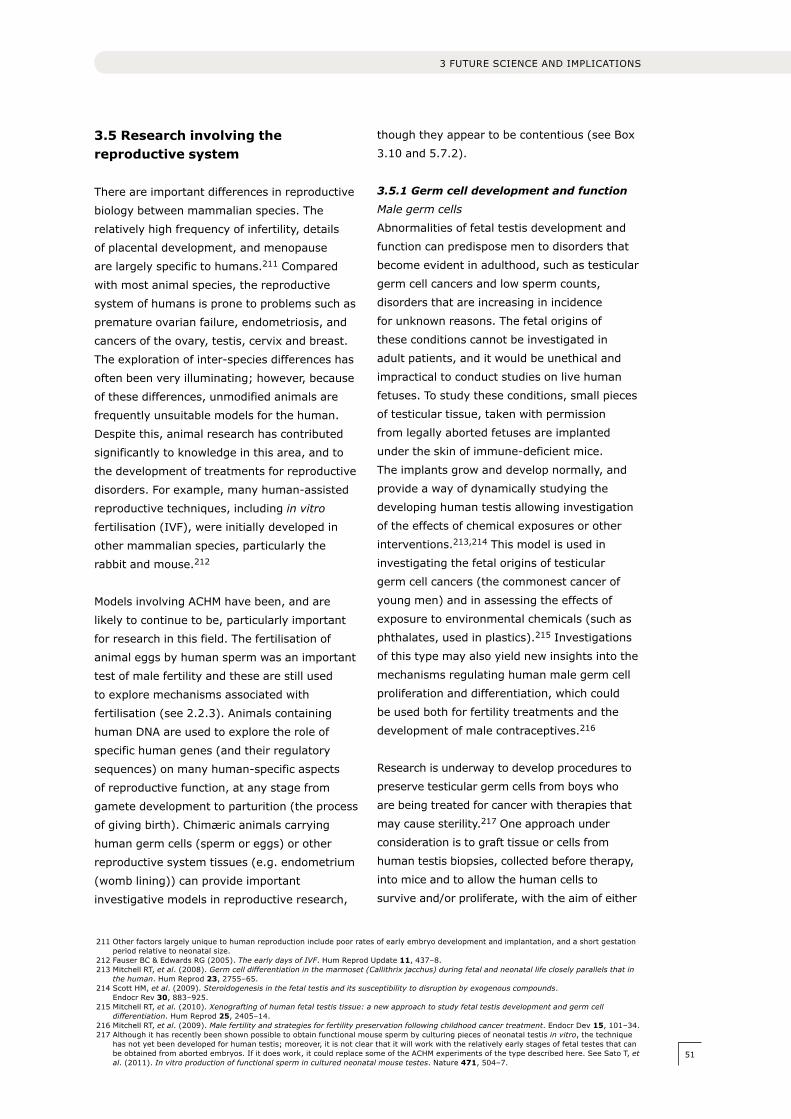
51
3.5 Research involving the reproductive system
There are important differences in reproductive
biology between mammalian species. The
relatively high frequency of infertility, details
of placental development, and menopause
are largely specific to humans.211 Compared
with most animal species, the reproductive
system of humans is prone to problems such as
premature ovarian failure, endometriosis, and
cancers of the ovary, testis, cervix and breast.
The exploration of inter-species differences has
often been very illuminating; however, because
of these differences, unmodified animals are
frequently unsuitable models for the human.
Despite this, animal research has contributed
significantly to knowledge in this area, and to
the development of treatments for reproductive
disorders. For example, many human-assisted
reproductive techniques, including in vitro
fertilisation (IVF), were initially developed in
other mammalian species, particularly the
rabbit and mouse.212
Models involving ACHM have been, and are
likely to continue to be, particularly important
for research in this field. The fertilisation of
animal eggs by human sperm was an important
test of male fertility and these are still used
to explore mechanisms associated with
fertilisation (see 2.2.3). Animals containing
human DNA are used to explore the role of
specific human genes (and their regulatory
sequences) on many human-specific aspects
of reproductive function, at any stage from
gamete development to parturition (the process
of giving birth). Chimæric animals carrying
human germ cells (sperm or eggs) or other
reproductive system tissues (e.g. endometrium
(womb lining)) can provide important
investigative models in reproductive research,
though they appear to be contentious (see Box
3.10 and 5.7.2).
3.5.1 Germ cell development and function
Male germ cells
Abnormalities of fetal testis development and
function can predispose men to disorders that
become evident in adulthood, such as testicular
germ cell cancers and low sperm counts,
disorders that are increasing in incidence
for unknown reasons. The fetal origins of
these conditions cannot be investigated in
adult patients, and it would be unethical and
impractical to conduct studies on live human
fetuses. To study these conditions, small pieces
of testicular tissue, taken with permission
from legally aborted fetuses are implanted
under the skin of immune-deficient mice.
The implants grow and develop normally, and
provide a way of dynamically studying the
developing human testis allowing investigation
of the effects of chemical exposures or other
interventions.213,214 This model is used in
investigating the fetal origins of testicular
germ cell cancers (the commonest cancer of
young men) and in assessing the effects of
exposure to environmental chemicals (such as
phthalates, used in plastics).215 Investigations
of this type may also yield new insights into the
mechanisms regulating human male germ cell
proliferation and differentiation, which could
be used both for fertility treatments and the
development of male contraceptives.216
Research is underway to develop procedures to
preserve testicular germ cells from boys who
are being treated for cancer with therapies that
may cause sterility.217 One approach under
consideration is to graft tissue or cells from
human testis biopsies, collected before therapy,
into mice and to allow the human cells to
survive and/or proliferate, with the aim of either
211 Other factors largely unique to human reproduction include poor rates of early embryo development and implantation, and a short gestation period relative to neonatal size.
212 Fauser BC & Edwards RG (2005). The early days of IVF. Hum Reprod Update 11, 437–8.213 Mitchell RT, et al. (2008). Germ cell differentiation in the marmoset (Callithrix jacchus) during fetal and neonatal life closely parallels that in
the human. Hum Reprod 23, 2755–65.214 Scott HM, et al. (2009). Steroidogenesis in the fetal testis and its susceptibility to disruption by exogenous compounds.
Endocr Rev 30, 883–925. 215 Mitchell RT, et al. (2010). Xenografting of human fetal testis tissue: a new approach to study fetal testis development and germ cell
differentiation. Hum Reprod 25, 2405–14.216 Mitchell RT, et al. (2009). Male fertility and strategies for fertility preservation following childhood cancer treatment. Endocr Dev 15, 101–34.217 Although it has recently been shown possible to obtain functional mouse sperm by culturing pieces of neonatal testis in vitro, the technique
has not yet been developed for human testis; moreover, it is not clear that it will work with the relatively early stages of fetal testes that can be obtained from aborted embryos. If it does work, it could replace some of the ACHM experiments of the type described here. See Sato T, et al. (2011). In vitro production of functional sperm in cultured neonatal mouse testes. Nature 471, 504–7.
3 FUTURE SCIENCE AND IMPLICATIONS
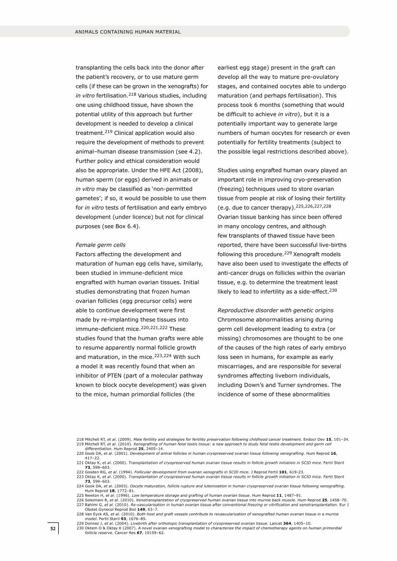
5252
ANIMALS CONTAINING HUMAN MATERIAL
218 Mitchell RT, et al. (2009). Male fertility and strategies for fertility preservation following childhood cancer treatment. Endocr Dev 15, 101–34.219 Mitchell RT, et al. (2010). Xenografting of human fetal testis tissue: a new approach to study fetal testis development and germ cell
differentiation. Hum Reprod 25, 2405–14.220 Gook DA, et al. (2001). Development of antral follicles in human cryopreserved ovarian tissue following xenografting. Hum Reprod 16,
417–22.221 Oktay K, et al. (2000). Transplantation of cryopreserved human ovarian tissue results in follicle growth initiation in SCID mice. Fertil Steril
73, 599–603.222 Gosden RG, et al. (1994). Follicular development from ovarian xenografts in SCID mice. J Reprod Fertil 101, 619-23.223 Oktay K, et al. (2000). Transplantation of cryopreserved human ovarian tissue results in follicle growth initiation in SCID mice. Fertil Steril
73, 599–603.224 Gook DA, et al. (2003). Oocyte maturation, follicle rupture and luteinization in human cryopreserved ovarian tissue following xenografting.
Hum Reprod 18, 1772–81.225 Newton H, et al. (1996). Low temperature storage and grafting of human ovarian tissue. Hum Reprod 11, 1487–91.226 Soleimani R, et al. (2010). Xenotransplantation of cryopreserved human ovarian tissue into murine back muscle. Hum Reprod 25, 1458–70.227 Rahimi G, et al. (2010). Re-vascularisation in human ovarian tissue after conventional freezing or vitrification and xenotransplantation. Eur J
Obstet Gynecol Reprod Biol 149, 63–7.228 Van Eyck AS, et al. (2010). Both host and graft vessels contribute to revascularization of xenografted human ovarian tissue in a murine
model. Fertil Steril 93, 1676–85.229 Donnez J, et al. (2004). Livebirth after orthotopic transplantation of cryopreserved ovarian tissue. Lancet 364, 1405–10.230 Oktem O & Oktay K (2007). A novel ovarian xenografting model to characterize the impact of chemotherapy agents on human primordial
follicle reserve. Cancer Res 67, 10159–62.
transplanting the cells back into the donor after
the patient’s recovery, or to use mature germ
cells (if these can be grown in the xenografts) for
in vitro fertilisation.218 Various studies, including
one using childhood tissue, have shown the
potential utility of this approach but further
development is needed to develop a clinical
treatment.219 Clinical application would also
require the development of methods to prevent
animal–human disease transmission (see 4.2).
Further policy and ethical consideration would
also be appropriate. Under the HFE Act (2008),
human sperm (or eggs) derived in animals or
in vitro may be classified as ‘non-permitted
gametes’; if so, it would be possible to use them
for in vitro tests of fertilisation and early embryo
development (under licence) but not for clinical
purposes (see Box 6.4).
Female germ cells
Factors affecting the development and
maturation of human egg cells have, similarly,
been studied in immune-deficient mice
engrafted with human ovarian tissues. Initial
studies demonstrating that frozen human
ovarian follicles (egg precursor cells) were
able to continue development were first
made by re-implanting these tissues into
immune-deficient mice.220,221,222 These
studies found that the human grafts were able
to resume apparently normal follicle growth
and maturation, in the mice.223,224 With such
a model it was recently found that when an
inhibitor of PTEN (part of a molecular pathway
known to block oocyte development) was given
to the mice, human primordial follicles (the
earliest egg stage) present in the graft can
develop all the way to mature pre-ovulatory
stages, and contained oocytes able to undergo
maturation (and perhaps fertilisation). This
process took 6 months (something that would
be difficult to achieve in vitro), but it is a
potentially important way to generate large
numbers of human oocytes for research or even
potentially for fertility treatments (subject to
the possible legal restrictions described above).
Studies using engrafted human ovary played an
important role in improving cryo-preservation
(freezing) techniques used to store ovarian
tissue from people at risk of losing their fertility
(e.g. due to cancer therapy).225,226,227,228
Ovarian tissue banking has since been offered
in many oncology centres, and although
few transplants of thawed tissue have been
reported, there have been successful live-births
following this procedure.229 xenograft models
have also been used to investigate the effects of
anti-cancer drugs on follicles within the ovarian
tissue, e.g. to determine the treatment least
likely to lead to infertility as a side-effect.230
Reproductive disorder with genetic origins
Chromosome abnormalities arising during
germ cell development leading to extra (or
missing) chromosomes are thought to be one
of the causes of the high rates of early embryo
loss seen in humans, for example as early
miscarriages, and are responsible for several
syndromes affecting liveborn individuals,
including Down’s and Turner syndromes. The
incidence of some of these abnormalities
53
increases significantly with age.231,232,233,234
It can be difficult to study factors that may
predispose to these abnormal cells, especially
during the first meiotic division in oocytes as
this begins during fetal ovary development.
Progress is being made on in vitro growth
and maturation of primary follicles, which
could be used to study later stages of egg
cell division, but these methods are not yet
sufficiently robust.235 Animals carrying grafts
of human ovarian (or testicular) tissue may
allow detailed, longitudinal studies on factors
(hormonal, toxic or age-related) affecting
chromosome segregation.
3.5.2 Endometrial development and
pathology
Experimental approaches involving ACHM
are currently being established to study the
normal physiology of human endometrial
tissue, as well as its malfunction in conditions
such as endometriosis, a condition that may
affect more than 10% of women.236, 237 These
studies involve the engraftment (usually
into the peritoneal cavity) of small sections
of endometrial tissue, taken from a healthy
human donor, or a patient with endometriosis,
into ovariectomised, immune-deficient
mice.238 The mice are treated with a course
of endocrine hormones which imitate the
human female menstrual cycle. The engrafted
human tissue allows study of factors such
as tissue morphology and gene expression.
Bioluminescent or other markers can also be
introduced into the endometrial cells to allow
their growth to be monitored in vivo.239 The
effects of repeated hormone cycles could be
studied. These models are being used to screen
for molecules that might reduce endometrial
cell proliferation or might prevent attachment
and spread of endometriosis.240,241 Recently,
using such mouse models, at least three types
of drug have been claimed to reduce growth
of human endometrial tissue.242,243,244,245,246
Apart from giving promising leads towards
therapy, these mouse studies replace the use of
other animals, notably baboons, that are also
used in research on endometriosis.247
3.5.3 Implantation and placenta
development
Two important areas where ACHM may allow
future research are embryo implantation
into the lining of the uterus and placental
development.
There is a significant loss of human embryos
(perhaps as high as 70%) during pre-implantation
development and around implantation. This
very high rate appears specific to humans, and
although chromosomal abnormalities account for
a significant proportion (see 3.5.1), most causes
are unknown. A failure of interaction between
the embryo and the endometrium into which
it implants is probably also a common cause.
The underlying defect could be intrinsic to the
embryo, the endometrium or the hormone system
that makes the endometrium receptive.248 It
is, however, very difficult to carry out relevant
experiments on human material. Some research
231 Hunt P & Hassold T (2010). Female meiosis: coming unglued with age. Curr Biol 20, R699–702.232 Thomas NS, et al. (2010). De novo apparently balanced translocations in man are predominantly paternal in origin and associated with a
significant increase in paternal age. J Med Genet 47, 112–5.233 Thomas NS, et al. (2001). Maternal sex chromosome non-disjunction: evidence for X chromosome-specific risk factors. Hum Mol Genet 10,
243–50.234 Muhlhauser A, et al. (2009). Bisphenol A effects on the growing mouse oocyte are influenced by diet. Biol Reprod 80, 1066–71235.235 Jin SY, et al. (2010). A novel two-step strategy for in vitro culture of early-stage ovarian follicles in the mouse. Fertil Steril 93, 2633–9.236 Olive DL & Schwartz LB (1993). Endometriosis. N Engl J Med 328, 1759–69.237 Giudice LC & Kao LC (2004). Endometriosis. Lancet 364, 1789–99.238 Ovariectomy (removal of the ovaries) is used to remove the mouse’s own secretion of endocrine hormones. See Masuda H, et al. (2007).
Noninvasive and real-time assessment of reconstructed functional human endometrium in NOD/SCID/gamma c(null) immunodeficient mice. Proc Natl Acad Sci USA 104, 1925–30.
239 Defrere S, et al. (2009). Review: luminescence as a tool to assess pelvic endometriosis development in murine models. Reprod Sci 16, 1117–24.
240 Hull ML, et al. (2008). Endometrial–peritoneal interactions during endometriotic lesion establishment. Am J Pathol 173, 700–15.241 Collins NH, et al. (2009). Characterization of antiestrogenic activity of the Chinese herb, prunella vulgaris, using in vitro and in vivo (Mouse
Xenograft) models. Biol Reprod 80, 375–83.242 The drugs are: simvastatin, a cannabinoid agonist which inhibits the Akt signalling pathway; Raloxifene, a selective estrogen receptor
modulator; and an antibody based protein, ‘icon’, that inactivates a growth factor.243 Bruner-Tran KL, et al. (2009). Simvastatin protects against the development of endometriosis in a nude mouse model. J Clin Endocrinol
Metab 94, 2489–94.244 Leconte M, et al. (2010). Antiproliferative effects of cannabinoid agonists on deep infiltrating endometriosis. Am J Pathol 177, 2963–70.245 Chen YJ, et al. (2010). Oestrogen-induced epithelial–mesenchymal transition of endometrial epithelial cells contributes to the development
of adenomyosis. J Pathol 222, 261–70.246 Krikun G, et al. (2010). The immunoconjugate “icon” targets aberrantly expressed endothelial tissue factor causing regression of
endometriosis. Am J Pathol 176, 1050–6.247 Tirado-Gonzalez I, et al. (2010). Endometriosis research: animal models for the study of a complex disease. J Reprod Immunol 86, 141–7.248 Singh M, et al. (2011). Bridging endometrial receptivity and implantation: Network of hormones, cytokines, and growth factors. J Endocrinol.
In press.
3 FUTURE SCIENCE AND IMPLICATIONS
5454
ANIMALS CONTAINING HUMAN MATERIAL
on implantation is currently conducted in animals
such as the baboon, where mechanisms of
implantation are thought to be fairly similar
to those in humans. There are attempts to
derive in vitro systems to look at implantation
using cultures of endometrium, but these do
not replicate the complex three-dimensional
architecture or physiology of the womb.
ACHM experiments involving grafts of human
uterus or endometrium into animals, or, if
the identities of the human genes required
for receptivity are known, transgenic animals
expressing such genes within their uterus,
might allow human embryo implantation to be
studied. This would not be permitted under the
HFE Act (2008) (see 6.5); however, it might
be possible to use disabled embryos, such as
trophoblast vesicles or tetraploid embryos.
Different mammals often have very different
types of placenta. Some studies can be done
in rodents on placental cell types that appear
similar to those in the human placenta, and
some molecular and genetic pathways are
conserved, but to understand many details of
human placental development and physiology
fully requires studies in humans or closely
related species.249 Although it would be difficult
to study entire human placental development
in animals, it is possible to study the role of
specific human genes in transgenic animals, or to
introduce specific human placental cell types into
the placenta of animals in utero and to determine
their effects on placenta function and physiology.
3.5.4 Other studies involving reproductive
tissues and general concerns
ACHM may be appropriate to study a wide
range of questions about human reproduction,
from eclampsia and birth timing to menopause.
In such studies, human reproductive tissues
are usually implanted into the recipient animal
‘ectopically’ (e.g. under the skin of a mouse
rather than into its own reproductive system),
and there is very little possibility of the eggs or
sperm contacting another germ cell and being
fertilised. However, some experiments of this
type do result in the presence of functional
human sperm and/or egg cells in animals,
which raises the possibility that fertilisation
between human and animal germ cells (or
even between human eggs and sperm) may
inadvertently occur within an animal.
At least one study has reported grafting pieces
of human ovary under the membrane of mice
ovaries, creating the possibility that human
oocytes might enter the reproductive tract of
female mice.250 The females were not allowed
to mate, but if they had (with male mice), there
would be very little chance of hybrid embryo
development or implantation. Human sperm
do not normally penetrate the mouse zona
pellucida (a type of protective shell around
the egg), as species-specific molecules are
required. Transgenic mice expressing human
zona pellucida proteins are being used to
search for the relevant molecules.251,252 To
achieve cross-species fertilisation, for example
in tests of human male fertility using hamster
or mouse eggs, it is necessary to remove the
zona pellucida or to use ICSI.253 Such tests are
usually terminated at the two-cell embryo stage,
although ‘true-hybrid’ embryos may be allowed
to develop for 14 days (see Box 6.6).254 We have
briefly discussed the problems of studying human
embryo implantation and human placental
development (3.5.3). Would it be possible to
transplant a human uterus into an animal and
then use this to implant human embryos? Given
that uterus transplants are being considered
249 For example, at least two retroviral elements, HERV-W and HERV-FRD, both of which lead to the expression of viral envelope proteins (termed Syncytin and Syncytin2, respectively) in the human placenta, are specific to the primate lineage. The Syncytin proteins lead to cell fusion and to the formation of a specialised tissue, termed syncytiotrophoblast, which plays an important role in the maternal–fetal interchange of nutrients. Although some rodents also have syncytiotrophoblast, this appears to be due to the activity of rodent-specific retroviral genes. Other retroviral integrations are thought to have affected the expression of endogenous genes involved in human placental development, such as the Insulin-like 4 gene (INSL4).
250 Dath C, et al. (2010). Xenotransplantation of human ovarian tissue to nude mice: comparison between four grafting sites. Hum Reprod 25, 1734–43.
251 xu YN, et al. (2010). DNA synthesis and epigenetic modification during mouse oocyte fertilization by human or hamster sperm injection. J Assist Reprod Genet. In press.
252 Yauger B, et al. (2011). Human ZP4 is not sufficient for taxon-specific sperm recognition of the zona pellucida in transgenic mice. Reproduction 141, 313–9.
253 Intra-cytoplasmic sperm injection (ICSI) involves injecting a single sperm directly into an egg in order to fertilise it.254 However, subsequent development is unlikely to occur owing to epigenetic defects, aneuploidy, and species-specific factors controlling
implantation.
55255 Grynberg M, et al. (2011). Uterine transplantation: a promising surrogate to surrogacy? Ann N Y Acad Sci 1221, 47–53.256 Correspondence from Fisher, E.
3 FUTURE SCIENCE AND IMPLICATIONS
Box 3.10 Public views on research involving human reproductive tissues
The creation of animal models including human reproductive tissue was a very sensitive area
for public participants. Compared with other human tissues, the use of animal models involving
human reproductive cells was regarded as acceptable by the fewest number of participants in
the quantitative survey (42%).
‘ … that is so far out there, just awful. Perhaps if there was no sperm left on earth, but otherwise
no way.’
Dialogue discussions identified several possible explanations for these responses, including:
• The cultural significance of reproductive cells (through associations with sex, the production
of children, birth experiences and development, and familial characteristics).
• A suggestion that even small changes to a single reproductive cell might produce profound
effects (reproductive cells were seen as easy to ‘abuse’, and contrasted with the brain,
where ‘changing a few cells might not matter’).
• A view that the consequences of research involving human reproductive cells might be
experienced not only by the animal involved, but potentially by resulting human offspring.
as an alternative to surrogacy in humans,
this is not necessarily a remote possibility.255
Careful thought would need to be given about
such experiments from scientific and ethical
perspectives. Licensing of any animal experiment
where there is a chance of human embryo or
true hybrid development should address the
precautions taken to avoid this (see 8.2.2).
3.6 Research involving human appearance or behavioural traits
Current ACHM do not show overt human-like
appearance or behaviours; the alterations are
seen at a biochemical or pathological level.
Transgenic mice have the appearance of ordinary
mice; chimæric goats engrafted with human
stem cells look like ‘ordinary’ goats. Even the
most extensive of current genetic modifications,
such as the addition of a human chromosome to
mice in the Down’s syndrome model (see 3.2.),
do not markedly alter the appearance of the
animals to a casual human observer.256
Participants in the public dialogue expressed
particular concern that the incorporation of
human material into experimental animals
might result in the creation of animals with
‘human-like’ appearance or characteristics
(see Box 3.11). There are some cardinal
phenotypic features that are intuitively
recognised as essentially human, such as facial
appearance and skin texture, and behaviours
including speech. Experiments that confer
these properties on animals may be expected
to attract public interest (see for example the
reaction to the Vacanti Earmouse, Box 3.12).
The societal and ethical bases of such concern
are discussed in Chapter 5.
Box 3.11 Public views on research involving human-like appearance
Research involving external body parts, such as the use of human hair, skin, or the possible
development of human-like limbs on animals, was often met with distaste by dialogue
participants. This type of response was attributed to participants’ ability to imagine and visualise
the resulting animal as ‘unnatural’. The physical appearance of animals was found to be an
important way in which participants identified animals as different ‘kinds’, and changes to
external features might be seen to blur these well-recognised visible distinctions between species.
5656
ANIMALS CONTAINING HUMAN MATERIAL
3.6.1 Human external appearance
Studies involving the transplantation of human
skin onto animals are undertaken for several
research purposes. Exposure of human skin
to radiation (such as ultraviolet B in sunlight)
can lead to DNA damage and skin cancer, and
mouse models have been developed for use in
research to understand the mechanistic basis of
cancers of this kind. Human skin with different
pigmentation types, and skin from cancer-
prone patients with the disease Xeroderma
pigmentosum, have been transplanted onto
immune-deficient mice, allowing the effects
of radiation exposure to be investigated, and
potential therapeutics tested.257
Mice are used in the study of psoriasis, a
human skin condition that results in the
development of scaly, red patches on the skin.
Some forms of psoriasis result from disorders
of the immune system, and mice transplanted
with skin grafts from psoriatic patients have
been used to understand the malfunctioning
relationship between the epidermal (skin) cells
and the immune system.258 Mice with human
skin grafts have also been used to improve
grafting techniques (e.g. for use with burns
patients) and to investigate approaches to
reduce the immune rejection of skin grafts.259
In such studies only a small area of human skin
is grafted onto the recipient mice.
Transgenic animal models are contributing
to understanding of the genetic basis of limb
development. For example, the Prx1 gene codes
for a DNA-regulating protein important for the
growth of limb bones. The regulatory sequences
affecting Prx1 expression are now known in
several species, including the mouse and bat. In
studies to investigate their function, the mouse
regulatory sequences were exchanged for the
equivalent regulatory regions from the bat.
The resulting transgenic mice had elongated
forelimbs.260 Mutation of a human gene
regulatory sequence results in the development
of extra digits (‘pre-axial polydactyly’). Mice
carrying the same genetic mutation are
also born with extra digits. The mice have
contributed significantly to the identification of
the developmental basis of this condition, which
is now known to result from extra expression
of a protein in a small patch on one side of the
developing limb.261 Animal and human limbs are
composed of similar cell types making similar
proteins; the different shapes of human and
animal limbs presumably reflect differences in
gene regulation. Testing this could in theory lead
to transgenic animals with human-like hands
or feet. This could give basic understanding
that has clinical importance, but it may cause
some disquiet and it would, of course, have
consequences for the animals, including
mismatch between hard-wired behavioural
patterns and what the new limbs can do.
In future, genes underlying the development
of other body parts, perhaps including facial
features, may also be studied in animals.
Such experiments may require consideration
from both socio-ethical and animal welfare
perspectives. An animal may be distressed
by an unusual body part, may suffer rejection
by its own species, or elicit unusual response
from those charged with its care (see 4.1.2).
Attempts should be made to anticipate such
effects, in the design and licensing of the work.
Recognisable fragments of teeth, hair and other
tissues can sometimes arise in naturally occurring
tumours, known as ‘teratomas’, which are
occasionally found in humans and other species.
They arise from remnants of very early stem
cells, capable of differentiating into different body
tissues. Similar teratomas are often created in
stem cell research (see 3.3.1), when human or
other embryonic stem cells are implanted into
mice, for example to test the cells’ developmental
potential, resulting in the presence of, for
example, fragments of human tooth or hair
(within the tumour) in the mouse.262
257 Sun xZ, et al. (2008). Animal models of xeroderma pigmentosum. Adv Exp Med Biol 637, 152–60.258 Guerrero-Aspizua S, et al. (2010). Development of a bioengineered skin-humanized mouse model for psoriasis: dissecting epidermal-
lymphocyte interacting pathways. Am J Pathol 177, 3112–24.259 Issa F, et al. (2010). Ex vivo-expanded human regulatory T cells prevent the rejection of skin allografts in a humanized mouse model.
Transplantation 90, 1321–7.260 Cretekos CJ, et al. (2008). Regulatory divergence modifies limb length between mammals. Genes Dev 22, 141–51.261 Lettice LA, et al. (2008). Point mutations in a distant sonic hedgehog cis-regulator generate a variable regulatory output responsible for
preaxial polydactyly. Hum Mol Genet 17, 978–85.
57
3.6.2 Human behavioural traits
It is hard to argue that many behavioural traits
are individually unique to humans, although
large brains and manual dexterity allow us
to generate sophisticated tools, which then
influence aspects of our behaviour, such as
writing and reading, playing and appreciating
music, and playing sports.
Recent research to investigate language-
related disorders identified a mutation in a
gene (known as FOXP2), which was found to
be associated with an inherited form of speech
and language disorder in humans. The FOXP2
sequence was found to be different between
humans and Great Apes (and other mammals),
leading to the suggestion that these changes
may be partly responsible for the acquisition
of speech during human evolution.263
Furthermore, when the human equivalent
sequences were introduced into mice, they
developed vocalisations different from those of
non-modified mice.264,265 These studies provide
some evidence to suggest roles for genes such
as FOXP2 in the processes underpinning speech
and language development.
However, it is important to distinguish
vocalisation (making sound) from speech
and language (the complex human system
of communication). Parrots are capable of
complex vocalisation and ‘mimickry’. The
capacity to make sounds is not the same as
the possession of language, which involves
many cognitive processes (e.g. memory
symbolisation, a shared communicative
structure of signs and a process of learning in
interaction with adults at crucial developmental
stages). Evidence for true language acquisition,
even in higher NHP species such as the
chimpanzee, is controversial and inconclusive
(see Box 3.6). It is likely that more genes
underpinning speech development will be
identified in future. However, even if all the
genes underlying these processes could be
introduced into an NHP, it remains a matter of
speculation whether the brain of the modified
animal would then be capable of language
acquisition. Although in some studies carefully
trained NHPs have developed some aspects of
communication (see Box 3.6), is it not clear
that even a modified NHP brain would have the
capacity for complex human communication in
its true sense.266
Creating characteristics such as speech
and behaviour in animals would be very
complex, probably requiring manipulation of
environmental as well as biological factors.
Authorisation of such work would need to be
justified by considerable potential benefit and
the lack of satisfactory alternative research
strategies. Measures to determine and respond
to public sensitivity should be considered before
licensing such research.
262 Cao Y et al. (1997). Transplantation of chondrocytes utilizing a polymer-cell construct to produce tissue-engineered cartilage in the shape of a human ear. Plast Reconstr Surg 100(2), 297-302.
263 Hardin J (1998). Producing tissue-engineered cartilage in the shape of a human ear. Plast Reconstr Surg 101(6), 1745.264 Reimers-Kipping S, et al. (2011). Humanized Foxp2 specifically affects cortico-basal ganglia circuits. Neuroscience 175, 75–84.265 Enard W (2011). FOXP2 and the role of cortico-basal ganglia circuits in speech and language evolution. Curr Opin Neurobiol. In press.266 Human communication conveys meaning and intent, which requires a concept of the mental state of others to whom you are communicating.
There are reported instances of human children who have not developed language. Such findings suggest that, although the capacity for human language might have a biological basis, its realisation depends on immersion in complex human communities from birth. For example,
3 FUTURE SCIENCE AND IMPLICATIONS
Box 3.12 The Vacanti ear-mouse
The ‘Vacanti ear-mouse’, which appeared to have a human ear grown on its back, was created
in 1997. The mouse was created to demonstrate a method of fabricating cartilage structures for
transplantation into human patients. The ‘ear’ was actually a cartilage structure, grown by seeding
cow cartilage cells into a biodegradable, ear-shaped, polyester fabric mould, which was then
implanted under the mouse’s skin. Although the Vacanti mouse did not contain any human tissue,
and was not functional, its human-like appearance evoked a strong public interest and is still widely
remembered today (erroneously) as an example of an animal containing a human organ. 262,263

5858
ANIMALS CONTAINING HUMAN MATERIAL
see ‘Genie’ – http://en.wikipedia.org/wiki/Genie_(feral_child) and ‘Oxana’ http://www.mymultiplesclerosis.co.uk/misc/feral-children.html.

59
4 WELFARE AND SAFETY ASPECTS OF ACHM
4.1 Welfare
The protection of animals is central to the
operation of the UK’s Animals (Scientific
Procedures) Act 1986 (ASPA), which is intended
to ensure that animals used in research are not
subject to unnecessary pain, suffering, distress
or lasting harm (see 6.2.1, and for a wider
international perspective see 7.3.1).267 Under
ASPA, all experiments involving ‘protected’
animals must be licensed, and they can be
licensed only if there are no scientifically
suitable alternatives that replace animal use,
reduce the number of animals needed or refine
the procedures used to cause less suffering
(principles known as the ‘3Rs’ see 6.2.1).268
Decisions to license research must take
into account the likely benefits (to humans,
other animals or the environment), weighed
against the likely welfare costs to the animals
involved.269 Additional requirements apply
to particular research, such as that involving
genetically altered animals or species including
NHPs (see Box 6.1).270 This long-standing
framework underpins the close governance
of animal research in the UK, which is more
carefully scrutinised than other uses of animals
such as in agriculture, or as companion animals
(pets). (See Box 4.1 for public views on animal
welfare.)
Application of animal welfare principles is
an obligation on individuals and institutions
under the Home Office licensing system,
and is monitored both locally, for example
by ‘named animal care and welfare officers’,
and by the Home Office inspectorate.
Further improvements are encouraged and
taken forward in the UK through the work
of the National Centre for the Replacement,
Refinement and Reduction of Animals in
Research (NC3Rs), and other bodies.271
An important aspect of such work is the
development of guidelines for best practice, in
areas such as welfare assessment of genetically
modified rodents, and in defining the welfare
needs of particular animal species.272 We emphasise that research involving ACHM should be subject to scrutiny, and advancement from the perspective of animal welfare, in a manner no different from other animal research.
Here we introduce two aspects of animal
welfare relating specifically to ACHM:
• Thepossibilitythatthecreationoruseof
ACHM raises specific welfare concerns.
• ThepotentialofACHMresearchto
contribute to advancement of the 3Rs
(replacement, reduction and refinement,
see above).
Further consideration is included in Chapter 5
(5.5).
4.1.1 ACHM and animal welfare
In principle, the use of ACHM that closely model
human biology increases the likely benefit
of the research and so contributes to the
refinement of experimental techniques. ACHM
use can support animal welfare principles by
enabling researchers to use species likely to
experience less pain, suffering or harm, or to
reduce the numbers of animals used in some
experimental situations.273
4 Welfare and safety aspects of ACHM
267 New legislation, intended in part to bring harmonisation in animal welfare practices across Europe, has recently been adopted (see 7.3.1). 268 See Guidance on the Operation of the Animals (Scientific Procedures) Act 1986, Section 2.3. http://www.archive.official-documents.co.uk/
document/hoc/321/321.htm269 Animal Procedures Committee (2003). Review of the cost-benefit assessment in the use of animals in research.
http://www.homeoffice.gov.uk/publications/agencies-public-bodies/apc/key-reports/270 Research involving NHPs is only permissible where there is strong scientific justification, and where no other species are suitable for the
purposes of the programme of work, or where it is not practicable to obtain animals of any other species that are suitable for those purposes. See Guidance on the Operation of the Animals (Scientific Procedures) Act 1986, Section 5.22.
271 The National Centre for the Replacement, Refinement and Reduction of Animals in Research (NC3Rs) is an independent scientific organisation, tasked by Government with supporting the UK science base through the application of the 3Rs. See www.nc3rs.org.uk
272 For example see, ‘Mouse Welfare Terms’ http://www.mousewelfareterms.org/doku.php?id=home developed by the Medical Research Council Harwell and Wellcome Trust Sanger Institute Cambridge; Wells et al. (2006) Full report of GA mouse welfare assessment working group. Lab Animals 40,111–114; Ellegaard L, et al. (2010). Welfare of the minipig with special reference to use in regulatory toxicology studies. J Pharmacol Toxicol Methods 62, 167–83; RETHINK http://www.rethink-eu.dk/index.php?page=one&id=8
273 Use of a lower species (phylogenetic reduction) is often considered to be refinement, but such a judgement can only be made if assessment of the available scientific evidence suggests that the lower species is less sentient/likely to suffer less. See http://www.nc3rs.org.uk/category.asp?catID=78

6060
ANIMALS CONTAINING HUMAN MATERIAL
Research involving ACHM, particularly mice
with humanised organs such as the liver, or the
immune system, can be used as an alternative
to NHPs in investigating infectious diseases.
For example, they have facilitated studies of
HIV and hepatitis, to which unmodified mice
are not susceptible, and for which NHPs have
previously been used (see 2.3.3). ACHM
approaches also have potential application in
drug development and testing; for example
the use of transgenic mice susceptible to polio
virus through incorporation of the human CD155
gene has been approved as an alternative to
NHP use in polio vaccine testing.274 A protocol
developed using a chimæric mouse model which
recapitulates forms of human cancer facilitates a
significant reduction in the number of mice used
compared with previous approaches. This is due
to the more faithful development of cancer in the
chimæric model, and elimination of the need for
breeding programmes.275 The recent increase
in the development of antibody therapies (see
2.3.2) has resulted in an increase of the use of
NHPs in toxicity testing of these therapeutics.
This is an important avenue for future study in
which mice with humanised immune systems
may reduce (though not fully replace) NHP
use. For other types of human condition, for
example those affecting cognitive abilities, it
may be that NHP models incorporating human
material are so much better than similar rodent
models that they will allow an overall reduction
and refinement, and lead more rapidly to
treatments. An example where this might be the
case (although no treatments are yet available)
is Huntington’s disease.276
We anticipate that the use of animals
containing human material is likely to present
further avenues for advancement of the 3Rs.
We support their development and use, while
emphasising the view put forward in evidence
that ‘… the development of an ‘improved’ model
needs to be followed by a rigorous and critical
appraisal of the value of existing models by
research funders, scientists and regulators.’277
However, we do not anticipate that research
involving ACHM would decrease the overall
use of animals in medical research in the short
term, in part because the development of ACHM
will open up new research avenues.
4.1.2 Specific welfare concerns
We have considered whether the incorporation
of human genetic or cellular material into an
animal might in itself have the potential to
cause a distinct dimension of ‘pain, suffering
or lasting harm’ to the animal involved. Our
general conclusion is that, although individual
experiments may give rise to particular types of
animal suffering, the techniques in themselves do
not raise distinct types of animal welfare concern.
Social aspects of animal welfare are
increasingly taken into account, and research
animals are housed in appropriate and species-
specific environments, which often involve
‘group-housing’ (e.g. of NHPs). We considered
whether, by conferring a human characteristic
onto an animal (such as appearance, e.g.
through engraftment of human skin (see
3.6), or a behavioural trait) an animal might
suffer distress or harm, resulting from the
actions of others of its own species, or those
responsible for its care. Although the potential
for suffering brought about in this way is
plausible, it does not represent a ‘unique’
dimension of suffering that is specific to the
creation of ACHM, because similar situations
can arise (and need to be taken into account
in assessing welfare issues) in other types of
research. Evidence submitted to the study
indicated that there is: ‘no rationale, specifically
on animal welfare grounds, for moving to
regulate this type of research differently from
other animal research’ and that ‘research
involving ACHM is not significantly different to
other areas of animal research from an animal
274 Humans and primates express a protein (CD155) on their neurons which confers susceptibility to infection by the polio virus. Batches of live polio vaccine for human use cannot be tested to determine their activity (virulence) on species that lack the CD155 protein (including mice) and are therefore tested on NHPs. See Shultz LD, et al. (2007). Humanized mice in translational biomedical research. Nat Rev Immunol 7, 118–30; Mendelsohn CL, et al. (1989). Cellular receptor for poliovirus: molecular cloning, nucleotide sequence, and expression of a new member of the immunoglobulin superfamily. Cell 56, 855–65; Dragunsky EM, et al. (2006). Further development of a new transgenic mouse test for the evaluation of the immunogenicity and protective properties of inactivated poliovirus vaccine. J Infect Dis 194, 804–7.
275 Zhou Y, et al. (2010). Chimeric mouse tumor models reveal differences in pathway activation between ERBB family- and KRAS-dependent lung adenocarcinomas. Nat Biotechnol 28, 71–8.
276 Yang SH & Chan AW (2011). Transgenic Animal Models of Huntington‘s Disease. Curr Top Behav Neurosci 7, 61–85.277 Written evidence from the Royal Society for the Protection of Cruelty to Animals (RSPCA).
61
welfare perspective.’278,279 Although we do
not currently see any reason for this aspect
of animal welfare to be treated differently in
ACHM experiments compared with other animal
experimentation, this matter should be kept
under review as techniques evolve.
We considered whether, through incorporation
of human neurons into its brain, an animal
might in some way be made more ‘self-aware’
and therefore capable of experiencing a greater
degree of suffering (see 3.4.1 and 5.6.2).
The same issues would potentially apply to
any situation where neural cells from a more
self-aware species are introduced into one
that is less self-aware, such as chimpanzee
into macaque, or macaque into marmoset.
However, as humans are probably the most
self-aware species (at least we like to think
so), then ACHM pose the greatest risk of this
happening. We are not aware of any evidence
that self-awareness has been altered in such
experiments, but researchers and regulators
should be aware of the possibilities.
The effect of animal experimentation on
those directly responsible for the day-
to-day care of research animals is often
underestimated.280,281 Although ACHM in
general are unlikely to pose additional concerns
in this respect, it is conceivable that some
individual carers might react differently to
animals containing large amounts of human
material, or with altered appearance or
behaviour, whether or not the animals were
actually more ‘human-like’. There could be
positive or negative effects on either the
animals or their carers. This is a topic that
could be explored, especially as there is an
increasing tendency for animal technicians to
become more directly involved in the design
and interpretation of experiments.
Box 4.1 Public concern for animal welfare
The views of participants in the public dialogue on animal welfare emerged in several ways.
Although the dialogue was not intended to explore attitudes to the general use of animals
in research, animal welfare concerns were consistently expressed, and participants often
transferred broad concerns for the welfare of research animals directly onto research using ACHM.
Overall, as described in Box 3.1, participants expressed conditional support for ACHM. Animal
welfare was one of the considerations which they took into account when thinking about whether
such research would be justified. (See Box 5.1 for more discussion of these considerations.)
In the quantitative survey, animal welfare was the reason most often given by those who found
introducing human material into animals unacceptable. When participants were asked about
the welfare aspects relating specifically to ACHM, there were a few suggestions that a new kind
of suffering might result from the creation of ACHM. These included concerns that modifying
an animal’s external organs to cause them to appear human in some way might cause the
animal distress, or that research involving the brain might alter an animal’s perception of its own
circumstances and so increase its suffering. However, for the most part, participants did not feel that
the creation of ACHM would produce greater suffering than other types of research involving animals.
‘It‘s a great deal of suffering. The fact that it has human material makes no difference really.’
This concerned but fundamentally supportive view of animal experimentation, if carried out for
medical advancement, is in agreement with recent trends in public polling on the topic – see
The 2010 Ipsos MORI Report on Public Attitudes towards Animal Experimentation.282
278 Oral evidence from the Royal Society for the Protection of Cruelty to Animals (RSPCA).279 Oral evidence from Robinson V., National Centre for the Replacement, Refinement and Reduction of Animals in Research280 Herzog H (2002). Ethical aspects of relationships between humans and research animals. ILAR J 43, 27–32.281 Coleman K (2011). Caring for nonhuman primates in biomedical research facilities: scientific, moral and emotional considerations.
Am J Primatol 73, 220–5.282 Ipsos MORI (2011). Views on Animal Experimentation (BIS research) Alternatives to Animal Experimentation (NC3R research).
http://www.ipsos-mori.com/researchspecialisms/socialresearch/specareas/nhspublichealth/attitudestowardsanimalexperimentation.aspx
4 WELFARE AND SAFETY ASPECTS OF ACHM

6262
ANIMALS CONTAINING HUMAN MATERIAL
4.2 Safety
4.2.1 Introduction
Research involving ACHM is subject to safety
controls that apply to all biomedical research.
These include practices set out in legislation
and guidance to protect against hazards
to human health and to the environment
(e.g. principles of occupational safety and
hygiene, and good laboratory practice).283
Further precautions apply to studies involving
genetically altered animals, relating to their
containment or deliberate release into the
environment (see 6.2.4).
We are grateful for the advice of experts
outside the working group, which has aided us
in this consideration.284
4.2.2 Safety issues considered
Some ACHM experiments will raise safety
issues related to the individual experiments
being proposed, for example those where
animals are made susceptible to infectious
agents normally confined to humans, including
viruses, bacteria, parasites and prions (see
2.3.3).285 Such hazards must be considered
and managed in order to protect those handling
the animals, the animals themselves (from
inadvertent infection) and the public, notably
from infection or the escape of animals which
might act as a reservoir of infection. Neither
the issues nor the methods of managing them
are different in ACHM experiments from those
regularly encountered when dealing with other
types of experiment involving infectious agents
or other biohazards in animals or cell cultures.
For example, ferrets are susceptible to human
influenza viruses, but they are routinely used to
study the viruses in facilities with a high level of
containment. Similarly, how work with human
cells or tissue in vitro is conducted will depend
on the nature of any hazards that might be
generated by the experiments proposed, but
the minimum conditions used are those that
protect both the cells and the researchers from
adventitious infection. All such experiments
should be assessed in advance for potential risk
by researchers and regulators, and managed
accordingly; exactly the same considerations
apply to ACHM work.
We considered whether there are additional,
generic safety issues applicable to research
involving ACHM. The major potential issues
identified arise from the fact that complex
genomes (both human and animal) carry within
them integrated viral genomes (endogenous
retroviruses or proviruses). These may be
quiescent and only able to replicate under
certain conditions; indeed many are inactivated
because during evolution either the viruses
have lost an essential component that enables
them to replicate satisfactorily, or the host
has lost cell-surface receptors or intracellular
factors essential for viral entry and infection
of new cells. Specific intracellular host factors
(known as restriction factors) can also produce
resistance to infectious agents such as viruses;
these are species specific and can confer
species resistance to particular infectious
agents such as HIV.286 This is a further barrier
to cross-species transmission of infectious
agents. It is, however, known that infectious
agents can, occasionally, change their host
specificity (e.g. owing to mutation and/or
recombination with other related viruses)
such that they become able to infect species
previously not liable to infection. Such events
are likely to have been involved in influenza
strains moving from birds to humans, or
from pigs to humans (swine ‘flu). HIV might
have moved into humans from NHP hosts by
similar mechanisms in the wild. We therefore
considered whether ACHM experiments could
lead to an increased likelihood of reactivation
of quiescent viruses or to changes of host
specificity of infectious agents.
283 See Health and Safety Executive. The Scientific Advisory Committee on Genetic Modification (SACGM) Compendium of guidance, Part 3 (Containment and control of activities involving genetically modified microorganisms). http://www.hse.gov.uk/biosafety/gmo/acgm/acgmcomp/index.htm; Organisation for Economic Co-operation and Development (OECD) Principles on Good Laboratory Practice http://www.oecd.org/officialdocuments/displaydocumentpdf/?cote=env/mc/chem(98)17&doclanguage=en
284 Correspondence from Lever A., Stoye J., Bradley A., Weiss R., and Weissmann C.285 A prion is an infectious agent composed of protein in a misfolded form.286 Lever AM & Jeang KT (2011). Insights into cellular factors that regulate HIV-1 replication in human cells. Biochemistry 50, 920–31.

63
4.2.3 Inter-species chimæras
Making chimæras involves mixing cells or
tissues containing whole genomes (including
their integrated viruses) of different species. We
considered whether such an intimate admixture
of human and animal cells and tissues might
lead to reactivation of infectious particles,
such as retroviruses or other pathogens; or to
alter their host specificity so that they become
infectious to humans. Very similar issues have
been extensively discussed in the debate around
the transplanting of animal tissues into humans,
referred to as xenotransplantation.
Humans and animals have lived in close
proximity for a long time, and although
examples of viral transfer between human
and other species are well known, they are
relatively infrequent.287 Animal tissues have
been introduced into people (e.g. pig heart
valve transplants, baboon hearts), although
in small numbers. What limited evidence is
available on humans who have received living
pig cells indicates that no infection by porcine
viruses has taken place.288,289,290 A number
of studies have, however, shown transient
appearances of foreign virus in humans or
animals who had received cellular material from
other species, suggesting that this issue must
be kept under careful review.291,292,293,294 Any
move from experimental into clinical systems
will, as with any new therapy, need very careful
assessment of safety including infectious risk.
There have been years of experience, and
large numbers of experiments, grafting human
tissues such as tumours into other species.
Human tumour tissue transplanted into
immunodeficient mice is known to become
infected by endogenous mouse retrovirus. We
know of no proven incidents of transmitted
infectious events hazardous to man.295
Mice with human immune systems or mostly
human livers have been produced for studying
specific infections, and have therefore been
closely monitored. To our knowledge there
have been no reports that these animals have
developed any problems due to activation of
proviruses or to novel infections.
Inter-specific cell hybrids involve an even closer
association of cells than is generally the case
in chimæras because they involve the fusion
of whole cells, which can be from a range of
species including animal–human combinations.
These have been cultured in laboratories for
decades, without any generic safety issues of
this sort arising.
Chimæras that comprise a mixture of many
different cell types, both human and animal,
may possibly pose a slightly greater risk than
the examples above. This is partly because
specific molecules on the cell surface (referred
to as receptors) to which viruses and other
pathogens attach are often cell-type specific
(e.g. influenza viruses tend to infect cells lining
the upper respiratory tract, other pathogens
target cells in the gut). The greater the range
of cell types present from the two species,
the greater the chance of any virus finding
its appropriate receptor. Moreover, cell fusion
does occur naturally in some tissues, such as
placenta and muscle, so that inter-species
chimæras may also contain inter-specific
cell hybrids, increasing the chance of viral
recombination events.
Factors relating to the animal host may affect
the probability of an adverse event occurring.
For example, the longer cell types from two
species co-exist the more opportunity there
may be for rare events to occur, so that
chimæras with a long life span may deserve
287 For example, see Shukla P, et al. (2011). Cross-species infections of cultured cells by hepatitis E virus and discovery of an infectious virus–host recombinant. Proc Natl Acad Sci USA 108, 2438–43.
288 Ekser B, et al. (2009). Xenotransplantation of solid organs in the pig-to-primate model. Transpl Immunol 21, 87–92.289 Paradis K, et al. (1999). Search for cross-species transmission of porcine endogenous retrovirus in patients treated with living pig tissue.
The XEN 111 Study Group. Science 285, 1236–41.290 Weiss RA (1998). Transgenic pigs and virus adaptation. Nature 391, 327–8.291 Teotia SS, et al. (2005). Prevention, detection, and management of early bacterial and fungal infections in a preclinical cardiac
xenotransplantation model that achieves prolonged survival. xenotransplantation 12, 127–33.292 Michaels MG, et al. (2004). Baboon bone-marrow xenotransplant in a patient with advanced HIV disease: case report and 8-year follow-up.
Transplantation 78, 1582–9.293 Michaels MG, et al. (2001). Detection of infectious baboon cytomegalovirus after baboon-to-human liver xenotransplantation. J Virol 75, 2825–8.294 Stoye JP & Coffin JM (1995). The dangers of xenotransplantation. Nat Med 1, 1100.295 We are aware of claims that some cases of prostate cancer and myalgic encephalopathy have been associated with murine derived retrovirus.
These claims remain scientifically contentious and it is not clear that, even if true, they are related to ACHM. See Urisman A, et al. (2006). Identification of a novel Gammaretrovirus in prostate tumors of patients homozygous for R462Q RNASEL variant. PLoS Pathog 2, e25; Hue S, et al. (2010). Disease-associated XMRV sequences are consistent with laboratory contamination. Retrovirology 7, 111.
4 WELFARE AND SAFETY ASPECTS OF ACHM
6464
ANIMALS CONTAINING HUMAN MATERIAL
closer attention, especially as aged animals can
show reduced immune function. Such events
might be more likely to occur in animals that
are immune deficient.
It is conceivable that human cells isolated from
animal–human chimæras and grown in the
laboratory might have acquired replication-
competent retroviruses from the animal host.
Such animal viruses do not usually cause
problems to humans when they are made
by animal cells, because they have animal-
type coat modifications (alpha-gal epitopes)
that would lead to them being detected and
destroyed or severely damaged by anti-alpha-
gal antibodies that are present in humans.
However, if the viruses had moved from the
animal to the human cells within a chimæric
animal and these human cells were then isolated
and grown in culture, the viruses could be
competent to infect human cells.296,297,298
Any future attempts to use material derived from
chimæric animals for therapeutic purposes would
need to be very carefully assessed for safety (as
is the case with any proposed new therapeutic)
and particularly for risk of viral transmission.
Researchers studying pathogens are more likely
to consider the infection risks than those who
do not. It follows that there needs to be some
general awareness of potential infection risks
when chimæric animals have been modified in a
way that may make them susceptible to human
pathogens, but where the study of the latter is
not the primary purpose. For example, human
respiratory tract cells introduced into animals
to study disease such as cystic fibrosis may be
susceptible to strains of influenza that could be
passed to them by humans, and subsequently
passed back. Transmission directly between
humans during an epidemic is more likely, but
the animals would also need protection.
On balance, we consider the overall risk of an
event of this type to be small, though not zero.
The types of risk are, however, not unique to
ACHM and there are well established methods
for risk management. It is important that
researchers and regulators bear these risks in
mind, particularly when contemplating novel
classes of experiment, and act appropriately to
manage any possible hazards.
4.2.4 Transgenic and genetically altered
animals
Transgenic experiments in which unusually
large amounts of genomic material (such as a
whole chromosome) are transferred between
species (see 3.2) raise similar issues as
chimaeras, as it will be difficult to know a priori
whether the sequences contain proviruses that
could be activated or genes that are critical
for pathogen infection. However, the great
majority of transgenic experiments do not raise
these issues because the transfer of one or
a few specific known gene sequences should
not lead to transfer of viral sequences into an
unusual environment, unless it is part of the
experimental design.
Modification of cell surfaces can produce or
modify viral or other pathogen receptors,
leading animal (or human) cells to alter
their ‘tropism’ (ability to be infected by the
pathogen).299 This approach has been used
deliberately to develop animals expressing
specific human receptors, to study human-
specific viruses and infectious agents (see
2.3.3). For example, transgenic mice have
been made that express the human cell-surface
receptor for polio virus, so that the modified
mice become susceptible.300 Mice susceptible to
hepatitis virus have also been developed
(see 3.3), and a similar approach for the study
of HIV is under investigation.301
296 Hara K, et al. (2008). Neural progenitor NT2N cell lines from teratocarcinoma for transplantation therapy in stroke. Prog Neurobiol 85, 318–34.
297 Newman MB, et al. (2005). Tumorigenicity issues of embryonic carcinoma-derived stem cells: relevance to surgical trials using NT2 and hNT neural cells. Stem Cells Dev 14, 29–43.
298 Nelson PT, et al. (2002). Clonal human (hNT) neuron grafts for stroke therapy: neuropathology in a patient 27 months after implantation. Am J Pathol 160, 1201–6.
299 Tissue tropism is a term used in virology to define the cells and tissues of a host which support growth of a particular virus. Bacteria and other parasites may also be referred to as having a tissue tropism.
300 Ren RB, et al. (1990). Transgenic mice expressing a human poliovirus receptor: a new model for poliomyelitis. Cell 63, 353–62; Koike S, et al. (1994). Characterization of three different transgenic mouse lines that carry human poliovirus receptor gene–-—influence of the transgene expression on pathogenesis. Arch Virol 139, 351–63; Dragunsky E, et al. (2003). Transgenic mice as an alternative to monkeys for neurovirulence testing of live oral poliovirus vaccine: validation by a WHO collaborative study. Bull World Health Organ 81, 251–60.
301 Shultz LD, et al. (2007). Humanized mice in translational biomedical research. Nat Rev Immunol 7, 118–30.
65
We have described the generation of strains of
mice with humanised immune systems (2.3.3)
and have considered whether these systems
may allow rodent viruses to become selected
for the ability to escape human immune
systems, and so encourage their ability to
cross species barriers. Expert consensus is that
this is an extremely unlikely scenario. All such
mouse strains should in any event be kept in
appropriate containment.
Virus inactivation can occur by the same
mechanism as the hyperacute rejection of
xenografts.302 Lysis of animal retroviruses is
triggered by the binding of human anti-alpha-
gal antibodies to alpha-gal epitopes expressed
on the viral envelope (outer shell of the virus).
Virus grown in vitro in non-primate cells is
inactivated by human blood serum, but the
same virus cultured in human cells is not. This
is because the virus makes its envelope by
budding out from the cells it grows in – only
when alpha-gal is present on the host cells
is the viral envelope sensitive to antibody-
dependent, complement-mediated lysis by
components of human serum. It follows that
modifications to the alpha-gal system to make
pig xenografts resistant to hyperacute rejection
may make enveloped pig viruses resistant
to destruction by humans.303,304 Two of the
three complement regulatory proteins are also
receptors for human viral pathogens: CD46 is
a cell-surface receptor for measles virus, and
CD55 can serve as a binding receptor for Echo
and Coxsackie B picornaviruses.305 Transgenic
animals expressing human CD46 and CD55
would therefore be vulnerable to infection from
humans with these viruses (this is a welfare
concern for the animals), but a greater concern
is that such transgenic animals may increase
the opportunities for animal viruses to adapt to
a human host range. For example, in transgenic
pigs that express both pig and human forms
of the CD55, picornaviruses that use the
porcine CD55 equivalent might readily adapt to
recognise human CD55. These viruses would
be pre-adapted for transmission to a xenograft
recipient, and for human–human transmission.
Where the genes under manipulation carry
any risk of modifying viral receptors or aspects
of the intracellular environment in a way that
risks affecting endogenous pathogens, the
same precautions are required as in other
experiments involving potentially infectious
agents: prior risk assessment and appropriate
risk management, including containment
strategies. In our view, provided proper
vigilance is exercised in the design and
licensing of relevant ACHM experiments,
current knowledge makes it unlikely that
important safety issues of this sort would arise
accidentally. These considerations only apply
to a small minority of ACHM experiments,
but it is very important that proper vigilance
is maintained in the design and regulation of
these experiments.
In summary, although the use of humanised
animals could theoretically lead to adaptation
or recombination of viruses, we concur with
broader guidance that such risk is low:
‘… if an animal line was produced which was
modified to contain a receptor for a human
virus, these animals may act as a novel
reservoir for human disease. Although the
possibility of such additional hazards to humans
must always be considered, it is recognised
that, in most cases, the activities will not pose
any extra hazards to humans.’306
We also consider that any risk to other animals
(especially those outside any research facility)
is very low.
Any manipulation that is known to, or could, alter viral or other pathogen recognition sites, or in any other way affect susceptibility to pathogens or
302 Magre S, et al. (2004). Reduced sensitivity to human serum inactivation of enveloped viruses produced by pig cells transgenic for human CD55 or deficient for the galactosyl-alpha(1-3) galactosyl epitope. J Virol 78, 5812–9. In this study amphotropic murine leukaemia virus, porcine endogenous retrovirus, and vesicular stomatitis virus were tested.
303 Destruction in this context refers specifically to antibody-dependent, complement-mediated lysis of enveloped virus particles.304 Weiss RA (1998). Transgenic pigs and virus adaptation. Nature 391, 327–8.305 Ibid.306 See Health and Safety Executive. The Scientific Advisory Committee on Genetic Modification (SACGM) Compendium of guidance,
Part 5 (Genetic modification of animals), Clause 38. http://www.hse.gov.uk/biosafety/gmo/acgm/acgmcomp/index.htm
4 WELFARE AND SAFETY ASPECTS OF ACHM
6666
ANIMALS CONTAINING HUMAN MATERIAL
that deliberately involves the activation of human and animal proviruses within the same ACHM (such that they could recombine) should be carefully risk-assessed by researchers and regulators and appropriate control mechanisms should be put in place (see 8.5).307
4.2.5 Accidental or deliberate release
We have considered potential issues relating
to the accidental or deliberate release of ACHM
into the environment. Accidental release would
be mainly relevant to animals that are less
easily contained, such as rodents, those with
small free-living eggs or larval forms, or those
with flight.308 The release of large or non-
endemic animals would be more apparent and
recapture more likely.
Chimæric animals containing human cells are
very unlikely to pose any specific hazard, unless
they are also infected with an animal or human
pathogen as part of a research programme or
are very likely to pick up such a pathogen in the
wild. We do not consider such ACHM to pose
risks different from conventionally infected
animals used in research.
Animals containing human DNA sequence
may transmit these modifications to offspring.
However, there are well-established protocols
for containing genetically modified animals,
which would equally apply to ACHM (see 6.2.4).
Competition to breed outside a contained
environment is usually high and evidence
suggests that laboratory strains are less able
to compete and breed in the wild.309 If there
was concern around a specific human DNA
alteration, and a risk of interbreeding in the
wild, then inclusion of a genetic alteration
to prevent survival or fertility should also be
considered in designing and reviewing the
experimental protocol.310
Good practice requires that ACHM should be
kept under appropriate containment, and any
deliberate release should only be contemplated
after full risk assessment, and with appropriate
regulatory permission (see 6.2.4).
4.2.6 Other considerations
ACHM and the food chain
We have considered whether it is feasible that
ACHM may be consumed by other organisms (by
intention, or accident) and whether there may be
safety concerns associated with ACHM entering
the animal or human food chain. For example,
the possibility of genetically engineering cows
to express human milk proteins has been
considered and some progress reported.311,312
There are general arguments related to the use
of genetically altered animals in agriculture,
beyond the scope of the current study
(see 1.1), which we do not replicate here. As a
specific subset of such animals, it is not evident
that the consumption of animals (e.g. sheep
or goats) carrying human DNA would merit
concern from a safety perspective above that of
genetically modified animals in general, unless
the particular genetic modification itself created
a hazard. We therefore see no additional
considerations that should be applied to such
animals, except in limited cases that relate to
the specific modifications involved.313
Although we have considered only safety issues in
this section, we stress that deliberate introduction
of any such materials into the human food chain
could only be contemplated after full public
discussion of all the issues involved; and with
appropriate evaluation and authorisation under
the relevant European frameworks for genetically
modified and novel foods. These are administered
in the UK by the Food Standards Agency and
enforced by local authorities.314
307 It is critical that the provenance of human material to be used clinically is known and considered during the risk assessment.308 Such as aquatic species including Ciona (sea squirt), fish and frogs, insects (e.g. Drosophila) and birds.309 See Meagher S, et al. (2000). Male-male competition magnifies inbreeding depression in wild house mice. Proc Natl Acad Sci USA 97, 3324–
9; Jimenez JA, et al. (1994). An experimental study of inbreeding depression in a natural habitat. Science 266, 271–3.310 For example, the animal could be modified to become dependent on administration of a drug.311 Wang J, et al. (2008). Expression and characterization of bioactive recombinant human alpha-lactalbumin in the milk of transgenic cloned
cows. J Dairy Sci 91, 4466–76.312 Yang B, et al. (2011). Characterization of bioactive recombinant human lysozyme expressed in milk of cloned transgenic cattle. PLoS One 6,
e17593.313 For example, animals that have been modified to render them susceptible to carry human pathogens, or human prions, would require very
stringent control.314 There are two pieces of relevant European legislation in this area: Regulation (EC) No 1829/2003 on genetically modified food and feed,
which would apply to genetically altered animals, and Regulation (EC) No 258/97 concerning novel foods and novel food ingredients, which would apply to chimæras.
67
Biological weapons
ACHM could, in theory, be applied to the
development of biological weapons or to
development of antidotes or countermeasures,
but it is not obvious that it creates important
novel hazards, nor do we see that it raises
concepts that have not already been covered
elsewhere in this report.
Concerns about the safety of ACHM raised by
participants in the public dialogue are set out in
Box 4.2.
Box 4.2 Public concerns about the safety of the use of ACHM
Participants’ safety concerns around ACHM fell into two categories: immediate and future risks.
Immediate risks related to unintended release of modified animals and the consequences for
humans, animals or the environment. Concerns included:
• Triggering disease epidemics (some participants related this to the origin of HIV through
human-primate contact).
• ‘Contamination’ of the food chain.
• Permanent alteration or loss of existing species due to breeding with released animals.
• Unpredicted impacts of modified animals on existing flora, fauna and the ecosystem.
Future risks concerned events such as the creation of species for terrorism or warfare, which
participants felt might ultimately result from the decision to permit certain types of research
now (sometimes described as the ‘slippery slope’ argument).
4 WELFARE AND SAFETY ASPECTS OF ACHM

6868
ANIMALS CONTAINING HUMAN MATERIAL
69
5 ETHICAL AND SOCIAL CONCERNS
5.1 Ethical principles and biomedical research
Biomedical research seeks to determine
the normal processes of life, to advance the
understanding of health, and to identify and
develop new methods of promoting health and
preventing illness. This research deals with
conditions which affect humans and therefore
at some stage entails investigations of human
subjects; ideally, the research ultimately leads
to new interventions that need to be tested on
human subjects before they can enter clinical
practice. So the involvement of human subjects
in medical research is inescapable. But it is also
constrained by the rights and interests of the
human subjects, and where medical research
poses serious risks to humans it is important to
minimise these risks by undertaking other kinds
of research before research involving human
subjects is undertaken.
5.1.1 The contested domain of animal
research and our working assumption
The way of pursuing this objective which is
under examination here, involves the use of
animals which have been modified to contain
human genetic or cellular material. It may
be objected at once that the acceptability
of such research can be challenged on the
grounds that all research involving the use
of animals is unethical, except where the
research involves procedures which benefit the
animals involved. We do not attempt to enter
directly into these arguments here; for a recent
survey of the issues and arguments in this
highly contested area, we commend the 2005
report by the Nuffield Council on Bioethics on
‘The ethics of research involving animals’315
and two previous reports led by the Academy
of Medical Sciences ‘The use of non-human
primates in research’ (2006)316 and ‘Inter-
species embryos’ (2007).317,318 But in Chapter
6 we describe current legislation and practice
in the UK under which some types of animal
research (such as the use of Great Apes) are
not undertaken, and in which use of animals is
licensed only where principles such as the 3Rs
(see 6.2.1) are followed and it is judged that
the potential benefits of the research outweigh
the harm done to the animals involved; and we
assume here that these practices are broadly
acceptable. We recognise that not everyone
will agree with this assumption (see Box 5.2);
but our aim is to focus specifically on the issues
raised by the use of animals which include
human genetic or cellular material (ACHM) and
these issues are best addressed in the context
of present practices.
5.1.2 Three ethical perspectives:
utilitarianism, deontology and virtue ethics
Although we start here from the assumption
that the use of animals in the course of medical
research is morally acceptable where its
benefits outweigh the harm done, and thus
from a position that in this respect addresses
moral questions from a broadly utilitarian
perspective, we accept that moral thought often
includes ‘deontological’ duties to others whose
basis lies in their status and our relationships
with them rather than in the relative value of
the consequences of action. So the approach
taken here is to be understood to allow for
consideration of similar duties to animals
which would place limits on the ways in which
animals may be used in medical research.319
We also recognise the importance of the ethical
perspective characteristic of virtue ethics, which
invites us to reflect on the kind of person we aim
to be, in addition to considering the justifications
for and defences of the actions we undertake.
This perspective is manifest in the ways in which
we think about other people; for we do not
just evaluate the acceptability of their actions
– we also care about their character, their
motivations, dispositions and aspirations. This,
then, is an ethical perspective which approaches
5 Ethical and social concerns
315 Nuffield Council on Bioethics (2005). The ethics of research involving animals. Nuffield Council on Bioethics, London.316 Weatherall D (2006). The use of non-human primates in research. http://www.acmedsci.ac.uk/images/project/nhpdownl.pdf317 Academy of Medical Sciences (2007). Inter species embryos. http://www.acmedsci.ac.uk/p48prid51.html318 See also Box 5.2 for the views obtained through our public dialogue.319 For further discussion of ‘deontological’ considerations of this kind, see Fiester A (2009). Ethical issues in transgenesis.
In Taupitz J. & Neschka M., Chimbrids. Springer, Berlin.
7070
ANIMALS CONTAINING HUMAN MATERIAL
the moral questions raised by the use of animals
in research, not only by reference to the
rights and wrongs of the research, but also by
reference to what it shows about the character
and relationships of those involved in it and of
the societies which practice it.
5.2 The significance of the distinction between animals and humans
We begin by reflecting briefly on traditional
attitudes to the distinction between humans
and animals. Over the last two million years of
human history people have been profoundly
affected by their encounters and relationships
with animals, especially those on which they
have come to depend for their way of life
and those which threaten it. The distinctions
that have been drawn between humans and
animals, and among groups of animals, have
been central to the values and culture of almost
all human societies of which we have records.
Some of these distinctions may be arbitrary,
such as that between animals which we eat
and those we refuse to eat; many just reflect
human interests, such as the categorisation of
some animals as pets and others as vermin. But
the understanding of the relationship between
humans and animals always has a special
status: in many cultures it defines what it is to
be human, informing social rituals and taboos,
shaping what humans may do, and determining
those to whom special responsibilities are
owed by defining the limits of those who are
considered human. The fact that the ways this
distinction is made may sometimes seem to us to
be irrational, unstable or hard to define does not
rob it of importance, though it indicates that its
significance is often a matter of social practice,
and hence of cultural and historical specificity.320
5.2.1 The special ‘dignity’ of man
We use palaeontology and molecular genetics
to distinguish between the species to which
we belong, Homo sapiens sapiens, and other
hominid apes; with continuing debate about
the status of Neanderthal man and other earlier
creatures we see as significantly near-relations
of ours. The ethical and symbolic significance
of this distinction, and that between humans
and animals generally, is normally explained
by reference to capacities which are central
to our sense of what gives special value to
human life, such as the capacity for rationality
and self-consciousness, for free will and moral
sensibility, or for language and culture. And
one term, ‘dignity’, has come to symbolise the
thought that human life has a special value.
Kant famously maintained that only humans
have the kind of self-conscious rationality which
gives them dignity as ‘ends-in-themselves’
and entitles them to respect from others;321
and following Kant, ‘human dignity’ is regularly
invoked in declarations and charters of human
rights (see 7.4.1).322
5.2.2 Challenging the moral boundary
between animals and humans
But these explanations, and the boundaries
that come with them, can be challenged. In
the 19th century Jeremy Bentham argued
that it is the capacity for suffering that is of
fundamental ethical significance, and that once
this is recognised the moral boundary between
humans and animals should be erased:
‘The day may come, when the rest of the animal
creation may acquire those rights which never
could have been withholden from them but by
the hand of tyranny. … It may come one day to
be recognized, that the number of the legs, the
villosity of the skin, or the termination of the
os sacrum, are reasons equally insufficient for
abandoning a sensitive being to the same fate.
What else is it that should trace the insuperable
line? Is it the faculty of reason, or, perhaps, the
faculty of discourse? But a full-grown horse or
dog is beyond comparison a more rational, as
well as a more conversable animal, than an infant
of a day, or a week, or even a month, old. But
suppose the case were otherwise, what would it
avail? the question is not, Can they reason? nor,
Can they talk? but, Can they suffer?’ 323
320 The classic account of this topic is Douglas M (1966). Purity and danger: an analysis of concepts of pollution and taboo. Routledge Classics, London.
321 Kant I (1785). Groundwork of the metaphysics of morals.322 See, for example, articles 1 and 2 of the 1997 UNESCO Declaration on the Human Genome and Human Rights.
http://www.unesco.org/new/en/social-and-human-sciences/themes/bioethics/human-genome-and-human-rights/323 See Chapter 17, section IV, note 122 of Bentham J (1823). Introduction to the principles of morals and legislation.
71
Bentham did not convert his contemporaries
to his radical point of view. But in our own
time Bentham’s challenge has been renewed
by philosophers such as Peter Singer and Tom
Regan, and there is no doubt that through
their writings they have managed to broaden
support for a Benthamite animal-rights
movement.324 Many important issues are raised
here concerning the ways in which animals
are viewed and treated in contemporary
life, in agriculture, in domestic contexts, in
protected natural habitats as well as in the
course of medical research; and we recognise
the importance of the continuing debates on
these issues. As we have indicated earlier in this
report we do not seek to enter into these broad
debates, our focus is on the question of whether
the use of ACHM makes a significant difference
to the acceptability of research involving them.
But there is one project championed by Singer
which merits some attention here, – his ‘Great
Ape project’ which aims to secure a legal
status for Great Apes comparable to that of
humans.325 For it is an explicit aim of Singer’s
project to establish a ban on the use of Great
Apes in medical research.
Research involving Great Apes has not in fact
been undertaken in the UK in the past 50
years (unlike research on human subjects);
nonetheless the issue of a complete ban
remains controversial. Opponents of a complete
ban such as Colin Blakemore argue that the
use of Great Apes for research needs to be
retained as an option for cases where there
is a pressing medical need involving a serious
disease whose control requires research that
cannot be carried out in any other way.326 In
this report we accept that there are powerful
moral reasons for being very reluctant to use
Great Apes for medical research; but we argue
that it is reasonable to hope that the issue of
a complete ban can be set to one side by the
use of other transgenic animals containing
human materials (see 4.1). Nonetheless the
fundamental issue between animal-rights
advocates and their opponents is whether there
is a moral boundary between humans and
(other) Great Apes. Where Singer’s Great Ape
Project is explicitly founded on the claim that
there is no such boundary, Blakemore took the
opposite position: ‘I worry about the principle
of where the moral boundaries lie. There is only
one very secure definition that can be made
and that is between our species and others.’327
In our discussion below of the use of primates
in medical research, we too find ourselves
drawn into this debate.
5.3 Humanised animals in fiction
The phrase ‘humanised animal’ is often used
in scientific literature to describe transgenic
animals or chimæras in which human genetic
material or cells have been incorporated.
For those who know the origin of the phrase
‘humanised animal’ the use of this description
will be disconcerting. It was coined by H G Wells
to describe the results of the cruel activities of
the fictional vivisectionist Dr. Moreau whose
project of creating ‘humanised animals’ is
described in The Island of Dr. Moreau.328 But
because the ‘humanisation’ inherent in the
work of today’s researchers is not at all like
that attempted by H G Wells’ Dr. Moreau, who
sought to turn animals of other species into
quasi-humans, any direct association between
the two would be misguided and unfair.
5.3.1 Frankenstein and his ‘monster’
Wells’s book is not well-known these days.
But popular discussions often allude to Mary
Shelley’s Frankenstein. Unlike Wells’s Dr
Moreau, Shelley’s Victor Frankenstein is not
represented as engaged in a deliberately
vicious project – instead he is carried along
by a thoughtless, obsessive wish to bring life
back to a human corpse, or rather to a creature
assembled from several human corpses.
324 Singer’s most famous work in this area is Singer P (1976). Animal liberation. Cape, London. Tom Regan’s writings include Regan T (1983). The case for animal rights. University of California Press, Berkeley.
325 Singer P & Cavalieri P eds (1994). The Great Ape project: equality beyond humanity. Fourth Estate, London.326 For a recent statement to this effect, see the transcript of Blakemore’s ARZone discussion (19 February 2011):
http://arzone.ning.com/profiles/blogs/transcript-of-prof-colin327 Owen J & Lean G (2006). Leave our apes alone. The Independent.
http://www.independent.co.uk/environment/leave-our-apes-alone-481035.html328 Wells H G (1962). The island of Dr. Moreau. Penguin Books, Harmondsworth.
5 ETHICAL AND SOCIAL CONCERNS

7272
ANIMALS CONTAINING HUMAN MATERIAL
The horrendous consequences of his success
are then the substance of Mary Shelley’s
extraordinary story. Although Frankenstein’s
‘monster’ is not a humanised animal, Shelley’s
depiction of the monster’s thoughts and
feelings, and of the attitudes of the humans
whom the monster encounters to his advances,
brilliantly captures a natural fear concerning
humanised animals, especially humanised
primates: the fear that although through their
humanisation they become so close subjectively
to humans to merit treatment as humans,
their appearance and behaviour gives rise to
revulsion and horror as a result of which they
turn against their human creators.
5.3.2 Children’s fiction
These stories by Wells and Shelley are of
course just the tip of the iceberg when it comes
to fictional explorations of variations of the
boundary between humans and animals. From
Aesop’s Fables to Maurice Sendak’s ‘Where the
Wild Things Are’, stories for children have been
populated with animals, familiar or imaginary,
which take on human capacities for thought
and feeling and also human virtues and
vices.329 Quite why stories about animals are
so well-suited as ways of introducing children
to human characters and situations is a deep
question for child psychology which we do
not attempt to investigate here.330 But there
is no doubt that our attitudes to animals and
sympathies for them are affected by these
stories, even when we recognise that they are
fanciful and that we are prone to the ‘pathetic
fallacy’331 of projecting human sentiments into
animals that are not capable of them.
The temptation when faced with fictional and
mythical explorations of humanised animals is
to regard them as intriguing exercises of the
imagination, often charming though sometimes
frightening, but not especially revealing when
it comes to a serious understanding of animals,
which requires instead more austere scientific
research. But that may be too quick. It is often
through our relationships with the animals
with which we share our homes, our ‘pets’,
that we learn to appreciate something of their
subjectivity even when we recognise the truth
of Montaigne’s famous remark ‘When I play
with my cat, who knows if I am not a pastime
to her more than she is to me’.332 Our capacity
to understand and engage with each other
draws upon an intuitive ‘theory of mind’ which
is more a matter of empathetic simulation than
of overt reasoning,333 and there is every reason
to suppose that a similar capacity is engaged
in our direct relationships with animals.334
So although fiction no doubt exaggerates the
empathetic projection of human sentiments
into animals, it draws on a capacity which is
fundamental to our understanding of
each other.
5.3.3 Kafka’s animals
Two stories by Kafka exemplify these
types of fiction.335 In his well-known story,
‘Metamorphosis’, the hero, Gregor, is
mysteriously transformed into a cockroach; and
the story then imaginatively explores Gregor’s
terrifying predicament and the attitudes of his
family to the giant cockroach who shares their
small apartment. Wonderful though this story
is, it tells us nothing about cockroaches. But
Kafka wrote another short story, whose title,
‘A Report to the Academy’ is nicely appropriate
for this report. In this story Kafka writes from
the point of view of a humanised chimpanzee
about the life of a circus ape who has learnt to
speak. It is not a comforting story and Kafka
clearly writes to make one wonder what ‘it
might be like’ for a chimpanzee to be in this
situation.336 So there is a ‘Kafkaesque concern’
which we need to take seriously, alongside what
one might call the ‘Frankenstein fear’ that the
medical research which creates ‘humanised’
animals is going to generate ‘monsters’.
329 Sendak M (1963). Where the wild things are. Harper & Row, New York.330 Bruno Bettelheim developed an influential Freudian approach to this issue in Bettelheim B (1976). The uses of enchantment. Knopf, New
York. For a critical discussion of Bettelheim’s position see Zipes J (1979). Breaking the magic spell: radical theories of folk and fairy tales. University of Texas Press, Austin.
331 The phrase is Ruskin’s; see volume 3, part 4, of Ruskin J (1856). Modern painters. Smith, Elder, London.332 Montaigne M (1580). An apology for Raymond Sebond (Essays Book 2, Chapter 12).333 For discussion of the issues here see Carruthers P & Smith PK (eds) (1996). Theories of theories of mind. Cambridge University Press,
Cambridge.334 The issues here are explored in Haraway D (2008). When species meet. University of Minnesota Press, Minnesota.335 Both stories are included in Kafka F (1996). The metamorphosis and other stories. Dover, New York.336 ibid.
73
5.4 ‘Playing God’
‘God made all the animals and then he made
man to be in charge of animals and take charge
of the world. We have the ability to do that.’
Public dialogue participant, London.
One of the themes of Mary Shelley’s novel is
that the terrible consequences of Frankenstein’s
success in acquiring a God-like power to
overcome death show the need for humility in
the exercise of power gained through scientific
research. In a similar way, it might be said that
by creating animals with significant human
genetic and cellular components contemporary
scientists are ‘playing God’. This is not a
specifically religious objection, although some
may make it on religious grounds; the phrase
carries a more general sense that scientists
are possessed of a certain hubris, a false belief
in their own powers and their own rights to
exercise them in pursuit of their own projects,
hence abusing their capacities without proper
consideration of the consequences, in this case
the transgression of the boundaries between
humans and other animals.
5.4.1 Humanity’s stewardship responsibility
There are two ways in which this complaint can
be made more specific. From one direction,
it might be said that by creating humanised
animals scientists threaten the distinctive
dignity of man; from the other direction, it
might be argued that the process of humanising
an animal undermines the integrity of the
animal’s inherent life-form. We discuss the
first point in this section and come back to the
second in the next section. But in both cases
we start from the thought that humans have a
general ethical responsibility to act as ‘stewards’
of the natural world, valuing and caring for the
environment, including plants, fish and animals,
instead of just treating them as a resource to
be exploited for the benefit of one species,
mankind. We take it that the exercise of this
stewardship responsibility can be thought of as
a virtue which should inform our relationships
with the natural world, bringing with it duties
that are appropriate to these relationships.
This report is not the place for a detailed
exploration of these duties whose exercise
enters into a great number of activities, but
we take it that they do not preclude research
which leads to the creation of animals which
cross the boundaries between species, as long
as the research is conducted in a way which
attends to the interests of the animals involved
and to the health of the broader environment.
However, when one of the species is man, an
extra deontological moral claim comes into play,
the ‘dignity’ of humans (see 5.2.1); and the first
claim above was that by humanising animals
and thus blurring the distinction between
animals and humans, scientists threaten the
special dignity of man.
5.4.2 Humanised animals and human dignity
We have already observed that the presumption
that humans have this distinctive status can be
questioned by comparing humans with other
animals, especially Great Apes; and we return
to this point below. But setting it aside for the
moment, it has long been accepted that the
dignity of man does not rule out many ways
in which animal and human materials are
combined. After all, most humans eat meat or
drink milk. Of course, some people are vegans
on moral grounds, but these grounds are not
that the very idea of combining human and
animal materials is wrong, but that it is wrong
to kill animals for human consumption, that
dairy farming is exploitative and so on. Again,
humans are not demeaned by the incorporation
of parts of non-human animals (such as heart
valves from pigs) through xenotransplantation,
though it is possible to object to this practice
on other grounds.337 Similarly, therefore, the
creation by another form of xenotransplantation
of animals which include significant human
elements cannot be held to threaten human
dignity just because it humanises the animals
involved. In particular, the creation of reliable
animal models for human disease poses no
threat to human dignity. Perhaps this practice
imposes unacceptable harm on the animals
involved; but that is a different argument which
will be considered in the next section.
337 For a further recent work about human–animal xenotransplantation, see Blackman M (1997). Pig heart boy. Doubleday, London.
5 ETHICAL AND SOCIAL CONCERNS
7474
ANIMALS CONTAINING HUMAN MATERIAL
5.4.3 Extending human dignity
But what, one might suggest more
speculatively (and this is the Kafkaesque
concern of the previous section), about the
creation of animals, especially primates, with
the types of capacity that are more central to
human dignity, such as a capacity for practical
reasoning, a sense of their own identity and the
ability to understand and engage with others?
On reflection, however, what this possibility
would undermine is not the dignity of human
life, but its supposed distinctive dignity, in a
way that extends the central claim of Singer’s
Great Ape project that there is no moral
boundary between humans and Great Apes
(see 5.2.2 above). For the more such enhanced
primates come to have the capacities that have
been regarded as characteristically human,
the more unacceptable it would be to maintain
a firm moral boundary between them and
ourselves.338
In the present context, this conclusion cuts
two ways. It refutes the complaint that it is an
insult to human dignity to create animals which
include significant human materials. But it also
suggests that it would be right to hold that such
enhanced primates should be accorded much
the same moral status that we take ourselves
to have, and thus that there are deontological
grounds for opposing their use for research,
at least in any way in which we would not use
humans for research. In section 5.6 we return
to these difficult issues.
5.5 Animal welfare
We now turn to the second point raised earlier,
that the process of humanising an animal
interferes with it in a way which is destructive
of its integrity. In Chapters 2 and 3 we reviewed
the ways in which current medical research
involves the use of animals which include
significant amounts of human material. Much
of this research is directed to the development
of animal models for human disorders to
make it possible to undertake fundamental
research into the causes of these conditions
and possible treatments for them which cannot
be properly carried out on human subjects. In
the course of this research, therefore, animals
such as mice are modified in such a way that
they become susceptible to disorders such as
variant Creutzfeldt–Jakob disease, Huntington’s
disease, Parkinson’s, diabetes, Down’s
syndrome, β-thalassaemia, human cancers and
so on. While this list shows the potential of this
approach to medical research, from the point
of view of animal welfare it is depressing: for
the research precisely involves finding ways
of transmitting the worst of human disorders
to animals that are not normally afflicted by
them. While no doubt the animals are treated
‘humanely’ (a strange word in this context), the
whole process is intended to transform these
animals into living laboratories for research into
these human disorders.
In thinking further about this, there are
two questions which one can raise. The first
question arises from a utilitarian ethical
perspective and looks both to the interests
of the animals involved and to the interest
of the humans who might benefit; it asks
whether medical research, which involves
ACHM, makes things distinctively worse for
the animals involved as compared with other
forms of medical research which use animals
and compares this with the benefits that might
accrue to humans. The second question arises
from the ‘stewardship’ virtue ethics perspective
described earlier (see 5.4.1) and looks to the
relationship between humans and animals
implicit in this kind of medical research; it asks
whether humanising animals, so that they
can be used as models for human disorders,
introduces a new level of exploitation into the
relationship between humans and animals
which is unjustified by the correlated benefits to
humans.
5.5.1 Comparing welfare
‘A mouse feels the same pain. I’m not saying
protect the millions of them. But I feel pain is
pain to be honest’ Public Dialogue, London.
338 See Harris J (2011). Taking the “human” out of human rights. The Cambridge Quarterly of Healthcare Ethics 20(1), 9–20.
75
‘It’s a great deal of suffering. The fact that
it has human material makes no difference’.
Public Dialogue, Newcastle.
The familiar way of answering the first question
is to apply the approach which is characteristic
of the existing rules which govern the use of
animals in medical research and concentrate
primarily on the levels of suffering to which
the animals are exposed. Thinking about this
requires comparisons which cannot be precise,
but the salient points appear to be
the following:
1. The specific techniques involved in creating
transgenic and chimæric animals involving
human material do not themselves
bring any great suffering to the animals
involved, nor is their quality of life seriously
compromised by these transformations, at
least as compared with that which is normal
for experimental animals (see 4.1.2).
2. But, the use of these animals for research
which could not otherwise be conducted
into human disorders, including in principle
the worst that we experience, does often
impose significant suffering on the animals.
3. Equally some current animal research
necessarily involves the infliction of
suffering on animals, and a minority of
research very great suffering, including that
mandated by our human safety regulations.
4. In fact (see 4.1.1), research indicates that
it should be possible to undertake some
types of research and testing (including
some toxicity testing) on transgenic mice
rather than on species such as primates
whose suffering is of more concern to us
because of their greater cognitive capacity,
but which are currently the best indicators
of human reactions.
The last two points here are significant, for
from the point of view of animal welfare, it is
extreme suffering that is most objectionable,
and if this new research makes it possible to
limit the need for tests which involve it, or to
mitigate the suffering involved in them, then
that is an important animal welfare benefit (we
return to this point in the next section). The
second point above should then be set against
this benefit, but it is hard to see that it implies
that this work significantly increases the level of
suffering experienced by the animals involved
as compared with that experienced by animals
in other kinds of medical research.
In considering the impact of this research
on the animals involved, however, it is not
sufficient to take account of the familiar
question about the level of suffering involved,
since further questions about animal welfare
are raised by the process of humanisation
itself. But as long as the condition mentioned
in the first point above is met, namely that the
quality of life of these humanised animals, for
example that of breeding colonies of transgenic
mice, is not seriously compromised by their
humanisation, at least as compared with that
which is normal for experimental animals,
this kind of research does not appear to bring
with it any new animal welfare consideration.
What it does open up instead is the challenge
inherent in the second question above,
namely that humanising animals so that they
can be used as models for human disorders
introduces a new level of exploitation into the
relationship between humans and animals
which runs contrary to the values inherent in
our stewardship responsibility to animals.
5.5.2 Stewardship, humanisation and
exploitation
The process of humanising an animal is not
necessarily harmful to it: it could be part of
a process of enhancement which endows
the animal with greater physical abilities or
resistance to disease. Yet although there are no
doubt possibilities of this kind, especially where
primates are concerned (to which we return
in the next section), it would be disingenuous
to pretend that a significant part of the work
described in Chapters 2 and 3 is of this kind.
The type of humanisation of animals we are
considering here is undertaken primarily to
facilitate medical research for the benefit of the
human species.
5 ETHICAL AND SOCIAL CONCERNS
7676
ANIMALS CONTAINING HUMAN MATERIAL
The issue which this challenge throws into relief
is that of the ethical significance of the ‘human’
dimension of the process of humanisation when
it is considered in the context of the assumption
that the use of animals for medical research is
in principle acceptable under certain conditions
(see 5.1.1). Is it the introduction of significant
human materials into animals which is thought
to make the process especially exploitative? Or
is it just the fact that the process is undertaken
primarily for the benefit of humans? If the
former claim is made, then it needs to be
explained why the presence of the human
materials (cells or genes) is by itself of decisive
significance. Suppose that it is discovered that
there are ways of genetically modifying mice
which do not involve the insertion of human
genes but which provide equally valuable
models for human disorders, and that all the
animal welfare issues are much as they are for
humanised mice: would that kind of practice
be ethically preferable to that which we are
considering here?339 We find it hard to see what
reason one could have for such a preference
beyond the symbolic absence of human
materials from the animals in the hypothetical
case; yet given that the animal welfare issues
are supposed to be the same, it is hard to see
why this justifies a moral distinction between
the two cases (and if one thinks that it does,
suppose that the hypothetical procedure leads
to a greater cost in animal welfare; which
procedure is then preferable?). If, alternatively,
it is just the fact that the primary goal of this
research is the promotion of human welfare that
is supposed to make it exploitative, then there
is no reason to hold that this kind of research
is ethically more problematic than other types
of medical research which use animals for the
benefit of research into human disorders.
There is another way in which the ethical
significance of the ‘human’ dimension of the
process of humanisation might be elucidated,
namely by supposing that where it involves
neuronal cells, it transfers significant human
psychological capacities and abilities to the
animals involved. But we set that aside for now
since we shall discuss the issue to which this
hypothesis gives rise in the next section.
5.5.3 A preliminary conclusion
The conclusion that we have arrived at so far
is that the practice of humanising animals for
the purpose of medical research does not bring
significant new ethical problems as compared
with other kinds of medical research which
use animals. As we have explained, as far
as animal welfare is concerned, there are in
fact grounds to hope that the new practice
will make it possible to decrease the amount
of suffering required for some tests (and we
say more about this in the next section). The
further charge was that humanising animals
specifically to benefit humans introduces a
new level of exploitation into the relationship
between humans and animals. On examination,
however, this charge does not stand up: once
the symbolic value of the introduction of
human materials into animals is set aside, the
basis of the charge is that the whole practice
is undertaken for the benefit of humans. That
should indeed be admitted, but in this respect
the new kind of research is not different from
others which use animals for medical research
without humanising them.
5.5.4 Our conflicting responsibilities
One might respond that the conclusion to be
drawn from this argument is that the whole
practice of using animals for medical research
whose primary goal is the treatment of human
disorders is exploitative and runs counter to
the stewardship responsibility which ideally
guides man’s dealings with animals. But
that response opens up the general issue of
justifying this practice, an issue which we have
here set to one side. Our basic presumption is
that alongside our stewardship responsibility to
animals there is a general social responsibility
to facilitate medical research. Thus we face in
this area a conflict of responsibilities where the
use of animals for medical research provides
the best, and perhaps the only, acceptable
way of attempting to understand, diagnose
and treat some terrible human disorders. It is,
339 In fact, as is noted in 2.2.1, in some cases ACHM-type animals contain no human cells or DNA even though their genome has been modified to mimic relevant parts of the human genome.

77
we think, reasonable to believe that success
in this endeavour would bring a very great
benefit, just as withholding or postponing that
benefit would risk bringing significant suffering
and premature death to very large numbers
of people; and our working assumption is that
this benefit is sufficient to justify the harm done
to the animals involved. We recognise that
not everyone shares this assumption, and we
ourselves accept that it would be wonderful to
be able to make progress in medical research
without harming either animals or human
subjects. But in our judgement that is not the
world we inhabit.
5.6 Non-human primates
‘I don’t have a problem with it until it gets to
the brain – liver, heart, etc. are all fine. It’s
the brain which makes people humans’ Public
Dialogue, Newcastle.
We are ourselves primates. For this reason the
use in medical research of NHPs as substitutes
for humans gives rise to a dilemma. Their
biological proximity to us implies that they
generally provide more reliable models for
human disorders and reactions than other
animals, which makes them especially suitable
for use in medical research; yet it also implies
that their capacities and abilities are more
similar to ours than those of other animals,
and as a result some of the deontological
considerations we have for not conducting
medical experiments on unconsenting humans
apply also to them. It is not our task to explore
and debate this dilemma, though we commend
the discussion of it in the Academy’s 2006
report on ‘The use of non-human primates
in research’, undertaken by a working group
chaired by Sir David Weatherall.340 For us the
question is just what difference is made by the
development and use of animals containing
significant amounts of human material, which is
not a question directly addressed in that report.
5.6.1 Substitutes for NHPs
One striking fact highlighted in the Weatherall
report is that the great majority (about 75%)
of the NHPs currently used in medical research
in the UK are used for the purpose of testing
the toxicity of drugs.341 The explanation for this
is that testing drugs on primates has been a
much more reliable guide to the effects of drugs
on humans than testing the drugs on other
animals, such as mice.342 But, as we mentioned
above, the situation is now changing, and it is
reasonable to hope that suitable humanised
mice, or similar animals, could be developed as
effective substitutes for NHPs for the purpose
of many toxicity tests. Such a change could
therefore eventually lead to a reduction in the
number of NHPs used for this type of medical
research, which we take to be an important
potential change for the better because the
primate’s greater cognitive abilities imply that
it is likely to experience greater suffering and
distress in toxicity tests than a mouse.
Similar reasons apply to the potential
substitution of transgenic humanised mice
for NHPs in research concerning diseases
such as HIV, tuberculosis and hepatitis. And
here the benefit of substitution is especially
important, since in some cases (e.g. hepatitis)
the dilemma of primate research applies
especially sharply: on the one hand, it is only
the primates biologically closest to humans,
chimpanzees, which provide a naturally
effective model for the human disease; but
just because they are so close to humans,
with highly developed cognitive abilities and
affective sensibilities, their use for medical
research is morally very problematic and has
not been undertaken in the UK for the last 50
years.343 Hence the possibility of carrying out
research with mice and other similar animals
containing human material should make it
possible to take forward research concerning
these devastating diseases without incurring
the moral injury of inflicting them on NHPs.
340 Weatherall D (2006). The use of non-human primates in research http://www.acmedsci.ac.uk/images/project/nhpdownl.pdf.341 Ibid Chapter 8.342 It is important to recall that the value of pre-clinical testing is limited by differences between species. In March 2006, a study of the antibody
TGN1412, which had been pre-clinically tested in species including NHPs, caused severe adverse reactions in six trial participants. An expert inquiry into the trial concluded ‘… the pre-clinical development studies that were performed … did not predict a safe dose for use in humans, even though current regulatory requirements were met.’ http://www.dh.gov.uk/prod_consum_dh/groups/dh_digitalassets/@dh/@en/documents/digitalasset/dh_073165.pdf
343 See 3.4 for more detailed discussion and comparison of the abilities of humans, Great Apes and other primates.
5 ETHICAL AND SOCIAL CONCERNS
7878
ANIMALS CONTAINING HUMAN MATERIAL
5.6.2 The challenge of using NHPs for
research into neurodegenerative disorders
But there are areas of medical research
where substitution of this kind is not likely
to be helpful, especially that concerning
neurodegenerative disorders. Because the
brains of mice are very much simpler than
those of primates, it is judged very unlikely
that they will provide satisfactory models
for these human disorders. In this area,
therefore, medical research is beginning to use
monkeys such as marmosets and macaques
(evolutionarily further from humans than the
Great Apes) both for fundamental research
and as models for human disorders such as
Parkinson’s and Alzheimer’s, and in some cases
this research has involved the introduction of
human neural stem cells into NHPs.344 A related
development has been work which showed the
possibility of germline inheritance of genetic
modifications introduced into marmosets,
thus holding out the possibility of creating
a breeding colony of transgenic humanised
monkeys.345 The issue which this kind of
research now raises is whether this kind of
‘neural humanisation’ of an NHP endows it with
added cognitive abilities or affective sensibilities
which make it improper to use it for potentially
distressing medical research, such as that into
Parkinson’s or Alzheimer’s disease.346
As ever, the dilemma of primate research
opens up: by humanising these monkeys
to make them useful as models of human
neurodegenerative disorders, one may endow
them with capacities and abilities which make
it even more problematic to carry out the
research. It is not possible to resolve this
dilemma at present. To be confident about a
judgement, one needs answers to the following
questions concerning this proposed neural
humanisation of NHPs:
1. Will it be possible to create useful models
for human disorders such as Parkinson’s
and thereby facilitate research which
cannot now be undertaken?
2. Could an NHP, once modified as a model for
a disorder such as Parkinson’s, lead a life
whose quality is acceptable when assessed
by the normal standards for experimental
animals?
3. But (assuming that the answer to (2) is
positive) will the neural humanisation of
an NHP enhance its cognitive and affective
abilities in such a way that these become
comparable to those of Great Apes?
Despite some early work in which transgenic
rhesus macaques were developed to model
Huntington’s disease, it is too early to answer
the first question.347 But it ought to be possible
to answer the second question once this
work has progressed. If it turned out that the
monkeys were seriously impaired by their
neural adaptation, or that the quality of life
of breeding colonies of transgenic humanised
monkeys were significantly impaired by their
humanisation (perhaps by their becoming more
aware of their confinement), then these would
be powerful reasons for halting the research.
But assuming that the answer to the second
question is positive, we are led to the third,
speculative question; and if the answer to
this turned out to be positive, then, from the
other direction, there would also be reason for
halting the research, since it would imply that
the reasons we have for not licensing medical
research which uses chimpanzees and other
Great Apes apply also to research which uses
these genetically enhanced monkeys.
It is not straightforward to envisage how this
third question is to be settled. One can be
confident that the introduction of some human
neural stem cells would not endow a monkey348
with a human-type self-consciousness, since
that requires a capacity for higher-order
thoughts associated with language, and it is
fanciful to suppose that this capacity might
be produced in a monkey simply by the
introduction of some human neural stem cells
into its brain. But once one recognises that
344 Redmond DE et al. (2010). Cellular repair in the parkinsonian nonhuman primate brain. Rejuvenation Res 13, 188–94.345 Sasaki E et al. (2009). Generation of transgenic non-human primates with germline transmission. Nature 459, 523–27. As critics have
observed, Sasaki’s research was not without costs to the animals involved: to get a single case of germline transmission he used eighty modified marmoset embryos (the modification was the inclusion of the enhanced green fluorescent protein transgene).
346 For a preliminary discussion of these issues, see Olsson I & Sandøe P (2010). What‘s wrong with my monkey? Ethical perspectives on germline transgenesis in marmosets. Transgen Res 19(2), 181–6.
347 Yang S et al. (2008). Towards a transgenic model of Huntingdon‘s disease in a non-human primate. Nature 453, 921–4.348 In this section, the term ‘monkey’ is used to refer to primates other than both man and the Great Apes.

79
the important comparison here is with Great
Apes, then the uncertainties that affect our
understanding of their cognitive abilities also
affect procedures for comparing their abilities to
those of enhanced monkeys.349 Hence if work
of this kind with monkeys proceeds it would be
important to study some neurally humanised
monkeys before potentially damaging medical
research on them is undertaken so that an
informed assessment of their abilities can be
undertaken.350
5.7 Public concerns
The public dialogue we carried out brought out
several areas of concern (see Boxes 5.1–5.3
in this chapter and others throughout the
report).351 One was that this research should
be carried out in a way which advances the
public good and not primarily the interests of
business enterprises which have invested in it.
This point is indeed implicit in our discussion:
given that the practice of using animals for
medical research is justified (insofar as it is)
by its benefits for human health, the practice
clearly needs to be organised in a way which
ensures that these benefits are available to the
public without excessive cost. Another area
of concern was research involving the brain,
especially those of monkeys; some participants
expressed the kind of unease concerning the
transfer of human capabilities to monkeys
which we have just discussed here. But there
were two further areas of concern which we
have not addressed.
5.7.1 Humanising the appearance of
an animal
One concern arose from the possibility of
humanising the external appearance of an
animal in such a way that it strongly resembled
some aspect of a human being, an example
would be endowing a primate with human-type
skin in order to learn something about human
skin disorders that could not be investigated in
any other way.352
Many participants expressed strong distaste
concerning possibilities of this kind, even when
they were content with experiments which
humanised the internal organs of animals of
the same kind (see 3.6 and Box 3.11). Hence
the issue here is whether this reaction itself
provides a strong reason for not permitting
the research in question in a situation in which
the research is potentially important and it has
been established that the condition (e.g. the
humanised skin), including its appearance, is
not distressing to the primate itself or to others
with which it is living.
In thinking about this, the issue is what
significance one should attach to the distaste
at the visible appearance of a humanised
animal. One suggestion might be that this
distaste, or repugnance, reveals an ethical
truth, the profound error of blurring the
boundary between humans and animals.353
The objection to this suggestion, however, is
that once it is acknowledged that the same
distaste is not manifested towards substantial
internal humanisations of an animal, the
reaction appears to be irrational. Instead one
can compare this distaste at the humanised
appearance of an animal with the common
reaction of unease at the sight of human
disfigurement. This is a primitive reaction which
has no inherent ‘wisdom’. Nonetheless, given
the likely hostility to research which endows
animals such as primates with a humanised
appearance, there are pragmatic reasons
of public policy for requiring that special
consideration be given to proposals for research
of this kind.354
349 The discussion of this issue in 3.4 makes these uncertainties very clear.350 These considerations connect with those discussed by Greely and others in their paper: Greely HT et al. (2007). Thinking about the human
neuron mouse. Am J Bioeth 7, 27–40 in connection with the speculative ‘Mouse Neuron Project’ first proposed in 2000 by Dr Irving Weissman (see Box 3.4). Whereas working with mice was never likely to yield a useful model for human neurodegenerative disorders, it is quite possible that monkeys will provide useful models; so it is important to begin ‘thinking about the human neuron monkey’. Some preliminary considerations were discussed in Greene M et al. (2005). Moral issues of human-non-human primate neural grafting. Science 309, 385–6.
351 Ipsos MORI (2010). Exploring the boundaries: public dialogue on animals containing human material http://www.acmedsci.ac.uk/index.php?pid=209.
352 Ibid p29.353 This suggestion takes its lead from Leon Kass’s thesis of ‘the wisdom of repugnance’; Kass L (2002). Life, liberty, and the defense of dignity.
Encounter Books, San Francisco.354 For further discussion of animal welfare issues of this kind, see Coors ME et al. (2010). The ethics of using transgenic non-human primates to
study what makes us human. Nature Rev Genet 11, 658–62.
5 ETHICAL AND SOCIAL CONCERNS
8080
ANIMALS CONTAINING HUMAN MATERIAL
5.7.2 Research involving reproductive cells
The other area of public concern arose from
research which involves introducing human
reproductive tissues and cells into animals
(see 3.5 and Box 3.10).355 Although it was
not clear quite what kinds of research gave
rise to this concern, it is easy to understand
anxieties about the possibility of creating
human–animal hybrid embryos. In fact the
main area of research here involves the grafting
of human reproductive tissues such as ovarian
tissue into mice or other animals in order to
understand reproductive biology, the causes
of infertility, and to develop methods for
preserving the reproductive potential of young
people, for example those whose therapeutic
treatment poses a threat to the viability of their
reproductive system (see 3.5).356 By itself this
technique is not ethically problematic: on the
contrary the research aims to provide a way of
enabling those who are undergoing an invasive
treatment to recover their reproductive ability
once the treatment is over and the tissues in
question are replaced in their own bodies. So
the issue here is whether there is a significant
chance that while these human reproductive
tissues are lodged within a mouse or similar
animal, some human germ cells might migrate
within the host animal to that animal’s own
reproductive system and then lead to the
creation of a hybrid human–animal embryo. In
principle it appears that an event of this kind
could occur, albeit unlikely. So far as we know,
no such event has occurred in the context of
current research; but we share the public’s
concern that this should not happen. There
will be many ways of rationalising opposition
to the creation of such an embryo, but for us it
is sufficient to observe that it could never lead
to the birth of a biologically coherent animal.
So research that involves placing human
reproductive tissues in non-human animals
needs to be conducted in a way which avoids
the risk of fertilisation inside the animal.
5.8 Conclusion
‘Going into the discussion I think I was very
against any kind of animal research, but
having heard about what it is and what it is
for, I have completely reversed my position’.
Public dialogue – interview with Newcastle
respondent.
We accept that the use of animals for medical
research remains controversial, and we have
not attempted here to justify the practice.
Our attention has been directed at the
distinctive ethical issues raised by the use
of animals which include human genetic or
cellular material. In discussing these we have
addressed a variety of concerns – including
utilitarian concerns about animal welfare,
deontological concerns arising from the
capacities which underlie human dignity, and
considerations arising from our stewardship
responsibility towards animals. We have not
prioritised any one of these ethical perspectives
in our attempt to capture the complexity of
the cross-cutting ethical considerations that
are in play in this issue. Our conclusion is that
this work does not give rise to principled new
concerns which provide reasons for curtailing
it, and indeed that it offers the prospect of
reducing the use of primates and similar
animals in damaging experiments such as
toxicity tests. Nonetheless, this work does
have some troubling features which can be
justified only by the prospect of facilitating
the development of effective treatments
for serious human disorders. In the few
areas we have highlighted, such as neural
experimentation with monkeys in order to
advance the understanding and treatment of
neurodegenerative disorders, such work needs
to be accompanied by a careful assessment of
the abilities of any humanised NHPs and of the
ways in which their involvement in research
affects their quality of life.
355 See page 31 of Ipsos MORI (2010) ‘Exploring the boundaries‘. Public dialogue on animals containing human material. http://www.acmedsci.ac.uk/index.php?pid=209
356 For a survey of some recent work, see Dath C et al. (2010). Xenotransplantation of human ovarian tissue to nude mice: comparison between four grafting sites. Hum Reprod 25(7), 1734–43.
81
Box 5.1 Conditional support for research involving ACHM
The majority of participants in the public dialogue accepted and were supportive of research
using animals containing human materials (see Box 3.1). However, this support came with
conditions attached – the majority of participants gave their support on the understanding that
it is conducted to improve human health or combat disease.
In considering examples of research, participants were found to ‘trade-off’ the anticipated
benefits or purpose of the research against concerns about the process.
The purpose of the research was judged on its perceived value against two main factors:
• Tangibility:researchwithmoreimmediateorcertainbenefitsreceivedmostsupport.
• Severityofthehealthissue:researchaddressingcommonterminal,debilitatingor
painful diseases found greatest acceptance, followed by research into conditions causing
disfigurement or impacting on quality of life.
Key concerns that participants set against the value of the research included:
• Novelty:animalmodificationsthatwereseenasextensionsofexistingtechniqueswere
generally more accepted than new approaches, or the creation of new entities.
• Thetypeofentitycreated:in vitro research caused fewer concerns than research involving
whole animals (see Box 3.1).
• Tissuetype:human-likemodificationsofananimal’sbrain,reproductivesystem,or
external features were less accepted than modification of internal organs (see Box 3.1).
• Experimentalspecies:particularconcernswereexpressedinrelationtotheuseofpigsand
monkeys (and especially chimpanzees).
• Animalwelfareconcernswereimportantformanyparticipants(seeBox4.1).
• Safety:perceivedcurrentandfutureriskswerebothaconcern(seeBox4.2).
• Animal–humanboundaries:someexamplesraisedethicalconcerns,suchashowpartly
human experimental remains should be treated, and whether animals with elements of
human capacity (particularly cognition) should gain human rights.
• Whowouldbenefit:itwasimportanttosomeparticipantsthatresearchbenefitswouldbe
distributed equitably.
Box 5.2 Opposition to research involving ACHM
The dialogue identified a group of participants who did not find research involving animals
containing human material acceptable, even to address human health problems. Survey data
indicated that this view is held by around 15% of the British population. Around two-thirds of
this group in the survey also opposed any form of animal research, and a similar proportion
did not trust UK regulation of research involving animals containing human material. Workshop
participants who opposed research involving animals containing human material expressed
doubt whether such research would deliver benefits, or would achieve its aims.
In the qualitative survey the most frequent reasons for finding such research unacceptable
among this group were concerns for animal welfare, that it was against their personal views or
that it was unnatural.
5 ETHICAL AND SOCIAL CONCERNS

8282
ANIMALS CONTAINING HUMAN MATERIAL
Box 5.3 Focus group findings and demographics
Three groups whose views were anticipated to be more distinct than those of the wider public
were included in the dialogue:
• Patients and carers of those with serious illness (potential beneficiaries of medical
research). Although concerned for animal welfare, this group welcomed all research with
clear medical objectives and strongly supported the continuation of research using ACHM.
• Those who indicated religious faith played an important role in their daily life. An underlying
view that human life has a pre-eminent value strongly influenced this group. Participants
were highly supportive of research seen to enhance human life, and did not voice specific
theological objections to research involving ACHM.
• Those with strong concern for animal welfare. This group broadly opposed research
involving ACHM. Besides welfare concerns and a belief that animal experiments are
unethical, the group expressed wider concerns including that research benefits would not
be fairly distributed. Alternative priorities, including addressing poverty, global warming
and causes of disease, were suggested.
The dialogue did not find sufficient evidence to indicate that views varied between participants
of different ethnicities, or from different regions of the UK. However, there were some
differences in views on animals containing human material research across demographics:
• Gender: survey data indicated men were more likely to find research acceptable than
women.
• Age: older people were slightly more supportive of the research than younger people.
• Educational level: participants with higher education were more likely to express strongly
polarised views, either in favour of or opposing the research. Survey data indicated that
those with higher qualifications were more likely to find such research acceptable.
83
6 LEGAL AND REGULATORY CONSIDERATIONS
6.1 Introduction
No single piece of legislation specifically governs
the creation or use of ACHM in medical research
within the UK. However, several pieces of UK law
are relevant to particular aspects of this research.
The most significant is the Animals (Scientific
Procedures) Act 1986 (ASPA) which regulates
the use of animals in research. Also relevant
are the Human Fertilisation and Embryology Act
1990 (as amended in 2008) (HFE Act), which
governs research involving human gametes,
human embryos and human admixed embryos,
and the Human Tissue Act 2004 (HT Act), which
governs the use of human tissue containing
cells and human DNA.
Research involving ACHM will generally
fall under one or more of these pieces of
legislation, and therefore be within the remit of
one or more UK regulatory body, depending on
the specific nature of the experiments involved.
It may also be subject to other UK laws in some
instances, including regulations relating to the
use of genetically modified organisms, property
and intellectual property (patent) law, and
the Data Protection Act (DPA). In addition to
rules, standards and procedures defined in law,
research involving ACHM is also governed by
professional guidelines or codes of conduct.
The complexity of the regulatory background
is mirrored in the number of Government
Departments with some function related
to research using ACHM. The Department
of Health supports health research and its
translation into better healthcare. Its role
as sponsor for the independent bodies that
regulate the use of human embryos and human
tissue sits alongside this broader function.
Responsibility for ensuring a sustainable
science base rests with the Department for
Business, Innovation and Skills. In contrast
to Department of Health and Department for
Business, Innovation and Skills, facilitating
biomedical research is not a core objective of
the Home Office but it has a specific role in
the regulation of the research use of animals
so its activities impact on the work of many
researchers in the biomedical field. Other
Government departments also have a role in
relation to safety issues (see 4.2.5, 4.2.6).
Some consideration of the UK regulation of
research involving ACHM was undertaken in
the context of a wider review of the regulation
of transgenic and cloned animals, by a working
group of the Animal Procedures Committee
(APC) in 2001.357
This chapter reviews the current UK regulatory
environment for the creation and use of ACHM,
and considers the interfaces between the relevant
legislative instruments.358 The factors that the
public involved in the dialogue felt were important
for the regulation of ACHM are outlined in Box 6.8.
6.2 Overview of the current UK legal and regulatory environment
6.2.1 Animals (Scientific Procedures)
Act 1986
Scope and purpose
Scientific experimentation conducted in the
UK using ‘protected animals’ is regulated by
the Animals (Scientific Procedures) Act 1986
(ASPA), the principal purpose of which is to
ensure that animals used in research are
not subject to unnecessary pain, suffering,
distress or lasting harm.359,360 ASPA operates a
6 Legal and regulatory considerations
357 Animal Procedures Committee (2001). Report on Biotechnology. http://www.homeoffice.gov.uk/publications/agencies-public-bodies/apc/key-reports/biotechnology?view=Binary
358 The pathway of regulation and governance of research involving human participants, their tissue or data is addressed in a report from the Academy of Medical Sciences (2011). A new pathway for the regulation and governance of health research. http://www.acmedsci.ac.uk/p47prid88.html
359 The Animals (Scientific Procedures) Act 1986 is available at http://www.legislation.gov.uk/ukpga/1986/14/contents. For the associated Guidance on the Operation of the Animals (Scientific Procedures) Act 1986 see http://www.archive.official-documents.co.uk/document/hoc/321/321.htm
360 ‘Pain, suffering, distress and lasting harm’ encompass any material disturbance to normal health (defined as the physical, mental and social well-being of the animal). They include disease, injury and physiological or psychological discomfort, whether immediately (such as at the time of an injection) or in the longer term (such as the consequences of the application of a carcinogen). Guidance on the operation of ASPA, Section 2.14.
8484
ANIMALS CONTAINING HUMAN MATERIAL
licensing and inspection system, which governs
experimental or other scientific procedures
applied to ‘protected animals’.361
‘Protected animals’ are defined as ‘any living
vertebrate, other than man’, and ‘Octopus
vulgaris’ (the common octopus).362 The Act
applies to these types of animal if they are
used, or survive into, any of the following
stages of their development:
• Mammals,birdsandreptiles:fromhalf-way
through the gestation or incubation period.
• Fish,amphibiaandOctopus vulgaris: from
the time at which they become capable of
independent feeding.363
Vertebrates and Octopus vulgaris that do not
survive beyond these developmental stages,
and all other invertebrates, are not ‘protected
animals’ under ASPA. Use of these life forms in
research is not specifically regulated beyond
the Genetically Modified (GM) (contained
use) regulations, the GM (deliberate release)
regulations, and other general health and
safety requirements (see 6.2.4).
Application of ASPA to ACHM research
Although research involving ACHM is not
explicitly described within ASPA or its
associated guidance, in practice, almost all
such research is governed by ASPA because
it involves ‘regulated procedures’ applied to
‘protected animals’. Moreover, the regulatory
safeguards established under ASPA apply to
animals genetically altered for the purposes
of research and their progeny, howsoever
produced, throughout their lives.364,365
ASPA licensing system
ASPA operates through a three-part licensing
system.366 The Act sets out an exhaustive list
of the purposes for which project licences may
be granted (Box 6.1).
The decision to license research is based on
an analysis in which the potential benefits (to
human welfare or knowledge, to the welfare
of other animals or to the environment) are
weighed against the likely welfare costs to
the animals involved.367 Research can only
be authorised if there are no scientifically
suitable alternatives that replace animal use,
reduce the number of animals needed or refine
the procedures used to cause less suffering
– principles known as the 3Rs. Additional
conditions apply for research involving
particular species or purposes (Box 6.1).
The focus of ASPA and its implementation is
on animal welfare and the 3Rs. The legislation
was designed and is principally intended to
ensure the protection of animals rather than
to examine ethics, societal issues, or emerging
research. Although these wider issues are
considered in the weighing process described
above, ASPA was not designed with the complex
ethics and societal issues described in Chapter 5
in mind. As respondents to our call for evidence
indicated, all animals used in research under
ASPA are treated in a manner appropriate to
their welfare needs, whether or not they contain
human material: ‘animal technicians … and
researchers will assess the health of animals in
their care equally, regardless of whether human
materials have been incorporated into the
animals’ bodies or not’.368
361 These are a defined as ‘regulated procedures’. Guidance on the operation of ASPA, Sections 2.13–2.23.362 The term ‘man’ is not defined in this context, but could be considered to include certain predominantly human human–animal entities. For
discussion see 6.2.2.363 For example, licences are required for research involving embryonated bird eggs if the embryo is allowed to survive into the second half of
the incubation period. Guidance on the operation of ASPA, Section 2.8.364 Animal Scientific Procedures Committee (2007): Consideration for the discharge of GA animals from Animal (Scientific Procedures) Act 1986.
A genetically altered animal is defined as an animal in which the heritable DNA has been intentionally altered, or which carries a genetic mutation recognised as harmful, or the progeny of such an animal. This includes animals produced by genetic modification (as defined in the Genetically Modified Organisms (Contained Use) Regulations 2000); animals produced by induced mutagenesis; animals created by nuclear transfer procedures; animals created by the use of certain selective breeding strategies; harmful mutant lines arising from spontaneous mutations. It excludes animals with changes that are not heritable, such as somatic gene therapy or DNA immunisation.
365 It is in theory possible for such animals to be released from the requirements of ASPA once the research has been completed if the Home Office is satisfied this is appropriate on animal welfare grounds and has satisfied itself on any environmental or health and safety issues. In practice this has never happened, though it has been discussed by the APC (see 6.2.4 and Guidance on the operation of ASPA, Section 8.14). The approval of Defra would also be required to release such animals from the controls of the GM regulations.
366 Those carrying out any regulated procedure must hold a personal licence, all procedures must be part of a programme of research specified in a project licence, and research must be carried out at a designated scientific procedure establishment. See Guidance on the operation of ASPA, Section 2.36.
367 See Guidance on the operation of ASPA, Sections 5.10–5.12.368 Written evidence from Wellcome Trust Sanger Institute.
85
Enforcement of ASPA and the role of the APC
Enforcement of ASPA, including the issue of
licences, is the direct responsibility of the
Secretary of State for the Home Office. The
Animal and Scientific Procedures Division
of the Home Office operates the licensing
system on the Secretary of State’s behalf, as
well as providing the primary source of policy
advice. The Animals (Scientific Procedures)
Inspectorate provides advice to the Secretary
of State as to whether, and on what terms,
licences should be granted, and provides the
primary assessment of licence applications.369
The Animal Procedures Committee (APC) is an
advisory non-departmental public body, set up
to provide strategic advice to the Secretary of
State on policy, practice, ethics, science and
animal welfare related to ASPA.370 Neither
the Inspectorate nor the APC have executive
powers. Their advice to the Secretary of State
is not legally binding, though failure of the
Secretary of State to have regard to it may be
subject to judicial review.
The APC and the ASPA system more broadly
operate on a case-by-case basis rather than
through the development and application of
policy. Typically, the Committee considers
fewer than ten applications per year. The
Committee reviews any applications referred to
it by the Inspectorate and can review further
applications on request. It automatically
reviews all applications that fall within four
categories agreed with the Home Secretary
(see Box 6.2). These four categories are
principally based around animal welfare issues
of particular sensitivity or concern, although the
fourth (‘applications of any kind raising novel
or contentious issues, or giving rise to serious
societal concerns’), which is not defined, may
be interpreted more broadly.
In conducting a review, the APC must have
‘regard both to the legitimate requirements
of science and industry and to the protection
of animals against avoidable suffering and
unnecessary use in scientific procedures’.371
The Committee does not have any of the
broader functions conferred on some of
the statutory regulators (for example the
functions of issuing guidance, monitoring new
developments and engaging with external
stakeholders of the Human Fertilisation and
Embryology Authority and the Human
Tissue Authority).
Some consideration was given to ACHM by the
APC’s Biotechnology working group in 2001, in
the context of a wider review.372 Their report
highlighted concerns that emerged through
consultation about experiments involving the
humanisation of animals. It recommended that
research involving some chimæric and hybrid
forms should not be licensed. ‘The true worry
is about the creation of creatures with overtly
human properties, or conversely the production
of human-born entities with ‘animal’ properties.
… Concern may be partly for the fate of such
hybrids. But there may be a deeper repugnance
at the thought of chimæras and hybrids: the
wrong may not be in how we would treat them
if they did exist but in their existing at all
…’373 However, these recommendations have
not been developed into specific rules and
advice by the APC and decisions by the Home
Secretary about such research continues to be
issued on an ad hoc basis.
Local ethical review processes
In addition to the licensing system, ASPA
requires every designated user and breeding/
supplying establishment involved in animal
research to have a local ethical review process.
The purposes of the ethical review process
are to ensure that all use of animals in an
establishment is ‘carefully considered and
justified, that proper account is taken of the
3Rs, and that high standards of accommodation
369 The Inspectorate is also responsible for conducting inspections of premises where regulated procedures are performed, and where animals are bred or kept, to monitor standards and compliance with the Act. See Guidance on the Operation on ASPA, Sections 2.90–2.92.
370 See http://www.homeoffice.gov.uk/agencies-public-bodies/apc/371 See Guidance on the Operation on ASPA, Section 2.93.372 Animal Procedures Committee (2001). Report on Biotechnology. http://www.homeoffice.gov.uk/publications/agencies-public-bodies/apc/
key-reports/biotechnology?view=Binary. The working group was established to consider ‘the adequacy and appropriateness of the present regulatory regime under ASPA in regard to transgenic and cloned animals’ in the light of current and likely scientific developments at that time.
373 Ibid. The report recommended that ‘no licences should be issued for the production of embryo aggregation chimæras especially not cross-species chimæras between humans and other animals, nor of hybrids which involve a significant degree of hybridisation between animals of very dissimilar kinds.’
6 LEGAL AND REGULATORY CONSIDERATIONS
8686
ANIMALS CONTAINING HUMAN MATERIAL
and care are achieved’.374 Whilst the ethical
review process is intended to be specific and
appropriate to each individual establishment,
common aims and functions are defined in
Home Office guidance (see Box 6.3).
Responsibility for operation of the ethical review
process rests with a named ‘certificate holder’
at each establishment. Membership of any
ethical review group should, where practicable,
include a veterinary surgeon, representatives
from those who provide day-to-day animal
care, project and personal licence holders,
and one or more lay persons, and involve both
establishment staff and others.375
Although the local ethical review process
considers some ethical matters, we understand
that there is variability between ethical review
processes. Concern was expressed to us that
some focus more on ensuring the practicalities
of conducting proposed research within an
establishment (e.g. funding and capacity),
than considering societal or ethical implications
in their broadest context. In this case, it will
mainly fall to the Inspectorate to identify
broad societal or ethical concerns relating to a
particular research project, and to bring these
to the attention of the APC or the Secretary
of State.
Implementation of the European Directive on
the protection of animals
The revised European Directive (2010/63/EU)
on the protection of animals used for scientific
purposes was adopted in October 2010 and is
to be transposed into the national legislation
of all Member States by 2013.376 The influence
of the Directive on the UK’s current legislation
(ASPA) and regulatory system will be explored
during Government consultation. We anticipate
three areas, of relevance to the current study,
which may give rise to discussion and could
potentially result in changes to ASPA:
• Regulation of fetal mammals. The Directive
applies to ‘fetal forms of mammals as from
the last third of their normal development’,
whereas ASPA applies to ‘mammals … from
halfway through the gestation or incubation
period’.377
• ‘Animal welfare bodies’. The Directive
requires that each breeder, supplier and
user of research animals sets up an animal
welfare body.378
• ‘National committee for the protection
of animals’ used for scientific purposes.
The Directive requires that each Member
State establishes such a committee ‘for
the protection of animals used for scientific
purposes’. Such committees should (among
other things) provide advice and ensure the
sharing of best practice both nationally and
internationally.379 The development of this
committee was discussed in the 2009/10
review of the APC.380 Proposed aspects of
the roles of this committee are not part
of the functions of the APC in its current
form.381
374 See Guidance on the Operation of ASPA, Appendix J, 2.375 See Guidance on the Operation of ASPA, Appendix J, 5.376 Directive 2010/63/EU on the protection of animals used for scientific purposes is available at
http://eur-lex.europa.eu/LexUriServ/LexUriServ.do?uri=OJ:L:2010:276:0033:0079:EN:PDF377 Ibid Article 1, 3a (ii).378 Ibid Articles 26 and 27.379 Ibid Article 49.380 Omand D., (2010). Report of the 2009/10 NDPB Review of the Animal Procedures Committee. http://www.homeoffice.gov.uk/publications/
apc/publications-2010/review-apc-0910?view=Binary Recommendation 22.381 Functions of the ‘national committee for the protection of animals’ perhaps not clearly covered by the current APC include advising animal-
welfare bodies on matters dealing with the acquisition, breeding, accommodation, care and use of animals in procedures and ensuring sharing of best practice; exchanging information on the operation of animal-welfare bodies and project evaluation; and sharing best practice within the European Union.
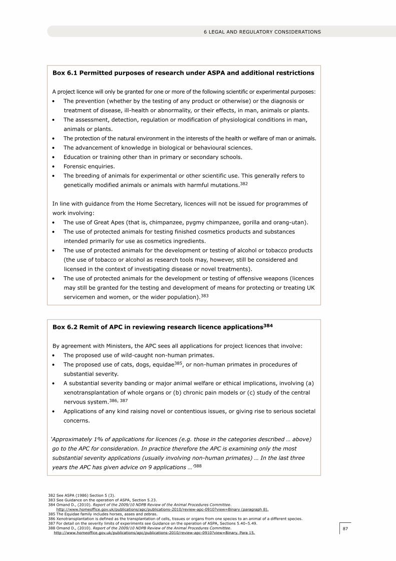
87
Box 6.1 Permitted purposes of research under ASPA and additional restrictions
A project licence will only be granted for one or more of the following scientific or experimental purposes:
• Theprevention(whetherbythetestingofanyproductorotherwise)orthediagnosisor
treatment of disease, ill-health or abnormality, or their effects, in man, animals or plants.
• Theassessment,detection,regulationormodificationofphysiologicalconditionsinman,
animals or plants.
• The protection of the natural environment in the interests of the health or welfare of man or animals.
• Theadvancementofknowledgeinbiologicalorbehaviouralsciences.
• Educationortrainingotherthaninprimaryorsecondaryschools.
• Forensicenquiries.
• Thebreedingofanimalsforexperimentalorotherscientificuse.Thisgenerallyrefersto
genetically modified animals or animals with harmful mutations.382
In line with guidance from the Home Secretary, licences will not be issued for programmes of
work involving:
• TheuseofGreatApes(thatis,chimpanzee,pygmychimpanzee,gorillaandorang-utan).
• Theuseofprotectedanimalsfortestingfinishedcosmeticsproductsandsubstances
intended primarily for use as cosmetics ingredients.
• Theuseofprotectedanimalsforthedevelopmentortestingofalcoholortobaccoproducts
(the use of tobacco or alcohol as research tools may, however, still be considered and
licensed in the context of investigating disease or novel treatments).
• Theuseofprotectedanimalsforthedevelopmentortestingofoffensiveweapons(licences
may still be granted for the testing and development of means for protecting or treating UK
servicemen and women, or the wider population).383
Box 6.2 Remit of APC in reviewing research licence applications384
By agreement with Ministers, the APC sees all applications for project licences that involve:
• Theproposeduseofwild-caughtnon-humanprimates.
• Theproposeduseofcats,dogs,equidae385, or non-human primates in procedures of
substantial severity.
• Asubstantialseveritybandingormajoranimalwelfareorethicalimplications,involving(a)
xenotransplantation of whole organs or (b) chronic pain models or (c) study of the central
nervous system.386, 387
• Applicationsofanykindraisingnovelorcontentiousissues,orgivingrisetoserioussocietal
concerns.
‘Approximately 1% of applications for licences (e.g. those in the categories described … above)
go to the APC for consideration. In practice therefore the APC is examining only the most
substantial severity applications (usually involving non-human primates) … In the last three
years the APC has given advice on 9 applications …’388
382 See ASPA (1986) Section 5 (3).383 See Guidance on the operation of ASPA, Section 5.23.384 Omand D., (2010). Report of the 2009/10 NDPB Review of the Animal Procedures Committee.
http://www.homeoffice.gov.uk/publications/apc/publications-2010/review-apc-0910?view=Binary (paragraph 8).385 The Equidae family includes horses, asses and zebras.386 xenotransplantation is defined as the transplantation of cells, tissues or organs from one species to an animal of a different species.387 For detail on the severity limits of experiments see Guidance on the operation of ASPA, Sections 5.40–5.49.388 Omand D., (2010). Report of the 2009/10 NDPB Review of the Animal Procedures Committee.
http://www.homeoffice.gov.uk/publications/apc/publications-2010/review-apc-0910?view=Binary. Para 15.
6 LEGAL AND REGULATORY CONSIDERATIONS
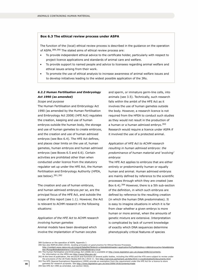
8888
ANIMALS CONTAINING HUMAN MATERIAL
Box 6.3 The ethical review process under ASPA
The function of the (local) ethical review process is described in the guidance on the operation
of ASPA.389,390 The stated aims of ethical review process are:
• Toprovideindependentethicaladvicetothecertificateholder,particularlywithrespectto
project licence applications and standards of animal care and welfare.
• Toprovidesupporttonamedpeopleandadvicetolicenseesregardinganimalwelfareand
ethical issues arising from their work.
• Topromotetheuseofethicalanalysistoincreaseawarenessofanimalwelfareissuesand
to develop initiatives leading to the widest possible application of the 3Rs.
389 Guidance on the operation of ASPA, Appendix J.390 See also RSPCA/LASA (2010). Guiding principles on good practice for Ethical Review Processes.
http://content.www.rspca.org.uk/cmsprd/Satellite?blobcol=urldata&blobheader=application%2Fpdf&blobkey=id&blobnocache=false&blobtable=MungoBlobs&blobwhere=1232992110664&ssbinary=true
391 The Human Fertilisation and Embryology Act (2008) Act is available at http://www.legislation.gov.uk/ukpga/2008/22/contents. For information on the HFEA see http://www.hfea.gov.uk/
392 At the time of publication, the structure and functions of several public bodies, including the HFEA and the HTA were subject to review under the provisions of the UK Public Bodies Bill [HL] 2010-11. See http://services.parliament.uk/bills/2010-11/publicbodieshl/documents.html
393 The HFE (Special Exemptions) Regulations (2009) provide an exemption from the requirement under the HFE Act for a licence to store gametes for research purposes. See http://www.legislation.gov.uk/uksi/2009/1918/contents/made
394 See HFE Act 1990 as amended, sub section 4A(6).
6.2.2 Human Fertilisation and Embryology
Act 1990 (as amended)
Scope and purpose
The Human Fertilisation and Embryology Act
1990 (as amended by the Human Fertilisation
and Embryology Act 2008) (HFE Act) regulates
the creation, keeping and use of human
embryos outside the human body, the storage
and use of human gametes to create embryos,
and the creation and use of human admixed
embryos (see Box 6.4). The HFE Act defines,
and places clear limits on the use of, human
gametes, human embryos and human admixed
embryos (see Boxes 6.5 and 6.6). Certain
activities are prohibited other than when
conducted under licence from the statutory
regulator set up under the HFE Act, the Human
Fertilisation and Embryology Authority (HFEA,
see below).391,392
The creation and use of human embryos,
and human admixed embryos per se, are the
principal focus of the HFE Act, and outside the
scope of this report (see 1.1). However, the Act
is relevant to ACHM research in the following
situations:
Application of the HFE Act to ACHM research
involving human gametes
Animal models have been developed which
involve the implantation of human oocytes
and sperm, or immature germ-line cells, into
animals (see 3.5). Technically, such research
falls within the ambit of the HFE Act as it
involves the use of human gametes outside
the body. However, a research licence is not
required from the HFEA to conduct such studies
as they would not result in the production of
a human or a human admixed embryo.393
Research would require a licence under ASPA if
it involved the use of a protected animal.
Application of HFE Act to ACHM research
resulting in human admixed embryos: the
predominance of human material and ‘evolving’
embryos
The HFE Act applies to embryos that are either
entirely or predominantly human or equally
human and animal. Human admixed embryos
are mainly defined by reference to the scientific
processes through which they are created (see
Box 6.4).394 However, there is a 5th sub-section
of the definition, in which such embryos are
defined by reference to the resulting creation
(in which the human DNA predominates). It
is easy to imagine situations in which it is far
from clear whether a given embryo is more
human or more animal, when the amounts of
genetic mixture are extensive. Interpretation
is complicated by lack of current knowledge
of exactly which DNA sequences determine
phenotypically critical features of species
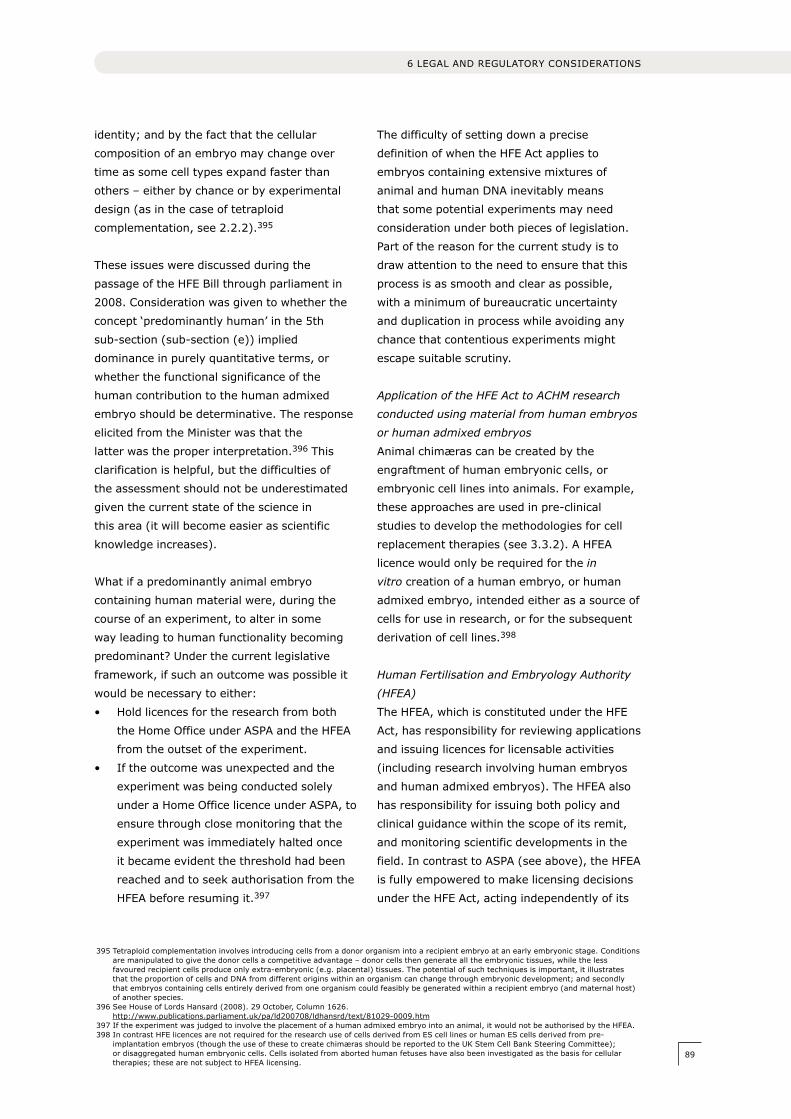
89
identity; and by the fact that the cellular
composition of an embryo may change over
time as some cell types expand faster than
others – either by chance or by experimental
design (as in the case of tetraploid
complementation, see 2.2.2).395
These issues were discussed during the
passage of the HFE Bill through parliament in
2008. Consideration was given to whether the
concept ‘predominantly human’ in the 5th
sub-section (sub-section (e)) implied
dominance in purely quantitative terms, or
whether the functional significance of the
human contribution to the human admixed
embryo should be determinative. The response
elicited from the Minister was that the
latter was the proper interpretation.396 This
clarification is helpful, but the difficulties of
the assessment should not be underestimated
given the current state of the science in
this area (it will become easier as scientific
knowledge increases).
What if a predominantly animal embryo
containing human material were, during the
course of an experiment, to alter in some
way leading to human functionality becoming
predominant? Under the current legislative
framework, if such an outcome was possible it
would be necessary to either:
• Holdlicencesfortheresearchfromboth
the Home Office under ASPA and the HFEA
from the outset of the experiment.
• Iftheoutcomewasunexpectedandthe
experiment was being conducted solely
under a Home Office licence under ASPA, to
ensure through close monitoring that the
experiment was immediately halted once
it became evident the threshold had been
reached and to seek authorisation from the
HFEA before resuming it.397
The difficulty of setting down a precise
definition of when the HFE Act applies to
embryos containing extensive mixtures of
animal and human DNA inevitably means
that some potential experiments may need
consideration under both pieces of legislation.
Part of the reason for the current study is to
draw attention to the need to ensure that this
process is as smooth and clear as possible,
with a minimum of bureaucratic uncertainty
and duplication in process while avoiding any
chance that contentious experiments might
escape suitable scrutiny.
Application of the HFE Act to ACHM research
conducted using material from human embryos
or human admixed embryos
Animal chimæras can be created by the
engraftment of human embryonic cells, or
embryonic cell lines into animals. For example,
these approaches are used in pre-clinical
studies to develop the methodologies for cell
replacement therapies (see 3.3.2). A HFEA
licence would only be required for the in
vitro creation of a human embryo, or human
admixed embryo, intended either as a source of
cells for use in research, or for the subsequent
derivation of cell lines.398
Human Fertilisation and Embryology Authority
(HFEA)
The HFEA, which is constituted under the HFE
Act, has responsibility for reviewing applications
and issuing licences for licensable activities
(including research involving human embryos
and human admixed embryos). The HFEA also
has responsibility for issuing both policy and
clinical guidance within the scope of its remit,
and monitoring scientific developments in the
field. In contrast to ASPA (see above), the HFEA
is fully empowered to make licensing decisions
under the HFE Act, acting independently of its
395 Tetraploid complementation involves introducing cells from a donor organism into a recipient embryo at an early embryonic stage. Conditions are manipulated to give the donor cells a competitive advantage – donor cells then generate all the embryonic tissues, while the less favoured recipient cells produce only extra-embryonic (e.g. placental) tissues. The potential of such techniques is important, it illustrates that the proportion of cells and DNA from different origins within an organism can change through embryonic development; and secondly that embryos containing cells entirely derived from one organism could feasibly be generated within a recipient embryo (and maternal host) of another species.
396 See House of Lords Hansard (2008). 29 October, Column 1626. http://www.publications.parliament.uk/pa/ld200708/ldhansrd/text/81029-0009.htm
397 If the experiment was judged to involve the placement of a human admixed embryo into an animal, it would not be authorised by the HFEA.398 In contrast HFE licences are not required for the research use of cells derived from ES cell lines or human ES cells derived from pre-
implantation embryos (though the use of these to create chimæras should be reported to the UK Stem Cell Bank Steering Committee); or disaggregated human embryonic cells. Cells isolated from aborted human fetuses have also been investigated as the basis for cellular therapies; these are not subject to HFEA licensing.
6 LEGAL AND REGULATORY CONSIDERATIONS
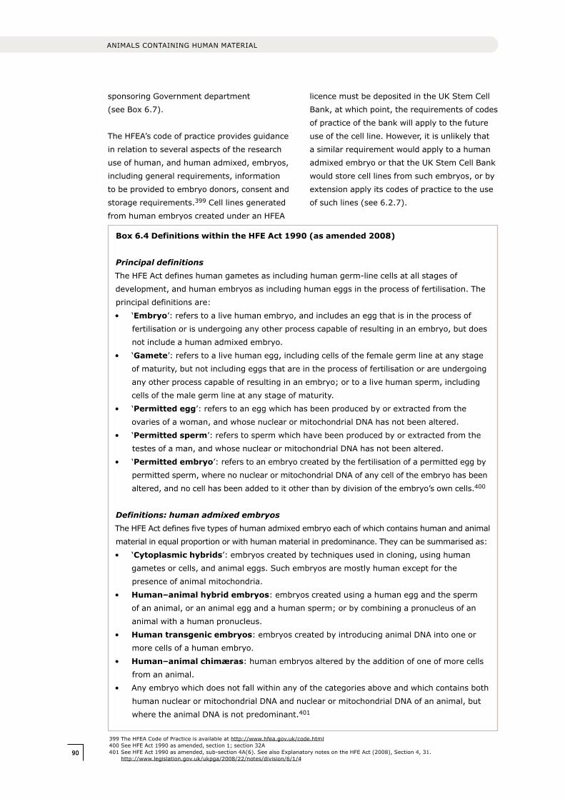
9090
ANIMALS CONTAINING HUMAN MATERIAL
sponsoring Government department
(see Box 6.7).
The HFEA’s code of practice provides guidance
in relation to several aspects of the research
use of human, and human admixed, embryos,
including general requirements, information
to be provided to embryo donors, consent and
storage requirements.399 Cell lines generated
from human embryos created under an HFEA
licence must be deposited in the UK Stem Cell
Bank, at which point, the requirements of codes
of practice of the bank will apply to the future
use of the cell line. However, it is unlikely that
a similar requirement would apply to a human
admixed embryo or that the UK Stem Cell Bank
would store cell lines from such embryos, or by
extension apply its codes of practice to the use
of such lines (see 6.2.7).
399 The HFEA Code of Practice is available at http://www.hfea.gov.uk/code.html400 See HFE Act 1990 as amended, section 1; section 32A401 See HFE Act 1990 as amended, sub-section 4A(6). See also Explanatory notes on the HFE Act (2008), Section 4, 31.
http://www.legislation.gov.uk/ukpga/2008/22/notes/division/6/1/4
Box 6.4 Definitions within the HFE Act 1990 (as amended 2008)
Principal definitions The HFE Act defines human gametes as including human germ-line cells at all stages of
development, and human embryos as including human eggs in the process of fertilisation. The
principal definitions are:
• ‘Embryo’: refers to a live human embryo, and includes an egg that is in the process of
fertilisation or is undergoing any other process capable of resulting in an embryo, but does
not include a human admixed embryo.
• ‘Gamete’: refers to a live human egg, including cells of the female germ line at any stage
of maturity, but not including eggs that are in the process of fertilisation or are undergoing
any other process capable of resulting in an embryo; or to a live human sperm, including
cells of the male germ line at any stage of maturity.
• ‘Permitted egg’: refers to an egg which has been produced by or extracted from the
ovaries of a woman, and whose nuclear or mitochondrial DNA has not been altered.
• ‘Permitted sperm’: refers to sperm which have been produced by or extracted from the
testes of a man, and whose nuclear or mitochondrial DNA has not been altered.
• ‘Permitted embryo’: refers to an embryo created by the fertilisation of a permitted egg by
permitted sperm, where no nuclear or mitochondrial DNA of any cell of the embryo has been
altered, and no cell has been added to it other than by division of the embryo’s own cells.400
Definitions: human admixed embryosThe HFE Act defines five types of human admixed embryo each of which contains human and animal
material in equal proportion or with human material in predominance. They can be summarised as:
• ‘Cytoplasmic hybrids’: embryos created by techniques used in cloning, using human
gametes or cells, and animal eggs. Such embryos are mostly human except for the
presence of animal mitochondria.
• Human–animal hybrid embryos: embryos created using a human egg and the sperm
of an animal, or an animal egg and a human sperm; or by combining a pronucleus of an
animal with a human pronucleus.
• Human transgenic embryos: embryos created by introducing animal DNA into one or
more cells of a human embryo.
• Human–animal chimæras: human embryos altered by the addition of one of more cells
from an animal.
• Anyembryowhichdoesnotfallwithinanyofthecategoriesaboveandwhichcontainsboth
human nuclear or mitochondrial DNA and nuclear or mitochondrial DNA of an animal, but
where the animal DNA is not predominant.401
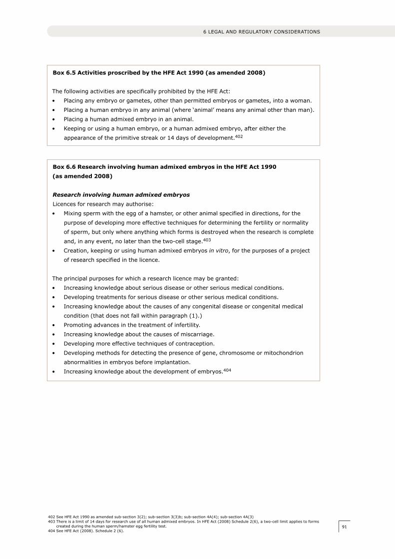
91
Box 6.5 Activities proscribed by the HFE Act 1990 (as amended 2008)
The following activities are specifically prohibited by the HFE Act:
• Placinganyembryoorgametes,otherthanpermittedembryosorgametes,intoawoman.
• Placingahumanembryoinanyanimal(where‘animal’meansanyanimalotherthanman).
• Placingahumanadmixedembryoinananimal.
• Keepingorusingahumanembryo,orahumanadmixedembryo,aftereitherthe
appearance of the primitive streak or 14 days of development.402
Box 6.6 Research involving human admixed embryos in the HFE Act 1990 (as amended 2008)
Research involving human admixed embryosLicences for research may authorise:
• Mixingspermwiththeeggofahamster,orotheranimalspecifiedindirections,forthe
purpose of developing more effective techniques for determining the fertility or normality
of sperm, but only where anything which forms is destroyed when the research is complete
and, in any event, no later than the two-cell stage.403
• Creation,keepingorusinghumanadmixedembryosin vitro, for the purposes of a project
of research specified in the licence.
The principal purposes for which a research licence may be granted:
• Increasingknowledgeaboutseriousdiseaseorotherseriousmedicalconditions.
• Developingtreatmentsforseriousdiseaseorotherseriousmedicalconditions.
• Increasingknowledgeaboutthecausesofanycongenitaldiseaseorcongenitalmedical
condition (that does not fall within paragraph (1).)
• Promotingadvancesinthetreatmentofinfertility.
• Increasingknowledgeaboutthecausesofmiscarriage.
• Developingmoreeffectivetechniquesofcontraception.
• Developingmethodsfordetectingthepresenceofgene,chromosomeormitochondrion
abnormalities in embryos before implantation.
• Increasingknowledgeaboutthedevelopmentofembryos.404
402 See HFE Act 1990 as amended sub-section 3(2); sub-section 3(3)b; sub-section 4A(4); sub-section 4A(3)403 There is a limit of 14 days for research use of all human admixed embryos. In HFE Act (2008) Schedule 2(6), a two-cell limit applies to forms
created during the human sperm/hamster egg fertility test.404 See HFE Act (2008). Schedule 2 (6).
6 LEGAL AND REGULATORY CONSIDERATIONS
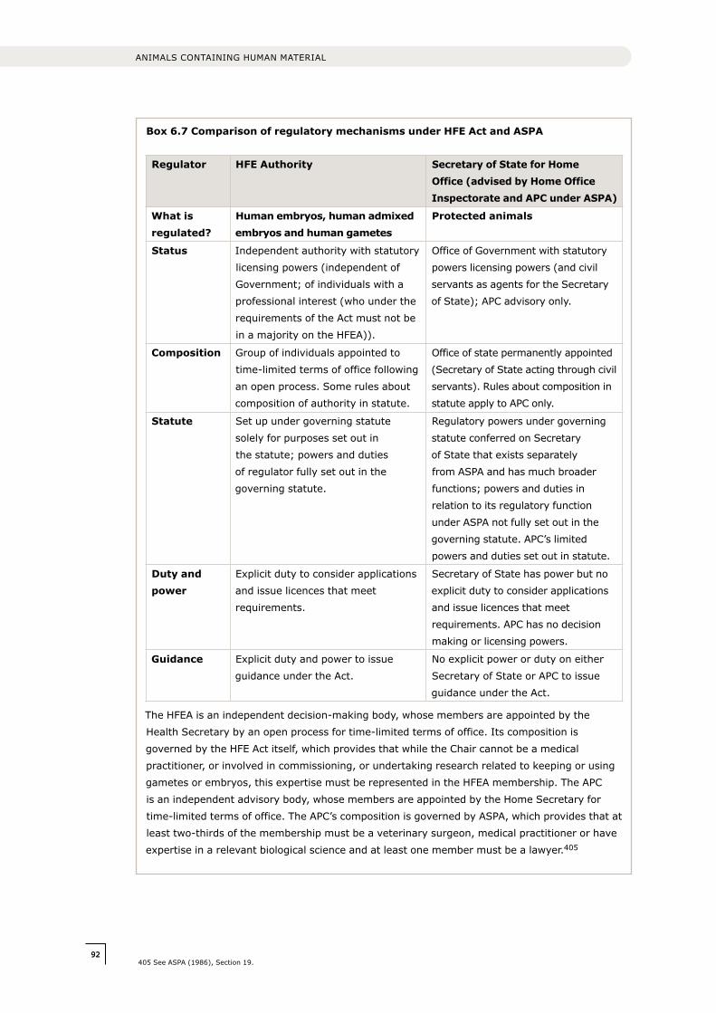
9292
ANIMALS CONTAINING HUMAN MATERIAL
Box 6.7 Comparison of regulatory mechanisms under HFE Act and ASPA
Regulator HFE Authority Secretary of State for Home Office (advised by Home Office Inspectorate and APC under ASPA)
What is regulated?
Human embryos, human admixed embryos and human gametes
Protected animals
Status Independent authority with statutory
licensing powers (independent of
Government; of individuals with a
professional interest (who under the
requirements of the Act must not be
in a majority on the HFEA)).
Office of Government with statutory
powers licensing powers (and civil
servants as agents for the Secretary
of State); APC advisory only.
Composition Group of individuals appointed to
time-limited terms of office following
an open process. Some rules about
composition of authority in statute.
Office of state permanently appointed
(Secretary of State acting through civil
servants). Rules about composition in
statute apply to APC only.
Statute Set up under governing statute
solely for purposes set out in
the statute; powers and duties
of regulator fully set out in the
governing statute.
Regulatory powers under governing
statute conferred on Secretary
of State that exists separately
from ASPA and has much broader
functions; powers and duties in
relation to its regulatory function
under ASPA not fully set out in the
governing statute. APC’s limited
powers and duties set out in statute.
Duty and power
Explicit duty to consider applications
and issue licences that meet
requirements.
Secretary of State has power but no
explicit duty to consider applications
and issue licences that meet
requirements. APC has no decision
making or licensing powers.
Guidance Explicit duty and power to issue
guidance under the Act.
No explicit power or duty on either
Secretary of State or APC to issue
guidance under the Act.
The HFEA is an independent decision-making body, whose members are appointed by the
Health Secretary by an open process for time-limited terms of office. Its composition is
governed by the HFE Act itself, which provides that while the Chair cannot be a medical
practitioner, or involved in commissioning, or undertaking research related to keeping or using
gametes or embryos, this expertise must be represented in the HFEA membership. The APC
is an independent advisory body, whose members are appointed by the Home Secretary for
time-limited terms of office. The APC’s composition is governed by ASPA, which provides that at
least two-thirds of the membership must be a veterinary surgeon, medical practitioner or have
expertise in a relevant biological science and at least one member must be a lawyer.405
405 See ASPA (1986), Section 19.
93
6.2.3 Human Tissue Act 2004
Scope and purpose
The Human Tissue Act 2004 (the HT Act) is
the legal framework in England, Wales and
Northern Ireland regulating the storage and
use of human organs and tissue from the
living, and the removal, storage and use of
tissue and organs from the deceased, for
health-related purposes and public display.406
The Act is principally intended to ensure that
appropriate consent is in place to enable the
lawful retention and use of body parts, organs
and tissue, for ‘scheduled purposes’, which
include medical research. The Act also prohibits
certain forms of DNA analysis without consent
throughout the UK.
The HT Act applies to human bodies and human
tissue that consist of, or contain, human cells
other than: hair and nails from living people;
human gametes and embryos; and other
human material created outside the human
body (e.g. human cell lines).407 It prohibits
the possession of ‘bodily material’ (from a
living or deceased human body, consisting of
or including human cells, including hair, nails
and gametes) with the intention of analysing its
DNA without consent.408 Except to the extent
of the prohibition above, DNA itself (extracted
human DNA, where no whole cells remain) is
not regulated by the Act.
Application of the HT Act to ACHM research
The requirements of the HT Act apply to the
creation of chimæric animals using human
tissue in some circumstances. For example,
where human tissue is removed directly from
the body of an identifiable living person, and
inserted into an animal the HT Act requirements
concerning consent and licences for any storage
of such tissue would apply.409,410 The HT Act
would not apply to the creation of transgenic
animals using ‘human-like DNA sequence’
(since extracted or artificially synthesised
human DNA is not regulated by the HT Act), nor
would it apply to the creation of chimæras using
human cell lines (since cell lines are outside the
scope of the Act).
Human Tissue Authority
The Human Tissue Authority (HTA), regulates
and licences the use and storage of human
tissue under the HT Act.411 The HTA’s remit
does not include ethical approval, which is
necessary for research involving human tissue
in some circumstances and governed by the
National Research Ethics Service (NRES).412
6.2.4 Health and safety law, including GM
Regulations
ACHM research is subject to general health
and safety requirements including the Health
and Safety at Work Act (1974) and Carriage of
Dangerous Goods legislation. Some types of
ACHM research are also subject to the controls
set out in the Genetically Modified Organisms
(GMO) (Contained Use) Regulations and
the GMO (Deliberate Release) Regulations,
regulated by the Health and Safety Executive
(HSE) and the Department for Environment
Food and Rural Affairs (Defra) respectively.413
The GM regulations are designed to control
risks from GMOs to human health (both
the contained use and deliberate release
regulations) and the environment (the
deliberate release regulations only). They apply
to biological organisms, cellular (including
animal cells in culture) and non-cellular
material, other than humans and human
embryos, which have been genetically altered
other than as a result of a naturally occurring
process and which are capable of replicating
406 The Human Tissue Act (2004) is available at http://www.legislation.gov.uk/ukpga/2004/30/contents. Removal of material from the living is regulated separately. The equivalent legislation in Scotland is the Human Tissue (Scotland) Act 2006 (which only applies to post not ante mortem tissue) in Scotland. http://www.legislation.gov.uk/asp/2006/4/contents
407 There is an exception in that the HT Act (2004) applies to stem cell lines intended for human application.408 Unlike the rest of the HT Act, this provision extends to the whole of the UK, including Scotland.409 In addition to any requirements under ASPA (1986).410 Though there are various exceptions to requirements that may be relevant, including (a) a storage licence is not required (1) for tissue
stored incidentally to transportation for less than a week or (2) for tissue stored solely for use in a NHS research ethics committee (‘REC’) approved project; (b) consent is not required (1) for use of tissue imported into England, Wales and Northern Ireland or (2) for use of tissue taken from a living person used in anonymised (to the researcher) form for a REC approved project.
411 For detail on the wider remit of the HTA see http://www.hta.gov.uk/412 For detail on NRES see http://www.nres.npsa.nhs.uk/413 For detail on the GMO regulations see http://www.hse.gov.uk/biosafety/gmo/law.htm and http://www.defra.gov.uk/environment/quality/
gm/; Certain decisions are reserved to Scottish Ministers in Scotland.
6 LEGAL AND REGULATORY CONSIDERATIONS
9494
ANIMALS CONTAINING HUMAN MATERIAL
or transferring genetic material. Thus, the
regulations apply to transgenic ACHM.414 We
presume the application of the regulations to
any particular chimæra will depend on whether
their genetic material can be said to have been
altered other than as a result of a naturally
occurring process (since each cell in a chimæra
contains an unmodified genome of one of the
precursor animals) and whether any change is
capable of transmission, (since the chimærism
may not involve the germ cells).
Users of GM animals in contained facilities must
notify their facilities to the Health and Safety
Executive (HSE), carry out risk assessments
addressing both risks to human health and
to the wider environment, ensure necessary
controls are in place to minimise such risks and
notify or seek the consent of (depending on the
risk level) the HSE in relation to GM activities.
GMOs cannot be released from containment
without the approval of Defra following
assessment by official assessors to ensure
there are no risks to human health or the
environment. Deliberate release of GM animals
governed by both the GM regulations and
ASPA also requires the approval of the Home
Secretary (though release of GM animals has
never been so authorised). Accidental release
must be notified to the HSE.415
6.2.5 Intellectual property rights
Intellectual property law does not regulate
the conduct of research involving animals
containing human material, but can strongly
influence whether research takes place, and
may impede (or create the conditions to
enable) research and development activity. For
example, a pharmaceutical company making
a transgenic animal expressing a human
protein is likely to seek to patent the animal.
UK legislation makes provision for biological
materials to be patented, including: ‘inventions
which concern plants or animals’ 416 and ‘an
element isolated from the human body …
including the sequence or partial sequence of
a gene, even if the structure of that element is
identical to that of a natural element’. However
‘processes for modifying the genetic identity of
animals which are likely to cause them suffering
without any substantial medical benefit to man
or animal’, and also animals resulting from such
processes cannot be patented. 417
6.2.6 Data Protection Act 1998
The Data Protection Act 1998 (DPA) is the
principal legislation relevant to the use of
medical information in research in the UK.418
It regulates the use of ‘personal data’, that
is, data relating to an individual who can
be uniquely ‘identified from those data, or
from a combination of those data and other
information which is in the possession of, or
is likely to come into the possession of a data
controller’. Although the DPA is generally
unlikely to apply to ACHM research, it would
apply if a particular individual could be uniquely
identified from a section of genetic code/
sequence, obtained through the sequencing of
human DNA, when combined with other data
in the possession of the same researcher. It
seems likely that this could be the case in some
circumstances, in which event the provisions of
the Act including the requirements relating to
consent, fair processing and right of access (by
the individual concerned) would apply.
6.2.7 Non-legislative requirements in
the UK
In addition to statutory legislation, scientific
and medical research is subject to, and
guided by, a complex raft of non-legislative
guidance, which varies in some cases between
the four different administrations within
the UK.419 Some touches on the creation
of human admixed embryos, but beyond
414 See the Scientific Advisory Committee on Genetic Modification (SACGM) compendium of guidance. Part 5 Genetic modification of animals. http://www.hse.gov.uk/biosafety/gmo/acgm/acgmcomp/index.htm
415 As at March 2011, the HSE were not aware of any incidents of accidental release of GM animals posing any risk to human health.416 If the technical feasibility of the invention is not confined to a particular plant or animal variety.417 The relevant UK legislation is the Patents Act (1977), as amended by the Patents Regulations 2000 (SI 2000/2037) – which implemented
the provisions of Articles 1 to 11 of the European Directive 98/44/EC on the legal protection of biotechnological inventions. Patent law is overseen by the Intellectual Property Office, an Executive Agency of the Department for Business Innovation and Skills. See http://www.ipo.gov.uk/
418 The Data Protection Act (1998) is available at http://www.legislation.gov.uk/ukpga/1998/29/contents419 The pathway of regulation and governance of research involving human participants, their tissue or data is addressed in Academy of Medical
Sciences (2008). A new pathway for the regulation and governance of health research. http://www.acmedsci.ac.uk/p47prid88.html
95
that, to our knowledge no guidance relates
specifically to the creation or use of ACHM as
such.420 Guidance and other non-legislative
requirements that have particular impact on
ACHM research include the NHS research
governance framework (RGF, below) and its
equivalent in Scotland, HFEA and HTA codes of
practice, professional codes, stem cell banks’
codes of practice, guidance from funding
bodies (both public and charitable), grant
conditions, and publishing requirements. This
guidance supports the formal legislation in the
development and maintenance of good practice
among the research community, including in
relation to ACHM research. In many cases it is
sufficiently flexible to enable ethical, societal
and other issues relating to ACHM research to
be identified and considered, notwithstanding
that ACHM was not in the contemplation of
the draftsmen.
NHS Research Governance Framework (RGF)
The RGF outlines the principles of good
governance for research carried out in the NHS,
including the different permissions required
(e.g. those of the HFEA or the HTA), and more
generic requirements which can apply to health
research involving NHS facilities, patients,
their tissue or data. Although the RGF does
not apply to animal research as such, it does
set out a regulatory framework, including the
requirement for NHS research ethics approval,
that is applicable to ACHM research insofar
as it involves the use of human tissue or
patient data.421 A similar framework applies in
Scotland.
Stem cell guidance
ACHM research that involves the introduction
of human stem cells into an animal is guided by
the general requirements of the Department of
Health Code of Practice for the use of human
stem cell lines and the codes of practice of the
UK Stem Cell Bank. If the research involves the
use of human embryonic stem cells generated
under an HFEA licence and supplied by the UK
Stem Cell Bank, the Stem Cell Bank Steering
Committee must approve the release of the
cell line from the bank, and the owner of the
line would be required to license its use subject
to the HFEA and UK Stem Cell Bank Codes of
Practice, which set out general requirements
concerning the use of human ESCs (though
any resulting animal cell lines could not be
deposited in the Stem Cell Bank)
(see also 6.2.2). 422
420 For example, see the HFEA code of practice http://www.hfea.gov.uk/3468.html421 Department of Health (2005). Research governance framework for health and social care: Second edition
http://www.dh.gov.uk/en/Publicationsandstatistics/Publications/PublicationsPolicyAndGuidance/DH_4108962; the equivalent in Scotland is the research governance framework for health and community care. http://www.cso.scot.nhs.uk/publications/ResGov/Framework/RGFEdTwo.pdf
422 For details on the UK Stem Cell Bank see http://www.ukstemcellbank.org.uk/; their Code of Practice is available at http://www.ukstemcellbank.org.uk/codesofpractice/codeofpracticefortheuseofhumanstemcelllines.cfm
6 LEGAL AND REGULATORY CONSIDERATIONS

9696
ANIMALS CONTAINING HUMAN MATERIAL
Box 6.8 Public views on research regulation
Most dialogue participants were aware that medical research is regulated in the UK, though
they had little knowledge of how regulation is brought about or the organisations involved.
A majority of the workshop participants expressed confidence that, in the UK, the regulation of
research involving animals containing human material would be adequate, properly enforced,
and reflective of their concerns and principles. This finding was echoed in the survey data, in
which 44% of participants agreed that they would trust regulation of such research in the UK
(29% said they would distrust such regulation, the remainder were neutral or unsure).
Participants’ main concerns about the research regulation related to:
• Thepossibilitythatpermittingsomeresearchofthistypemightleadscientiststoseekto
conduct unacceptable research in future (a ‘slippery slope’ argument).
• Knowledgeofsituationswhereregulatoryerrorswerethoughttohaveoccurred
(participants cited the release of foot-and-mouth disease at Pirbright in 2007).
• Asuggestionthat‘rogue’scientistswouldevadeauthoritiesandregulation.
• TheviewthatresearchofthiskindwouldnotbeadequatelyregulatedbeyondtheUKand
so ‘malpractice’ would take place elsewhere.
‘You trust your doctor and your scientists. Not in other countries but the UK is fine’.
Participants indicated several factors which they felt were important for future regulation of
research involving animals containing human material, these included:
• Ageneralprincipleoftransparencyshouldbeapplied,inthatinformationonresearchof
this type should be available in the public domain.
• Regulationshouldbeconductedbyindependent/impartialpeople,andamixtureofdifferent
interests should be represented (e.g. public members, independent scientists, specifically
appointed regulators).
• Regulationshouldfocusonanimalwelfare,ensuringthatanimalsufferingandthenumbers
of animals used are minimised.
• Regulationshouldaimtoeliminaterisks,includingtheunintendedreleaseofenvironmental
contaminants or disease-causing factors.
• Regulationshouldbeenforcedinamannerthatpreventsevasionby‘rogue’scientists.
• Regulationshouldbeappropriatetothetypeofanimalcreatedandthehumantissueand
organs involved.

97
6.3 Summary
As we have noted, ACHM research conducted
within the UK is principally regulated by
ASPA, though the focus of ASPA and its
implementation is on animal welfare and 3Rs,
rather than wider ethical considerations.
Although there is some grading within the
ASPA system in the form of the four categories
of applications that have been identified as
requiring review by the APC, the categorisation
is principally designed around animal welfare
issues rather than broader considerations,
and in the case of the fourth category, which
potentially addresses broader issues, lacks
definition (see Box 6.2). Given the evolving
nature of the science associated with ACHM
research, we see considerable benefit in further developing a graded approach to licensing and regulatory oversight, which is principle based and transparent, seeks to define different levels of sensitivity and differentiates the degrees of scrutiny required accordingly. We propose a possible
approach in Chapter 8 (see 8.2).
Recognising the specialist knowledge required to evaluate likely (and sometimes uncertain) outcomes in this complex field of science, as well as the socially sensitive nature of the judgements to be made, we would also consider that a national expert body, which includes the relevant expertise, is needed to advise on ACHM research (see 8.3). In order to build and maintain trust and ensure accountability to the public, the body needs to operate
transparently, be outward facing and engage with the public and the scientific community. To ensure consistency and transparency, it needs to have the power to develop guidelines. There would also be
considerable importance and value in it playing
a broader function, including the role of sharing
knowledge and best practice attributed to the
national committee required under the 2010
EU Directive.
As we have set out, the regulatory environment
is complex. There are several pieces of UK
legislation relevant to the regulation of ACHM.
In some cases, more than one regulatory
regime applies to a specific piece of ACHM
research, or the research is at the borders of
specific regimes. Aside from complexity, this
also creates the possibility of inconsistency
between regulatory regimes. To manage this
effectively, a key feature of the UK regulatory
environment in the future needs to be that all relevant stakeholders (Home Office, HFEA, HTA and others) develop a coordinated, consistent approach to regulating the field of research, work together under an agreed framework of operation to continue to monitor scientific developments and consider jointly how to address borderline cases (see 8.6). Borderline cases include
experiments that involve animal embryos
containing human cells or genes that are
close to the boundary of human admixed
embryos under the HFE Act as well as certain
ACHM experiments that involve a degree of
uncertainty as to outcome. Regulatory guidance
is likely to be particularly helpful in such cases.
6 LEGAL AND REGULATORY CONSIDERATIONS
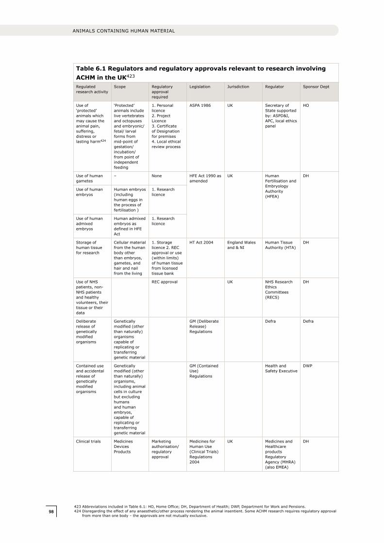
9898
ANIMALS CONTAINING HUMAN MATERIAL
Table 6.1 Regulators and regulatory approvals relevant to research involving ACHM in the UK423
Regulated research activity
Scope Regulatory approval required
Legislation Jurisdiction Regulator Sponsor Dept
Use of ‘protected’ animals which may cause the animal pain, suffering, distress or lasting harm424
‘Protected’ animals include live vertebrates and octopuses and embryonic/ fetal/ larval forms from mid-point of gestation/ incubation/ from point of independent feeding
1. Personal licence 2. Project Licence 3. Certificate of Designation for premises 4. Local ethical review process
ASPA 1986 UK Secretary of State supported by: ASPD&I, APC, local ethics panel
HO
Use of human gametes
– None HFE Act 1990 as amended
UK Human Fertilisation and Embryology Authority (HFEA)
DH
Use of human embryos
Human embryos (including human eggs in the process of fertilisation )
1. Research licence
Use of human admixed embryos
Human admixed embryos as defined in HFE Act
1. Research licence
Storage of human tissue for research
Cellular material from the human body other than embryos, gametes, and hair and nail from the living
1. Storage licence 2. REC approval or use (within limits) of human tissue from licensed tissue bank
HT Act 2004 England Wales and & NI
Human Tissue Authority (HTA)
DH
Use of NHS patients, non-NHS patients and healthy volunteers, their tissue or their data
REC approval UK NHS Research Ethics Committees (RECS)
DH
Deliberate release of genetically modified organisms
Genetically modified (other than naturally) organisms capable of replicating or transferring genetic material
GM (Deliberate Release) Regulations
Defra Defra
Contained use and accidental release of genetically modified organisms
Genetically modified (other than naturally) organisms, including animal cells in culture but excluding humans and human embryos, capable of replicating or transferring genetic material
GM (Contained Use) Regulations
Health and Safety Executive
DWP
Clinical trials Medicines Devices Products
Marketing authorisation/ regulatory approval
Medicines for Human Use (Clinical Trials) Regulations 2004
UK Medicines and Healthcare products Regulatory Agency (MHRA)(also EMEA)
DH
423 Abbreviations included in Table 6.1: HO, Home Office; DH, Department of Health; DWP, Department for Work and Pensions.424 Disregarding the effect of any anaesthetic/other process rendering the animal insentient. Some ACHM research requires regulatory approval
from more than one body – the approvals are not mutually exclusive.
99
7 INTERNATIONAL PERSPECTIVE
7.1 Introduction
In Chapter 6 we described the law and
regulation applicable to the creation and use
of ACHM in biomedical research in the UK.
However, like much biomedical research,
research involving ACHM is an international
activity. It frequently involves international
collaboration, takes place across national
boundaries, and involves funders or researchers
who are often free to choose the location in
which their research is conducted. In this
chapter, we outline the regulation of this
research from an international perspective and
consider some of the challenges this poses.
As far as we are aware, very few countries
have specifically considered the regulation of
research involving ACHM. As in the UK, to our
knowledge there are no specific national laws,
regulation or guidance documents addressing
ACHM research, though a range of laws and
regulatory frameworks, particularly those
governing the use of animals, cover different
aspects of this research.
A similar pattern of legislation is evident
at European Union level; whilst there is no
specific European legislation on ACHM, there
are European equivalents of many (though not
all) UK and other national European laws which
are of relevance. Internationally, and within
European states, broad principles that may
be applied to ACHM research are addressed
in legal instruments (largely in the context of
human cloning).
Guidelines developed by international groups
address aspects of ACHM research, particularly
the use of human stem cells to create inter-
species chimæras. Adoption of these guidelines,
though largely voluntary, provides a basis for the
development of international best practice in this
field, which would be of particular value given the
degree of diversity in national laws and regulation.
7.2 National regulation and international research
Research involving ACHM, like other forms of
medical research, is principally governed by
national law and regulation. Although these may
derive from international instruments (such
as European Directives), legislation relevant
to research is, in the main, implemented and
enforced at national level, and research is
therefore predominantly governed solely by
the laws of the country where it takes place.
Occasionally, research is regulated extra-
territorially; for example in some cases,
researchers are subject to the laws of the
country of which they are citizens, even when
they conduct research elsewhere. However,
this is relatively unusual (for example, none of
the regulations discussed in Chapter 6 apply
to research conducted solely outside the UK,
even where conducted by UK citizens). It is
considerably more likely that the conditions
and requirements imposed by funding and
professional bodies operate extra-territorially.425
Research involving ACHM conducted across
different national locations needs to be designed
to take into account legal and regulatory
divergence, with research in each national area
potentially being subject to different legal and
regulatory limits and controls. Although, as this
chapter will show, there is some harmonisation
(for example, the recent European Directive was
intended to promote greater standardisation
around the use of animals in research) this is
relatively limited. There remains considerable
diversity across nations beyond the European
Union, both in regulation of the research use of
animals, and other aspects of ACHM research
(e.g. the use of human tissue).
Guidelines developed by international groups
can encourage common standards and aid
researchers working across national borders
in navigating divergent governance systems.
7 International perspective
425 For example, research involving stem cells funded by the US National Institutes of Health (NIH) must accord with their guidance even if conducted in the UK (see http://stemcells.nih.gov/policy/2009guidelines.htm). The UK’s Medical Research Council has supplementary terms and conditions for research that it funds that involves human stem cells (see AC24 in http://www.mrc.ac.uk/Utilities/Documentrecord/index.htm?d=MRC001898).
100100
ANIMALS CONTAINING HUMAN MATERIAL
However, these are currently limited in relation
to ACHM research, and only address specific
aspects or types of ACHM.426
National divergence poses a risk of researchers
locating their research in certain countries in
order to avoid particular national restrictions.
The development of international standards
backed by collaboration between national and
international policymakers may help to reduce
this risk, as well as facilitate cross-border
research. It would also be beneficial for national
regulators to collaborate, and to encourage
international data-sharing, where evidence
from incremental research studies has been
acquired (see Box 3.8).
7.3 Europe
7.3.1 European Union
Some of the UK legislation governing research
involving ACHM originates from European Union
law (two notable exceptions are the Human
Fertilisation and Embryology Act, which has
no EU equivalent, and the Human Tissue Act,
which only reflects EU law in certain limited
respects).427 The application of EU Directives
on the protection of animals used for scientific
purposes, the use of genetically modified
organisms, data protection and patents to
ACHM research, is summarised below.
EU legislation for the protection of animals
Directive 86/609/EEC on the protection of
animals used for scientific purposes was revised
in September 2010 by Directive 2010/63/
EU, which is due to be implemented in all EU
member states by 2013 (see also 6.2.1).428,429
The revised Directive is the principal European
legal framework relating to ACHM research.
It applies to regulated scientific procedures
involving non-human vertebrates, including
larval forms capable of independent feeding
and fetal forms of mammals in the last third
of their development, and to cephalopods
(a class of molluscs including octopi and squid).
The Directive places clear limits on the scientific
procedures that can be carried out using such
animals, and places emphasis on the welfare
principles of the 3Rs (reduction, refinement
and replacement, see 4.1.1). A minority of
ACHM research may be outside the scope of
the Directive, as it does not apply to research
involving vertebrate animals at less than
two-thirds of gestation or invertebrate animals
(see 6.2.1).430
Creation and use of genetically modified entities
ACHM research may also be within the
remit of European Directives intended to
safeguard against environmental, and health
and safety risks associated with the use,
storage and containment, and disposal or
release of genetically modified organisms
and microorganisms. Directive 2009/41/
EC lays down measures for the contained
use of genetically modified microorganisms
(microbiological entities both cellular and
non-cellular, in which genetic material has
been altered other than naturally) (GMMs).431
Directive 2001/18/EC (as amended in 2008)
lays down requirements concerning the
deliberate release into the environment of
genetically modified organisms (biological
entities other than human beings capable of
replicating or transferring genetic material, in
which the genetic material has been altered
other than naturally) (GMOs).432 In particular, it
requires users to conduct risk assessments and
426 Examples of international guidelines include CURE Report China (http://www.mrc.ac.uk/Utilities/Documentrecord/index.htm?d=MRC006303); Swiss Commission for Research Partnership with Developing Countries KFPE (1998) (http://www.kfpe.ch/download/Guidelines_e.pdf); and further examples in ‘The ethics of research related to healthcare in developing countries: a follow-up discussion paper’ available at http://www.nuffieldbioethics.org/research-developing-countries-follow)
427 The European Tissue Directive 2004/23/EC applies only to human tissue for human application, and is not relevant to the use of human tissue for research (http://eur-lex.europa.eu/LexUriServ/site/en/oj/2004/l_102/l_10220040407en00480058.pdf).
428 Council Directive 86/609/EEC of 24 November 1986 on the approximation of laws, regulations and administrative provisions of the Member States regarding the protection of animals used for experimental and other scientific purposes is available at http://eur-lex.europa.eu/LexUriServ/LexUriServ.do?uri=CELEx:31986L0609:en:HTML
429 Directive 2010/63/EU on the protection of animals used for scientific purposes is available at http://eur-lex.europa.eu/LexUriServ/LexUriServ.do?uri=OJ:L:2010:276:0033:0079:EN:PDF
430 See 6.2.1 for a discussion of the implementation of Directive 2010/63/EU in the UK.431 Directive 2009/41/EC on the contained use of genetically modified microorganisms is available at http://www.bmwf.gv.at/fileadmin/user_
upload/forschung/gentechnik/2009-41-EC.pdf432 Directive 2001/18/EC 2001 on the deliberate release into the environment of genetically modified organisms is available at http://eur-lex.
europa.eu/LexUriServ/LexUriServ.do?uri=CELEx:32001L0018:EN:HTML
101
to notify and seek the consent of the competent
national authority prior to GMO release.433
Patent law
Directive 98/44/EC on the legal protection of
biotechnological interventions limits the legal
protection of inventions including biological
material, and may therefore affect some
ACHM research (see 6.2.5).434 In addition to
generic requirements, the Directive sets limits
to patentability on moral grounds, specifically
outlawing the patenting of inventions the
exploitation of which would be contrary to
‘ordre public or morality’ and ‘processes, the
use of which offend against human dignity
…’.435,436 This definition includes processes for
modifying the genetic identity of animals which
are likely to cause them suffering without any
substantial medical benefit to man or animal,
and animals resulting from such processes.437
Objections to patents on the grounds of these
provisions have been raised in a number of
cases. The issue was explored in relation
to a challenge to the oncomouse patent
for example, though the arguments were
ultimately rejected by the European Patent
Office and the patent upheld.438,439
Protection of personal data
ACHM research conducted within the European
Union which involves human tissue or data
from which a living individual can be identified,
is subject to the requirements of the Data
Protection Directive (Directive 95/46/EC).440
In particular, the Directive includes a
requirement that the individual concerned
is made aware of and has, subject to limited
exceptions, given their consent for the use of
their tissue or data.
7.3.2 Council of Europe
Convention on Human Rights and
Biomedicine (1999)
The Council of Europe Convention on Human
Rights and Biomedicine sets out international
standards concerning the protection of human
rights in relation to biology and medicine.441
The Convention does not specifically address
ACHM research, but includes principles that
may be considered of relevance. Notably,
it places emphasis on the protection of ‘the
dignity and identity of all human beings’ and
the importance of ‘the need to respect the
human being both as an individual and as a
member of the human species’, recognising
‘the importance of ensuring the dignity
of the human being’ and ‘that the misuse
of biology and medicine may lead to acts
endangering human dignity’.442 The absence
of any definition of ‘human being’ has enabled
considerable diversity of interpretation of the
Convention across Europe.443
7.4 International
7.4.1 International legal instruments
A range of international legal instruments
are of broad relevance to medical research,
including that involving the use of human
433 In England and Wales the competent authority enforcing jurisdiction over the GMO (Contained Use Regulations) includes the Health and Safety Executive (HSE), the Secretary of State and the Department for Environment Food and Rural Affairs (Defra). In Scotland the competent authorities includes HSE, the Scottish Executive Environment and Rural Affairs Department and the Scottish Ministers. Northern Ireland has its own separate competent authority. The UK Government and Devolved Administrations have established joint arrangements for assessing applications for the deliberate release of GMOs. This involves consultation with the Advisory Committee on Releases to the Environment (ACRE), the HSE, the Food Standards Agency (FSA), and as appropriate, the statutory nature conservation bodies, such as English Nature. For the HSE’s guidance see http://www.hse.gov.uk/biosafety/gmo/acgm/acgmcomp/part5.pdf
434 Directive 98/44/EC on the legal protection of biotechnological inventions is available at http://eur-lex.europa.eu/LexUriServ/LexUriServ.do?uri=OJ:L:1998:213:0013:0021:EN:PDF
435 Reference to ‘ordre public’ is made in Article 6 of Directive 98/44/EC (which is implemented through Article 53(a) of the European Patent Convention regulations).
436 Recital 38 of Directive 98/44/EC continues ‘…such as processes to produce chimæras from germ cells or totipotent cells of humans and animals, are obviously also excluded from patentability’.
437 Article 6 of Directive 98/44/EC defines as unpatentable processes for cloning human beings, processes for modifying the germ line genetic identity of human beings and uses of human embryos for industrial or commercial purposes.
438 EPO Case No. T0315/03 (transgenic animals/HARVARD); 6 July 2004 http://www.epo.org/law-practice/case-law-appeals/pdf/t030315ex1.pdf439 A second case (Brüstle vs. Greenpeace EV, (Case no. C34/10) which had yet to be considered by the European Court of Justice at the time
of writing) was to be the first case before the Court to involve consideration of Article 6(2)(c) of EU Directive 98/44 (non-patentability of use of human embryos for industrial or commercial purposes as being contrary to ordre public). The ‘opinion of the attorney general’ in the case was published on 10 March 2011.
440 Directive 95/46/EC on the protection of individuals with regard to the processing of personal data and on the free movement of such data is available at http://eur-lex.europa.eu/LexUriServ/LexUriServ.do?uri=CELEx:31995L0046:EN:NOT
441 The Convention for the Protection of Human Rights and Dignity of the Human Being with regard to the Application of Biology and Medicine: Convention on Human Rights and Biomedicine is available at http://conventions.coe.int/Treaty/en/Treaties/html/164.htm. The UK has not acceded to this convention.
442 Ibid Preamble and Article 1.443 For example, it leaves open whether a human embryo or fetus could be considered a human being and a rights holder under the Convention,
and by extension the application of the Convention in relation to assisted reproduction and use of human embryos in research.
7 INTERNATIONAL PERSPECTIVE
102102
ANIMALS CONTAINING HUMAN MATERIAL
genetic or cellular material.444 However, the
only international legal instruments that might
be applied to ACHM research of which we are
aware are non-binding instruments, intended
principally to address human cloning.
This UNESCO Universal Declaration on the
human genome and human rights (1977) was
the first international legal text to address the
relationship of biotechnological development
and human rights with the human genome.445
The non-binding declaration states that ‘No
research, or its applications, should prevail
over the respect for human rights, fundamental
freedoms, and human dignity of individuals or
groups of people’ and that ‘Practices contrary to
human dignity, such as the reproductive cloning
of human beings, shall not be permitted.’446
A second non-binding declaration, the UN
Declaration on human cloning (2005), was
adopted by a weak majority vote in which UN
states were called on to ‘adopt the measures
necessary to prohibit the application of genetic
engineering techniques that may be contrary
to human dignity’.447 The application of these
Declarations to the creation or use of ACHM is
a matter of interpretation (e.g. it is disputable
whether the creation of ACHM would be
considered ‘contrary to human dignity’). However,
as UN declarations, they are likely to influence
some states’ national laws, policies and practices.
7.4.2 International guidance
At an international level, the instruments that
are of most direct relevance to ACHM research
are guidelines developed by funding bodies,
scientists and other groups. These include
guidance on the use of human ES cells, and
other human stem cell types. In these areas
the development of international guidelines is
relatively mature.
International Society for Stem Cell Research
(ISSCR) Guidelines for the Conduct of Human
Embryonic Stem Cell Research (2006)
The ISSCR guidelines specify rigorous
ethical standards for scientists working
with human ES cells and seek to promote
responsible, transparent and uniform practices
worldwide.448 They set out a categorisation
of research involving stem cells and prescribe
the required nature of regulatory review and
oversight for each category of research (see
Box 7.1). Research involving the incorporation
of human ES cells and other human stem
cells into animals (i.e. the creation of human–
animal chimæras) is addressed, and specific
forms of research which should not be pursued
at present are identified (see Box 7.1). The
guidelines also:
• Encouragethedepositionofderivedhuman
stem cell lines in national or international
depositories that allow open distribution, to
facilitate the wider dissemination of these
valuable research tools.
• Setoutguidanceforprocurementoftissue
for human ES cell research, and specify
minimum requirements for obtaining
informed consent of donors.
• Indicatethatfundingorganisationsshould
pledge to comply with the guidelines, and
that publishers should require a statement
of compliance with them.
Hinxton Group
In early 2004, members of the Stem Cell
Policy and Ethics Program (SCOPE) at the
Johns Hopkins Berman Institute of Bioethics
brought together an international and
interdisciplinary group to explore the ethical
and policy challenges of transnational scientific
collaboration raised by variations in national
regulations governing embryo research and
stem cell science. Drawn from delegates at an
444 For example, the Declaration of Helsinki of the World Medical Association on Ethical Principles for Medical Research Involving Human Subjects, adopted in 1964 (as amended), the International Ethical Guidelines for Biomedical Research Involving Human Subjects of the Council for International Organizations of Medical Sciences, adopted in 1982 (as amended) and the UNESCO Universal Declaration on Bioethics and Human Rights adopted in 2005.
445 The Universal Declaration on the Human Genome and Human Rights is available at http://portal.unesco.org/en/ev.php-URL_ID=13177&URL_DO=DO_TOPIC&URL_SECTION=201.html
446 Ibid Articles 10–11.447 The United Nations Declaration on Human Cloning (2005) is available at http://daccess-dds-ny.un.org/doc/UNDOC/GEN/N04/493/06/PDF/
N0449306.pdf?OpenElement. For the associated press release, see http://www.un.org/News/Press/docs/2005/ga10333.doc.htm448 International Society for Stem Cell Research Guidelines (2006) are available at http://www.isscr.org/guidelines/ISSCRhESCguidelines2006.pdf
103
449 The three meetings of the Hinxton Group (www.hinxtongroup.org) have dealt with: ‘Transnational cooperation in stem cell research’, ‘Science, ethics and policy challenges of pluripotent stem cell-derived gametes’ and ‘Policies and practices governing data and materials sharing and intellectual property in stem cell science’.
450 The International Society for Stem Cell Research Guidelines (2006) are available at http://www.isscr.org/guidelines/ISSCRhESCguidelines2006.pdf
451 Ibid Extracts: Section10.1. In accordance with the ISSCR guidelines, ‘Each institution, academic or commercial, that engages in human stem cell research shall determine an appropriate Stem Cell Research Oversight (SCRO) procedure, either internal or external, by which their researchers will be subject to review, approval, and monitoring of their human stem cell research activities.’ The requirements for this procedure are detailed in the guidelines (Sections 8–9).
452 Ibid Section 10.2e.453 Ibid Section 10.3b and 10.3c.454 National Institutes of Health Guidelines on Human Stem Cell Research (2009) are available at http://stemcells.nih.gov/
policy/2009guidelines.htm455 Ibid Section IV.
Box 7.1 Categorisation of experiments, from the ISSCR guidelines for the conduct of human embryonic stem cell research450
Category 1: Experiments that are permissible after review under existing mandates and by
existing local committees, and are determined to be exempt from full Stem Cell Research
Oversight (SCRO) review.451
Category 2: Forms of research that are permissible only after additional and comprehensive
review by a specialised mechanism or body established to address the issues pertinent to stem
cell research (i.e. the SCRO function). This category includes:
• Forms of research that generate chimæric animals using human cells. Examples of such
forms of research include, but are not limited to introducing totipotent or pluripotent
human stem cells into non-human animals at any stage of post-fertilisation, fetal, or
postnatal development.
• In general, chimærism of the cerebral cortex or the germ-line are of greatest concern.452
Category 3: Research that should not be pursued at this time because of broad international
consensus that such experiments lack a compelling scientific rationale or raise strong ethical
concerns. Such forms of research include:
• Research in which any products of research involving human totipotent or pluripotent cells
are implanted into a human or NHP uterus.
• Research in which animal chimæras incorporating human cells with the potential to form
gametes are bred to each other.453
initial meeting in 2004, the Hinxton Group is an
informal collection of individuals interested in
ethical and well-regulated science, coordinated
by a US/UK steering committee.449
National Institutes of Health Guidelines on the
use of Human Stem Cells (2009)
These guidelines apply to research involving
human embryonic stem cells and certain uses
of human induced pluripotent stem cells.454
Although designed in relation to research
funded by the US National Institutes of Health
(NIH), they have wider influence and place clear
practical limits on certain categories of ACHM
research. In the US, the guidelines limit the use
of NIH funding for research involving human ES
cell lines to those approved lines listed on the
NIH Registry and prohibit NIH funding of:
• ReserachinwhichhumanEScellsor
human iPS cells are introduced into NHP
blastocysts.
• Researchinvolvingthebreedingofanimals
where the introduction of human ES cells or
human iPS cells may contribute to the germ
line.455
Final Report of The National Academies’ Human
Embryonic Stem Cell Research Advisory
Committee and 2010 Amendments to The
National Academies’ Guidelines for Human
Embryonic Stem Cell Research.
Guidance from the US National Academies
intercalates with, and extends, the NIH
7 INTERNATIONAL PERSPECTIVE

104104
ANIMALS CONTAINING HUMAN MATERIAL
guidelines to provide guidelines for
non-federally funded research involving human
ES cells and other human stem cell types.456
The National Academy of Sciences (NAS)
guidance acts as the principal reference on the
limits of permissible research uses of embryonic
stem cell lines (only briefly addressed in
the NIH guidelines) and sets out specific
recommendations applicable to research
using inter-species chimæras involving human
embryonic stem cells (Box 7.2) and other
stem cell types (Box 7.3). These guidelines
established a categorisation for certain types of
ACHM; we suggest a similar approach would be
of value in the UK (see 8.2).
A number of other reports have considered
aspects of research involving ACHM at national
or European level (Box 7.4).
456 National Academies (2010). Final report of the National Academies‘ human embryonic stem cell research advisory committee and 2010 amendments to the National Academies‘ guidelines for human embryonic stem cell research. Appendix C: National Academies‘ guidelines for human embryonic stem cell research amended as of May 2010. Available at http://books.nap.edu/openbook.php?record_id=12923&page=19
457 Ibid Sections 6.4–6.7.458 Ibid Sections 1.3a–1.3c.
Box 7.2 Extracts from NAS guidance on the research use of human ES cells
The US National Academy of Sciences (NAS) guidance sets out requirements in relation to
particular uses of human ES cells. These include:
• All protocols involving the combination of human ES cells with non-human embryos,
fetuses, or adult vertebrate animals must be submitted to the local Institutional Animal
Care and Use Committee (IACUC) for review of animal welfare issues and to the Embryonic
Stem Cell Research Oversight (ESCRO) committee for consideration of the consequences
of the human contributions to the resulting chimæras.
• Transplantation of differentiated derivatives of human ES cells or even human ES cells
themselves into adult animals will not require extensive ESCRO committee review. If there
is a possibility that the human cells could contribute in a major organised way to the brain
of the recipient animal, however, the scientific justification for the experiments must be
strong, and proof of principle using non-human (preferably primate) cells, is desirable.
• Experiments in which human ES cells, their derivatives, or other pluripotent cells are introduced
into non-human fetuses and allowed to develop into adult chimæras need more careful
consideration because the extent of human contribution to the resulting animal may be higher.
Consideration of any major functional contributions to the brain should be a main focus of review.
• Introduction of human ES cells into non-human mammalian blastocysts should be considered
only under circumstances in which no other experiment can provide the information needed.457
Defined categories of human ES cell research, include:
• Permissible after ESCRO committee review: Research involving the introduction of human ES cells into non-human animals other
than humans or primates at any stage of embryonic, fetal, or postnatal development.
Research involving the introduction of human ES cell into NHPs at any stage of fetal or
postnatal development.
• Currently prohibited: Research in which human ES cells are introduced into NHP blastocysts or in which any
embryonic stem cells are introduced into human blastocysts.
No animal into which human ES cells have been introduced such that they could
contribute to the germ line should be allowed to breed.
Guidance indicates that particular attention should be paid to at least three factors: the
extent to which the implanted cells colonise and integrate into the animal tissue; the degree
of differentiation of the implanted cells; and the possible effects of the implanted cells on the
function of the animal tissue.458

105
Box 7.3 NAS guidance on the use of non-embryo-derived human pluripotent stem cells and multipotent neural stem cells
Proposals for use of human pluripotent stem cells in animals should be considered in one of the
following categories:
• Permissible after currently mandated reviews and proper documentation. Experiments that involve only transplantation into postnatal animals with no likelihood of
contributing to the central nervous system or germ line.
• Permissible after additional review by an ESCRO committee. Experiments in which there is a significant possibility that the implanted human pluripotent
stem cells could give rise to neural or gametic cells and tissues. Such experiments would
include generation of all preimplantation chimæras as well as neural transplantation into
embryos or perinatal animals.
• Should not be conducted at this time: (1) Experiments that involve transplantation of human pluripotent stem cells into human
blastocysts.
(2) Research in which human pluripotent stem cells are introduced into NHP embryos,
pending further research that will clarify the potential of such introduced cells to contribute
to neural tissue or to the germ line.
• Prohibition on Breeding: No animal into which human pluripotent stem cells have been introduced such that they
could contribute to the germ line should be allowed to breed.459
Multipotent neural stem cells‘It is also relevant to note that neural stem cells, although not pluripotent, are multipotent and
may have the potential to contribute to neural tissue in chimeric animals. ESCRO committees
should decide whether they wish to review and monitor such experiments with neural stem
cells in a similar fashion.’ 460
459 Ibid Section 7.5.460 Ibid Section 7.4.
7 INTERNATIONAL PERSPECTIVE
106106
ANIMALS CONTAINING HUMAN MATERIAL
Box 7.4 Other initiatives
• Human–animal combinations in stem cell research. In 2010 a working group of
the Singapore Bioethics Advisory Committee reviewed national ethical, legal and social
issues related to research involving cytoplasmic hybrids and human–animal chimæras
involving human stem cells. Ethical issues and regulatory policies in other major scientific
jurisdictions were also examined. The group recommended a prohibition on breeding
animals into which human pluripotent stem cells had been introduced, and emphasised
the need, where research involves the introduction of pluripotent human stem cells into
animals, to avoid the creation of entities in which human sentience or consciousness might
occur.461
• German Ethics Council Opinion on human–animal mixed-species entities. The
German Ethics Council’s opinion is under consideration following a public survey and
meeting of international experts on ‘human–animal mixed-species entities’ in 2010.462
• Chimbrids. The ‘Chimæras and hybrids in comparative European and International
research’ study involved researchers from 15 European states and six further nations
in 2005–2007. Scientific, ethical, philosophical and legal aspects of research involving
inter-species mixtures were addressed. It was recommended that ‘chimbrid’ research
proposals should be independently examined by an interdisciplinary body; and particular
experiments to be subject to prohibition or special consideration were identified.463
• ESTOOLS Ethics Workshop 2. This multi-national group of European stem cell
researchers held a workshop in Lund, Sweden, in October 2008 which considered ‘ethical
aspects of research on inter-species embryos and iPS cells’, including ethical and regulatory
aspects of inter-species embryo research.464
• Man or mouse? Ethical aspects of chimæra research. In 2006–7 the Danish Ethical
Council for Animals and Danish Council of Ethics conducted a joint study which included
ethical discussion of research involving human–animal chimæras. Modification of Danish
regulation was recommended to ensure that chimæras ‘difficult to place biologically,
ethically and legally’ would not be created; however, these recommendations have not yet
been enacted.465
• The Cultural, Ethical and Spiritual Dimensions of the Use of Human Genes in Other Organisms. The New Zealand Bioethics Council’s 2003–4 study included a programme
of public consultation across broad demographics. Its recommendations included that
genetic manipulations, intended to produce social or mental capacities in animals that are
recognisably human-like, or produce significant morphological changes in life forms to
make them more similar to human life forms, should not be pursued.466,467
461 The Bioethics Advisory Committee Singapore (2010). Human–animal combinations in stem cell research. http://www.bioethics-singapore.org/uploadfile/62913%20PMFull%20HAC%20Report.pdf
462 The German Ethics Council (2010). http://www.ethikrat.org/press/press-releases/2010/press-release-02-2010463 The Coordination Action Chimbrids (Chimæras and Hybrids in Comparative European and International Research: scientific, ethical,
philosophical and legal aspects) (2009). Taupitz J., & Weschka M, Springer. See http://www.jura.uni-mannheim.de/imgbchimbrids/index.php?option=com_content&task=view&id=12&Itemid=31
464 ESTOOLS is the largest grouping of human embryonic and induced pluripotent stem cell researchers in Europe. Spanning 10 countries, the project brings together the combined expertise of 21 academic and commercial research teams. For the report of the Ethics Workshop 2 see http://www.estools.eu/assets/files/Uploaded_Files_1/Lund%20workshop/ESTOOLS%202nd%20Ethics%20workshop%20October%202008%20Lund%20Report.pdf
465 The Danish Council of Ethics (2008) Man or mouse? Ethical aspects of chimæra research http://etiskraad.dk/upload/publications-en/stem-cell-research/man-or-mouse/index.htm.
466 The Bioethics Council of New Zealand (2004). The Cultural, Ethical and Spiritual Dimensions of the Use of Human Genes in Other Organisms http://ndhadeliver.natlib.govt.nz/ArcAggregator/frameView/IE1074184/http://www.bioethics.org.nz/. The Council disbanded in 2009.
467 For completion we note that a draft US Senate Human Chimæra Prohibition Act was introduced in 2005 following a recommendation in a report by the President’s Council on Bioethics (President’s Council on Bioethics (2004). Reproduction and Responsibility: The Regulation of New Biotechnologies; http://bioethics.gov/reports/reproductionandresponsibility/chapter10.html). The Bill sought to prohibit the creation human chimæras, including attempts to create, transfer or receive them. The Bill was endorsed in the 2006 State of the Union Address but did not become law. A draft Human–animal Hybrid Prohibition Act, introduced in 2009, did not become law.
107
As in many fields of science, much research
involving ACHM depends on collaborative
working between groups of scientists working
in different legal jurisdictions. Whilst intra- and
international scientific collaboration is vital to
the success of such research, this can create
regulatory challenges where work is conducted
under different legislative frameworks.
We endorse the views of bodies such as the
Hinxton group in encouraging international
coordination, which stated in relation to human
ES cell research: ‘Steps should be taken to
develop consensus in ethical standards and
practices in hESC research for international
collaboration to proceed with confidence and
for research from anywhere in the world that
adheres to these standards and practices
to be accepted as valid and valuable by the
scientific community and academic journals. To
achieve this goal, it will be necessary to specify
what these standards and practices should be
through the international efforts of scientists,
philosophers, bioethicists, lawyers, clinicians,
journal editors and regulators involved in this
field, in collaboration and consultation with
the public. This process of identification of
international ethical standards and practices
should include concerted efforts to engage
people throughout the world in honest and
realistic conversations about the science and
ethics of stem cell research and its emerging
applications.’468
We believe this statement has equal validity in
relation to ACHM research, particularly given
the diversity across national regulation and
practice outlined in this Chapter. We therefore strongly encourage initiatives to raise awareness and promote consistency in research practice at an international level, which could be led by regulators, policy-makers, national and international bioethics bodies, medical research councils or the research community itself. The UK is well placed to take a lead in encouraging such dialogue (see 8.7).
468 Hinxton Group Consensus statement 2006: http://www.hinxtongroup.org/au_trans_cs.html
7 INTERNATIONAL PERSPECTIVE

108108
ANIMALS CONTAINING HUMAN MATERIAL
109
8 CONCLUSIONS AND RECOMMENDATIONS
8.1 Overview
We have reviewed the types of research
conducted using animals incorporating human
gene sequences or human cells. The overall
purposes of such work are to study the function
of human genes and cells, to create improved
animal models of human disease, and to
develop, produce and test novel therapeutic
products. Not all such experiments are
successful, as in all types of science, but this
research has yielded important new knowledge
and significant insights with promise for the
future, as well as methods and products that
have considerable clinical value.
8.1.1 ACHM and animal research
Consideration of the research use of ACHM
must always be set in the general context of
animal research, which is tightly regulated in
the UK under the Animal (Scientific Procedures)
Act (ASPA), such that any suffering inflicted
on a protected animal must be justified by the
potential value of the research, and animal
welfare principles, as commonly embodied
in 3Rs, must be applied.469 Comparable
national regulation exists in many scientifically
advanced countries, and is incorporated in the
European Directive (2010/63/EU). We see no
reason to either relax or tighten UK standards
in the case of ACHM. However, we have
considered whether any additional scrutiny
might be required for ACHM research.
8.1.2 ACHM history and prospects
Research involving ACHM has a long history.
No specific safety or regulatory concerns
have emerged from such research to date,
although a few issues have prompted ethical
debate (see 8.5 for discussion of safety issues).
Developments in transgenesis and particularly
in stem cell research lead us to anticipate a
major increase in the use of these techniques to
investigate the biological effects of normal and
abnormal human genes and cells in animals: to
study their roles in development, normal function
and human disease processes; to test the safety
and efficacy of novel therapeutics (particularly
biological therapeutics); and to produce clinically
useful proteins, cells and tissues.
These approaches hold promise for advancing
biomedical and biological research but, as with
virtually all scientific developments, we repeat our
caution that not all avenues explored will prove
fruitful; and that the timescales between initial
research and applicable health interventions are
long (up to decades), variable and impossible to
predict with confidence. The use of ACHM can
also offer approaches which may advance the 3Rs
principles, improving the effectiveness of animal
use by making individual experiments more
informative about human biology.470
8.1.3 ACHM ethical and societal aspects
The great majority of experiments that we can
currently anticipate do not present novel ethical
issues and should continue to be satisfactorily
regulated under the existing framework
governing all animal research. They include
familiar experiments such as the creation of
transgenic rodents containing relatively small
numbers of human genes, tissue grafting, and
the transfer of tissue-specific stem cells to
humanise individual organs.
Evidence we received, the public dialogue, the
published literature and our own deliberations,
identify a limited number of research areas
which may require greater scrutiny. These
include research that may raise issues of
ethical and social acceptability or have
unusual implications for the animals involved.
Experiments that approach these sensitive areas
may, however, be of substantial medical and
scientific importance. We therefore propose that
such research projects should remain eligible for
consideration for licensing by the appropriate
regulatory authorities (see sections 8.3 and
8.6), but subject to additional expert scrutiny.
8 Conclusions and recommendations
469 The 3Rs principles are that experiments involving animals can be licensed only if there are no scientifically suitable alternatives that replace animal use, reduce the number of animals needed or refine the procedures used to cause less suffering (see 4.1 and 6.2.1).
470 This is not to imply that we expect overall use of animals in medical research to diminish in the short term as a result of research involving ACHM, in part because their development will open up new avenues of research involving animal experimentation.

110110
ANIMALS CONTAINING HUMAN MATERIAL
8.2 Categorisation of ACHM
We propose that experiments involving ACHM
could be usefully classified into three categories:471
8.2.1 Category 1
The great majority of ACHM experiments, as
outlined in section 8.1.3 above, which do not
present issues beyond those of the general use
of animals in research, should be subject to the
same oversight and regulation under ASPA as
other animal research.
8.2.2 Category 2
A limited number of types of ACHM research,
outlined below in this section (8.2.2), should
be permissible subject to additional specialist
scrutiny by the national expert body we
propose in section 8.3. Such experiments
should be approached with caution. Strong
scientific justification should be provided to
the national expert body, who should closely
consider the ethical and any safety issues in
addition to the potential value of the research.
Authorisation may require studies to adopt an
incremental (graduated) approach as described
in section 8.2.4 and Box 3.8. Proposed studies
should be assessed on a case-by-case basis, at
least until experience allows the formulation of
guidelines. Although we would expect this list
to evolve over time as knowledge advances, the
major types of research that we would currently
include in this category are:
• Substantialmodificationofananimal’s
brain that may make the brain function
potentially more ‘human-like’, particularly
in large animals.
• Experimentsthatmayleadtothe
generation or propagation of functional
human germ cells in animals.
• Experimentsthatcouldbeexpected
to significantly alter the appearance or
behaviour of animals, affecting those
characteristics that are perceived to
contribute most to distinguishing our species
from our close evolutionary relatives.
• Experimentsinvolvingtheadditionof
human genes or cells to NHPs. We recognise
that research on NHPs is appropriate, and in
some types of research probably essential
if it is to lead to clinical benefit, but such
research should remain under a high degree
of regulatory scrutiny.472
8.2.3 Category 3
A very narrow range of experiments should
not, for now, be licensed because they either
lack compelling scientific justification or raise
very strong ethical concerns. The list of such
experiments should be kept under regular
review by the proposed national expert body,
but should at present include:
• Allowingthedevelopmentofanembryo,
formed by pre-implantation mixing of NHP
and human embryonic or pluripotent stem
cells, beyond 14 days of development
or the first signs of primitive streak
development, (whichever occurs first),
unless there is persuasive evidence that
the fate of the implanted (human) cells
will not lead to ‘sensitive’ phenotypic
changes in the developing fetus.473,474
This supplements the 14 day provision
applied to human admixed embryos under
the HFE Act, so that mixed embryos that
are judged to not quite meet the criteria
for being ‘predominantly human’, should
nevertheless be regulated on the basis
of the likely phenotypic effect on the
embryos created. Currently, any mixed
origin embryo judged to be ‘predominantly
human’ is regulated by HFEA and cannot
be kept beyond the 14 day stage, whereas
an embryo judged to be predominantly
animal is unregulated until the mid-point
of gestation (likely to be increased to two-
thirds on implementation of the European
Directive 2010/63/EU) and can in principle
be kept indefinitely. As to whether or not an
admixed embryo is predominantly ‘human’
is an expert judgement, including an
assessment of likely phenotype, but neither
471 A graded approach already operates to some degree under ASPA. Project licenses including certain types of experiment, including those that raise ‘novel or contentious’ issues, must be referred to the Animal Procedures Committee for review (see Box 6.2). The principle of a graded approach has also been enunciated by the International Society for Stem Cell Research (see 7.4.2), the US National Academy of Sciences (Box 7.2-3), and in reference to the ‘human neuron mouse’ by Greely et al. (see 3.4).
472 For example, stem cell therapeutic approaches may need to be tested on NHPs because their greater similarity (cell cycle time, brain structure, molecular homology) to humans will provide better assessment of colonisation and neural contact development.
473 This applies whether the embryo is implanted within an animal uterus or maintained as an intact embryo in vitro.474 Equivalent statutory restrictions are applicable to human and human admixed embryos under the HFE Act (see 6.2.2).
111
the precise eventual composition of an
individual embryo nor the phenotypic effect
of the admixture will be easily predictable
in the current state of knowledge.
• Transplantationofsufficienthuman-derived
neural cells into an NHP as to make it
possible, in the judgement of the national
expert body, that there could be substantial
functional modification of the NHP
brain, such as to engender ‘human-like’
behaviour. Assessing the likely phenotypic
effect of such experiments will be informed
by prior work on other species (possibly
including stem cell transfer between
NHPs) or by data on the effects of ‘graded’
transplantation of human cells into NHPs.
• Breedingofanimalsthathave,ormay
develop, human-derived germ cells in
their gonads where this could lead to the
production of human embryos or true
hybrid embryos within an animal.475
8.2.4 Graduated licensing
Since the outcome of many of the experiments
outlined in category 2 (8.2.2) will be somewhat
unpredictable until initial studies have been
conducted, we recommend consideration of
graduated licensing. By this we mean licensing
limited initial experiments, involving small
numbers of animals, starting with those
species considered least likely to experience
pain, suffering, or long-lasting harm, and with
careful monitoring of the outcomes according to
agreed measurable criteria, before further work
is permitted.476 Given the exploratory nature
of the work, there should be active dialogue
between investigator and the national expert
body, and the results of such experiments should
in turn inform the future regulatory process for
similar experiments. In Chapter 3 (Box 3.8)
we outline an example of this approach in
neuroscience, but the principles are generic.
8.2.5 Flexibility of regulation
The types of experiment in these categories,
and the boundaries which are set, are virtually
certain to evolve with time, new knowledge
and changing social norms. Regulators should
monitor and respond to changes in societal
views and scientific knowledge, and regulatory
mechanisms should be sufficiently flexible to
accommodate such change.
8.3 National expert body
The limited number of such experiments, the
specialist knowledge required to evaluate
their likely outcomes and the socially sensitive
nature of the judgements to be made, dictate
that oversight of research involving ACHM
should be carried out by a single, national,
expert, review body. We recommend that the Home Office ensures that a national expert body with a duty to advise on the use of ACHM in research is put in place.
We recommend that this national expert body should:• Be multidisciplinary, involving
people with knowledge of ethics, the humanities, social sciences, law and the biological sciences as well as people without specific expertise in these fields, and be able to co-opt additional expertise when relevant.477
• Be transparent, making its proceedings, deliberations, reasoning, conclusions and recommendations available for public scrutiny.
• Be outward facing so that interested persons are aware of its function and feel able to input into its work programme.
• Be actively involved in public engagement and consultation; and maintain regular forward-looking dialogue with the scientific community. This will enable it to anticipate future scientific
directions. A major strength of this approach
would be the ability to ensure that scientific
work in this area proceeds with reasonable
475 Placement of human embryos into animals is prohibited by the HFE Act, and this seems likely to be interpreted to include placement of human embryos into animals modified to contain human uterine tissue.
476 We do not intend this to lead to the duplication of animal experiments. Where there is satisfactory evidence from previous experiments this should be taken into account and not repeated.
477 Given the special issues associated with experiments on NHPs, we recommend that the national expert body should include, either in its membership or as an advisor, an independent scientist with experience in NHP research who should be present to advise the group when such issues are discussed.
8 CONCLUSIONS AND RECOMMENDATIONS
112112
ANIMALS CONTAINING HUMAN MATERIAL
public understanding and support, and is
not unduly influenced by extreme views.
Responses from public participants in our
dialogue indicated that the UK public would
be receptive to such an approach.
• Have the power to develop guidelines to promote consistency and transparency in the regulatory process.
To ensure a consistent approach in ethical and
animal welfare matters (see Chapters 4 and 5),
we consider it desirable that research involving
ACHM is considered by the same body that
advises Government on other aspects of animal
research. We are aware that, in implementing
the EU Directive 2010/63/EU, the UK is
required to establish a ‘national committee for
the protection of animals used for scientific
purposes’.478 We anticipate this body will
succeed the currently constituted Animal
Procedures Committee. We recommend that the Home Office ensures that the body which meets the requirement of the ‘national committee for the protection of animals used for scientific purposes’ in the UK has within its remit and competence the function of the national expert body for ACHM.
8.4 Welfare
We have commented that research involving
ACHM does not have a generally increased
potential for causing animal suffering compared
with other experiments permitted under
existing regulation, and that the development
and use of ACHM could contribute to 3Rs
principles. There may, however, be a few
specific situations in which modification of the
appearance or behaviour of a normally social
animal may cause it to experience distress,
including as a result of the actions of others of
its own species, or of its human carers. Such
effects can also occur in other experimental
situations. This type of harm should be
taken into account in the overall assessment
of potential animal suffering in ACHM
experiments, as it would with similar changes
induced by other experimental procedures. We
emphasise that research involving ACHM should
be subject to scrutiny, and advancement from
the perspective of animal welfare, in a manner
no different from other animal research.
8.5 Safety
We have considered a variety of safety issues
that could arise from experiments involving
ACHM. There are some hazards that are
specific to the purpose and nature of individual
research protocols, such as those altering an
animal’s susceptibility to human infections,
which must be appropriately regulated and
managed according to established procedures.
We have also considered more generic issues,
predominantly relating to the risk of activating
endogenous viruses or altering the host
range of infectious agents. The risk levels are
thought to be very low, but not zero.479 Any
manipulation which is known to, or could,
alter viral or other pathogen recognition sites,
or in any other way affect susceptibility to
pathogens, or which deliberately involves the
activation of human and animal proviruses
within the same ACHM (such that they could
recombine) should be carefully risk-assessed
and appropriate control mechanisms put in
place. It is critical that the provenance of
human material to be used clinically is known
and considered during the risk assessment.
The nature of the risks, and ways of mitigating
them, are similar to those regularly used for
other research involving potentially infectious
materials. We recommend that, for those classes of ACHM where it is relevant, a risk assessment should be undertaken and appropriate containment levels specified. The risk assessment is the responsibility of investigators, research institutions, and regulators; and should where relevant take the advice of an independent virologist.
478 Article 49, Directive 2010/63/EU on the protection of animals used for scientific purposes. Available at http://eur-lex.europa.eu/LexUriServ/LexUriServ.do?uri=CELEx:31986L0609:en:HTML
479 Notably when human cells are isolated from ACHM and then maintained in culture or introduced into humans.
113
8.6 Interfaces between regulatory authorities
Research involving human embryos is regulated
by the HFEA under the HFE Act (see 6.2.2). As
was recognised during the passage of this Act,
there are situations in which this regulation
of human embryo research and the matters
discussed in the current report interface very
closely, and may partly overlap. Chimæric
embryos containing both human and animal stem
cells are examples, because whether they are
considered ‘human’ for the purposes of regulation
depends on the proportion of human cells, their
distribution and, most importantly, their expected
effect on the phenotype of the resultant embryo.
The proportions and distribution of cells of
different species in a single structure may evolve
over time; such change may be unanticipated or
result from experimental design; and the state
of current knowledge is such that predicting
phenotypic effects may be difficult. In each case,
an expert judgement will have to be made, as
to whether and how to proceed. The technical
potential to create transgenic animals containing
ever larger amounts of human DNA sequence
raises similar issues.
The existing UK legislative structure is such
that some awkward cases may fall at the
boundary of jurisdiction. We recommend that the Home Office and the Department of Health work closely together to ensure that there are no regulatory gaps, overlaps, or inconsistencies, between the two regulatory systems. They should
bear in mind that animal embryos are not
regulated until the middle of gestation (likely to
be increased to two-thirds of gestation under
the new European Directive), although we
recognise that maternal animals carrying these
embryos may be regulated under ASPA.
We consider it essential that the Home Office and the HFEA (or, as appropriate, the Department of Health) work together to develop and maintain a smooth, functionally integrated operational
interface at the boundaries of their areas of responsibility. This should be supported by clear guidance to the research community, to ensure the timely and appropriate adjudication of innovative scientific projects without undue bureaucracy. Such an interface may well involve the expert advisory bodies in the two systems, as well as officials acting for the agencies concerned.
The Home Office (and, where relevant, the
Department of Health) should consult, as
appropriate, with other bodies who may
sometimes have a role in the regulation of
ACHM, namely, the Human Tissue Authority, the
Health and Safety Executive, the Department
for the Environment, Food and Rural Affairs and
the Steering Committee of the National Stem
Cell Bank.
8.7 International regulation
We have considered other recent (non-
UK) national and international studies
which have examined aspects of the use
of ACHM in research (Chapter 7). To date,
consideration of ACHM research from policy,
societal, ethical and regulatory perspectives
is limited. We have also noted that this field
of science, like so many, could take place
across several jurisdictions with differing
regulatory requirements, allowing funders
and researchers to exercise choice about the
location of their research. We recommend raising international awareness of ACHM, promoting international consistency in research practice involving their use, and exploring the development of international standards or guidance. This might be achieved through international collaboration amongst regulators, policy-makers, national and international bioethics bodies and medical research councils, or initiatives within the research community. This is an area in which the UK should provide leadership.
8 CONCLUSIONS AND RECOMMENDATIONS

114114
ANIMALS CONTAINING HUMAN MATERIAL
8.8 Summary
In short, we advocate a tiered approach to
regulation such that the great majority of
uncontentious experiments proceed as under
current ASPA regulation, while a small number
of categories of experiment are referred for
more expert scrutiny, with graduated licensing
allowing progress to be made under regular
review. A very limited number of experiments
should not be licensed at the current time.
The graduated licensing process should be
interfaced with the corresponding processes
that regulate human embryos so that the
regulators are aware of each other’s activities
and so that there is no gap or unnecessary
overlap between their jurisdictions.
115
Working group membership
This report was prepared by a working group of the Academy of Medical Sciences. Members
participated in a personal capacity, not as representatives of the organisations listed. A summary of
working group members’ interests is given below.
Chair
Professor Martin Bobrow CBE FRS FMedSci is Professor Emeritus of Medical Genetics at the
University of Cambridge. His research interests are primarily in genetic disease and molecular
diagnostics, with clinical specialism in genetics, including genetic diagnosis and genetic counselling.
Currently a non-executive Director of the Cambridge University Hospitals NHS Foundation Trust,
and Chair of the Muscular Dystrophy Campaign, Professor Bobrow has been Chairman of Unrelated
Living Donor Regulating Authority (ULTRA) and the Advisory Committee on Radiation in the
Environment (COMARE), Deputy Chairman of Wellcome Trust and the Nuffield Council on Bioethics,
and a member of the MRC Council, Human Genetics Advisory Commission and NHS Central R&D
Committee. In continuity with the current study, Professor Bobrow also chaired the Academy of
Medical Sciences’ ‘Inter-species embryos’ study, which informed revision of medical research
aspects of the UK Human Fertilisation and Embryology Act (2008).
Members
Professor Thomas Baldwin is a Professor of Philosophy at the University of York; he is also the
Editor of Mind, the leading UK philosophy journal. His research focuses on 20th century philosophy,
both analytical and continental (he has just completed a critical edition of some unpublished
writings by G E Moore and is now writing a book about J-P Sartre). He currently teaches
metaphysics, political philosophy and ethics. He is a member of the Human Genetics Commission
and of the Government’s Expert Advisory Committee on obesity. He has been Deputy Chairman
of the Human Fertilisation and Embryology Authority, and a member of the UK Stem Cell Bank
Steering Committee and of the Nuffield Council on Bioethics.
Reverend Dr Michael Banner is Dean and Fellow of Trinity College, Cambridge. He was
previously Professor of Public Policy and Ethics in the Life Sciences at the University of Edinburgh,
and of Moral and Social Theology at King’s College London. He currently Chairs the Cambridge
University (Animal Procedures) Licence Review Committee, and is a member of the Human Tissue
Authority. He has been Chairman of the Home Office Animal Procedures Committee, the Shell Panel
on Animal Testing, the Government Committee of Enquiry on the Ethics of Emerging Technologies
in Breeding Farm Animals, and the Department of Health CJD Incidents Panel, Director of the UK
Economic and Social Research Council’s Genomics Research Forum, and a member of the Royal
Commission on Environmental Pollution, and the Agriculture and Environment Biotechnology
Commission. He is currently writing a book on animals and ethics for Oxford University Press.
Professor Peter Brophy FRSE FMedSci is Director of the Centre for Neuroregeneration
and Professor of Anatomy at the University of Edinburgh. His research specialties are in the
molecular and cell biology of myelination and demyelination, particularly axon–glia interaction
and the genetics of inherited peripheral neuropathy using transgenesis and gene targeting in
mice. Professor Brophy chaired the 2008 International Gordon Conference on Myelin and the
Committee on Stokes Professorship Awards, Science Foundation Ireland, and currently chairs
Annex I Report preparation
ANNEx I REPORT PREPARATION
116116
ANIMALS CONTAINING HUMAN MATERIAL
the Scientific Advisory Board for the INSERM ‘Institut du Fer à Moulin’, Paris. He has also been a
member of Wellcome Trust’s Neurosciences and Mental Health Panel, the French ‘Agence Nationale
de Recherche’, the Canadian Government’s Foundation for Innovation Neuroscience Panel and a
research panel member for bodies including Action Research and the Multiple Sclerosis Society.
Ms Tara Camm is a UK qualified solicitor, who has spent over 15 years in the not-for-profit
sector, bringing her legal training and expertise to bear on a diverse range of strategic, policy
and operational issues affecting not-for-profit organisations in the UK and internationally. She
is currently General Counsel and Company Secretary for Plan International, one of the largest
non-governmental organisations in the world promoting child rights to relieve child poverty.
Previously Principal Solicitor for Wellcome Trust, Ms Camm has significant interest and experience
in biomedical science law. She led the legal work for the creation of the UK Biobank, including
its Ethics and Governance Framework and Council, and has had extensive involvement in the
development of UK and international guidance and legislation affecting biomedical science,
including in the UK, the Human Tissue Act (2004), the Mental Capacity Act (2005) and the Human
Fertilisation and Embryology Act (2008).
Professor Dame Kay Davies DBE CBE FRS FMedSci is Head of Department of Physiology,
Anatomy and Genetics, and Director of the MRC Functional Genomics Unit at the University of
Oxford. Her research interests centre on the molecular genetic analysis of human muscular and
neurological diseases, particularly muscular dystrophy, motor neuron disease and ataxia. She also
has an active interest in the ethical implications of genetics research and the public understanding
of science, and considerable experience of the use of biotechnology companies as a conduit for
translating the results of experimental science into new therapeutics and diagnostics. Professor
Davies is Executive Editor of the journal Human Molecular Genetics, and a member of Wellcome
Trust Board of Governors.
Professor John Harris FMedSci is Lord Alliance Professor of Bioethics, and Director of the
Institute for Science, Ethics and Innovation, at The University of Manchester. His specialities are in
the ethics of scientific and technological innovation, including areas of genetics, transplantation,
human enhancement and reproduction; he leads the Wellcome Strategic Programme in ‘The
Human Body, its Scope Limits and Future’. Currently joint Editor-in-Chief of The Journal of Medical
Ethics, Professor Harris is a member of several editorial boards including that of the Cambridge
Quarterly of Healthcare Ethics. A member of the Human Genetics Commission, he was formerly a
member of the Medical Ethics Committee of the British Medical Association, and the Government
Advisory Committee on Genetic Testing. Professor Harris was a Founder Director of the
International Association of Bioethics, and has been consultant to bodies including
the European Parliament and the World Health Organization.
Professor Roger Lemon FMedSci is Sobell Chair of Neurophysiology and Head of the Sobell
Department of Motor Neuroscience and Movement Disorders at the Institute of Neurology, University
College London. His main research interest is in the control of skilled hand movements by the brain,
including the impacts on these movements of damage to the cortex, for example as a result of stroke
or in cerebral palsy. He sits on the Ethical Review Panel of the UK Centre for Macaques, is a member
of the Council of Understanding Animal Research, Chairs the Expert Group of the EU Animals Directive
of the European Science Foundation, and is Associate Editor at the Journal of Neuroscience, Guarantor
and Associate Editor at Brain and a Receiving Editor of Neuroscience Research.

117
Dr Robin Lovell-Badge FRS FMedSci is Head of Division of Stem Cell Biology and Developmental
Genetics, at the MRC's National Institute for Medical Research. His research specialties are in
genetics, early embryonic development, sex determination and the development of the mammalian
nervous system, as well as the biology and use of stem cells. Dr Lovell-Badge’s work in the wider
communication of science has included school lectures, National Institute for Medical Research
programmes, media interviews, and parliamentary and public debates on embryo and stem cell
research and genetics. Dr Lovell-Badge is President of the Institute of Animal Technologists, a
Visiting Professor at the University of Hong Kong and an honorary professor at University College
London. He is a member of the Academy of Medical Sciences’ Communications Group and has
advisory board membership including the Scientific and Clinical Advances Advisory Committee
of the Human Fertilisation and Embryology Authority, and the Science Media Centre. He is also a
member of the organising committee of the Hinxton Group.
Professor Jack Price is Professor of Developmental Neurobiology at King’s College London. He
got his first degree with the Open University, then a PhD in Neurobiology from University College
London. Following post-doctoral training at Massachusetts Institute of Technology, he ran a
research group at the National Institute for Medical Research for eight years. He was then Director
of Molecular Neuroscience at SmithKline Beecham Pharmaceuticals, until taking up his present
position of Professor of Developmental Neurobiology in 1998. He became Head of the newly formed
Centre for the Cellular Basis of Behaviour in 2006. He has worked on neural stem cells in various
guises for about twenty years and has more recently been pursuing an interest in the development
of psychiatric disorders. He is also currently Consultant and Director of Cell Biology for ReNeuron
Ltd., a UK biotechnology company developing stem cells for therapeutic and drug-discovery
applications.
Professor Terence Rabbitts FRS FMedSci works at the Leeds Institute of Molecular Medicine
where he was Scientific Director until 2010. His research interests centre on the molecular
analysis and modelling of chromosome abnormalities in human cancer, immunogenetics and the
development of cancer biotherapies. Professor Rabbitts was formerly the joint Head of the Division
of Protein and Nucleic Acid Chemistry at the Medical Research Council Laboratory of Molecular
Biology, Cambridge. He chaired the Scientific Advisory Boards of Cambridge Antibody Technology
and Quadrant HealthCare, and was a Domantis scientific advisory board member. He is currently
a member of the scientific advisory boards of Oryzon, DiThera and the Institute of Genetics and
Molecular Medicine, Edinburgh. He is a member of the Academy of Medical Sciences’ Council,
the European Molecular Biology Organization and has been awarded the Colworth Medal of the
Biochemical Society and the CIBA Prize.
Professor Martin Raff CBE FRS FMedSci is Emeritus Professor of Biology at the Medical
Research Council Laboratory for Molecular Cell Biology, University College London. His research
interests were in cell biology, with focus on developmental neurobiology and mammalian cell
proliferation and differentiation. A Fellow of the Academia Europaea, foreign member of the
American Academy of Arts and Sciences and the National Academy of Sciences, and a member of
the Lasker Awards jury, Professor Raff is also co-author of ‘Molecular biology of the cell’. He is a
Director of the Company of Biologists, and is a member of scientific advisory boards in America and
Europe, including Wellcome Trust Centre for Human Genetics, the Weatherall Institute of Molecular
Medicine, and the Medical Research Council Clinical Sciences Centre within the UK. Professor Raff
has been President of the British Society of Cell Biology and Chairman of the UK Life Sciences
Committee.
ANNEx I REPORT PREPARATION
118118
ANIMALS CONTAINING HUMAN MATERIAL
Professor Trevor Robbins FRS FMedSci was elected to the Chair of Experimental Psychology
(and Head of Department) at the University of Cambridge in October 2002. He is a Fellow of
the British Psychological Society, the Academy of Medical Sciences, and the Royal Society. He
has been President of the British Association for Psychopharmacology (1994–1996) and the
European Behavioural Pharmacology Society (1992–1994), winning the latter Society’s inaugural
Distinguished Scientist Award in 2001. He was the F. Kavli Distinguished International Lecturer at
the Society for Neuroscience meeting in 2005 and he gave the Staglin Mental Health Music Festival
Keynote address in 2008. He was recently jointly given the prestigious Distinguished Scientific
Achievement Award for 2011 by the American Psychological Association. He has been a member
of the MRC Council and chaired the Neuroscience and Mental Health Board from 1996 until 1999.
Currently, he directs the MRC/Wellcome Trust-funded ‘Behavioural and Clinical Neuroscience
Institute’, the mission of which is to enhance translation from basic to clinical neuroscience.
Professor Nikolas Rose is the Martin White Professor of Sociology and Director of the BIOS
Centre for the study of Bioscience, Biomedicine, Biotechnology and Society at the London School
of Economics and Political Science. His current research concerns the social, ethical, cultural and
legal implications of biological and genetic psychiatry and behavioural neuroscience, examining
in particular the emergence of novel ways of governing human mental life and conduct, and their
consequences. He is also working with colleagues at Imperial College London in the joint Imperial–
LSE Centre for Synthetic Biology and Innovation. He has published on areas including the social
and political history of the human sciences, the history of empirical thought in sociology, and
changing rationalities and techniques of political power. He is a member of the Nuffield Council on
Bioethics, Chair of the European Neuroscience and Society Network, Editor of BioSocieties and a
Visiting Professor at the Institute of Psychiatry, King’s College, London.
Professor Christopher Shaw FMedSci is Professor of Neurology and Neurogenetics at the
Institute of Psychiatry, King’s College London. He is also Head of the Department of Clinical
Neurosciences and Director of the MRC Centre for Neurodegeneration Research and Director of
the Maurice Wohl Clinical Neurosciences Institute. He is also an Honorary Consultant Neurologist
at King’s College and Guy’s Hospitals. His early training in General Medicine and Clinical Neurology
was conducted in New Zealand. He was awarded a Wellcome Trust New Zealand Health Research
Council Fellowship to come to the UK and study Neurobiology in the Neurology Unit of Cambridge
University from 1992 to 1995. From that time he was a Neurologist at King’s College Hospital and
running a research laboratory in the Institute of Psychiatry. His major area of clinical and research
interest is in the genetic and molecular basis of motor neuron disease. He runs a clinic at King’s
College Hospital for people with motor neuron disease.
Professor Veronica van Heyningen CBE FRS FRSE FMedSci is a Group Leader and joint
Section Head of the Medical and Developmental Genetics Section at the MRC Human Genetics Unit
in Edinburgh. Her research focuses on human eye anomalies such as aniridia and anophthalmia/
microphthalmia to define gene networks implicated in disease and normal development. Her
broader interests include exploration of the mechanisms of mutation, long-range regulation of gene
expression and phenotype modulation. Professor van Heyningen has been a member of UK Human
Genetics Commission, and is the current President of the UK Genetics Society.
119
Observers
Representatives from the study sponsors and Government stakeholders were invited to join
working group meetings as observers to clarify factual points. They were not present for the
discussions of the study’s conclusions and recommendations. The observers were:
Dr Joseph Chan, Animals (Scientific Procedures) Inspectorate, Home Office
Dr John Connolly, Head of Advanced Therapies, Department of Health
Mr Andrew Earnshaw, Senior Policy Manager, Advanced Therapies, Department of Health
Ms Eve Jacques, Corporate Affairs Group, Medical Research Council
Ms Nancy Lee, Senior Policy Adviser, Wellcome Trust
Dr Frances Rawle, Head of Corporate Governance and Policy, Medical Research Council
Mr Carl Reynolds, Dialogue and Engagement Specialist, Department of for Business,
Innovation and Skills Sciencewise Expert Resource Centre
Dr Neil Watt, Animals (Scientific Procedures) Inspectorate, Home Office
Secretariat
Dr Laura Boothman (Lead Secretariat), Policy Officer, Academy of Medical Sciences
Ms Catherine Luckin, Policy Officer, Academy of Medical Sciences
Dr Rachel Quinn, Director, Medical Science Policy, Academy of Medical Sciences
Review group membership
The report was reviewed by a group on behalf of the Academy’s Council. Reviewers were asked to
consider whether the report met the terms of reference and whether the evidence and arguments
presented in the report were sound and supported the conclusions. Reviewers were not asked to
endorse the report or its findings. Review group members were:
Professor Ronald Laskey CBE FRS FMedSci (Chair)
Emeritus Professor of Embryology, University of Cambridge
Professor Sir Richard Gardner FRSEmeritus Professor, Department of Biology, University of York
Lord Richard Harries of Pentregarth FMedSci Former Bishop of Oxford
Dr Stephen InglisDirector of the National Institute for Biological Standards and Control (NIBSC)
Professor Ian KimberChair, Board of National Centre for the Replacement, Refinement and Reduction of Animals in
Research (NC3Rs)
Professor Ian McConnell FRSE FMedSciEmeritus Professor of Veterinary Science, Department of Veterinary Medicine,
University of Cambridge
Dr Paul WhitingHead of Molecular and Cellular Biology and Site Head, Regenerative Medicine, Pfizer
ANNEx I REPORT PREPARATION

120120
ANIMALS CONTAINING HUMAN MATERIAL
121
ANNEx II CONSULTATION AND EVIDENCE GATHERING
Call for evidence
The Academy issued an open call for evidence to inform the study. Those who submitted written
evidence are listed below.
Organisations
Animal Procedures Committee (APC)
The Anscombe Bioethics Centre
AstraZeneca
Biotechnology and Biological Sciences Research Council
British Pharmacological Society Animal Welfare and Integrative Pharmacology Committee
British Union for the Abolition of Vivisection
Church of England Mission and Public Affairs Council
Department of Health
Fund for the Replacement of Animals in Medical Experiments
Genetic Interest Group
Human Fertilisation and Embryology Authority
Human Tissue Authority
Institute of Animal Technology
Medical Research Council (MRC)
National Centre for the Replacement, Refinement and Reduction of Animals in Research (NC3Rs)
Northeast England Stem Cell Institute
Nuffield Council on Bioethics
Royal Society for the Prevention of Cruelty to Animals (RSPCA)
Safer Medicines Trust
Scottish Council on Human Bioethics
Wellcome Trust Sanger Institute
Individuals
Professor Richard Anderson, University of Edinburgh
Dr Sian Beynon-Jones, University of York
Elio Caccavale, University of Dundee; Professor Richard Ashcroft, Queen Mary University of
London; and Professor Michael Reiss, Institute of Education (joint submission)
Ms J Deeks
Professor Robert Dingwall, Ms Michelle Hudson and Ms Kathleen Job, University of Nottingham
(joint submission)
Professor Hank Greely, Stanford University
Dr Mark Greene, University of Delaware
Dr Gill Haddow, INNOGEN, University of Edinburgh
Dr Alison Harvey, King’s College London
Dr D Jones
Dr Jonathan Kelley, University of Nevada
Annex II Consultation and evidence gathering
122122
ANIMALS CONTAINING HUMAN MATERIAL
Dr Edward Moore OStJ
Dr Barbara Nicholas, formerly secretariat to New Zealand Bioethics Council
Miss J M Pick
Sir Robert Worcester KBE DL, Ipsos MORI
Additional evidence gathering
The following individuals provided oral evidence to the working group:
Sir Patrick Bateson FRS, Emeritus Professor of Ethnology, Department of Zoology,
University of Cambridge
Professor Allan Bradley FRS FMedSci, Director Emeritus, Wellcome Trust Sanger Institute
Mr Phil Banks, APC Secretariat
Dr John Connolly, Head of Advanced Therapies, Department of Health
Professor Elizabeth Fisher FMedSci, Professor of Molecular Genetics, Department of
Neurodegenerative Disease, University College London
Dr Simon Glendinning, APC member
Dr Maggie Jennings, Head of Research Animals Department, RSPCA
Professor Keith Kendrick, APC member
Dr Sophie Petit-Zeman, Head of External Relations, Association of Medical Research Charities
Dr Vicky Robinson, Chief Executive, NC3Rs
Dr Victor Tybulewicz FMedSci, Head of Division of Immune Cell Biology, MRC National Institute for
Medical Research
Mr Martin Walsh, Head of Policy, Animals (Scientific Procedures) Division, Home Office
We are particularly grateful for the advice and assistance of:
Professor Robin Weiss FRS FMedSci, University College London
Dr Jonathan Stoye, MRC National Institute for Medical Research
The following individuals submitted evidence, information or relevant publications through
correspondence with the working group and the secretariat:
Professor Robin Ali FMedSci, University College London
Professor Jeffrey Almond FMedSci, Sanofi Pasteur
Professor Peter Andrews, University of Sheffield
Dr Roger Barker, University of Cambridge
Antony Blackburn-Starza
Dr Gary Burns MBE, AstraZeneca
Professor Hilary Critchley FMedSci, University of Edinburgh
Anne Lykkeskov, The Danish Council of Ethics
Dr John Dick, University of Toronto
Professor Stephen Dunnett FMedSci, Cardiff University
Dr Kristina Elvidge, Muscular Dystrophy Campaign
Dr Maurizio Salvi, European Group on Ethics
Professor Sir Martin Evans FRS, Cardiff University
Dr Simon Fisher, University of Oxford
Professor Richard Flavell FRS, Yale School of Medicine
Professor Robin Franklin, University of Cambridge
Dr Carrie Friese, London School of Economics

123
Dr Jonathan Gawn, Health and Safety Laboratory
Professor Daniel Geschwind, University of California, Los Angeles
Professor Roger Gosden
Professor Melvyn Greaves FRS FMedSci, Institute of Cancer Research
Dr Christine Hauskeller, University of Exeter
Professor Douglas Higgs FRS FMedSci, University of Oxford
Julian Hitchcock, Field Fisher Waterhouse
Calvin WL Ho, Bioethics Advisory Committee Singapore
David Jones, Medicines and Healthcare products Regulatory Agency
Mary Kirwan, Chase Paymentech
Professor Andrew Lever FMedSci, University of Cambridge
Professor Alison Murdoch, Newcastle University
Professor Trevor Owens, University of Southern Denmark
Professor Andrew Parker, University of Oxford
Professor David Rubinsztein FMedSci, University of Cambridge
Dr Sebastian Sethe, Lawford Davies Denoon
Professor Richard Sharpe, MRC Centre for Reproductive Health
Professor Pamela Shaw FMedSci, University of Sheffield
Dr William C Skarnes, Wellcome Trust Sanger Institute
Professor Peter St George-Hyslop FRS FMedSci, University of Cambridge
Dr Glyn Stacey, UK Stem Cell Bank
Professor Francis Stewart, Biotechnology Center, Technische Universität, Dresden
Professor Jerome Strauss, Virginia Commonwealth University
Professor Swee Lay Thein FMedSci, King’s College London
Professor Adrian Thrasher FMedSci, University College London
Professor John Todd FRS FMedSci, University of Cambridge
Professor Arthur Toga, University of California, Los Angeles
Dr Irving Weissmann Institute for Stem Cell Biology and Regenerative Medicine, Stanford Cancer
Center and Ludwig Center, Stamford
Professor Charles Weissmann ForMemRS FMedSci, The Scripps Research Institute
Professor Bruce Whitelaw, University of Edinburgh
We are very grateful to all those who have contributed information to the study, including those
who submitted evidence anonymously and anyone that we have inadvertently omitted from
this list.
ANNEx II CONSULTATION AND EVIDENCE GATHERING

124124
ANIMALS CONTAINING HUMAN MATERIAL

125
ANNEx III OVERVIEW OF DIALOGUE METHODOLOGY AND EVALUATION
Background and objectives
To ensure that the working group’s discussions
and recommendations were informed by
public concerns and aspirations alongside the
scientific evidence and the social, ethical and
legal perspectives, the Academy commissioned
a programme of public dialogue. ‘Exploring
the boundaries’ was designed and managed
by a consortium led by Ipsos MORI and
including Dialogue by Design and the British
Science Association. It was supported by
the Sciencewise Expert Resource Centre
programme, funded by the Department for
Business, Innovation and Skills. Oversight was
provided by a group consisting of members of
the working group, the Department of Health
and Sciencewise Expert Resource Centre.
A comprehensive report of the dialogue
methodology and findings has been published
separately.480
The dialogue focused specifically on public
awareness of, and attitudes towards, research
using ACHM, distinguishing this from more
general use of animals in research. The purpose
of the ‘Exploring the boundaries’ dialogue
was to provide a forum in which individuals
could explore their concerns and aspirations
around this unfamiliar topic. In introducing the
subject, the dialogue set out to identify areas
of consensus, disagreement or uncertainty
on a broad range of issues raised by current
and possible future uses of ACHM. It was
designed to actively seek the views of a range
of different audiences, including patients and
carers, those with strong views on animal
welfare and individuals for whom religious faith
is important. The dialogue sought to provide an
in-depth assessment of the attitudes towards
research using ACHM of these varied audiences,
but also to indicate the views of the public
overall towards such work.
The dialogue programme was a core aspect of
the study’s evidence-gathering process. The
working group considered its findings alongside
the other evidence throughout their discussions
and in considering their recommendations.
The working group members were involved
in providing oversight, including contributing
towards the development of the dialogue
materials, and attending the events.
Aims and objectives
The overall aim of the dialogue was to engage
members of the public on the issues raised
by the current and future uses of research
involving ACHM. The objectives of the dialogue
were to:
• Provideopportunitiesformembersof
the public to discuss and explore their
aspirations and concerns relating to
the scientific, social, ethical, safety or
regulatory aspects of research involving
ACHM.
• Identifyareasofconsensus,disagreement
or uncertainty on a broad range of issues
raised by current and possible future
scientific developments, and explore both
initial views and changes in opinion.
• Informthefinalrecommendationsmadeby
the Academy for public policy and research
needs.
A secondary objective was to enable the
Academy and the wider science community
to build on previous experience in public
dialogue, to pioneer innovative approaches in
public engagement where appropriate, and
to develop knowledge and understanding of
public dialogue and its potential for future
applications.
Annex III Overview of dialogue methodology and evaluation
480 Ipsos MORI (2010). Exploring the Boundaries: report on a public dialogue into animals containing human material http://www.acmedsci.ac.uk/download.php?file=/images/page/128619890736.pdf
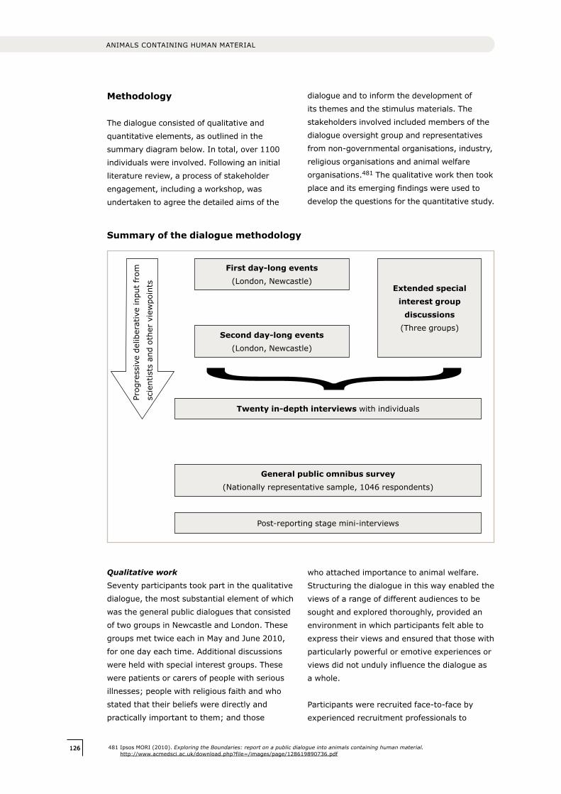
126126
ANIMALS CONTAINING HUMAN MATERIAL
Methodology
The dialogue consisted of qualitative and
quantitative elements, as outlined in the
summary diagram below. In total, over 1100
individuals were involved. Following an initial
literature review, a process of stakeholder
engagement, including a workshop, was
undertaken to agree the detailed aims of the
dialogue and to inform the development of
its themes and the stimulus materials. The
stakeholders involved included members of the
dialogue oversight group and representatives
from non-governmental organisations, industry,
religious organisations and animal welfare
organisations.481 The qualitative work then took
place and its emerging findings were used to
develop the questions for the quantitative study.
481 Ipsos MORI (2010). Exploring the Boundaries: report on a public dialogue into animals containing human material. http://www.acmedsci.ac.uk/download.php?file=/images/page/128619890736.pdf
Twenty in-depth interviews with individuals
General public omnibus survey (Nationally representative sample, 1046 respondents)
Post-reporting stage mini-interviews
First day-long events (London, Newcastle)
Second day-long events (London, Newcastle)
Extended special interest group
discussions (Three groups)
Qualitative work
Seventy participants took part in the qualitative
dialogue, the most substantial element of which
was the general public dialogues that consisted
of two groups in Newcastle and London. These
groups met twice each in May and June 2010,
for one day each time. Additional discussions
were held with special interest groups. These
were patients or carers of people with serious
illnesses; people with religious faith and who
stated that their beliefs were directly and
practically important to them; and those
who attached importance to animal welfare.
Structuring the dialogue in this way enabled the
views of a range of different audiences to be
sought and explored thoroughly, provided an
environment in which participants felt able to
express their views and ensured that those with
particularly powerful or emotive experiences or
views did not unduly influence the dialogue as
a whole.
Participants were recruited face-to-face by
experienced recruitment professionals to
Summary of the dialogue methodology
Prog
ress
ive
delib
erat
ive
inpu
t fr
om
scie
ntis
ts a
nd o
ther
vie
wpo
ints
127
ensure that a mixed and broadly representative
group of people took part. A modest cash
incentive was paid to encourage a diverse range
of participants.
The dialogue process was designed to reveal
how participants responded to information
about research involving ACHM. The emphasis
was on encouraging participants to reflect
on information that was provided and on
their interactions with each other and with
the scientists and facilitators present. ‘Focus
group’-style sessions were used, in which
participants could share their views, first
spontaneously, then after reflection. Their
eventual conclusions were the focus of analysis.
A sub-sample of 20 participants from both
the general public dialogue and special
interest groups were interviewed individually
by telephone during July and August 2010.
Interviewees for this stage were selected in
part because the views they expressed during
the day were distinct, which enabled issues
raised by preliminary analysis of the qualitative
data to be more thoroughly investigated and to
confirm the validity of these views, for example
that individuals were not unduly influenced by
others in the group.
Data analysis
Notes were taken throughout each dialogue
session and insights that each facilitator gained
were then shared during analysis meetings
at the end of each day and at the end of the
dialogue process as a whole. During the general
public dialogue events, an observational
researcher also made notes on body language,
facial expressions and evidence of behaviours,
without taking part in the facilitation, as well
as carrying out ad hoc interviews to explore
participants’ thoughts and feelings. The
purpose of this was to understand more subtle
and unspoken reactions alongside the main
discussions and to help build hypotheses as to
participants’ thoughts and feelings throughout
the process as their views developed.
Quantitative work
The quantitative findings in the report provide
an indication of the views of the wider British
public. The findings of the qualitative dialogue
informed the survey questions. The findings
were collected through the Ipsos MORI weekly
computer-assisted personal interviewing
(CAPI) survey, which is a cross-sectional
representative survey of individuals aged
over 15 years across Great Britain, performed
face-to-face by trained interviewers. 1046
participants completed the ‘Exploring the
boundaries’ survey in July 2010.
Findings
The findings of the public dialogue are
highlighted throughout this report and are
outlined and discussed in detail in the full report
produced by Ipsos MORI.482
Evaluation
The Academy commissioned Laura Grant
Associates to conduct an independent
evaluation of the dialogue. The evaluation
aimed to provide an independent assessment
of the dialogue programme’s credibility,
effectiveness and success against its
deliverables and objectives, throughout the
programme and at its conclusion; and to assess
its contribution to the overall Sciencewise
Expert Resource Centre aim of creating
excellence in public dialogue to inspire and
inform better policy in science and technology.
A comprehensive report of the full evaluation
methodology and findings produced by Laura
Grant Associates is available online.483
482 Ipsos MORI (2010). Exploring the Boundaries: report on a public dialogue into animals containing human material. http://www.acmedsci.ac.uk/download.php?file=/images/page/128619890736.pdf
483 Laura Grant Associates (2010). Exploring the Boundaries: A dialogue on Animals Containing Human Material Evaluation Report. http://www.acmedsci.ac.uk/download.php?file=/images/page/129111803577.pdf
ANNEx III OVERVIEW OF DIALOGUE METHODOLOGY AND EVALUATION

128128
ANIMALS CONTAINING HUMAN MATERIAL
129
ANNEx IV GLOSSARY OF TERMS AND ABBREVIATIONS
This glossary is intended to assist readers with the terminology and abbreviations used in this report; it is not presented as a definitive list of terms. Cross-references (e.g. see 2.2)
refer to the sections of this report.484
ACHM: Animals containing human material. See 2.2, page 18.
Admixed: ‘Admixture’ is the process of mingling one substance with another. The Human
Fertilisation and Embryology Act (as amended in 2008) defines five classes of ‘human admixed
embryos’ containing both human and animal material, with the human contribution predominating.
See Box 6.4, page 90.
Adult stem cell: Another term for ‘tissue-specific’ stem cell. See Box 3.3, page 38.
Amino acid: One of a group of chemical compounds that are the basic units of proteins.
Amniocentesis: A prenatal diagnostic technique in which a sample of amniotic fluid is withdrawn
and examined for information about the fetus. (The ‘amnion’ is the innermost membrane enclosing
the fetus.)
Amniotic stem cell: See Box 3.3, page 38.
Animal: In this text ‘animal’ (rather than ‘non-human animal’) is used to refer to animals of all
species in the animal kingdom except humans. (In correct scientific taxonomy, humans are both
primates and animals.)
Animal model: A living animal in which normal and abnormal biological processes can be studied,
to gain insight into human health and disease. The more closely the process being modelled
resembles the process in humans, the more scientifically valuable the model is likely to be.
Antibody: An antibody is a large protein found in blood and tissues, used by the immune system
to identify and neutralise foreign material such as cells, bacteria and viruses. The antibody
recognises a unique part of the foreign target, termed an antigen. Antibodies are produced as part
of the immune response.
Antigen: A foreign substance that, when introduced into a living organism, stimulates the
production of an antibody.
Aneuploid: Used to describe cells, tissues or organisms in which the number of chromosomes
is abnormal in that it differs from the euploid. ‘Euploid’ entities are those in which each of the
chromosomes of the set is represented in equal number (e.g. two copies of each chromosome is
termed ‘diploid’; one of each is ‘haploid’). See also haploid and diploid.
ASPA: Animals (Scientific Procedures) Act (1986). See 6.2.1, page 83.
APC: Animal Procedures Committee. See 6.2.1, page 83.
Annex IV Glossary of terms and abbreviations
484 Terms are drawn from sources including Department of Health (2010). Code of Practice for the use of Human Stem Cell Lines. http://workspace.imperial.ac.uk/clinicalresearchgovernanceoffice/Public/Code%20of%20practice%20for%20stem%20cell%20lines.pdf; Nuffield Council on Bioethics (2005). The ethics of research involving animals. Nuffield Council on Bioethics, London; The National Institutes of Health resource for stem cell research glossary, http://stemcells.nih.gov/info/glossary.asp; The Standard Oxford English Dictionary; Understanding Animal Research, http://www.understandinganimalresearch.org.uk/
130130
ANIMALS CONTAINING HUMAN MATERIAL
ASPD, ASPI: Animals Scientific Procedures Division (ASPD) and Inspectorate (ASPI) of the
Home Office.
Autologous: Derived from the same individual.
Autophagy: Literally, ‘feeding upon oneself’. A biological process by which a cell digests internal
components, for example to break down and recycle cellular components or to rid itself of toxins.
BIS: Department for Business, Innovation and Skills.
B-lymphocyte, B cell: A type of lymphocyte (a form of white blood cell), which forms part of the
immune system and produces antibodies in response to antigens.
Bioluminesence: The emission of light by living organisms such as fireflies and deep-sea
creatures. In biomedicine, cells or tissues can be made to emit light in this way as a form of marker
(see GFP).
Blastocyst: An early embryo consisting of a hollow ball of 50–100 cells reached after about 4 or
5 days of embryonic development (depending on species), just prior to implantation in the uterus.
The outer ‘trophectoderm’ cells give rise to part of the placenta, while a distinct group of about
15–20 ‘inner cell mass’ cells give rise to the embryo proper and other extraembryonic tissues (e.g.
placenta, yolk sac).
cDNA: Complementary deoxyribonucleic acid. A DNA strand that has been produced by ‘reverse
copying’ a messenger ribonucleic acid (mRNA) (either by a laboratory technique or by a retrovirus).
cDNA has the same sequence as DNA, except that it lacks introns.
Cell: The fundamental, usually microscopic, structural and functional unit of all living organisms,
which consists of a small quantity of cytoplasm enclosed within a membrane, typically contains
a nucleus and other organelles and internal compartments, and is capable of utilising energy,
synthesising proteins and other biomolecules, and (usually) of self-replicating.
CNS: Central nervous system. The largest part of the nervous system, including the brain and
spinal cord.
Cephalopod: An animal within the most highly organised class of molluscs. Cephalopods are
characterised by a distinct head with ‘arms’ or tentacles attached. Examples are Cuttlefish and
Octopuses.
Chimæra, chimæric animal: An animal comprised of whole cells from two different organisms.
See 2.2.2, page 18.
Chloride channel: A protein ‘pore’ that enables the transit of chloride ions into and out of cells,
across the cell membrane.
Chromosome: One of the threadlike structures containing identical sister strands of DNA, protected
by proteins and carrying genetic information, that can be microscopically visible within a cell.
131
Cleavage stage embryo: An embryo prior to formation of a blastocyst, undergoing ‘cleavage’
divisions where there is little or no cell growth; thus during cleavage each cell of the one, two, four,
and then eight cell embryo becomes progressively smaller.
Cognition, cognitive capacity: In its broadest sense, human ‘cognition’ can be defined as the
‘faculty of knowing’, to include aspects such as knowledge, reason, intelligence, understanding,
sensation and perception (as distinguished from feeling and volition). See 3.4, page 46.
Complement: A protein complex found in blood and other body fluids, which forms part of the
adaptive immune system. When combined with an antigen–antibody complex, complement
produces a series of reactions (the ‘complement cascade’) to bring about cell lysis.
Congenital: Present from the time of (and often before) birth.
Cord blood stem cell: See Box 3.3, page 38.
Cytoplasm: The gel-like substance enclosed by the cell membrane. It contains many important
molecules and organelles concerned with cell metabolism and movement.
Cytoplasmic hybrid: Cytoplasmic hybrid cells (cybrids) are those created by combining the
nucleus (with a minimal amount of cytoplasm and mitochondria) of a cell of one species with the
cytoplasm (including the mitochondria) of another species. Cytoplasmic hybrid embryos are those
created by transferring a somatic cell nucleus from one species into the enucleated oocyte of
another species.
Deontological: Relating to an ethical approach based on rules and duties. (Deontology is the
study of duty, a branch of knowledge that deals with moral obligations.)
Defra: Department for Environment, Food and Rural Affairs.
DH: Department of Health.
Differentiation: The process by which cells become progressively more specialised towards their
final function, both during development and adult maintenance.
Diploid: The state in which each type of chromosome (except the sex chromosomes) is
represented twice. This is the normal state of all cells of the body, except the germ cells (sperm
and eggs), which have only a single (haploid) set of chromosomes.
DNA: Deoxyribonucleic acid. A type of double-stranded nucleic acid molecule that encodes the
genetic instructions used by almost all living organisms.
DNA base pair: Two nucleotides (the structural units of which DNA is composed), on opposite
complementary DNA strands, which are connected by a hydrogen bond.
DNA regulatory region: A section of DNA that functions as a ‘switch’ to control gene expression.
They may lie either side of the gene or even within its introns, and they are often highly conserved
in evolution. Regulatory proteins (such as transcription factors) bind to regulatory regions. See Box
2.2, page 21.
ANNEx IV GLOSSARY OF TERMS AND ABBREVIATIONS
132132
ANIMALS CONTAINING HUMAN MATERIAL
DPA: Data Protection Act (1998).
Double-blind: A clinical trial or experiment, conducted by one person on another, in which
information (such as whether a substance being administered is active or placebo) that may lead
to bias in the results is concealed from both the tester and the subject. This method is used to
eliminate subjective bias.
Ectoderm: The outermost of the three primary germ layers. It gives rise to the epidermis (outer
part of the skin, including hair, nails, and sebaceous glands) the central nervous system (brain
and spinal cord) and to sensory systems, such as the eye, olfactory system and inner ear. See also
endoderm and mesoderm.
Ectopic: The location of cells or tissues at an abnormal site in the body. For example, ectopic
pregnancy involves the implantation of a fertilised egg outside the uterus.
EG cell: Embryonic germ cell. See Box 3.3, page 38.
EMEA: European Medicines Agency.
Embryo: the first stages in the development of an animal, usually the result of fertilising an egg
with a sperm. In humans, the embryo is usually referred to as a fetus from about the eighth week
of fertilisation. In other mammals, ‘fetus’ may be used to refer to older embryos, but there is no
strict definition of when fetal stages begin.
Embryonic stem cell: See Box 3.3, page 38.
Emphysema: A long-term, progressive disease of the lungs that primarily causes shortness of
breath.
Endoderm: There are two types of endoderm; ‘primitive’ or ‘extra-embryonic’ endoderm (also
sometimes called the ‘hypoblast’) forms the lower or outer layer of the early embryo (around
the time of implantation in mammals) and contributes to the yolk sac, but contributes little to
the embryo proper. ‘Definitive’ or ‘embryonic’ endoderm develops during gastrulation where it
displaces the extra-embryonic endoderm and gives rise to the larynx, lungs, gut and associated
organs, such as the thyroid and liver. See also ectoderm and mesoderm.
Endogenous: Having a cause (or origin) inside the body or self, not attributable to any external or
environmental factor.
Endometriosis: a condition resulting from the development of endometrial (womb lining) tissue in
an abnormal location outside the uterus.
Engraft: To insert a piece of material (e.g. cells, tissues or an organ) into an organism as a graft.
(Autologous grafts involve movement of material from one location to other within the same body.
Secondary chimæras are created by grafting cells tissue or organs from one animal into another.)
Enucleate: A cell lacking a nucleus; or the process of removing the nucleus of a cell.

133
Enteric nervous system: The part of the nervous system that directly controls the
gastrointestinal system (gut).
Enzyme: A protein that catalyses (increases the speed of) a specific biochemical reaction.
Epiblast: The upper layer of cells present in the embryo just prior to gastrulation. These cells are
pluripotent, and give rise to all three primary germ layers (ectoderm, mesoderm and endoderm) as
well as to germ cells and to extra-embryonic mesoderm.
Epigenetic: (‘Over’ or ‘above’ genetics). Epigenetic factors are heritable changes in phenotype
or gene expression, which result from mechanisms other than changes to the underlying DNA
sequence. For example, characteristics resulting from alterations in DNA methylation or changes in
chromosomal proteins.
Epitope: Part of an antigen, to which a particular antibody binds with a high degree of specificity.
EPO: European Patent Office.
ERP: Ethical review process. See 6.2, page 83.
ESC, ES cell: Embryonic stem cell. See Box 3.3, page 38.
ESCRO: Embryonic Stem Cell Research Oversight Committee. See Box 7.2, page 104.
Exogenous: Originating outside the body.
Exon: See intron.
Express, expression: Gene expression is the process by which information from a gene is used to
synthesise a functional gene product. This involves transcription to produce an RNA molecule called
messenger RNA (mRNA). mRNA is exported from the cell nucleus to the cytoplasm where its code
is translated into proteins by assembling amino acids in the right order. The polypeptide chains
produced are ultimately folded into proteins. Some genes only produce RNA products that fulfill
different important functions in cells. See Box 2.2, page 21.
Extra-embryonic tissue: Tissue that contributes to the growth or development of an embryo
without forming part of the embryo itself. Placental and yolk sac tissues are extra-embryonic.
Extra-embryonic stem cell: See Box 3.3, page 38.
Fetus: See embryo.
Fibroblast: A type of cell ubiquitously found in connective (supporting) tissues in most organs.
Fibroblasts play a role during wound healing or tissue repair. They are the type of cell that most
commonly grows in tissue culture, emerging from explants of pieces of most body tissues.
Fetal stem cell: See Box 3.3, page 38.
ANNEx IV GLOSSARY OF TERMS AND ABBREVIATIONS
134134
ANIMALS CONTAINING HUMAN MATERIAL
Fluorescence: Coloured light emitted by some chemicals, including some proteins, in response to
the action of light (especially violet and ultraviolet rays) upon them.
Gamete: A mature haploid sexual reproductive cell, for example a sperm or egg, which can unite
with another gamete to form a new organism. See germ cell.
Gastrulation: A phase early in the embryonic development of most animals, during which the
single layer of cells called the blastula (or in higher vertebrates the epiblast), is reorganised into a
three-layered patterned structure that will go on to form the three primary tissues of the embryo
proper (ectoderm, mesoderm, endoderm). In human embryonic development it begins at around
14 days after fertilisation, in the mouse at about 7 days.
Gene: The basic unit of heredity in living organisms, now known to consist of a sequence of DNA
(or RNA in certain viruses) containing a code for an RNA molecule that in many cases encodes a
protein. The gene also includes any associated regulatory sequences. See Box 2.3, page 23.
Gene product: A substance produced by the expression of a gene, for example a protein molecule.
Gene replacement therapy: The insertion of gene copies within some of an individual’s cells for
the purpose of treating disease. See Box 2.3, page 23.
Genotype: The genetic constitution of an individual.
Genetic sequence: The order of nucleotide bases (the individual units of which DNA is composed)
in a section of a DNA molecule.
Genetically altered: A cell, or organism, in which the DNA sequence has been modified.
See 2.2.1, page 18.
Genome: The complete DNA sequence of an individual, or a representative sequence for a species.
Germ cell: A sex cell or gamete (e.g. an egg or sperm); a reproductive cell that fuses with one
from the opposite sex in fertilisation to form a single-celled zygote. The term is also used to refer
to the progenitors of eggs and sperm during development.
Germ-line: The lineage of special cells set aside early in development that eventually differentiate
into mature germ cells. (Cells that are not part of the germ-line are referred to as somatic cells.)
Gestation: The process of carrying young in the womb.
GFP: Green fluorescent protein. A protein that occurs naturally in some marine organisms. GFP
fluoresces bright green under blue light, and is widely use as a marker in biomedical research. Its
gene can be readily transfected into cells of many species, and confers the fluorescent property on
the cells, which make the fluorescent protein and can then be easily visualised under appropriate
illumination.
Glia: The supportive non-neuronal tissue of the nervous system, composed of different types of
glial cell.
135
GM, GMO: Genetically modified, genetically modified organism.
Gonad: Any organ in an animal that produces gametes (e.g. a testis or an ovary).
Haematopoietic: A haematopoietic cell is one that is able to produce blood cells.
Haematopoietic stem cell: A stem cell that can give rise to all types of blood cell.
Haploid: A cell in which each type of chromosome is represented once (half the diploid number).
Hepatocyte: A type of cell that makes up the majority of liver tissue.
HFE Act: UK Human Fertilisation and Embryology Act. Unless specified, this term is used to refer
to the 1990 Act as amended in 2008.
HFEA: Human Fertilisation and Embryology Authority.
HLA: Human leukocyte antigen. The HLA genes are the human versions of the major
histocompatibility complex genes (MHC) found in most vertebrates. These genes encode cell-
surface antigen-presenting proteins, antigens and other proteins. The major HLA antigens are
the most important determinants of ‘tissue type’, which must be matched for optimal organ
transplantation.
Homologous: Sharing a common ancestral origin; entities that are homologous have a similar
structure. This term is used to describe genes that have a similar DNA sequence, or proteins
that have the same (or very similar) structure. It is often used in describing genes or proteins of
common evolutionary origin, found in different species.
Human (man): Individuals of the species Homo sapiens; human beings. Although in correct
taxonomy, humans are both primates and animals, in this text ‘animal’ (rather than ‘non-human
animal’) is used to refer to animals of all species except humans; and non-human primate (NHP) is
used to refer to primates except humans.
HSC: Haematopoietic stem cell. See Box 3.3, page 38.
hESC, hES cell: Human embryonic stem cell. See stem cell.
hiPSC, hIPS cell: Human induced pluripotent stem cell. See stem cell.
HSE: Health and Safety Executive.
HTA: Human Tissue Authority.
HT Act: Human Tissue Act (2004).
Human lineage-specific sequence: A section of the genome that is unique to humans, or to
humans and their near ancestors. See 3.2, page 32.
ANNEx IV GLOSSARY OF TERMS AND ABBREVIATIONS
136136
ANIMALS CONTAINING HUMAN MATERIAL
Humanised: An aspect of the biology of an animal (including for example a gene, protein, organ,
element of external appearance or behavioural characteristic) that has been modified so that it
more closely resembles that of the human.
Humanised antibody: An antibody produced by an animal (typically a mouse) whose antibody-
producing genes have been replaced by human DNA sequence, causing it to produce antibody
molecules that resemble those of the human. See 2.3.2, page 25.
Hybrid: An animal or plant that is the offspring of individuals of different kinds (usually, different
species) (see 2.2.3); hybrid embryos (see Box 6.4); inter-species cell hybrids are cells created by
the in vitro fusion of (usually somatic) cells from two different species (see 2.2.3), page 20.
Hyperacute response: A type of immune response that can occur rapidly after the
transplantation of cells or tissues from one species into another (e.g. xenotransplantation of organs
from pigs into humans). It is mediated by the binding of host antibodies to the donor graft causing
damage to, and rejection of, the transplanted tissue.
ICSI: Intra-cytoplasmic sperm injection. The injection of a sperm directly into an egg.
Immature germ-line cell: A cell of the germ-line that has not fully differentiated into a mature gamete.
Immortalised cell line: A cell line that has the ability to grow through an indefinite number
of divisions in cell culture. Some stem cell types and many cancer cell lines are immortal; such
cell lines can also be produced deliberately in the laboratory, usually as the result of genetic
manipulation. See 3.3.1, page 34.
Immune system (adaptive): The adaptive (or specific) immune system, found in vertebrates,
is composed of highly specialised cells and processes that recognise and destroy foreign proteins,
cells or micro-organisms entering the body. This is the body’s first defence against infections. The
adaptive immune response provides the ability to recognise and remember specific antigens and so
to generate immunity.
Immunodeficiency: A lack, or deficiency, of a functional immune system. The term ‘immuno-
compromised’ is used similarly. See 2.3.3, page 27.
Immuno-suppressive drug: A drug that prevents or inhibits immune system function.
In vitro: Literally, ‘in glass’; an experiment performed in a test tube, culture dish, or other non-
living environment.
In vivo: An experiment conducted within a living organism.
iPSC, iPS cell: Induced pluripotent stem cell. See Box 3.3, page 38.
Intron: A segment of a DNA molecule, which separates the exons (protein coding sections) of a
gene. An intron does not code for protein. See Box 2.3, page 23.
Ion channel: A protein ‘pore’ that enables the transit of ions into and out of cells across the
cell membrane.
137
IVF: In vitro fertilisation. The fertilisation of an egg by a sperm outside the body.
ISSCR: International Society for Stem Cell Research.
kb: Kilobase. A unit of length for measuring DNA or RNA sequences, equivalent to the length of
1000 bases.
Latent: Latent diseases are those in which the usual symptoms are not yet manifest. For infectious
disease, this may be because the causative microorganisms are lying dormant within the body until
circumstances are suitable for the development of overt disease.
Limbal stem cell: A stem cell found towards the edge of the cornea of the eye.
Longitudinal: Refers to a study that involves repeated observations on the same subject over a
long period of time.
Lymphatic system: A network of vessels through which lymph (a colourless fluid containing white
blood cells, vital in immune system function) drains from the tissues into the blood.
Lysis: The disintegration, for example of a cell, brought about by the breakdown of the containing
wall or membrane.
Lytic: Relating to, or causing, lysis. For example, in lytic viral infection a virus replicates within a
cell and, in the process of its release, destroys the cell.
Macular degeneration: A condition that results in a loss of vision in the centre of the visual field,
owing to the deterioration of the macula (an area near the centre of the retina in the eye).
Matrigel™: A gelatinous protein mixture that resembles the extracellular environment found in
many tissues and is used by cell biologists as a substrate for cell culture.
Meiosis: Part of the process of gamete formation, involving two cell divisions, in the course of
which the diploid chromosome number becomes reduced to the haploid.
Mesenchyme: Undifferentiated loose connective tissue that is derived mostly from embryonic
mesoderm. It contains cells capable of developing into various tissues such as bone, cartilage and
blood vessels.
Mesenchymal stem cell: see Box 3.3, page 38.
Mesoderm: The middle of the primary germ layers, which gives rise to many of the internal
tissues, such as bone, cartilage, muscle, blood, dermis and connective tissues. See also ectoderm
and endoderm.
Mesodermal stem cell: A stem cell of a type found in or derived from the mesoderm (the middle
layer of cells or tissues of the embryo).
ANNEx IV GLOSSARY OF TERMS AND ABBREVIATIONS

138138
ANIMALS CONTAINING HUMAN MATERIAL
Metabolism: The chemical processes that occur within a living organism to maintain life, including
both the synthesis of substances and their breakdown to produce energy.
Mitosis: The normal form of cell division in body tissues, resulting in two diploid daughter cells.
See also meiosis.
Molecular phylogenetics: The study of similarities of DNA sequence, to gain information on the
evolutionary relationships between organisms and species. See Box 2.1, page 17.
Monoclonal antibody: An antibody produced in the laboratory from a single clone (a genetically
identical population) of cells. Monoclonal antibodies from the same clone are identical, so they
recognise the same epitope on the antigen.
Monoclonal antibody therapy: A medical treatment that makes use of the highly specific binding
of a monoclonal antibody to a specific biological target. The antibody itself can act as a therapeutic
agent (e.g. by blocking receptors) or can carry with it an active drug molecule.
Mosaic animal: An animal containing two or more genetically distinct cell types that have arisen
from the same zygote. Mosaic animals can occur as a result of naturally occurring mutations, or
manipulations such as retroviral transfer. They are distinct from chimæras. See Box 2.3, page 23.
MHRA: Medicines and Healthcare products Regulatory Agency.
mRNA: Messenger ribonucleic acid. The molecule that carries the information from DNA to act as
a template for protein synthesis (some mRNAs are non-protein-coding and have other functions).
See Box 2.2, page 21.
MRI: Magnetic resonance imaging. A technique for producing images of bodily organs by
measuring the response to high-frequency radiowaves in a strong magnetic field.
MSC: Mesenchymal stem cell. See Box 3.3, page 38.
Multipotent: See potency and Box 3.3, page 38.
Mutation: The modification of a DNA sequence that has the potential to lead to a change in the
function of a gene. Mutations may be caused by mistakes in copying of DNA during cell division,
or by exposure to DNA-damaging agents in the environment. Mutations can be harmful, beneficial
or, most commonly, of no consequence. They can be caused by the alteration of single base units
in DNA, or the deletion, insertion or rearrangement of larger sections of genes or chromosomes.
Mutations can only be passed on to offspring if they occur in cells that make eggs or sperm.
NAS: National Academy of Sciences. One of the four United States National Academies.
Neuron: A specialised cell that transmits nerve impulses; a nerve cell.
Neural stem cell: A stem cell of a type found in the brain, from early development to adulthood.
Niche: The cellular microenvironment providing support and stimuli necessary to sustain stem cell
self-renewal and to control stem cell differentiation. See 3.3, page 38.

139
NHP: Non-human primate. In this text ‘NHP’ is used to refer to species of primates except humans.
‘Great Apes’ is used to refer to chimpanzee, pygmy chimpanzee, gorilla and orang-utan (see Box
6.1, page 83), and ‘monkey’ to refer to NHPs other than humans and Great Apes (e.g. 5.6.2),
page 78.
NRES: UK National Research Ethics Service, which provides ethical assessment of proposed
medical research involving human subjects.
Nucleotide: The structural unit of nucleic acids, DNA and RNA.
Nucleus (cell nucleus): A membrane-bound structure, often spherical, present in most living
cells, which contains the DNA in the form of chromosomes.
Oocyte: A cell of the female germ-line that may undergo division to form an ovum (a mature female
reproductive cell, egg). However, the term oocyte is often loosely used in place of ovum or egg.
Olfactory: Relating to the sense of smell.
Oligodendrocyte precursor: A cell that can develop into an oligodendrocyte, a kind of glial cell
that produces myelin (a substance that provides an insulating sheath around many nerve fibres) in
the central nervous system.
Oncogene: A gene that in certain circumstances can transform a cell into a tumour cell.
Oncology: The study and treatment of cancer.
Open-label: Describes a clinical trial in which both the researchers and participants know
the treatment that is being administered. This contrasts with the single blind method where
participants are not aware of what treatment they are receiving, and the double-blind trial
where neither experimenter nor the subject know whether active treatment or placebo is being
administered.
Organism: An individual animal, plant or single-celled life form.
Ovariectomy: Surgical removal of one or both ovaries.
Perinatal: Relating to the time immediately before and after birth. In humans this is usually a
period of several weeks; in rodents, a few days.
PET: Positron emission tomography. A medical imaging technique that produces a three-
dimensional image of internal body structures. During a PET scan, the recording system detects
rays emitted by a radioactive substance that is introduced into the body on a biologically active
molecule.
Phenotype: The physical manifestation of an organism, which results from the expression of the
genotype together with non-genetic influences.
Plasticity: The ability of a cell or organism to adapt to changes in its environment.
ANNEx IV GLOSSARY OF TERMS AND ABBREVIATIONS
140140
ANIMALS CONTAINING HUMAN MATERIAL
Pluripotent: See potency and Box 3.3, page 38.
Polypeptide: A molecule made up of several amino acids joined together in a chain. Proteins
consist of one or more polypeptides.
Potency (or potential): Generic terms to denote the range of specialised cells that a stem
cell may/can give rise to. Stem cell potency can be more specifically described as unipotent,
multipotent or pluripotent. See Box 3.3, page 38. (A totipotent cell is one that can give rise to all
cell types in an animal or human, including extra-embryonic tissues, and (according to a commonly
accepted definition), carries the information required to organise development of the embryo
correctly. The only cells that are totipotent are therefore the fertilised egg and those of early
cleavage stage embryos. These do not self-renew, therefore they are not stem cells.)
Pre-clinical: Denotes research, or the stages of the drug development process, conducted before
that in the clinic. It may include approaches such as in vitro research, computer simulation and
research using animals.
Primitive streak: A structure that forms during the early stages of mammalian embryogenesis;
its appearance is one of the first signs of gastrulation.
Prion: A misfolded form of protein believed to act as an infectious agent. Diseases including bovine
spongiform encephalopathy (BSE, in cattle), scrapie (in sheep) and Creutzfeldt–Jakob disease
(CJD, in humans) are thought to be prion-mediated.
Progenitor cell type: A generic term for any dividing cell with the capacity to differentiate. It
includes putative stem cells in which self-renewal has not yet been demonstrated.
Pronucleus: Either of a pair of nuclei from gametes (in the haploid state following meiosis) in
the egg after fertilisation (or activation) but before they come together to form the (diploid)
chromosome complement of the zygote.
Protein: Large molecules composed of one or more long chains of amino acids (polypeptides).
Proteins are an essential part of living organisms, as both structural and functional components of
all body tissues.
Provirus: The genetic material of a virus as incorporated into the genome of a host cell.
Quiescent: In a state or period of inactivity or dormancy.
Receptor: A molecule within a cell (frequently in a cell membrane), which binds and responds
specifically to a particular transmitter (cell signalling molecule), hormone, antigen or other
biologically active molecule.
Recombinant DNA: DNA formed artificially by combining sections of DNA, often from different
organisms.
141
Regenerative medicine: Approaches aimed at creating living, functional tissues to repair or
replace the function of cells, tissues or organs lost because of damage or congenital defects. Many
such approaches involve the use of stem cells.
Restricted: Limitation of the potency of a stem cell, meaning that it cannot give rise to some types
of specialised cells in the body.
Retrovirus: An RNA virus that inserts a DNA copy of its genome into the host cell to replicate, for
example human immuodeficiency virus (HIV).
RGF: Research Governance Framework.
SCRO: Stem Cell Research Oversight Committee. See Box 7.2, page 104.
Self-renewal: Cycles of division that generate at least one daughter cell equivalent to the mother
cell, with latent capacity for differentiation. The defining property of stem cells. See 3.3, page 34.
Somatic cell: Any cell that forms part of the body of an organism, not including germ cells.
SCNT: Somatic cell nuclear transfer. The transfer of the nucleus from a somatic (e.g. fetal or adult
body) cell into an oocyte from which the nucleus (or the nuclear DNA) has been removed. The
basis of the technique used to clone mammals, famously including Dolly the sheep.
Species: A species is one of the basic units of biological classification and a taxonomic rank. A
species is often defined as a group of organisms capable of interbreeding and producing fertile
offspring. Although in many cases this definition is adequate, more precise or differing measures
are often used, such as similarity of DNA or morphology. See Box 2.1, page 17.
Sperm: An abbreviated term used to denote a male sex cell of an animal. In scientific
terminology, the developing male sex cells are named at different stages (e.g. ‘spermatogonium’,
‘spermatocyte’, ‘spermatid’), and a mature, motile male sex cell is referred to as a ‘spermatozoon’.
Spermatagonial stem cell: See Box 3.3, page 38.
Splicing: The modification of a primary messenger RNA (mRNA) transcript to remove the non-
coding ‘introns’ and join up the coding ‘exons’ to make a functional mRNA, usually one able to be
translated to form proteins.
Stem cell: A stem cell is a cell that can continuously produce unaltered daughters and has the
ability to produce daughter cells that have different, more restricted properties. In this text, the
term ‘stem cell’ is sometimes used to encompass other progenitor cells types. Human stem cells
are abbreviated ‘h’, e.g; human ES cell (human embryonic stem cell); human iPS cell (human
induced pluripotent stem cell).
Telomere: Repetitive nucleotide sequences at the ends of chromosomes that serve as a ‘capping’
structure. Telomeres are shortened with each cell division. Short telomeres are consequently
considered a sign of ageing.
ANNEx IV GLOSSARY OF TERMS AND ABBREVIATIONS
142142
ANIMALS CONTAINING HUMAN MATERIAL
Teratocarcinomas: A form of malignant teratoma occurring especially in the testis.
Teratoma: A type of tumour that contains several different tissue types. See 3.6.1, page 56.
Tetraploid: A cell or nucleus in which four homologous sets of chromosomes are represented, in
contrast to the two sets (diploid) normally found in somatic cells.
Tissue: A distinct type of material of which the body is composed, consisting of specialised cells
and their products, for example connective tissue, muscle.
Tissue-specific (or adult) stem cell: See Box 3.3, page 38.
Toxicity testing: A stage in the development of therapeutic products, in which they are tested
for their potential to cause unanticipated or harmful effects to the body. Toxicity tests are often
conducted on animals to establish dose–toxicity relationships and maximum safe dosage levels
before clinical trials are conducted in man.
Transfection: A process in which DNA is introduced into a cell containing a nucleus, and integrates
into the recipient cell’s nuclear DNA.
Transgenic: A cell, embryo or animal created by the insertion of some additional genetic material
from another genome. See Box 2.3, page 23.
Transgene: A sequence of genetic material taken one organism (or artifically synthesised) and
inserted into the genome of another cell, embryo, or animal. See Box 2.3, page 23.
Translation: The process by which a sequence of nucleotides in a mRNA molecule is read and
‘translated’ by cellular machinery to a specific sequence of amino acids, during synthesis of a
protein.
Trophectoderm, trophoblast: A layer of tissue on the outside of a mammalian blastocyst, which
supplies the embryo with nourishment and later forms the major part of the placenta.
Tumour: A swelling of a part of the body, generally without inflammation, caused by an abnormal
growth of tissue.
Tumourigenic: Tumour-causing.
UNESCO: United Nations Educational, Scientific and Cultural Organization.
Unipotent: See potency and Box 3.3, page 38.
Vaccine: An antigenic substance prepared from the causative agent of a disease or a synthetic
substitute, used to produce immunity against disease.
Vascularisation: The process of developing blood vessels.
143
Vector: A biological construct (e.g. a plasmid) used as a vehicle for transferring genetic material
into a cell. See Box 2.2, page 21.
Vertebrate: An animal possessing a backbone or spinal column, such as a mammal, bird, reptile,
amphibian or fish.
Xenograft: A tissue graft or organ transplant from a donor of one species into a recipient of
another. (‘xeno-’ from Greek meaning ‘stranger’.)
X-ray: An electromagnetic wave of high energy, which is able to pass through many materials
opaque to light; a photographic or digital image of the internal composition of something,
especially a part of the body, produced by x-rays being passed through it.
Zygote: The diploid cell resulting from the union of haploid male and female gametes.
ANNEx IV GLOSSARY OF TERMS AND ABBREVIATIONS

145
Recent reports from the Academy of Medical Sciences
A new pathway for the regulation and governance of health research
http://www.acmedsci.ac.uk/p99puid209.html
Biomedical research - a platform for increasing health and wealth in the UK
http://www.acmedsci.ac.uk/p99puid212.html
Academia, industry and the NHS: collaboration and innovation
http://www.acmedsci.ac.uk/p101puid202.html
Redressing the balance: the status and valuation of teaching in academic careers
http://www.acmedsci.ac.uk/p99puid181.html
Reaping the rewards: a vision for UK medical science
http://www.acmedsci.ac.uk/p99puid172.html
Rejuvenating ageing research
http://www.acmedsci.ac.uk/p99puid161.html
Building clinical academic capacity and the allocation of resources across academic specialties
http://www.acmedsci.ac.uk/p99puid150.html
Research in general practice: bringing innovation into patient care
http://www.acmedsci.ac.uk/p101puid163.html
The Bologna Process: will it affect UK biomedicine and clinical science
http://www.acmedsci.ac.uk/p101puid179.html

146146
ANIMALS CONTAINING HUMAN MATERIAL
Academy of Medical Sciences41 Portland Place London, W1B 1QH Tel: +44 (0)20 3176 2150