animal health monitor
TRANSCRIPT
To subscribe to the Animal Health Monitor, contact: Julie Hughes by “CTRL” clicking [email protected]
Animal Health Monitor September, 2021
Volume 14, Issue 1
Reports (Page 2—7)
• AgriService BC
• Blackhead in Turkeys in BC
• Bluetongue in BC California Bighorn
Sheep Herd
• Small Scale Pig Production in BC
Fact Sheet (Page 8—10)
• Domestic and Wild Sheep and Goats and
Risk of Mycoplasma Ovipneumoniae
Test Updates (Page 11—12)
• Fecal Parasitology at AHC
Tech Tips (Page 13 –14)
• Sample Submission to the Bacteriolo-
gy Section of the PAHB
Mission Statement: To provide our clients with important information regarding the work being
done by the Animal Health Centre and Regulatory
Unit, as well as events and issues relevant to
animal health in BC.
Editorial by Dr. Shauna-Lee Chai, Acting Executive Director, Plant and Animal Health Branch
The only constant in life is change.
Heraclitus
As I reflect on the past few months since the last edition of the Animal Health
Monitor, there are changes afoot at the Plant and Animal Health Branch that I
would like to make you aware of; top of mind for many of us at the Branch is the
retirement of our Executive Director – Ursula Viney.
Ursula has been with the Ministry of Agriculture, Food and Fisheries for eight
years, working in the Kelowna and Abbotsford offices. Her attention to financial
details and process management has benefited our Ministry tremendously. Her
leadership at the Animal Health Centre has helped to shift the role and the visi-
bility of the lab to one that contributes more meaningfully to Ministry and pro-
vincial priorities. Ursula will be missed. She is retiring from the Ministry to move
to Ontario to join her family.
In the 8-month interim, until we recruit a permanent Executive Director, I have
been temporarily appointed to the role for a 4-month period: August 30 - Decem-
ber 31, 2021. January 1 – April 30, 2022 will also ring in a new temporary ap-
pointment of Shannon Tucker as Executive Director of the Branch.
Despite the changes in leadership, we remain the same great place to diagnose,
monitor and manage animal pests and diseases in British Columbia. Last month
alone, 2,562 people found the Plant and Animal Health Branch on Google, and
we worked through 828 submissions (plants and animals).
My usual role at the Branch is the Director of the Plant Health Unit, where we
diagnose, survey and address plant pests and diseases that impact the crop indus-
try. As Acting Executive Director, my vision for the Branch is to maintain emi-
nence in all our specific fields within animal and plant health and to share our
science with British Columbians and the world at large.
The COVID-19 pandemic and wildfires here in B.C. have stretched us and taught us the value and possibilities of working collaboratively, across teams and work units. So, wherever you find yourself today, it is my wish that you will work with a mindset of abundance and kindness.
All authors are employees of the Ministry of Agriculture, Food & Fisheries unless otherwise noted.

To subscribe to the Animal Health Monitor, contact: Julie Hughes by “CTRL” clicking [email protected]
AgriService BC
AgriServiceBC (https://www2.gov.bc.ca/gov/content/industry/
agriservice-bc) is a complimentary service offered by the BC Ministry
of Agriculture, Food, and Fisheries. It is designed to serve and support
B.C.’s agrifood sector by connecting farmers, food processors and new
entrants to agricultural services with the programs and information
that can help them succeed. Through dedicated email or phone, peo-
ple can get in touch with knowledgeable staff who can answer a variety
of questions related to livestock and poultry, crop production, aqua-
culture, food processing, and much more. The AgriServiceBC e-bulletin
is published 1-2 times per month and is a great way to stay up to date
about upcoming webinars, events, programs and resources. To sign
up, go to: https://agriservicebc.campayn.com/contact_list_form/
signup/88410
Blackhead (Histomonas meleagridis) in Turkeys
in BC Doris Leung (Veterinary Specialist), Gigi Lin (Veterinarian –
Canadian Poultry Consultants Ltd.), Victoria Bowes (Avian
Pathologist), and Tony Redford (Veterinary Pathologist)
Background
Blackhead disease, also known as Histomoniasis, is a parasitic disease
of importance caused by the protozoal organism Histomonas melea-
gridis. It affects gallinaceous birds including turkeys, chickens, game-
birds, and peafowl. Blackhead disease may cause high mortality rates
in turkey flocks, with variable mortality rates of 10 to 100%. The dis-
ease is usually less severe in chickens, with mortality rates of 10 to
20%, but infected flocks may have reduced egg production and poor
health. As there are currently no approved vaccines or treatments for
licensed use in Canada, it is important to have good biosecurity prac-
tices in place to limit the introduction and subsequent spread of Histo-
monas within the flock.
Transmission of Histomonas meleagridis is complicated. There are three
possible ways that a turkey could become infected with the disease:
1) direct uptake of Histomonas meleagridis into the lower digestive
tract of the bird through a process called “cloacal drinking”
which occurs when birds are sitting on the litter;
2) ingestion of cecal worm eggs (Heterakis gallinarum) that are in-
fected with Histomonas meleagridis; and
3) ingestion of common earthworms that contain Histomonas
meleagridis, or other insects that act as mechanical vectors of
the protozoa.
It should be noted that naked Histomonas organisms are quite fragile
to the external environment but can persist within its cecal worm host
and remain infective for several years. Turkeys infected with Histomo-
niasis show signs of disease at around 7 to 12 days post infection.
Clinical signs include reduced appetite, depression, drooped wings, ruffled feathers, and sulfur -yellow droppings. Contra-ry to its name, turkeys with blackhead disease generally do not show dark (cyanotic) discolouration of their heads.
The case definition of blackhead is defined as a combination
of cheesy cores in cecal pouches and multiple target-like
white spots throughout the liver (Figures 1, 2). Together, the
cecal and liver lesions are considered diagnostic for Histomo-
niasis. In the absence of obvious white liver lesions, caseous
(cheesy) cecal cores are noted microscopically with histomon-
ad protozoal organism present (Figure 3).
Figure 1. Caseous yellow-green exudates develop into
cheesy cores within the cecum of turkeys infected with
Histomoniasis. Image courtesy of Dr. Gigi Lin, Canadian
Poultry Consultants.
Figure 2. Necrotic, target-like lesions in the liver are noted in turkeys infected with Histomoniasis.
Image courtesy of Dr. Gigi Lin, Canadian Poultry Consult-ants.
Reports || 2

To subscribe to the Animal Health Monitor, contact: Julie Hughes by “CTRL” clicking [email protected]
Blackhead (Histomonas meleagridis) in Turkeys in BC cont’d
Figure 3. A microscopic view of a blackhead infected cecum.
Note the numerous round pink Histomonas organisms sur-
rounded by a clear halo. There is an intense inflammatory
reaction in the adjacent tissue. Image courtesy of Dr. Victoria
Bowes, Animal Health Centre Laboratory.
Since 2020, the BC turkey industry has experienced multi-farm
outbreaks of blackhead disease, with significant negative eco-
nomic consequences for turkey growers, processors, and the
hatchery. In response to the outbreak, a collaborative study
began in January 2021 in partnership with stakeholders in
industry and government.
The goals of the study are to understand the epidemiology of
the blackhead in the province, identify on-farm risk factors and
reservoirs, and ultimately, to create an effective blackhead
disease prevention and control strategy to improve flock health,
productivity, as well as profitability of the BC turkey industry.
Blackhead Engagement Strategies
Several engagement strategies have been underway over the past
six months. Two web seminars were hosted to share relevant
and timely information to BC turkey growers. Presentations
from the Ministry of Agriculture, Food, and Fisheries (MAFF),
Canadian Poultry Consultants (CPC), and other field experts
provided information on topics that included blackhead etiolo-
gy, disease prevention, and general biosecurity recommenda-
tions. Please contact Michel Benoit of BC Turkey Farmers at
[email protected] if you would like a copy of the seminar
recording.
Moreover, a questionnaire was introduced as a retrospective
investigation to identify on-farm risk factors of blackhead in BC
turkey farms. The questionnaire was administered to growers
identified as “cases” and “controls” based on the case definition
discussed previously. “Cases” are growers that had blackhead
disease from January to December 2020, whereas “controls” are growers that did not have blackhead infected flock in the same period. The questionnaire included questions related to turkey production, animal husbandry, farm and barn renovations, biose-curity, litter management, vector control, and more. There were additional questions in the last section of the questionnaire relat-ed to blackhead management if the farm was identified as a “case”. The questionnaire was sent electronically, and MAFF staff epidemiologist Dr. Doris Leung collected answers from growers through in-depth phone interviews. The results of this questionnaire will be made available to growers in upcoming months. We would like to thank all growers for participating in this questionnaire.
Current Situation
In 2020, the Animal Health Centre (AHC) laboratory received 21 turkey submissions that were diagnosed with Histomoniasis (Figure 4) by either necropsy and/ or histopathology with the presence of histomonads organisms. Infected premises were in Abbotsford and Langley. Summer months accounted for an increased number of cases, with submitted birds and fixed tissue samples from June to August 2020 accounting for 43% of the cases (9/21). The average age of birds with blackhead disease is 62 days of age.
Figure 4. Epidemic curve of 2020 Blackhead cases by month of onset.
As of August 2021*, 8 turkey submissions received by the AHC
laboratory were diagnosed with Histomoniasis (Figure 5). Infected
premises were in Abbotsford and Langley. Similar to 2020,
summer months accounted for an increased number of cases, with
submitted birds and fixed tissue samples from June to August
2021 accounting for 75% of the cases (6/8). The average age of
birds with blackhead disease is 57 days of age. It should be noted
that all infected premises had blackhead outbreaks previously.
*Data collected until August 10, 2021.
Reports || 3

To subscribe to the Animal Health Monitor, contact: Julie Hughes by “CTRL” clicking [email protected]
Wear new coveralls and boots with each barn visit. Clean
and disinfect personal protective equipment (PPE) often.
Perform barn visits from youngest to oldest flocks. If there
are sick or diseased flocks, ensure that you visit this flock
last.
Ensure external personnel (e.g. catching crew, cleaning
personnel) adhere to similar biosecurity protocols regarding
PPE and cleaning and disinfection of equipment or
machinery.
Restrict outside visitors whenever possible.
• Active repairs and maintenance of the barn:
Cracks and crevices on the floor and wall should be filled
and sealed routinely. This also helps to minimize pest
habitat.
Gaps on doorways are being sealed properly to prevent
vermin or insects from entering the barn.
• Bird movement:
Birds are being moved indoor only.
Ensure equipment used to move birds are adequately
disinfected prior to and after use.
• Shaving/ litter management:
Do not re-use contaminated or old litter.
Do not have on-site shaving storage.
Fully remove all litter with each stage, not just with
brooders. Manure is cleaned out with full blow down, and
power washed. Ensure that the concrete floor is fully dried
prior to disinfectant application.
Ensure concrete floor is fully dried and/or heat treated prior
to placement of fresh shavings.
Litter condition should be dry with minimal condensation.
New litter should be treated with acid, dried chlorine
dioxide, etc. prior to bird placement. Ensure a thick layer of
litter for each flock.
• Animal husbandry
Create a routine deworming program with your veterinarian
to bolster bird intestinal health.
Blackhead (Histomonas meleagridis) in Turkeys in BC cont’d
Figure 5. Epidemic curve of 2021* Blackhead cases by month
of onset.
Best Practices Recommendations
• Exercise appropriate biosecurity to protect turkey flocks:
Ensure adequate use of disinfectants (e.g. at labelled
dosage, rotate different class of disinfectant with every
cycle).
Ensure adequate use of physical and chemical pesticides
/ rodenticides (e.g. at labelled dosage, rotate differ ent
class of insecticide with each cycle). Use pesticides
liberally in barn prior to bird placement if there are
historical insect infestations (e.g. with darkling beetles).
Have extended downtime between flocks, with at least 2
weeks downtime for each barn to allow for thorough
cleanout and disinfection.
Have a mandatory vehicle disinfection station for all
vehicles entering the premise.
Have well-defined biosecurity lines prior to barn
entrance. Footbath solutions must be changed every 2
to 3 days to maximize disinfectant activity and to
remove any organic debris.
No sharing of equipment or machinery with other
farms, especially with different poultry species.
Equipment and machinery should be housed indoors.
Do not move equipment or machinery between barns.
Machinery used inside the barn should not be used for
outdoor pasture or gardening.
Good draining around barn perimeters. Apply salt and
pesticide to the perimeters to prevent earthworms and
other insects from entering barn.
Reports || 4

To subscribe to the Animal Health Monitor, contact: Julie Hughes by “CTRL” clicking [email protected]
Blackhead (Histomonas meleagridis) in Turkeys in BC cont’d
Build partitions near barn doors to prevent drafts and to
act as barriers to prevent birds from ingesting earth
worms or other insects outside of the barn.
Ensure birds do not succumb to heat stress with misters
or evaporative cooling if needed.
Minimize exposure to environmental stressors that can
be detrimental to bird intestinal health. (e.g. avoid feed
interruption from feed outage, feed pens filled with
litter, high CO2 concentration in the barn)
• Management practices if you suspect blackhead in your
flock:
Seek veterinary advice ASAP if birds appear ill. Your
veterinarian may perform a necropsy and/or send
samples to the AHC laboratory for confirmatory
diagnosis of blackhead.
Prompt removal of mortalities and contaminated litter
ideally off-farm.
Ensure proper hand washing hygiene after handling
mortalities and sick birds.
Ensure equipment or machinery used to handle dead or
sick birds is adequately cleaned and disinfected.
Cull birds that appear lethargic or depressed.
Create partitions to physically segregate sick and healthy
birds to reduce the spread of blackhead within the barn.
Move the flock to a clean barn/ clean litter if possible. Additional information upon request; please contact Doris Leung at [email protected]
Bluetongue in B.C. California Bighorn Sheep Herd Glenna McGregor (Veterinary Pathologist), Katie Galliazzo (Industry Advisor for Livestock)
On August 11, 2021, a deceased California bighorn sheep from the
Grandby Herd, located near Grand Forks, B.C., was reported to the
Ministry of Forests, Lands, Natural Resource Operations and Rural
Development (FLNRORD). By August 20th, upwards of 6 bighorn
sheep had been reported dead and 3 relatively fresh carcasses of
adult female bighorn sheep were recovered and brought to the
Animal Health Centre for post-mortem examination.
All animals were in good body condition. One of the carcasses was
too severely decomposed for post-mortem examination. In the other
two sheep, the tongues were swollen and dark with extensive submu-
cosal hemorrhage and congestion. The nasal mucosa was diffusely
expanded by a large amount of dark purple gelatinous material
(submucosal edema and hemorrhage). There was a large amount of
stable foam filling the trachea throughout its entire length
(pulmonary edema). The lungs were edematous and congested with
mottled areas of hemorrhage. Approximately a litre of serosangui-
nous fluid filled the abdominal and thoracic cavities. There were
ecchymoses at the base of the heart. Lymph nodes were diffusely
enlarged, dark and hemorrhagic. In both, there was marked bilateral
perirenal hemorrhage. Histologically there was hemorrhage, edema
and a subtle acute fibrinous vasculitis in multiple organs as well as a
severe necrosuppurative glossitis (figure 1). PCR for Bluetongue per-
formed at the Animal Health Centre was positive on the spleen, and
this was confirmed on August 26, 2021 by National Centre for For-
eign Animal Disease on both the spleen and bone marrow from the
three submitted animals. Viral isolation and genotyping are pend-
ing. PCR for Epizootic Hemorrhagic Disease was negative on spleen.
PCR for Mycoplasma ovipneumoniae was negative on nasal swabs.
Mortalities in the infected herd continue at a high level. Of the 12
sheep FLNRORD had collared in this population, 8 have died;
however, the exact toll on the uncollared animals and the whole
population is unknown. Prior to this outbreak, the total population
size was 230 to 240 animals. White tailed deer also appear to be
dying in the area; the number affected is unknown.
Bluetongue is a viral infection caused by an Orbivirus, in the family
Reoviridae. It is spread by the bites of some species of Culicoides
midges that are infected by feeding on infected animals, and then,
following a viral replication period of 6-8 days in the midge’s salivary
gland, can transmit the infection to other susceptible animals. There
are 27 recognized serotypes. Serotypes 2, 10, 11, 13 and 17 are
considered endemic in the US and are immediately notifiable to the
CFIA. All other serotypes are exotic to North America and are
federally reportable in Canada. Serotyping of the virus in this case is
still pending.
Reports || 5

To subscribe to the Animal Health Monitor, contact: Julie Hughes by “CTRL” clicking [email protected]
Bluetongue in B.C. California Bighorn Sheep Herd cont’d Midges are the only significant natural transmitters of blue-
tongue virus (BTV); thus, distribution and prevalence of the
disease is governed by ecological factors impacting geographical
distribution and abundance of the midges. Because of this,
infection with BTV is common in a broad band across the world,
which until recently stretched from roughly 35°S to 40°–50°N;
however, in the last few decades, BTV has been extending farther
north in many parts of the world. In B.C., we have sporadic
outbreaks in the Okanagan every several years. These generally
correspond to outbreaks in Washington and are thought to be
due to windborne midges blowing up from the US rather than
due to established midge populations within the province. These
generally only occur in the late summer or early fall as the
weather needs to be warm (15 to 35°C) for the virus to replicate
within the midge, and midge activity ceases with the first hard
frost. There is no evidence that bluetongue is able to survive
winter in Canada. It does not establish persistent infections in
ruminants and so can only overwinter in areas where the midges
are able to overwinter. Outbreaks in B.C. with the level of mor-
tality seen this year are uncommon.
Bluetongue can infect a variety of wild and domestic ruminants
including sheep, goats, cattle, bison, camelids, deer, bighorn
sheep, elk, mountain goats, and pronghorns, but generally only
causes severe disease in sheep and occasionally white-tailed deer.
It does not infect humans and does not pose a risk to human
health.
For livestock, keeping animals inside at dawn and dusk when the
midges tend to be most active may decrease the risk of transmis-
sion. There are no vaccines for Bluetongue licensed in Canada.
The midges that transmit Bluetongue are currently only found in
the south Okanagan, this does not pose a risk to livestock else-
where in the province at this time. If anyone has animals with
consistent clinical signs or sudden death of multiple animals,
they should contact their veterinarian or the Animal Health
Centre.
For more information please see the BC Wildlife Health Website
at https://www2.gov.bc.ca/gov/content/environment/plants-
animals-ecosystems/wildlife/wildlife-health/wildlife-diseases, or
the CFIA factsheet at : https://inspection.canada.ca/animal-
health/terrestrial-animals/diseases/reportable/bluetongue/
fact-sheet/eng/1306116803992/1306121522520
Figure 1: A microscopic view of a tongue from a bighorn sheep with Bluetongue.
Figure 2: Clinical signs of bluetongue infection in ruminants such as sheep include swelling and hyperaemia of the mouth and lips.
Reports || 6

To subscribe to the Animal Health Monitor, contact: Julie Hughes by “CTRL” clicking [email protected]
Small Scale Pig Production in B.C. Tom Droppo (Industry Specialist - Dairy & Pork)
B.C. has an estimated 2,000 small scale pig
producers. Farms range in size from one to 300 pigs. A national
survey of small scale pig producers was conducted in early 2021,
and a Final Survey Report released June 2021. This article
highlights key findings from that survey.
565 small scale producers representing all provinces responded to
the survey. Survey questions focused on operation features,
management, awareness of pig diseases and health, and sources of
information.
Distribution of number pigs managed by the 565 producer respondents:
198 (or 35%) had 1-4 pigs 109 (or 19%) had 5-9 pigs 258 (or 46%) had 10 or more pigs 94% of producers reported having other livestock in
addition to pigs on their farm, while 14% of producers house other animals in the same enclosure as their pigs.
74% of producers get their pigs from other swine operations, such as local farms., while 12% indicated they get their pigs from public markets.
67% of producers house their pigs outdoors in fenced areas; however, the majority of barriers are wire mesh suggesting that risky interactions with wildlife can occur.
25% of producers indicated they don’t do anything specific regarding illness & disease reduction.
38% of producers are ‘not at all concerned’ about their pigs developing a disease, while only 23% of producers are ‘at least somewhat concerned’ about their pigs developing a disease.
25% of producers have not heard of foot & mouth disease (FMD) or African swine fever (ASF), and 50% have not heard of porcine reproductive & respiratory syndrome (PRRS).
60% of producers have a veterinarian, but 26% feel they don’t need one.
Deadstock disposal methods: 49% by burial, 25% compost, and 21% by scavenging (Western Canada).
56% of producers feed their pigs table scraps or food waste from their kitchen, and 3.7% feed meat or meat products to their pigs
47% of producers do not perceive any risks associated with feeding food waste from their kitchen or from grocery stores, bakeries, and restaurants.
Producers were most likely to get their information by word-of-mouth from people they consider knowledgea-ble: 63% from other pig producers, 53% from veterinarians, and 50% from pig websites.
In 2014, federal legislation was enacted that required all pig
producers, regardless of size, to comply with new national
traceability requirements. This requires every pig producer to
have a Premises ID, all pigs with Animal ID, and any
movement of pigs off-farm to be reported to PigTrace, the
national pig database. As of August 2021, only 2/3 (or 66%)
of small lot pig producers in B.C. have a Premises I.D.
The national survey reveals the need for education outreach
to raise awareness of best management practices to improve
biosecurity and protect the health of pigs raised by small scale
producers. Further information on small scale pig production
can be found at B.C. Pork (Small Lot Pork Producers | BC
Pork or at B.C. Ministry of Agriculture, Food and Fisheries
Pork - Province of British Columbia (gov.bc.ca) .
Reports || 7

To subscribe to the Animal Health Monitor, contact: Julie Hughes by “CTRL” clicking [email protected]
Domestic and wild sheep and goats, and risk of
Mycoplasma ovipneumoniae
What is Mycoplasma ovipneumoniae? • Mycoplasma ovipneumoniae (M. ovi) is a bacterial species that is commonly found in the nasal
cavity and sinuses of apparently healthy domestic sheep and goats.
• It is transmitted to wild sheep and goat populations via nose-to-nose contact and, less commonly, aerosol/droplet transmission. In wild sheep, infection often causes large all-age die-offs followed by years of poor lamb recruitment, resulting in large population declines.
• In experimental studies, M. ovi has been
shown to slightly halt weight gains in domestic
sheep and may predispose animals to more
serious bacterial pneumonia.
• Thinhorn sheep in British Columbia have had little exposure to domestic animal pathogens
and are considered more at risk than Bighorn sheep. Exposure to these pathogens is
expected to result in significant and widespread losses from either domestic goat or sheep
M. ovi strains.
Some sheep and
goats carry M. ovi
in their noses
Roaming big-
horns pick up the
bacteria when
they visit or en-
counter domestic
Ewes may carry
bacteria and infect
lambs for many
years. With little
immunity, they
often die before
Some ewes
develop long-
term “chronic”
M. ovi-positive animals
spread the pathogen
throughout their herds,
What happens during a M. ovi outbreak in wild sheep:
Sources: 1. Cassirer, E. F et al. (2018). “Pneumonia in bighorn sheep: Risk and Resilience. Journal of Wildlife Management. 82(1): 32-45. 2. Manlove, K. R. et al. (2019). “Risk factors and productivity losses associated with Mycoplasma ovipneumnoniae infections in United States domestic sheep operations.” Preventive Veterinary
Medicine 168: 30-38. 3. Plowright, R. K. et al. (2017). “Age-specific infectious period shapes dynamics of pneumonia in bighorn sheep.” Ecology Letters 20(10): 1325-1336.
Fact Sheet || 8

To subscribe to the Animal Health Monitor, contact: Julie Hughes by “CTRL” clicking [email protected]
Domestic and wild sheep and goats, and risk of Mycoplasma ovipneumoniae cont’d
How do I know if my sheep or goats are carrying M. ovi?
If you are interested in assessing for M. ovi in your domestic sheep flock or goat herd, learning more about it is the first step. You may then wish to test your animals. It is strongly advised to discuss your concerns with your local veterinarian. Please contact the Animal Health Centre for more information about obtaining and handling samples, as well as for advice on testing strategies tailored to your flock/herd.
The M. ovi PCR test is available at the Plant and
Animal Health Branch of the Ministry of Agri-
culture.
Additional Diagnostic Testing & Services
A full range of fee-for-service diagnostic
testing, including Bacteriology, Histopathology,
Molecular Diagnostics, Necropsy, Serology and
Virology are accepted from veterinarians, live-
stock producers, the general public and other
government agencies.
1767 Angus Campbell Road
Abbotsford, B.C. V3G 2M3
Phone: 604-556-3003
Toll free: 1-800-661-9903
Fax: 604-556-3010
E-mail: [email protected]
Animal Health Centre
Testing must be accompanied by recognized farm biosecurity measures such as pre-testing and quarantine of new arrivals. Secure fencing is crucial to maintain separation from wild sheep and goats.
Polymerase Chain Reaction (PCR) tests • The best test for an individual animal is PCR on a nasal
swab. This test looks for the genetic material (DNA) of M. ovi on the sample tested.
• Animals can shed this organism from their noses in-termittently or in low numbers, so repeated testing is often needed to confirm that an individual animal is not infected.
• Seek advice from the Animal Health Centre and work with your veterinarian for interpretation of testing and if repeated testing is needed.
Can I remove M. ovi from my flock or herd? There are currently no known, easy ways to eliminate M. ovi from an infected flock/herd. Early weaning
and/or test and cull have shown some success but can be challenging due to the intermittent shedding
of the bacteria and may involve culling a large percentage of the herd/flock.
If you have clinical symptoms of respiratory disease in your flock/herd, consult your veterinarian to
determine the best diagnostic, and treatment approach.
Sources: 1. Cassirer, E. F et al. (2018). “Pneumonia in bighorn sheep: Risk and Resilience. Journal of Wildlife Management. 82(1): 32-45. 2. Manlove, K. R. et al. (2019). “Risk factors and productivity losses associated with Mycoplasma ovipneumnoniae infections in United States domestic sheep operations.” Preventive Veterinary
Medicine 168: 30-38. 3. Plowright, R. K. et al. (2017). “Age-specific infectious period shapes dynamics of pneumonia in bighorn sheep.” Ecology Letters 20(10): 1325-1336.
Fact Sheet || 9
To subscribe to the Animal Health Monitor, contact: Julie Hughes by “CTRL” clicking [email protected]
Domestic and wild sheep and goats, and risk of Mycoplasma ovipneumoniae cont’d
Currently there are no antibiotics proven to eliminate M. ovi carriage and shed in domestic sheep and goat flocks. Antibiotic treatment of a flock or herd to try to clear M. ovi that is not cur-rently experiencing clinical signs of respiratory disease is not recommended by the Province of BC at this time. Please contact the Animal Health Centre if you have any concerns or questions.
Because M. ovi is so difficult to get rid of once a flock/herd is infected, if your flock or herd is M. ovi
negative having good biosecurity practices, limiting and/or pre-testing and quarantine of all new
arrivals (consult your veterinarian and/or the Animal Health Centre for testing recommendations) is
strongly recommended.
What can I do to help protect wild sheep and goats from M. ovi carried by my flock/herd?
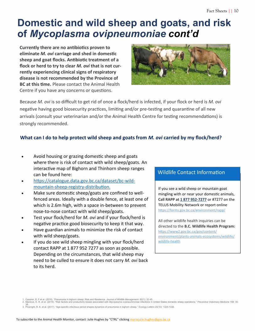
• Avoid housing or grazing domestic sheep and goats where there is risk of contact with wild sheep/goats. An interactive map of Bighorn and Thinhorn sheep ranges can be found here:
• https://catalogue.data.gov.bc.ca/dataset/bc-wild-mountain-sheep-registry-distribution.
• Make sure domestic sheep/goats are confined to well-fenced areas. Ideally with a double fence, at least one of which is 2.6m high, with a space in-between to prevent nose-to-nose contact with wild sheep/goats.
• Test your flock/herd for M. ovi and if your flock/herd is negative practice good biosecurity to keep it that way.
• Have guardian animals to minimize the risk of contact with wild sheep/goats.
• If you do see wild sheep mingling with your flock/herd contact RAPP at 1 877 952 7277 as soon as possible. Depending on the circumstances, that wild sheep may need to be culled to ensure it does not carry M. ovi back to its herd.
If you see a wild sheep or mountain goat
mingling with or near your domestic animals,
Call RAPP at 1 877 952-7277 or #7277 on the
TELUS Mobility Network or report online https://forms.gov.bc.ca/environment/rapp/
All other wildlife health inquiries can be
directed to the B.C. Wildlife Health Program: https://www2.gov.bc.ca/gov/content/
environment/plants-animals-ecosystems/wildlife/
wildlife-health
Wildlife Contact Information
1. Cassirer, E. F et al. (2018). “Pneumonia in bighorn sheep: Risk and Resilience. Journal of Wildlife Management. 82(1): 32-45. 2. Manlove, K. R. et al. (2019). “Risk factors and productivity losses associated with Mycoplasma ovipneumnoniae infections in United States domestic sheep operations.” Preventive Veterinary Medicine 168: 30-
38. 3. Plowright, R. K. et al. (2017). “Age-specific infectious period shapes dynamics of pneumonia in bighorn sheep.” Ecology Letters 20(10): 1325-1336.
Fact Sheets || 10

To subscribe to the Animal Health Monitor, contact: Julie Hughes by “CTRL” clicking [email protected]
Fecal Parasitology at the AHC Jocelyn Montague
(Registered Veterinary Technician—Post-Mortem)
A microscopic view of a tapeworm infection in a sheep, strongyles also
present. Photo credit AHC
Fecal examination is a common laboratory procedure used to
diagnose parasitic infections in domestic and wild animals. At the
Animal Health Centre, we offer the following tests on fresh fecal
samples:
Fecal Flotation: Centrifugal fecal flotation with Sheather’s sugar solution is the standard test to concentrate parasite eggs and cysts. Fecal flotation will detect most parasites including Strongyles, Ascarids, Tapeworms, Trichuris, Capillaria and Coccidia. This is a qualitative test to determine which (if any) parasite species are present. The ova are counted, and the test is reported as 1+ (1-24), 2+ (25-99), 3+ (100-299), 4+ (300+) or Negative (None seen)
Fecal Egg count: The Modified McMaster test is used to count strongyle eggs in a measured amount of feces and is recommended to estimate the extent of parasite egg contamination on pastures and/or efficacy of treatment. This test is reported in Eggs per gram (EPG). The fecal egg count has a low sensitivity of 25-50 PG
and may not pick up low concentrations of parasite ova.
This test is routinely performed on all sheep and goat fecal samples submitted to AHC that are positive for strongyles and may also be performed upon request for horses, cattle, and other animals. The Modified McMaster is only validated for strongyles, other ova will not be counted.
Baermann Test: The Baermann test is used to isolate larvae from fecal samples and is most often used to diagnose lung-
worm infections. It is very important the fecal sample be fresh as Strongyle type eggs may hatch or if left outdoors feces may be
invaded with free -living nematodes, making identification
difficult.
Direct Smear for Cryptosporidium: This test is performed on fresh feces in our Bacteriology laboratory to diagnose cryptosporidium infection. A small amount of feces is smeared on a slide and stained for microscopic examination.
.
Test Updates || 11

To subscribe to the Animal Health Monitor, contact: Julie Hughes by “CTRL” clicking [email protected]
Fecal Parasitology at the AHC cont’d
Sample Collection:
Proper collection and submission of samples to the laboratory is
important for accurate diagnosis of parasitic infection. Feces
should be fresh to preserve quality and prevent hatching and/or
death/degeneration of fragile parasite eggs and oocysts. Collect
several grams (approximately a tablespoon) of fresh feces in a clean
container or ziplock bag. Remove as much air from specimen
container as possible. Label clearly with date, animal’s name and
whether or not sample is pooled from a group of several animals.
Samples should be collected immediately after observing
defecation and refrigerated if to be held more than 1 or 2 hours
before examination. If sending samples through mail or courier,
an icepack should be included to keep sample cool. Freezing
should be avoided to prevent distortion of eggs.
Please call 604-556-3007 to speak with one of our Registered
Veterinary Technicians if you have any questions about the fecal
testing offered at AHC.
REFERENCES
Foreyt, William J. Veterinary Parasitology. 5th edition, 2001
Zajac, Anne M. and Conboy, Gary A. Veterinary Clinical Parasit-ology. 8th edition, 2012.
Test Updates || 12

To subscribe to the Animal Health Monitor, contact: Julie Hughes by “CTRL” clicking [email protected]
Sample Submission to the Bacteriology
Section of the PAHB Jaime Battle (Laboratory Scientist,
Bacteriology)
Sample submission is arguably the most important part of the
entire laboratory testing process.
Leaking, old, messy, contaminated, desiccated or otherwise
inappropriately packaged/collected samples will result in
laboratory delays, loss of sample integrity, contaminated and
inaccurate results or in worst case scenarios, refusal of sample/
inability to perform testing required.
Different laboratory sections also have very specific requirements
for the submission of samples for testing in that section. For
example, samples submitted in bacterial culture media, cannot be
used for virology/PCR testing as many of the components of the
media/agar will prohibit these tests.
I will only deal with individual fluid/tissue/environmental
samples here, whole animals are a different story and are covered
under Post Mortem room policies and procedures.
1) When submitting SWABS: Please specify the site the
swab is from, ie. Trachea, Cloaca, Yolk Sac, etc. This aids
in the type of testing performed and classifies the
direction further testing will take. Antibiotic Sensitivities
are NOT performed on any environmental samples and
any unlabelled swabs will not be assumed to come from
an actual animal.
2) Do not submit any samples in a glove. This creates a
huge potential for cross contamination of both the
sample and the environment in the lab. Urine sample
containers, collection vials, etc are all easily accessible
and simple to use.
3) Labeling is incredibly important. Please ensure your sub-
mission forms match the samples you are sending in. In
any cases of discrepancy, we will take the markings on the
sample itself as opposed to the information contained in
the history. THE SIMPLER THE BETTER. If you have a
large number of samples, simple numbering on the lab
side is preferable versus us transcribing a large amount of
information on site. Keep your spreadsheet data concise
and as simple as possible (1,2,3,4, etc as opposed to com-
plicated combinations of numbers and other characters)
4) Liquids (and other samples) in Whirl Pak Bags: This is a
particular point of contention in any laboratory that
receives samples in these bags. The manufacturer instruc-
tions are very clear and using ANY other procedure for
use of these bags results in both a compromised sample
and no small amount of frustration to laboratory
personnel in sample handling and processing.
Removing the top perforation to open the bag is
KEY!!!! It retains the integrity of the bag itself by not
introducing damage to the seals and closure
mechanism, and it also removes the extra bulk so there
is better sealing when closed properly (leaving that
plastic strip in place provides bulk and space that will
result in liquid leakage, it is simply NOT how the
system is designed to work).
Tech Tips || 13

To subscribe to the Animal Health Monitor, contact: Julie Hughes by “CTRL” clicking [email protected]
Sample Submission to the Bacteriology Section of the PAHB
cont’d
The outside of your sample containers should also be clean. It’s
irrelevant if you collected a pristine sample if it will inevitably get
contaminated by virtue of us having to get inside to retrieve it
(ie gloves)
If you have any questions about sample submission, how to use certain submission containers, please do not hesitate to follow the manufacturer instructions or contact the lab for guidance.
The quality of the results we are able to provide to you, the client,
is very much influenced by the quality of the samples we receive.
Garbage samples equal potentially garbage results.
This is by no means a comprehensive list, but should give a good guideline on how to submit samples for Bacteriological/Fungal testing.
Tech Tips || 14