angiotensin ii type 1 receptor blockade ameliorates tubulointerstitial injury induced by chronic...
TRANSCRIPT
Kidney International, Vol. 61 (2002), pp. 951–958
Angiotensin II type 1 receptor blockade amelioratestubulointerstitial injury induced by chronicpotassium deficiency
SHIN-ICHI SUGA, MARILDA MAZZALI, PATRICIO E. RAY, DUK-HEE KANG,and RICHARD J. JOHNSON
Division of Nephrology, University of Washington Medical Center, Seattle, Washington, and Children’s Research Institute,Washington, D.C., USA; Division of Hypertension, National Cardiovascular Center Research Institute, Suita, Osaka, Japan;and Baylor College of Medicine, Houston, Texas, USA
Angiotensin II type 1 receptor blockade ameliorates tubulo- Potassium (K�) deficiency is one of the commonlyinterstitial injury induced by chronic potassium deficiency. encountered electrolyte abnormalities, occurring most
Background. Chronic potassium (K�) deficiency, one of the frequently in patients who use diuretics, or who havewell-known causes of renal tubulointerstitial injury, is associ-
vomiting, diarrhea or undergo nasogastric suction. Theated with an alteration in vasoactive mediators including persis-renal manifestations of K� deficiency are diverse andtent generation of renal cortical angiotensin (Ang) II despiteinclude functional changes such as increased ammo-the suppression of plasma Ang II, and suppression of urinary
nitrite/nitrate excretion. We tested the hypothesis that K�- niagenesis, metabolic alkalosis, nephrogenic diabetesdeficiency–induced renal tubulointerstitial injury could be me- insipidus, chloride wasting, sodium retention [1], anddiated by Ang II or a reduction in nitric oxide. structural changes consisting of renal hypertrophy and
Methods. Rats were fed a K�-deficient diet (0.01% K�)tubulointerstitial injury [2, 3]. K� deficiency also is asso-alone, or with either losartan or l-arginine (L-Arg) in drinkingciated with intrarenal hemodynamic changes, includingwater. Control rats were fed with a normal K� diet (0.36%
K�). At the end of 10 weeks, kidneys were excised and renal decreased renal blood flow and increased renal vasocon-injury was evaluated. striction with only a minor reduction in glomerular filtra-
Results. Serum K� was similarly depressed in all three tion rate (GFR), for which angiotensin (Ang) II andgroups receiving the K�-deficient diet. Rats on the K�-deficient thromboxane A2 have been postulated as possible media-diet alone developed renal hypertrophy and tubulointerstitial
tors [4, 5].fibrosis with an increase in tubular osteopontin expression,Despite the frequency with which K� deficiency oc-macrophage infiltration and type III collagen deposition. Ad-
ministration of losartan significantly reduced renal hypertrophy curs, little is known about the intrarenal events that leadand prevented tubulointerstitial injury in the cortex, although to these structural changes. Since the existence of chronicsome medullary injury occurred. In contrast, administration of hypoxia and/or ischemia is regarded as one of the keyL-Arg did not attenuate tubulointerstitial injury in the cortex, events in the progression of chronic tubulointerstitialdespite a complete recovery of urinary nitrate excretion. Mild
injury [6], we recently hypothesized that chronic K� de-but significant improvement of tubular osteopontin expressionficiency could induce alterations in local vasoactive medi-and macrophage infiltration were observed in the medulla of
L-Arg-treated hypokalemic rats. ators favoring vasoconstriction that could result in intra-Conclusions. These results indicate that hypokalemic renal renal ischemia and subsequent tubulointerstitial injury
injury is mediated, at least in part, by Ang II via the Ang [7]. By analyzing chronic hypokalemic rats, we docu-II type 1 receptor, with a lesser contribution mediated by a mented alterations in vasoactive mediators including in-reduction in nitric oxide. Losartan may be beneficial in pre-
creased cortical angiotensin-converting enzyme (ACE)venting hypokalemic tubulointerstitial injury.expression and unsuppressed cortical Ang II generationdespite systemic suppression of the renin-angiotensinsystem (RAS), suggesting the activation of RAS in theKey words: hypokalemia, tubulointerstitial disease, angiotensin II, ni-
tric oxide, losartan, electrolyte abnormality. cortex, an increase in renal endothelin-1, a decrease inrenal kallikrein, and a decrease in urinary nitrite/nitrateReceived for publication August 8, 2001[7]. In the present study, we tested the hypothesis thatand in revised form October 19, 2001
Accepted for publication October 22, 2001 K� deficiency–induced renal injury could be mediatedby Ang II or a reduction in nitric oxide (NO). 2002 by the International Society of Nephrology
951
Suga et al: Ang II and hypokalemic nephropathy952
METHODS grade 1, �10% tubulointerstitial changes present; grade2, 10–25% tubulointerstitial involvement; grade 3, 25–Experimental protocol50% tubulointerstitial involvement; grade 4, 50–75% tu-Studies were designed to examine the role of Ang IIbulointerstitial involvement; and grade 5, 75–100% tubu-and NO in the pathogenesis of hypokalemic tubulointer-lointerstitial involvement. For each biopsy, the entirestitial injury. Male Sprague-Dawley rats (230 to 270 g,cortical and outer medullary regions were evaluated andN � 24; Simonsen Labs, Gilroy, CA, USA) were divideda mean score per biopsy was calculated. The secondinto four groups (N � 6, in each) and fed either a K�-method was to measure a percent area occupied by os-deficient diet (0.01% K�, 0.26% NaCl; Zeigler Brothers,teopontin-positive tubules, based on observations thatGardners, PA, USA) or a diet with normal K� contentosteopontin expression by injured tubules is a sensitive(0.36% K�, 0.26% NaCl) for 10 weeks.marker of tubulointerstitial injury [9]. Using computer-Group I, LK. Rats on a K�-deficient diet alone withassisted image analysis software (Optimas, v 6.2; Mediano additives in the drinking water.Cybernetics, Silver Springs, MD, USA) and digitizedGroup II, LK � losartan (Los). Rats on a K�-deficientimages, the percent area occupied by osteopontin-posi-diet were treated with the Ang II type 1 receptor (AT1)tive tubules (including the entire cortical and outer med-antagonist, losartan (Merck and Co., Inc., West Point,
PA, USA), at a dose of 10 mg/kg per day in the drinking ullary regions, exclusive of glomeruli) was measured perwater from the beginning of the K�-deficient diet for 10 field (4 mm2) at �25 and the mean percent area wasweeks. Water intake was measured three times a week calculated for each biopsy. We also measured interstitialso that the concentration of losartan could be adjusted. fibrosis by the percent area of type III collagen positive
Group III, LK � Arg. Rats on a K�-deficient diet were interstitium, obtained by the same analytical method attreated with 1% of l-arginine (L-Arg; Sigma Chemical �25. In addition, the number of macrophages (ED-1Co., St. Louis, MO, USA) in the drinking water from positive cells/mm2) in the cortex and medulla was quanti-the beginning of the K�-deficient diet for 10 weeks. fied at �50.
Group IV, NK. Rats on a normal K� diet with noadditives in the drinking water. Since we previously Urinary nitrite/nitrate assayshowed that losartan and L-Arg treatment in control
The urinary concentration of nitrite/nitrate, stable endrats did not induce significant renal histological changesproducts of NO, was measured as previously reported[8], these control groups were omitted in the present[7]. Briefly, urine samples were first incubated with As-study.pergillus nitrate reductase (Sigma Chemical Co.) in theAt week 10, animals were housed separately in meta-presence of NADPH for one hour to convert nitrate inbolic cages and urine was collected for 16 hours. Bloodthe samples to nitrite. After the incubation, the totalwas drawn from the tail vein on a different day of thenitrite content was measured using the Griess reagentsame week. At the end of week 10, rats were sacrificedfollowing the manufacturer’s instruction (Clontech, Paloand kidneys were excised.Alto, CA, USA).
Renal histologic studiesEIA for PGE2Methyl Carnoy’s fixed tissue was processed and paraf-
fin embedded, and 4-�m sections were stained with the The urinary concentration of prostaglandin E2 (PGE2)periodic acid-Schiff reagent (PAS). An indirect immuno- was measured using commercial enzyme immunoassayperoxidase method was used to identify the following kit (EIA kit; Cayman Chemical, Ann Arbor, MI, USA).antigens [7]: osteopontin with OP 199, a goat anti-rat The cross-reactivity to PGE3, PGE1 and 6-keto-PGF1�osteopontin antibody (gift of C. Giachelli, University was 43%, 19% and 1%, on a molar basis, respectively.of Washington, Seattle, WA, USA); macrophages withED-1, a monoclonal IgG1 to rat macrophages (Harlan Additional measurementsBioproducts, Indianapolis, IN, USA) and type III colla-
Serum and urinary creatinine and K� concentrationsgen with a goat anti-human type III collagen antibodywere measured by Cobas autoanalyzer (Roche Diagnos-(Southern Biotechnology Associates, Birmingham, AL,tics, Nutley, NJ, USA).USA).
Several parameters were used to evaluate tubulointer-Statistical methodsstitial injury. The first method was a blinded semiquanti-
Values are expressed as mean � SE. A comparisontative scoring system (0 through 5) of PAS-stained sec-between groups was made by ANOVA with the Fisher’stions based on the presence of tubular cellularity, basementprotected least significant difference test for multiplemembrane thickening, dilation, atrophy, sloughing or
interstitial widening as follows [7]: 0, no changes present; comparisons.

Suga et al: Ang II and hypokalemic nephropathy 953
Table 1. Systemic parameters for rats on K�-deficient diet
Group I Group II Group III Group IVLK LK�Los LK�Arg NK
Serum K� at week 10 mEq/L 2.34�0.16a 2.45 �0.12a 2.60 �0.35a 3.82�0.12Initial body weight g 245�2 245�3 243�3 244�3Body weight at week 10 g 330�7a 329 �6a 295 �7ab 412�9Kidney weight at week 10 g 1.59�0.07a 1.42 �0.03ab 1.67 �0.04a 1.12�0.02Creatinine clearance at week 10 mL/min 1.05�0.15a 1.46 �0.20a 1.37 �0.13a 2.03�0.10
Groups are defined in the Methods section.a P � 0.05 vs. NKb P � 0.05 vs. LK
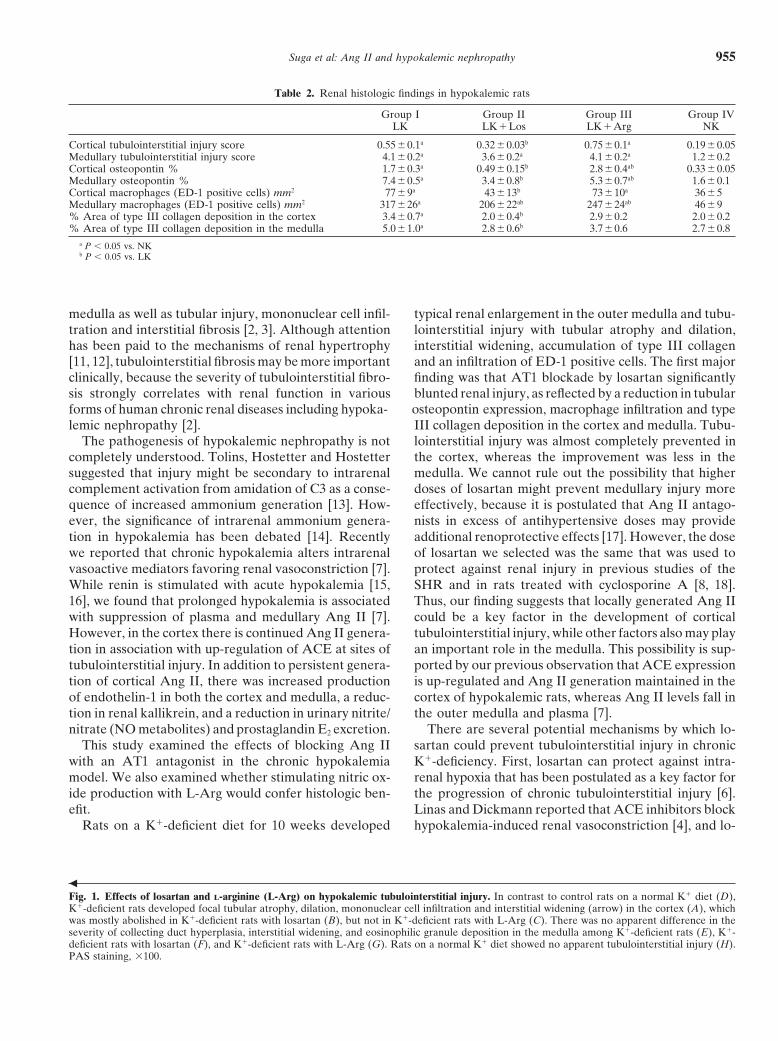
RESULTS of losartan significantly suppressed the increase in tubu-lar osteopontin expression both in the cortex and me-General featuresdulla (Fig. 2B). The striped pattern of osteopontin posi-
Marked hypokalemia was induced by a K�-deficient tive tubules that radiated into the cortex of control K�-diet alone (2.34 � 0.16 mEq/L). Neither losartan nor deficient rats was completely prevented by losartan.L-Arg affected the severity of hypokalemia (Table 1). L-Arg did not improve but rather increased tubular os-K�-deficient rats (groups I to III) gained less body weight teopontin expression in the cortex. However, it inhibitedthan control rats on a normal K� diet (group IV). In the increase in osteopontin expression mildly but signifi-contrast, kidney weights were markedly greater in K�- cantly in the medulla (Fig. 2C and Table 2).deficient rats than in controls, as already described [3]. The number of macrophages (ED-1 positive cells) wasLK�Los animals had a less increase in kidney weight elevated in the cortex and medulla of hypokalemic ratscompared to the other two groups on the K�-deficient (Table 2). Similar to the suppression of tubular osteopon-diet (groups I and III). Renal function, as assessed by tin expression, losartan prevented the cortical macro-creatinine clearance, decreased significantly in all three phage infiltration and reduced the accumulation of mac-groups administered the K�-deficient diet. Although the rophages in the medulla. Macrophage infiltration wasdifference was not significant, the LK�Los group tended not affected in the cortex of L-Arg treated rats, althoughto have a higher mean creatinine clearance than the LK some reduction was observed in the medulla (Table 2).group (P � 0.080; Table 1). An increase in the deposition of type III collagen was
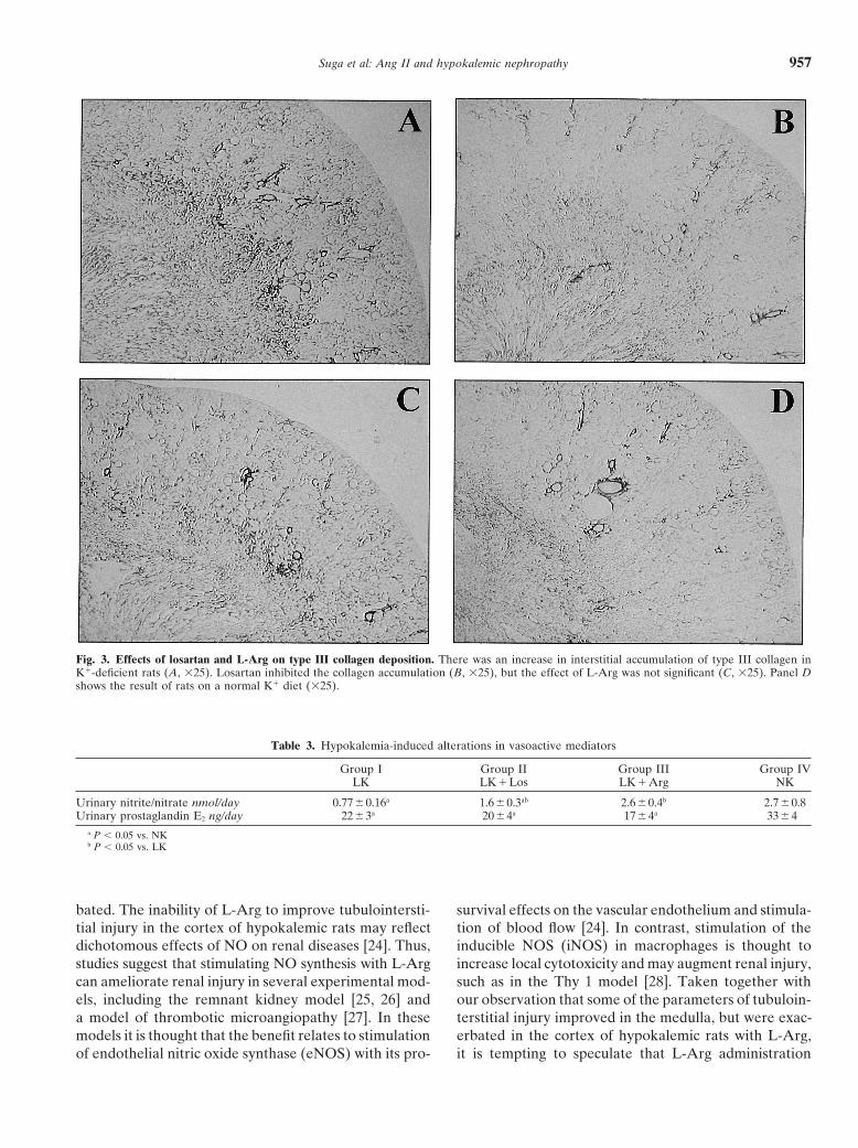
present in the cortical and medullary interstitium of hy-Effects of losartan and L-Arg on hypokalemicpokalemic rats (Fig. 3A and Table 2). Losartan amelio-tubulointerstitial injuryrated the collagen deposition significantly in both the
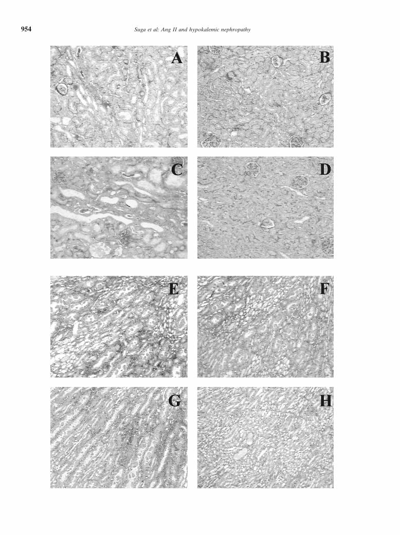
Hypokalemic rats developed prominent tubulointer- cortex and medulla (Fig. 3B), whereas L-Arg had nostitial injury with no apparent glomerular injury. There significant effect (Fig. 3C and Table 2).was focal atrophy or dilation of tubules with mild intersti-tial accumulation of mononuclear cells and interstitial Effects of losartan and L-Arg on hypokalemia-inducedexpansion in both the cortex and medulla (Fig. 1A). In alteration in vasoactive mediatorsthe outer medulla, there was also diffuse swelling and Urinary nitrite/nitrate excretion was reduced by 70%hyperplasia of collecting duct epithelial cells (Fig. 1E). in K�-deficient rats at week 10 (Table 3). The decreaseLosartan treatment significantly reduced a tubulointer- in urinary nitrite/nitrate was partially recovered by thestitial injury score in the cortex (Fig. 1B and Table 2). administration of losartan and fully restored by L-Arg.In the medulla, losartan tended to ameliorate the score We also measured urinary excretion of PGE2, a potent(Fig. 1F and Table 2), but the effect was weak and not vasodilator produced in the outer medulla. A reductionstatistically significant. Administration of L-Arg had no of urinary PGE2 (Table 3) was observed at week 10,apparent effect on the tubulointerstitial injury score in being consistent with previous reports of chronic K�-either the cortex or medulla (Fig. 1 C, G, and Table 2). deficiency [7, 10]. Neither losartan nor L-Arg had any
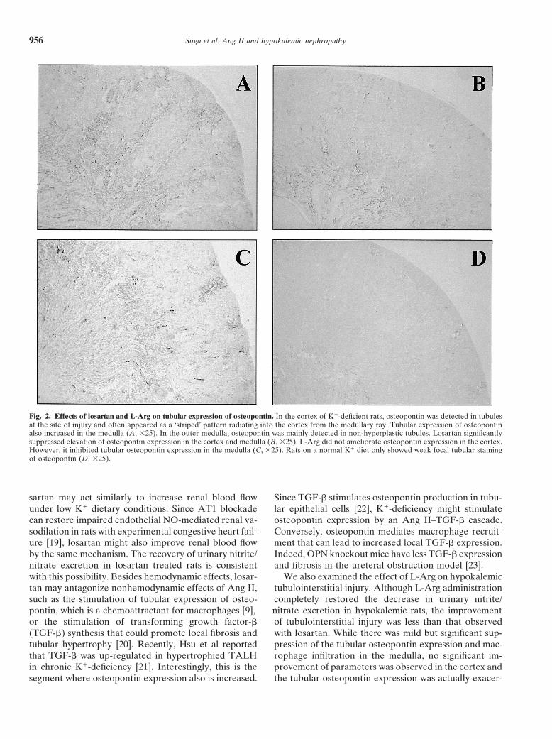
Immunohistologic studies documented increased ex- significant effect on the decrease in urinary PGE2 excre-pression of osteopontin, a sensitive marker of injured tion (Table 3).tubules, in K�-deficient rats (Fig. 2A). Osteopontin ex-pression was most prominent in distal tubules of the
DISCUSSIONouter medulla corresponding to the thick ascending limbof Henle (TALH; Fig. 2A). In the cortex, osteopontin Chronic K� deficiency in both experimental animalsexpression was distributed in a striped pattern radiating and humans causes renal enlargement consisting of hy-
pertrophy and hyperplasia of tubular cells in the outerinto the cortex from the outer medulla. Administration
Suga et al: Ang II and hypokalemic nephropathy954
Suga et al: Ang II and hypokalemic nephropathy 955
Table 2. Renal histologic findings in hypokalemic rats
Group I Group II Group III Group IVLK LK�Los LK�Arg NK
Cortical tubulointerstitial injury score 0.55�0.1a 0.32 �0.03b 0.75 �0.1a 0.19�0.05Medullary tubulointerstitial injury score 4.1�0.2a 3.6 �0.2a 4.1 �0.2a 1.2�0.2Cortical osteopontin % 1.7 �0.3a 0.49 �0.15b 2.8 �0.4ab 0.33�0.05Medullary osteopontin % 7.4 �0.5a 3.4 �0.8b 5.3 �0.7ab 1.6�0.1Cortical macrophages (ED-1 positive cells) mm2 77�9a 43�13b 73�10a 36�5Medullary macrophages (ED-1 positive cells) mm2 317 �26a 206 �22ab 247 �24ab 46�9% Area of type III collagen deposition in the cortex 3.4�0.7a 2.0 �0.4b 2.9 �0.2 2.0�0.2% Area of type III collagen deposition in the medulla 5.0�1.0a 2.8 �0.6b 3.7 �0.6 2.7�0.8
a P � 0.05 vs. NKb P � 0.05 vs. LK
medulla as well as tubular injury, mononuclear cell infil- typical renal enlargement in the outer medulla and tubu-lointerstitial injury with tubular atrophy and dilation,tration and interstitial fibrosis [2, 3]. Although attention
has been paid to the mechanisms of renal hypertrophy interstitial widening, accumulation of type III collagenand an infiltration of ED-1 positive cells. The first major[11, 12], tubulointerstitial fibrosis may be more important
clinically, because the severity of tubulointerstitial fibro- finding was that AT1 blockade by losartan significantlyblunted renal injury, as reflected by a reduction in tubularsis strongly correlates with renal function in various
forms of human chronic renal diseases including hypoka- osteopontin expression, macrophage infiltration and typeIII collagen deposition in the cortex and medulla. Tubu-lemic nephropathy [2].
The pathogenesis of hypokalemic nephropathy is not lointerstitial injury was almost completely prevented inthe cortex, whereas the improvement was less in thecompletely understood. Tolins, Hostetter and Hostetter
suggested that injury might be secondary to intrarenal medulla. We cannot rule out the possibility that higherdoses of losartan might prevent medullary injury morecomplement activation from amidation of C3 as a conse-
quence of increased ammonium generation [13]. How- effectively, because it is postulated that Ang II antago-nists in excess of antihypertensive doses may provideever, the significance of intrarenal ammonium genera-
tion in hypokalemia has been debated [14]. Recently additional renoprotective effects [17]. However, the doseof losartan we selected was the same that was used towe reported that chronic hypokalemia alters intrarenal
vasoactive mediators favoring renal vasoconstriction [7]. protect against renal injury in previous studies of theSHR and in rats treated with cyclosporine A [8, 18].While renin is stimulated with acute hypokalemia [15,
16], we found that prolonged hypokalemia is associated Thus, our finding suggests that locally generated Ang IIcould be a key factor in the development of corticalwith suppression of plasma and medullary Ang II [7].
However, in the cortex there is continued Ang II genera- tubulointerstitial injury, while other factors also may playan important role in the medulla. This possibility is sup-tion in association with up-regulation of ACE at sites of
tubulointerstitial injury. In addition to persistent genera- ported by our previous observation that ACE expressionis up-regulated and Ang II generation maintained in thetion of cortical Ang II, there was increased production
of endothelin-1 in both the cortex and medulla, a reduc- cortex of hypokalemic rats, whereas Ang II levels fall inthe outer medulla and plasma [7].tion in renal kallikrein, and a reduction in urinary nitrite/
nitrate (NO metabolites) and prostaglandin E2 excretion. There are several potential mechanisms by which lo-sartan could prevent tubulointerstitial injury in chronicThis study examined the effects of blocking Ang II
with an AT1 antagonist in the chronic hypokalemia K�-deficiency. First, losartan can protect against intra-renal hypoxia that has been postulated as a key factor formodel. We also examined whether stimulating nitric ox-
ide production with L-Arg would confer histologic ben- the progression of chronic tubulointerstitial injury [6].Linas and Dickmann reported that ACE inhibitors blockefit.
Rats on a K�-deficient diet for 10 weeks developed hypokalemia-induced renal vasoconstriction [4], and lo-
�
Fig. 1. Effects of losartan and L-arginine (L-Arg) on hypokalemic tubulointerstitial injury. In contrast to control rats on a normal K� diet (D),K�-deficient rats developed focal tubular atrophy, dilation, mononuclear cell infiltration and interstitial widening (arrow) in the cortex (A), whichwas mostly abolished in K�-deficient rats with losartan (B), but not in K�-deficient rats with L-Arg (C). There was no apparent difference in theseverity of collecting duct hyperplasia, interstitial widening, and eosinophilic granule deposition in the medulla among K�-deficient rats (E), K�-deficient rats with losartan (F), and K�-deficient rats with L-Arg (G). Rats on a normal K� diet showed no apparent tubulointerstitial injury (H).PAS staining, �100.
Suga et al: Ang II and hypokalemic nephropathy956
Fig. 2. Effects of losartan and L-Arg on tubular expression of osteopontin. In the cortex of K�-deficient rats, osteopontin was detected in tubulesat the site of injury and often appeared as a ‘striped’ pattern radiating into the cortex from the medullary ray. Tubular expression of osteopontinalso increased in the medulla (A, �25). In the outer medulla, osteopontin was mainly detected in non-hyperplastic tubules. Losartan significantlysuppressed elevation of osteopontin expression in the cortex and medulla (B, �25). L-Arg did not ameliorate osteopontin expression in the cortex.However, it inhibited tubular osteopontin expression in the medulla (C, �25). Rats on a normal K� diet only showed weak focal tubular stainingof osteopontin (D, �25).
sartan may act similarly to increase renal blood flow Since TGF-� stimulates osteopontin production in tubu-lar epithelial cells [22], K�-deficiency might stimulateunder low K� dietary conditions. Since AT1 blockade
can restore impaired endothelial NO-mediated renal va- osteopontin expression by an Ang II–TGF-� cascade.Conversely, osteopontin mediates macrophage recruit-sodilation in rats with experimental congestive heart fail-
ure [19], losartan might also improve renal blood flow ment that can lead to increased local TGF-� expression.Indeed, OPN knockout mice have less TGF-� expressionby the same mechanism. The recovery of urinary nitrite/
nitrate excretion in losartan treated rats is consistent and fibrosis in the ureteral obstruction model [23].We also examined the effect of L-Arg on hypokalemicwith this possibility. Besides hemodynamic effects, losar-
tan may antagonize nonhemodynamic effects of Ang II, tubulointerstitial injury. Although L-Arg administrationcompletely restored the decrease in urinary nitrite/such as the stimulation of tubular expression of osteo-
pontin, which is a chemoattractant for macrophages [9], nitrate excretion in hypokalemic rats, the improvementof tubulointerstitial injury was less than that observedor the stimulation of transforming growth factor-�
(TGF-�) synthesis that could promote local fibrosis and with losartan. While there was mild but significant sup-pression of the tubular osteopontin expression and mac-tubular hypertrophy [20]. Recently, Hsu et al reported
that TGF-� was up-regulated in hypertrophied TALH rophage infiltration in the medulla, no significant im-provement of parameters was observed in the cortex andin chronic K�-deficiency [21]. Interestingly, this is the
segment where osteopontin expression also is increased. the tubular osteopontin expression was actually exacer-
Suga et al: Ang II and hypokalemic nephropathy 957
Fig. 3. Effects of losartan and L-Arg on type III collagen deposition. There was an increase in interstitial accumulation of type III collagen inK�-deficient rats (A, �25). Losartan inhibited the collagen accumulation (B, �25), but the effect of L-Arg was not significant (C, �25). Panel Dshows the result of rats on a normal K� diet (�25).
Table 3. Hypokalemia-induced alterations in vasoactive mediators
Group I Group II Group III Group IVLK LK�Los LK�Arg NK
Urinary nitrite/nitrate nmol/day 0.77�0.16a 1.6 �0.3ab 2.6 �0.4b 2.7�0.8Urinary prostaglandin E2 ng/day 22�3a 20�4a 17�4a 33�4
a P � 0.05 vs. NKb P � 0.05 vs. LK
bated. The inability of L-Arg to improve tubulointersti- survival effects on the vascular endothelium and stimula-tion of blood flow [24]. In contrast, stimulation of thetial injury in the cortex of hypokalemic rats may reflect
dichotomous effects of NO on renal diseases [24]. Thus, inducible NOS (iNOS) in macrophages is thought toincrease local cytotoxicity and may augment renal injury,studies suggest that stimulating NO synthesis with L-Arg
can ameliorate renal injury in several experimental mod- such as in the Thy 1 model [28]. Taken together withour observation that some of the parameters of tubuloin-els, including the remnant kidney model [25, 26] and
a model of thrombotic microangiopathy [27]. In these terstitial injury improved in the medulla, but were exac-erbated in the cortex of hypokalemic rats with L-Arg,models it is thought that the benefit relates to stimulation
of endothelial nitric oxide synthase (eNOS) with its pro- it is tempting to speculate that L-Arg administration
Suga et al: Ang II and hypokalemic nephropathy958
8. Thomas SE, Andoh TF, Pichler RH, et al: Accelerated apoptosismight stimulate iNOS more in the cortex and lead tocharacterizes cyclosporine-associated interstitial fibrosis. Kidney
tubular injury, while an eNOS-dependent, tissue protec- Int 53:897–908, 19989. Giachelli CM, Pichler R, Lombardi D, et al: Osteopontin expres-tive pathway may be predominant in the medulla in K�-
sion in angiotensin II-induced tubulointerstitial nephritis. Kidneydeficient rats. Alternatively, stimulation of NO may serveInt 45:515–524, 1994
to antagonize hypokalemia-induced intrarenal vasocon- 10. Beck N, Shaw JO: Thromboxane B2 and prostaglandin E2 in theK�-depleted rat kidney. Am J Physiol 240:F151–F157, 1981striction preferentially in the renal medulla, resulting in
11. Flyvbjerg A, Marshall SM, Frystyk J, et al: Insulin-like growthimprovement of renal injury in the medulla of hypoka- factor I in initial renal hypertrophy in potassium-depleted rats.lemic rats treated with L-Arg. Further studies with selec- Am J Physiol 262:F1023–F1031, 1992
12. Mujais SK: Collecting duct changes in potassium depletion: Effectstive activation or inhibition of each NOS isoform areof ACE inhibition. Am J Physiol 266:F419–F424, 1994necessary to clarify the roles of NO in hypokalemic ne- 13. Tolins JP, Hostetter MK, Hostetter TH: Hypokalemic nephrop-
phropathy. athy in the rat. Role of ammonia in chronic tubular injury. J ClinInvest 79:1447–1458, 1987In conclusion, chronic K�-deficiency–induced tubulo-
14. Throssell D, Brown J, Harris KP, Walls J: Metabolic acidosisinterstitial injury is mediated, at least in part, by Ang II does not contribute to chronic renal injury in the rat. Clin SciColch 89:643–650, 1995via the AT1 receptor. NO production may have some
15. Linas SL: Mechanism of hyperreninemia in the potassium-depletedprotective role in the medulla of hypokalemic rats. Theserat. J Clin Invest 68:347–355, 1981
studies suggest that AT1 blockade may be beneficial in 16. Ray PE, Suga SI, Liu XH, et al: Chronic potassium depletioninduces renal injury, salt sensitivity, and hypertension in youngthe prevention of hypokalemic tubulointerstitial injury.rats. Kidney Int 59:1850–1858, 2001
17. Fogo AB: The role of angiotensin II and plasminogen activatorACKNOWLEDGMENTS inhibitor-1 in progressive glomerulosclerosis. Am J Kidney Dis
35:179–188, 2000Support for this study was provided by United States Public Health 18. Fornes P, Richer C, Vacher E, et al: Losartan’s protective effects
Science Grants DK-47659, HL/DK68607, and DK-52121, and a grant in stroke-prone spontaneously hypertensive rats persist durablyfrom the Kanae Medical Research Foundation. Dr. Shin-ichi Suga was after treatment withdrawal. J Cardiovasc Pharmacol 22:305–313,supported by a Uehara Memorial Foundation Research Fellowship and 1993a Banyu Fellowship Award in Lipid Metabolism and Atherosclerosis 19. Abassi ZA, Gurbanov K, Mulroney SE, et al: Impaired nitricsponsored by Banyu Pharmaceutical Co., Ltd., and the Merck Com- oxide-mediated renal vasodilation in rats with experimental heartpany Foundation. The authors thank Dr. C. Giachelli, Univ. of Wash- failure: Role of angiotensin II. Circulation 96:3655–3664, 1997ington, Seattle for donating the antibody for osteopontin. 20. Wolf G, Mueller E, Stahl RA, Ziyadeh FN: Angiotensin II-
induced hypertrophy of cultured murine proximal tubular cells isReprint requests to Dr. Shin-ichi Suga, Division of Hypertension, mediated by endogenous transforming growth factor-beta. J Clin
National Cardiovascular Center Research Institute, Suita, Osaka, Japan. Invest 92:1366–1372, 1993E-mail: [email protected]: 21. Tsao T, Fawcett J, Fervenza FC, et al: Expression of insulin-like
growth factor-I and transforming growth factor-beta in hypoka-lemic nephropathy in the rat. Kidney Int 59:96–105, 2001REFERENCES 22. Malyankar UM, Almeida M, Johnson RJ, et al: Osteopontinregulation in cultured rat renal epithelial cells. Kidney Int 51:1766–1. Mujais SK, Katz AI: Potassium deficiency, in The Kidney, edited1773, 1997by Seldin DW, Giebisch G, Philadelphia, Lippincott Williams &
23. Ophascharoensuk V, Giachelli CM, Gordon K, et al: Obstruc-Wilkins, 2000, pp 1615–1646tive uropathy in the mouse: Role of osteopontin in interstitial2. Riemenschneider T, Bohle A: Morphologic aspects of low-potas-fibrosis and apoptosis. Kidney Int 56:571–580, 1999sium and low-sodium nephropathy. Clin Nephrol 19:271–279, 1983
24. Klahr S, Morrissey J: Renal disease: The two faces of nitric oxide.3. Muehrcke RC, Rosen S: Hypokalemic nephropathy in rat andLab Invest 72:1–3, 1995man. A light and electron microscopic study. Lab Invest 13:1359– 25. Andoh TF, Gardner MP, Bennett WM: Protective effects of1373, 1964 dietary L-arginine supplementation on chronic cyclosporine neph-4. Linas SL, Dickmann D: Mechanism of the decreased renal blood rotoxicity. Transplantation 64:1236–1240, 1997
flow in the potassium-depleted conscious rat. Kidney Int 21:757– 26. Reyes AA, Purkerson ML, Karl I, Klahr S: Dietary supplemen-764, 1982 tation with L-arginine ameliorates the progression of renal disease
5. Whinnery MA, Kunau RT Jr: Effect of potassium deficiency on in rats with subtotal nephrectomy. Am J Kidney Dis 20:168–176,papillary plasma flow in the rat. Am J Physiol 237:F226–F231, 1979 1992
6. Fine LG, Orphanides C, Norman JT: Progressive renal disease: 27. Shao J, Miyata T, Yamada K, et al: Protective role of nitric oxide inthe chronic hypoxia hypothesis. Kidney Int 53(Suppl 65):S74–78, a model of thrombotic microangiopathy in rats. J Am Soc Nephrol1998 12:2088–2097, 2001
7. Suga S, Phillips MI, Ray PE, et al: Hypokalemia induces renal 28. Peters H, Border WA, Noble NA: L-Arginine supplementationinjury and alterations in vasoactive mediators that favor salt sensi- increases mesangial cell injury and subsequent tissue fibrosis intivity. Am J Physiol (Renal Physiol) 281:F620–F629, 2001 experimental glomerulonephritis. Kidney Int 55:2264–2273, 1999