aneurysms subarachnoidal hemorrhage …. vascular/vas25. aneurysms, sah.pdf · – internal elastic...
TRANSCRIPT
ANEURYSMS, SUBARACHNOIDAL HEMORRHAGE Vas25 (1)
Aneurysms, Subarachnoidal Hemorrhage Last updated: September 5, 2017
ANEURYSM ............................................................................................................................................... 1 ETIOPATHOPHYSIOLOGY, PATHOLOGY .................................................................................................. 1
Aneurysm growth .................................................................................................................. 3
Giant saccular aneurysms ...................................................................................................... 3 Multiplicity ............................................................................................................................ 3
Location ................................................................................................................................. 4 Pediatric aneurysms ............................................................................................................... 4
EPIDEMIOLOGY ...................................................................................................................................... 4 Risk Factors ........................................................................................................................... 4
CLINICAL FEATURES .............................................................................................................................. 4
Site specific clinical features ................................................................................................. 5 DIAGNOSIS ............................................................................................................................................. 5
CT .......................................................................................................................................... 5 CTA ....................................................................................................................................... 6
MRI ....................................................................................................................................... 6
MRA ...................................................................................................................................... 6 Angiography .......................................................................................................................... 6
SCREENING ............................................................................................................................................ 7 TREATMENT ........................................................................................................................................... 7
Ruptured aneurysms .............................................................................................................. 7
Unruptured (incidental) aneurysms ....................................................................................... 7 FOLLOW-UP............................................................................................................................................ 8
SUBARACHNOID HEMORRHAGE (SAH) ................................................................................................... 8 ETIOLOGY .............................................................................................................................................. 8
EPIDEMIOLOGY ...................................................................................................................................... 9
PATHOLOGY ........................................................................................................................................... 9 CLINICAL FEATURES .............................................................................................................................. 9
Grading clinical severity ..................................................................................................... 10 DIAGNOSIS ........................................................................................................................................... 10
Noncontrast CT ................................................................................................................... 11
Lumbar Puncture ................................................................................................................. 12
Angiography ........................................................................................................................ 13
MRI ..................................................................................................................................... 13 Transcranial Doppler ........................................................................................................... 14
Funduscopy ......................................................................................................................... 14 Cardiac Manifestations ........................................................................................................ 14
COMPLICATIONS .................................................................................................................................. 14 Vasospasm ...................................................................................................................................... 14
Clinical Features .................................................................................................................. 15
Monitoring ........................................................................................................................... 15 Diagnosis ............................................................................................................................. 15 Prevention ............................................................................................................................ 16 Treatment ............................................................................................................................. 16
Rebleeding (rerupture) ................................................................................................................... 16
Seizures .......................................................................................................................................... 16 Acute OBSTRUCTIVE hydrocephalus ......................................................................................... 16 Delayed COMMUNICATING hydrocephalus ...................................................................................... 16 Non-neurological ............................................................................................................................ 17
SAH-induced hyponatremia ................................................................................................ 17
Acute neurogenic pulmonary edema ................................................................................... 17 Neurogenic stunned myocardium (s. Takotsubo cardiomyopathy) ..................................... 17 Other .................................................................................................................................... 17
TREATMENT ......................................................................................................................................... 17 SAH per se treatment ..................................................................................................................... 17
Regimen ............................................................................................................................... 17 EVD ..................................................................................................................................... 17 Diet ...................................................................................................................................... 17
IV fluids ............................................................................................................................... 18 Headache ............................................................................................................................. 18 BP goals ............................................................................................................................... 18
Oxygenation ........................................................................................................................ 18 Antiepileptics ....................................................................................................................... 18 Fever control ........................................................................................................................ 18 Not to use ............................................................................................................................. 18
Vasospasm treatment ..................................................................................................................... 18
For all patients – vasospasm prevention .............................................................................. 18 Symptomatic vasospasm ..................................................................................................... 19
Aneurysm treatment ....................................................................................................................... 20 Timing of treatment ............................................................................................................. 20 Methods ............................................................................................................................... 20
Surgical Clipping ................................................................................................................. 21
Open Alternatives to Surgical Clipping .............................................................................. 22
Endovascular Surgery .......................................................................................................... 22 Special Situations ........................................................................................................................... 25
Recanalization (of previously treated aneurysm) ................................................................ 25 Multiple aneurysms ............................................................................................................. 25
Aneurysm coexisting with AVM ........................................................................................ 25
Aneurysm causing CN3 palsy ............................................................................................. 25 Carotid stenosis associated with ICA aneurysm ................................................................. 25
Aneurysm perforation with coil .......................................................................................... 25 Carotid aneurysm within cavernous sinus ........................................................................... 25
Recanalization in previous stent-assist coiled aneurysm .................................................... 25
Cervical ICA aneurysms ..................................................................................................... 26
ICA terminus aneurysms ..................................................................................................... 26 AChA aneurysms ................................................................................................................ 26
AComA aneurysms ............................................................................................................. 26
Aneurysm with atheroma .................................................................................................... 26 FOLLOW UP .......................................................................................................................................... 26
PROGNOSIS .......................................................................................................................................... 26 SAH ................................................................................................................................................ 26
Unruptured aneurysms ................................................................................................................... 26
Clipping ............................................................................................................................... 26
ANEURYSM
- pathologic focal blood vessel dilatation (Lat. aneurysma - dilatation).
aneurysm is prone to rupture.
some lie entirely within subarachnoid space; others are buried in brain substance.
ETIOPATHOPHYSIOLOGY, PATHOLOGY
ANEURYSMS, SUBARACHNOIDAL HEMORRHAGE Vas25 (2)
FALSE aneurysms (s. pseudoaneurysms) – encapsulation of perivascular hematoma – cavities
that lack any components of vessel wall, but communicate with vessel lumen; cavity is lined by blood
clot (periadventitial hematoma is seen on imaging).
caused by penetrating vessel injuries (most commonly; pseudoaneurysm grows in hours),
periadventitial infections or malignant process (rare).
N.B. intracerebral hematoma may harbor and simultaneously obscure traumatic
aneurysm - angiography is diagnostic procedure of choice (esp. indicated for
penetrating head injuries!; absolutely indicated in all stab wounds to head)
TRUE aneurysms – dilatations of vascular lumen caused by weakness of vessel wall (at least
adventitia is present in aneurysm wall).
1. Saccular (“berry”) aneurysms (> 90%) – rounded outpouchings (i.e. neck with dome)
why intracranial arteries are susceptible to aneurysm development:
1) walls lack external elastic lamina
2) very thin adventitia
3) lie unsupported in subarachnoid space
sac is composed of only intima and adventitia;
– media ends at junction of aneurysm neck with parent vessel.
– intima is typically normal or thickened hyalinized (subintimal cellular proliferation is
common).
– internal elastic membrane is reduced or absent (normal internal elastic membrane can
withstand pressures over 600 mmHg without bulging - as long as membrane remains
intact, defects in media are inconsequential).
– hemosiderin-laden leukocytes may infiltrate adventitia.
sac lumen often contains thrombotic debris.
atherosclerotic changes in parent vessel are common.
etiology:
1) developmental (congenital) - focal defects in media (present at birth); over period of
years arterial pressure balloons out vessel wall (i.e. aneurysm is "congenital" in sense
that defect in arterial wall is present from birth, but actual aneurysm develops over years
after birth).
Familial inheritance pattern has been noted in < 2% of intracranial aneurysms
– congenital media defects in vessels of circle of Willis are found in 80% normal
vessels at autopsy.
– congenital abnormalities (e.g. fenestrations of vertebrobasilar junction, persistent
trigeminal arteries, vessel asymmetry / hypoplasia, coarctation of aorta) increase
incidence of saccular aneurysms.
– AUTOSOMAL DOMINANT POLYCYSTIC KIDNEY DISEASE (ADPKD) - most common
genetic abnormality associated with multiple intracranial aneurysms (present in 5-
40% ADPKD patients) - all patients should undergo screening MRA.
– chromosomal loci associated with intracranial aneurysm:
8q - likely acts via SOX17 (formation and maintenance of endothelial cell)
9p - likely acts via CDKN2A (may have similar role to SOX17).
2) hemodynamically induced degenerative vascular injury - hemodynamic shear
stresses (esp. at bifurcation points) cause occurrence, growth, thrombosis, and rupture of
aneurysms.
N.B. apex of vessel bifurcation is site of maximum hemodynamic stress!
3) high flow-related (caused by high-flow states) – aneurysms occur along PROXIMAL* and
DISTAL** vessels feeding AVM.
*do not increase risk of hemorrhage
**thin-walled intranidal aneurysms exposed to arterial pressure -
likely site for AVM hemorrhage (i.e. AVMs that bleed often
have intra-nidal aneurysms)
4) traumatic (< 1% all aneurysms) - caused by blunt vessel injuries. see p. TrH1 >>
5) oncotic - direct tumor invasion (e.g. meningioma) or implantation of metastatic emboli*
(e.g. left atrial myxoma, choriocarcinoma) → vessel wall infiltration → disruption.
*metastatic implants often involve peripheral cerebral vessels at
gray-white junction
6) vasculopathy / vasculitis-related - fibromuscular dysplasia, connective tissue disorders
(SLE*, Marfan, Ehlers-Danlos, Osler-Weber-Rendu syndromes, pseudoxanthoma
elasticum, Takayasu arteritis).
*aneurysms can be saccular, fusiform, or bizarre-looking mixture
of both.
7) drug-related: COCAINE, HEROIN, EPHEDRINE, METHAMPHETAMINE – can induce cerebral
vasculitis - necrotizing angiitis (histologically similar to periarteritis nodosa) with focal
arterial ectasias.
2. Fusiform (s. dolichoectatic) aneurysms (no identifiable neck):
1) atherosclerotic (7% all aneurysms) - unusual form of atherosclerosis damages media →
arterial stretching and elongation that may extend over considerable length (may have
serpentine, giant, bizarre shapes).
occur in older patients.
affect PROXIMAL arteries (vertebrobasilar system is commonly affected).
perforating branches often arise from entire length of aneurysm.
intraluminal clots are common (→ occlusion of small ostia of perforating vessels →
infarcts).
bleed rarely (mass effects are much more common).
2) mycotic (infectious) (0.5-3% all aneurysms) - any aneurysm resulting from infectious
process that involves (destroys) arterial wall:
a) septic embolus (e.g. IV drug abuse, bacterial endocarditis) → extension from
lumen to adventitia; most common on DISTAL vessels (due to embolic nature).
b) meningitis → extension from periphery to lumen (e.g. aneurysms of basal
circulation associated with fungal infections).
frequently multiple (20%), very friable - greater propensity to bleed! (rupture is fatal
in 80% patients!)
3. Dissecting aneurysms – when intramural hematoma extends into subadventitial plane (e.g. in
fibromuscular dysplasia, trauma).
elongated, ovoid, or saccular.
most affect extracranial segments of ICA, VA.
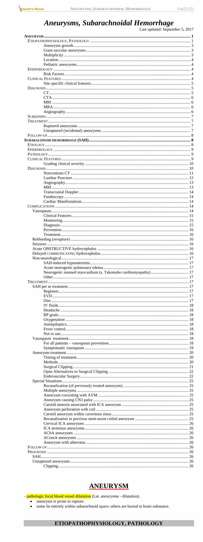
A. Dissected base of brain - aneurysm of ACA (arrow).
B. Dissected circle of Willis - large aneurysm.
ANEURYSMS, SUBARACHNOIDAL HEMORRHAGE Vas25 (3)
C. Section through berry aneurysm showing hyalinized fibrous vessel wall (H&E):
Source of picture: Ramzi S. Cotran “Robbins Pathologic Basis of Disease”, 6th ed. (1999); W. B. Saunders Company; ISBN-13: 978-
0721673356 >>
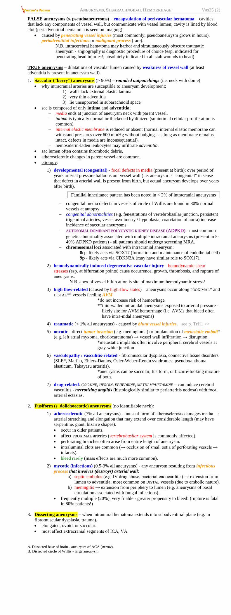
Berry aneurysm on ACA:
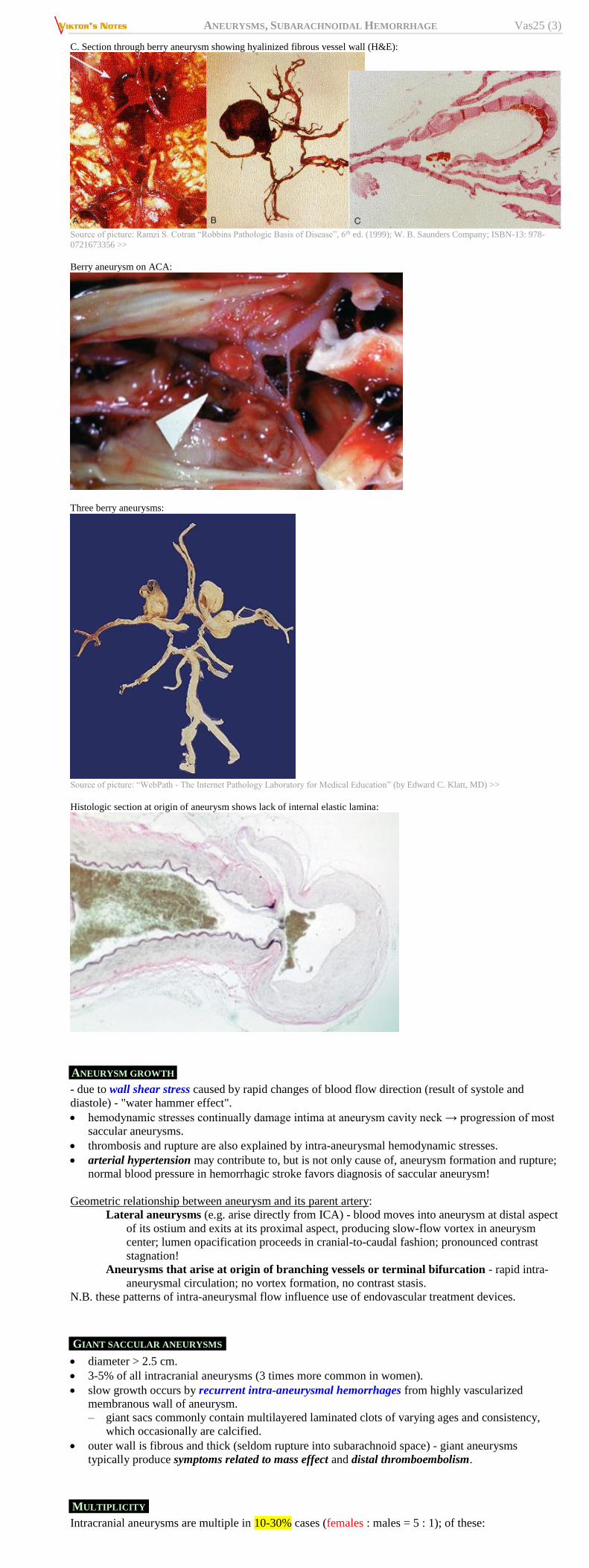
Three berry aneurysms:
Source of picture: “WebPath - The Internet Pathology Laboratory for Medical Education” (by Edward C. Klatt, MD) >>
Histologic section at origin of aneurysm shows lack of internal elastic lamina:
ANEURYSM GROWTH
- due to wall shear stress caused by rapid changes of blood flow direction (result of systole and
diastole) - "water hammer effect".
hemodynamic stresses continually damage intima at aneurysm cavity neck → progression of most
saccular aneurysms.
thrombosis and rupture are also explained by intra-aneurysmal hemodynamic stresses.
arterial hypertension may contribute to, but is not only cause of, aneurysm formation and rupture;
normal blood pressure in hemorrhagic stroke favors diagnosis of saccular aneurysm!
Geometric relationship between aneurysm and its parent artery:
Lateral aneurysms (e.g. arise directly from ICA) - blood moves into aneurysm at distal aspect
of its ostium and exits at its proximal aspect, producing slow-flow vortex in aneurysm
center; lumen opacification proceeds in cranial-to-caudal fashion; pronounced contrast
stagnation!
Aneurysms that arise at origin of branching vessels or terminal bifurcation - rapid intra-
aneurysmal circulation; no vortex formation, no contrast stasis.
N.B. these patterns of intra-aneurysmal flow influence use of endovascular treatment devices.
GIANT SACCULAR ANEURYSMS
diameter > 2.5 cm.
3-5% of all intracranial aneurysms (3 times more common in women).
slow growth occurs by recurrent intra-aneurysmal hemorrhages from highly vascularized
membranous wall of aneurysm.
– giant sacs commonly contain multilayered laminated clots of varying ages and consistency,
which occasionally are calcified.
outer wall is fibrous and thick (seldom rupture into subarachnoid space) - giant aneurysms
typically produce symptoms related to mass effect and distal thromboembolism.
MULTIPLICITY
Intracranial aneurysms are multiple in 10-30% cases (females : males = 5 : 1); of these:
ANEURYSMS, SUBARACHNOIDAL HEMORRHAGE Vas25 (4)
75% have 2 aneurysms
15% have 3 aneurysms
10% have > 3 aneurysms (females : males = 11 : 1)
multiple aneurysms are common with vasculopathies (e.g. FMD).
many are at mirror sites bilaterally.
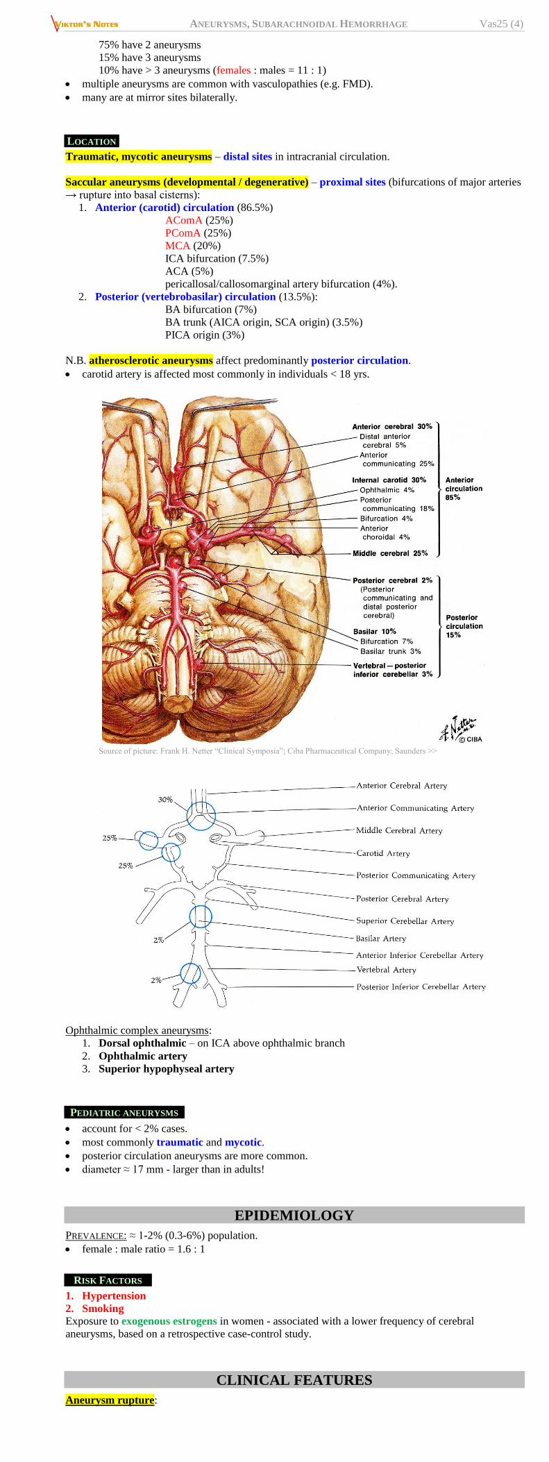
LOCATION
Traumatic, mycotic aneurysms – distal sites in intracranial circulation.
Saccular aneurysms (developmental / degenerative) – proximal sites (bifurcations of major arteries
→ rupture into basal cisterns):
1. Anterior (carotid) circulation (86.5%)
AComA (25%)
PComA (25%)
MCA (20%)
ICA bifurcation (7.5%)
ACA (5%)
pericallosal/callosomarginal artery bifurcation (4%).
2. Posterior (vertebrobasilar) circulation (13.5%):
BA bifurcation (7%)
BA trunk (AICA origin, SCA origin) (3.5%)
PICA origin (3%)
N.B. atherosclerotic aneurysms affect predominantly posterior circulation.
carotid artery is affected most commonly in individuals < 18 yrs.
Source of picture: Frank H. Netter “Clinical Symposia”; Ciba Pharmaceutical Company; Saunders >>
Ophthalmic complex aneurysms:
1. Dorsal ophthalmic – on ICA above ophthalmic branch
2. Ophthalmic artery
3. Superior hypophyseal artery
PEDIATRIC ANEURYSMS
account for < 2% cases.
most commonly traumatic and mycotic.
posterior circulation aneurysms are more common.
diameter ≈ 17 mm - larger than in adults!
EPIDEMIOLOGY
PREVALENCE: ≈ 1-2% (0.3-6%) population.
female : male ratio = 1.6 : 1
RISK FACTORS
1. Hypertension
2. Smoking
Exposure to exogenous estrogens in women - associated with a lower frequency of cerebral
aneurysms, based on a retrospective case-control study.
CLINICAL FEATURES
Aneurysm rupture:
ANEURYSMS, SUBARACHNOIDAL HEMORRHAGE Vas25 (5)
a) minor aneurysmal hemorrhage (WARNING LEAK, s. SENTINEL BLEED) - may be clinically
silent (or headache with meningeal irritation); may precede rupture with wide variation in
latency.
b) SAH (significant morbidity and mortality) - most common presentation of intracranial
aneurysm! see below
c) intraparenchymal hematoma (more common with distal aneurysms) ← direct rupture of
aneurysm into brain, secondary rupture of subarachnoid hematoma into brain parenchyma.
d) intraventricular hemorrhage (13-28%) - sources are ACA-AComA (40%), ICA (25%),
MCA (21%), VBA (14%).
e) subdural hematoma (2-5%) ← tearing of arachnoid by jet of blood.
most aneurysms do not cause symptoms until they rupture.
aneurysms typically become symptomatic in 40-60 years; peak incidence of SAH is 55-60 yrs.
risk of rupture ≈ up to 1% per year;
aneurysms < 7 mm or aneurysms in anterior circulation ≈ 0.05% per year
aneurysms at other locations, aneurysms > 10 mm, aneurysms in patients who had bled from
prior aneurysm ≈ 0.5% per year.
risk factors for aneurysm growth and rupture:
1) cigarette smoking
2) female sex
3) younger age
4) hypertension
5) aneurysm size (La Place law states that tension is determined by radius of aneurysm and
pressure gradient across wall of aneurysm) - rupture rate is directly related to aneurysm size
(≤ 5 mm - 2% risk of rupture; 6-10 mm - 40% have already ruptured upon diagnosis).
traumatic aneurysms occasionally cause epistaxis.
Nonhemorrhagic symptoms (relatively uncommon):
More common with giant aneurysms (diameter > 2.5 cm)
1. Mass effect:
1) cranial neuropathies (e.g. CN3 palsy due to PComA aneurysm - requires urgent
treatment!!!).
2) visual loss (ophthalmic artery aneurysm compresses CN2)
3) pituitary dysfunction (intrasellar aneurysms)
4) seizures 5) subacute, unilateral, periorbital headaches (aneurysmal expansion, thrombosis, intramural
hemorrhage)
6) brain stem compression (respiratory dysfunction, cardiovascular instability)
2. Emboli → TIAs / cerebral infarction (esp. with large partially thrombosed MCA aneurysms)
H: anticoagulation.
N.B. it might be sign of sentinel bleeds when thrombi start forming and shedding!!! –
pay close attention to such patients (anticoagulation might be dangerous?)
N.B. symptomatic aneurysms have significantly higher risk of rupture (6% per year).
SITE SPECIFIC CLINICAL FEATURES
AComA aneurysms (usually silent until rupture) – suprachiasmatic pressure may cause altitudinal
visual field deficits, abulia / akinetic mutism, amnestic syndromes, hypothalamic dysfunction, leg
paraparesis (!)
ACA aneurysms (usually silent until rupture) – frontal lobe syndromes, anosmia, motor deficits.
MCA aneurysms – hemiparesis (face & hand), hemisensory loss, aphasia / visual hemineglect,
visual field defects, pain in or behind eye and in low temple.
PComA aneurysms – CN3 palsy (if acute – due to rapid aneurysm growth or sentinel bleed – both
need urgent treatment!!!), hemiparesis, progressive retro-orbital headaches.
30% of acute CN3 palsies are due to PComA aneurysms
ICA aneurysms:
SUPRACLINOID aneurysms – ophthalmoplegia (CN3), variable visual defects and optic atrophy
(CN2), chiasmal compression (bilateral temporal hemianopsia), hypopituitarism, anosmia.
INTRACAVERNOUS aneurysms (extradural - do not cause SAH) – ophthalmoplegia (CN3, 4, 6),
facial sensory loss / facial pain mimicking trigeminal neuralgia (CN5), retroorbital pain.
basilar tip aneurysms – bitemporal hemianopsia, oculomotor palsy, vertical gaze paresis, coma.
VA, PICA aneurysms – vertigo, lateral medullary syndrome, ataxia, bulbar dysfunction, spinal
involvement, occipital and posterior cervical pain.
DIAGNOSIS
Angiography – criterion standard; preferred in patients with SAH.
CTA – preferred for unruptured intracranial aneurysms.
MRA – alternative to CTA (esp. in screening for aneurysm or following after coiling*).
*CTA would have lots of metal artefacts and difficult
visualization of any recurrences
CT
Noncontrast CT can visualize large aneurysms (≥ 10 mm) or that contain calcium (mural calcification
is uncommon, but both punctate and curvilinear types have been identified).
Accuracy of high-resolution axial CT in diagnosis of aneurysms ≥ 3 mm is ≈ 97%
bone erosion in long-standing lesions near skull base.
patent aneurysms - well-delineated, isodense ÷ slightly hyperdense mass in suprasellar
subarachnoid space or sylvian fissure; IV contrast → enhance intensely and uniformly.
partially thrombosed aneurysm - patent lumen inside thickened (often partially calcified) wall that
is lined with laminated clot.
– residual lumen and outer rim of aneurysm may enhance strongly with IV contrast.
– atherosclerotic debris in aneurysm wall appears hypodense on CT.
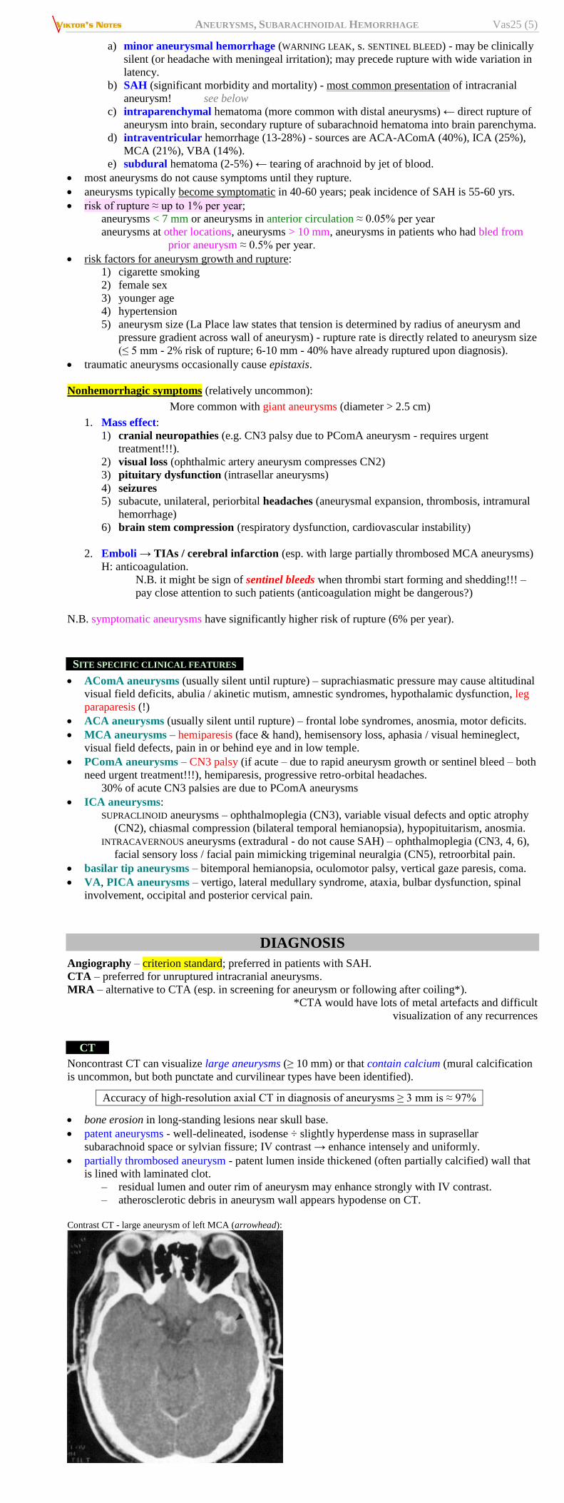
Contrast CT - large aneurysm of left MCA (arrowhead):
ANEURYSMS, SUBARACHNOIDAL HEMORRHAGE Vas25 (6)
CTA
sensitivity 97% in detecting aneurysms.
important in detection of vasospasm.
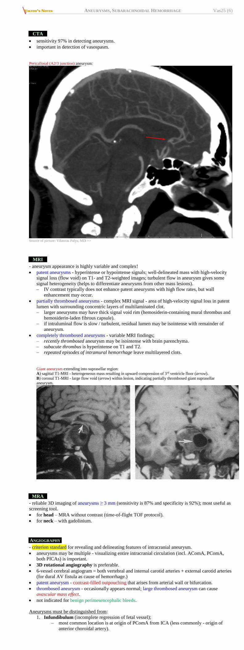
Pericallosal (A2/3 junction) aneurysm:
Source of picture: Viktoras Palys, MD >>
MRI
- aneurysm appearance is highly variable and complex!
patent aneurysms - hyperintense or hypointense signals; well-delineated mass with high-velocity
signal loss (flow void) on T1- and T2-weighted images; turbulent flow in aneurysm gives some
signal heterogeneity (helps to differentiate aneurysms from other mass lesions).
– IV contrast typically does not enhance patent aneurysms with high flow rates, but wall
enhancement may occur.
partially thrombosed aneurysms - complex MRI signal - area of high-velocity signal loss in patent
lumen with surrounding concentric layers of multilaminated clot.
– larger aneurysms may have thick signal void rim (hemosiderin-containing mural thrombus and
hemosiderin-laden fibrous capsule).
– if intraluminal flow is slow / turbulent, residual lumen may be isointense with remainder of
aneurysm.
completely thrombosed aneurysms - variable MRI findings;
– recently thrombosed aneurysm may be isointense with brain parenchyma.
– subacute thrombus is hyperintense on T1 and T2.
– repeated episodes of intramural hemorrhage leave multilayered clots.
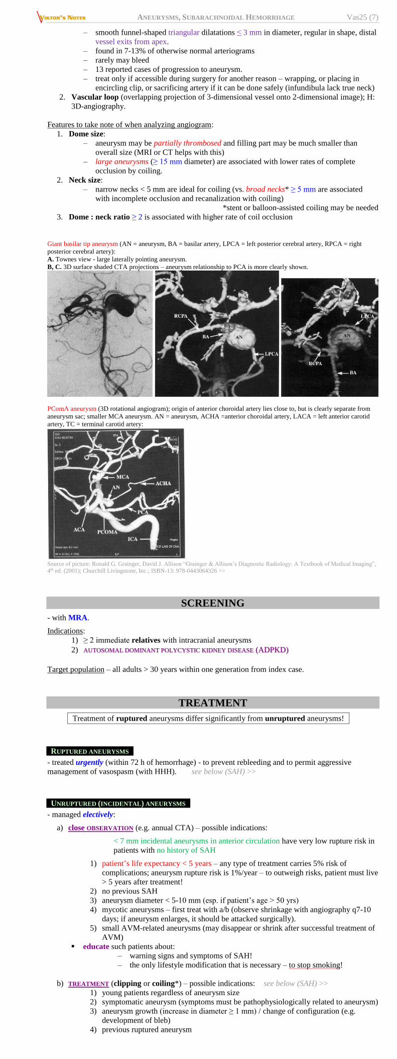
Giant aneurysm extending into suprasellar region:
A) sagittal T1-MRI - heterogeneous mass resulting in upward compression of 3rd ventricle floor (arrow).
B) coronal T1-MRI - large flow void (arrow) within lesion, indicating partially thrombosed giant suprasellar
aneurysm.
MRA
- reliable 3D imaging of aneurysms ≥ 3 mm (sensitivity is 87% and specificity is 92%); most useful as
screening tool.
for head – MRA without contrast (time-of-flight TOF protocol).
for neck – with gadolinium.
ANGIOGRAPHY
- criterion standard for revealing and delineating features of intracranial aneurysm.
aneurysms may be multiple - visualizing entire intracranial circulation (incl. AComA, PComA,
both PICAs) is important.
3D rotational angiography is preferable.
6-vessel cerebral angiogram = both vertebral and internal carotid arteries + external carotid arteries
(for dural AV fistula as cause of hemorrhage.)
patent aneurysm - contrast-filled outpouching that arises from arterial wall or bifurcation.
thrombosed aneurysm - occasionally appears normal; large thrombosed aneurysm can cause
avascular mass effect.
not indicated for benign perimesencephalic bleeds.
Aneurysms must be distinguished from:
1. Infundibulum (incomplete regression of fetal vessel);
– most common location is at origin of PComA from ICA (less commonly - origin of
anterior choroidal artery).
ANEURYSMS, SUBARACHNOIDAL HEMORRHAGE Vas25 (7)
– smooth funnel-shaped triangular dilatations ≤ 3 mm in diameter, regular in shape, distal
vessel exits from apex.
– found in 7-13% of otherwise normal arteriograms
– rarely may bleed
– 13 reported cases of progression to aneurysm.
– treat only if accessible during surgery for another reason – wrapping, or placing in
encircling clip, or sacrificing artery if it can be done safely (infundibula lack true neck)
2. Vascular loop (overlapping projection of 3-dimensional vessel onto 2-dimensional image); H:
3D-angiography.
Features to take note of when analyzing angiogram:
1. Dome size:
– aneurysm may be partially thrombosed and filling part may be much smaller than
overall size (MRI or CT helps with this)
– large aneurysms (≥ 15 mm diameter) are associated with lower rates of complete
occlusion by coiling.
2. Neck size:
– narrow necks < 5 mm are ideal for coiling (vs. broad necks* ≥ 5 mm are associated
with incomplete occlusion and recanalization with coiling)
*stent or balloon-assisted coiling may be needed
3. Dome : neck ratio ≥ 2 is associated with higher rate of coil occlusion
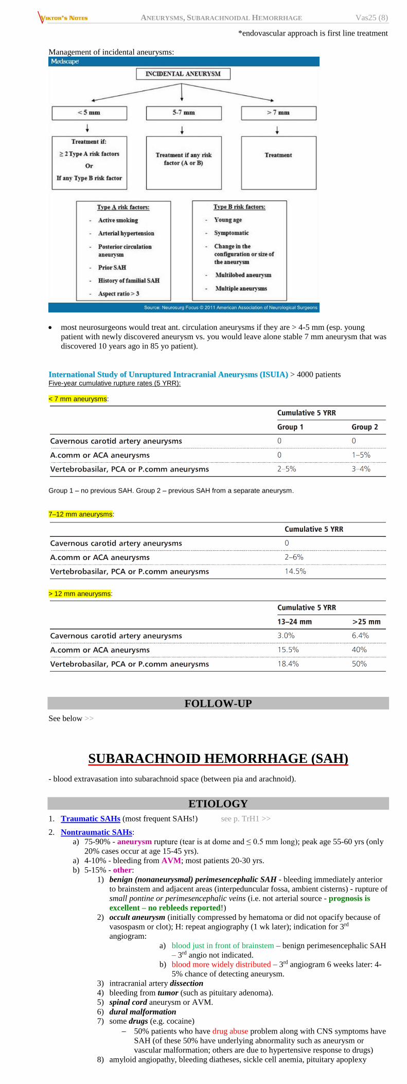
Giant basilar tip aneurysm (AN = aneurysm, BA = basilar artery, LPCA = left posterior cerebral artery, RPCA = right
posterior cerebral artery):
A. Townes view - large laterally pointing aneurysm.
B, C. 3D surface shaded CTA projections – aneurysm relationship to PCA is more clearly shown.
PComA aneurysm (3D rotational angiogram); origin of anterior choroidal artery lies close to, but is clearly separate from
aneurysm sac; smaller MCA aneurysm. AN = aneurysm, ACHA =anterior choroidal artery, LACA = left anterior carotid
artery, TC = terminal carotid artery:
Source of picture: Ronald G. Grainger, David J. Allison “Grainger & Allison’s Diagnostic Radiology: A Textbook of Medical Imaging”,
4th ed. (2001); Churchill Livingstone, Inc.; ISBN-13: 978-0443064326 >>
SCREENING
- with MRA.
Indications:
1) ≥ 2 immediate relatives with intracranial aneurysms
2) AUTOSOMAL DOMINANT POLYCYSTIC KIDNEY DISEASE (ADPKD)
Target population – all adults > 30 years within one generation from index case.
TREATMENT
Treatment of ruptured aneurysms differ significantly from unruptured aneurysms!
RUPTURED ANEURYSMS
- treated urgently (within 72 h of hemorrhage) - to prevent rebleeding and to permit aggressive
management of vasospasm (with HHH). see below (SAH) >>
UNRUPTURED (INCIDENTAL) ANEURYSMS
- managed electively:
a) close OBSERVATION (e.g. annual CTA) – possible indications:
< 7 mm incidental aneurysms in anterior circulation have very low rupture risk in
patients with no history of SAH
1) patient’s life expectancy < 5 years – any type of treatment carries 5% risk of
complications; aneurysm rupture risk is 1%/year – to outweigh risks, patient must live
> 5 years after treatment!
2) no previous SAH
3) aneurysm diameter < 5-10 mm (esp. if patient’s age > 50 yrs)
4) mycotic aneurysms – first treat with a/b (observe shrinkage with angiography q7-10
days; if aneurysm enlarges, it should be attacked surgically).
5) small AVM-related aneurysms (may disappear or shrink after successful treatment of
AVM)
educate such patients about:
– warning signs and symptoms of SAH!
– the only lifestyle modification that is necessary – to stop smoking!
b) TREATMENT (clipping or coiling*) – possible indications: see below (SAH) >>
1) young patients regardless of aneurysm size
2) symptomatic aneurysm (symptoms must be pathophysiologically related to aneurysm)
3) aneurysm growth (increase in diameter ≥ 1 mm) / change of configuration (e.g.
development of bleb)
4) previous ruptured aneurysm
ANEURYSMS, SUBARACHNOIDAL HEMORRHAGE Vas25 (8)
*endovascular approach is first line treatment
Management of incidental aneurysms:
most neurosurgeons would treat ant. circulation aneurysms if they are > 4-5 mm (esp. young
patient with newly discovered aneurysm vs. you would leave alone stable 7 mm aneurysm that was
discovered 10 years ago in 85 yo patient).
IInntteerrnnaattiioonnaall SSttuuddyy ooff UUnnrruuppttuurreedd IInnttrraaccrraanniiaall AAnneeuurryyssmmss ((IISSUUIIAA)) > 4000 patients Five-year cumulative rupture rates (5 YRR): < 7 mm aneurysms:
Group 1 – no previous SAH. Group 2 – previous SAH from a separate aneurysm. 7–12 mm aneurysms:
> 12 mm aneurysms:
FOLLOW-UP
See below >>
SUBARACHNOID HEMORRHAGE (SAH)
- blood extravasation into subarachnoid space (between pia and arachnoid).
ETIOLOGY
1. Traumatic SAHs (most frequent SAHs!) see p. TrH1 >>
2. Nontraumatic SAHs:
a) 75-90% - aneurysm rupture (tear is at dome and ≤ 0.5 mm long); peak age 55-60 yrs (only
20% cases occur at age 15-45 yrs).
a) 4-10% - bleeding from AVM; most patients 20-30 yrs.
b) 5-15% - other:
1) benign (nonaneurysmal) perimesencephalic SAH - bleeding immediately anterior
to brainstem and adjacent areas (interpeduncular fossa, ambient cisterns) - rupture of
small pontine or perimesencephalic veins (i.e. not arterial source - prognosis is
excellent – no rebleeds reported!)
2) occult aneurysm (initially compressed by hematoma or did not opacify because of
vasospasm or clot); H: repeat angiography (1 wk later); indication for 3rd
angiogram:
a) blood just in front of brainstem – benign perimesencephalic SAH
– 3rd angio not indicated.
b) blood more widely distributed – 3rd angiogram 6 weeks later: 4-
5% chance of detecting aneurysm.
3) intracranial artery dissection
4) bleeding from tumor (such as pituitary adenoma).
5) spinal cord aneurysm or AVM.
6) dural malformation 7) some drugs (e.g. cocaine)
50% patients who have drug abuse problem along with CNS symptoms have
SAH (of these 50% have underlying abnormality such as aneurysm or
vascular malformation; others are due to hypertensive response to drugs)
8) amyloid angiopathy, bleeding diatheses, sickle cell anemia, pituitary apoplexy
ANEURYSMS, SUBARACHNOIDAL HEMORRHAGE Vas25 (9)
relationship between hypertension and aneurysmal SAH is "uncertain". hypertension per se is not significant risk factor, but aneurysms do rupture under
conditions associated with sudden BP rise (coitus, athletic events, etc)
evidence for association with smoking is indirect.
EPIDEMIOLOGY
INCIDENCE has not decreased over decades (unlike for other stroke categories), but survival
improved;
– 10-fold variation in age-adjusted incidence: from 2.0 cases / 100 000 population / year in China
to 22.5 cases / 100 000 / year in Finland (a little bit less in Japan)
– rate of aneurysmal SAH in western populations: 6-8 / 100,000 population / year
– incidence increases with age and peaks at 50 yrs.
80% SAHs occur in people aged > 40 years
15% in people aged 20-40 years
4.5% in people aged 10-20 years
0.5% in children < 10 years
significant risk factors for aneurysmal SAH:
1) smoking (risk of SAH increased 3-6-fold; risk increased 6-fold if positive family history of
aneurysmal SAH).
2) hypertension (conflicting data – see above)
3) North American blacks (2.1 times greater risk than in whites)
4) females (1.24 : 1)
5) 3rd trimester of pregnancy*- SAH is leading cause of maternal mortality (5-25% maternal
deaths during pregnancy):
75% from aneurysms (esp. older multiparous women)
25% from AVMs (esp. younger nulliparous women)
*recent reviews have suggested that, contrary to conclusions from prior
studies, risk of SAH is not increased during pregnancy.
6) hormone replacement therapy in postmenopausal women
7) heavy alcohol consumption (controversial)
8) reduced lung function (reduced FEV1, reduced FEV1/FVC) - significantly associated with
increased incidence of SAH - comparable with effects of hypertension and smoking "Our hypothesis is that matrix degradation of vessel walls, which is major reason for SAH, and
degradation of lung tissue, which is major reason for reduced FEV1, share common mechanisms"
9) long-term (esp. > 3 years) low-dose ASPIRIN has protective effective against SAH and does
not increase risk of intracerebral hemorrhage.
aneurysm size matters; AComA aneurysm size correlates linearly with risk of rupture.
oral contraceptives, hormone replacement therapy, hypercholesterolemia, physical activity are not
significantly related!
PATHOLOGY
surrounding brain parenchyma - brownish pigmentation and fibrous adhesions.
aneurysm size may be diminished postmortem.
ruptured fundus may be visualized with calcifications of aneurysm wall and intraluminal
thrombus.
SAH may be complicated by:
intracerebral hemorrhage (20-40%)
intraventricular hemorrhage (13-28%)
subdural hematoma (2-5%) - over convexity (most commonly due to PComA aneurysm) or
interhemispheric (distal ACA).
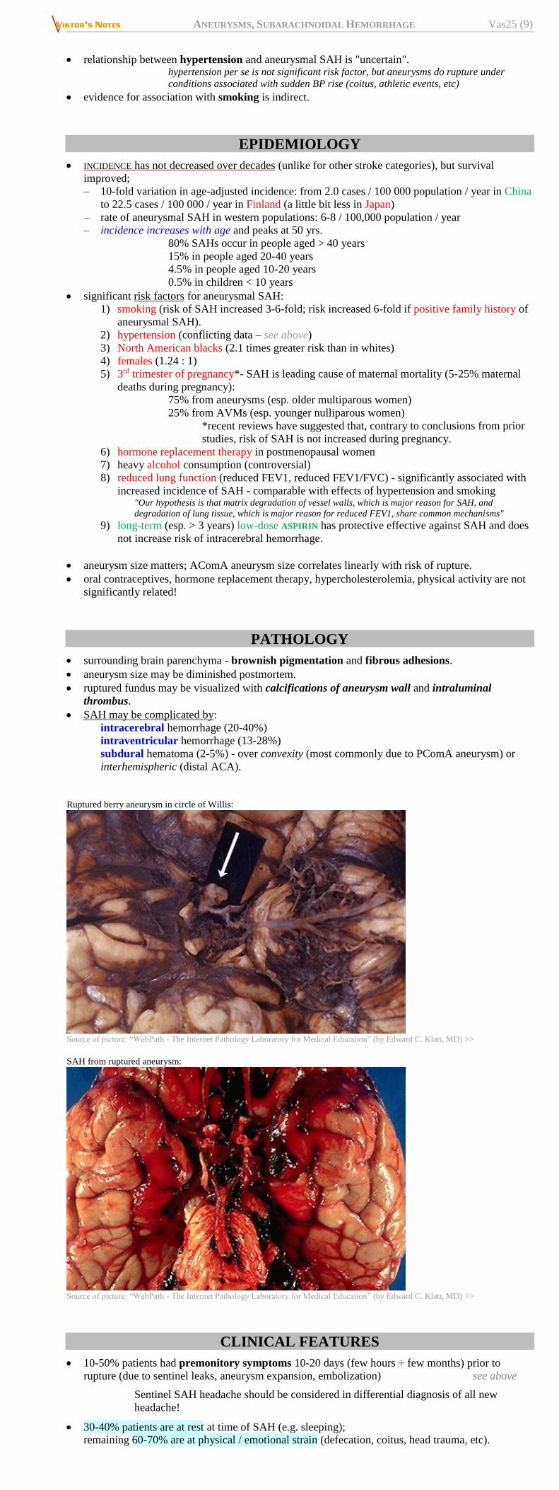
Ruptured berry aneurysm in circle of Willis:
Source of picture: “WebPath - The Internet Pathology Laboratory for Medical Education” (by Edward C. Klatt, MD) >>
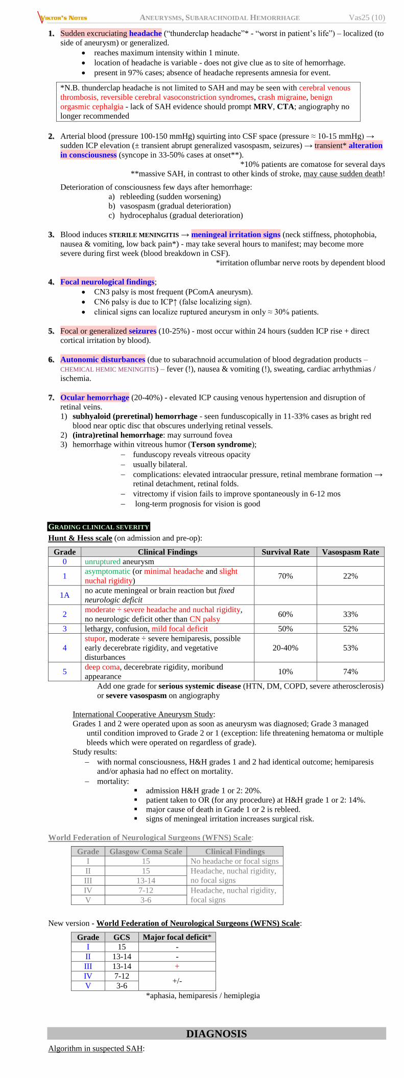
SAH from ruptured aneurysm:
Source of picture: “WebPath - The Internet Pathology Laboratory for Medical Education” (by Edward C. Klatt, MD) >>
CLINICAL FEATURES
10-50% patients had premonitory symptoms 10-20 days (few hours ÷ few months) prior to
rupture (due to sentinel leaks, aneurysm expansion, embolization) see above
Sentinel SAH headache should be considered in differential diagnosis of all new
headache!
30-40% patients are at rest at time of SAH (e.g. sleeping);
remaining 60-70% are at physical / emotional strain (defecation, coitus, head trauma, etc).
ANEURYSMS, SUBARACHNOIDAL HEMORRHAGE Vas25 (10)
1. Sudden excruciating headache (“thunderclap headache”* - “worst in patient’s life”) – localized (to
side of aneurysm) or generalized.
reaches maximum intensity within 1 minute.
location of headache is variable - does not give clue as to site of hemorrhage.
present in 97% cases; absence of headache represents amnesia for event.
*N.B. thunderclap headache is not limited to SAH and may be seen with cerebral venous
thrombosis, reversible cerebral vasoconstriction syndromes, crash migraine, benign
orgasmic cephalgia - lack of SAH evidence should prompt MRV, CTA; angiography no
longer recommended
2. Arterial blood (pressure 100-150 mmHg) squirting into CSF space (pressure ≈ 10-15 mmHg) →
sudden ICP elevation (± transient abrupt generalized vasospasm, seizures) → transient* alteration
in consciousness (syncope in 33-50% cases at onset**).
*10% patients are comatose for several days
**massive SAH, in contrast to other kinds of stroke, may cause sudden death!
Deterioration of consciousness few days after hemorrhage:
a) rebleeding (sudden worsening)
b) vasospasm (gradual deterioration)
c) hydrocephalus (gradual deterioration)
3. Blood induces STERILE MENINGITIS → meningeal irritation signs (neck stiffness, photophobia,
nausea & vomiting, low back pain*) - may take several hours to manifest; may become more
severe during first week (blood breakdown in CSF).
*irritation oflumbar nerve roots by dependent blood
4. Focal neurological findings;
CN3 palsy is most frequent (PComA aneurysm).
CN6 palsy is due to ICP↑ (false localizing sign).
clinical signs can localize ruptured aneurysm in only ≈ 30% patients.
5. Focal or generalized seizures (10-25%) - most occur within 24 hours (sudden ICP rise + direct
cortical irritation by blood).
6. Autonomic disturbances (due to subarachnoid accumulation of blood degradation products –
CHEMICAL HEMIC MENINGITIS) – fever (!), nausea & vomiting (!), sweating, cardiac arrhythmias /
ischemia.
7. Ocular hemorrhage (20-40%) - elevated ICP causing venous hypertension and disruption of
retinal veins.
1) subhyaloid (preretinal) hemorrhage - seen funduscopically in 11-33% cases as bright red
blood near optic disc that obscures underlying retinal vessels.
2) (intra)retinal hemorrhage: may surround fovea
3) hemorrhage within vitreous humor (Terson syndrome);
funduscopy reveals vitreous opacity
usually bilateral.
complications: elevated intraocular pressure, retinal membrane formation →
retinal detachment, retinal folds.
vitrectomy if vision fails to improve spontaneously in 6-12 mos
long-term prognosis for vision is good
GRADING CLINICAL SEVERITY
Hunt & Hess scale (on admission and pre-op):
Grade Clinical Findings Survival Rate Vasospasm Rate
0 unruptured aneurysm
1 asymptomatic (or minimal headache and slight
nuchal rigidity) 70% 22%
1A no acute meningeal or brain reaction but fixed
neurologic deficit
2 moderate ÷ severe headache and nuchal rigidity,
no neurologic deficit other than CN palsy 60% 33%
3 lethargy, confusion, mild focal deficit 50% 52%
4
stupor, moderate ÷ severe hemiparesis, possible
early decerebrate rigidity, and vegetative
disturbances
20-40% 53%
5 deep coma, decerebrate rigidity, moribund
appearance 10% 74%
Add one grade for serious systemic disease (HTN, DM, COPD, severe atherosclerosis)
or severe vasospasm on angiography
International Cooperative Aneurysm Study:
Grades 1 and 2 were operated upon as soon as aneurysm was diagnosed; Grade 3 managed
until condition improved to Grade 2 or 1 (exception: life threatening hematoma or multiple
bleeds which were operated on regardless of grade).
Study results:
with normal consciousness, H&H grades 1 and 2 had identical outcome; hemiparesis
and/or aphasia had no effect on mortality.
mortality:
admission H&H grade 1 or 2: 20%.
patient taken to OR (for any procedure) at H&H grade 1 or 2: 14%.
major cause of death in Grade 1 or 2 is rebleed.
signs of meningeal irritation increases surgical risk.
World Federation of Neurological Surgeons (WFNS) Scale:
Grade Glasgow Coma Scale Clinical Findings
I 15 No headache or focal signs
II 15 Headache, nuchal rigidity,
no focal signs III 13-14
IV 7-12 Headache, nuchal rigidity,
focal signs V 3-6
New version - World Federation of Neurological Surgeons (WFNS) Scale:
Grade GCS Major focal deficit*
I 15 -
II 13-14 -
III 13-14 +
IV 7-12 +/-
V 3-6
*aphasia, hemiparesis / hemiplegia
DIAGNOSIS
Algorithm in suspected SAH:
ANEURYSMS, SUBARACHNOIDAL HEMORRHAGE Vas25 (11)
CT without contrast
positive ↓ positive ↓ negative
angiography ← LP
NONCONTRAST CT
Acute SAH - areas of increased density in subarachnoid spaces.
IV contrast may obscure SAH detection!
False-negative CT:
a) hemorrhage from lesion in spinal cord.
b) CT performed too early (only small amount of blood being present).
c) subarachnoid blood disappeared before imaging.
CT sensitivity 95-98% (maximum sensitivity is within 12-48 hours)
sensitivity decreases to 80% at 72 hours, 50% at 1 week
Negative CT does not preclude SAH!!!
For subtle SAH, look in occipital horns of lateral ventricles and dependent
portions of sylvian fissures.
If CT is negative, but SAH is still suspected → lumbar puncture or repeat CT later or FLAIR MRI
In patients with minimal symptomatology (e.g. sentinel leaks), lumbar puncture
is considerably more accurate than CT!
N.B. you have only one chance with LP – if you repeat it, it will be flase-
positive from small amount of blood from first LP
Bleeding site localization:
N.B. blood may quickly spread diffusely throughout CSF spaces, providing little
clue to its site of origin.
– suprasellar cistern blood is common from many bleeding sites.
– blood within cistern of lamina terminalis, anterior interhemispheric fissure – AComA.
– blood within sylvian fissure – MCA or PcomA.
– blood predominantly in prepontine or peduncular cistern – basilar apex or SCA.
– lateral ventricle blood – AComA
– 3rd ventricle blood – basilar tip.
– 4th ventricle blood – posterior fossa aneurysms (esp. at PICA takeoff) – almost pathognomonic
for PICA!!!!
– surrounding edema and inflammation may be appreciated with IV contrast following
noncontrast CT.
blood in basal cisterns, sylvian fissure, or intrahemispheric fissure - saccular aneurysm rupture;
blood over convexities or within superficial brain parenchyma - AVM or mycotic aneurysm rupture.
HIJDRA score - amount of cisternal and ventricular blood seen on CT.
FISHER grade - amount of blood seen on CT (predicts vasospasm):
Fisher 1 - no blood detected
Fisher 2 - diffuse thin (< 1 mm) layer, no clots
Fisher 3 - localized clot and/or vertical layer (≥ 1 mm) – high risk of vasospasm!
Fisher 4 - intracerebral or intraventricular clot with diffuse or no SAH.
Only Fisher 3 is associated with clinical vasospasm (amount of blood in cisterns
and fissures is important prognosticator for vasospasm!)
– "vertical layer" refers to blood within "vertical" subarachnoid spaces including
interhemispheric fissure, insular cistern, ambient cistern
– reflux of blood into ventricles frequently indicates obstruction of CSF circulation, and is
associated with high incidence of hydrocephalus
Things that can mimic SAH on CT:
1. Pus
2. Contrast (IV and especially intrathecal)
3. Pachymeningeal thickening seen in spontaneous intracranial hypotension
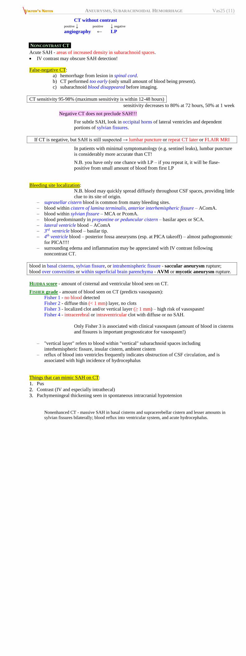
Nonenhanced CT - massive SAH in basal cisterns and supracerebellar cistern and lesser amounts in
sylvian fissures bilaterally; blood reflux into ventricular system, and acute hydrocephalus.
ANEURYSMS, SUBARACHNOIDAL HEMORRHAGE Vas25 (12)
Acute diffuse SAH within suprasellar cistern, ambient cistern, and frontal and temporal sulci. There is dilation of both
temporal horns of lateral ventricles associated with communicating hydrocephalus:
Source of picture: H. Richard Winn “Youmans Neurological Surgery”, 6th ed. (2011); Saunders; ISBN-13: 978-1416053163 >>
SDH due to ruptured right PComA aneurysm:
Source of picture: Viktoras Palys, MD
LUMBAR PUNCTURE
- most sensitive test for SAH - indicated if CT is negative - nonclotting hemorrhagic CSF with
xanthochromic supernatant (may be absent within first few hours!; 100% present after ≥ 12 hours).
Parameter "Traumatic Tap" SAH
Xanthochromia Absent Onset: 4-6 hr
Still present in
40% at 4 wks
RBC count (serial tubes) Decreasing Constant
Blood clot formation Rapid Slower
opening pressure↑ (remains for many days), proteins↑
proportion of WBCs to RBCs is that of peripheral blood (≈ 1:1000) → WBC count increases after
24 hours (chemical meningitis); also [glucose] decreases.
LP should not be performed if CT demonstrates SAH.
N.B. if aneurysm is unsecured, excessive lowering of CSF pressure increases
transmural pressure across wall of aneurysm → rebleed (remove only minimum volume
of CSF needed for diagnostic studies).
you have only one chance; repeat LP may be xantochromic from previous LP.
ANEURYSMS, SUBARACHNOIDAL HEMORRHAGE Vas25 (13)
differential of xanthochromia: jaundice, high protein levels in CSF.
ANGIOGRAPHY
- indicated in all patients after SAH diagnosis!
explore all 4 vessels.
signs of ruptured aneurysm (if ≥ 1 aneurysm is found - which aneurysm needs to be treated
acutely):
1) contrast extravasation (pathognomonic but extremely rare)
2) larger aneurysm will be site of rupture more frequently than smaller one – most important
(practically) criterion! (all others below – only soft signs)
3) mass effect adjacent to aneurysm (focal parenchymal or cisternal hematoma)
4) irregularly shaped aneurysm (lobulation, smaller daughter dome)
5) focal vasospasm (but subarachnoid blood quickly spreads along basal cisterns)
N.B. acute angiography occasionally yields negative results (e.g. due to thrombosis or vasospasm) →
repeat angiography 1 and 4* weeks later.
*3 angiograms still give 4% false-negatives; 4-week angio is not indicated if
SAH blood pattern is compatible with benign perimesencephalic bleed
Before calling angiogram negative, one must:
1) visualize both PICA origins (via one VA injection if there is enough reflux down
contralateral VA).
2) visualize AComA*; if both ACAs fill from one side, this is satisfactory.
* perform cross compression AP study with carotid injection
(first, rule-out plaque in carotid to be compressed), or use a
higher injection rate to facilitate flow
3) see no infundibulum (see above) co-localized to SAH.
after first negative angiogram, order cervical MRI to rule out cervical AVM / AVF as source of
SAH.
trial balloon occlusion of parent artery - for giant and fusiform aneurysms that may need to be
surgically “trapped” because they lack defined neck for surgical clipping.
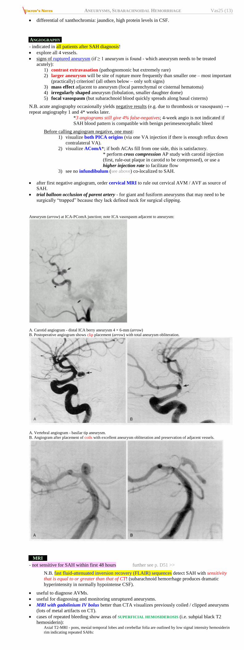
Aneurysm (arrow) at ICA-PComA junction; note ICA vasospasm adjacent to aneurysm:
A. Carotid angiogram - distal ICA berry aneurysm 4 × 6-mm (arrow)
B. Postoperative angiogram shows clip placement (arrow) with total aneurysm obliteration.
A. Vertebral angiogram - basilar tip aneurysm.
B. Angiogram after placement of coils with excellent aneurysm obliteration and preservation of adjacent vessels.
MRI
- not sensitive for SAH within first 48 hours further see p. D51 >>
N.B. fast fluid-attenuated inversion recovery (FLAIR) sequences detect SAH with sensitivity
that is equal to or greater than that of CT! (subarachnoid hemorrhage produces dramatic
hyperintensity in normally hypointense CSF).
useful to diagnose AVMs.
useful for diagnosing and monitoring unruptured aneurysms.
MRI with gadolinium IV bolus better than CTA visualizes previously coiled / clipped aneurysms
(lots of metal artifacts on CT).
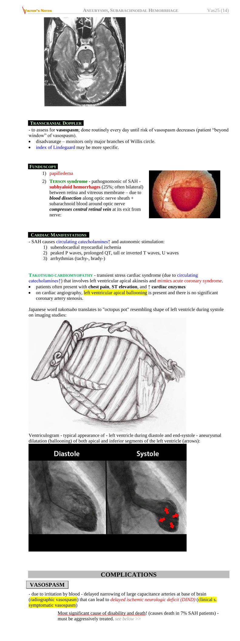
cases of repeated bleeding show areas of SUPERFICIAL HEMOSIDEROSIS (i.e. subpial black T2
hemosiderin): Axial T2-MRI - pons, mesial temporal lobes and cerebellar folia are outlined by low signal intensity hemosiderin
rim indicating repeated SAHs:
ANEURYSMS, SUBARACHNOIDAL HEMORRHAGE Vas25 (14)
TRANSCRANIAL DOPPLER
- to assess for vasospasm; done routinely every day until risk of vasospasm decreases (patient “beyond
window” of vasospasm).
disadvanatge – monitors only major branches of Willis circle.
index of Lindegaard may be more specific.
FUNDUSCOPY
1) papilledema
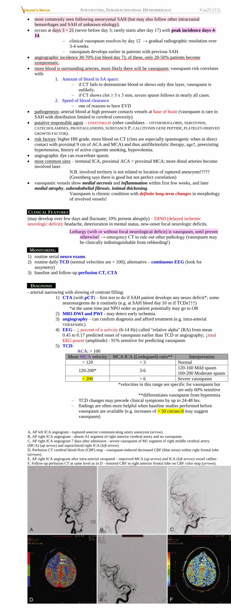
2) TERSON syndrome - pathognomonic of SAH -
subhyaloid hemorrhages (25%; often bilateral)
between retina and vitreous membrane – due to
blood dissection along optic nerve sheath +
subarachnoid blood around optic nerve
compresses central retinal vein at its exit from
nerve:
CARDIAC MANIFESTATIONS
- SAH causes circulating catecholamines↑ and autonomic stimulation:
1) subendocardial myocardial ischemia
2) peaked P waves, prolonged QT, tall or inverted T waves, U waves
3) arrhythmias (tachy-, brady-)
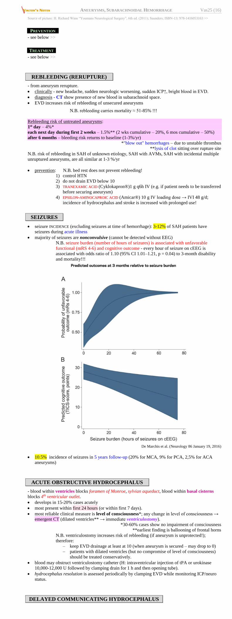
TAKOTSUBO CARDIOMYOPATHY - transient stress cardiac syndrome (due to circulating
catecholamines↑) that involves left ventricular apical akinesis and mimics acute coronary syndrome.
patients often present with chest pain, ST elevation, and ↑ cardiac enzymes
on cardiac angiography, left ventricular apical ballooning is present and there is no significant
coronary artery stenosis.
Japanese word takotsubo translates to "octopus pot" resembling shape of left ventricle during systole
on imaging studies:
Ventriculogram - typical appearance of - left ventricle during diastole and end-systole - aneurysmal
dilatation (ballooning) of both apical and inferior segments of the left ventricle (arrows):
COMPLICATIONS
VASOSPASM
- due to irritation by blood - delayed narrowing of large capacitance arteries at base of brain
(radiographic vasospasm) that can lead to delayed ischemic neurologic deficit (DIND) (clinical s.
symptomatic vasospasm)
Most significant cause of disability and death! (causes death in 7% SAH patients) -
must be aggressively treated. see below >>
ANEURYSMS, SUBARACHNOIDAL HEMORRHAGE Vas25 (15)
most commonly seen following aneurysmal SAH (but may also follow other intracranial
hemorrhages and SAH of unknown etiology).
occurs at days 3 ÷ 21 (never before day 3; rarely starts after day 17) with peak incidence days 4-
14.
– clinical vasospasm resolves by day 12 → gradual radiographic resolution over
3-4 weeks
– vasospasm develops earlier in patients with previous SAH.
angiographic incidence 30-70% (on bleed day 7); of these, only 20-50% patients become
symptomatic.
more blood is surrounding arteries, more likely there will be vasospasm; vasospasm risk correlates
with:
1. Amount of blood in SA space:
– if CT fails to demonstrate blood or shows only thin layer, vasospasm is
unlikely.
– if CT shows clot ≥ 5 x 3 mm, severe spasm follows in nearly all cases.
2. Speed of blood clearance
– one of reasons to have EVD
pathogenesis: arterial blood at high pressure contacts vessels at base of brain (vasospasm is rare in
SAH with distribution limited to cerebral convexity).
putative responsible agent – ENDOTHELIN (other candidates – OXYHEMOGLOBIN, SEROTONIN,
CATECHOLAMINES, PROSTAGLANDINS, SUBSTANCE P, CALCITONIN GENE PEPTIDE, PLATELET-DERIVED
GROWTH FACTOR).
risk factors: higher HH grade, more blood on CT (clots are especially spasmogenic when in direct
contact with proximal 9 cm of ACA and MCA) and thus antifibrinolytic therapy, age↑, preexisting
hypertension, history of active cigarette smoking, hypovolemia.
angiographic dye can exacerbate spasm.
most common sites – terminal ICA, proximal ACA > proximal MCA; more distal arteries become
involved later.
N.B. involved territory is not related to location of ruptured aneurysm!????
(Greenberg says there is good but not perfect correlation)
vasospastic vessels show medial necrosis and inflammation within first few weeks, and later
medial atrophy, subendothelial fibrosis, intimal thickening.
Vasospasm is chronic condition with definite long-term changes in morphology
of involved vessels!
CLINICAL FEATURES
(may develop over few days and fluctuate; 10% present abruptly) – DIND (delayed ischemic
neurologic deficit): headache, deterioration in mental status, new-onset focal neurologic deficits.
Lethargy (with or without focal neurological deficit) is vasospasm, until proven
otherwise! → emergency CT to rule out other pathology (vasospasm may
be clinically indistinguishable from rebleeding!)
MONITORING
1) routine serial neuro exams
2) routine daily TCD (normal velocities are < 100); alternative – continuous EEG (look for
assymetry)
3) baseline and follow-up perfusion CT, CTA
DIAGNOSIS
- arterial narrowing with slowing of contrast filling:
1) CTA (with pCT) – first test to do if SAH patient develops any neuro deficit*; some
neurosurgeons do it routinely (e.g. at SAH bleed day 10 or if TCDs↑↑↑)
*at the same time put NPO order as patient potentially may go to OR
2) MRI-DWI and PWI - may detect early ischemia.
3) angiography – can confirm diagnosis and afford treatment (e.g. intra-arterial
VERAPAMIL)
4) EEG – ↓ percent of α activity (6-14 Hz) called "relative alpha" (RA) from mean
0.45 to 0.17 predicted onset of vasospasm earlier than TCD or angiography; ↓total
EEG power (amplitude) - 91% sensitive for predicting vasospasm
5) TCD:
ACA: > 100
Mean MCA velocity MCA:ICA (Lindegaard) ratio** Interpretation
< 120 < 3 Normal
120-200* 3-6 120-160 Mild spasm
160-200 Moderate spasm
> 200 > 6 Severe vasospasm
*velocities in this range are specific for vasospasm but
are only 60% sensitive
**differentiates vasospasm from hyperemia
– TCD changes may precede clinical symptoms by up to 24-48 hrs.
– findings are often more helpful when baseline studies performed before
vasospasm are available (e.g. increases of > 50 cm/sec/d may suggest
vasospasm)
A, AP left ICA angiogram - ruptured anterior communicating artery aneurysm (arrow).
B, AP right ICA angiogram - absent A1 segment of right anterior cerebral artery and no vasospasm.
C, AP right ICA angiogram 7 days after admission - severe vasospasm of M1 segment of right middle cerebral artery
(MCA) (up arrow) and supraclinoid right ICA (left arrow).
D, Perfusion CT cerebral blood flow (CBF) map - vasospasm-induced decreased CBF (blue areas) within right frontal lobe
(arrows).
E, AP right ICA angiogram after intra-arterial verapamil - improved MCA (up arrow) and ICA (left arrow) vessel caliber.
F, Follow-up perfusion CT at same level as in D - restored CBF in right anterior frontal lobe on CBF color map (arrows).
ANEURYSMS, SUBARACHNOIDAL HEMORRHAGE Vas25 (16)
Source of picture: H. Richard Winn “Youmans Neurological Surgery”, 6th ed. (2011); Saunders; ISBN-13: 978-1416053163 >>
PREVENTION
- see below >>
TREATMENT
- see below >>
REBLEEDING (RERUPTURE)
- from aneurysm rerupture.
clinically - new headache, sudden neurologic worsening, sudden ICP↑, bright blood in EVD.
diagnosis - CT show presence of new blood in subarachnoid space.
EVD increases risk of rebleeding of unsecured aneurysms
N.B. rebleeding carries mortality ≈ 51-85% !!!
Rebleeding risk of untreated aneurysms:
1st day – 4%*
each next day during first 2 weeks – 1.5%** (2 wks cumulative – 20%, 6 mos cumulative – 50%)
after 6 months – bleeding risk returns to baseline (1-3%/yr)
*"blow out" hemorrhages – due to unstable thrombus
**lysis of clot sitting over rupture site
N.B. risk of rebleeding in SAH of unknown etiology, SAH with AVMs, SAH with incidental multiple
unruptured aneurysms, are all similar at 1-3 %/yr
prevention: N.B. bed rest does not prevent rebleeding!
1) control HTN
2) do not drain EVD below 10
3) TTRRAANNEEXXAAMMIICC AACCIIDD (Cyklokapron®)1 g q6h IV (e.g. if patient needs to be transferred
before securing aneurysm)
4) EEPPSSIILLOONN--AAMMIINNOOCCAAPPRROOIICC AACCIIDD (Amicar®) 10 g IV loading dose → IVI 48 g/d;
incidence of hydrocephalus and stroke is increased with prolonged use!
SEIZURES
seizure INCIDENCE (excluding seizures at time of hemorrhage): 3-12% of SAH patients have
seizures during acute illness
majority of seizures are nonconvulsive (cannot be detected without EEG)
N.B. seizure burden (number of hours of seizures) is associated with unfavorable
functional (mRS 4-6) and cognitive outcome - every hour of seizure on cEEG is
associated with odds ratio of 1.10 (95% CI 1.01–1.21, p = 0.04) to 3-month disability
and mortality!!!
De Marchis et al. (Neurology 86 January 19, 2016)
10.5% incidence of seizures in 5 years follow-up (20% for MCA, 9% for PCA, 2,5% for ACA
aneurysms)
ACUTE OBSTRUCTIVE HYDROCEPHALUS
- blood within ventricles blocks foramen of Monroe, sylvian aqueduct, blood within basal cisterns
blocks 4th ventricular outlet.
develops in 15-20% cases acutely
most present within first 24 hours (or within first 7 days).
most reliable clinical measure is level of consciousness*; any change in level of consciousness →
emergent CT (dilated ventricles** → immediate ventriculostomy).
*30-60% cases show no impairment of consciousness
**earliest finding is ballooning of frontal horns
N.B. ventriculostomy increases risk of rebleeding (if aneurysm is unprotected!);
therefore:
– keep EVD drainage at least at 10 (when aneurysm is secured – may drop to 0)
– patients with dilated ventricles (but no compromise of level of consciousness)
should be treated conservatively.
blood may obstruct ventriculostomy catheter (H: intraventricular injection of tPA or urokinase
10,000-12,000 U followed by clamping drain for 1 h and then opening tube).
hydrocephalus resolution is assessed periodically by clamping EVD while monitoring ICP/neuro
status.
DELAYED COMMUNICATING HYDROCEPHALUS
ANEURYSMS, SUBARACHNOIDAL HEMORRHAGE Vas25 (17)
- blood in subarachnoid space obliterates arachnoidal villi;
develops in 8-45% cases
develops ≥ 10 days after SAH - incontinence, gait instability, cognitive deterioration (abulia).
prophylaxis: EVD (clears blood from CSF).
treatment: ventriculoperitoneal shunt.
N.B. usually it is temporary condition and prolonged EVD/LD helps to avoid shunt in some patients;
some experts would even use intermittent LP with multiple passes to create CSF leak in spine as
temporary “safety valve” until condition resolves.
NON-NEUROLOGICAL
SAH-INDUCED HYPONATREMIA
develops in 10-34% cases
majority of cases are due to cerebral salt wasting (atrial natriuretic peptide*↑?); the rest – due to
SIADH *not brain natriuretic peptide
may be first sign of vasospasm!
treatment: see p. 2514 >>
SIADH – fluid restriction
CSW – fluid repletion with normal saline or slightly hypertonic (1.5-3%) NaCl at rates
above maintenance requirements, hydrocortisone / fludrocortisone, salt tabs.
hyponatremic patients have 3 times incidence of delayed cerebral infarction than normonatremic
patients!
ACUTE NEUROGENIC PULMONARY EDEMA
unrelated to HHH therapy.
almost universal in severe SAH.
H: gentle diuresis, dobutamine, PEEP.
Patients undergoing triple-H therapy can develop cardiogenic pulmonary edema as they "fall
off' Starling curve with volume expansion!
NEUROGENIC STUNNED MYOCARDIUM (S. TAKOTSUBO CARDIOMYOPATHY)
- myocardial hypokinesis (↓ ejection fraction) not attributable to coronary artery disease or myocardial
abnormalities.
putative mechanism: hypothalamic ischemia → local (myocardial) catecholamine surge (peak 2
days to 2 weeks post SAH).
clinically: hypotension (may be compensated by ↑SVR), CHF, arrhythmias.
reverses completely in most cases within about 5 days; 10% progress to MI.
diagnosis: as MI on echocardiography and EKG but cardiac enzymes tend to be lower than
expected for degree of myocardial impairment.
treatment: DDOOBBUUTTAAMMIINNEE (for SBP < 90 and low SVR), MMIILLRRIINNOONNEE (for SBP > 90 and normal or
increased SVR, esp. if patient is on chronic β-blockers).
OTHER
Neurogenic sympathetic hyperactivity → arrhythmias (90% patients), myocardial ischemia (cardiac
myonecrosis)
most prevalent in first 48 hours.
HHH therapy to prevent cerebral ischemia may exacerbate myocardial ischemia; therapy for
myocardial ischemia (nitrates) may increase ICP and exacerbate cerebral ischemia.
2D echocardiography is more sensitive in detecting myocardial ischemia than is ECG.
CAVERNOUS-CAROTID aneurysm rupture typically produces carotid-cavernous fistula (rather than
SAH).
Heparin-induced thrombocytopenia, DVT - occur infrequently but not uncommonly.
TREATMENT
Three factors that improve outcome:
1) early referral to hospital that has physicians experienced in treating aneurysms.
2) urgent aneurysm treatment
3) aggressive treatment of vasospasm.
SAH per se treatment
Vasospasm prevention (nimodipine, pravastatin, Mg sulfate) see below >>
REGIMEN
– dark quiet environment in ICU; visitors are limited to immediate family;
– strict bed rest (no out of bed for any reason, limit number of daily visitors) for 2
weeks → gradually return to normal activities over 3rd week.
Bed rest does not prevent rebleeding!
– elevate head of bed at 30°
– avoid straining during defecation
– midazolam IV prior to procedures that may increase ICP.
– frequent turning and calf compressions.
EVD
If GCS < 14 (esp. if blood in ventricles) – insert EVD (drain continuously at 10 cmH2O*; when
aneurysm is secured – drain at 0 cmH2O) *Greenberg recommends 15-25 mmHg
don’t start challenging EVD until day 6-7.
don’t start challenging EVD if on vasopressors.
don’t start challenging EVD until CSF output becomes < 100 mL/8 hr.
elevated ICPs require mannitol, etc.
DIET
No nicotine patch for smokers!
antacids (e.g. RANITIDINE)
antiemetics (e.g. PROMETHAZINE, ONDANSETRON)
Avoid phenothiazines - lower seizure threshold!
stool softeners (e.g. DOCUSATE); bedside commode requires less straining than bed pan and is
preferred for patients who are able to get out of bed.
ANEURYSMS, SUBARACHNOIDAL HEMORRHAGE Vas25 (18)
IV FLUIDS
– use only isotonic solutions (to minimize cerebral edema).
early aggressive fluid therapy to head off cerebral salt wasting: NS + 20 mEq KC/L at 2 ml/kg/hr
(typically 140-150 ml/hr)
if Hct < 40%, give 500 ml of 5% albumin over 4 hrs upon admission.
HEADACHE
– the best is Fioricet® (Dr. Rivet avoids it due to caffeine – risk of vasospasm)
in general, medications are not helpful with headache.
avoid NSAIDs (bleeding risk; however, if aneurysm is secured, IIBBUUPPRROOFFEENN is OK), avoid sedatives
and opioids (unless patient is with clear sensorium)
in acute crisis, FFEENNTTAANNYYLL is OK – short acting and does not release histamine (does not elevate
ICP)!
Avoid Demerol® - lowers seizure threshold!
glucocorticoids may help reduce head & neck ache (irritative effect of subarachnoid blood); no
good evidence that they are neuroprotective - routine use is controversial.
BP GOALS
SBP goal: unsecured aneurysm < 140; secured (clipped / coiled) 100-160; up to 180-220 (if
secured and in vasospasm)
maintain BP in range that allows for sufficient cerebral perfusion* yet limits risk of rebleeding;
– β-blockers (labetalol) / Ca-antagonists (nicardipine) IV are agents of choice → start
long-acting ACEI.
– most clinicians avoid nitrates (NITROPRUSSIDE, NITROGLYCERIN) which elevate ICP;
*state of consciousness may be used as guide to level of cerebral
perfusion - administer hypotensive medications up to level that
patient begins to experience drowsiness.
Always avoid HYPOVOLEMIA!
Arterial-line indications: hemodynamically unstable, stuporous or comatose, difficult to control
hypertension, requiring frequent labs (e.g. ventilator patients).
pulmonary-artery catheter (aka Swann-Ganz catheter) is out of favor.
OXYGENATION
- must be adequate.
some experts target for early moderate hyperoxemia in intubated patients; studies show no benefit
on outcomes.
ANTIEPILEPTICS
- controversial
generalized seizure may be devastating in presence of tenuous aneurysm – seizures increase risk of
rebleeding!
AEDs are given by many authorities at least for 1 week post-op
Keppra® (LLEEVVEETTIIRRAACCEETTAAMM): start with 500 mg PO or IV q 12 hours
alternative - PPHHEENNYYTTOOIINN (load with 17 mg/kg, maintenance of 100 mg TID)
some prophylaxis is provided by barbiturates (e.g. PPHHEENNOOBBAARRBBIITTAALL) when given for sedation or
burst suppression.
FEVER CONTROL
normothermia, particularly in immediate post event period (14 days), is crucial, and lack of
maintenance may negatively affect outcomes.
very aggressive fever control (beyond Tylenol and external cooling measures) may improve
outcomes without causing significant harm (maybe pneumonia↑).
NOT TO USE
antifibrinolytics (e.g. tranexamic acid) reduce rebleeding from 19-24% to 9-11%, but do not
improve or even worsen outcome:
1) delay clot lysis → vasospasm, hydrocephalus → cerebral ischemia↑
2) all sort of ischemic complications
Vermeulen M, Lindsay KW, Murray GD, et al. Antifibrinolytic treatment in subarachnoid
hemorrhage. N Engl J Med 1984 Aug 16; 311: 432-7
A randomised, placebo-controlled, double-blind study in 479 patients with
subarachnoid haemorrhage showed a significant (p < 0.001) reduction in the rate of
rebleeding (from 24% in placebo-treated patients to 9% in those who received
intravenous tranexamic acid 6 g/day for the first week and 4 g/day thereafter for up to 4
weeks, with some patients receiving oral therapy at a dosage of 6 g/day for the third
and fourth weeks). Treatment was started within 72 hours of haemorrhage in all
patients. Overall outcome was not improved after 3 months, however, because of an
increase in the incidence of cerebral ischaemia (15% in the placebotreated group vs
24% in patients who received active treatment).
Kassell NF, Torner JC, Adams Jr HP. Antifibrinolytic therapy in the acute period following
aneurysmal subarachnoid hemorrhage: preliminary observations from the Cooperative
Aneurysm Study. J Neurosurg 1984; 61: 225-30
In an analysis of 672 patients participating in the International Cooperative Study on
the Timing of Aneurysm Surgery, in which patients who received antifibrinolytic
therapy (tranexamic acid or EACA) and those who did not were compared, inhibition
of fibrinolysis was associated with a significant reduction in the rate of rebleeding (11.7
vs 19.4%; p = 0.01). However, significant increases in rates of ischaemic deficit (32.4
vs 22.7%; p = 0.01) and hydrocephalus (13.5 vs 6.8%; p = 0.02) were also reported.
Findings of other studies published during the 1970s and early 1980s have been inconsistent in
terms of clinical benefit of tranexamic acid in these patients.
anticoagulants and antiplatelets are contraindicated; exceptions:
1) heparin in prophylactic doses
2) antiplatelets after stent-assisted coiling (but usually it is not used in case patient will need
second surgery – shunt placement); some experts start AASSPPIIRRIINN after all coilings (even if
patient has EVD or may need EVD in near future), especially if there is some coil
protrusion into vessel lumen.
caffeine (e.g. Fioricet), nicotine patch, vasopressin – vasoconstrictor properties.
VASOSPASM treatment
FOR ALL PATIENTS – VASOSPASM PREVENTION
NNIIMMOODDIIPPIINNEE - Ca2+ channel blocker - improves outcome!
60 mg PO q4h or 30 mg PO q2hr (to avoid periodic dips in BP)
ANEURYSMS, SUBARACHNOIDAL HEMORRHAGE Vas25 (19)
start within 96 h of SAH; no effect on reversing chronic vasospasm once that has started.
N.B. does not alter radiographic vasospasm, and there is no statistically significant
difference in mortality, however, outcome is improved!
for 21 d or until patient is discharged home in good neurological condition, whichever occurs first.
early impressions that NIMODIPINE prevents vasospasm have not been confirmed (i.e. actual
mechanism of action unknown but may involve brain protection against ischemia - blocking Ca2+
influx into damaged neurons).
if capsule cannot be swallowed, hole can be made at both ends of capsule with 18-G needle, and
contents extracted into syringe → empty contents into nasogastric tube in situ and wash down tube
with 30 mL isotonic saline.
contraindications: systolic BP < 90 mmHg; sick sinus syndrome; 2-3° AV block (except when
using pacemaker).
dosage is halved for liver failure.
drug interactions:
β-blockers - increased depressant effects on myocardium and AV conduction;
FENTANYL - may cause severe hypotension;
CIMETIDINE - may increase blood [nimodipine].
NNEEWWTTOONN ((NNiimmooddiippiinnee mmiiccrrooppaarrttiicclleess ttoo EEnnhhaannccee rreeccoovveerryy WWhhiillee rreedduucciinngg TTOOxxiicciittyy aafftteerr
ssuubbaarraacchhNNooiidd hheemmoorrrrhhaaggee)) ssttuuddyy -
EG-1962 (Edge Therapeutics, Inc.) - polymer-based microparticle containing nimodipine.
delivered as single dose, delivers high and sustained concentrations of nimodipine over 21-day
period.
SSttaattiinnss (e.g. PRAVASTATIN 40 mg/d, SIMVASTATIN 80 mg/d)
MMAAGGNNEESSIIUUMM SSUULLFFAATTEE 64 mmol/d IVI for 14 days – neuroprotective agent; beneficial for
treatment of eclampsia, which shares pathophysiological mechanisms with delayed cerebral ischemia
after aneurysmal SAH
Magnesium for Aneurysmal Subarachnoid Hemorrhage trial (MASH-2): safe but no benefit
compared with placebo (and no subgroup of patients who might benefit from magnesium).
IV Magnesium May Do More Harm Than Good in Subarachnoid Hemorrhage:
http://www.medscape.com/viewarticle/723797?src=mpnews&spon=26&uac=121060BZ
Prophylactic intravenous magnesium sulfate for treatment of aneurysmal subarachnoid
hemorrhage: a randomized, placebo-controlled, clinical study.:
http://www.medscape.com/medline/abstract/20228677?cid=med&src=nlbest
Intravenous Magnesium Sulphate for Aneurysmal Subarachnoid Hemorrhage (IMASH): a
randomized, double-blinded, placebo-controlled, multicenter phase III trial.:
http://www.medscape.com/medline/abstract/20378868?cid=med&src=nlbest
CCLLAAZZOOSSEENNTTAANN (PIVLAZ®) - intravenous endothelin receptor antagonist.
currently, in pivotal Phase III study.
CCOONNSSCCIIOOUUSS--22: No Benefit of Clazosentan on Vasospasm After SAH:
http://www.medscape.com/viewarticle/737674?src=mpnews&spon=26
FFAASSUUDDIILL - Rho kinase (ROCK) inhibitor - approved in Japan for treatment of cerebral vasospasm
after aneurysm rupture.
may reduce lesion burden in patients with cerebral cavernous malformation (CCM), study in mice
suggests.
Clot removal during surgery or via EVD drainage (up to subarachnoid irrigation with thrombolytic
agents)
Triple H therapy – not recommended!
IIMMPPRROOVVEESS ((IInntteennssiivvee MMaannaaggeemmeenntt ooff BBlloooodd PPrreessssuurree oorr VVoolluummee EExxppaannssiioonn iinn SSuubbaarraacchhnnooiidd
HHeemmoorrrrhhaaggee)) ttrriiaall - volume expansion or blood pressure augmentation for prevention of delayed
cerebral ischemia; did not result in significant differences in neuropsychological outcomes at 6 months
vs. patients with normovolemia and normotension;
– blood pressure augmentation was associated with worse neurobehavioral outcome;
– hypervolemia caused 4-fold increase in risk of adverse event.
SYMPTOMATIC VASOSPASM
1. Aggressive HHH s. hyperdynamic therapy (aims to maintain cerebral perfusion pressure in
setting of impaired cerebrovascular autoregulation):
1) hypertension (DDOOPPAAMMIINNEE, NNOORREEPPIINNEEPPHHRRIINNEE) – keep MAP 70-130 mmHg, systolic BP high
(e.g. 180-220 mmHg);
if tachycardia> 140-150 add PPHHEENNYYLLEEPPHHRRIINNEE.
if SBP still low (esp. if SVR > 800 and PCWP 12-14 mmHg*), add DDOOBBUUTTAAMMIINNEE.
*in severe vasospasm may increase to 18-21 (risk of pulmonary edema!)
2) hypervolemia – NS* > 200 mL/hr (plus, boluses of colloid, e.g. ALBUMIN**); if UO > 200
mL/hr Greenberg recommends DDAVP.
*change to ½NS if [Na] > 150
**ALISAH study: ALBUMIN in doses up to 1.25 g/kg/d ×7 days is tolerated
and may be neuroprotective in SAH.
DEXTRAN and HETASTARCH are not used - may induce coagulopathy!
3) hemodilution (optimal hematocrit is 30-33%); for Hct < 25% give packed RBCs;
MANNITOL 20% at 0.25 gm/kg/hr may improve rheological properties.
HHH therapy increases risk of rebleeding if aneurysm is still not treated!
N.B. use of pRBC transfusion to treat anemia is reasonable, although optimal
hemoglobin goal is still undetermined (maintain Hct ≈ 30%)
central venous pressure (CVP) should be maintained at 10-12 mmHg.
Swan-Ganz catheterization is indicated - target pulmonary capillary wedge pressure
(PCWP) to 14-20 mmHg (in normovolemia ≈ 10-12 mmHg).
may reduce delayed cerebral ischemia by as much as 50%.
2. Drop EVD drainage to 0.
Refractory vasospasm → endovascular methods:
a) transluminal balloon angioplasty – method of choice! (contraindicated if stroke already
happened)
b) intra-arterial medications (for vasospasm in distal vasculature, where balloons may not
access)
VVEERRAAPPAAMMIILL – 8 mg injected over 2 min; takes 30 min for full effect; effect lasts 24 hrs.
NNIICCAARRDDIIPPIINNEE – 10-40 mg per procedure.

ANEURYSMS, SUBARACHNOIDAL HEMORRHAGE Vas25 (20)
PPAAPPAAVVEERRIINNEE 200-300 mg infused over 30 mins – effect short-lived, more adverse side
effects than angioplasty; largely abandoned because of limited success.
Protocol example
TCD↑ (close to 200) → triple H; if symptomatic – maximum therapy for 1 hour; if still symptomatic
→ angiography
ANEURYSM treatment
Unruptured (incidental) aneurysms – see above >>
All ruptured aneurysms are treated to avoid disastrous rebleeding (rare exceptions - hemodynamic
instability, extreme old age, clinical condition approaching brain death).
TIMING OF TREATMENT
ruptured aneurysm is treated ASAP (ideally within 24 h of SAH) (to prevent rebleeding* and to
allow HHH therapy for vasospasm) even in patients with grade 5.
– in absence of compressing hematoma, it is not necessary to operate during nighttime.
*e.g. pregnant patients - risk of rebleeding during delivery when
aneurysms are unclipped
formerly, surgery was delayed until 2-3rd week after SAH; arguments against early surgery:
1) inflammation and brain edema are most severe immediately following SAH.
2) presence of solid clot complicates surgery.
3) risk of intraoperative rupture of fragile aneurysm.
4) risk of vasospasm due to mechanotrauma to vessels.
Although operative mortality (of early surgery) is higher,
overall patient mortality rate is lower (than of delayed surgery)
do not delay treatment, if patient presents ≤ 10 days after SAH.
treatment after 10 days is associated with worse outcome (regardless of treatment modality)
– if there is delay in aneurysm treatment plus significant risk for rebleeding (and no
compelling medical contraindications) → short-term (< 72 hours) TTRRAANNEEXXAAMMIICC AACCIIDD or
AAMMIINNOOCCAAPPRROOIICC AACCIIDD (Class IIa, Level B)
– patient presents ≤ 7 days after SAH – coiling has better results (after day 9, coiling =
clipping).
IInntteerrnnaattiioonnaall CCooooppeerraattiivvee SSttuuddyy oonn tthhee TTiimmiinngg ooff AAnneeuurryyssmm SSuurrggeerryy SSttuuddyy - the first large-scale
study to look at this issue. Between December 1980 and July 1983 a total of 3521 patients (83%, ie.
2922 had aneurysm surgery) were recruited out of 8879 patients with SAH. 68 centers in 14 countries
(24 in USA). Only 6 months of follow up.
Exclusion criteria: Delayed admission > 3 days since bleed; multiple bleeds prior to admission; no confirmation of aneurysm Results Many demographic results including the age, sex, site, and size of the aneurysm(s), and the presence of pre-existing hypertension are reported in the results. In 51 % of patients, surgery was performed on day 0–3. About 75–80 % of patients were considered in ‘good condition’ at the time of admission but at 6 months, only 58 % had recovered to their premorbid state without neurological deficit. 9% were moderately disabled, 5 % severely disabled, 2 % vegetative, and 26 % died. Leading causes of death or disability, in descending order, were vasospasm (13.5 % ), direct effect of the bleed on brain parenchyma (10.6 % ), re-bleeding (7.5 % ), operative complications (4 % ), intracerebral haemorrhage (2 % ), hydrocephalus (1.7 % ), and other less common causes. The most important results are probably the prognostic factors, as determined by a univariate analysis. These included: ◆ Level of consciousness ( p < 0.001): 75 % who were alert on admission had a good recovery, compared to 11 % who were comatose ◆ Age inversely related to outcome (26 % between 70 and 87 years had a good outcome) ◆ No significant sex differences ◆ Smaller aneurysms (<12 mm) had more favourable results ◆ Outcome better if middle cerebral or internal carotid aneurysm (compared to vertebrobasilar or anterior circulation) ◆ Other good prognostic indicators included lower admission blood pressure, clot distribution on CT, absence of pre-existing medical conditions, absence of vasospasm, admission motor response, and orientation. Surgical results ◆ At 6 months, 69 % who had surgery had a good result, versus 14 % dead. Compare this to the 58 % good recovery overall. This effect was strongly related to age (90 % good result in the 18–29 years age group versus 56 % in the 60–69 years age group). Factors associated with good surgical outcome were similar to the overall prognostic factors. ◆ Patients who were alert pre-operatively had a more favourable prognosis (overall) if their operation occurred between days 0–3 or after day 10. Operatively mortality, however, only reduced after day 10. ◆ Patients who were drowsy pre-operatively had better outcomes when operated after day 10. Conclusions The main conclusions are that 75 % of those admitted within 3 days were in good condition, with a 58 % good recovery at 6 months, and 25 % death rate. Vasospasm and re-bleeding were the major causes of death or disability, aside from the initial effects of the bleed. There were a number of prognostic factors including admission Glasgow coma scale (GCS) and age. The study concluded that there is considerable room for improvement. This study had a very large impact on the management of patients with suspected aneurysmal SAH. It confirmed that patients with poor grade and older age, with pre-existing medical conditions have a very poor prognosis, and that these patients should not be operated on before day 10. It also had a profound effect on the timing of surgery of good grade patients, prompting an international change in practice to early surgery by day 3. The main difference today is that we now have the option of endovascular treatment. This is often performed on poorer grade patients within the vasospastic period, largely because it is less risky to do so. However, as a large number of aneurysms are still treated surgically, this study still has great relevance.
Early surgery decreases risk of rebleeding but does not prevent vasospasm.
Risk of waiting for delayed surgery: 12% rebleeding, 30% vasospasm with DIND.
Study concluded that overall management results of early surgery (< 3 POD) are equivalent to delayed
surgery (> 10 POD).
METHODS
ANEURYSMS, SUBARACHNOIDAL HEMORRHAGE Vas25 (21)
N.B. whereas coiling is somewhat safer than clipping for both ruptured and unruptured
aneurysms (at least in acute perioperative period), clipping is slightly more durable.
– coiling (vs. clipping) is associated with less risk of vasospasm.
Advantages
Coiling (vs. clipping) carries lower risk of vasospasm (due to endothelial damage during angiographic
manipulation → less endothelin release?)
Complications (5% after coiling, 10% after clipping)
Seizures:
8.3% - after coiling
13.6% - after clipping
MCA aneurysm location increased risk of seizures in both groups.
Morbidity and mortality (strongly correlates with patient’s age):
clipping of unruptured aneurysms – morbidity 4-10.9%, mortality 1-3% (higher for
ruptured aneurysms, but still < 5%).
coiling of aneurysms – morbidity 3.7-5.3%, mortality 1.1-1.5%.
Long term outcomes ar similar - decision to clip or coil
should be made on individual basis
– long-term rebleed rates might be slightly higher in coiling patients; thus, in patients < 40 yo
clipping might be better.
IISSAATT ((IInntteerrnnaattiioonnaall SSuubbaarraacchhnnooiidd AAnneeuurryyssmm TTrriiaall)) – rates of rebleeding are higher after coiling
but poor outcomes (mortality, dependence) are more common after clipping. Molyneux A , Kerr R , Stratton I , Sandercock P , Clarke M , Shrimpton J , Holman R ;
International Subarachnoid Aneurysm Trial (ISAT) Collaborative Group . International
Subarachnoid Aneurysm Trial (ISAT) of neurosurgical clipping versus endovascular coiling in
2143 patients with ruptured intracranial aneurysms: a randomised trial. Lancet 2002 ; 3 60 :
1267 – 1274.
class 1 evidence that has a greater impact on treatment of ruptured aneurysms than any
other study; trial has led to a huge shift from surgery to endovascular treatment in some
centers.
2143 patients (1073 coiled, 1070 clipped).
SURGICAL CLIPPING
- clip across neck of aneurysm - to exclude aneurysm from circulation (without occluding normal
vessels).
Dandy performed first successful clipping in 1937, using clip designed by Harvey Cushing.
all clips after 2000 are made of titanium – MRI compatible.
Indications:
1) wide aneurysm neck (as is often in MCA bifurcation aneurysms); alternative –
pCONus device
2) vessel branching off from dome
– better eliminates symptomatic mass effect created by aneurysm!
– if aneurysm contacts cranial nerve (causing neuropathy) it is more important to eliminate
pulsations that mass itself.
Approach:
ANTERIOR CIRCULATION aneurysms:
a) pterional (fronto-temporal) approach.
opening of Sylvian fissure damages small veins → “brain retraction” ischemic
changes in postoperative CT
b) minimally invasive approach – performed in Day Surgery unit.
POSTERIOR CIRCULATION aneurysms:
head of BA (BA bifurcation above dorsum sellae) - modified pterional approach.
head of BA (BA bifurcation below dorsum sellae) - subtemporal approach.
BA trunk - posterior subtemporal approach.
lower BA trunk and midline VA - far lateral inferior approach.
VA where it pierces dura - midline suboccipital approach.
Prep and drape neck for rapid carotid proximal control – absolutely for ophthalmic aneurysms,
strongly recommended for PComA aneurysms.
for PComA aneurysms, check CTA preop – if fetal circulation, cannot sacrifice PComA!
Details: also see p. Op350 >>
first identify parent artery (for possible temporary clipping in event of aneurysmal rupture).
use operative microscope to dissect aneurysm neck free from feeding vessels without rupturing
aneurysm.
if lots of blood clots around – irrigate with saline.
aneurysmal sac volume can be decreased (to soften aneurysm and facilitate manipulation):
a) temporary clips (with low closing force) on parent artery ± aspiration of aneurysm
sac (suction device placed over cotton pad)
b) systemic hypotension
incise arachnoid overlying aneurysm with tip of #11 blade.
with small aneurysm dissector or spatula aneurysm walls are dissected away from perforating
vessels.
mobilize aneurysm in all directions - for visualization of any perforating vessels (that might
inadvertently be incorporated by clip misplacement).
clips are in various types, shapes, sizes, and lengths and currently are MRI compatible titanium.
– giant aneurysms or aneurysms with calcified neck require specialized clips with added
strength (tandem or booster clips - add force to closing of original clip).
use clip as small as possible.
jaws of clip applier are of same metal as clip, so as to avoid transfer of different metal type to clip.
avoid placing clip too close to parent artery (may cause tear in aneurysmal sac).
– if tear does occur → repair with clip graft (risk of damage to perforating vessels).
– suturing in close proximity to aneurysm can result in damage to parent artery.
ANEURYSMS, SUBARACHNOIDAL HEMORRHAGE Vas25 (22)
if aneurysm is located at vessel bifurcation making T (like MCA bifurcation or ICA terminus) –
apply clip parallel (not perpendicular) to T top – to minimize constriction.
for left PComA aneurysms, use pistol-grip aneurysm applier.
INTRAOPERATIVE FLUORESCEIN ANGIOGRAPHY is used (confirms aneurysm occlusion and patency of
nearby vessels); alternative - immediate POSTOPERATIVE ANGIOGRAPHY.
gently open basal cisterns → carefully remove as much of subarachnoid blood as possible
(suction, lavage, ± cisternal infusion of rt-PA).
advantages - reducing likelihood of vasospasm & hydrocephalus;
disadvantages - clot aspiration is usually suboptimal + risk of iatrogenic trauma to pial
surfaces and small vessels.
after successful obliteration of ruptured aneurysm, patient remains at significant risk for
vasospasm, hydrocephalus, and medical complications – must remain in ICU for at least 7-10 days.
Measures to avoid INTRAOPERATIVE ANEURYSMAL RUPTURE (principal surgical complication):
1) minimal brain retraction - paramount in aneurysm surgery! - use one blade only self-
retaining retractor (e.g. Yasargil, Greenberg, Sugita)
2) induced systemic hypotension, diuretics
3) lumbar CSF drainage
4) hyperventilation
5) hypothermia (± circulatory arrest)
Key moment in aneurysm surgery – establishment of proximal vascular control!
Measures for CEREBRAL PROTECTION (surgery often requires temporary reduction / suspension of
cerebral blood flow, either regionally or globally):
1) continuous EEG monitoring
2) hyperosmolar agents: 20% MMAANNNNIITTOOLL 1 g/kg IV 5 min prior to temporary clipping of
parent artery - protects ischemic cerebral tissues from infarction for 30 min.
3) anesthetic agents: IISSOOFFLLUURRAANNEE - preferred anesthetic agent for aneurysm surgery -
effective in protecting against infarction during temporary ischemia.
4) barbiturates - better protective effects than other anesthetic agents - redistribute blood
flow to ischemic regions ("reversed steal" phenomenon): TTHHIIOOPPEENNTTAALL (EEG burst
suppression is dosage end point).
5) hypothermia with cardiopulmonary bypass - aneurysm can be opened and debulked in
bloodless field; disadvantage – small perforating branches become transparent and blend
unperceptively with aneurysm wall so that origin of perforator near neck cannot be
visualized (safe dissection may be impossible); H: low perfusion rather than total
circulatory arrest.
– bypass can be achieved by open thoracotomy or cannulation of femoral artery and
vein.
– times of estimated safe circulatory arrest: (1) 37°C = 3.5 min, (2) 19°C = 31 min, (3)
13°C = 65 min; therefore, as much dissection as possible is completed before arrest is
instituted.
Perioperatively
AED (e.g. LLEEVVEETTIIRRAACCEETTAAMM) for 7 days.
Outcomes, prognosis after clipping – see below >>
OPEN ALTERNATIVES TO SURGICAL CLIPPING
(if aneurysm cannot be clipped because of nature of aneurysm or poor medical condition):
a) wrapping (for dissecting aneurysms, fusiform aneurysms) with plastic resins (slightly
better results) or muscle or fascia or gauze (cotton or muslin) – only modest benefits.
b) trapping (for fusiform aneurysms) - distal and proximal arterial interruption with direct
surgery (ligation or occlusion with clip); trial balloon occlusion assesses which cases
necessitate extracranial-intracranial (EC-IC) bypass to maintain flow distal to trapped
segment.
c) Hunterian principle (proximal ligation of parent artery) (reduces intravascular pressure
on aneurysm) - for unclippable broad-based or giant aneurysms (esp. ICA in cavernous
sinus).
– clamp is applied ≈ 2 cm below CCA bifurcation.
– adjustable clamps (gradual occlusion) allow time for collateral circulation to
increase.
– if patient fails to tolerate CCA occlusion → extracranial-intracranial bypass can
augment collateral flow.
d) aneurysm excision (for giant MCA aneurysms) → end-to-end or branch reconstruction of
parent artery.
ENDOVASCULAR SURGERY
A. Balloon occlusion of feeding artery (e.g. for oncotic aneurysms) - pioneered in mid 1970s by
Serbinenko at Moscow Institute of Neurosurgery.
B. Direct obliteration of aneurysmal lumen:
a) detachable balloon (silicon and latex)
b) liquid embolic agents (glues) c) GUGLIELMI detachable coil (GDC) - primary treatment modality of aneurysms in many
centers! - can be used to treat most aneurysms (initially used for aneurysms not amenable to
surgical clipping).
FDA approved in 1995.
soft flexible* platinum microcoils are detached from stainless steel guide by passing
very small direct current that causes electrolysis at solder junction - separation occurs
within 2-10 minutes after satisfactory coil placement (some newer generation coils
involve detachment strategies that do not involve electrolysis – takes only 20-30
seconds).
*can be contoured to configuration of aneurysm
coil sizes range 2-20 mm in diameter and 2-30 cm in length.
coil induces electrothrombosis - positively charged electrode attracts negatively
charged blood components (WBCs, RBCs, platelets, fibrinogen).
platinum is 3-4 times more thrombogenic than stainless steel.
technical limitations - aneurysms with wide necks* or complex morphologies.
*coils can protrude into parent vessel and can compromise it
(H: balloon-assisted and stent-assisted coil placements)
high rates of recurrence secondary to coil compaction – see below >>
d) NeuroVasx's cPAX Aneurysm Treatment System - FDA approved; advantages:
can detach at any point chosen by clinician (platinum coils have fixed detachment
zone)
continuous filling capability (decreases number of devices and amount of time required
for treatment
polymeric material allows for noninvasive MRI and CT without metallic artifact.
Indications for coiling:
a) poor clinical grade
b) medically unstable
c) increased surgical risk (e.g. cavernous sinus and many basilar tip aneurysms)
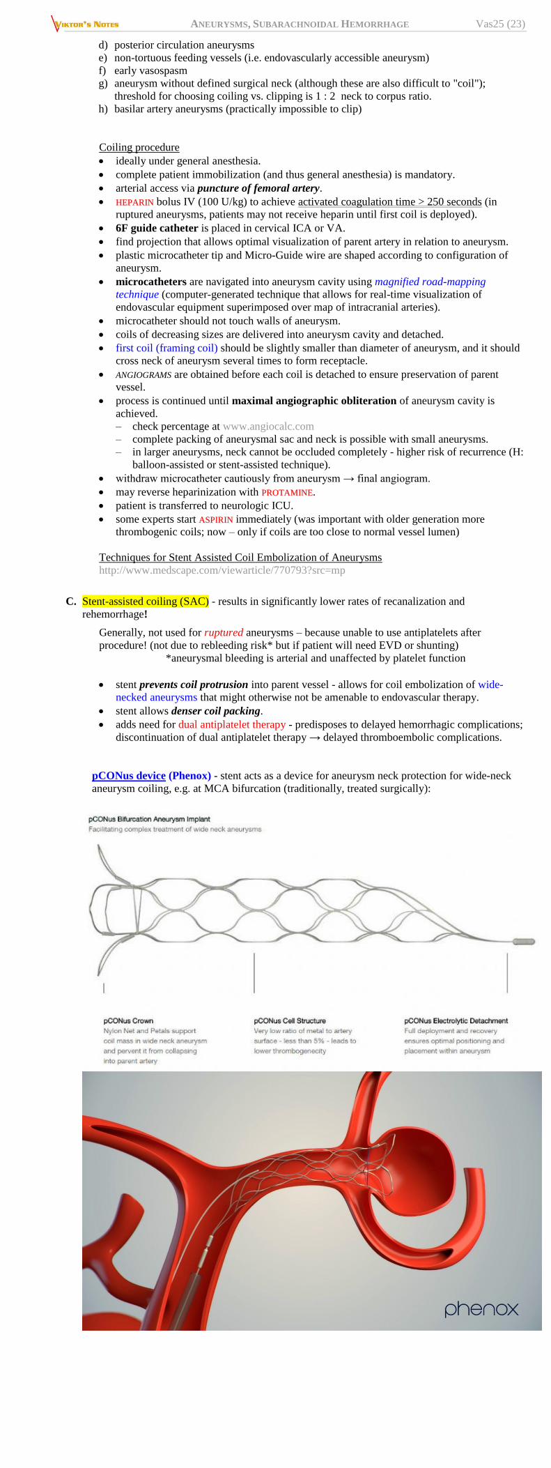
ANEURYSMS, SUBARACHNOIDAL HEMORRHAGE Vas25 (23)
d) posterior circulation aneurysms
e) non-tortuous feeding vessels (i.e. endovascularly accessible aneurysm)
f) early vasospasm
g) aneurysm without defined surgical neck (although these are also difficult to "coil");
threshold for choosing coiling vs. clipping is 1 : 2 neck to corpus ratio.
h) basilar artery aneurysms (practically impossible to clip)
Coiling procedure
ideally under general anesthesia.
complete patient immobilization (and thus general anesthesia) is mandatory.
arterial access via puncture of femoral artery.
HHEEPPAARRIINN bolus IV (100 U/kg) to achieve activated coagulation time > 250 seconds (in
ruptured aneurysms, patients may not receive heparin until first coil is deployed).
6F guide catheter is placed in cervical ICA or VA.
find projection that allows optimal visualization of parent artery in relation to aneurysm.
plastic microcatheter tip and Micro-Guide wire are shaped according to configuration of
aneurysm.
microcatheters are navigated into aneurysm cavity using magnified road-mapping
technique (computer-generated technique that allows for real-time visualization of
endovascular equipment superimposed over map of intracranial arteries).
microcatheter should not touch walls of aneurysm.
coils of decreasing sizes are delivered into aneurysm cavity and detached.
first coil (framing coil) should be slightly smaller than diameter of aneurysm, and it should
cross neck of aneurysm several times to form receptacle.
ANGIOGRAMS are obtained before each coil is detached to ensure preservation of parent
vessel.
process is continued until maximal angiographic obliteration of aneurysm cavity is
achieved.
– check percentage at www.angiocalc.com
– complete packing of aneurysmal sac and neck is possible with small aneurysms.
– in larger aneurysms, neck cannot be occluded completely - higher risk of recurrence (H:
balloon-assisted or stent-assisted technique).
withdraw microcatheter cautiously from aneurysm → final angiogram.
may reverse heparinization with PPRROOTTAAMMIINNEE.
patient is transferred to neurologic ICU.
some experts start AASSPPIIRRIINN immediately (was important with older generation more
thrombogenic coils; now – only if coils are too close to normal vessel lumen)
Techniques for Stent Assisted Coil Embolization of Aneurysms
http://www.medscape.com/viewarticle/770793?src=mp
C. Stent-assisted coiling (SAC) - results in significantly lower rates of recanalization and
rehemorrhage!
Generally, not used for ruptured aneurysms – because unable to use antiplatelets after
procedure! (not due to rebleeding risk* but if patient will need EVD or shunting)
*aneurysmal bleeding is arterial and unaffected by platelet function
stent prevents coil protrusion into parent vessel - allows for coil embolization of wide-
necked aneurysms that might otherwise not be amenable to endovascular therapy.
stent allows denser coil packing.
adds need for dual antiplatelet therapy - predisposes to delayed hemorrhagic complications;
discontinuation of dual antiplatelet therapy → delayed thromboembolic complications.
pCONus device (Phenox) - stent acts as a device for aneurysm neck protection for wide-neck
aneurysm coiling, e.g. at MCA bifurcation (traditionally, treated surgically):
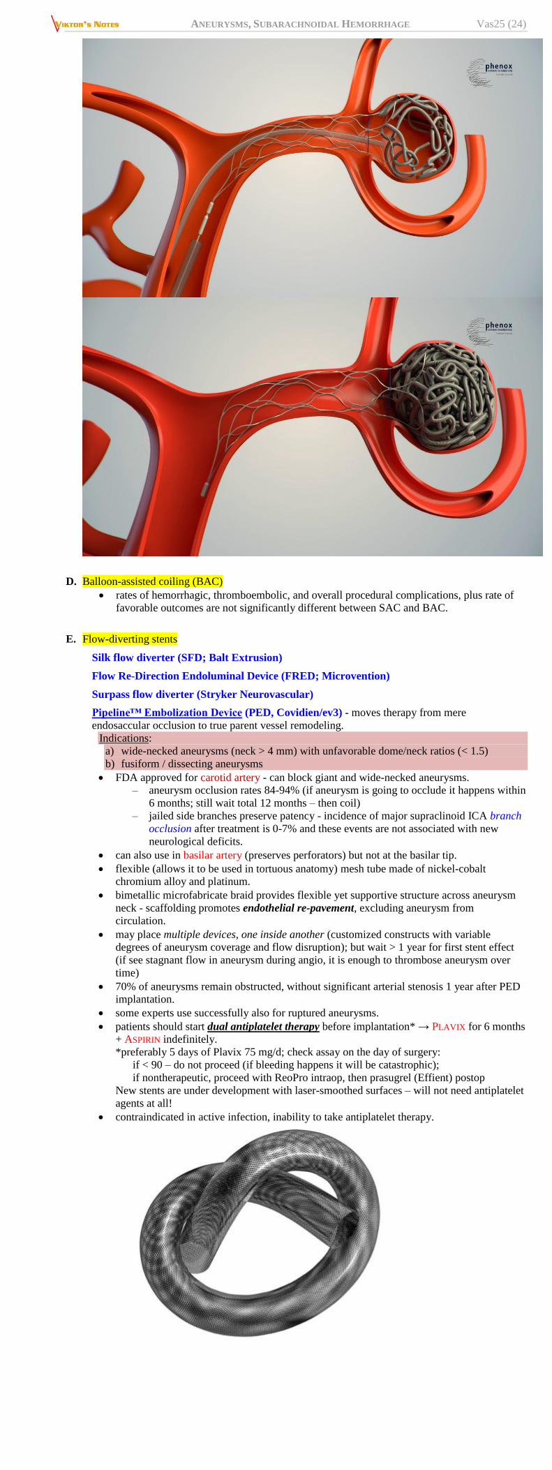
ANEURYSMS, SUBARACHNOIDAL HEMORRHAGE Vas25 (24)
D. Balloon-assisted coiling (BAC)
rates of hemorrhagic, thromboembolic, and overall procedural complications, plus rate of
favorable outcomes are not significantly different between SAC and BAC.
E. Flow-diverting stents
Silk flow diverter (SFD; Balt Extrusion)
Flow Re-Direction Endoluminal Device (FRED; Microvention)
Surpass flow diverter (Stryker Neurovascular)
Pipeline™ Embolization Device (PED, Covidien/ev3) - moves therapy from mere
endosaccular occlusion to true parent vessel remodeling.
Indications:
a) wide-necked aneurysms (neck > 4 mm) with unfavorable dome/neck ratios (< 1.5)
b) fusiform / dissecting aneurysms
FDA approved for carotid artery - can block giant and wide-necked aneurysms.
– aneurysm occlusion rates 84-94% (if aneurysm is going to occlude it happens within
6 months; still wait total 12 months – then coil)
– jailed side branches preserve patency - incidence of major supraclinoid ICA branch
occlusion after treatment is 0-7% and these events are not associated with new
neurological deficits.
can also use in basilar artery (preserves perforators) but not at the basilar tip.
flexible (allows it to be used in tortuous anatomy) mesh tube made of nickel-cobalt
chromium alloy and platinum.
bimetallic microfabricate braid provides flexible yet supportive structure across aneurysm
neck - scaffolding promotes endothelial re-pavement, excluding aneurysm from
circulation.
may place multiple devices, one inside another (customized constructs with variable
degrees of aneurysm coverage and flow disruption); but wait > 1 year for first stent effect
(if see stagnant flow in aneurysm during angio, it is enough to thrombose aneurysm over
time)
70% of aneurysms remain obstructed, without significant arterial stenosis 1 year after PED
implantation.
some experts use successfully also for ruptured aneurysms.
patients should start dual antiplatelet therapy before implantation* → PLAVIX for 6 months
+ ASPIRIN indefinitely.
*preferably 5 days of Plavix 75 mg/d; check assay on the day of surgery:
if < 90 – do not proceed (if bleeding happens it will be catastrophic);
if nontherapeutic, proceed with ReoPro intraop, then prasugrel (Effient) postop
New stents are under development with laser-smoothed surfaces – will not need antiplatelet
agents at all!
contraindicated in active infection, inability to take antiplatelet therapy.
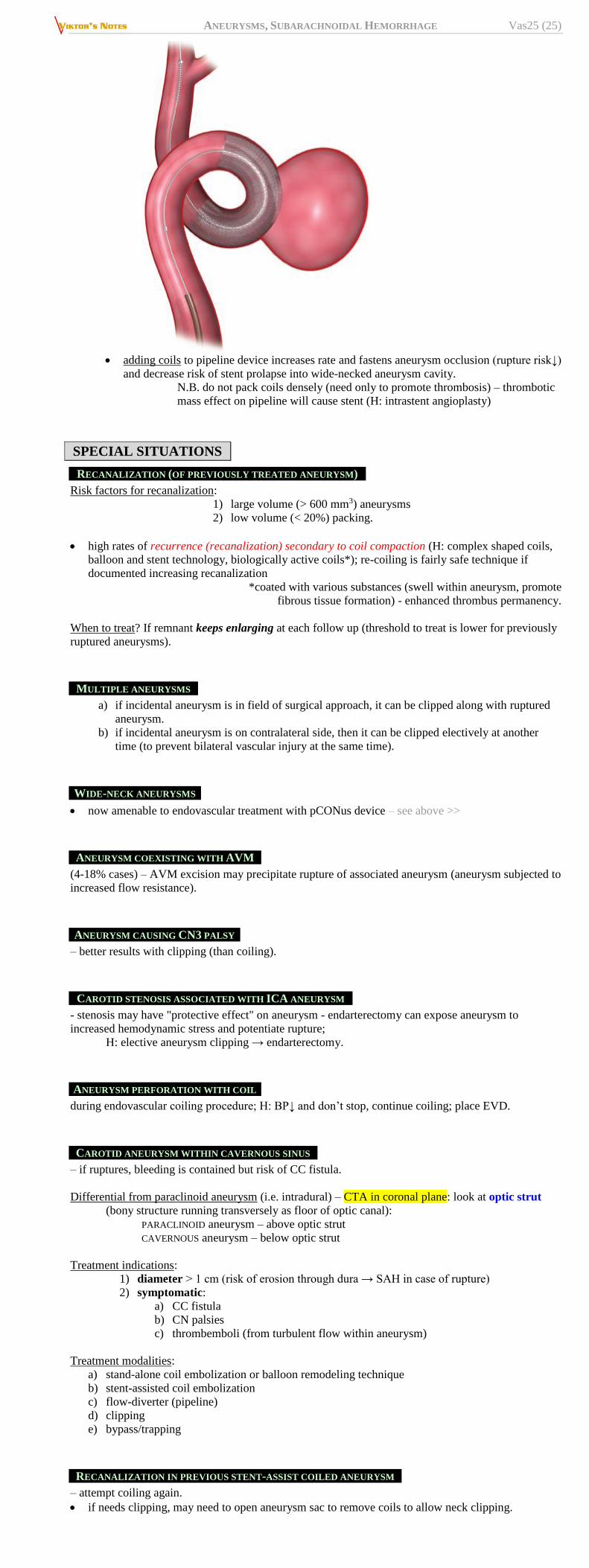
ANEURYSMS, SUBARACHNOIDAL HEMORRHAGE Vas25 (25)
adding coils to pipeline device increases rate and fastens aneurysm occlusion (rupture risk↓)
and decrease risk of stent prolapse into wide-necked aneurysm cavity.
N.B. do not pack coils densely (need only to promote thrombosis) – thrombotic
mass effect on pipeline will cause stent (H: intrastent angioplasty)
SPECIAL SITUATIONS
RECANALIZATION (OF PREVIOUSLY TREATED ANEURYSM)
Risk factors for recanalization:
1) large volume (> 600 mm3) aneurysms
2) low volume (< 20%) packing.
high rates of recurrence (recanalization) secondary to coil compaction (H: complex shaped coils,
balloon and stent technology, biologically active coils*); re-coiling is fairly safe technique if
documented increasing recanalization
*coated with various substances (swell within aneurysm, promote
fibrous tissue formation) - enhanced thrombus permanency.
When to treat? If remnant keeps enlarging at each follow up (threshold to treat is lower for previously
ruptured aneurysms).
MULTIPLE ANEURYSMS
a) if incidental aneurysm is in field of surgical approach, it can be clipped along with ruptured
aneurysm.
b) if incidental aneurysm is on contralateral side, then it can be clipped electively at another
time (to prevent bilateral vascular injury at the same time).
WIDE-NECK ANEURYSMS
now amenable to endovascular treatment with pCONus device – see above >>
ANEURYSM COEXISTING WITH AVM
(4-18% cases) – AVM excision may precipitate rupture of associated aneurysm (aneurysm subjected to
increased flow resistance).
ANEURYSM CAUSING CN3 PALSY
– better results with clipping (than coiling).
CAROTID STENOSIS ASSOCIATED WITH ICA ANEURYSM
- stenosis may have "protective effect" on aneurysm - endarterectomy can expose aneurysm to
increased hemodynamic stress and potentiate rupture;
H: elective aneurysm clipping → endarterectomy.
ANEURYSM PERFORATION WITH COIL
during endovascular coiling procedure; H: BP↓ and don’t stop, continue coiling; place EVD.
CAROTID ANEURYSM WITHIN CAVERNOUS SINUS
– if ruptures, bleeding is contained but risk of CC fistula.
Differential from paraclinoid aneurysm (i.e. intradural) – CTA in coronal plane: look at optic strut
(bony structure running transversely as floor of optic canal):
PARACLINOID aneurysm – above optic strut
CAVERNOUS aneurysm – below optic strut
Treatment indications:
1) diameter > 1 cm (risk of erosion through dura → SAH in case of rupture)
2) symptomatic:
a) CC fistula
b) CN palsies
c) thrombemboli (from turbulent flow within aneurysm)
Treatment modalities:
a) stand-alone coil embolization or balloon remodeling technique
b) stent-assisted coil embolization
c) flow-diverter (pipeline)
d) clipping
e) bypass/trapping
RECANALIZATION IN PREVIOUS STENT-ASSIST COILED ANEURYSM
– attempt coiling again.
if needs clipping, may need to open aneurysm sac to remove coils to allow neck clipping.
ANEURYSMS, SUBARACHNOIDAL HEMORRHAGE Vas25 (26)
CERVICAL ICA ANEURYSMS
– treatment not indicated.
ICA TERMINUS ANEURYSMS
high rate of recurrence after treatment.
ACHA ANEURYSMS
AChA supplies posterior limb of internal capsule and optic radiation.
treatment of AChA aneurysms poses particular challenges - complex anatomy of aneurysm +
relatively small diameter of AChAs.
limitation of vessel manipulation should be used to improve outcomes.
ACOMA ANEURYSMS
aneurysms are known to rupture at nearly any size – risk proportional to size!
ANEURYSM WITH ATHEROMA
even after clipping contrast may go in (neck is too stiff): H: place second reinforcing clip
(piggyback on first clip).
FOLLOW UP
After coiling
1. 1-vessel angiogram - 6 months after coiling (if it was stent-assisted coil embolization, patient may
discontinue PPLLAAVVIIXX, but will remain on AASSPPIIRRIINN for life)
2. 4-vessel angiogram - 18 months (1.5 yrs) + 42 months (3.5 yrs) after coiling.
3. Later – MRA / CTA every 4 years for life
N.B. MRI with gadolinium IV bolus better than CTA or MRA visualizes previously coiled /
clipped aneurysms (lots of metal artifacts on CT).
At some point perform simultaneous angiography and MRA so that in the future, aneurysm
recurrence can be monitored using MRI.
After clipping (there is currently no standard protocol); recurrence risk is 0.26-0.053% / year
2 weeks – clinical
6 weeks – clinical
1 year – angiography
every 4-5 years – CTA
Risk for de novo aneurysm is 0.84-1.8% / year
PROGNOSIS
risk and treatment of recanalization – see above >>
SAH
1/4 of patients with aSAH die, and roughly half of survivors are left with some
persistent neurological deficit.
overall MORTALITY is 45% (range: 32-67%):
10-15% die before reaching medical attention
20-25% die within 24 hours
50% deaths occur within 1 month
prognosis is worse for:
1) women
2) blacks
3) age > 70 yrs
4) smokers – smoking increases risk of aneurysm rupture 3-6 times!
5) posterior circulation aneurysms
6) higher clinical grades
7) aneurysms (vs. AVMs)
causes of death:
1) direct effect of aneurysm rupture (25%)
2) rebleeding (17.6-25%) - mortality ≈ 51-85% !!!
≈ 15-20% aneurysmal SAHs rebleed in first 2 weeks – see above
3) delayed ischemia due to vasospasm (7-32%)
4) neurogenic pulmonary edema
5) neurogenic stunned myocardium
25-50% survivors have moderate to severe neurological deficits.
66% of those who have successful aneurysm clipping never return to the same quality of life as
before SAH.
UNRUPTURED ANEURYSMS
CLIPPING
10.1% of craniotomies result in a complication leading to a modified Rankin Scale score >1 at 12
months.
significant risk factors for complications:
1) age (odds ratio, 1.04; 95% CI, 1.02-1.06)
2) size (odds ratio, 1.12; 95% CI, 1.09-1.15)
3) posterior circulation location (odds ratio, 2.95; 95% CI, 1.82-4.78).
cumulative 10-year risk of retreatment or rupture - 3.0% (95% CI, 1.3-7.0).
BIBLIOGRAPHY for ch. “Neurovascular Disorders” → follow this LINK >>
Viktor’s Notes℠ for the Neurosurgery Resident
Please visit website at www.NeurosurgeryResident.net