and gd nickel-zinc ferrite doped rare earth elements of sc
TRANSCRIPT
Page 1/20
Structure and magnetic properties of coprecipitatednickel-zinc ferrite doped rare earth elements of Sc, Dy,and GdLi Shiwen ( [email protected] )
Guangxi University https://orcid.org/0000-0001-8907-8311Pan Jiatong
Guangxi UniversityGao Feng
Guangxi UniversityZeng Deqian
Guangxi UniversityFeng Qin
Guangxi UniversityHe Chunling
Guangxi UniversityGjergj Dodbiba
University of Tokyo: Tokyo DaigakuYuezou Wei
Guangxi UniversityToyohisa Fujita
Guangxi University https://orcid.org/0000-0002-2697-6867
Research Article
Keywords: Structure, Magnetic property, Nickel zinc ferrite, Rare earth, Coprecipitation
Posted Date: March 23rd, 2021
DOI: https://doi.org/10.21203/rs.3.rs-277352/v1
License: This work is licensed under a Creative Commons Attribution 4.0 International License. Read FullLicense
Version of Record: A version of this preprint was published at Journal of Materials Science: Materials in Electronicson April 20th, 2021. See the published version at https://doi.org/10.1007/s10854-021-05928-0.

Page 2/20
AbstractThis research is the basic study of temperature-sensitive ferrite characteristics prepared by coprecipitation withdoping different typical sizes of rare earth elements. Ni 0.5 Zn 0.5 Re x Fe 2-x O 4 (NZRF) (X = 0.02, 0.05, 0.07 and0.09) nanoparticles (NPs) doped by Sc, Dy and Gd prepared by chemical coprecipitation method. The structure andproperties of Ni 0.5 Zn 0.5 Re x Fe 2-x O 4 were analyzed by various characterization methods. XRD results showthat the grain size of Ni 0.5 Zn 0.5 Re x Fe 2-x O 4 is from 10.6 to 12.4 nm, which is close to the average grain sizeof 13.9 nm observed on TEM images. It is also found that the ferrite particles are spherical and slightlyagglomerated in TEM images. FTIR measurements between 400 and 4000 cm -1 have con�rmed the intrinsiccation vibration of the spinel structure. The concentrations of nickel, zinc, iron, and rare earth elements have beendetermined by ICP-AES, and all ions have participated in the reaction. The magnetic properties of Sc, Dy, and Gd 3+doped NZRF NPs at room temperature are recorded by a physical property measurement system (PPMS-9). It isfound that the magnetization can be changed by adding rare-earth ions. When X = 0.07, Gd 3+ doped Ni 0.5 Zn 0.5Fe 2 O 4 (NZF) exhibits the highest saturation magnetization. The magnetic properties of NZGd 0.07 vary the mostwith temperature. The thermomagnetic coe�cient of NZGd 0.07 nanoparticles stabilized to 0.18 emu/gK at 0-100℃. Hence, NZGd 0.07 with low Curie temperature and the high thermomagnetic coe�cient can be used toprepare temperature-sensitive ferro�uid. All the samples exhibit very small coercivity and almost zero remanences,which indicates the superparamagnetism of the synthesized nanoparticles.
1. IntroductionIn recent years, due to its unique properties, nano ferrite has been widely used in many technical �elds such asmicrowave devices, biosensors, catalysis, biomedicine and magnetic recording, magnetic drug delivery, and soon [1]. Ferrite is a composite oxide mainly composed of iron oxide and other iron or rare-earth group oxides. Thechemical formula of spinel ferrite is generally represented by MeFe2O4, and Me is usually divalent metal ions, such
as Mg2+, Co2+, Ni2+, Mn2+, Cu2+, Cd2+, Fe2+, etc. [2]. The magnetic property of zinc ferrite and nickel-zinc ferrite aretypical soft ferrite. NZF is a kind of functional material widely used in modern communication, the Internet,electrical appliances, biomedical, computer circuit, and anti-electromagnetic interference technology [3-6]. N.Boda [7] studied the low-temperature properties of Sm, Er substituted nickel ferrite and cobalt ferrite, and foundthat the synthesized nickel ferrite showed superparamagnetic behavior, while cobalt ferrite showed soft magneticbehavior. Therefore, this kind of material is expected to be used in the soft magnetic manufacturing industry. P. H.Nam [8] studied the in�uence of dipole interaction between particles on the heating e�ciency of cobalt zinc ferriteunder an alternating magnetic �eld, and found that Co0.5Zn0.5Fe2O4 nano ferrite has become a potential materialfor thermotherapy. N. Kaur [9] has synthesized Mn-Zn ferrite NPs with good magnetism and adjustable Curietemperature, and it is considered that Mn-Zn ferrite can be used as the stable temperature-sensitive ferro�uid(magnetic �uid) for heat exchange devices.
The magnetism of ferrite is formed by the direct electron spin exchange between adjacent magnetic atoms. Thestructural formula of spinel ferrite can be written as [Znx
2+Fe1-x3+]A[Me1-x
2+Fe1+x3+]BO4, and the subscripts are
tetrahedron (A) and octahedron (B). As shown in Fig. 1, the cell of spinel consists of 8 molecules, including 8divalent metals, 16 of 3-valent metals, and 32 oxygen. O2- is connected with B site and A site sublattices to form aferrite face-centered cubic structure[10]. The distance between the two magnetic ions is relatively far, and there areoxygen ions in the middle. Due to the existence of oxygen ions, the ferromagnetic electron spin exchange isformed. This type of exchange is called super exchange in ferromagnetic theory. Because of the super exchange,

Page 3/20
the magnetic moments of the magnetic ions on both sides of the oxygen ions are arranged in the oppositedirection. The antiferromagnetism of many metal oxides comes from this structure. If the magnetic momentsarranged in the opposite direction are not equal and there is a residual magnetic moment, the magnetic property iscalled ferrimagnetism, or ferrite magnetism.
The magnetic, electrical, catalytic, and optical properties of ferrite depend on the distribution of divalent andtrivalent ions at A and B sites [11]. In addition, these properties also vary with the geometry and size ofnanoparticles. Most of the Me2+ ions occupy the octahedral position, while Zn2+ ions tend to stabilize in thetetrahedral position. When x = 0, it is inverse spinel structure, in which Fe3+ accounts for half in A and B, such asFe3O4 and NiFe2O4; when x = 1, it is orthospinel structure, such as ZnFe2O4; when 0 < x < 1, the spinel structure ismixed, such as MnFe2O4 and MgFe2O4 [12]. The quality of ferrite can be improved by adding non-magnetic/diamagnetic ions with the same appropriate valence state at points A and point B. Lanthanide (Ce - Y)as a dopant in ferrite is particularly noteworthy [13].
The ionic radius of rare-earth ions (RE3+) (0.086 - 0.106 nm) is much larger than that of Fe3+ ions (0.064 nm). Inspinel structure, the space of A site is smaller than that of B site (rA = 0.03 nm, rB = 0.055 nm); therefore, rare earthions tend to occupy B site. R.K. Singh [14] studied the properties of Ni-Zn ferrite doped with La, Pr, and Sm by citricacid precursor method. The results showed that the size of ferrite doped with rare earth decreased, the saturationmagnetization and coercivity decreased. However, only a small concentration of rare-earth ions can enter the spinellattice because their ionic radius is larger than that of Fe3+. The high concentration of rare-earth ions can causestructural disorder and form an impurity phase at the grain boundary. S.E. Jacobo [15] synthesized Gd substitutednickel ferrite by combustion method. It was found that only a small part of Gd3+ entered the lattice, and thesaturation magnetization increased slightly. It is well known that the magnetic behavior of ferromagnetic oxides islargely controlled by the iron-iron interaction (spin coupling of 3d electrons). When RE3+ replaces Fe3+ ions in spinelferrite, RE3+ interact with Fe3+ to form 3d-4f electron spin coupling. The magnetic and electrical properties of ferritecan be improved by the strong interaction between RE3+ and Fe3+ (3d-4f coupling) [16]. R.A. Pawar [17] studied theproperties of Cobalt zinc ferrite doped with Gd. Due to the strong exchange between Gd (4f7) and Fe (3d5), thesaturation magnetization and coercivity of cobalt zinc ferrite were improved.
The magnetic behavior of spinel ferrite is strongly affected by many factors, including the change of crystal size,magnetic moment (nB), preferred site occupation of different ions, and local strain. In addition, disordered cationdistribution and super exchange interaction between different ions also affect magnetic properties [18]. Thedistribution of metal cations at A and B sites depends on the sample preparation method, particle size, type, andthe number of cations involved. Common synthetic methods include co-precipitation [19], sol-gel [20], citric acidprecursor [21], Microemulsion [22] and solid-state chemical reaction method [23]. Among these methods,coprecipitation is one of the methods for preparing ferrite, which can be used in industry at present [24]. Thepreparation process is to make a solution of a certain proportion of metal salts in accordance with the ratio andthen add appropriate precipitators (such as OH-, CO3
2-, C2O42-, etc.) to precipitate metal ions out [25]. The chemical
coprecipitation method can easily control the particle size of prepared NPs, and its operation is simple, and itscrystallinity is high [26]. Nanoscale particles can be obtained by controlling the mixing speed, stirring time, workingtemperature, concentration of precursor, and pH value of the reaction mixture. T.J. Shinde [27] et al. studied theproperties of Nd-substituted zinc ferrite and found that the average particle size of ferrite decreased with the

Page 4/20
increase of Nd3+ content. The room temperature resistivity of zinc ferrite substituted by Nd3+ is 102 times that ofzinc ferrite.
In this paper, in order to meet their application in targeted ferro�uid heat exchanger and drug delivery at near roomtemperature, magnetic ferromagnetic materials need to have a smaller size, appropriate susceptibility andcoercivity, and high-temperature sensitivity, so that the ferro�uid can be used as a hyperthermia medium in analternating magnetic �eld to have a better thermal effect. Sc, Gd, and Dy rare earth doped nickel-zinc ferrites weresynthesized by and coprecipitation method. The morphology and magnetic properties of rare earth doped NZFwere studied with temperature. So that ferrite materials have a wider range of applications.
2. Preparation And Characterization2.1 Synthesis of ferrite nanoparticles
Nickel chloride (NiCl2), ferric chloride hexahydrate (FeCl3·6H2O), zinc chloride (ZnCl2), sodium hydroxide (NaOH),rare earth chloride, and anhydrous ethanol are purchased from Guoyao Chemical Reagent Co., Ltd. All chemicalsare analytical grade, and no further puri�cation is required. Deionized water is used throughout the experiment.FeCl3·6H2O (3 M), NiCl2 (3 M), ZnCl2 (3 M), ScCl3 (0.3 M) solutions were diluted in 100 ml deionized wateraccording to the appropriate proportion (stoichiometric ratio), and then mixed and stirred for 30 minutes to obtaina homogeneous solution. The mixed solution was placed in a magnetic stirrer, heated to 80 ℃, and continuouslystirred. 3 M sodium hydroxide was added to the salt solution mixture drop by drop. The pH was adjusted to 10 - 11.This is due to that non-magnetic and paramagnetic particles will form simultaneously at a pH higher than 13. Thereaction was then promoted by stirring for 2 hours and ensuring the complete formation of spinel ferrite [26]. Themixed solution gradually turned brown and precipitated. Thoroughly clean the precipitated ferrite particles withdeionized water several times to remove any impurities present in the particles or solvents and reduce the pH to 7.The slurry was obtained by suction �ltration and dried in a drying oven for one day and one night. After it wastaken out and the powder was ground to use performance study.
2.2 Measurement of structure and magnetic property for ferrite particles
The nanoparticles’ crystal structures were investigated by X-ray diffraction (XRD). The composition and structureof the samples were analyzed by Cu-Kα1 (λ = 0.154 nm) at room temperature by RIGAKUD/max 2500 V X-raydiffractometer. The voltage of the X-ray tube is 40 kV; the current of the X-ray tube is 150 mA; continuous scanning.Scanning speed: 10 °/min; scanning angle: 20° - 90°. The chemical components of prepared ferrites were analyzedwith coupled plasma atomic emission spectrometry (ICP-AES). The intrinsic vibrations of metal complexes weremeasured by Fourier transform infrared spectroscopy (FT-IR). The Fourier transform infrared spectrometer(IRTRACer-100) performs scanning analysis in 400 - 4000 wavelengths. The surface morphology and particle sizeof the doped ferrite samples were analyzed by transmission electron microscope (TEM, FEI Tecnai G2 F20). Theelemental analysis of particles was measured by energy dispersive spectroscopy (EDS).
Magnetic analysis of the sample was carried out using a physical property measurement system (PPMS-9)vibrating sample Magnetometer (VSM). In addition, the temperature sensitivity of the samples was measuredunder the magnetic �eld of 2 T in the range of 225 - 400 K.
3. Results And Discussion
Page 5/20
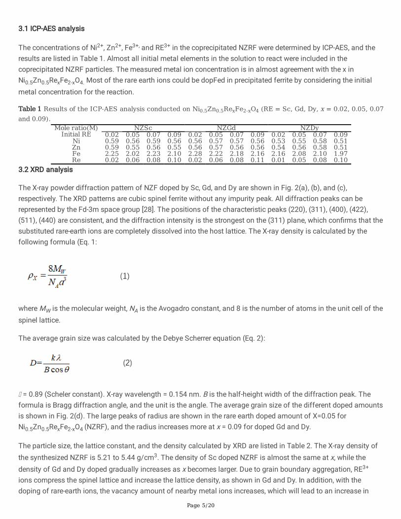
3.1 ICP-AES analysis
The concentrations of Ni2+, Zn2+, Fe3+, and RE3+ in the coprecipitated NZRF were determined by ICP-AES, and theresults are listed in Table 1. Almost all initial metal elements in the solution to react were included in thecoprecipitated NZRF particles. The measured metal ion concentration is in almost agreement with the x inNi0.5Zn0.5RexFe2-xO4. Most of the rare earth ions could be dopFed in precipitated ferrite by considering the initialmetal concentration for the reaction.
Table 1 Results of the ICP-AES analysis conducted on Ni0.5Zn0.5RexFe2-xO4 (RE = Sc, Gd, Dy, x = 0.02, 0.05, 0.07and 0.09).
Mole ratio(M) NZSc NZGd NZDyInitial RE 0.02 0.05 0.07 0.09 0.02 0.05 0.07 0.09 0.02 0.05 0.07 0.09
Ni 0.59 0.56 0.59 0.56 0.56 0.57 0.57 0.56 0.53 0.55 0.58 0.51Zn 0.59 0.55 0.56 0.55 0.56 0.57 0.56 0.56 0.54 0.56 0.58 0.51Fe 2.25 2.02 2.23 2.10 2.28 2.22 2.18 2.16 2.16 2.08 2.10 1.97Re 0.02 0.06 0.08 0.10 0.02 0.06 0.08 0.11 0.01 0.05 0.08 0.10
3.2 XRD analysis
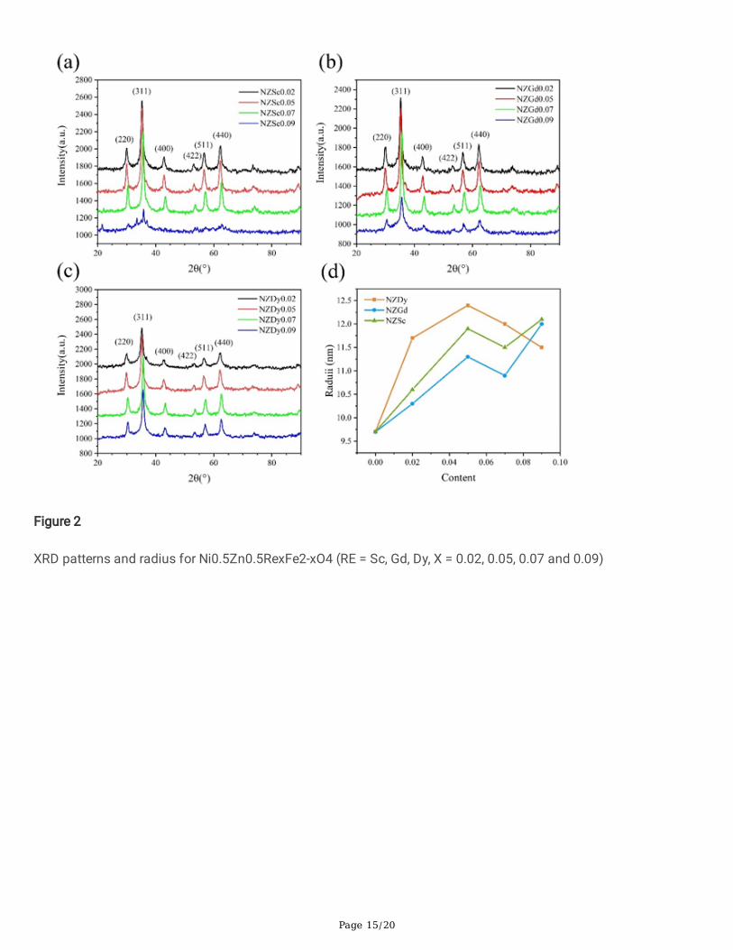
The X-ray powder diffraction pattern of NZF doped by Sc, Gd, and Dy are shown in Fig. 2(a), (b), and (c),respectively. The XRD patterns are cubic spinel ferrite without any impurity peak. All diffraction peaks can berepresented by the Fd-3m space group [28]. The positions of the characteristic peaks (220), (311), (400), (422),(511), (440) are consistent, and the diffraction intensity is the strongest on the (311) plane, which con�rms that thesubstituted rare-earth ions are completely dissolved into the host lattice. The X-ray density is calculated by thefollowing formula (Eq. 1:
where MW is the molecular weight, NA is the Avogadro constant, and 8 is the number of atoms in the unit cell of thespinel lattice.
The average grain size was calculated by the Debye Scherrer equation (Eq. 2):
= 0.89 (Scheler constant). X-ray wavelength = 0.154 nm. B is the half-height width of the diffraction peak. Theformula is Bragg diffraction angle, and the unit is the angle. The average grain size of the different doped amountsis shown in Fig. 2(d). The large peaks of radius are shown in the rare earth doped amount of X=0.05 forNi0.5Zn0.5RexFe2-xO4 (NZRF), and the radius increases more at x = 0.09 for doped Gd and Dy.
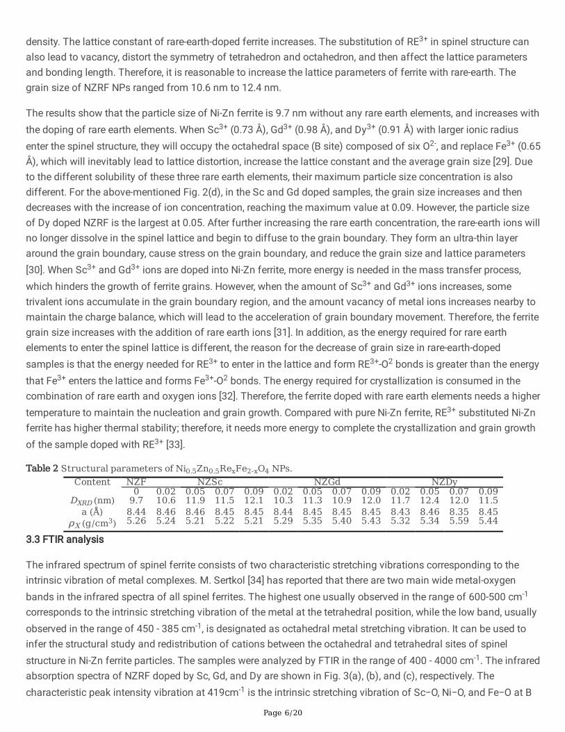
The particle size, the lattice constant, and the density calculated by XRD are listed in Table 2. The X-ray density ofthe synthesized NZRF is 5.21 to 5.44 g/cm3. The density of Sc doped NZRF is almost the same at x, while thedensity of Gd and Dy doped gradually increases as x becomes larger. Due to grain boundary aggregation, RE3+
ions compress the spinel lattice and increase the lattice density, as shown in Gd and Dy. In addition, with thedoping of rare-earth ions, the vacancy amount of nearby metal ions increases, which will lead to an increase in
Page 6/20
density. The lattice constant of rare-earth-doped ferrite increases. The substitution of RE3+ in spinel structure canalso lead to vacancy, distort the symmetry of tetrahedron and octahedron, and then affect the lattice parametersand bonding length. Therefore, it is reasonable to increase the lattice parameters of ferrite with rare-earth. Thegrain size of NZRF NPs ranged from 10.6 nm to 12.4 nm.
The results show that the particle size of Ni-Zn ferrite is 9.7 nm without any rare earth elements, and increases withthe doping of rare earth elements. When Sc3+ (0.73 Å), Gd3+ (0.98 Å), and Dy3+ (0.91 Å) with larger ionic radiusenter the spinel structure, they will occupy the octahedral space (B site) composed of six O2-, and replace Fe3+ (0.65Å), which will inevitably lead to lattice distortion, increase the lattice constant and the average grain size [29]. Dueto the different solubility of these three rare earth elements, their maximum particle size concentration is alsodifferent. For the above-mentioned Fig. 2(d), in the Sc and Gd doped samples, the grain size increases and thendecreases with the increase of ion concentration, reaching the maximum value at 0.09. However, the particle sizeof Dy doped NZRF is the largest at 0.05. After further increasing the rare earth concentration, the rare-earth ions willno longer dissolve in the spinel lattice and begin to diffuse to the grain boundary. They form an ultra-thin layeraround the grain boundary, cause stress on the grain boundary, and reduce the grain size and lattice parameters[30]. When Sc3+ and Gd3+ ions are doped into Ni-Zn ferrite, more energy is needed in the mass transfer process,which hinders the growth of ferrite grains. However, when the amount of Sc3+ and Gd3+ ions increases, sometrivalent ions accumulate in the grain boundary region, and the amount vacancy of metal ions increases nearby tomaintain the charge balance, which will lead to the acceleration of grain boundary movement. Therefore, the ferritegrain size increases with the addition of rare earth ions [31]. In addition, as the energy required for rare earthelements to enter the spinel lattice is different, the reason for the decrease of grain size in rare-earth-dopedsamples is that the energy needed for RE3+ to enter in the lattice and form RE3+-O2 bonds is greater than the energythat Fe3+ enters the lattice and forms Fe3+-O2 bonds. The energy required for crystallization is consumed in thecombination of rare earth and oxygen ions [32]. Therefore, the ferrite doped with rare earth elements needs a highertemperature to maintain the nucleation and grain growth. Compared with pure Ni-Zn ferrite, RE3+ substituted Ni-Znferrite has higher thermal stability; therefore, it needs more energy to complete the crystallization and grain growthof the sample doped with RE3+ [33].
Table 2 Structural parameters of Ni0.5Zn0.5RexFe2-xO4 NPs.Content NZF NZSc NZGd NZDy
0 0.02 0.05 0.07 0.09 0.02 0.05 0.07 0.09 0.02 0.05 0.07 0.09DXRD (nm) 9.7 10.6 11.9 11.5 12.1 10.3 11.3 10.9 12.0 11.7 12.4 12.0 11.5
a (Å) 8.44 8.46 8.46 8.45 8.45 8.44 8.45 8.45 8.45 8.43 8.46 8.35 8.45ρX (g/cm3) 5.26 5.24 5.21 5.22 5.21 5.29 5.35 5.40 5.43 5.32 5.34 5.59 5.44
3.3 FTIR analysis
The infrared spectrum of spinel ferrite consists of two characteristic stretching vibrations corresponding to theintrinsic vibration of metal complexes. M. Sertkol [34] has reported that there are two main wide metal-oxygenbands in the infrared spectra of all spinel ferrites. The highest one usually observed in the range of 600-500 cm-1
corresponds to the intrinsic stretching vibration of the metal at the tetrahedral position, while the low band, usuallyobserved in the range of 450 - 385 cm-1, is designated as octahedral metal stretching vibration. It can be used toinfer the structural study and redistribution of cations between the octahedral and tetrahedral sites of spinelstructure in Ni-Zn ferrite particles. The samples were analyzed by FTIR in the range of 400 - 4000 cm-1. The infraredabsorption spectra of NZRF doped by Sc, Gd, and Dy are shown in Fig. 3(a), (b), and (c), respectively. Thecharacteristic peak intensity vibration at 419cm-1 is the intrinsic stretching vibration of Sc−O, Ni−O, and Fe−O at B

Page 7/20
site (octahedron) in ferrite, while the characteristic peak at 570 cm-1 corresponds to the position of Fe−O and Zn−Oat A site (tetrahedron), therefore spinel structure can be considered in the prepared samples [35]. The peakspectrum at 1620 cm-1 is −OH, which is caused by the adsorption of −OH on the surface of nanoparticles by thecoprecipitation method. The broadband absorption peak at about 3400 cm-1 can be attributed to the presence ofthe −OH group in the sample due to the symmetrical and antisymmetric stretching of water. In addition, the peakvalue at 2378 cm-1 is H−O−H bending vibration caused by free or absorbed water. Bands related to the structure ofmetal chloride were observed at 1479 and 1365 cm-1. In the case of rare-earth-doped ferrite, the intensity of thesetwo absorption peaks is related to their ability to form surface chloride structure [36].
3.4 TEM analysis
The grain nucleation and crystal growth of ferrite are changed by doping RE3+. When the doping amount reaches acertain amount, the ferrite grain morphology will be destroyed. The surface morphology and particle size of Gd3+
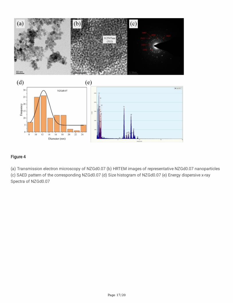
doped Ni-Zn ferrite samples were analyzed by TEM (FEI TECNAI G2 F20). The photograph of particles is shown inFig. 4(a), HRTEM images of representative NZGd0.07 nanoparticles, and SAED pattern of the correspondingNZGd0.07 are shown in Fig. 4(b) and (c), respectively. In Fig. 4(b), the measured width of the lattice fringes is 0.255nm, which corresponds to the spacing of the (311) plane shown in XRD. The observed microcrystalline latticefringes con�rm the high crystallinity of nanoparticles in ferrite samples.
In Fig. 4(c), six of them correspond to (200), (311), (400), (422), (511), and (440) planes in ferrite crystals. Thearrangement order of the diffraction ring is consistent with that observed in XRD data, so the phase purity of theferrite sample is determined by plane calibration. Similarly, the ring designated in the SAED mode indicates thepolycrystalline nature of the sample.
The particle size distribution calculated by the software is shown in Fig. 4(d). It can be observed from Fig. 4(a) thatthe particles are cubic and spherical, with sizes ranging from 8 nm to 25 nm, with slight to moderateagglomeration. The distribution size of nanoparticles was expressed by the histogram, and the average particlesize was 13.93 nm, which was slightly larger than the previous XRD calculation results. Meanwhile, it also veri�edthat the samples had an agglomeration, so the observed particle size was larger than the average grain sizecalculated by Scherer's formula. EDS of NZGd0.07 detected Ni, Zn, Fe, and Gd at the same particle area as shown inFig. 4(e), no other impurities were found.
3.5 Magnetic analysis
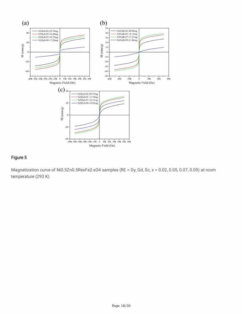
Magnetization (Mr), remanent magnetization (Mr), and coercive force (Hc) obtained by the hysteresis loops (M-H)of Sc, Gd, Dy doped Ni-Zn ferrites at room temperature (293 K) are shown in Fig. 6(a), (b) and (c), respectively. Inthe magnetic �eld range of ± 0.5 T, a PPMS-9 VSM was used to measure the magnetization at room temperature.Synthesized ferrites particles exhibit values of MS varying in the ranges of 12.2-36.1 emu/g, 12.4 - 41.4 emu/g and21.2 - 33.6 emu/g for Ni0.5Zn0.5ScxFe2-xO4, Ni0.5Zn0.5GdxFe2-xO4 and Ni0.5Zn0.5DyxFe2-xO4 , respectively. The valuesof remanent magnetization (Mr) are in the intervals of 0.03 - 0.72 emu/g, 0.01 - 0.78 emu/g, and 0.05 - 0.47 emu/gfor Sc, Gd, and Dy substituted NZF, respectively.
MS was obtained by M and 1/H2 curve. The saturation magnetization (MS), residual magnetization (Mr), coercivity(HC), Mr/MS, and magnetic moment of all samples are listed in Table 3. It can be found that the saturationmagnetization is improved and large after doping rare earth elements. This result is consistent with that of Ce-
Page 8/20
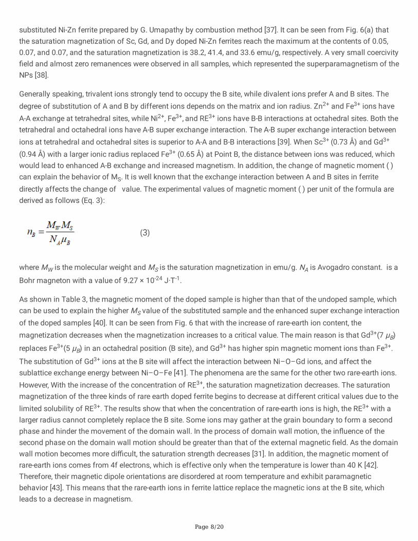
substituted Ni-Zn ferrite prepared by G. Umapathy by combustion method [37]. It can be seen from Fig. 6(a) thatthe saturation magnetization of Sc, Gd, and Dy doped Ni-Zn ferrites reach the maximum at the contents of 0.05,0.07, and 0.07, and the saturation magnetization is 38.2, 41.4, and 33.6 emu/g, respectively. A very small coercivity�eld and almost zero remanences were observed in all samples, which represented the superparamagnetism of theNPs [38].
Generally speaking, trivalent ions strongly tend to occupy the B site, while divalent ions prefer A and B sites. Thedegree of substitution of A and B by different ions depends on the matrix and ion radius. Zn2+ and Fe3+ ions haveA-A exchange at tetrahedral sites, while Ni2+, Fe3+, and RE3+ ions have B-B interactions at octahedral sites. Both thetetrahedral and octahedral ions have A-B super exchange interaction. The A-B super exchange interaction betweenions at tetrahedral and octahedral sites is superior to A-A and B-B interactions [39]. When Sc3+ (0.73 Å) and Gd3+
(0.94 Å) with a larger ionic radius replaced Fe3+ (0.65 Å) at Point B, the distance between ions was reduced, whichwould lead to enhanced A-B exchange and increased magnetism. In addition, the change of magnetic moment ( )can explain the behavior of MS. It is well known that the exchange interaction between A and B sites in ferritedirectly affects the change of value. The experimental values of magnetic moment ( ) per unit of the formula arederived as follows (Eq. 3):
where MW is the molecular weight and MS is the saturation magnetization in emu/g. NA is Avogadro constant. is a
Bohr magneton with a value of 9.27 × 10⁻²⁴ J·T-1.
As shown in Table 3, the magnetic moment of the doped sample is higher than that of the undoped sample, whichcan be used to explain the higher MS value of the substituted sample and the enhanced super exchange interactionof the doped samples [40]. It can be seen from Fig. 6 that with the increase of rare-earth ion content, themagnetization decreases when the magnetization increases to a critical value. The main reason is that Gd3+(7 μB)
replaces Fe3+(5 μB) in an octahedral position (B site), and Gd3+ has higher spin magnetic moment ions than Fe3+.
The substitution of Gd3+ ions at the B site will affect the interaction between Ni–O–Gd ions, and affect thesublattice exchange energy between Ni–O–Fe [41]. The phenomena are the same for the other two rare-earth ions.However, With the increase of the concentration of RE3+, the saturation magnetization decreases. The saturationmagnetization of the three kinds of rare earth doped ferrite begins to decrease at different critical values due to thelimited solubility of RE3+. The results show that when the concentration of rare-earth ions is high, the RE3+ with alarger radius cannot completely replace the B site. Some ions may gather at the grain boundary to form a secondphase and hinder the movement of the domain wall. In the process of domain wall motion, the in�uence of thesecond phase on the domain wall motion should be greater than that of the external magnetic �eld. As the domainwall motion becomes more di�cult, the saturation strength decreases [31]. In addition, the magnetic moment ofrare-earth ions comes from 4f electrons, which is effective only when the temperature is lower than 40 K [42].Therefore, their magnetic dipole orientations are disordered at room temperature and exhibit paramagneticbehavior [43]. This means that the rare-earth ions in ferrite lattice replace the magnetic ions at the B site, whichleads to a decrease in magnetism.

Page 9/20
The coercivity of ferrite is a microstructure property, which is affected by strain, defect, porosity, and magneto-crystalline anisotropy [43]. The anisotropy constant K1 depends on the rare earth ion content x. According to theStoner Wohlfarth model, the coercivity and anisotropy constant K1 are connected by the following relationship (Eq.4) [44].
μ0 is the vacuum permeability. According to Stoner Wohlfarth's theory, coercivity increases with the decrease ofmagnetization. In accordance with the conclusion obtained in this paper, the relationship between HC and MS isinversely proportional. It can be seen from the �gure that the coercivity of ferrite doped with rare earth elementsincreases because RE3+ have a large single-ion anisotropy. When rare earth ions partially replace Fe3+ ions, themagnetic anisotropy and the coercivity become greater. With the increase of rare-earth doping content, thecoercivity �rst decreases and then increases. On the one hand, with the increase of grain size, larger grain sizeoften contains more domain walls. When the magnetic moment and magnetic anisotropy of a single particledecrease, a stronger magnetic �eld is needed for magnetization reversal. This behavior continues until the criticalsize becomes a single domain, beyond which the single domain particle will split into multiple domains [41]. In aweak external magnetic �eld, the domain wall motion and magnetization rotation will lead to the magnetizationprocess of multi-domain samples. This will reduce coercivity.
The ratio Mr/Ms is called squareness ratio (SQR), which determines the magnetic hardness of materials and theexistence of group exchange between grains [45]. It is reported that when 0.05 < Mr / Ms < 0.5, the sample has asingle domain magnetostatic coupling [46]. The squareness ratio of NZRF spinel ferrite is calculated, and theresults have been shown in Table 3. The squareness ratio ranges from 0.001 to 0.019, indicating the multi-domainstructure of the particles in which domain wall movement allows for an easier change in orientation with theapplied �eld.
The variation of magnetization with temperature in the range of 225 - 400 K was recorded at an applied magnetic�eld of 2 T. The magnetization versus temperature of NZF doped by Sc, Gd, and Dy are shown in Fig. 7(a), (b), and(c), respectively. In the rare earth doped ferrites, most magnetization change versus temperature shows in NZSc0.05,NZGd0.07, and NZDy0.07 for each rare earth doping. According to the dependence of saturation magnetization ontemperature, Curie temperature of Ni-Zn ferrite has been roughly estimated at Tc = 425 K. It can be seen from Fig. 7that with the increase of temperature, the decrease of magnetization is due to the disorder of the magnetic spinphase caused by the increase of heat energy [47]. Thermomagnetic coe�cient KT (KT = dM/dT) is calculated fromthe �rst derivative of the temperature-dependent magnetization curves of each component. As can be seen fromFig. 7(d), when the temperature is close to the Curie temperature, the thermomagnetic coe�cient KT decreasesrapidly with the temperature increase. The magnetic properties of NZGd0.07 vary the most with temperature, andthe thermomagnetic coe�cient KT increases from 0.13 emu/gK to 0.24 emu/gK. The magnetic properties ofNZGd0.07 are more varied than Dy doped cobalt-zinc ferrite obtained by S. Urcia-Romer [48]. The stability offerro�uid at operating temperature and the vapor pressure of carrying liquid determine the practical application offerro�uid. As can be seen from Fig. 7(d), the thermomagnetic coe�cient of NZGd0.07 nanoparticles stabilized to0.18 emu/gK at 0 - 100 ℃. As a result, ferro�uid can operate at temperatures around 0 - 100 ℃ without losing

Page 10/20
much stability and can be used for energy conversion applications. In addition, when ferro�uid is used to treatcancer, it quickly reaches the speci�ed temperature and remains stable without harming human tissues.
Table 3 Saturation magnetization (MS), remanence (Mr), coercivity (Hc) of nickel-zinc ferrite doped with differentconcentrations of different rare earth elements at room temperature.
Content NZF NZSc NZGd NZDy0 0.02 0.05 0.07 0.09 0.02 0.05 0.07 0.09 0.02 0.05 0.07 0.09
Ms(emu/g) 27.4 31.1 38.2 34.6 12.2 29.9 38.6 41.4 15.0 21.2 31.7 33.6 26.3Mr(emu/g) 0.33 0.36 0.72 0.70 0.03 0.35 0.74 0.78 0.01 0.05 0.42 0.47 0.29
Hc(Oe) 14.73 17.02 15.47 15.26 16.00 16.34 15.22 15.14 15.39 16.23 15.52 15.32 15.40Mr/Ms 0.012 0.011 0.019 0.020 0.002 0.012 0.019 0.019 0.001 0.002 0.013 0.014 0.011nB(μB) 1.17 1.33 1.62 1.47 0.52 1.28 1.68 1.82 0.53 0.91 1.38 1.48 1.17
4. ConclusionDy, Gd, and Sc doped Ni-Zn ferrites were successfully prepared by the coprecipitation method. The effects ofdoping Dy3+, Gd3+, Sc3+ ions on the structure and properties of Ni-Zn ferrite were analyzed in detail. Rare-earthelement doping plays an important role in changing the structure and magnetic properties of Ni-Zn ferrite. XRDanalysis con�rmed that the samples were cubic spinel structure, single-phase. Rare earth doping increases thegrain size of ferrite. The grain size of ferrite without doping is 9.7 nm, while that of rare-earth-doped ferrite is from10.6 to 12.4 nm. The grain size of doped ferrite increases and shows the �rst peak at 0.05 RE content. At the sametime, it was observed by TEM that the Ni-Zn ferrite particles' average grain size was 13.93 nm, which was similar tothe XRD results. The intrinsic stretching vibration of B site (octahedral) RE−O, Ni−O, and Fe−O and the intrinsicstretching vibration of Fe−O and Zn−O at A site tetrahedron) were observed at 419 cm-1 and 570 cm-1, respectively,which con�rmed the formation of metal oxides. The magnetization results showed that the MS of Ni-Zn ferrite wasincreased by doping rare earth elements, and the maximum saturation magnetization of NZGd0.07 was 41.4emu/g. When the saturation magnetization of ferrite increases, the coercivity decreases. The coercivity of allferrites is about 16 Oe, and the remanent magnetization is almost zero, which indicates the superparamagnetic ofthe synthesized ferrite. The increase of MS and Mr is also attributed to the increase of grain size, the enhancementof super exchange interaction, and the increase of . With the rise in temperature, the magnetization of Ni-Zn ferritedecreases. The magnetic properties of NZGd0.07 vary the most with temperature, and the thermomagneticcoe�cient KT increases from 0.13 emu/gK to 0.24 emu/gK. NZGd0.07 with low Curie temperature and the highthermomagnetic coe�cient can be used to prepare temperature-sensitive ferro�uid. It can be used in heatexchanger, magnetic hyperthermia, switch and so on at near room temperature.
DeclarationsThe authors report no declarations of interest.
Author Contributions
Shiwen Li: Conceptualization, Methodology, Analysis, Data curation, Writing, Jiaotong Pan: data curation, FengGao, Deqian Zeng, Feng Qin and Chunlin He: Discussion of the experiment, Gjergj Dodbiba: Correction of writing,Yezou Wei: Conceptualization, Toyohisa Fujita: Supervision
Acknowledgments

Page 11/20
One part of this work was supported by the Natural Science Foundation of China [NSFC 21976039].
References[1] P. Thakur, R. Sharma, V. Sharma, P.B. Barman, M. Kumar, D. Barman, S.C. Katyal, P. Sharma, Gd doped Mn-Znsoft ferrite nanoparticles: Superparamagnetism and its correlation with other physical properties, Journal ofMagnetism and Magnetic Materials, 432 (2017) 208-217.
[2] A.C.F.M.C. A, E.T. B, M.R.M. B, R.H.G.A.K. B, Synthesis, microstructure and magnetic properties of Ni–Zn ferrites -ScienceDirect, Journal of Magnetism and Magnetic Materials, 256 (2003) 174-182.
[3] T.J. Shinde, A.B. Gadkari, P.N. Vasambekar, Magnetic properties and cation distribution study of nanocrystallineNi–Zn ferrites, Journal of Magnetism and Magnetic Materials, 333 (2013) 152-155.
[4] G.V. Bazuev, O.I. Gyrdasova, S.I. Novikov, A.Y. Kuznetsov, Synthesis, structure, and magnetic properties of rare-earth-doped Ni0.75Zn0.25Fe2O4 nickel zinc ferrite, Inorganic Materials, 52 (2016) 932-938.
[5] J.-L. Mattei, D. Souriou, A. Chevalier, Magnetic and dielectric properties in the UHF frequency band of half-denseNi-Zn-Co ferrites ceramics with Fe-excess and Fe-de�ciency, Journal of Magnetism and Magnetic Materials, 447(2018) 9-14.
[6] N. Sanchayita, R. Anirban, D. Dipankar, D. Sukhen, M. Sampad, Structural and magnetic properties of erbium (Er3+ ) doped nickel zinc ferrite prepared by sol-gel auto-combustion method, Journal of Magnetism and MagneticMaterials, 466 (2018) S0304885317311472-.
[7] N. Boda, G. Boda, K.C.B. Naidu, M. Srinivas, K.M. Batoo, D. Ravinder, A.P. Reddy, Effect of rare earth elements onlow temperature magnetic properties of Ni and Co-ferrite nanoparticles, Journal of Magnetism and MagneticMaterials, 473 (2019) 228-235.
[8] P.H. Nam, N.X. Phuc, D.K. Tung, V.Q. Nguyen, N.H. Nam, D.H. Manh, P.T. Phong, Effect of superparamagneticinteraction on the magnetic heating e�ciency of Co0.3Zn0.7Fe2O4 and Co0.5Zn0.5Fe2O4 nanoparticles, PhysicaB: Condensed Matter, 591 (2020).
[9] N. Kaur, B. Chudasama, Tunable Curie temperature of Mn0.6Zn0.4Fe2O4 nanoparticles, Journal of Magnetismand Magnetic Materials, 465 (2018) 164-168.
[10] S.I. Ahmad, S.A. Ansari, D. Ravi Kumar, Structural, morphological, magnetic properties and cation distributionof Ce and Sm co-substituted nano crystalline cobalt ferrite, Materials Chemistry and Physics, 208 (2018) 248-257.
[11] S.M. Ognjanovic, I. Tokic, Z. Cvejic, S. Rakic, V.V. Srdic, Structural and dielectric properties of yttriumsubstituted nickel ferrites, Materials Research Bulletin, 49 (2014) 259-264.
[12] N.I. Abu-Elsaad, A.S. Nawara, S.A. Mazen, Synthesis, structural characterization, and magnetic properties ofNi–Zn nanoferrites substituted with different metal ions (Mn2+, Co2+, and Cu2+), Journal of Physics andChemistry of Solids, 146 (2020).
[13] M.A. Almessiere, A.D. Korkmaz, Y. Slimani, M. Nawaz, S. Ali, A. Baykal, Magneto-optical properties of rare earthmetals substituted Co-Zn spinel nanoferrites, Ceramics International, 45 (2019) 3449-3458.
Page 12/20
[14] R.K. Singh, J. Shah, R.K. Kotnala, Magnetic and dielectric properties of rare earth substitutedNi0.5Zn0.5Fe1.95R0.05O4 (R = Pr, Sm and La) ferrite nanoparticles, Materials Science and Engineering: B, 210(2016) 64-69.
[15] S.E. Jacobo, M. Arana, P.G. Bercoff, Gadolinium substitution effect on the thermomagnetic properties of Niferrite ferro�uids, Journal of Magnetism and Magnetic Materials, 415 (2016) 30-34.
[16] K.K. Bamzai, G. Kour, B. Kaur, S.D. Kulkarni, Effect of cation distribution on structural and magnetic propertiesof Dy substituted magnesium ferrite, Journal of Magnetism and Magnetic Materials, 327 (2013) 159-166.
[17] R.A. Pawar, S.M. Patange, A.R. Shitre, S.K. Gore, S.S. Jadhav, S.E. Shirsath, Crystal chemistry and single-phasesynthesis of Gd3+substituted Co–Zn ferrite nanoparticles for enhanced magnetic properties, RSC Advances, 8(2018) 25258-25267.
[18] N. Hamdaoui, Y. Azizian-Kalandaragh, M. Khli�, L. Beji, Structural, magnetic and dielectric properties ofNi0.6Mg0.4Fe2O4 ferromagnetic ferrite prepared by sol gel method, Ceramics International, 45 (2019) 16458-16465.
[19] R. Raeisi Shahraki, M. Ebrahimi, S.A. Seyyed Ebrahimi, S.M. Masoudpanah, Structural characterization andmagnetic properties of superparamagnetic zinc ferrite nanoparticles synthesized by the coprecipitation method,Journal of Magnetism and Magnetic Materials, 324 (2012) 3762-3765.
[20] R. Kesavamoorthi, C.R. Raja, Substitution Effects on Rare-Earth Ions-Doped Nickel-Zinc Ferrite Nanoparticles,Journal of Superconductivity and Novel Magnetism, 30 (2016) 1207-1212.
[21] I. Soibam, S. Phanjoubam, H.B. Sharma, H.N.K. Sarma, C. Prakash, Magnetic studies of Li–Zn ferrites preparedby citrate precursor method, Physica B: Condensed Matter, 404 (2009) 3839-3841.
[22] D. Makovec, A. Košak, A. Žnidaršič, M. Drofenik, The synthesis of spinel–ferrite nanoparticles usingprecipitation in microemulsions for ferro�uid applications, Journal of Magnetism and Magnetic Materials, 289(2005) 32-35.
[23] P. Pahuja, R.K. Kotnala, R.P. Tandon, Effect of rare earth substitution on properties of barium strontium titanateceramic and its multiferroic composite with nickel cobalt ferrite, Journal of Alloys and Compounds, 617 (2014)140-148.
[24] L.B. de Mello, L.C. Varanda, F.A. Sigoli, I.O. Mazali, Coprecipitation synthesis of (Zn-Mn)-co-doped magnetitenanoparticles and their application in magnetic hyperthermia, Journal of Alloys and Compounds, 779 (2019) 698-705.
[25] P. Thakur, S. Taneja, D. Sindhu, U. Lüders, A. Sharma, B. Ravelo, A. Thakur, Manganese Zinc Ferrites: a ShortReview on Synthesis and Characterization, Journal of Superconductivity and Novel Magnetism, 33 (2020) 1569-1584.
[26] S. Amiri, H. Shokrollahi, Magnetic and structural properties of RE doped Co-ferrite (REåNd, Eu, and Gd) nano-particles synthesized by coprecipitation, Journal of Magnetism and Magnetic Materials, 345 (2013) 18-23.

Page 13/20
[27] T.J. Shinde, A.B. Gadkari, P.N. Vasambekar, Effect of Nd3+ substitution on structural and electrical propertiesof nanocrystalline zinc ferrite, Journal of Magnetism and Magnetic Materials, 322 (2010) 2777-2781.
[28] X. Wu, Z. Ding, N. Song, L. Li, W. Wang, Effect of the rare-earth substitution on the structural, magnetic andadsorption properties in cobalt ferrite nanoparticles, Ceramics International, 42 (2016) 4246-4255.
[29] X. Zhou, Y. Zhou, L. Zhou, J. Wei, J. Wu, D. Yao, Effect of Gd and La doping on the structure, optical andmagnetic properties of NiZnCo ferrites, Ceramics International, 45 (2019) 6236-6242.
[30] S. Ikram, F. Ashraf, M. Alzaid, K. Mahmood, N. Amin, S.A. Haider, Role of Nature of Rare Earth Ion Dopants onStructural, Spectral, and Magnetic Properties in Spinel Ferrites, Journal of Superconductivity and Novel Magnetism,(2020).
[31] Z. Liu, Z. Peng, C. Lv, X. Fu, Doping effect of Sm 3+ on magnetic and dielectric properties of Ni-Zn ferrites,Ceramics International, 43 (2017) 1449-1454.
[32] C.C. Naik, A.V. Salker, Structural, magnetic and dielectric properties of Dy3+ and Sm3+ substituted Co–Cuferrite, Materials Research Express, 6 (2019).
[33] Z. Bitar, W. Abdeen, R. Awad, Effect of Er~(3+) and Pr~(3+) on the structural, magnetic and dielectric propertiesof Zn-Co ferrite synthesised via coprecipitation method, Materials Research Innovations, 24 (2020) 104-112.
[34] M. Sertkol, Y. Köseoğlu, A. Baykal, H. Kavas, A. Bozkurt, M.S. Toprak, Microwave synthesis and characterizationof Zn-doped nickel ferrite nanoparticles, Journal of Alloys and Compounds, 486 (2009) 325-329.
[35] F. Moravvej-Farshi, M. Amishi, K.A. Nekouee, In�uence of different milling time on synthesized Ni–Zn ferriteproperties by mechanical alloying method, Journal of Materials Science: Materials in Electronics, 31 (2020) 13610-13619.
[36] M. Kumari, M.C. Bhatnagar, Study of the Effect of Pr Doping on Structural, Morphological and MagneticProperties of Nickel Ferrite, Journal of Superconductivity and Novel Magnetism, 32 (2018) 1027-1033.
[37] G. Umapathy, G. Senguttuvan, L.J. Berchmans, V. Sivakumar, P. Jegatheesan, In�uence of cerium substitutionon structural, magnetic and dielectric properties of nanocrystalline Ni–Zn ferrites synthesized by combustionmethod, Journal of Materials Science: Materials in Electronics, 28 (2017) 17505-17515.
[38] V. Jagadeesha Angadi, B. Rudraswamy, K. Sadhana, S.R. Murthy, K. Praveena, Effect of Sm3+ –Gd3+ onstructural, electrical and magnetic properties of Mn–Zn ferrites synthesized via combustion route, Journal ofAlloys and Compounds, 656 (2016) 5-12.
[39] X. Zhao, A. Sun, W. Zhang, Y. Han, X. Pan, Effects of Mg Substitution on the Structural and Magnetic Propertiesof Ni0.2MgxCo0.8−xFe2O4 Nanoparticle Ferrites, Journal of Superconductivity and Novel Magnetism, 32 (2019)2589-2598.
[40] M.A. Almessiere, Y. Slimani, A. Demir Korkmaz, S. Guner, A. Baykal, S.E. Shirsath, I. Ercan, P. Kogerler,Sonochemical synthesis of Dy3+ substituted Mn0.5Zn0.5Fe2-xO4 nanoparticles: Structural, magnetic and opticalcharacterizations, Ultrasonics sonochemistry, 61 (2019) 104836-104836.

Page 14/20
[41] A. Kumar, P.S. Rana, M.S. Yadav, R.P. Pant, Effect of Gd3+ ion distribution on structural and magneticproperties in nano-sized Mn–Zn ferrite particles, Ceramics International, 41 (2015) 1297-1302.
[42] V. Verma, R.K. Kotnala , V. Pandey, P.C. Kothari , L. Radhapiyari, B.S. Matheru, The effect on dielectric losses inlithium ferrite by cerium substitution, Journal of Alloys and Compounds 466 (2008) 404-407.
[43] S. Joshi, M. Kumar, H. Pandey, M. Singh, P. Pal, Structural, magnetic and dielectric properties of Gd3+substituted NiFe2O4 nanoparticles, Journal of Alloys and Compounds, 768 (2018) 287-297.
[44] S. Aslam, M. Shahzad Shifa, Z. Abbas Gilani, H.M. Noor ul Huda Khan Asghar, M. Nauman Usmani, J. UrRehman, M. Azhar Khan, A. Perveen, M. Khalid, Structural, optical and magnetic elucidation of co-doping of Nd3+and Pr3+ on lithium nanoferrite and its technological application, Results in Physics, 12 (2019) 1334-1339.
[45] J. Hu, Y. Ma, X. Kan, C. Liu, X. Zhang, R. Rao, M. Wang, G. Zheng, Investigations of Co substitution on thestructural and magnetic properties of Ni-Zn spinel ferrite, Journal of Magnetism and Magnetic Materials, 513(2020).
[46] C.C. Chauhan, A.R. Kagdi, R.B. Jotania, A. Upadhyay, C.S. Sandhu, S.E. Shirsath, S.S. Meena, Structural,magnetic and dielectric properties of Co-Zr substituted M-type calcium hexagonal ferrite nanoparticles in thepresence of α-Fe2O3 phase, Ceramics International, 44 (2018) 17812-17823.
[47] T.A. Nhlapo, T. Moyo, The effect of particle size on structural and magnetic properties of Sm3+ ion substitutedZn-Mn nanoferrites synthesized by glycol-thermal method, Journal of Magnetism and Magnetic Materials, 513(2020).
[48] S. Urcia-Romero, O. Perales-Pérez, G. Gutiérrez, Effect of Dy-doping on the structural and magnetic propertiesof Co–Zn ferrite nanocrystals for magnetocaloric applications, Journal of Applied Physics, 107 (2010).
Figures
Figure 1
Crystallographic texture of Ni-Zn spinel ferrite (Prepared by software VESTA)
Page 15/20
Figure 2
XRD patterns and radius for Ni0.5Zn0.5RexFe2-xO4 (RE = Sc, Gd, Dy, X = 0.02, 0.05, 0.07 and 0.09)

Page 16/20
Figure 3
FTIR spectra of the Ni0.5Zn0.5RexFe2-xO4 samples (RE = Dy, Gd, Sc x = 0.02, 0.05, 0.07, 0.09)
Page 17/20
Figure 4
(a) Transmission electron microscopy of NZGd0.07 (b) HRTEM images of representative NZGd0.07 nanoparticles(c) SAED pattern of the corresponding NZGd0.07 (d) Size histogram of NZGd0.07 (e) Energy dispersive x-raySpectra of NZGd0.07
Page 18/20
Figure 5
Magnetization curve of Ni0.5Zn0.5RexFe2-xO4 samples (RE = Dy, Gd, Sc, x = 0.02, 0.05, 0.07, 0.09) at roomtemperature (293 K)

Page 19/20
Figure 6
Variations of MS and HC values for, Ni0.5Zn0.5RexFe2-xO4 samples (RE = Dy, Gd, Sc, x = 0.02, 0.05, 0.07, 0.09)with respect to the content at room temperature (293 K)

Page 20/20
Figure 7
Magnetization measurements as a function of temperature for Ni0.5Zn0.5RexFe2-xO4 samples (RE = Dy, Gd, Sc, x= 0.02, 0.05, 0.07, 0.09) under 2T
Supplementary Files
This is a list of supplementary �les associated with this preprint. Click to download.
renamed20670.pptx