anatomy of wrist and hand
DESCRIPTION
Clinical Anatomy of wrist and hand -bones - muscles (its origin, insertion, nerves and actions) -blood supplyTRANSCRIPT
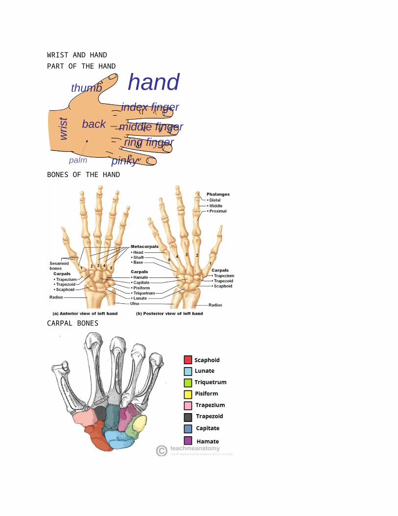
WRIST AND HANDPART OF THE HAND
BONES OF THE HAND
CARPAL BONES
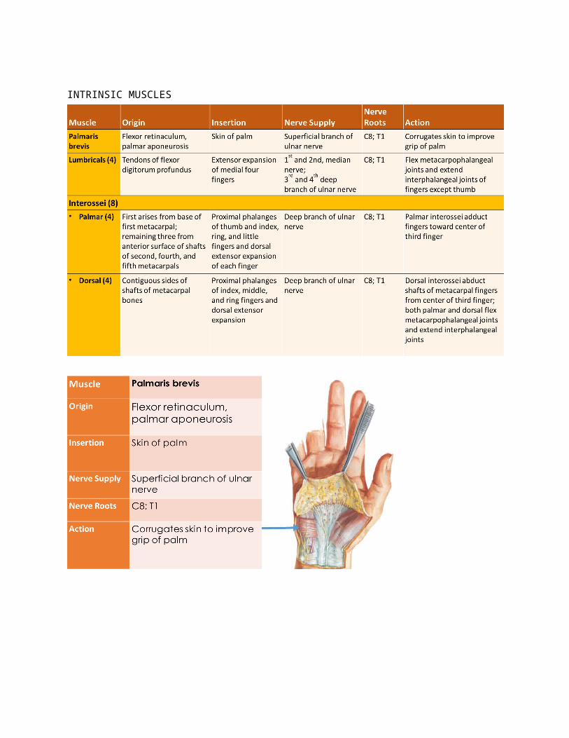
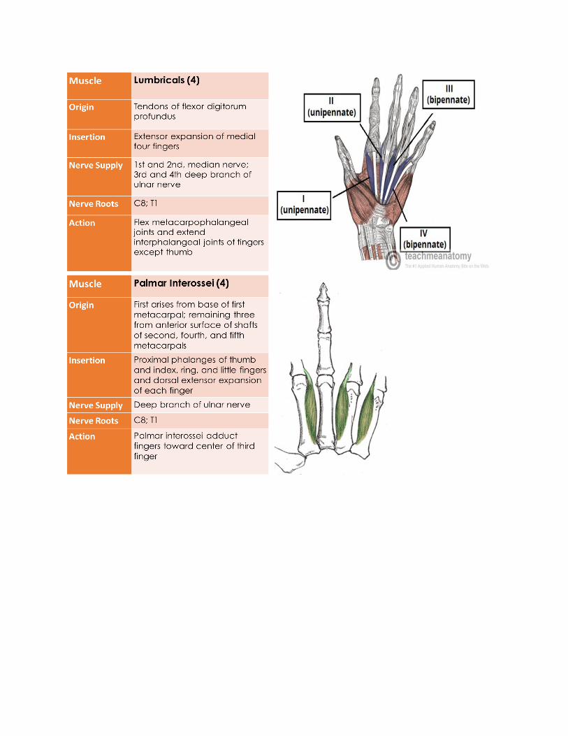
INTRINSIC MUSCLES
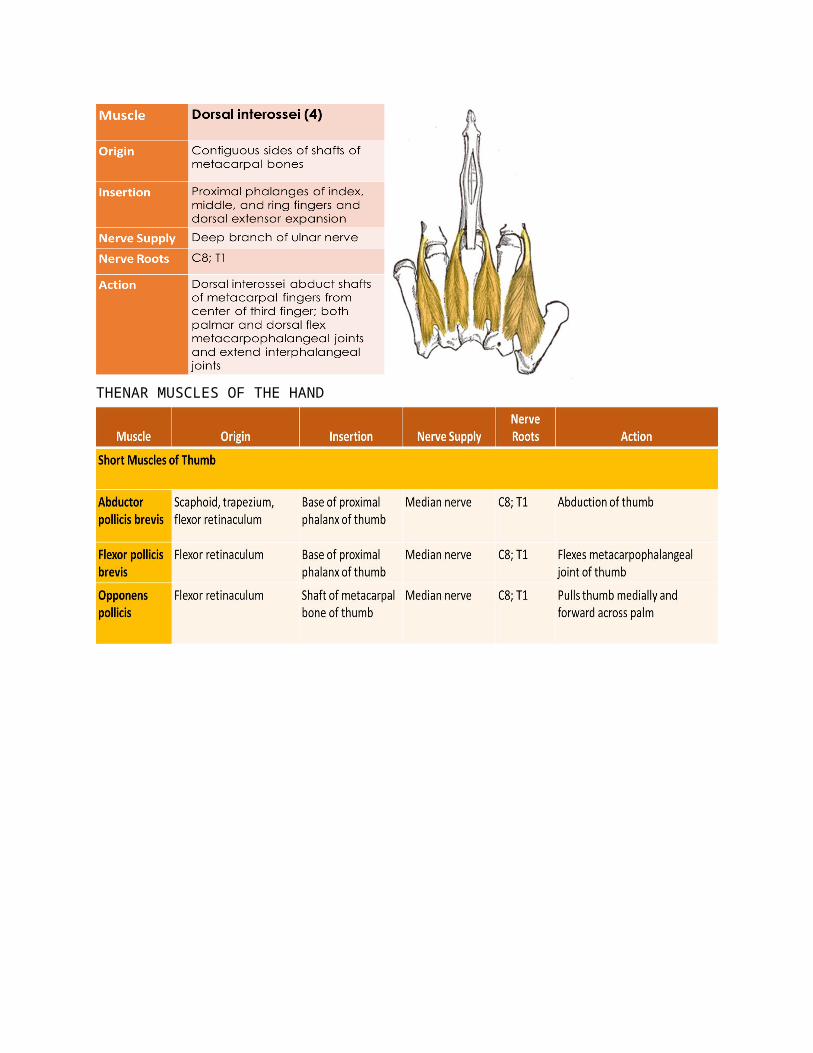
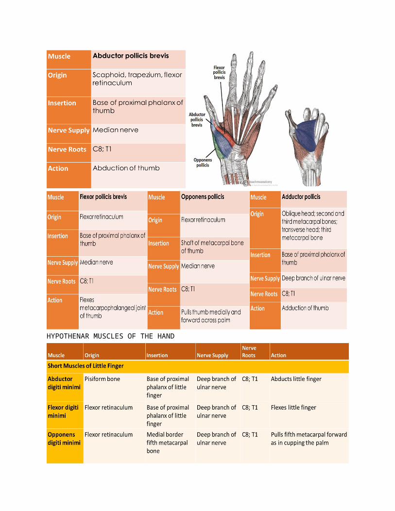
THENAR MUSCLES OF THE HAND
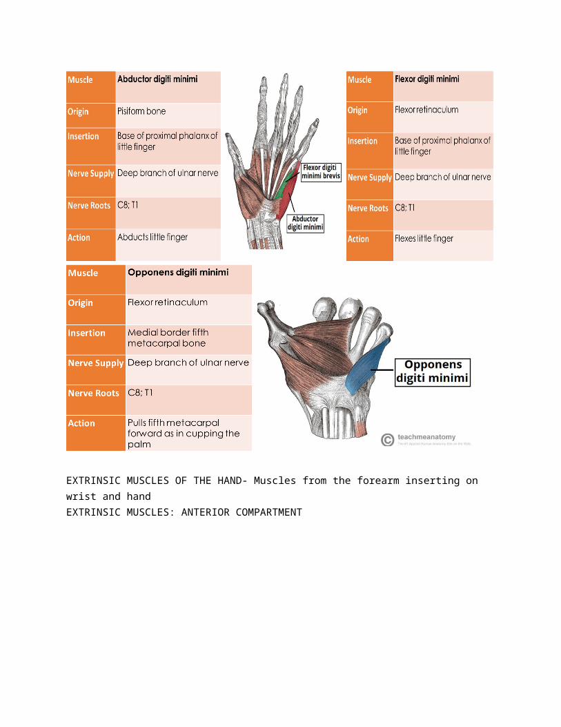
HYPOTHENAR MUSCLES OF THE HAND
EXTRINSIC MUSCLES OF THE HAND- Muscles from the forearm inserting on wrist and handEXTRINSIC MUSCLES: ANTERIOR COMPARTMENT
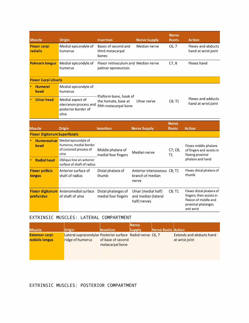
EXTRINSIC MUSCLES: LATERAL COMPARTMENT
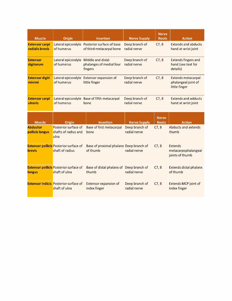
EXTRINSIC MUSCLES: POSTERIOR COMPARTMENT
WRIST: ANTERIOR ASPECT• Superficial to the Flexor Retinaculum
(medial to lateral)• FCU tendon• Ulnar nerve• Ulnar artery• Palmar cutaneous branch of
ulnar nerve• Palmaris longus tendon• Palmar cutaneous branch of
median nerve• Passing below the Flexor Retinaculum
(medial to lateral)• FDS tendons (4) and FDP tendons (4) – common sheath• Median nerve

• Flexor pollicis longus tendon• Flexor carpi radialis (thru a split in the retinaculum)
WRIST: POSTERIOR ASPECT• Superficial to the Extensor Retinaculum
• Dorsal cutaneous branch of the ulnar nerve• Basilic vein• Cephalic vein• Superficial branch of radial nerve
• Beneath the Extensor Retinaculum• ECU tendon• EDM tendon• Extensor digitorum and Extensor indicis tendons – common sheath• Extensor pollicis longus tendon• Extensor carpi radialis longus• Abductor pollicis longus and extensor pollicis brevis
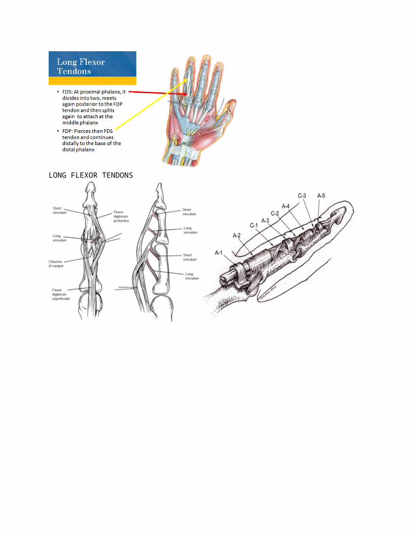
LONG FLEXOR TENDONS
LONG EXTENSOR TENDONS• Fan out from external
retinaculum• Together with deep fascia,
they form the subfascial space.
• Tendons of middle, ring and small fingers are connected by fibrous bands.
• Tendon to the index finger is joined medially by the extensor indicis.
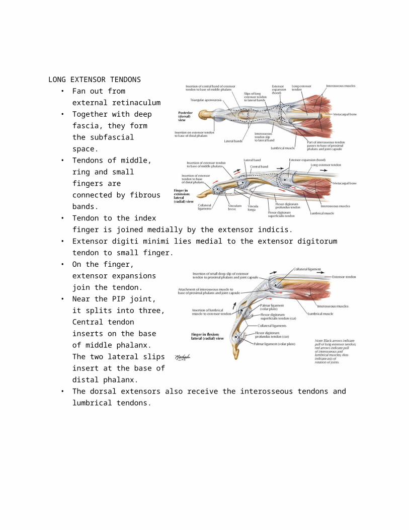
• Extensor digiti minimi lies medial to the extensor digitorum tendon to small finger.• On the finger, extensor expansions join the tendon.• Near the PIP joint, it splits into
three, Central tendon inserts on the base of middle phalanx. The two lateral slips insert at the base of distal phalanx.
• The dorsal extensors also receive the interosseous tendons and lumbrical tendons.
VASCULAR SUPPLY TO THE HAND

PALMAR ARCHES
RADIAL ARTERY AT THE PALM• Leaves the dorsum between 1st and 2nd metacarpals.• Continues as deep palmar arch between oblique and
transverse head of adductor pollicis• Curves medially between the long flexors and interosseous
muscles to form the deep palmar arch• Has anastomosis at the wrist joint and also digital branches of
the superficial palmar arch• Branches
Arteria radialis indicis – lateral side of index finger Arteria princeps pollicis – lateral and medial sides of
the thumb
RADIAL ARTERY OF THE DORSUM
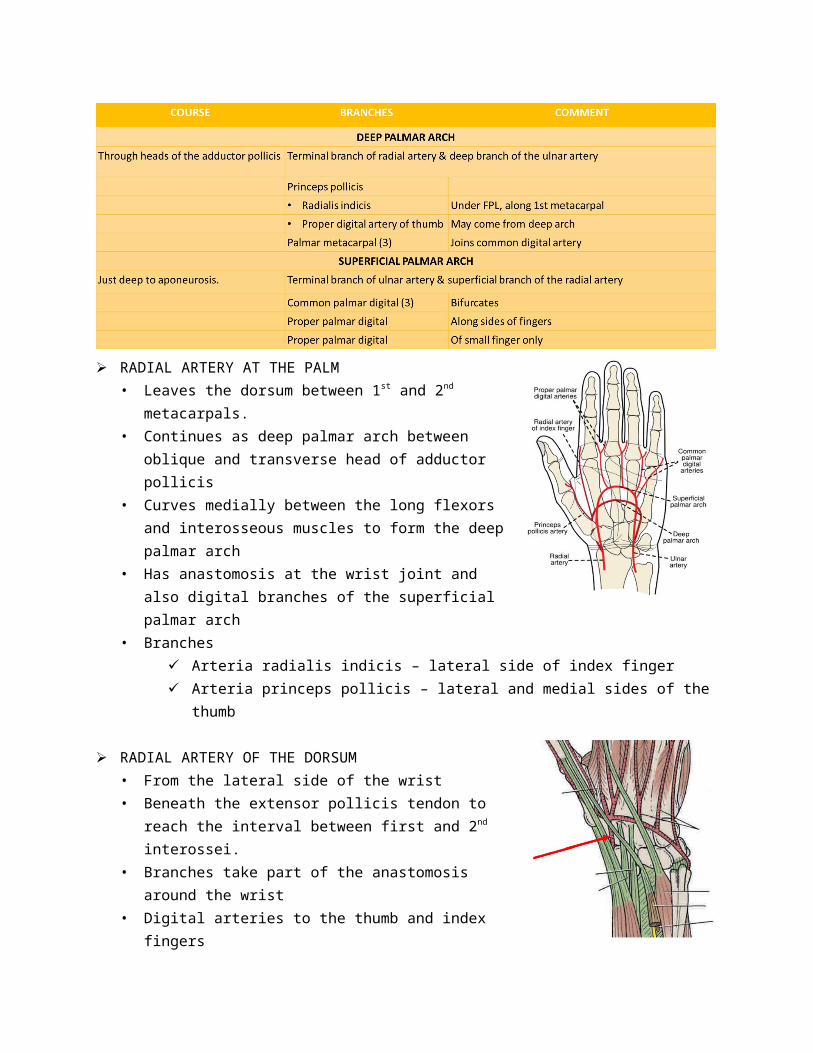
• From the lateral side of the wrist• Beneath the extensor pollicis tendon to reach the interval between first and 2nd interossei.• Branches take part of the anastomosis around the wrist• Digital arteries to the thumb and index fingers
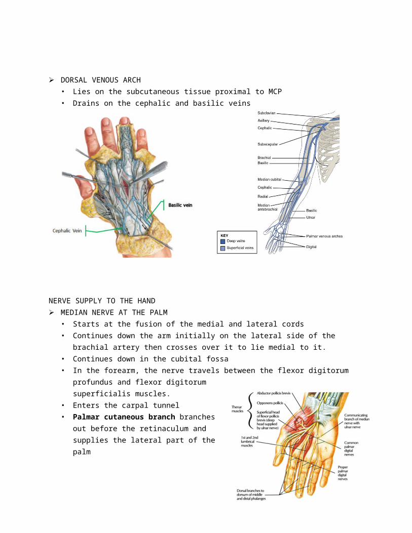
DORSAL VENOUS ARCH• Lies on the subcutaneous tissue proximal to MCP• Drains on the cephalic and basilic veins
NERVE SUPPLY TO THE HAND MEDIAN NERVE AT THE PALM
• Starts at the fusion of the medial and lateral cords• Continues down the arm initially on the lateral side of the brachial artery then crosses over it to
lie medial to it.• Continues down in the cubital fossa• In the forearm, the nerve travels between the flexor digitorum profundus and flexor digitorum
superficialis muscles. • Enters the carpal tunnel• Palmar cutaneous branch branches out before
the retinaculum and supplies the lateral part of the palm
• After the carpal tunnel, it divided into medial and lateral branches
• Muscular branch – recurrent course to about one fingerbreadth distal to the scaphoid tubercle and supply the abductor pollicis brevis, flexor pollicis brevis and opponens pollicis and the 1st lumbricals
• Cutaneous branch to the lateral three and half digits and 2nd lumbricals
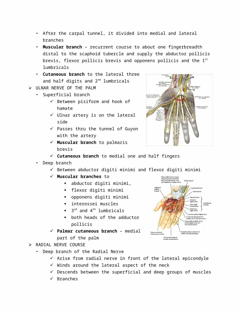
ULNAR NERVE OF THE PALM• Superficial branch
Between pisiform and hook of hamate Ulnar artery is on the lateral side Passes thru the tunnel of Guyon with the artery Muscular branch to palmaris brevis Cutaneous branch to medial one and half
fingers• Deep branch
Between abductor digiti minimi and flexor digiti minimi
Muscular branches to abductor digiti minimi, flexor digiti minimi opponens digiti minimi interossei muscles 3rd and 4th lumbricals both heads of the adductor pollicis
Palmar cutaneous branch – medial part of the palm
RADIAL NERVE COURSE• Deep branch of the Radial Nerve
Arise from radial nerve in front of the lateral epicondyle Winds around the lateral aspect of the neck Descends between the superficial and deep groups of muscles Branches
Muscular – ECRB, supinator, Extensor digitorum, extensor digiti minimi, ECU, Abductor pollicis longus, Extensor pollicis brevis and longus, and extensor indicis
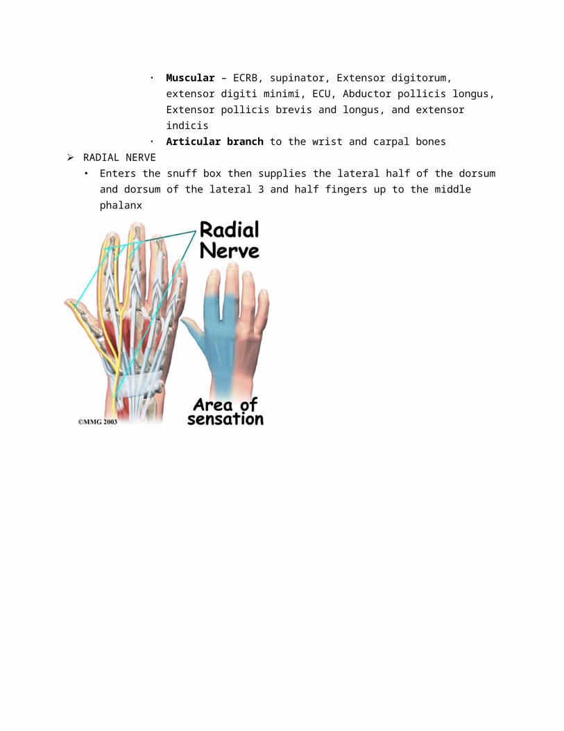
Articular branch to the wrist and carpal bones RADIAL NERVE
• Enters the snuff box then supplies the lateral half of the dorsum and dorsum of the lateral 3 and half fingers up to the middle phalanx
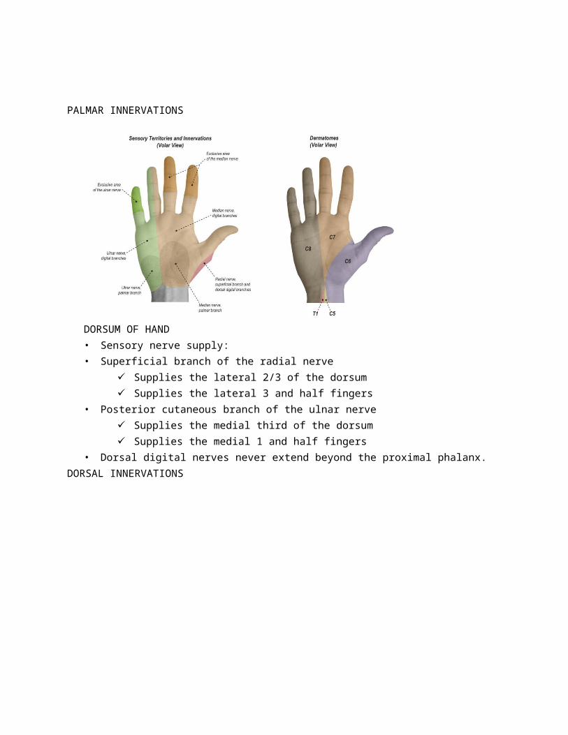
PALMAR INNERVATIONS
DORSUM OF HAND• Sensory nerve supply: • Superficial branch of the radial nerve
Supplies the lateral 2/3 of the dorsum Supplies the lateral 3 and half fingers
• Posterior cutaneous branch of the ulnar nerve Supplies the medial third of the dorsum Supplies the medial 1 and half fingers
• Dorsal digital nerves never extend beyond the proximal phalanx.DORSAL INNERVATIONS
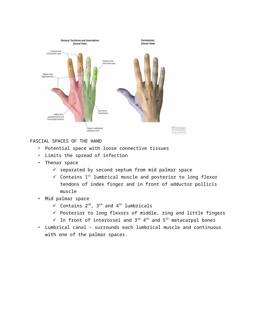
FASCIAL SPACES OF THE HAND
• Potential space with loose connective tissues• Limits the spread of infection• Thenar space
separated by second septum from mid palmar space Contains 1st lumbrical muscle and posterior to long flexor tendons of index finger and in
front of adductor pollicis muscle• Mid palmar space
Contains 2nd, 3rd and 4th lumbricals Posterior to long flexors of middle, ring and little fingers In front of interossei and 3rd 4th and 5th metacarpal bones
• Lumbrical canal – surrounds each lumbrical muscle and continuous with one of the palmar spaces.
PALM OF THE HAND: SKIN• Palmaris brevis
From flexor retinaculum and palmar aponeurosis to skin of the palm
Nerve: superficial branch of the ulnar nerve Action: corrugate the skin at the base of the
hypothenar eminence thus improving the grip• Sensory supply
Palmar cutaneous branch of median nerve Palmar cutaneous branch of ulnar nerve Thenar eminence – lateral cutaneous nerve of the
forearm or superficial branch of radial nerve
PALM OF THE HAND: PALMAR APONEUROSIS
• Triangular, at the center of palm• Apex attached to distal border of the palmaris longus• Base divides the base of finger (four slips)• Each slip has two bands, one passing superficial to the skin, the other deep to the root of the
finger• Deep band divides into two around the flexor tendons and fuse with the fibrous flexor sheath
and deep transverse ligaments.• Medial and lateral borders are continuous with the thenar and hypothenar muscle fascias• Function: firm attachment of the skin, improve grip and protect the tendons
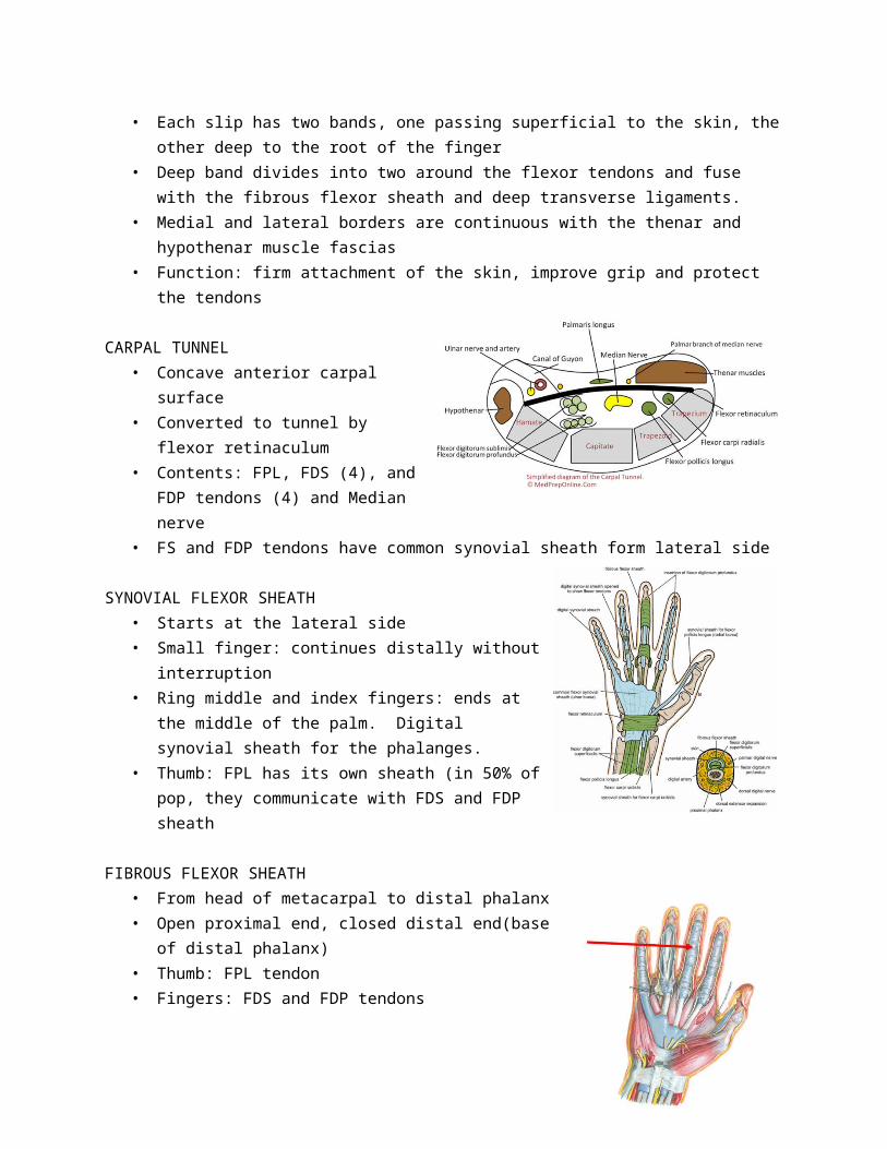
CARPAL TUNNEL• Concave anterior carpal surface• Converted to tunnel by flexor
retinaculum• Contents: FPL, FDS (4), and FDP tendons
(4) and Median nerve• FS and FDP tendons have common
synovial sheath form lateral side
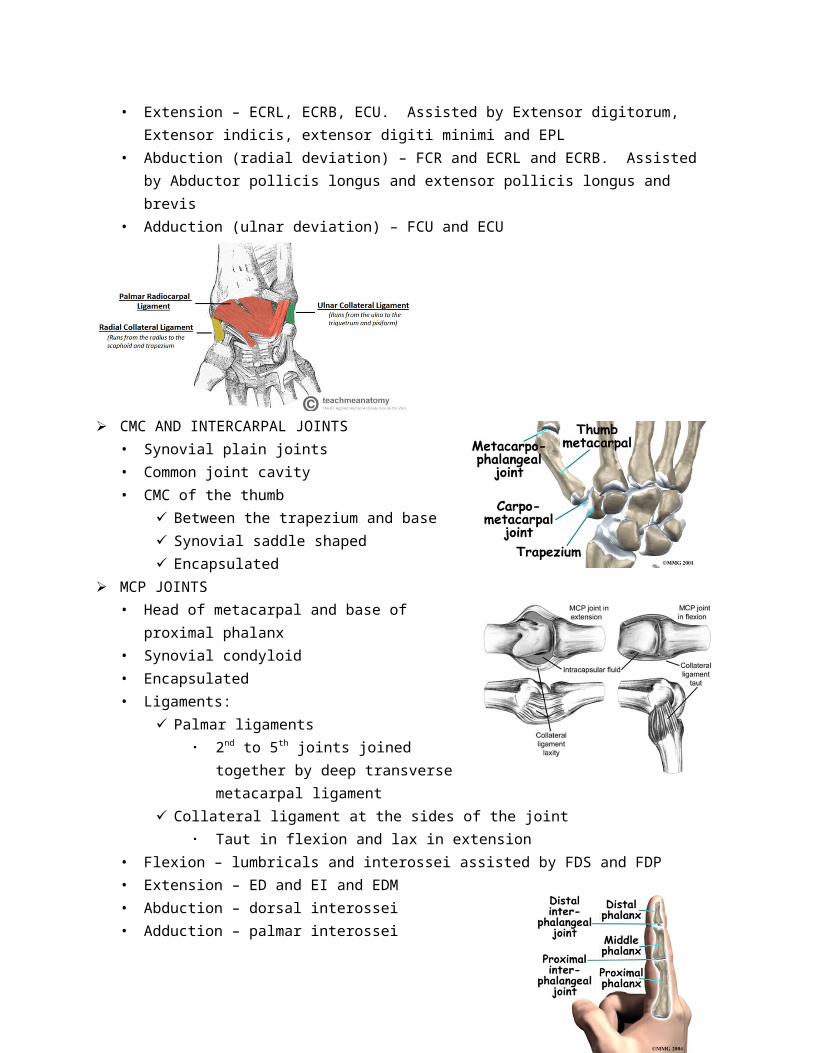
SYNOVIAL FLEXOR SHEATH• Starts at the lateral side• Small finger: continues distally without interruption• Ring middle and index fingers: ends at the middle of the
palm. Digital synovial sheath for the phalanges.• Thumb: FPL has its own sheath (in 50% of pop, they
communicate with FDS and FDP sheath
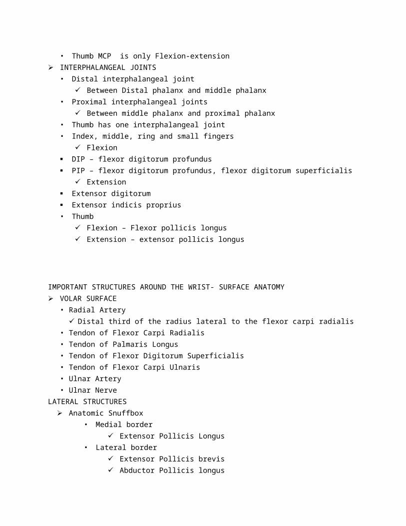
FIBROUS FLEXOR SHEATH• From head of metacarpal to distal phalanx• Open proximal end, closed distal end(base of distal phalanx)• Thumb: FPL tendon• Fingers: FDS and FDP tendons
JOINTS OF THE WRIST AND HAND DISTAL RADIOULNAR JOINT
• Articulation: between round head of ulna to ulnar notch of the radius• Synovial pivot joint

• Enclosed in capsule• Ligaments: anterior and posterior ligaments• Articular disc: triangular with apex at the styloid process and base at the lower border of the
ulnar notch• With synovial membrane• Nerve supply: Anterior interosseus nerve and deep branch of the radial nerve• Pronation – pronator teres and pronator quadratus• Supination – biceps brachii and supinator
More powerful than pronation• Relations:
Anterior: tendons of FDP Posterior: tendons of extensor digiti minimi.
WRIST JOINT- RADIOCARPAL• Between the distal end of the radius and articular disc above and scaphoid, lunate and triquetral
bone• Synovial ellipsoid joint
Proximal is ellipsoid concave and distal surface is ellipsoid convex.• With capsule – distal end of radius to proximal carpus• Ligaments – Anterior and posterior ligaments• Medial ligaments attached to ulnar styloid and triquetral bones• Lateral ligaments from radial styloid to scaphoid bones• With Synovial membrane• Nerve supply – Anterior interosseous nerve and deep branches of the radial nerve.• Flexion – FCR, FCU and palmaris longus• Extension – ECRL, ECRB, ECU. Assisted by Extensor digitorum, Extensor indicis, extensor digiti
minimi and EPL• Abduction (radial deviation) – FCR and ECRL and ECRB. Assisted by Abductor pollicis longus and
extensor pollicis longus and brevis• Adduction (ulnar deviation) – FCU and ECU
CMC AND INTERCARPAL JOINTS• Synovial plain joints • Common joint cavity• CMC of the thumb
Between the trapezium and base Synovial saddle shaped Encapsulated
MCP JOINTS• Head of metacarpal and base of proximal phalanx• Synovial condyloid• Encapsulated• Ligaments:
Palmar ligaments 2nd to 5th joints joined together by deep
transverse metacarpal ligament Collateral ligament at the sides of the joint
Taut in flexion and lax in extension• Flexion – lumbricals and interossei assisted by FDS and FDP• Extension – ED and EI and EDM• Abduction – dorsal interossei• Adduction – palmar interossei• Thumb MCP is only Flexion-extension
INTERPHALANGEAL JOINTS• Distal interphalangeal joint
Between Distal phalanx and middle phalanx• Proximal interphalangeal joints
Between middle phalanx and proximal phalanx• Thumb has one interphalangeal joint• Index, middle, ring and small fingers
Flexion DIP – flexor digitorum profundus PIP – flexor digitorum profundus, flexor digitorum superficialis
Extension Extensor digitorum Extensor indicis proprius• Thumb
Flexion – Flexor pollicis longus Extension – extensor pollicis longus
IMPORTANT STRUCTURES AROUND THE WRIST- SURFACE ANATOMY VOLAR SURFACE
• Radial Artery Distal third of the radius lateral to the flexor carpi radialis

• Tendon of Flexor Carpi Radialis• Tendon of Palmaris Longus• Tendon of Flexor Digitorum Superficialis• Tendon of Flexor Carpi Ulnaris• Ulnar Artery• Ulnar Nerve
LATERAL STRUCTURES Anatomic Snuffbox
• Medial border Extensor Pollicis Longus
• Lateral border Extensor Pollicis brevis Abductor Pollicis longus
• Floor Radial styloid Scaphoid Trapezium 1st metacarpal
• Contents Radial Artery Superficial branch of radial nerve Cephalic vein
DORSUM Lunate
FUNCTIONAL POSITION OF THE HAND• Semiprone• Wrist more extended than at rest• All finger flexed at the same degree• Index and thumb in apposition
MOVEMENTS OF THE HAND
CLINICAL CONDITIONSCOMPARTMENT SYNDROMES
• Carpal tunnel syndrome
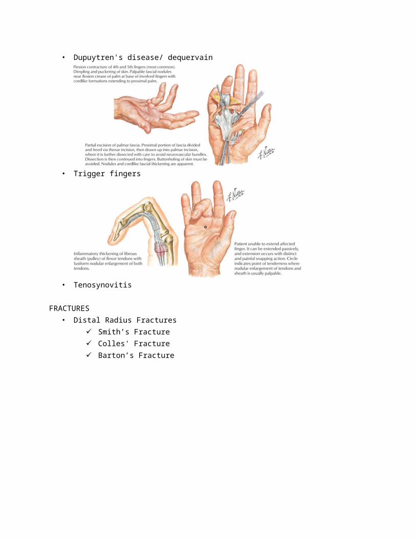
• Dupuytren's disease/ dequervain
• Trigger fingers
• Tenosynovitis
FRACTURES• Distal Radius Fractures
Smith’s Fracture Colles' Fracture Barton’s Fracture
• Carpal Bone fractures
Scaphoid
• Metacarpal fracturesBase of Metacarpal
Metacarpal Shaft
• Phalangeal fractures

DEFORMITIES• Heberden's Nodes
Distal interphalangeal joints• Bouchard’s Nodes
Proximal interphalangeal joints• Mallet Deformity
• Boutonniere’s Deformity Rupture of the extensor central slip Volar displacement of the lateral slips
Changes the direction of the pull from PIP extension to PIP flexion
Lateral slips still functions as DIP extensor
NERVE INJURIES
Ulnar Nerve• Motor - Intrinsic muscles of the hand• Sensory – Medial 1 and half fingers
Median Nerve (Ape hand deformity)• Motor – Thenar Muscles, 1ST and 2ND Lumbricals• Sensory – Lateral 3 and half fingers
Radial Nerve• Motor - none• Sensory – lateral dorsum of the hand