anatomy of the leaf and stem of gossypium
TRANSCRIPT

ANATOMY OF THE LEAF AND STEM OF GOSSYPIUM '
By IRMA E. WEBBER 2 Collaborator^ Division of Cotton and Other Fiber Crops and Diseases^ Bureau of
Plant Industry y United States Department of Agriculture
INTRODUCTION
Because of the great commercial importance of cotton fibers, their structure at various stages of development has been carefully studied. An indication of the work that has been done along this line may be gained from the papers of Dischendorfer {6),^ Youngman and Pande {63), and Kerr {21), which give a number of literature citations. Structure of the seed {I4, v, 3; 32; 33; 34; 35; 36; 37) and that of the boll {I4, ^' 3; 4O) are intimately connected with fiber production and have received considerable attention. The anatomy of the cotton flower has also been investigated to some extent (7; 10; I4, v- 3-4)-
The structure of the vegetative organs of Gossypium, although closely connected with the various plant functions and hence influenc- ing fiber yield, has received comparatively little attention. Some anatomical characteristics of seedlings of G, hirsutum X G, barbadense were depicted by Heim de Balsac {14yVA), The relation of the various reproductive and vegetative parts of mature cotton plants of different types has been repeatedly described (4, 6, 16, 38), and an attempt has been made by Zaitzev {54) to ascertain the gross morphological characteristics of Old World cottons with a haploid chromosome num- ber of 13 and New World cottons with a haploid chromosome number of 26, but descriptions of the histology of the vegetative organs are wholly inadequate. A brief account of the anatomy of the various vegetative organs of an unnamed species of Gossypium was given by Flatters {9), rather detailed reports of the microscopic structure of the roots {11) and the transition region {44) of G. hirsutum L. have been made, the ontogeny of the main stem and fruiting branches has been traced {10) y and some histological characteristics of leaves and stems of G, herbaceum L., G. barbadense L. {14j v, 3), and G. hirsutum X barbadense {I4, v, 2) have been figured; but there is no consecutive account of the anatomical characteristics of the vegetative organs of the main groups of species or of the genus as a whole. Since there is considerable disagreement concerning the taxonomy of the genus and since microscopic and endomorphic characters when considered in conjunction with macroscopic exomorphic ones are often helpful in solving taxonomic problems, the present anatomical study of the genus Gossypium was undertaken.
MATERIAL AND METHODS
Although this investigation was limited to nonfruiting branches, special attention was paid to anatomical characteristics that might be useful in distinguishing between American wild cottons with 13 haploid chromosomes {12, 19, 20) which have been excluded from the genus
1 Received for publication October 13, 1937; issued August 1938. 2 The material examined was made available for study through the courtesy of T. H. Kearney and J. M.
Webber, of this Division. 3 Italic numbers in parentheses refer to Literature Cited, p. 283.
Journal of Agricultural Eesearch, Vol. (57, No. 4 Washington, D. C. Aug. 15,1938
Key No. G-1102 (269)

270 Journal of Agricultural Research voi. 67, NO. 4
Gossypium by some taxonomists, Old World cottons with 13 haploid chromosomes, and New World cottons with 26 haploid chromosomes. For comparative purposes a limited number of Gossypium hybrids and representatives of other genera of the Hibisceae were also included in the study. The species examined and the groups to which they belong are:
Old World cottons with 13 haploid chromosomes: Gossypium africanum Watt, G. anomalum Wawra and Peyr., G. arhoreum L., G. cernuum Tod., G. herhaceum L., G. irdermedium Tod., G. nanking Meyen, G. neglectum Tod., G. sanguineum Hasskl., G. stocksii Mast., G. sturtii F. Muell., G, transvaalense Watt.
American wild cottons with 13 haploid chromosomes: Gossypium armourianum Kearney, G, davidsonii Kellogg, G, harknessii Brandeg., G. klotzschianum Anderss., G, thurheri Tod. (Thurheria thespesioides A. Gray).
New World cottons with 26 haploid chromosomes: Gossypium barhadense L., G. brasiliense Macf., G, contextum Cook and Hubbard, G. darwinii Watt, G. hirsutum L., G. hopi Lewton, G, peruvianum Cav., G. purpurascens Poir., G. schoUii Watt, G, tomentosum Nutt.
Doubtful species of Gossypium and members of related genera: Gossypium kirkii Mast., Alyogyne hakeaefolia (Giord.) Alef., Erioxylum aridum Rose and Standl., Hibiscus brackenridgei A. Gray, H. tiliaceus L., Kokia drynarioides (Seem.) Lewton, K. rockii Lewton, Lagunaria patersonii G. Don, Thespesia lampas (Cav.) Dalz., T, populnea Soland., and Shantzia garckeana Lewton.
All of the material examined was taken from plants grown in the field at the Rubidoux Laboratory,* Eiverside, Calif. Much of the material was sectioned when fresh, but some of it was first killed and fixed in formalin-acetic alcohol and embedded in paraffin. Safranine was used to stain some sections; the remainder were left unstained.
LEAF ANATOMY
EPIDERMIS
The ordinary epidermal cells over chlorenchyma may be straight- walled or very sinuate in surface view. In most species their lateral walls are much less sinuous in the upper epidermis than in the lower epidermis, where stomata are more numerous (pi. 1, ^, 5). However, in some species the lateral walls of epidermal cells are characteristically rather straight on both upper and lower leaf surfaces ((7, D). On vertically transcurrent veins the epidermal cells are longer and narrower than elsewhere on the lamina, the long axis of the cell being parallel to the course of the vein (E), Over lysigenous cavities in the mesophyll, the epidermal cells are concentrically arranged and generally smaller than the surrounding cells (i^-
According to Flatters (P, p. 4.8) ''The epidermis of the upper [leafj surface consists of closely-packed cells with resin-cells distributed among them * * *.'' What he here designates as ''resin-cells" is not clear to the writer. In his figure of a cross section of a stem the lysigenous cavities are so labeled, but their absence in the epidermis
* Maintained cooperatively by the U. S. Department of Agriculture and the California Agricultural Experiment Station.
EXPLANATORY LEGEND FOR PLATE 1.
Epidermis of Gossypium leaves. X 312. A~J, Surface views showing comparative shape and size of ordi- nary epidermal cells and stomata. A and B, Q. brasiliense: A, From upper side of leaf; B, from lower side of leaf. C and D, 0. hurknessii: C, From upper side of leaf; D, from lower side of leaf. E and F, Ö. peruvianum: E, Epidermis of a vein; F, epidermis over a lysigenous cavity in the mesophyll. Q-J, Epidermis from lower surfaces of leaves: G, G. hirsutum var. Acala; H, G. hirsutum X G. cernuum; I, G. armourianum; J, G. hirsutum X G. armourianum. K-M, Leaf sections showing position of stomata: K, G. contextum, lower epidermis; L, G. arhoreum, upper epidermis; M, G. harknessii, upper epidermis.

Anatomy of the Leaf and Stem of Gossypium PLATE I
FOR EXPLANATORY LEGEND SEE OPPOSITE PAGE.

Anatomy of the Leaf and Stem of Gossypium PLATE 2
: ^-
If
% '
)'^ .>
f -*i^
G FOR EXPLANATORY LEGEND SEE OPPOSITE PAGE.

Aug. 15,1938 Anatomy oj Leaf and Stem oj Gossypium 271
of leaves suggests that he either did not distinguish clearly between epidermal and subepidermal tissue, or applied the term ^^resin-cells'' to some other structure as well as to the lysigenous cavities. In the latter case, ordinary epidermal cells with colored contents, mucilage cells, or minute, depressed capitate hairs may be meant.
Ordinary epidermal cells with colored, usually purplish or reddish, cell sap are of scattered occurrence in most species of Gossypium. In green-leaved plants they commonly adjoin the guard cells of s tomata and are not nearly so numerous as the hyaline epidermal cells. In reddish-leaved plants, such as G. purpurascens and the Red Acala variety of G, hirsutum, they are not restricted to the vicinity of stomata and may be more numerous than hyaline epidermal cells. The inheritance of red plant color in cotton plants has beeu discussed by Ware (^P).
Although mucilage cells were reported by Dumont (8) as completely wanting in Gossypium and as very rare throughout the Hibisceae, according to Kuntze (^2) they are characteristic of both the upper and the lower epidermis of the leaves of all Hibisceae. They are apparently lacking or inconspicuous in the epidermis of Kokia, Shantziaj Erioxylum, and most species of Gossypium, but fairly numerous in that of G. kirkii (pis. 2, 5, a; 3, M, a), G. klotzschianum (pi. 3, Bj a), Thespesia, Hibiscus, and Lagunaria. As a rule, they are considerably larger than the adjoining cells, and more common in the upper than in the lower epidermis. The distribution and structure of mucilage cells in a number of malvaceous genera, chiefly representative of tribes other than the Hibisceae, have been discussed by Trecul {^6), Walliczek U8), and Nestler {28),
Heim de Balsac {H, v. S) depicted stomata in the lower but not the upper epidermis of the leaves of Gossypium herbaceum and G. barba- dense, while stomata were reported by Flatters (9) as few or absent in the upper epidermis of the leaves of Gossypium and by Kuntze (22) as lacking in the upper epidermis of G. drynarioides Seem. The latter species is now referred to the genus Kokia and was found by the writer to have a few widely scattered stomata on the upper leaf surface. In all species of Gossypium examined, stomata are present on both upper and lower leaf surfaces but are more numerous on the lower surface. In some species the difference between the average number of stomata per square millimeter of the upper and the lower epidermis of a leaf is very marked, while in others it is comparatively slight. For example, when stomatal counts were made near the centers of mature leaf blades in areas free from large veins, the average number of stomata per square millimeter of upper and lower leaf surfaces were, respectively, 122 and 159 in G. harknessii, and 40 and 218 in Ö. peruvianum. The correlation indicated by Kuntze for most Malvaceae between a dense coating of hairs and the presence of stoma- ta in the upper epidermis, or dense hairy covering and relatively high stomatal number in the lower epidermis, does not hold true in Gossyp- ium. In leaves of the species examined by the writer the average number of stomata per square millimeter of the upper epidermis ranged
EXPLANATORY LEGEND EOR PLATE 2.
Leaf epidermis of Gossypium and related genera showing various types of hairs. X 80. A, G. intermedium; B, G. kirkii; C, G. sanguineum; D, G. tomentosum; E, G. peruvianum; F, G. thurberi; G, Thespesia populnea: H, Lagunaria patersonii. a, Mucilage cell; b, capitate hair; c, stellate hair; d, simple hair; e, peltate scale.
89150—38 3

272 Journal of Agricultural Research voi. 57, NO. 4
from 40 in G. peruvianum to 170 in 0, intermedium, while average stomatal numbers per square millimeter of the lower epidermis varied from 80 in G. tomentosum to 280 in G, anomalum.
Within the genus Gossypium the stomatal apparatus of leaves varies from roundish (pi. 1, jff, /) to elliptic {A, B, C, D, E, Ö, J) in surface view, the latter form dominating. Twin and malformed stomata are occasionally present, being somewhat more numerous in certain hybrids than in their parents. The normal stomatal apparatus of mature leaves varies in average length from about 24/^ to 32jLt and in average width from about 16JLI to 24jLt. Within a species there are no appreciable differences in size or shape between stomata of the upper leaf surface and those of the lower leaf surface, some variation generally occurring on both surfaces. Because of the similarity of stomata in most of the species, stomatal size in interspecies hybrids of Gossypium is generally of Httle significance. At times, however, when a species with relatively large stomata {G) and one with small stomata (/) are crossed, stomatal size of the hybrid is clearly inter- mediate between that of the parents (J). Some such hybrids show a wider range in stomatal size than most of the species (JS), Stomata may be level with the ordinary epidermal cells (L), slightly above the general epidermal surface {K), or slightly below it (M), those on a single leaf often being in more than one position.
Exclusive of epidermal outgrowths, the thickness of the epidermis on mature leaves of Gossypium varies considerably. In some species and hybrids, notably G. armourianum (pi. 3, A), G harknessii, G. thurberi ((7), G. armourianum X G. harknessiij G. thurberi X G, sturtii, G. sturtii X G, harknessii, and G. hirsutum X G. sturtii, the upper and lower epidermis of a leaf are of approximately the same thickness. In such plants the average thickness of the leaf epidermis over chlo- renchyma varies from about 16/x to 32jLt. In the majority of species the upper epidermis of a leaf is thicker than the lower epidermis. The difference in thickness may be relatively slight as in G. barbadense, G. peruvianum (E), and G. anomalum (/), or considerable as in G. klotzschianUm (J5), G. hirsutum (F), and 6?. herbaceum (J), In leaves with upper and lower epidermis of different thicknesses, the upper epidermis varies in average thickness from about 14jLt to 32//, and the thickness of the lower epidermis ranges from about lOjn to 32)Lt. Over large veins the epidermal cells often have thicker outer walls than elsewhere on the lamina.
Three types of epidermal outgrowths occur on the leaves and stems of Gossypium, The ontogeny of each hair type has been traced by Youngman and Pande {58) and shows a marked similarity to that of comparable trichomes described by Kauter (30) in Malva.
Multicellular capitate hairs, sometimes called glandular hairs (^S), club-shaped bodies (53), or septate papillae (45), were observed on both the upper and the lower leaf surfaces of all species of Gossypium examined. They are more numerous than other types of trichomes on the mature leaves of G. armourianum, G. harknessii, G. barbadense, G, brasiliense, G. contextum, G. darwinii, G. hirsutum, G, hopi, G.
EXPLANATORY LEGEND EOR PLATE 3.
Cross sections of leaf blades showing structure of the epidermis and mesophyll. X 80. A, Gossypium armouri- anum; B, (?. klotzschianum; C, O. thurberi; D, Erioxylum aridum; E, G. peruvianum; F, G. hirsutum; G, G. tomentosum; H, G. barbadense; I, G. anomalum; J, G. herbaceum; K, G- cernuum; i, G. sturtii; M, G. kirkii. a, Mucilage cell; 6, capitate hair; c, stellate hair ; d, lysigenous cavity in mesophyll.

Anatomy of the Leaf and Stem of Gossypium
I
FOR EXPLANATORY LEGEND SEE OPPOSITE PAGE.

Anatomy of the Leaf and Stem of Goasypium PLATE 4
FOR EXPLANATORY LEGEND SEE OPPOSITE PAGE.
Aug. 15,1938 Anatomy of Leaf and Stem of Gossypium 273
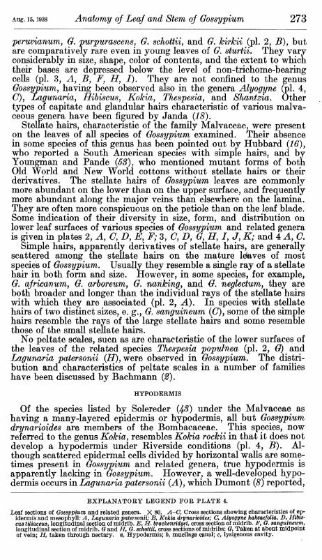
peruvianum, G. purpurascens, G. schottii, and G, kirkii (pi. 2, J5), but are comparatively rare even in young leaves of G. sturtiL They vary considerably in size, shape, color of contents, and the extent to which their bases are depressed below the level of non-trichome-bearing cells (pi. 3, Aj B, F, H, I). They are not confined to the genus Gossypium, having been observed also in the genera Alyogyne (pi. 4, (7), Lagunaria, Hibiscusj Kokia, Thespesia, and Shantzia, Other types of capitate and glandular hairs characteristic of various malva- ceous genera have been figured by Janda {18).
Stellate hairs, characteristic of the family Malvaceae, were present on the leaves of all species of Gossypium examined. Their absence in some species of this genus has been pointed out by Hubbard {16), who reported a South American species with simple hairs, and by Youngman and Pande {53), who mentioned mutant forms of both Old World and New World cottons without stellate hairs or their derivatives. The stellate hairs of Gossypium leaves are commonly more abundant on the lower than on the upper surface, and frequently more abundant along the major veins than elsewhere on the lamina. They are often more conspicuous on the petiole than on the leaf blade. Some indication of their diversity in size, form, and distribution on lower leaf surfaces of various species of Gossypium and related genera is given in plates 2, A, C, D, E, F; 3, O, D, G, H, /, J, K; and 4 A, C.
Simple hairs, apparently derivatives of stellate hairs, are generally scattered among the stellate hairs on the mature leaves of most species of Gossypium, Usually they resemble a single ray of a stellate hair in both form and size. However, in some species, for example, Ö. africanum, G. arboreum, G. nanking, and G, neglectum, they are both broader and longer than the individual rays of the stellate hairs with which they are associated (pi. 2, A). In species with stellate hairs of two distinct sizes, e. g., G. sanguineum (O), some of the simple hairs resemble the rays of the large stellate hairs and some resemble those of the small stellate hairs.
No peltate scales, sucn as are characteristic of the lower surfaces of the leaves of the related species Thespesia popuJnea (pi. 2, G) and Lagunaria patersonii {FT), were observed in Gossypium, The distri- bution and characteristics of peltate scales in a number of families have been discussed by Bachmann {2).
HYPODERMIS
Of the species listed by Solereder {48) under the Malvaceae as having a many-layered epidermis or hypodermis, all but Gossypium drynarioides are members of the Bombacaceae. This species, now referred to the genus Kokia, resembles Kokia rockii in that it does not develop a hypodermis under Riverside conditions (pi. 4, B). Al- though scattered epidermal cells divided by horizontal walls are some- times present in Gossypium and related genera, true hypodermis is apparently lacking in Gossypium. However, a well-developed hypo- dermis occurs in Lagunaria patersonii {A), which Dumont {8) reported,
EXPLANATORY LEGEND FOR PLATE 4.
Leaf sections of Gossypium and related genera. X 80. A-C, Cross sections showing characteristics of ep- idermis and mesophyll: A, Lagunaria patersonii; B, Kokia drynarioides; C, Alyogyne hakeaefolia. B, Hibis- cus tiliaceus, longitudinal section of midrib. E, H. brackenridgei, cross section of midrib. F, O. sanguineum, longitudinal section of midrib. Q and H, G. schottii, cross sections of midribs: G, Taken at about midpoint of vein; H, taken through nectary, a, Hypodermis; b, mucilage canal; c, lysigenous cavity.

274 Journal of Agricultural Research voi. 57, No. 4
under the name Lagunea syuameaj as having much elongated epidermal cells often divided transversely.
MESOPHYLL
The predominance of bifacial leaf structure in the Malvaceae has been pointed out by Solereder (43). Aside from the bombacaceous species he includes, he reports centric leaf structure only in Malva parviflora. In describing the anatomy cf cotton leaves, Flatters {9j p. 43) states that the upper part of the mesophyll is composed of two layers of palisade cells. *'The lower half of the mesophyll is made up of loosely arranged irregular cells with large air spaces be- tween them * * *." The inapplicability of Flatters' description to the mesophyll of all species of Gossypium and the commoner occur- rence of centric leaf structure than was indicated by Solereder (48) are evident from the account of structural differences in mesophyll within the genus Oossypium given by Magitt and Magitt (25), They stated that palisade parenchyma underlies the epidermis of only the upper surface of the leaf in American cottons and in hybrids between American and Asiatic cottons, but occurs beneath the epidermis on both upper and lower leaf surfaces in Asiatic cottons. This distinc- tion between Old World and New World cottons does not apply to plants grown in the field at Riverside. Specimens examined by the writer showed that leaves of some species of the Old World group (pi. 3, If Kj L)j of the American group with 26 pairs of chromosomes {Gj H), and of the wild American group with 13 pairs of chromosomes (A) develop palisade underlying the epidermis of both the upper and the low^er surface. Within each of these three groups of Gossyp- ium ^ the leaves of some species are characterized by mesophyll com- posed of palisade tissue underlying the upper epidermis and spongy parenchyma underlying the lower epidermis (5, C, E, F, J). A less common type of centric leaf structure than that occurring in Gos- sypium was observed in the cotton relative Alyogyne hakeaefolia (pl-4, (7).
In structure as well as in distribution, the palisade tissue of Gos- sypium leaves shows greater diversity than was indicated by Flatters (ß). In the leaves of ö. armourianum (pi. 3, A) and G, harknessii there are areas in which palisade tissue extends from the upper to the lower epidermis and is commonly four or five cells thick. The pali- sade layers are more commonly one cell than two cells thick in leaves with palisade tissue restricted to the upper side or with clearly dis- tinct upper and lower palisade layers. Palisade cells of plants grown in the field vary in length from about 30/x to 220JLI and are commonly longer in the upper layer of palisade parenchyma than in the lower layer, when the latter is present.
The spongy parenchyma of Gossypium leaves is often more compact than Flatters (9) indicated. At times the form and arrangement of spongy parenchyma cells so closely approximates that of weak pali- sade tissue that a distinction between the two types of tissue is purely arbitrary.
Oblate or spherical cavities, usually 50/x to 170/x in diameter and commonly with dark-purplish or brownish contents (pi. 3, (7, M), occur in the mesophyll of all species of Gossypium examined and in the mesophyll of Erioxylum aridum (Z>), Kokia drynarioides (pi. 4, B), K. rockii, Thespesia lampas, T, populnea, and Shantzia garckeana.

Aug. 15,1938 Anatomy of Leaj and Stem oj Gossypium 275
They were reported by Dumont (8) to be of schizogenous origin, but according to the earHer report of Von Höhnel {15) on similar cavities in the cotyledons of Oossypium herbaceum and the more recent work of Stanford and Viehoever (45) on various organs of G. hirsutumj they are formed lysigenously. Similar cavities have been referred to under a variety of names, including mucilage glands {14, o, 2), mucilage pockets {8, 22), resin cells {9), resin glands {36)^ black oil glands {23), internal glands {45), gossypol cavities {52), and mucilage canals {18), According to Stanford and Viehoever {45), the nature of the contents of the flattened cells surrounding the cavities is dependent upon whether they are developed in darkness or exposed to light.
VEINS
The veins of Gossypium leaves are mostly vertically transcurrent (pi. 3, /, L), but some of the smaller ones are embedded {G). The midribs and other large veins commonly project beyond the general level of the lamina on both upper and lower surfaces of the leaf {A-I). The relative thickness of the major veins and of the photosynthetic areas of leaves, as well as size and shape of major veins at comparable points in the leaves of different species, shows considerable diversity within the genus. The size of both the vascular bundles of the mid- ribs and the conducting elements composing them is likewise variable. Kuntze {22) reported the veins of G. herbaceum to be without bast, but Heim de Balsac^s {14, 'V. 3) diagrammatic sketch of the midrib of this species showed groups of pericyclic fibers. The veins of most species of Gossypium commonly lack fibers at the outer edge of the phloem throughout the greater part of their length. However, it is not unusual to find a few bast fibers capping the phloem toward the base of the larger veins. In large veins (pi. 4, G) the vascular bundles or fibrovascular bundles are surrounded by parenchyma which extends to a coUenchymatic layer of variable thickness underlying the epidermis, while in small veins the parenchyma extends to the epider- mis. Druses, probably of calcium oxalate, are often present in the parenchyma of both large and small veins. Lysigenous cavities resembhng those occurring in the mesophyll are frequently present in the parenchyma of large veins (pis. 4, F; 5, A). Mucilage canals, such as are present in the parenchyma of the veins of species of Hibiscus (pi. 4, D, E), are apparently absent in Gossypium. As shown in plate 4, D and E, mucilage canals may occur in vein parenchyma above as well as below the vascular bundles, where they were observed by Kuntze {22).
NECTARIES
Nectaries are usually a conspicuous structural feature of the leaves of Gossypium, but they are apparently always lacking in G. tomentosum and are often absent in individual leaves or plants of other species. Unlike the extrafloral nectaries described by Tyler {47) as character- istic of one or both sets of involucres in Gossypium, the leaf nectaries are considered by Lewton {23) to be of little value in classification.
The leaf nectaries of Gossypium occur on the lower surface of from one to five major veins, being most common on the midrib. Only one nectary occurs on a vein, and it is usually close to the base of the vein in most species, but in G. kirkii it is characteristically well above the middle of the vein. Leaf nectaries vary considerably in size within

276 Journal of Agricultural Research voi. 57, No. 4
each of the three main groups of species. Nectary size does not always correspond to leaf size, the nectaries of G. sturtii being very long in proportion to leaf length, and those of hybrids involving the eglandular species G. tomentosum generally being very small in pro- portion to leaf size. Often the nectaries on major lateral veins are smaller than those on the midrib of the same leaf. Leaf nectaries likewise show wide variation in size in the close relatives of Gossypium^ those of Shantzia being noteworthy because of their great length.
The leaf nectaries of Gossypium are most commonly rounded- triangular in outline, but may be nearly circular, elliptical, elongate- quadrangular, or sagittate, the shape showing no correlation with the main divisions of the genus. Cross sections taken at approximately the middle of nectaries (pi. 5, A~I) show that the extent of depression of the glandular surface, the size and shape of glandular hairs, and the contour of the sides of the nectaries are also variable.
Janda (18) recently classified the extrafloral nectaries of Gossypium, Thespesia trilohata, and Cienfuegosia as depressed nectaries, or nec- taries with the secretory surface below the surrounding surface. In contrast, the nectaries of Hibiscus rosa-sinensis, H. schizopetalus, H. archeri, H. syriacuSj^ and Thespesia populnea were classified as surface nectaries, or nectaries with the secretory surface lying at or near the same level as the surrounding epidermis; and the nectaries of Kydia, Urena, Decaschistia, Dicellostyles, Julostylis, and some species of Hibiscus were described as hollow nectaries^ or nectaries with the secreting surface covering a cavity which has a small, slitlike orifice. Since Janda listed different species of Thespesia and Hibiscus as having different types of nectaries, it is not surprising that cross sec- tions of some Gossypium nectaries (pi. 5, (7, D) show that they might well be described as surface nectaries rather than depressed nectaries.
The ontogeny of the nectaries of cotton leaves has been traced by Schwendt {39, pp. 254-266) in Gossypium brasiliense and by Keed (31) in G. hirsutum. The histology of mature leaf nectaries of G. brasiliense, G. herbaceum, G. davidsonii, and G. microcarpum Tod. was compared by Schwendt (39), and the structure of leaf nectaries and involucral nectaries of G. purpurascens was compared by Janda (18). Although the leaf nectaries of different species of Gossypium vary in such characteristics as the size and number of glandular hairs, the number of simple or stellate hairs, and the size of cells composing their glandular and subglandular tissue, their general structure shows a marked similarity in all species. As in extrafloral nectaries dis- cussed by Solereder (43), the upper, expanded portion of the glandular hairs of Gossypium nectaries is generally more strongly developed than that of capitate hairs of similar structure found elsewhere on the same plant (pi. 4, G, H). Bordering the glandular hairs of a nectary, the epidermal ceUs are often vertically elongate and are sometimes divided horizontally. Underlying the epidermis of the nectaries are several cell layers of subglandular tissue. As shown in plate 4, G and H, the cells of the subglandular tissue are smaller than the paren- chyma cells of major veins outside the nectarial region. In some nectaries, druses of calcium oxalate are particularly abundant in the subglandular tissue.
PETIOLES
Within the genus Gossypium petioles of mature leaves vary greatly in size. According to Afzal (1) the petiole attains its full size at the

Anatomy of the Leaf and Stem of Gossypium PLATE 5
Cross sections of Gossypium leaf nectaries. X 2ñ. A, G. klotzuchianum; B, G. nanking: C, G. herbaceam D, G. africanum; E, G. stUTtii; F, G. darwinii; G, G. peruvianum; II, Q. thurberi; I, G. harknessii.

Anatomy of the Leaf and Stem of Gossypium PLATE 6
FOR EXPLANATORY LEGEND SEE OPPOSITE PAGE.

Aug. 15,1938 Anatomy of Leaf and Stem of Gossypium 277
same time as the lamina, and there is a high correlation between length of petiole and length of midrib. There is also a general tendency for long petioles to be of greater diameter than short petioles, but individual exceptions to this generalization are of fairly common occurrence. The shape of a petiole in cross section depends con- siderably on the point in the petiole at which the section is taken (pi. 6, D, E, F). An indication of the range in size and shape of the midsections of petioles of Gossypium may be gained from plate 6, A, B, (7, E, G, and H,
The epidermis of the petiole bears a marked resemblance to that of the major veins. The various types of epidermal hairs are well de- veloped on the petiole and are often more conspicuous than on the midrib. Underlying the epidermis is a multicellular layer of collen- chyma such as Plitt {29) described as characteristic of malvaceous petioles. The prominent ridges of the petioles of G. kirkii (pi. 6, B), which give them their quadrangular outline in cross section, are com- posed mostly of this tissue. Between the collenchyma and the well- developed fibers capping the phloem of the vascular bundles is a parenchymatic layer several cells deep. Oblate or spherical lysigenous cavities similar to those occurring in the parenchyma of veins are usually of scattered occurrence in this parenchymatic layer (A~H). At the midpoint of the petiole the fibrovascular system may take the form of a ring (Ay B), an arc open at the top ((7), or isolated bundles arranged in a ring {E), This diversity in arrangement of the vascular tissues of the petioles of Gossypium is greater than is in- dicated for the family Malvaceae by Dumont (8), Plitt {29), or Solereder {43).
While many species of Gossypium and related genera have dimorphic leaves {3), G. thurberi is notable for its diversity of leaf form. In this species the arrangement of the vascular tissues in cross sections taken at the midpoints of petioles of an entire leaf (pi. 6, G) and of leaves with two, three, four, and five lobes {H) showed but slight differences.
The central parenchyma of a petiole is usually homogeneous, al- though some of its cells frequently are conspicuous because of their dark-brownish contents (pi. 6, A), The central parenchyma of the petioles of the aberrant G, kirkii differs from that of the typical species of Gossypium examined in that it occasionally includes a few small medullary bundles and generally contains scattered spherical or oblate lysigenous cavities similar to those found in the lamina and in the cortex of the petiole {B). The occurrence of these cavities in the central parenchyma of the petiole of G. kirkii is of significance since Lewton {23) has pointed out that the distribution as well as the- presence or absence of such cavities should be regarded as an anatomi- cal character of value in classifying the Hibisceae.
EXPLANATORY LEGEND EOR PLATE 6.
Cross sections of Gossypium petioles and stems. A-H, Petioles (X 15): A, B, C, E, G, and if, Sections taken at approximately midpoint of petiole; D, section taken near top of petiole; F, section taken near base of petiole; A, Q. brasiliense; B, G. kirkii; C, G. armourianum; D-F, G. nanking; G, H, G. thurberi. I-O, Stems (X 7H): I-L, very young stems; M-O, older stems; /and M, G. purpurascens; J and N, G. arboreum; K and O, G. davidsonii; L, G. kirkii.

278 Journal of Agricultural Research voi. 57, No. 4
STEM ANATOMY SHAPE OF THE STEM AND ARRANGEMENT OF THE TISSUES
The stems of all species of Gossypium vary in cross-sectional shape at different stages in their development. As shown in plate 6, I-L, sections taken near the apices of the stems of Old World cottons, an American wild cotton, and a cultivated American cotton are angular and are generally incHned to be pentagonal in form. In contrast, sections of older stems (M-0) are rounded, often nearly circular, but sometimes broadly elliptic in outhne.
The arrangement of the tissues in stems of Gossypium may be seen in plate 6, I~0, and plate 7, A, B, C, E, In centripetal order the primary permanent tissues (pi. 7, B) are epidermis, cortex merged with pericycle, phloem, cambium, xylem, and pith. In older stems the epidermis is underlain by periderm (C), which later replaces it {E).
EPIDERMIS AND PERIDERM
The epidermis of the stem is quite similar to that of the petiole The characteristic types of epidermal hairs are usually well developed near the apex of a stem but are less numerous Avhere the epidermis is older. In a given plant, the capitate hairs near the apex of a stem are sometimes considerably larger than those over the green portions of the leaf blades, and approach those of the nectaries in size. A two- layered epidermis is present in the stems of some relatives of Gossyp- iuTUj notably Hibiscus splendens {2Jj) and Alyogyne hakeaefolia (pl.7,£>).
Periderm formation in the stems of Gossypium usually begins before the end of the first growing season. In numerous species of Gossypium the phellogen that gives rise to the periderm (pi. 7, C) forms in the outermost layer of cortical cells. A subepidermal phellogen is also characteristic of Erioxylum aridum and, according to Moeller (27) y of Hibiscus syriacus L. and Lavatera olbia L. Solereder (4S) reported that phellogen of epidermal origin is much less common in the Malvaceae, being known to occur only in the Ureneae, Sida pulchellay and most species of Hibiscus. At different ages the periderm of any given species is of rather different appearance. Different stages in the devel- opment of periderm in stems of G. harknessii are shown in plate 7, C and E. As has been reported by Kuntze (2^), the phellem cells of most species of Gossypium often have brownish contents. The com- paratively rare occurrence of oblate or spherical lysigenous cavities in the phelloderm of Gossypium stems has been discussed by Stanford and Viehoever (^5).
CORTEX AND PERICYCLE
The general occurrence in the primary cortex of the stems of Mal- vaceae of an outer and an inner parenchymatous zone separated by a collenchymatous zone was reported by Solereder (4^), These three
EXPLANATORY LEGEND FOR PLATE 7.
Stem sections. X 60. A and X>, Stem epidermis and cortex: A, Gossypium brasiliense; D, Alyogyne hakeae- folia. B, C, E, F, Q. harknessii; B, Young stem showing primary permanent tissues; C, somewhat older stem showing origin of periderm; E, somewhat older stem than C, showing replacement of epidermis by periderm and early stage in stratification of phloem; F, stratified phloem in old stem. Q and J, G. armourianum, xylem showing lysigenous cavities in rays: G, Cross section; J, tangential section. H, I, K, and L, Cross sections of xylem showing differences in size and distribution of xylem elements within species: ii and J, G. africanum; A"and L, Q. hirsutum var. Acala.

-Anatomy of the Leaf and Stem of Gossypium PLATE 7
FOR EXPLANATORY LEGEND SEE OPPOSITE PAGE.

Anatomy of the Leaf and Stem of Goseypiu PLATE 8
'U-h ̂L^'^'SILJC^ ', ^f^'^Êt ' "S^^KÊ^^I^K^Ê
m 0 j^äf^l 1 I^HH
FOR EXPLANATORY LEGEND SEE OPPOSITE PAGE.

Aug. 15,1938 Anatomy of Leaf and Stem of Gossypium 279
zones are usually present in the cortex of stems of Gossypium^ but, as shown in plate 7, A and B, they vary considerably in thickness and in the size and character of the cells composing them. Even greater variation in the characteristics of the three cortical zones is found in the Hibisceae. This may be seen by comparing the cortex of the stem of Alyogyne hakeaefolia (pi. 7, D) with that of species of Gossypium, Oblate or spherical lysigenous cavities, similar to those described in the leaves, were present in the cortex of all species of Gossypium examined. They occur in all three cortical zones, but are perhaps more common in the outer parenchymatous zone and the coUenchymatous zone (A) than in the inner parenchymatous zone. This distribution of lysigenous cavities is in contrast to that of mucilage canals in related genera, which are reported {8) to occur in the outer parenchymatous zone only in the Eumalveae and are usually formed in the inner parenchymatous zone.
In Gossypium and related genera (pi. 7, Z>), pericyclic fibers are developed during the first year's growth of, the stem. They show some differences in diameter and thickness of cell wall in stems of différent ages (5, Í7), and perhaps among different species. They ad- join the phloem in groups of variable size (B, C, E).
PHLOEM AND XYLEM
Moeller {27) and Solereder {^S) have called attention to the charac- teristic shape of the phloem portion of vascular bundles in the Mal- vaceae. As shown in plate 6, M-0, the phloem between the primary rays is considerably broader adjoining the cambium than at its outer edge, while in contrast the rays are broadened outward. Although Dumont iß) considered stratification of the phloem as one of the most characteristic features of the Malvaceae, he reported that this char- acteristic is obscure in the Hibisceae, and particularly so in the Gossypieae, because of the sparsity of phloem fibers, these being entirely absent in. Cienfugosia (Fugosia). In the various species of Gossypium examined, the phloem is distinctly stratified (pis. 6, M; 7, E, F) in all but very young stems. The groups of fibers are, how- ever, more conspicuous in some specimens than in others. The fairly common occurrence of oblate or spherical lysigenous cavities in the phloem rays of Gossypium has been noted by Stanford and Viehoever (4^). According to Hawkins, Matlock, and Hobart {IS), phloem growth in Acala cotton was stimulated by increased soil moisture, but to a lesser extent than was the xylem.
The xylem of the Hibisceae was described by Dumont {8) as very parenchymatous, with few but very large vessels. Webber's {60, 61, 62) descriptions of malvaceous woods indicate much more structural diversity in the woods of this tribe. A difference in porosi% or com- pactness of the wood of Old World and New World cottons was reported by Zaitzev {54)- As shown in plate 7, H, /, K, L, woods of both Old World and New World cottons often show wide variation Sections of pith. X 60. A, B, C, Cross sections of pith without lysigenous cavities or mucilage canals:
A, Gossypium peruvianum; B, 0. armourianum; C, Q. neglectum. D, E, F, G. kirkii, showing variation in size and form of lysigenous cavities in pith: D, Cross section; E and F, longitudinal sections.*» (? and H, Hibiscus brackenridgeî showing mucilage canals in pith: G, Cross section; //, longitudinal section. / and J, Thespesia lamps, pith showing nearly spherical lysigenous cavities with dark contents and mucilage canals: /, Longitudinal section; J, cross section. K, Kokia drynarioides, longitudinal section showing lysigenous cavities of different forms in pith. L, Erioxylum aridum, longitudinal section show- ing elongated lysigenous cavity in pith. MandiNT, Shantzia garckeana, longitudinal sections of pith: M, Showing lysigenous cavity with dark contents; N, showing mucilage canal.

280 Journal of Agricultural Research voi. 57, No. 4
in size of the xylem elements and abundance of vessels. Although Stanford and Viehoever (^5) failed to find oblate or spherical lysige- nous cavities in the xylem of Gossypium, they were noted by Webber {52) in the xylem rays of Q, mexicanum, G. morrilli, G, peruvianum, and G. schottii. They are also of frequent occurrence in the xylem rays of G. armourianum {G, J). As reported by Dumont {8), reddish- brown contents are often abundant in the ray cells and wood paren- chyma of the xylem of Gossypium,
PITH
The pith of Gossypium is entirely parenchymatous, lacking the stone cells and sclerenchyma fibers reported by Solereder (43) as characteristic of some Malvaceae. Within a single species the cells vary considerably in size, wall thickness, and contents at different stages in their development. Comparable differences occur between mature pith cells of different species (pi. 8, A, B, C). The pith of Alyogyne hakeaefolia resembles that of Gossypium^ and according to Mentovich (26) the pith is homogeneous in Hibiscus syriacus and H. sinensis. Although scattered cells with yellowish or brownish contents are common in all species of Gossypium^ lysigenous cavities with brownish contents were observed within the genus only in the very aberrant G. kirkii (pi. 8, D, E, F). Similar cavities occur also in the pith of the related species Erioxylum aridum (i), Shantzia garckeana (M), Kokia drynarioides {K), and Thespesia lampas (/, J). Although the lysigenous cavities in the pith of G. kirkii and Erioxylum aridum are often spherical or nearly so (£'), they are at times much more vertically elongate (Fj L) than those observed elsewhere in the stems and leaves of Gossypium and related genera. Some of these elongate cavities might be regarded as short ducts or canals. They are, however, considerably shorter than most of the mucilage canals occurring in the pith of Lagunaria patersonii, Hibiscus brackenridgei (G, H)j Thespesia lampas (7, J), Kokia drynarioides, and Shantzia garckeana {N), According to Dumont (5), who discussed the distribu- tion of mucilage canals in the Malvaceae, such ducts are abundant in the Eumalveae, Sideae, and Malopeae, but» comparatively rare in the Hibisceae. Within the latter tribe he observed them in Hibiscus splendens, H, cameroni, and H. palustris.
DISCUSSION
A histological examination of the leaves and stems of Old World cottons (n=13), American wild cottons (71=13), and New World cultivated cottons (n=26), has shown that within each of these groups there is considerable variation in certain structural character- istics that have hitherto been suggested as valuable in distinguishing between Old World and New World cottons. It seems probable that some of the reports of distinctive characters for the different groups were based on superficial examination of specimens or on the exam- ination of too limited a collection of specimens to warrant the gener- alizations that were made.
The type of pubescence has been suggested by Zaitzev (5^), who excludes the wild American cottons with 13 pairs of chromosomes from the genus Gossypium, as a means of distinguishing between Old World and New World cottons. He listed among distinctive morpho-
Aug. 15,1938 Anatomy of Leaf and Stem of Gossypium 281
logical characteristics of the two groups the universal absence in New World cottons of a "two-layered pubescence,—^i. e., pubescence of long and short hairs^' and the occasional presence of this character in Old World species. This distinction is very misleading since it is applicable only to macroscopic stellate hairs and simple hairs and not to minute capitate or glandular hairs, which appear to be present throughout the genus, even on leaves often described as entirely glabrous. When the presence of the short capitate hairs is considered, the pubescence of New World cottons is seen to be always two-layered and that of the Old World cottons sometimes three-layered.
Aside from the nature of the pubescence, no epidermal character has been found that may be used to distinguished any of the main groups of Gossypium. Although there is considerable diversity in epidermal structure within the genus, none of the structural features, such as relative number of stomata on the upper and lower surfaces of the leaf, size and shape of stomata, level of the guard cells in rela- tion to that of ordinary epidermal cells, or relative size and shape of the ordinary epidermal cells on the upper and lower leaf surfaces, is of value in delimiting the main groups of species, as there is consider- able variation in each of these characteristics within every group.
Although Magitt and Magitt (^ö) did not include American wild cottons in their study, they reported the distribution of palisade tissue as differing in the leaves of Old World and New World cottons. Nevertheless, species with bifacial leaves and with subcentric to centric leaf structure occur within each of the three groups. The distribution of palisade tissue in the leaves of American wild cottons is of particular interest in view of Skovsted^s (42) report on the rela- tionships within the group. He considered Erioxylum aridum, which he* renamed Gossypium aridum, G. thurberi {20), G, armourianum, and G. harknessii to be more closely related to one another than to G. davidsonii and G, klotzschianum. While the mesophyll of G, armourianum resembles that of G. harknessii in having well- developed palisade tissue on both sides of the leaf, the mesophyll of G. thurberi and Erioxylum aridum, which are also natives of dry areas, resembles that of G. davidsonii and G. klotzschianum in having spongy parenchyma adjacent to the lower epidermis of the leaf.
The shape of the stems in cross section is one of the characters enumerated by Zaitzev {54) as useful in distinguishing between Old World and New World cottons. He states that the cross section of the stem is circular in cottons of the Old World group, but oval or angular in New World cottons. This statement was evidently based on an examination of stems of different ages. As shown in plate 6, I-L, the stems of cottons belonging to both groups discussed by Zaitzev, and of American wild cottons as well, are angular, usually pentagonal in cross section near their apices. As the stems grow older they lose their angularity. Cross sections of 1-year-old and older stems are usually oval or nearly circular, and are often more nearly circular in New World than in Old World species. A '^square stem^^ is listed by Skovsted (41) among the characteristics that dis- tinguish Gossypium kirkii, the type of his genus Gossypioides, from most of the species of Gossypium, As shown in plate 6, i, very young stems of G. kirkii are star-shaped in cross section. As the stems grow older they become less noticeably ridged and nearly pentagonal in

282 Journal of Agricultural Research voi. 57, No. 4
cross-sectional outline. In view of the widespread occurrence of a similar shape in the sections of young stems of other species of Gos- sypium, the stem shape of G, kirkii is less pecuUar than Skovsted considered it to be. It should, however, be pointed out that the stems of G. kirkii retain their angularity longer than those of more typical species of Gossypium.
In view of the reported (43) epidermal origin of phellogen in most species of Hibiscus and the subepidermal origin of phellogen in Gos- sypium, determination of the place of origin of phellogen in G. kirkii might have some taxonomic bearing. Skovsted {4-1) has shown that this species in some respects resembles Hibiscus rather than Gossyp- ium. Unfortunately the origin of the phellogen cannot be determined in material of this species now available.
Zaitzev {Ö4) considered the xylem structure of Old World and New World cottons as sufficiently distinct to be useful in distinguishing between the two groups. He described the wood of Old World cottons as "compact'^ and that of New World cottons as '^porous.'' This terminology is confusing in view of the technical terms used in describing wood (17). The woods of all species of Gossypium are "porous^' in the sense that they have pores, i. e. vessels in cross section. Accordingly Zaitzev^s (54) descriptions of differences in porosity of the woods of different groups of Gossypium might be interpreted to refer to differences in the number of vessels only, but it seems probable that they refer instead to differences in texture, which is dependent on the size of wood elements. Regardless of whether difference in vessel number or difference in size of various wood elements was meant by Zaitzev, his distinction between the woods of Old World and New World cottons is not valid. As shown in plate 7, H, /, K, L, the woods of both New World and Old World cottons are diffuse-porous, but the number of pores in a unit of area and the size of the various wood elements are often highly variable in the same stem. Hawkins, Matlock, and Hobart {IS) have shown that the size of the wood elements and the thickness of their walls in Acala cotton {G. hirsutum) are influenced by available soil moisture.
The presence and distribution in the Hibisceae of oblate or spherical lysigenous cavities filled with brownish contents was regarded by Lewton {23) as a valuable characteristic in classifying this group. It is therefore noteworthy that such cavities may occur in xylem rays, central parenchyma of petioles, and pith of stems, in addition to the tissues in which their occurrence was reported by Stanford and Vie- hoever (45). In the pith of G. kirkii, in addition to the common oblate or spherical lysigenous cavities (pi. 8, JE), various transitional forms to short, canallike cavities with dark contents occur {F). Both typical oblate or spherical cavities and typical mucilage canals occur in the pith of Kokia, Shantzia, and Thespesia. Dumont's {8) distinc- tion between Euhibisceae with mucilage canals and Gossypieae with spherical cavities is therefore invalid.
The presence of lysigenous cavities in the pith of the stems and petioles of G. kirkii and their absence in all other species of Gossypium examined is of significance, since, for cytological reasons, the inclusion of this species in the genus Gossypium has been questioned (^i). This characteristic may therefore be considered as further grounds for excluding this species from the genus Gossypium. Since Skovsted

Aug. 15,1938 Anatomy oj Leaf and Stem of Gossypium 283
(41) has noted the resemblance of 0. kirkii to the Hibiscus sub tribe as well as to the Gossypium subtribe, the presence in this species of both spherical lysigenous cavities such as are common in Gossypium and elongated cavities approaching the form common in Hibiscus is of interest.
Erioxylum aridum has been referred to the genus Gossypium on cytological grounds {41, 42). It should therefore be noted that lysigenous cavities such as occur in the pith of Erioxylum (pi. 8, L) are not characteristic of the pith of Gossypium, although they occur in other tissues in the latter genus.
SUMMARY
The descriptions of leaf and stem structure of Gossypium presented in this paper are based on a histological examination of the leaves and stems of 12 Old World species, 5 wild American species, 10 cultivated or semiwild American species, 2 doubtful species, and 9 species be- longing to related genera.
There is more anatomical variation within each of these three groups, and less between the several groups, than previous descriptions indicated.
Since the distribution of spherical or oblate lysigenous'cavities has been thought to be of value in classifying the Hibisceae, their hitherto unreported occurrence in the xylem rays of G, armourianum and in the pith of G. kirkiij Erioxylum aridum, Kokia drynarioides, Shantzia garckeana, and Thespesia lampas is significant.
The presence of both spherical lysigenous cavities and elongate mucilage canals in the pith of Kokia, Thespesia, and Shantzia is of interest since the type of cavity present in either cortex or pith has been suggested as a means of distinguishing between the subtribes Euhibisceae and Gossypieae of the tribe Hibisceae.
The occurrence of spherical to elongate lysigenous cavities in the pith of G. kirkii may be considered as an additional reason for exclud- ing this species from the genus Gossypium, since no such cavities were observed in the pith of typical species of Gossypium.
In regard to Skovsted's proposed inclusion of Erioxylum aridum in the genus Gossypium, it should be pointed out that this species differs from all typical species of that genus that were examined in having lysigenous cavities in its pith.
LITERATURE CITED
(1) AFZAL, MOHAMMAD. 1933. DEVELOPMENT AND SHEDDING OF LEAVES OF COTTON. Indian
Jour. Agr. Sei. 3: 97-115, illus. (2) BACHMANN, OTTO.
1886. UNTERSUCHUNGEN üBER DIE SYSTEMATISCHE BEDEUTUNG DER SCHILDHAARE. Flora [Jena] 69: [387J-400, [403]-415, 428-448, illus.
(3) COOK, O. F. 1911. DIMORPHIC LEAVES OF COTTON AND ALLIED PLANTS IN RELATION TO
HEREDITY. U. S. Bur. Plant Indus. Bull. 221, 59 pp., illus. (4) and MEAD, ROWLAND M.
1911. ARRANGEMENT OF PARTS IN THE COTTON PLANT. U. S. Bur. Plant Indus. Bui. 222, 26 pp., illus.
(5) DANTAS, GARIBALDI. 1936, o ALGODOEiRO SUA ESTRUCTURA. Algodâo No. 23, pp. 32-36, illus.

284 Journal oj Agricultural Besearch voi. 57, No. 4
(6) DiSCHENDORFER, OTTO.
1925. ZUR KENNTNIS DER BAUMWOLLE ÄSER. Angew. Bot. 7: [57]-73, illus. [Original not seen.]
(7) DoAK, CLIFTON C.
1937. THE PISTIL ANATOMY OF COTTON AS RELATED TO EXPERIMENTAL CONTROL OF FERTILIZATION UNDER VARIED CONDITIONS OP POL- LINATION. Amer. Jour. Bot. 24: 187-194, illus.
(8) DuMONT, ARTHUR. 1888. RECHERCHES SUR L'ANATOMIE COMPARÉE DES MALVACEES, BOM-
BAcÉEs, TiLiAcÉEs, STERCULiAcÉEs. Ann. Sci. Nat., But. (7) 6: 129-246, illus.
(9) FLATTERS, ABRAHAM.
1906. THE COTTON PLANT! ITS DEVELOPMENT AND STRUCTURE AND THE
EVOLUTION AND STRUCTURE OF THE COTTON FIBRE. 92 PP.,
illus. London. (10) GORE, ULYS ROY.
1935. MORPHOGENETIC STUDIES ON THE INFLORESCENCE OF COTTON. BOT.
Gaz. 97: 118-138, illus. (11) and TAUBENHAUS, J. J.
1931. ANATOMY OF NORMAL AND ACID-INJURED COTTON ROOTS. Bot. Gaz. 92: 436-441, illus.
(12) HARLAND, S. C, and ATTECK, O. S. 1931. INTERGENERIC HYBRIDS BETWEEN GOSSYPIUM AND THURBERIA.
Amer. Nat. 65: 380-382. (13) HAWKINS, R. S., MATLOCK, R. L., and HOBART, CHARLES.
1933. PHYSIOLOGICAL FACTORS AFFECTING THE FRUITING OF COTTON WITH SPECIAL REFERENCE TO BOLL SHEDDING. Ariz. AgT. Expt. Sta. Tech. Bull. 46, pp. [361]-407, illus.
(14) HEIM DE BALSAC, F., and HEIM DE BALSAC, H. 1926-29, CONTRIBUTION à L'éTUDE BOTANIQUE DES COTONNIERS, ATLAS
ICONOGRAPHIQUE DU COTONNIER. ORGANISATION-STRUCTURE. Coton et Cuit. Coton. 1 (fase. 3): [79]-86, 163-169, 211-216, illus., 1926; 2: 62, 64, illus., 1927; 3: 67-72, 177-184, 251-254, illus., 1928; 4: 163-166, illus. 1929.
(15) HöHNEL, FRANZ R. v. 1881. ANATOMISCHE UNTERSUCHUNGEN üBER EINIGE SECRETIONSORGANE
DER PFLANZEN. Sitzber. Akad. Wiss. Wien, Math. Naturw. Kl. 84: 565-603, iUus.
(16) HUBBARD, J. W. 1931. FARM STUDY OF THE COTTON PLANT. U. S. Dept. Agi. Farmers'
Bull. 1661, 18 pp., illus. (17) INTERNATIONAL ASSOCIATION OF WOOD ANATOMISTS, COMMITTEE ON NO-
MENCLATURE. 1933. GLOSSARY OF TERMS USED IN DESCRIBING WOODS. Trop. Woods
36: 1-12. (18) JANDA, CHARLOTTE.
1937. DIE EXTRANUPTIALEN NEKTARIEN DER MALVACEEN. Oesterr. Bot. Ztschr. 86: [81]-130, illus.
(19) KEARNEY, THOMAS H. 1934. AMERICAN WILD COTTONS WITH THIRTEEN CHROMOSOMES. JOUR.
Heredity 25: 305-312, illus. (20)
1937. iNGENHouziA AND THURBERIA. Amer. Jour. Bot. 24: 298-300. (21) KERR, THOMAS.
1937. THE STRUCTURE OF THE GROWTH RINGS IN THE SECONDARY WALL OF THE COTTON HAIR. Protoplasma 27: [229]-241, illus.
(22) KuNTZE, GEORG. 1891. BEITRÄGE ZUR VERGLEICHENDEN ANATOMIE DER MALVACEEN. Bot.
Centbl. 45: [161]-168, [197]-202, [229]-234, [261]-268, [293]-299, [3251-329, illus.
(23) LEWTON, FREDERICK L. 1925. THE VALUE OF CERTAIN ANATOMICAL CHARACTERS IN CLASSIFYING
THE HiBiscEAE. Jour. Wash. Acad. Sei. 15: 165-172. (24) LINSBAUER, K.
1930. DIE EPIDERMIS. 283 pp., illus. Berlin. In Linsbauer, K., Hand- buch der Pflanzenanatomie, Abt. 1, Teil 2, Histologie, Bd. 4.
Aug. 15,1938 Anatomy of Leaf and Stem of Gossypium 285
(25) MAGITT, M., and MAGITT, E. 1929. STUDIEN UEBER DIE ANATOMIE DES BAUMWOLLENSTRAUCHS, II.
DAS PALISSADENPARENCHYM IM BLATT DES BAUMWOLLENSTRAUCHS. Jour. Soc. Bot. Russie 14: 191-198, illus. [In Russian. Ger- man summary, pp. 197-198.]
(26) MENTOVICH, FERENC VON. 1886. ZUR HISTOLOGIE DES PFLANZLICHEN MARKES, MIT BESONDERER
RÜCKSICHT AUF DIE DIKOTYLEN. Reviewed in Bot. CentbL 26: 67-75.
(27) MoELLER, JOSEPH. 1882. ANATOMIE DER BAUMRINDEN. 447 pp., ülus. Berlin.
(28) NESTLER, A. 1898. DIE SCHLEIMZELLEN DER LAUBBLÄTTER DER MALVACEEN. Oesterr.
Bot. Ztschr. 48: 94-99, illus. (29) PLITT, C.
1886. BEITRäGE ZUR VERGLEICHENDEN ANATOMIE DES BLATTSTEILS DER DICOTYLEDONEN. Diss. Marburg 2 (20) : 1-52, illus.
(30) RAUTER, J. 1872. ZUR ENTWICKLUNGSGESCHICHTE EINIGER TRICHOMGEBILDE. DENK-
schr. Akad. Wiss. Wien, Math. Naturw. Kl. 31 (2): 1-48, illus. (31) REED, E. L.
1917. LEAF NECTARIES OF GOSSYPIUM. Bot. Gaz. 63: 229-231, illus. (32) REEVES, R.G.
1935. ORIGIN OF THE FRINGE TISSUE OF THE COTTON SEED. Bot. GaZ. 97: 179-184, illus.
1936. COMPARATIVE ANATOMY OF THE SEEDS OF COTTONS AND OTHER MAL- VACEOUS PLANTS. I. MALVEAE AND URENEAE. Amer. Jour. Bot. 23: 291-296, illus.
(33)
(34) 1936. COMPARATIVE ANATOMY OF THE SEEDS OF COTTON AND OTHER MAL-
VACEous PLANTS. II. HiBiscEAE. Amer. Jour. Bot. 23: 394-405, illus.
(35) and BEASLEY, J. O. 1934. THE DEVELOPMENT OF THE COTTON EMBRYO. Jour. Agr. Research
51:935-944, illus. (36) and BEASLEY, J. O.
1937. LOCALIZATION OF PENTOSANS IN THE RESIN GLANDS OF THE COTTON EMBRYO. Jour. Agr. Research 54: 711-718.
(37) and VALLE, C. C. 1932. ANATOMY AND MICROCHEMISTRY OF THE COTTON SEED. Bot. GaZ.
93: 259-277, illus. (38) BOBBINS, WILFRED W^.
1917. THE BOTANY OF CROP PLANTS,* A TEXT AND REFERENCE BOOK. 681 pp., illus. Philadelphia.
(39) ScHWENDT, EDUARD. 1907. ZUR KENNTNIS DER EXTRAFLORALEN NEKTARIEN. Bot. Ceutbl.
Beihefte 22 (Abt. 1) : 245-286, illus. (40) SiNGH, T. C. N.
1931. NOTES ON THE EARLY STAGES IN THE DEVELOPMENT OF THE COTTON- FIBRE AND THE STRUCTURE OF THE BOLL AND SEED. Ann. Bot. [London] 45: 378-380, illus.
(41) SKOVSTED, A. 1935. CHROMOSOME NUMBERS IN THE MALVACEAE. I. Jour. GeUCtiCS 31:
[2631-296, illus. (42)
1937. CYTOLOGICAL STUDIES IN COTTON. IV. CHROMOSOME CONJUGATION IN INTERSPECIFIC HYBRIDS. Jour. Geuctics 34: 97-134, illus.
(43) SOLEREDER, HANS.
1908 SYSTEMATIC ANATOMY OF THE DICOTYLEDONS,* A HANDBOOK FOR LABORATORIES OF PURE AND APPLIED BOTANY. TraUS. by Boodle, L. A., and Fritsch, F. E., rev. by D. H. Scott. 2 v., illus. Oxford.
(44) SPIETH, ALDA MAY. 1933. ANATOMY OF THE TRANSITION REGION IN GOSSYPIUM. BOT. GAZ. 95:
338-347, illus.

286 Journal of Agricultural Research voi. 57, No. 4
(45) STANFORD, ERNEST E., and VIEHOEVER, ARNO. 1918. CHEMISTRY AND HISTOLOGY OF THE GLANDS OF THE COTTON PLANT,
WITH NOTES ON THE OCCURRENCE OF SIMILAR GLANDS IN RELATED PLANTS. Jour. Agr. Research 13: 419-436, illus.
(46) TRECUL, AUGUSTE. 1866-67. DES MUCILAGES CHEZ LES MALVACEES, LE TILLEUL, LES STERCU-
LIACÉES, LES CACTEES ET LES ORCHIDÉES INDIGÈNES. AdanSOnia 7: 248-255.
(47) TYLER, FREDERICK J. 1908. THE NECTARIES OF COTTON. U. S. Bur. Plant Indus. Bull. 131:
45-54, illus. (48) WALLICZEK, HEINRICH.
1893. STUDIEN üBER DIE MEMBRANSCHLEIME VEGETATIVER ORGANE. Jahrb. Wiss. Bot. 25: [209]-277, illus.
(49) WARE, J, O. 1927. THE INHERITANCE OF RED PLANT COLOR IN COTTON. Ark. Agr.
Expt. Sta. Bull. 220, 80 pp., illus. (50) WEBBER, I. E.
1934. BEARING OF UPRIGHT RAY CELLS IN THE WOOD OF HIBISCUS MUTABILIS L. ON USAGE OF THE TERM TILE CELL. Trop. Woods 37: 9-13, illus.
(51)
(52)
1934. THE WOOD OF HIBISCUS TiLiACEUS L. Trop. Woods 37: 14-18, illus.
1934. SYSTEMATIC ANATOMY OF THE WOODS OF THE MALVACEAE. Trop. Woods 38:15-36, illus.
(53) YouNGMAN, W., and PANDE, S. S. 1929. THE EPIDERMAL OUTGROWTHS OF THE GENERA THESPESIA AND
GOSSYPIUM; A MORPHOLOGICAL STUDY THROWING SOME LIGHT UPON THE EVOLUTION OF THE HAIRS CONSTITUTING COMMERCIAL COTTON. Ann. Bot. [London] 43: [711]-740, illus.
(54) ZAITZEV, GAVRIL S. 1928. A CONTRIBUTION TO THE CLASSIFICATION OF THE GENUS GOSSYPIUM L.
Turkestan Plant Breeding Sta., Tashkent, Trans. 12, 65 pp., illus. [In Russian. English summary, pp. 39-65.]