anatomy and taxonomic composition of the genus latisipho ... · anatomy and taxonomic composition...
TRANSCRIPT

Anatomy and taxonomic composition of the genus Latisipho Dall(Gastropoda: Buccinidae) from the Russian waters
A. R. KOSYAN
A.N. Severtsov Institute of Problems of Ecology and Evolution, Russian Academy of Sciences,Leninski prospect 33, Moscow 119071, RUSSIA; e-mail: [email protected]
ABSTRACT. Based on the shell structure, anatomicaland radular characters of seven species recorded fromthe Russian marine fauna and attributed to genera La-tisipho and Helicofusus, L. pharcidus is reduced to thejunior synonym of L. hypolispus, whereas L. errones,L. jordani, L. georgianus, and H. luridus – to thesynonyms of L. hallii.
In 1916, Dall described subgenus Latisipho wit-hin the genus Colus Röding, 1799, with the typespecies Chrysodomus (Sipho) hypolispus. He notedthat numerous species of Latisipho exist in the Be-ring Sea region, and are strongly contrasted withtypical Colus by their buccinoid form and stronglyrecurved short canal [Dall, 1918]. In the list of shell-bearing mollusks of the Northwest coast of America,Dall [1921] attributed ten species to Latisipho: La-tispho hypolispus (Dall, 1891), L. hallii (Dall, 1873),L. jordani (Dall, 1913), L. errones (Dall, 1919), L.georgianus (Dall, 1921), L. pharcidus (Dall, 1919),L. aphelus (Dall, 1890), L. halibrectus (Dall, 1919),L. clementinus (Dall, 1919), and L. dalmasius (Dall,1919). Some authors [Turgeon et al., 1998] consi-dered L. hypolispus as the junior synonym of L.aphelus. The photograph of the type specimen of L.aphelus given in Dall [1925] is too small, and un-satisfactory for making a conclusion about its simi-larity with L. hypolispus. That is why we prefer notto accept this synonymy until the examination of thetype of L. aphelus is possible. L. halibrectus is usu-ally included in Colus [Kantor, Sysoev, 2005]. L.clementinus, due to its small-sized, long-fusiformshell, is now attributed to Retimohnia [McLean,1995]. L. dalmasius was not referred to in the lite-rature since Dall’s description [1919]. Based on thephotograph of the type (by courtesy of USNM) weinclude it into the synonymy of L. hallii (see below).The rest six species, namely Latispho hypolispus, L.pharcidus, L. hallii, L. jordani, L. errones, and L.georgianus, were recorded in the recent most com-plete checklist of the Russian marine mollusks [Kan-tor, Sysoev, 2005]. The authors noted that the latterfour species could probably be synonyms becauseof the similarity of the shell sculpture.
The purpose of this paper was to revise the tax-onomic composition of the genus Latisipho from theRussian seas, based on anatomical and conchologi-cal characters of six mentioned species and the spe-cies, described within the other genus, but foundbeing closer to Latisipho, Helicofusus luridus Goli-kov in Golikov et Scarlato [1985]. There is a numberof works containing descriptions of shells and so-metimes radulae of Latisipho [Golikov, Gulbin,1977; Bouchet, Warén, 1985; Kosuge, 1991; Oku-tani, 2000], and the data on their ecology and dist-ribution [Golikov, Sirenko, 1998; Golikov et al.,2001; Kantor, Sysoev, 2005]. Nevertheless, thereare no data on the head-foot and mantle morphology,as well as the anatomy of digestive and reproductivesystems.
The species of the genus appeared to be veryvariable both conchologically and anatomically, andafter examination of a large number of specimens Icame to the conclusion that there are only two validspecies. The synonymy and descriptions of thesetwo species, namely L. hypolispus and L. hallii, aregiven below. I found it useful to include morpholo-gical descriptions of specimens, selected as concho-logically most similar to types of synonymized no-minal species recorded from Russian fauna [Kantor,Sysoev, 2005].
Materials and methods
The preserved material was obtained from theZoological Institute of Russian Academy of Scien-ces (RAS) – ZIN, Saint-Petersburg, Russia, P. P.Shirshov Institute of Oceanology of RAS – IO, Mos-cow, Russia, and the Zoological Museum of theMoscow State University – ZMMU, Moscow, Rus-sia. Parts of proboscises were serially sectioned at8 µm after embedding in paraplast, and stained withMasson triple stain. The radulae were removed bygross dissection, cleaned using diluted bleach(NaOCl), air-dried, coated with gold and examinedwith a Tescan Scanning Electron Microscope. Someradulae were embedded in glycerol and examinedusing light microscopy.
©Ruthenica, 2006Ruthenica, 2006, 16(1-2): 17-42.

Terminology of the stomach morphology isgiven after Kantor [2003].
Abbreviations: adg, opening of anterior duct ofdigestive gland; agl, ampule of gland of Leiblein;ao, anterior aorta; aoe, anterior oesophagus; ba,buccal artery; bc, bursa copulatrix; bh, body hae-mocoel; bm, buccal mass; cep.t, cephalic tentacles;cf, circular fold of skin around seminal papilla; cg,capsular gland; cm, columellar muscle; cm1, outerlayer of circular muscle fibers; cm2, inner layer ofcircular muscle fibers; cnt, connective tissue; ct,ctenidium; cte, transverse folds on the outer stomachwall; dg, digestive gland; dgl, duct of gland of Le-iblein; eye, eye; ep, epithelium; ft, foot; gl, glandof Leiblein; hd, head; int, intestine; kd, kidney; lm1,outer layer of longitudinal muscle fibers; lm2, innerlayer of longitudinal muscle fibers; lti, longitudinalfolds on the inner stomach wall; mo, mouth opening;mrr, medial radular retractor muscle; n, nerves; nd,nephridial duct; nr, nerve ring; odr, odontophoralretractor muscles; oeo, oesophageal opening; op,operculum; os, osphradium; ot, oesophageal tensors;oti, oblique folds on the inner stomach wall; p, penis;pdg, opening of posterior duct of digestive gland;pma, posterior mixing area; poe, posterior oesop-hagus; pr, proboscis; prp, propodium; prpg, pro-podial groove; prr, proboscis retractors; pw, pro-boscis wall; r, radula; rd, rhynchodaeum; re, rec-tum; s, siphon; sd, salivary duct; sg, salivary gland;so, genital orifice; sp, seminal papilla; st, stomach;vd, vas deferens; vl, valve of Leiblein.
Abbreviations of the shell parameters in mor-phological descriptions: AL, aperture length, H, he-ight of the shell, h, height of the last whorl.
Other abbreviations: USNM – National Museumof Natural History, Smithsonian Institution, Wa-shington DC.
Results
Order NeogastropodaFamily Buccinidae Rafinesque, 1815
Subfamily Colinae Gray, 1857Genus Latisipho Dall, 1916
Type species: Chrysodomus (Sipho) hypolispusDall, 1891 (OD)
Latisipho hypolispus (Dall, 1891)(Figs. 1, 2–6)
Chrysodomus (Sipho) hypolispus Dall, 1891: 188. – Dall,1895: 708, pl. 27, fig. 1.
Colus (Latisipho) hypolispus. – Dall, 1916: 7. – Dall,1921: 96.
Colus (Latifusus) pharcidus Dall, 1919: 314-315.
Colus hypolispus. – Abbot, 1974: 211, fig. 2330. – Tiba,Kosuge, 1981: 15.
Latisipho jordani. – Matsukuma et al., 1991: 83, pl.LXXXI, fig. 8 (sensu auct., non Dall, 1913).
Plicifusus (Latisipho) hypolispus. – Okutani, 2000: 466,pl. 232, fig. 68.
Type locality of Chrysodomus (Sipho) hypolis-pus: Bering Sea, Alaska, between Bristol Bay andPribiloff Islands, 56°50.00’N, 164°27.50’W, Albat-ross sta. 3254, 46 fms.
Type locality of Colus (Latifusus) pharcidus:Okhotsk Sea, Sakhalin Island, east of Aniva Cape,46°44’N, 144°02’E, USBF sta. 5015, 510 fms.
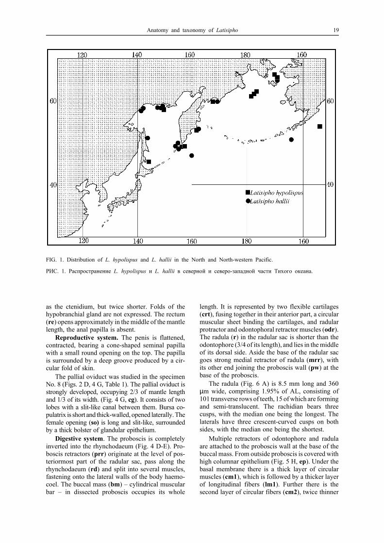
Distribution: Okhotsk, Bering and Chukchi seas,Alaska; 40-930 m (Fig. 1).
Material examined anatomically: No. 1 – ZIN52050/30, Bering Sea, “Pelamida” sta. 3, 65 m, coll. V.Goryachev, 30.08.1972; No. 2, No. 7 – ZIN 58219/2,Bering Sea, 65°13.8’N 169°21.2’W, 45 m, coll. V. Koltun,B. Sirenko, 20.08.1988; No. 4, No. 5 – ZIN 52050/41,64°22.55’N 169°10.9’W, 38 m, 22.08.1988; No. 3, No.6 – IO, Bering Sea, Anadyr Bay, 63°59.9’N 177°38.6’W,87 m, 12.10.1951; No. 8 – ZMMU 18587, Bering Sea,Anadyr Bay, DT 35, 90 m, 22.06.1986.
Description.Shell. More than 70 specimens were examined.
The shell is large or medium-sized, from moderatelythick and solid to thin and fragile; the shape changesfrom widely fusiform to oval. The shell is coveredwith dark-brown, brownish or light-olive smoothperiostracum, tight-fitting to the shell. The siphonalcanal is from strongly curved to the left to straight.The spiral sculpture is represented by irregular ob-lique spiral ridges, up to 10 on the penultimate whorl,separated by wide smooth interspaces without anyspiral striation. Weak spiral ribs may be observedonly near the siphonal canal. The axial sculpture isrepresented by incremental lines. The shell outlineis very variable (Figs. 2, 3).
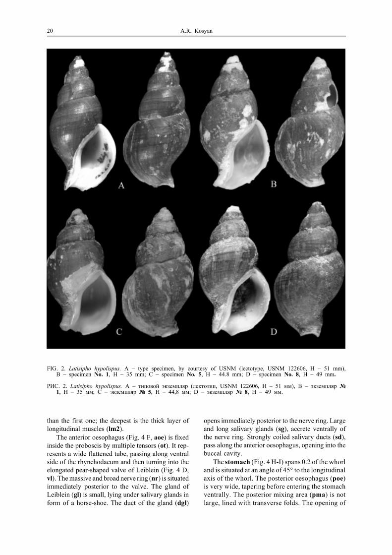
Morphology. Description of specimen No. 1(Fig. 2 B), having the shell most similar to that ofthe type specimen of L. hypolispus (Fig. 2 A):
H 35 mm, h 25.5 mm, AL 18.5 mm.External anatomy. The soft body consists of
3.5 whorls. The mantle occupies one whorl, thekidney – 0.2, the digestive gland and the gonad –2.5 (Fig. 4 A-B). The head (Fig. 4 B, hd) is ratherlarge, with the length slightly exceeding the width.Cephalic tentacles are very long and thick, bearingwell-noticeable black eyes on small lobes at the base.The foot is folded transversally. The wide propodi-um (prp) is separated by the deep propodial groove(prpg). The operculum (op) is oval, with terminalnucleus.
The mantle length is twice the width (Fig. 4 C).The siphon (s) is moderately long, slightly protru-ding beyond the mantle edge. The ctenidium (ct) islong, occupying 5/6 of the length and 1/3 of thewidth of the mantle. The osphradium (os) is as wide
18 A.R. Kosyan
as the ctenidium, but twice shorter. Folds of thehypobranchial gland are not expressed. The rectum(re) opens approximately in the middle of the mantlelength, the anal papilla is absent.
Reproductive system. The penis is flattened,contracted, bearing a cone-shaped seminal papillawith a small round opening on the top. The papillais surrounded by a deep groove produced by a cir-cular fold of skin.
The pallial oviduct was studied in the specimenNo. 8 (Figs. 2 D, 4 G, Table 1). The pallial oviduct isstrongly developed, occupying 2/3 of mantle lengthand 1/3 of its width. (Fig. 4 G, cg). It consists of twolobes with a slit-like canal between them. Bursa co-pulatrix is short and thick-walled, opened laterally. Thefemale opening (so) is long and slit-like, surroundedby a thick bolster of glandular epithelium.
Digestive system. The proboscis is completelyinverted into the rhynchodaeum (Fig. 4 D-E). Pro-boscis retractors (prr) originate at the level of pos-teriormost part of the radular sac, pass along therhynchodaeum (rd) and split into several muscles,fastening onto the lateral walls of the body haemo-coel. The buccal mass (bm) – cylindrical muscularbar – in dissected proboscis occupies its whole
length. It is represented by two flexible cartilages(crt), fusing together in their anterior part, a circularmuscular sheet binding the cartilages, and radularprotractor and odontophoral retractor muscles (odr).The radula (r) in the radular sac is shorter than theodontophore (3/4 of its length), and lies in the middleof its dorsal side. Aside the base of the radular sacgoes strong medial retractor of radula (mrr), withits other end joining the proboscis wall (pw) at thebase of the proboscis.
The radula (Fig. 6 A) is 8.5 mm long and 360µm wide, comprising 1.95% of AL, consisting of101 transverse rows of teeth, 15 of which are formingand semi-translucent. The rachidian bears threecusps, with the median one being the longest. Thelaterals have three crescent-curved cusps on bothsides, with the median one being the shortest.
Multiple retractors of odontophore and radulaare attached to the proboscis wall at the base of thebuccal mass. From outside proboscis is covered withhigh columnar epithelium (Fig. 5 H, ep). Under thebasal membrane there is a thick layer of circularmuscles (cm1), which is followed by a thicker layerof longitudinal fibers (lm1). Further there is thesecond layer of circular fibers (cm2), twice thinner
FIG. 1. Distribution of L. hypolispus and L. hallii in the North and North-western Pacific.
РИС. 1. Распространение L. hypolispus и L. hallii в северной и северо-западной части Тихого океана.
Anatomy and taxonomy of Latisipho 19
than the first one; the deepest is the thick layer oflongitudinal muscles (lm2).
The anterior oesophagus (Fig. 4 F, aoe) is fixedinside the proboscis by multiple tensors (ot). It rep-resents a wide flattened tube, passing along ventralside of the rhynchodaeum and then turning into theelongated pear-shaped valve of Leiblein (Fig. 4 D,vl). The massive and broad nerve ring (nr) is situatedimmediately posterior to the valve. The gland ofLeiblein (gl) is small, lying under salivary glands inform of a horse-shoe. The duct of the gland (dgl)
opens immediately posterior to the nerve ring. Largeand long salivary glands (sg), accrete ventrally ofthe nerve ring. Strongly coiled salivary ducts (sd),pass along the anterior oesophagus, opening into thebuccal cavity.
The stomach (Fig. 4 H-I) spans 0.2 of the whorland is situated at an angle of 45° to the longitudinalaxis of the whorl. The posterior oesophagus (poe)is very wide, tapering before entering the stomachventrally. The posterior mixing area (pma) is notlarge, lined with transverse folds. The opening of
FIG. 2. Latisipho hypolispus. A – type specimen, by courtesy of USNM (lectotype, USNM 122606, H – 51 mm),B – specimen No. 1, H – 35 mm; C – specimen No. 5, H – 44.8 mm; D – specimen No. 8, H – 49 mm.
РИС. 2. Latisipho hypolispus. A – типовой экземпляр (лектотип, USNM 122606, H – 51 мм), B – экземпляр №1, H – 35 мм; C – экземпляр № 5, H – 44,8 мм; D – экземпляр № 8, H – 49 мм.
20 A.R. Kosyan

FIG. 3. Latisipho hypolispus. A – type specimen of Latisipho pharcidus, by courtesy of USNM (syntype, USNM205243, H – 29 mm); B – specimen No. 2, H – 33 mm; C – specimen No. 3, H – 40.2 mm; D – ZMMU18587, Bering Sea, Anadyr Bay, 90 m, H – 49 mm; E – specimen No. 4, H – 48.7 mm; F – specimen No. 5,H – 44.8 mm.
РИС. 3. Latisipho hypolispus. A – типовой экземпляр Latisipho pharcidus (синтип, USNM 205243, H – 29 мм), B– экземпляр № 2, H – 33 мм; C – экземпляр № 3, H – 40,2 мм; D – ЗМ МГУ 18587, Берингово море,Анадырский зал., 90 м, Н – 49 мм; E – экземпляр № 4, H – 48,7 мм; F – экземпляр № 5, Н – 44,8 мм.
Anatomy and taxonomy of Latisipho 21

FIG. 4. Anatomy of Latisipho hypolispus No. 1. A–B – body, removed from the shell, C – mantle, D–E – organsof the body haemocoel (D – lateral view, E – ventral view), F – proboscis, opened dorsally, rhynchodaeum isturned aside, G – mantle of the female No. 8, H – stomach, general view, I – stomach, opened dorsally.
РИС. 4. Анатомия Latisipho hypolispus № 1. А–В – мягкое тело, С – мантия, D–E – органы туловищногогемоцеля (D – латеральный вид, Е – вентральный вид), F – хобот, вскрытый дорзально, G – мантия самки№ 8. H – желудок, общий вид, I – желудок, вскрытый дорзально.
22 A.R. Kosyan
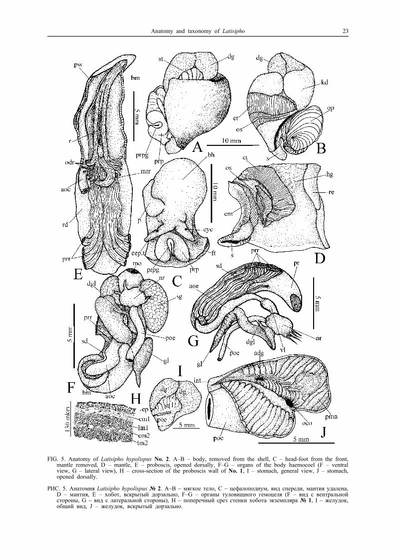
FIG. 5. Anatomy of Latisipho hypolispus No. 2. A–B – body, removed from the shell, C – head-foot from the front,mantle removed, D – mantle, E – proboscis, opened dorsally, F–G – organs of the body haemocoel (F – ventralview, G – lateral view), H – cross-section of the proboscis wall of No. 1, I – stomach, general view, J – stomach,opened dorsally.
РИС. 5. Анатомия Latisipho hypolispus № 2. A–B – мягкое тело, C – цефалоподиум, вид спереди, мантия удалена,D – мантия, E – хобот, вскрытый дорзально, F–G – органы туловищного гемоцеля (F – вид с вентральнойстороны, G – вид с латеральной стороны), H – поперечный срез стенки хобота экземпляра № 1, I – желудок,общий вид, J – желудок, вскрытый дорзально.
Anatomy and taxonomy of Latisipho 23

the posterior duct of digestive gland (pdg) is rathersmall, situated at some distance above the oesop-hageal opening (oeo). The inner stomach wall islined with divergent high oblique folds (oti), thelower of which are very large (lti). The small roun-ded opening of the anterior duct of the digestive
gland (adg) is located close to the region where theintestine (int) is entering the stomach. The lateralsulcus is absent. The outer stomach wall is linedwith large deep transversal folds (cte).
Specimen No. 2 (Fig. 3 B) has thin shell withstraight siphonal canal and light-olive periostracum,
FIG. 6. Radulae of Latisipho hypolispus.A – No. 1, B – No. 5, C – D – No. 3, E – No. 2, F – No. 4.
РИС. 6. Радулы Latisipho hypolispus. A – № 1, B – № 5, C – D – № 3, E – № 2, F – № 4.
24 A.R. Kosyan
and conhologically is similar to the type specimenof L. pharcidus (Fig. 3 A). Its description is givenbelow.
H 33 mm, h 25 mm, AL 17.7 mm; immaturemale:
External anatomy. Posterior coils of the bodyabove the stomach were torn off during extractionfrom the shell. The remaining part consists of 1.5whorls, the mantle occupies one whorl, the kidney– 0.2, and remaining parts are the digestive glandand the gonad (Fig. 5 A-B). The head (hd) is nearlysquare in form (the length without tentacles is equalto the width). The length of the thick contractedtentacles is twice that of the head. The eyes sit onthe small lobes of the tentacles at the distance of onethird of their length from the base. The foot (ft) isfolded transversally. The wide propodium (prp) isseparated by a deep propodial groove. The opercu-lum (op) is leaf-shaped, with terminal nucleus dis-lodged to the left.
The mantle length, unlike the previous speci-men, is equal to its width (Fig. 5 D). The siphon (s)is moderately long, protruding beyond the incrassatemantle edge. The ctenidium (ct) is long, crescent-curved, occupying 3/4 of mantle length. The osph-radium (os) is 2/3 of ctenidium length. Folds of thehypobranchial gland (hg) are weakly expressed. Therectum (re) opens in the middle of the mantle length,the anal papilla is absent.
Reproductive system. The penis (Fig. 5 C, p)is small, structured in the same way as in the previousspecimen.
Digestive system. The proboscis is completelyinverted into the rhynchodaeum (Fig. 5 F-G, pr).Proboscis retractors (prr) originate at the proboscisbase as a wide ventral band, pass along the rhyn-chodaeum, and then split into several muscles, fas-tening onto the lateral walls of the body haemocoel.The buccal mass (Fig. 5 E, bm) occupies the wholeproboscis length. The radula (r) is 10 mm long and600 µm wide, bearing 85 transverse rows of teeth,and comprising 3.39% of AL (Fig. 6 E). The rachi-
dian is tricuspidate, with the median cusp beingmuch smaller and narrower than the others. Thelaterals are tricuspidate, with median cusp the shor-test. Multiple retractors of odontophore (odr) andradula are attached to the proboscis wall at the baseof the buccal mass. The composition of the probosciswall is similar to that of above described specimen(Fig. 5 H).
The anterior oesophagus, valve of Leiblein andthe nerve ring are similar to those already described.The gland of Leiblein (gl) is large, with the longtapering duct (dgl) which opens slightly posteriorto the nerve ring. Salivary glands (sg) are medium-sized, beanlike, separate. Slightly coiled salivaryducts (sd) open into the buccal cavity.
The stomach occupies 0.25 of the whorl andruns along longitudinal axis of the whorl (Fig. 5 I).The posterior oesophagus and stomach are filledwith mud, and the internal structure (Fig. 5 J) ispoorly recognizable. The posterior oesophagus isvery wide, tapering before entering the stomach ven-trally. The posterior mixing area (pma) is not large,lined with transverse folds. The opening of the an-terior duct of digestive gland (adg) is rather large,situated ventrally not far from the beginning of theintestine (int). The posterior opening was not foundbecause of the poor preservation of stomach tissues.The inner stomach wall is lined with divergent ob-lique folds. The intestine in the region of its fallinginto the stomach is lined with transverse folds. Thelateral sulcus is absent. The outer stomach wall islined with large high transverse folds.
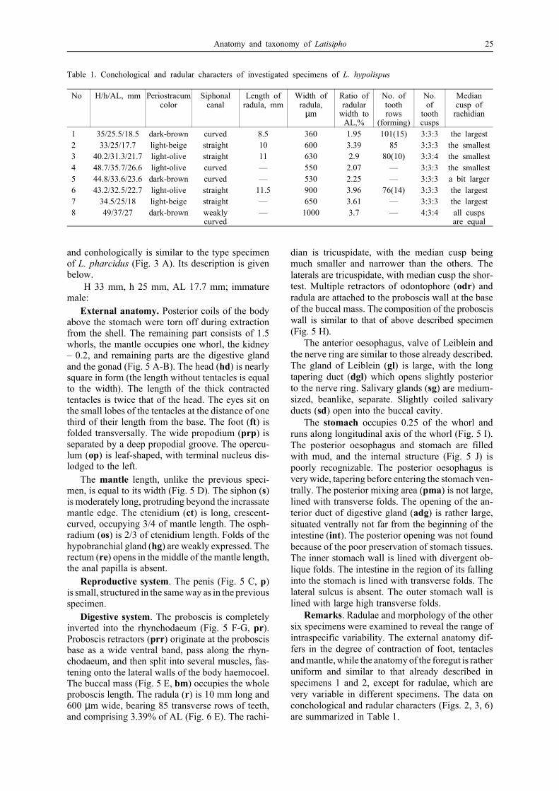
Remarks. Radulae and morphology of the othersix specimens were examined to reveal the range ofintraspecific variability. The external anatomy dif-fers in the degree of contraction of foot, tentaclesand mantle, while the anatomy of the foregut is ratheruniform and similar to that already described inspecimens 1 and 2, except for radulae, which arevery variable in different specimens. The data onconchological and radular characters (Figs. 2, 3, 6)are summarized in Table 1.
Table 1. Conchological and radular characters of investigated specimens of L. hypolispus
No H/h/AL, mm Periostracumcolor
Siphonalcanal
Length ofradula, mm
Width ofradula,
µm
Ratio ofradular
width toAL,%
No. oftoothrows
(forming)
No.of
toothcusps
Mediancusp of
rachidian
1 35/25.5/18.5 dark-brown curved 8.5 360 1.95 101(15) 3:3:3 the largest2 33/25/17.7 light-beige straight 10 600 3.39 85 3:3:3 the smallest3 40.2/31.3/21.7 light-olive straight 11 630 2.9 80(10) 3:3:4 the smallest4 48.7/35.7/26.6 light-olive curved — 550 2.07 — 3:3:3 the smallest5 44.8/33.6/23.6 dark-brown curved — 530 2.25 — 3:3:3 a bit larger6 43.2/32.5/22.7 light-olive straight 11.5 900 3.96 76(14) 3:3:3 the largest7 34.5/25/18 light-beige straight — 650 3.61 — 3:3:3 the largest8 49/37/27 dark-brown weakly
curved— 1000 3.7 — 4:3:4 all cusps
are equal
Anatomy and taxonomy of Latisipho 25

Latisipho hallii (Dall, 1873)(Figs. 1, 7–17)
Sipho hallii Dall, 1873: 59, pl. 2, fig. 3.Tritonofusus hallii. – Dall, 1902: 525, pl. 36, fig. 9.Latisipho nallii. – Dall, 1921: 96, pl. 8, fig. 7 (misspelling
of L. hallii).Colus hallii. – Abbot, 1974: 211 fig. 2332. – Tiba, Kosuge,
1981: 11.Colus (Latisipho) hallii. – Okutani, 2000: 464, pl. 232,
fig. 59.Tritonofusus jordani Dall, 1913: 588.Colus (Latisipho) jordani. – Dall, 1921: 96.Colus jordani. – Abbot, 1974: 211, fig. 2333.Colus (Latisipho) errones Dall, 1919: 321. – Dall, 1925:
12, pl. 3, fig. 6.Latisipho errones. – Golikov, Gulbin, 1977: 184.Colus errones. – Tiba, Kosuge, 1981: 5.Aulacofusus (Limatofusus) georgianus Dall, 1921: 95, pl.
8, fig. 3.Colus georgianus. – Abbot, 1974: 211, fig. 2318. – Tiba,
Kosuge, 1981: 9.Aulacofusus georgianus. – Golikov, Gulbin, 1977: 182.Colus (Aulacofusus) georgianus. – Golikov, Sirenko, 1998:
114, pl. 8, fig. F.Latisipho georgianus. – Kantor, Sysoev, 2005: 132.Helicofusus luridus Golikov in Golikov et Scarlato, 1985:
406, fig. 7.
Possible synonym:Colus (Latisipho) dalmasius Dall, 1919: 322. – Dall, 1925:
12, pl. 1, fig. 9.
Type locality of Sipho hallii: Sanborn Harbor,Nagai, Alaska.
Type locality of Tritonofusus jordani: PugetSound, Washington (for lectotype).
Type locality of Colus (Latisipho) errones: Be-ring Sea.
Type locality of Aulacofusus (Limatofusus) ge-orgianus: Gulf of Georgia, 60-200 fms.
Type locality of Helicofusus luridus: TerpenijaBay, Sakhalin Island, 48°48.7’N, 143°55.3’E, 53 m.
Type locality of Colus (Latisipho) dalmasius: offthe coast of British Columbia, 238 fms.
Distribution: Kurile Islands, Okhotsk Sea, BeringSea, Alaska to northern California; 2-1112 m (Fig.1).
Material examined anatomically: No. 1, No. 2 –ZIN 28389/2, Paramushir Island, 50°07’N, 156°37’E, 552m, 26.06.1988; No. 3 – ZIN 28420/4, Kamchatka, offLopatka Cape, 68 m, 31.07.1954; No. 4 – ZIN 55992/3,Okhotsk Sea, 51°36’N, 156°09’E, 112 m, 24.06.1988; No.5 – ZMMU 18587, Bering Sea, Anadyr Bay, DT 35, 90m, 22.06.1986; No. 6 – ZIN 52050/41, Bering Sea,64°22.55’N, 169°10.9’W, 38 m, 22.08.1988; No. 7 – IO,Bering Sea, Anadyr Bay, 63°59.9’N, 177°38.6’W, 87 m,12.10.1951; No. 8, No. 9 – ZMMU 18186, Okhotsk Sea,58°41’N, 150°00’E, 110 m, 28.08.1985; No. 10 – IO,53°25.3’N, 160°59.6’E, 58 m, 1952; No. 11 – ZMMU18214, Okhotsk Sea, 58°41’N, 150°E, 28.08.1985; No.12 – Sakhalin Island, 51°15’N, 143°51’E, 80 m, 18.10.04,coll. I.P. Smirnov; No. 13 – ZMMU, Sakhalin Bay,59°57.5’N 141°01.0’E, 95 m; No. 14 – ZIN ?/3 58°57’N,148°44’E, 28.06.1915; No. 15 – IO, “Vityaz” 10th cruise,Kamchatka, 54°35’N, 162°02’E, 445 m, 22.05.1952; No.
16 – IO, Okhotsk Sea, “Vityaz” 2d cruise, 59°10.0’N,148°31.0’E, 71 m, 04.09.1949.
Description.Shell. More than 50 specimens were examined.
The shell is large or medium-sized, with slightly orstrongly convex whorls, moderately thick and solid,or thin and fragile. The periostracum is dark-brown,olive, greenish-grey, or yellowish-olive, smooth,tight-fitting to the shell. The siphonal canal is usuallystraight, but sometimes slightly deflected to the left.The spiral sculpture is represented by multiple spiralribs separated by interspaces usually narrower, orsometimes wider than the ribs. The spiral striationis uniform throughout the shell; on the penultimatewhorl there are 20 to 30 ribs. The axial sculpture isrepresented by incremental lines (Figs. 7-8, ).
Morphology. Several specimens with differentshells similar to the type specimens of L. hallii, L.jordani, L. georgianus, L. errones, and Helicofususluridus (see Table 2) were studied.
Description of specimens with shells most simi-lar to those of the type specimens of L. hallii (Fig.7 A) and L. errones (Fig. 8 A) is given below.
No. 1. H 35.3 mm, h 26 mm, AL 18.8 mm, maturefemale (Fig. 8 C); No. 2. H 34.3 mm, h 27.4 mm,AL 20 mm, mature female:
External anatomy. Posterior coils of the bodyabove the stomach were torn off during extractionfrom the shell (Fig. 10 A). The remaining part con-sists of 1.5 whorls; the mantle occupies one whorl,the kidney (kd) – 0.3, the digestive gland and thegonad – the remainder. The head (Fig. 10 B, hd) islarge, with long, half-contracted tentacles, pressedto each other. The eyes are black and small, sittingon minor lobes of the first third of cephalic tentacles.The propodium (prp) of the contracted foot (ft) isnarrow, separated by a deep propodial groove(prpg). The operculum (op) is leaf-like, with termi-nal nucleus dislodged to the left.
The mantle is a bit longer than wide (Fig. 10 C).The siphon (s) is long and muscular. The ctenidium(ct) occupies 7/8 of the mantle length and 1/4 of itswidth. The osphradium (os) is twice shorter and abit narrower than the ctenidium. The hypobranchialgland (hg) consists of a number of large deep glan-dular folds situated in the posterior right quadrantof the mantle. The rectum (re) has the form of a thintube, half-covered by the capsular gland (cg), ope-ning in the middle of the mantle length. The analpapilla is absent.
Reproductive system (Fig. 10 C). The capsulargland is thick, well-developed, consisting of twolobes separated by a slit-like canal. Bursa copulatrix(bc) has thin epithelial walls, which make the con-nection between the thick glandular lobes and longand narrow female orifice. The female genital ope-ning (so) is surrounded by a thick bolster.
Digestive system. The proboscis is completely
26 A.R. Kosyan
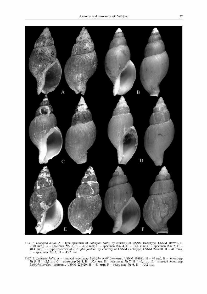
FIG. 7. Latisipho hallii. A – type specimen of Latisipho hallii, by courtesy of USNM (lectotype, USNM 108981, H– 40 mm); B – specimen No. 5, H – 42.2 mm; C – specimen No. 4, H – 37.4 mm; D – specimen No. 7, H –40.4 mm; E – type specimen of Latisipho jordani, by courtesy of USNM (lectotype, USNM 226420, H – 41 mm);F – specimen No 6, H – 43.2 mm.
РИС. 7. Latisipho hallii. A – типовой экземпляр Latisipho hallii (лектотип, USNM 108981, H – 40 мм), B – экземпляр№ 5, Н – 42,2 мм; C – экземпляр № 4, Н – 37,4 мм; D – экземпляр № 7, Н – 40,4 мм; E – типовой экземплярLatisipho jordani (лектотип, USNM 226420, H – 41 мм); F – экземпляр № 6, Н – 43,2 мм.
Anatomy and taxonomy of Latisipho 27
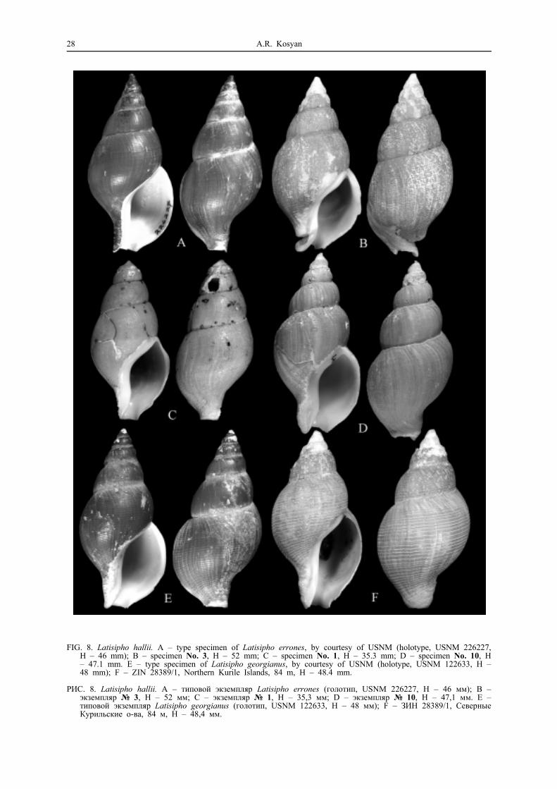
FIG. 8. Latisipho hallii. A – type specimen of Latisipho errones, by courtesy of USNM (holotype, USNM 226227,H – 46 mm); B – specimen No. 3, H – 52 mm; C – specimen No. 1, H – 35.3 mm; D – specimen No. 10, H– 47.1 mm. E – type specimen of Latisipho georgianus, by courtesy of USNM (holotype, USNM 122633, H –48 mm); F – ZIN 28389/1, Northern Kurile Islands, 84 m, H – 48.4 mm.
РИС. 8. Latisipho hallii. A – типовой экземпляр Latisipho errones (голотип, USNM 226227, H – 46 мм); B –экземпляр № 3, Н – 52 мм; C – экземпляр № 1, Н – 35,3 мм; D – экземпляр № 10, Н – 47,1 мм. E –типовой экземпляр Latisipho georgianus (голотип, USNM 122633, H – 48 мм); F – ЗИН 28389/1, СеверныеКурильские о-ва, 84 м, H – 48,4 мм.
28 A.R. Kosyan

nverted into the rhynchodaeum (Fig. 10 E-F). Pro-boscis retractors (prr) originate at the level of pos-teriormost part of the radular sac, pass along therhynchodaeum (rd) and split into several muscles,fastening onto the lateral walls of the body hae-mocoel. The buccal mass (bm) occupies 2/3 ofthe proboscis length. The radula (r) in the radularsac is of the same length as odontophore, and liesin the middle of its dorsal side. It is 9.8 mm longand 350 µm wide, consisting of 65 rows of teeth,comprising 1.86% of the aperture length (Fig. 11E-F). The rachidian is tricuspid, with a bit longermedian cusp. The laterals are less curved than inL. hypolispus, tricuspid, with the median cusp si-tuated very close to the inner cusp, and havingalmost the same length. In general the radula looksmore tuff and rude. Aside the base of the radularsac goes a strong medial retractor of radula (mrr),with its other end joining the fibers of one ofproboscis retractors. Multiple retractors of odon-tophore (odr) and radula attach to the probosciswall at the base of the buccal mass (Fig. 10 D).The proboscis wall from its inner side looks lon-gitudinally striated by the muscle fibers.
Anterior oesophagus is a wide flattened tube (Fig.10 D, aoe), passing along the ventral side of therhynchodaeum till the beginning of the proboscis,and then turning into the medium-sized pear-shapedvalve of Leiblein (Fig. 10 E, vl). The massive nervering (nr) is well-developed, situated immediatelyposterior to the valve. The gland of Leiblein (gl) isa long narrow brownish strip of glandular tissue. Inthe upper part it gradually tapers, turning into theduct of the gland (dgl), entering the posterior oeso-phagus at some distance posterior to the valve. Theposterior part is also tapered. Oval, medium-sized,separate salivary glands (sg) are located on bothsides of the nerve ring. Salivary ducts (sd) are thinand strongly coiled, running on both sides of theanterior oesophagus. The posterior oesophagus(poe) is a bit narrower than the anterior one. Thelarge anterior aorta (ao) also comes out of the nervering; it has spongy walls impregnated with whitespots of unknown origin.
The stomach (spm. No. 2) occupies 0.33 of thewhorl (Fig. 10 G, H). The posterior oesophagus fallsventrally into the stomach. The posterior mixing area(pma) is not large, lined with high transverse folds.The opening of the anterior duct of digestive gland(adg) is rounded, situated approximately in the mid-dle of the inner stomach wall. The opening of theposterior duct was not found. Three longitudinalfolds on the inner wall (lti) run from the anterioropening towards the opening of the oesophagus(oeo); the remaining part of the stomach is linedwith transverse folds, gradually decreasing towardsthe intestine. The outer wall is lined posteriorly withhigh and anteriorly – with low transverse folds.
Specimen No. 3 conhologically is similar to thetype specimen of L. errones (Fig. 8 A);
H 52 mm, h 37 mm, AL 27 mm (Fig. 8 B), maturefemale:
External anatomy (Fig. 12 A – B) is similar tothat of the previous specimens. Pale-yellowish bodylacks pigmentation.
The mantle length is equal to its width (Fig. 12C); organs of the mantle cavity have the same pro-portions as in the previous specimens. Folds of thehypobranchial gland are not expressed.
Reproductive system (Fig. 12 C). The pallialoviduct is strongly developed, occupying 2/3 of cte-nidium length and 1/3 of its width (Fig. 12 C, cg).It consists of two lobes with a slit-like canal betweenthem. The lobes abruptly terminate and turn into thecurved thin-walled bursa copulatrix (bcp). The latterends by the female opening (so), which is long andslit-like, with longitudinal folds seen on the innerside. The opening is surrounded by a thick bolsterof glandular epithelium.
Digestive system. Organs of body haemocoelare compactly packed in a similar way as in theprevious specimens (Fig. 12 E). The radula (r) isstrong, containing the remnants of food in its veryanterior part, 12.5 mm long and 430 µm wide, con-sisting of 88 rows of teeth (15 – forming), compri-sing 1.59% of AL (Fig. 11 A-B). The histologicalcomposition of the proboscis wall is the same as inL. hypolispus (Fig. 12 F). From the outside there areremnants of the covering columnar epithelium (ep).Under the epithelium there is a thick layer of circularmuscles (cm1), which is followed by a layer oflongitudinal fibers (lm1). The next is a very thinlayer of circular fibers (cm2). The deepest is thethickest layer of longitudinal muscles (lm2); it looksheterogeneous, because muscle fibers in its upperand lower parts are oriented a bit differently.
The valve of Leiblein (vl) is thick and short. Afterpassing through the nerve ring the anterior oesop-hagus strongly flattens. The gland of Leiblein (gl)is large, not tapered (obviously ragged in its posteriorpart), opening by a long broad duct (dgl) at a smalldistance posterior to the nerve ring. The anterioraorta (ao) comes out of the nerve ring and consistsof whitish connective tissue, impregnated with whitespots. Salivary glands (sg) are not large, beanlike,separate, situated on both sides of the anterior partof the proboscis.
The stomach occupies 0.33 of the whorl, beingsituated at an angle of 30° to the longitudinal axisof the whorl (Fig. 12 G, H). The posterior oesopha-gus is very wide, strongly narrowing prior to enteringthe stomach ventrally. The posterior mixing area(pma) is small, lined with poorly expressed trans-verse folds. The posterior opening of the duct ofdigestive gland (pdg) is rather large, situated abovethe oesophageal opening (oeo). The upper part of
Anatomy and taxonomy of Latisipho 29

FIG. 9. Anatomy of Latisipho hallii No. 1, 2. A – body, removed from the shell, B – head-foot, dorsal view, C –mantle, D – proboscis, opened dorsally, E–F – organs of the body haemocoel (E – from the right, F – from theleft), G – stomach, general view, H – stomach, opened dorsally, I – penis of the male No. 5, ventral view.
РИС. 9. Анатомия Latisipho hallii № 1, 2. A – мягкое тело, B – цефалоподиум, вид с дорзальной стороны, С– мантия. D – хобот, вскрытый дорзально, E–F – органы туловищного гемоцеля (Е – вид справа, F – видслева), G – желудок, общий вид, H – желудок, вскрытый дорзально, I – вентральный вид пениса самца №5, вид с вентральной стороны.
30 A.R. Kosyan

the inner stomach wall is lined with not high trans-verse folds, the lower – with minute oblique folds(oti). The large opening of the anterior duct of di-gestive gland (adg) is situated near the ventral chan-nel, not far from the beginning of the intestine. Theouter stomach wall is lined with multiple high tran-sverse folds (cte).
Specimen No. 4 is conchologically similar to thetype specimens of L. georgianus (Fig. 8 E) and L.jordani (Fig. 7 E); H 37.4 mm, h 27.5 mm, AL 20mm (Fig. 7 C); immature female:
External anatomy. Posterior coils of the bodyabove the stomach were torn off during extractionfrom the shell. The remaining part consists of 1.5
FIG. 10. Radulae of Latisipho hallii. A–B – No. 3, C–D – No. 10, E–F – No. 1.
РИС.10. Радулы Latisipho hallii. A–B – № 3, C–D – № 10, E–F – № 1.
Anatomy and taxonomy of Latisipho 31

FIG. 11. Anatomy of Latisipho hallii No. 3. A–B – body, removed from the shell, C – mantle, D – proboscis, openeddorsally, E – organs of the body haemocoel, lateral view, F – cross-section of the proboscis wall, G – stomach,general view, H – stomach, opened dorsally.
РИС. 11. Анатомия Latisipho hallii № 3. A–B – мягкое тело, С – мантия, D – хобот, вскрытый дорзально, Е –органы туловищного гемоцеля, латеральный вид, F – поперечный срез стенки хобота, G – желудок, общийвид, H – желудок, вскрытый дорзально.
32 A.R. Kosyan

FIG. 12. Anatomy of Latisipho hallii No. 4. A–B – body, removed from the shell, C – mantle, D–E – organs of thebody haemocoel (D – right-ventral view, E – lateral-left view), F – proboscis, opened dorsally, G – cross-sectionof the proboscis wall, H – stomach, opened dorsally, I – stomach, general view.
РИС. 12. Анатомия Latisipho hallii № 4. A–B – мягкое тело, С – мантия, D–E – органы туловищного гемоцеля(D – вид вентрально справа, Е – вид слева), F – хобот, вскрытый дорзально, G – поперечный срез стенкихобота, H – желудок, вскрытый дорзально, I – желудок, общий вид.
Anatomy and taxonomy of Latisipho 33

whorls, the mantle occupies one whorl, the kidney– 0.2, and remaining parts are the digestive glandand the gonad (Fig. 13 A–B). The body is pigmentedwith little black spots. The broad flattened head (hd)without tentacles is 1.5 times wider than long. Thelength of thick contracted tentacles is twice that ofthe head. The eyes sit on small lobes of the tentaclesat the distance of one third of their length from thebase. The foot (ft) is twice folded. The wide propo-dium (prp) is separated by a deep propodial groove.The operculum (op) is leaf-shaped, with terminalnucleus slightly dislodged to the left.
The mantle length is 1.5 times more than the
width (Fig. 13 C). The siphon (s) is long and mus-cular, transgressing far off the bounds of the mantleedge. The ctenidium (ct) is moderately large, occu-pying 3/4 of the mantle length. The osphradium (os)is twice shorter and narrower than the ctenidium.Folds of the hypobranchial gland (hg) are not ex-pressed. The rectum (re) opens in the middle of themantle; the anal papilla is absent.
Reproductive system. The capsular gland (Fig.13 C, cg) is poorly developed.
Digestive system. The proboscis is completelyinverted into the rhynchodaeum(Fig. 13 D-E). Thebuccal mass (bm) occupies the whole length of the
FIG. 13. Radulae of Latisipho hallii. A–B – No. 4, C–D – No. 6, E–F – No. 7, G – No. 9, H – No. 8.
РИС.13. Радулы Latisipho hallii. A–B – № 4, C–D – № 6, E–F – № 7, G – № 9, H – № 8.
34 A.R. Kosyan

FIG. 14. Latisipho hallii. A – specimen No. 9, H – 49.2 mm; B-C – specimen No. 12, H – 53.2 mm; D-E – specimenNo. 16, H – 47.6 mm; F-G – type specimen of Helicofusus luridus (holotype, ZIN 33739/1, H – 60.2 mm); H– specimen from the Okhotsk Sea, 151°50’-153°30’E, south off Alevin Cape, by courtesy of D.O. Alexeev, H –53.3 mm; I-J – specimen from ZMMU 18186, Okhotsk Sea, 58°41’N, 150°00’E, H – 54 mm; K – specimen fromKamchatka, 600 m, by courtesy of D.O. Alexeev, H – 70.5 mm.
РИС. 14. Latisipho hallii. A – экземпляр № 9, H – 49,2 мм. B-C – экземпляр № 12, H – 53,2 mm; D-E –экземпляр № 16, H – 47,6 мм; F-G – типовой экземпляр Helicofusus luridus (голотип, ЗИН 33739/1, H – 60,2мм); H – экземпляр из Охотского моря, 151°50’-153°30’E, к югу от мыса Алевина, любезно предоставленД.О. Алексеевым, H – 53,3 мм; I-J – экземпляр из ЗМ МГУ 18186, Охотское море, 58°41’N, 150°00’E, H –54 мм; K – экземпляр с Камчатки, 600 м, любезно предоставлен Д.О. Алексеевым, H – 70,5 мм.
Anatomy and taxonomy of Latisipho 35

rhynchocoel (Fig. 13 F). It is strongly contracted,making the radula (r) curved. The radula is 10 mmlong and 530 µm wide, bearing 81 transverse rowsof teeth (6 are forming) and comprising 2.65% ofAL (Fig. 14 A-B). The composition of the probosciswall (Fig. 13 G) is similar to that of L. hypolispus.
Anterior oesophagus (aoe) is a wide flattenedtube (Fig. 13 D-E). The valve of Leiblein (vl) is verylarge (1/4 of the length of anterior oesophagus). Thegland of Leiblein (gl) is also rather large. The shortduct of the gland (dgl) opens immediately posteriorto the nerve ring. Salivary glands (sg) are large,rounded, and separate. Salivary ducts are thin andcoiled, opening into the buccal cavity.
The stomach occupies 0.33 of the whorl and
runs along the longitudinal axis of the whorl (Fig.13H–I). The posterior oesophagus is very wide, tape-ring before entering the stomach ventrally. The pos-terior mixing area (pma) is not large, lined withtransverse folds. The opening of the posterior ductof digestive gland (pdg) is rather small, situated justabove the opening of the oesophagus (oeo). Theupper part of the inner stomach wall is lined withhigh transverse folds, the lower – with two rows ofminute divergent oblique folds. The outer wall islined with multiple transverse folds. The opening ofthe anterior duct of digestive gland was not found.
The penis was studied in specimen No. 5; H 42.2mm, h 32.4 mm, AL 24.6 mm, adult male (Fig. 7B). The penis is flattened, contracted, bearing a
FIG. 15. Latisipho hallii. A – specimen No. 11, H – 44.8 mm; B – specimen No. 14, H – 42.0 mm; C – specimenNo. 15, H – 40.0 mm; D – specimen No. 13, H – 47.5 mm.
РИС.15. Latisipho hallii. A – экземпляр № 11, H – 44.8 мм; B – экземпляр № 14, H – 42.0 мм; C – экземпляр№ 15, H – 40.0 mm; D – экземпляр № 13, H – 47.5 mm.
36 A.R. Kosyan

cone-shaped seminal papilla with a small round ope-ning on the top. The papilla is surrounded by a deepgroove produced by a circular fold of skin (Fig. 10I).
Specimens figured on the Fig. 14 have the spiralsculpture of sharp ribs separated by interspaces twicewider than the ribs. This kind of sculpture is char-acteristic for Helicofusus luridus (Fig. 14 F-G –holotype). There are several specimens representingthe transition of the sculpture from sharp spiral ribswith wide interspaces (Fig. 15 A-B) to flattenedspiral ribs with narrow interspaces (Fig. 15 C-D).Below the brief morphological description of speci-men No. 12 (Fig. 16) with sharp spiral ribs is given,with the notes on radulae of the other figured spe-cimens (Figs. 14, 15).
No. 12. H 53.2 mm, h 37.5 mm, AL 27 mm.External anatomy (Fig. 16 A) is the same as in
the previous described specimens.The mantle length (Fig. 16 D) is approximately
equal to its width; organs of the mantle cavity haveusual proportions.
Reproductive system. The capsular gland islarge and dorso-ventrally flattened (Fig. 16 D, cg).
Bursa copulatrix is medium-sized, folded ventrally,with a slit-like female orifice.
Digestive system. Organs of the body haemocoelare compactly packed in a way described above forthe other specimens of L. hallii. The buccal mass(bm) is of the same length as the proboscis (Fig. 16C). The radula (r) in the radular sac is equal toodontophore length, lying in the middle of its dorsalside. It is 14 mm long and 900 µm wide, comprising3.33% of AL, with 78 rows of teeth, 5 of which areforming. The rachidian is relatively broad, bearingthree equal cusps. The laterals are tricuspid with themedian cusp the shortest, similar to those of previousspecimens.
The valve of Leiblein (vl) is medium-sized, elon-gated. Salivary ducts (sd) are narrow and stronglycoiling, passing on the both sides of the anterioroesophagus, and separating from it in the beginningof the valve of Leiblein. The nerve ring and salivaryglands are strongly fused with the connective tissue,so it is impossible to distinguish one from another.The gland of Leiblein (gl) is medium-sized, slightlytapering towards the end. The short broad duct ofthe gland (dgl) opens in a small distance posteriorto the nerve ring.
FIG. 16. Anatomy of Latisipho hallii No. 12. A – anterior view of the head-foot, B – stomach, opened dorsally, C– proboscis, opened dorsally (rhynchodaeum opened dorsally and pulled backwards), D – mantle.
РИС. 16. Анатомия Latisipho hallii № 12. A – фронтальный вид головы и ноги, B – желудок, вскрытый дорзально,C – хобот, вскрытый дорзально (ринходеум отвернут назад), D – мантия.
Anatomy and taxonomy of Latisipho 37
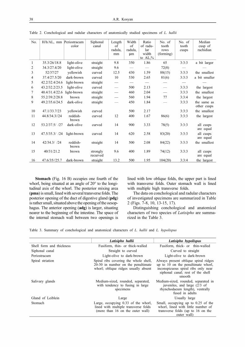
Stomach (Fig. 16 B) occupies one fourth of thewhorl, being situated at an angle of 20° to the longi-tudinal axis of the whorl. The posterior mixing area(pma) is small, lined with several transverse folds. Theposterior opening of the duct of digestive gland (pdg)is rather small, situated above the opening of the oesop-hagus. The anterior opening (adg) is larger, situatingnearer to the beginning of the intestine. The space ofthe internal stomach wall between two openings is
lined with low oblique folds, the upper part is linedwith transverse folds. Outer stomach wall is linedwith multiple high transverse folds.
The data on conchological and radular charactersof investigated specimens are summarized in Table2 (Figs. 7-8, 10, 13-15, 17).
Distinguishing conchological and anatomicalcharacters of two species of Latisipho are summa-rized in the Table 3.
Table 2. Conchological and radular characters of anatomically studied specimens of L. hallii
No. H/h/AL, mm Periostracum color
Siphonalcanal
Lengthof
radula,mm
Widthof
radula,µm
Ratioof radu-
larwidth
to AL,%
No. oftoothrows
(forming)
No. oftoothcusps
Mediancusp of
rachidian
1 35.3/26/18.8 light-olive straight 9.8 350 1.86 65 3:3:3 a bit larger2 34.3/27.4/20 light-olive straight 9.6 — — 72(8) — —3 52/37/27 yellowish curved 12.5 430 1.59 88(15) 3:3:3 the smallest4 37.4/27.5/20 dark-brown curved 10 530 2.65 81(6) 3:3:3 a bit smaller5 42.2/32.4/24.6 light-brown straight — — — — — —6 43.2/32.2/23.5 light-olive curved — 500 2.13 — 3:3:3 the largest7 40.4/31.4/22.6 light-brown straight — 460 2.04 — 3:3:3 the smallest8 55.2/39.2/28.8 brown straight — 560 1.94 77 3:3:4 the largest9 49.2/35.6/24.5 dark-olive straight — 450 1.84 — 3:3:3 the same as
other cusps10 47.1/33.7/23 yellowish curved 500 2.17 3:3:3 the smallest11 44.8/34.3//24 reddish-
browncurved 12 400 1.67 86(6) 3:3:3 the largest
12 53.2/37.5/ /27 dark-olive curved 14 900 3.33 78(5) 3:3:3 all cuspsare equal
13 47.5/35.3/ /24 light-brown curved 14 620 2.58 83(20) 3:3:3 all cuspsare equal
14 42/34.3/ /24 reddish-brown
straight 14 500 2.08 84(22) 3:3:3 the smallest
15 40/31/21.2 brown stronglyrecurved
9.6 400 1.89 74(12) 3:3:3 all cuspsare equal
16 47.6/35//25.7 dark-brown straight 13.2 500 1.95 104(20) 3:3:4 the largest
Table 3. Summary of conchological and anatomical characters of L. hallii and L. hypolispus
Latisipho hallii Latisipho hypolispusShell form and thickness Fusiform, thin- or thick-walled Fusiform, thick- or thin-walledSiphonal canal Straight to curved Curved to straightPeriostracum Light-olive to dark-brown Light-olive to dark-brownSpiral striation Spiral ribs covering the whole shell,
20-30 in number on the penultimatewhorl; oblique ridges usually absent
Always present oblique spiral ridgesup to 10 on the penultimate whorl,inconspicuous spiral ribs only near
siphonal canal; rest of the shellsmooth
Salivary glands Medium-sized, rounded, separated,with tendency to fusing in large
specimens
Medium-sized, rounded, separated injuveniles, and large (2/3 of
rhynchodaeum length), ventrallyfused in adults
Gland of Leiblein Large Usually largeStomach Large, occupying 0.33 of the whorl,
lined with multiple transverse folds(more than 16 on the outer wall)
Small, occupying up to 0.25 of thewhorl, lined with little number oftransverse folds (up to 16 on the
outer wall)
38 A.R. Kosyan

FIG. 17. Radulae of Latisipho hallii. A – No. 14, B – No. 15, C-D – No. 13, E-F – No. 9, G – No. 11, H – No.16. Scale bar – 200 µm.
РИС. 17. Радулы Latisipho hallii. A – № 14, B – № 15, C-D – № 13, E-F – № 9, G – № 11, H – № 16.Масштабный отрезок – 200 мкм.
Anatomy and taxonomy of Latisipho 39

Discussion
It is well known that the shell in buccinids isextremely variable [Golikov, 1963, 1980; Goryac-hev, 1978]. Even without special anatomical inves-tigation some authors [Foster, 1981; Kantor, Sysoev,2005] noted the strong similarity in shells of L. hallii,L. jordani, L. errones and L. georgianus, and madea suggestion about their belonging to one widelydistributed and variable species.
Based on spiral sculpture it is possible to divideseven studied species – six nominal species of La-tisipho and Helicofusus luridus – into two groups.In L. hallii, L. jordani, L. errones, L. georgianusand H. luridus the spiral sculpture is “of numerousflattened threads with narrow channelled interspa-ces, coarser on the canal” [Dall, 1919: 322], whileshells of L. hypolispus and L. pharcidus are sculp-tured “only by a few obsolete spirals” [Dall, 1891:188].
The similarity of types of L. hypolispus and L.pharcidus is not obvious. Although the spiral scul-pture is similar, the shell of the type specimen of L.pharcidus is thin and oval, with a straight siphonalcanal and light-olive periostracum, while that of L.hypolispus – solid and more fusiform, with curvedsiphonal canal and dark-brown periostracum. Afterexamining a large number of specimens having thespiral sculpture only of irregular spiral ridges, it wasfound that the thickness of the shell and the colourof the periostracum change significantly, producinga lot of transitions, and the siphonal canal can becurved to a variable extent. In Russian museumsthere is no material from the type locality of L.pharcidus (i.e. Sakhalin Island, east of Aniva Cape,930 m), nevertheless, we consider that present sam-ples from the Bering Sea and Kamchatka fall withinthe limits of intraspecific variability. Radulae alsoappear to be very variable, different even in twospecimens from the same sample and similar con-chologically (Table 1, No. 2 and No. 7). Varyingfeatures are the number of cusps on the lateral teethand the length of the median cusp of the rachidian.The ratio of radula width to the aperture length variesfrom 1.95 to 3.96. It seems that the larger values(2.9-3.96) are those of shells with straight siphonalcanals, while the smaller – of those with curvedcanals (1.95-2.25). Nevertheless, the specimen No.8 with weakly curved siphon represents the transitionbetween these two forms, having the ratio 3.7. Theanatomy of the foregut is uniform, and does notreveal any distinctions between the specimens ofdifferent forms. Based on all above mentioned, it ispossible to say that L. hypolispus and L. pharcidusrepresent forms of one extremely variable species.
Specimens with spiral ribs are also remarkablyvariable. Thus, shells can be thin and fragile, or thickand solid; with convex, or flattened whorls; with
curved or straight siphon, and periostracum of dif-ferent tint. The spiral sculpture itself varies greatly:the ribs can be sharp, separated by interspaces widerthan the ribs themselves (Fig. 14 F-G – Helicofususluridus), or, on the contrary, smooth and flattened,separated by grooves twice narrower than the rib(Figs. 7, 8, 15 C-D). The number of ribs is notconstant, varying from 20 (Fig. 7) to 30 (Fig. 8 A-C)on the penultimate whorl. The radula is extremelyvariable, but without a correlation to the shell vari-ability: it can be different in two similar specimensand similar in two different specimens (Fig. 14 C-Dand H; Fig. 11 C-D and 14 E-F). The ratio of radularwidth to the aperture length varies from 1.59 to3.33%, i.e. it is as variable as in L. hypolispus.
The anatomy of the foregut and reproductivesystem is similar within the group, and reveals veryfew differences between L. hypolispus and L. hallii(Table 3). The stomach of L. hypolispus is a bitsmaller than that of L. hallii: it occupies up to 0.25of the whorl, lined with little number of transversefolds (up to 16 on the outer wall), while the stomachof L. hallii occupies 0.33 of the whorl, and linedwith multiple transverse folds (more than 16 on theouter wall). Salivary glands are usually fused inadults of L. hypolispus and separate in L. hallii.Fusing of glands is not uncommon and occurs insome other Buccinoidea: Habevolutopsius and Lus-sivolutopsius (Volutopsiinae, Buccinidae) [Kantor,1990], Germonea (Prosiphiinae, Buccinulidae) [Ha-rasewych, Kantor, 2004].
Based on all above mentioned, it is necessary toreduce L. pharcidus to the junior synonym of L.hypolispus, and L. jordani, L. errones, L. georgianusand H. luridus – to the junior synonyms of L. hallii.
As the morphology of both species is similar,and the radula is extremely variable, they can beclearly destinguished only conchologically by spiralsculpture. In L. hypolispus the spiral sculpture is notuniform, represented by irregular oblique spiral rid-ges (up to 10 on the penultimate whorl), and weakinconspicuous spiral ribs near siphonal canal. In L.hallii all the shell is covered with regular frequentspiral ribs, 20-30 in number on the penultimatewhorl. The majority of samples containing L. hypo-lispus were from the Bering Sea region, and L. hallii– from the Okhotsk Sea (Fig. 1). Nevertheless, therewere a number of samples containing both species.
AcknowledgmentsI am deeply grateful to Dr. Yu.I. Kantor from Severtsov
Institute of Ecology and Evolution for his support andgood advice during the research and valuable commentson the manuscript. The photos of molluscan type specimenswere taken by him and kindly placed at my disposal. Ihighly appreciate the possibility to work with buccinidcollections provided by Dr. B.I. Sirenko and R.A. Kor-mushkina from the Zoological Institute, Dr. D.L. Ivanovand Dr. A.V. Sysoev from the Zoological Museum of
40 A.R. Kosyan

the Moscow State University, and Dr. D.O. Alexeev fromVNIRO. Thanks are to I.N. Marin for his active help inget-up of the paper.
This research was supported by INTAS grant No 04-83-3120.
ReferencesAbbott R. T. 1974. American seashells, marine mol-
lusks of the Atlantic and Pacific coasts of NorthAmerica. 2nd ed. 663 p.
Bouchet P., Waren A. 1985. Mollusca Gastropoda:Taxonomical notes on tropical deep water Buc-cinidae with descriptions of new taxa. Mémoiredu Muséum national d’Histoire naturelle, sérieA, Zoology, 133: 457-499.
Dall W. H. 1873. Descriptions of new species ofMollusca from the coast of Alaska, with noteson some rare forms. Proceedings of the Cali-fornian Academy of Sciences, 5: 57-62.
Dall W. H. 1891. Scientific results of explorationsby the U.S. Fish Comission steamer “Albatross”.XX. On some new or interesting west Americanshells obtained from the dredgings of the U.S.steamer “Albatross” in 1888, and from other re-sources. Proceedings of the United States Nati-onal Museum, 14(849): 173-191.
Dall W. H. 1895. Scientific results of explorationsby the U.S. Fish Comission steamer “Albatross”.XXXIV. Report on Mollusca and Brachiopodadredged in deep water, chiefly near the HawaiianIslands, with illustrations of hitherto unfiguredspecies from northwest America. Proceedings ofthe United States National Museum, 17(1032):675-733, pls. 23-32.
Dall W. H. 1902. Illustrations and descriptions ofnew, unfigured or imperfectly known shells, chi-efly American, in the U. S. National Museum.Proceedings of the United States National Mu-seum, 24(1264): 499-566, pls. 27-40.
Dall W. H. 1906. Early history of the generic nameFusus. Journal of Conchology, 11(10): 289-297.
Dall W. H. 1916. Prodrome of a revision of thechrysodomoid whelks of the boreal and arcticregions. Proceedings of the Biological Societyof Washington, 29: 7-8.
Dall W. H. 1913. Diagnoses of new shells fromthe Pacific Ocean. Proceedings of the UnitedStates National Museum, 45(2002): 587-597.
Dall W. H. 1918. Notes on Chrysodomus and othermollusks from the North Pacific Ocean. Proce-edings of the United States National Museum,54: 207-234.
Dall W. H. 1919. Descriptions of new species ofMollusca from the North Pacific Ocean in thecollection of the United States National Museum.Proceedings of the United States National Mu-seum, 54(2295): 293-371.
Dall W. H. 1921. Summary of the marine shell-bearing mollusks of the Northwest coast of Ame-rica, from San Diego, California, to the PolarSea, mostly contained in the collection of theUnited States National Museum, with illustrations
of hitherto unfigured species. United States Na-tional Museum Bulletin, 112: 1-217.
Dall W. H. 1925. Illustrations of unfigured typesof shells in the collection of the United StatesNational Museum. Proceedings of the United Sta-tes National Museum, 66(2554): 1-41, pls 1-36.
Foster N. R. 1981. A synopsis of the marine pro-sobranch gastropod and bivalve mollusks in Alas-kan waters. University of Alaska, Institute ofmarine Science, Report R81-3: 479 p.
Golikov A. N. 1963. The gastropod mollusks ofthe genus Neptunea Bolten. In: Fauna SSSR. Mol-lyuski, Leningrad, Nauka, 5(1): 183 p. [In Rus-sian].
Golikov A. N. 1980. Mollusks Buccininae of theWorld Ocean. In: Fauna SSSR. Mollyuski, Le-ningrad, Nauka, 5(2): 466 p. [In Russian].
Golikov A. N., Gulbin V. V. 1977. Prosobranchgastropods (Gastropoda, Prosobranchiata) of theshelf of Kurile Islands. II Orders Hamiglossa –Homoestropha. In: Fauna of the inshore zonesof Kurile Islands. Moscow, Nauka: 172-268 [InRussian].
Golikov A.N., Scarlato O.A. 1985. Shell-bearing gas-tropod and bivalve molluscs of the shelf of so-uthern Sakhalin and their ecology. In: Biocenosesand fauna of the shelf of south Sakhalin. Issle-dovaniya Fauny Morei, 30(38): 360-487 [In Rus-sian].
Golikov A. N., Sirenko B. I. 1998. Prosobranchgastropods of the continental slope of Kurile Is-lands. Ruthenica, 8(2): 91-135. [In Russian].
Golikov A. N., Sirenko B. I., Gulbin V. V., ChabanE. M. 2001. Checklist of shell-bearing gastropodsof the northwestern Pacific. Ruthenica, 11(2):153-174.
Goryachev V. N. 1978. Prosobranch gastropods ofthe genus Neptunea Röding from the Bering Sea.Moscow, Nauka, 90 p. [In Russian].
Harasewych M. G., Kantor Yu. I. 2004. The deep-seaBuccinoidea (Gastropoda: Neogastropoda) of theScotia Sea and adjacent abyssal plains and tren-ches. The Nautilus, 118: 1-42.
Kantor Yu. I. 1990. The gastropod mollusks of theWorld Ocean: subfamily Volutopsiinae. Moscow,Nauka, 178 p. [In Russian].
Kantor Yu. I. 2003. Comparative anatomy of thestomach of Buccinoidea (Neogastropoda). Journalof Molluscan Studies, 69: 203-220.
Kantor Yu. I., Sysoev A. V. 2005. Catalogue ofmollusks of Russia and adjacent countries. KMKScientific Press Ltd. Moscow, 627 p. [In Russian].
Kosuge S. 1991. Illustrations of type specimens of
Anatomy and taxonomy of Latisipho 41
Molluscs described by William Healey Dall(North-western Pacific gastropods), 29 pls.
Matsukuma A., Okutani T., Habe T. 1991. Worldseashells of rarity and beauty. National ScienceMuseum, Tokyo, 206 p.
McLean J. H. 1995. Four new genera for North-eastern Pacific prosobranch gastropods. The Na-utilus, 108(2): 39-41.
Okutani T. 2000. Marine molluscs of Japan. TokaiUniversity Press, 1173 p.
Tiba R., Kosuge S. 1981. North Pacific shells. 8.Genus Colus Roeding, 1798. Occasional Publi-cation of the Institute of Malacology of Tokyo:1-26.
Turgeon D. D., Quinn J. F., Bogan A. E., CoanE. V., Hochberg F. G., Lyons W. G., MikkelsenP. M., Neves R. J., Roper C. F. E., RosenbergG., Roth B., Scheltema A., Thompson F. G.,Veccione M., Williams J. D. 1998. Common andscientific names of aquatic invertebrates from theUnited States and Canada: Mollusks, 2nd ed.American Fisheries Society Special Publication
26. American Fisheries Society. Bethesda, Ma-ryland, USA, 526 p.
�
Анатомия и таксономический состав рода La-tisipho Dall (Gastropoda: Buccinidae) из рос-сийских водА. Р. КОСЬЯНИнститут проблем экологии и эволюции им. А. Н.Северцова РАН, Ленинский просп., 33, Москва 119071,e-mail: [email protected]
РЕЗЮМЕ. На основании анализа конхологичес-ких, анатомических и радулярных признаков шестиноминальных видов рода Latisipho и вида Helico-fusus luridus, отмеченных в морской фауне России,вид L. pharcidus сведен в младший синоним L. hy-polispus, а виды L. errones, L. jordani L. georgianusи Helicofusus luridus – в синонимы L. hallii.
42 A.R. Kosyan