anatomo-histology brain atrophy, flattening of gyri, widening of sulci, & cerebral ventricles...
Post on 20-Dec-2015
227 views
TRANSCRIPT

Anatomo-Histology
Brain atrophy, flattening of gyri,widening of sulci,
& cerebral ventricles
Loss of cholinergic neurons, in nucleus of Meynert, hippocampus & association cortices
Loss of adrenergic neurons, in locus ceruleus
Denudation of neurons, stripping of dendrites, damage to axons
Increased microglia
From Table8.10

Pathology
Accumulation of cell inclusions: lipofuscin, Hirano and Lewy bodies,
altered cytoskeletal Tau proteins, ubiquitin
Neurofibrillary tangles, neuritic plaques with amyloid,
Perivascular amyloid, distributed throughout the brain, but especially
in frontal, prefrontal lobes, Hippocampus,
association cortices

MetabolismDecreased oxidative metabolism, slower enzyme
activity (Ch. 7)
Free-radical accumulation (Ch. 5)
Impaired iron homeostasis (Ch. 7)
Other minerals, zinc, aluminum
Reduced level/metabolism/ activity of neurotransmitters
Increased amyloid peptide with accumulation of amyloid proteins
Increased prion protein
Altered immune response

TABLE 8-11 Genes Known to be Linked to Alzheimer’s Disease*
Chromosomal Location
Gene Type Age of Onset % Cases Familial
% Cases All
1
Presenilin 2
AD 40-70 yrs. 20 2-3
10
Risk factor > 60 yrs. ------ ------
14
Presenilin 1
AD 30-60 yrs. 40-60 5-10
19
Apolipoprotein E4
Risk factor > 60 yrs. ------ 40-50
21
APP Mutation ( APP) Down’s Syndrome
Trisomy
AD 45-60 yrs. 2-3 < 1
* Given the rapid progress in genetics, additional genes may be related to AD.

TABLE 8-9 Characteristics of Multi-Infarct Dementia
History of abrupt onset or stepwise deterioration History of transient ischemic attack or stroke Presence of hypertension or arrhythmia Presence of any neurologic focal symptoms or signs

Learning at all Ages Induces Successful
Aging

TABLE 8-6 Mechanisms of Effect s of Increased Education on Successful Aging Adequate income
Better access to medical care Better access to recreational activity Good nutrition Responsible health behaviors Moderate alcohol intake
Abstinence from smoking Possibility of increased brain reserve capacity More dendritic branching, more synapses Better cerebral blood flow Better neural cell efficiency, adaptability,
redundancy, survival and growth

TABLE 8-12 Basic Goals of Alzheimer’s Disease Management
• to maintain the patient's safety whil e allowing as much independence and dignity as ;possible
• to optimize the pati ’ent s function by treating underlying medical conditions and avoiding the use of drugs with side effects on the nervous syst ;em
• to prevent stressful situations that maycause or exacerbate catastrophic ;reactions
• to identify and manage complications that ma y arise from , ;agitation depression and incontinence
• to provide medica l and social in formation to the patient's family in addition to any needed counselin .g

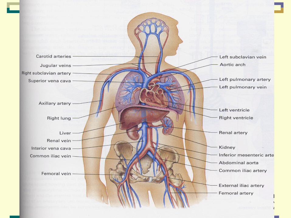
Aging of the Cardiovascular System
Chapter 16
P.S. Timiras

Major Functions of the Cardiovascular System
• Transports O2 & nutrients to the tissues
& returns C02 to the lungs and other products of metabolism to the kidney
• Regulates body temperature
• Distributes hormones and other agents that regulate cell function

Major Components of the Cardiovascular System
Heart
Pump that circulates the blood throughout the body
Vascular System
Transports blood to the body tissues
Central Nervous System (CNS)
Particularly the centers in the medulla that regulate the function of the heart and blood vessels

AtherosclerosisWhat?

Arteriosclerosis: Sclerosis: hardening of the arterial wall and narrowing
of the arterial lumen
Atherosclerosis:Same as arteriosclerosis PLUS presence of artheroma
(yellowish plaque containing lipids and cholesterol) on the
arterial wall

Atherosclerosis
UniversalProgressiveDeleterious
Irreversible (?)

Fig. 16-3: Natural history of atherosclerosis. Pathogenesis of human atherosclerotic lesions and their clinical manifestations.

Myocardial Infarction
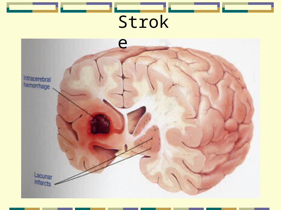
Stroke

Aneurysm

Atherosclerosis Affects the ArteriesArteries: the large arteries, the arterioles, & the capillaries
See Box 16-1, Fig. 16-1, Fig. 16-2 (pgs. 287-289)

Progressiveness of Atherosclerosis
• Onset at young age
• Progression through adulthood
• Culmination in old age with overt disease manifestation
• Consequences leading to severe disability & death

END

Extracellular cholesterol and cholesterol-filled macrophages (foam cells) accumulate in subendothelial space. Subsequent structural modifications of LDL particles render them more atherogenic. Oxidation of subendothelial LDL attracts monocytes, which enter subendothelium and change into macrophages. Macrophages may take up oxidized LDL to form foam cells.

Fibrous plaque larger than fatty streak and occupies more of the arterial lumen. Thickened cap synthesized by modified smooth muscle cells. Central core consists of extracellular cholesterol. Foam cells surrounding core derived primarily from smooth muscle cells. Fatty streaks may continue to form at periphery of plaque.

Total or partial occlusion of coronary artery due to plaque rupture and thrombosis can cause angina or frank myocardial infarction.
Plaques likely to rupture termed unstable. Rupture usually occurs in lipid-rich and foam cell-rich peripheral margins and may result in thrombosis and arterial occlusion.

Table 16-5: General Characteristics of Atherosclerotic Lesions
Early onset -- progressiveFocal lesionsEarly lesionsAdvance lesions
Damage, Repair, RegressionProgression of localized lesions influenced by:
Local factors: vessel structure and metabolism, blood turbulenceSystemic factors: diabetes, hypertension, stress, genetic predisposition