analysis of wastewater for use in agriculture - a laboratory manual
TRANSCRIPT
Analysis of Wastewater for Use in Agriculture -A Laboratory Manual of Parasitological
and Bacteriological Techniques
Rachel M. Ayres & D. Duncan MaraDepartment of Civil Engineering
University of LeedsLeeds, England
World Health OrganizationGeneva
1996
WHO Library Cataloguing in Publication Data
Ayres, Rachel M.Analysis of wastewater for use in agriculture: a laboratory manual of parasitological andbacteriological techniques/Rachel M. Ayres & D. Duncan Mara.
1. Microbiological techniques - laboratory manuals2. Waste disposal, Fluid - analysis - laboratory manuals3. Agriculture I. Mara, D. Duncan II. TitleISBN 92 4 154484 8 (NLM Classification: QW 25)
The World Health Organization welcomes requests for permission to reproduce or translate itspublications, in part or in full. Applications and enquiries should be addressed to the Office ofPublications, World Health Organization, Geneva, Switzerland, which will be glad to provide thelatest information on any changes made to the text, plans for new editions, and reprints andtranslations already available.
© World Health Organization 1996
Publications of the World Health Organization enjoy copyright protection in accordance with theprovisions of Protocol 2 of the Universal Copyright Convention. All rights reserved.
The designations employed and the presentation of the material in this publication do not implythe expression of any opinion whatsoever on the part of the Secretariat of the World HealthOrganization concerning the legal status of any country, territory, city or area or of its authorities,or concerning the delimitation of its frontiers or boundaries.
The mention of specific companies or of certain manufacturers’ products does not imply that theyare endorsed or recommended by the World Health Organization in preference to others of asimilar nature that are not mentioned. Errors and omissions excepted, the names of proprietaryproducts are distinguished by initial capital letters.
The authors alone are responsible for the views expressed in this publication.
Design and layout by WHO GRAPHICSPrinted in Finland
95/10419-WHO/GRA/Vammala-7000
The World Health Organization is a specialized agency of the United Nations with primaryresponsibility for international health matters and public health. Through this organization, whichwas created in 1948, the health professions of some 190 countries exchange their knowledgeand experience with the aim of making possible the attainment by all citizens of the world by theyear 2000 of a level of health that will permit them to lead a socially and economically productivelife.
By means of direct technical cooperation with its Member States, and by stimulating suchcooperation among them, WHO promotes the development of comprehensive health services,the prevention and control of diseases, the improvement of environmental conditions, thedevelopment of human resources for health, the coordination and development of biomedical andhealth services research, and the planning and implementation of health programmes.
These broad fields of endeavour encompass a wide variety of activities, such as developingsystems of primary health care that reach the whole population of Member countries; promotingthe health of mothers and children; combating malnutrition; controlling malaria and othercommunicable diseases including tuberculosis and leprosy; coordinating the global strategy forthe prevention and control of AIDS; having achieved the eradication of smallpox, promoting massimmunization against a number of other preventable diseases; improving mental health; providingsafe water supplies; and training health personnel of all categories.
Progress towards better health throughout the world also demands international cooperation insuch matters as establishing international standards for biological substances, pesticides, andpharmaceuticals; formulating environmental health criteria; recommending internationalnonproprietary names for drugs; administering the International Health Regulations; revising theInternational Statistical Classification of Diseases and Related Health Problems; and collectingand disseminating health statistical information.
Reflecting the concerns and priorities of the Organization and its Member States, WHOpublications provide authoritative information and guidance aimed at promoting and protectinghealth and preventing and controlling disease.
Ordering information
Analysis of Wastewater for Use in AgricultureA Laboratory Manual of Parasitological and Bacteriological TechniquesR.M. Ayres and D.D. Mara1996, iv + 31 pages [E, F, S*]ISBN 92 4 154484 8Sw.fr. 12.-/US $10.80; in developing countries: Sw.fr. 8.40Order no. 1150432

Acknowledgements
The authors are very grateful to Professor J. Schwartzbrod, University of Nancy, Nancy, France,for reviewing the text. They also wish to thank Dr Q. Bickle, London School of Hygiene andTropical Medicine, London, England, Dr L. Gibbons, International Institute of Parasitology, StAlbans, England, Mr M. Guy, Liverpool School of Tropical Medicine, Liverpool, England,Professor I. Miyazaki and Dr S. Habe, Kyushu University, Fukuoka, Japan, Dr F. Rochette,Janssen Pharmaceutica, Beerse, Belgium and Dr D. Spratt, CSIRO, Canberra, Australia forproviding fresh parasitic material for the photographic plates and permission to reproducematerial from existing texts.

1. Introduction
The use of wastewater for crop irrigation is becoming increasingly common, especially in arid andsemi-arid areas. Crop yields are higher as the wastewater contains not only water for cropgrowth, but also plant nutrients (mainly nitrogen and phosphorus). However, there is the risk thatwastewater irrigation may facilitate the transmission of excreta-related diseases. In the late1980s, the World Health Organization, the World Bank and the International Reference Centre forWaste Disposal sponsored a series of studies and meetings of experts to examine these healthrisks (International Reference Centre for Waste Disposal, 1985; Shuval et al., 1986; Prost, 1988;World Health Organization, 1989). From an appraisal of the available epidemiological evidence, itwas established that the major risks were:
- the transmission of intestinal nematode infections both to those working in the waste-water-irrigated fields and to those consuming vegetables grown in the fields; these infections are dueto Ascaris lumbricoides (the human roundworm), Trichuris trichiura (the human whipworm),and Ancylostoma duodenale and Necator americanus (the human hookworms); and
- the transmission of faecal bacterial diseases - bacterial diarrhoea and dysentery, typhoid andcholera - to the crop consumers.
Tab
le 1
Rec
om
men
ded
mic
rob
iolo
gic
al q
ual
ity
gu
idel
ines
fo
r tr
eate
d w
aste
wat
er u
sed
fo
r cr
op
irri
gat
ion
a
Cat
ego
ryR
euse
co
nd
itio
ns
Exp
ose
dg
rou
pIn
test
inal
nem
ato
des
b
(ari
thm
etic
mea
n n
o.
of
egg
s p
er li
trec )
Fae
cal c
on
form
s(g
eom
etri
c m
ean
no
. per
100
mlc )
Was
tew
ater
tre
atm
ent
exp
ecte
d t
o a
chie
ve t
he
req
uir
ed m
icro
bio
log
ical
qu
alit
yA
Irrig
atio
n of
cro
ps li
kely
to b
e ea
ten
unco
oked
,sp
orts
fie
lds,
pu
blic
park
s
Wor
kers
,co
nsum
ers,
publ
ic
≤1≤1
000d
A
serie
s of
st
abili
zatio
npo
nds
desi
gned
to
achi
eve
the
mic
robi
olog
ical
qu
ality
indi
cate
d,
or
equi
vale
nttr
eatm
ent
BIr
rigat
ion
of
cere
alcr
ops,
ind
ustr
ial
crop
s,fo
dder
cr
ops,
pa
stur
ean
dtr
eese
Wor
kers
≤1N
o st
anda
rdre
com
men
ded
Ret
entio
n in
st
abili
zatio
npo
nds
for
8-10
da
ys
oreq
uiva
lent
helm
inth
an
dfa
ecal
colif
orm
rem
oval
CLo
caliz
edirr
igat
ionf
ofcr
ops
in c
ateg
ory
B i
fex
posu
re
of
wor
kers
and
the
publ
ic
does
not o
ccur
Non
eN
ot a
pplic
able
Not
app
licab
leP
retr
eatm
ent
as r
equi
red
by
the
irrig
atio
nte
chno
logy
, bu
t no
t le
ssth
an
prim
ary
sedi
men
tatio
n
Sou
rce:
Wor
ld H
ealth
Org
aniz
atio
n (1
989)
.
a In
spec
ific
case
s, lo
cal e
pide
mio
logi
cal,
soci
ocul
tura
l an
d en
viro
nmen
tal
fact
ors
shou
ld b
e ta
ken
into
acc
ount
, an
d th
e gu
idel
ines
mod
ified
acco
rdin
gly.
bA
scar
isan
dT
richu
rissp
ecie
s an
d ho
okw
orm
s.c D
urin
g th
e irr
igat
ion
perio
d.d A
mor
e st
ringe
nt g
uide
line
(≤20
0fa
ecal
colif
orm
s pe
r 10
0 m
l) is
app
ropr
iate
for
pub
lic la
wns
, su
ch a
s ho
tel l
awns
, w
ith w
hich
the
pub
lic m
ayco
me
into
dire
ct c
onta
ct.
e In
the
case
of
frui
t tr
ees,
irr
igat
ion
shou
ld c
ease
tw
o w
eeks
bef
ore
frui
t is
pic
ked,
and
no
frui
t sh
ould
be
pick
ed o
ff th
e gr
ound
. S
prin
kler
irrig
atio
n sh
ould
not
be
used
.f A
lso
calle
d dr
ip o
r tr
ickl
e irr
igat
ion.
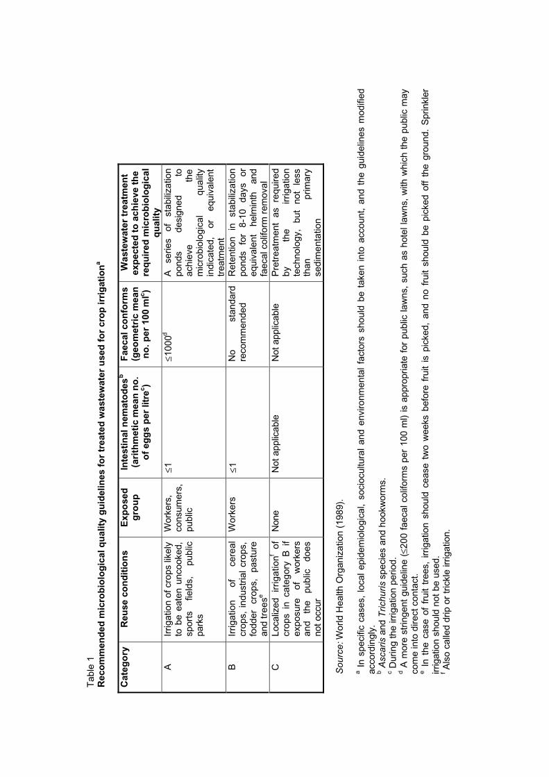
In order to prevent the transmission of these diseases, it has been recommended (World HealthOrganization, 1989) that:
- only treated wastewaters should be used for crop irrigation; and
- the treated wastewaters should comply with the microbiological quality guidelines given inTable 1.
This laboratory manual describes procedures for the examination of treated wastewater samplesto determine whether or not they comply with the guideline values given in Table 1. Theprocedures described have been selected because they are simple and require only the minimumof equipment; they can be readily carried out by laboratory technicians, even if they have noprevious microbiological expertise. Section 2 describes a method of counting the number ofintestinal nematode eggs in a wastewater sample, and section 3 describes two methods forcounting the numbers of faecal coliform bacteria. These parasitological and bacteriologicalmethods both focus on the analysis of samples at or near the quality guideline values given inTable 1, i.e. those containing about one intestinal nematode egg per litre and about 1000 faecalcoliform bacteria per 100 ml, although they can be simply adapted to count much highernumbers. Finally, section 4 contains recommendations for routine monitoring programmes.
2. Sanitary parasitology
2.1 The modified Bailenger method
Selection of method and comparison with others
The development of medical parasitology has led to a wide range of techniques for theenumeration of intestinal helminth eggs and larvae in faeces, and the basic principles of thesemethods have been adapted to the enumeration of helminth eggs in sludge and compost. Theenumeration of intestinal helminth eggs and larvae in wastewater, however, is much lessstraightforward. A great variety of human and animal parasite species, as well as free-livingspecies, may be present, varying in size, specific gravity and surface properties, and at muchlower concentrations than in faeces, sludge or compost.
Many methods for the enumeration of helminth eggs in wastewater are described in the literature.Each method has its own advantages and disadvantages: some have a high percentagerecovery, but are very time-consuming; many are not reported in sufficient detail for replication tobe possible, or their recovery rate is unknown; some require prohibitively expensive chemicals orare otherwise unsuitable for use in laboratories with limited equipment; and others only recover alimited range of species. It is clear that there is no one method that is universally useful, recoversall the helminth eggs of medical importance, and has a known rate of recovery.
All the available methods are based on one of two fundamental principles: either the parasites arefloated away from other debris in a solution of comparatively high relative density, or the fatty andother matter is separated in an interphase solution (normally ether or ethyl acetate) while theparasites sediment into a non-miscible buffer below. Both processes rely on centrifugal force. Thefactors which determine whether or not the concentration of particular species of parasite issuccessful are thought to be mainly the hydrophilic-lipophilic balance of the organism itself and itsrelative density in relation to that of the separating reagent (Bailenger, 1979). In practice, thismeans that the pH or the presence of heavy metals or alcohols in the reagents used may changethe surface properties of the parasite, and each species will respond differently to these changes:hence, no one method will concentrate all species with the same efficiency.
Bouhoum & Schwartzbrod (1989) compared a range of methods for faecal analysis with a view toadapting them for wastewater samples. Of the wide range of flotation solutions tested, they foundthat iodomercurate (Janeckso & Urbanyi, 1931) concentrated the greatest range of species ofparasitic helminth eggs, but concluded that the reagent was too corrosive and expensive forroutine use. Arthur’s method (described in Faust et al., 1938), in which saturated saccharose isused as a flotation solution, was found to deform eggs rapidly, while zinc sulfate solution (Faust etal., 1938) did not concentrate Trichuris spp. or Capillaria spp. very well. Bouhoum &Schwartzbrod (1989) concluded that Bailenger’s method (Bailenger, 1979), which they adaptedfor wastewater, was the best method overall: it requires relatively inexpensive reagents andsuccessfully concentrates the full range of species routinely found in wastewater.
This modified Bailenger method is generally useful, simple and cheap. However, its limitationsare well recognized (see below), and there is still a need for its further evaluation. Nevertheless,of all the methods available, it reliably recovers the eggs of the intestinal nematodes mentioned inTable 1, is replicable, and is already widely used in laboratories around the world. It is hoped thatthis manual will highlight the strengths and weaknesses of the method, standardize the way inwhich it is performed, and encourage the carrying out of the research still needed.
Advantages and disadvantages
The modified Bailenger method has the following advantages:
1. Sample collection and preparation are straightforward. Specialized containers are notrequired for sedimentation, and only the minimum of laboratory equipment is needed forsample processing. A few special chemical reagents are required but are usually both locallyavailable and inexpensive. McMaster slides are routinely used in parasitology laboratories,and should be readily available from laboratory supply companies.
2. Long periods of time spent at the microscope are very tiring and can lead to errors. Thetime required to examine each McMaster slide is usually only 1-2 minutes, so that operatorerror is reduced.
3. A subsample of each processed sample is examined for eggs. For greater accuracy and tocheck the homogenization, replicate samples can be examined and a mean egg count used;2-3 McMaster slides should be examined from each sample and the arithmetic mean countcalculated.
The method has the following disadvantages:
1. The percentage recovery of eggs when this method is used is not known, but it has beenshown that it compares favourably with that in all other techniques (Ayres et al., 1991;Bouhoum & Schwartzbrod, 1989). Bouhoum & Schwartzbrod showed that this method wouldsuccessfully recover a wide range of helminth eggs including Ascaris spp., Trichuris spp.,Capillaria spp., Enterobius vermicularis, Toxocara spp., Taenia spp. and Hymenolepis spp.,and Ayres et al. also routinely recovered hookworm eggs.
2. The method is not suitable for many of the operculated or trematode eggs, including thoseof Clonorchis sinensis, Diphyllobothrium latum, Fasciola hepatica, Fasciolopsis buski,Paragonimus westermani, P. pulmonalis, and Schistosoma spp. These all have intermediateaquatic hosts and are important in aquacultural (but not agricultural) reuse systems. Some ofthese eggs may float in the zinc sulfate flotation solution but sink again quickly or becomedistorted, making accurate identification difficult.
3. Ether is highly flammable and toxic. Rude, Peeler & Risty (1987) have shown that ether canbe replaced by ethyl acetate for the extraction of parasite eggs from faeces without any loss inefficiency. Ethyl acetate is much safer than ether; it has lower boiling and flash points and isless toxic. Its use is unlikely to affect the efficiency of the method for either raw or treatedwastewater.
Equipment and consumables
Reagents
The reagents required are the following: zinc sulfate solution (33%, relative density 1.18); ether(or ethyl acetate); acetoacetic buffer (pH 4.5) (15 g sodium acetate trihydrate, 3.6 ml glacial aceticacid, made up to 1 litre with distilled water); detergent solution (1 ml Triton X-100 or Tween 80,made up to 1 litre with tapwater).
Equipment
The following will be required: plastic containers for sample collection; a centrifuge (capable ofgenerating 1000 g) and centrifuge tubes with lids (50-ml and 15-ml tubes are preferable); Pasteurpipettes and teats; McMaster counting slides (1 or 2); a vortex mixer (not absolutely essential); asiphon; a 10-ml or 50-ml measuring cylinder or 10-ml graduated pipette.
Illustrated step-by-step guide
The method is very efficient for use with raw wastewater. However, the sample size must be
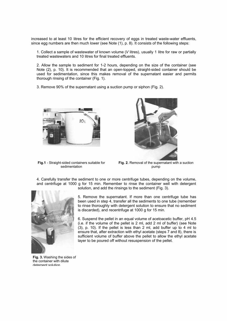
Fig. 3. Washing the sides ofthe container with dilutedetergent solution.
increased to at least 10 litres for the efficient recovery of eggs in treated waste-water effluents,since egg numbers are then much lower (see Note (1), p. 8). It consists of the following steps:
1. Collect a sample of wastewater of known volume (V litres), usually 1 litre for raw or partiallytreated wastewaters and 10 litres for final treated effluents.
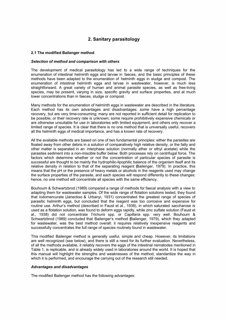
2. Allow the sample to sediment for 1-2 hours, depending on the size of the container (seeNote (2), p. 10). It is recommended that an open-topped, straight-sided container should beused for sedimentation, since this makes removal of the supernatant easier and permitsthorough rinsing of the container (Fig. 1).
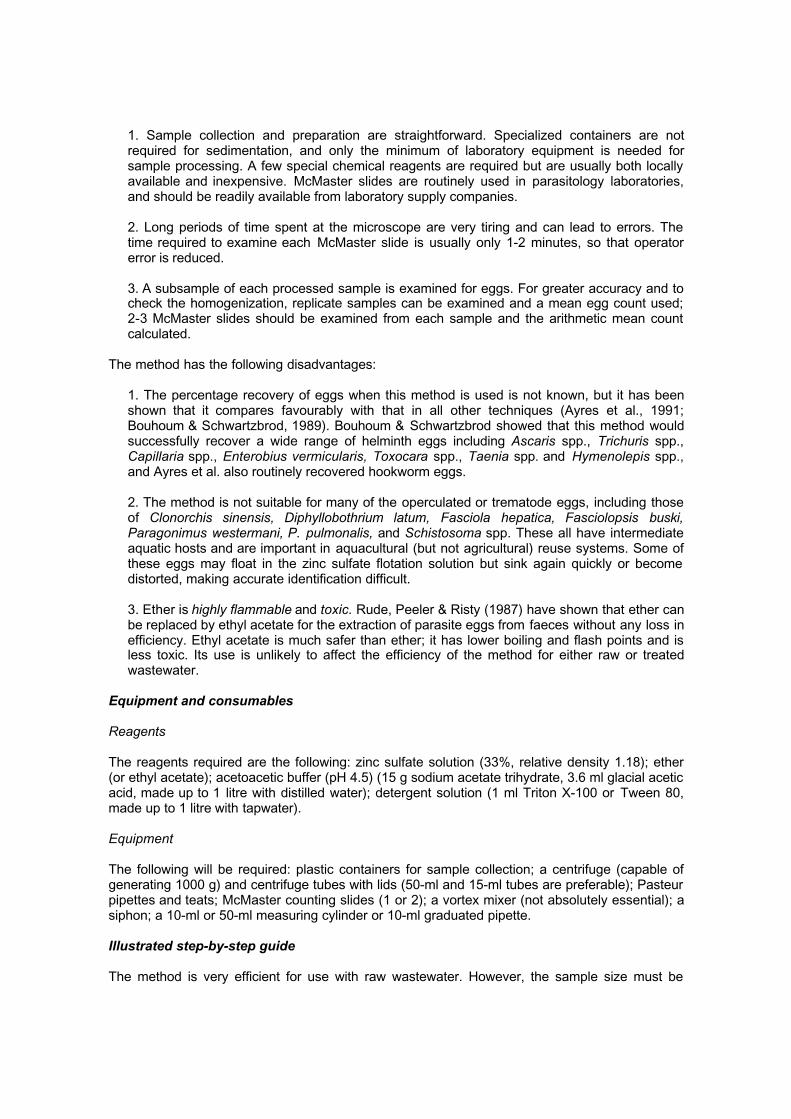
3. Remove 90% of the supernatant using a suction pump or siphon (Fig. 2).
Fig.1 - Straight-sided containers suitable forsedimentation
Fig. 2. Removal of the supernatant with a suctionpump
4. Carefully transfer the sediment to one or more centrifuge tubes, depending on the volume,and centrifuge at 1000 g for 15 min. Remember to rinse the container well with detergent
solution, and add the rinsings to the sediment (Fig. 3).
5. Remove the supernatant. If more than one centrifuge tube hasbeen used in step 4, transfer all the sediments to one tube (rememberto rinse thoroughly with detergent solution to ensure that no sedimentis discarded), and recentrifuge at 1000 g for 15 min.
6. Suspend the pellet in an equal volume of acetoacetic buffer, pH 4.5(i.e. if the volume of the pellet is 2 ml, add 2 ml of buffer) (see Note(3), p. 10). If the pellet is less than 2 ml, add buffer up to 4 ml toensure that, after extraction with ethyl acetate (steps 7 and 8), there issufficient volume of buffer above the pellet to allow the ethyl acetatelayer to be poured off without resuspension of the pellet.
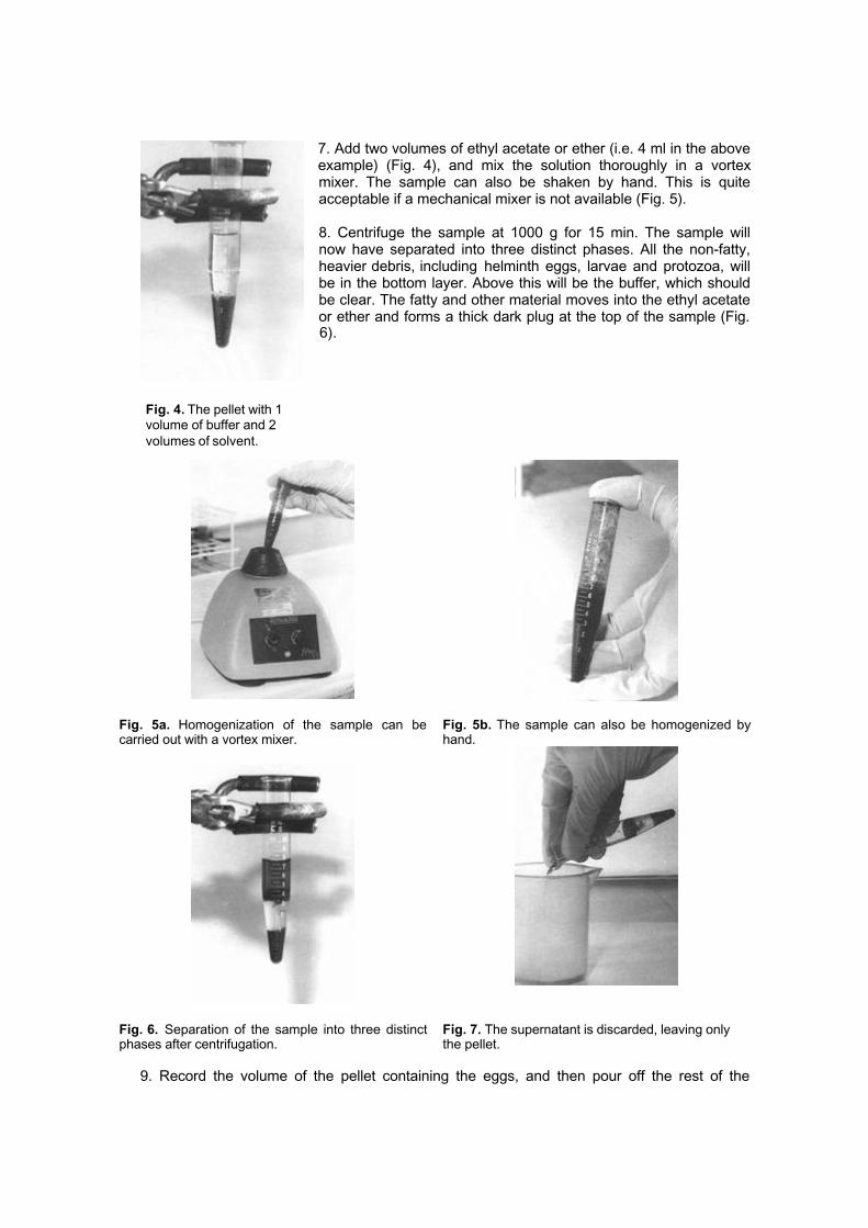
Fig. 4. The pellet with 1volume of buffer and 2volumes of solvent.
7. Add two volumes of ethyl acetate or ether (i.e. 4 ml in the aboveexample) (Fig. 4), and mix the solution thoroughly in a vortexmixer. The sample can also be shaken by hand. This is quiteacceptable if a mechanical mixer is not available (Fig. 5).
8. Centrifuge the sample at 1000 g for 15 min. The sample willnow have separated into three distinct phases. All the non-fatty,heavier debris, including helminth eggs, larvae and protozoa, willbe in the bottom layer. Above this will be the buffer, which shouldbe clear. The fatty and other material moves into the ethyl acetateor ether and forms a thick dark plug at the top of the sample (Fig.6).
Fig. 5a. Homogenization of the sample can becarried out with a vortex mixer.
Fig. 5b. The sample can also be homogenized byhand.
Fig. 6. Separation of the sample into three distinctphases after centrifugation.
Fig. 7. The supernatant is discarded, leaving onlythe pellet.
9. Record the volume of the pellet containing the eggs, and then pour off the rest of the

Fig. 8. The pellet, here 1ml, is suspended in 5volumes of zinc sulfatesolution.
supernatant in one smooth action (Fig. 7). It may be necessary to loosen the fatty plug first byrunning a fine needle around the side of the centrifuge tube.
10. Resuspend the pellet in five volumes of zinc sulfate solution, (i.e. if the volume of the pelletis 1 ml, add 5 ml of ZnSO4). Record the volume of the final product (X ml) (Fig. 8). Mix thesample thoroughly, preferably using a vortex mixer. Note that a minimum of 1.5 ml is requiredto fill a two-chambered McMaster slide.
11. Quickly remove an aliquot with a Pasteur pipette and transfer toa McMaster slide (see p. 11) for final examination (Fig. 9).
12. Leave the full McMaster slide to stand on a flat surface for 5min before examination. This allows all the eggs to float to thesurface.
13. Place the McMaster slide on the microscope stage andexamine under 10× or 40× magnification. Count all the eggs seenwithin the grid in both chambers of the McMaster slide (Fig. 10).For greater accuracy, the mean of two slides, or preferably three,should be recorded.
Fig. 9. Filling a McMaster slide: air bubbles must beavoided.
Fig. 10. Old-style McMaster slide: 0.15 ml is heldunder each grid.
14. Calculate the number of eggs per litre from the equation:
N = AX/PV
where:
N = number of eggs per litre of sampleA = number of eggs counted in the McMaster slide or the mean of counts from two or threeslidesX = volume of the final product (ml)P = volume of the McMaster slide (0.3 ml)V = original sample volume (litres)
Remember that, if a single-chamber McMaster slide is being used, P = 0.15 ml (Fig. 11).
<<I>> p09c.jpg Fig. 11. New-style McMaster slide: 0.15 ml is held under the single grid.
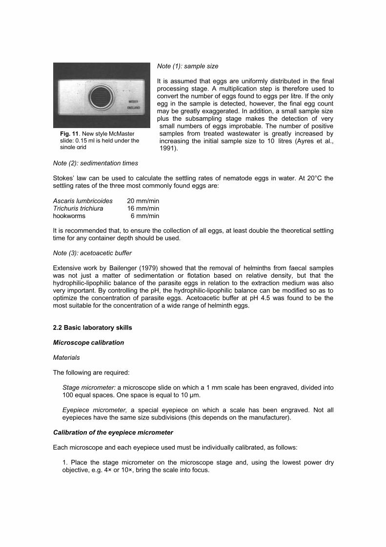
Fig. 11. New style McMasterslide: 0.15 ml is held under thesingle grid
Note (1): sample size
It is assumed that eggs are uniformly distributed in the finalprocessing stage. A multiplication step is therefore used toconvert the number of eggs found to eggs per litre. If the onlyegg in the sample is detected, however, the final egg countmay be greatly exaggerated. In addition, a small sample sizeplus the subsampling stage makes the detection of verysmall numbers of eggs improbable. The number of positivesamples from treated wastewater is greatly increased byincreasing the initial sample size to 10 litres (Ayres et al.,1991).
Note (2): sedimentation times
Stokes’ law can be used to calculate the settling rates of nematode eggs in water. At 20°C thesettling rates of the three most commonly found eggs are:
Ascaris lumbricoides 20 mm/minTrichuris trichiura 16 mm/minhookworms 6 mm/min
It is recommended that, to ensure the collection of all eggs, at least double the theoretical settlingtime for any container depth should be used.
Note (3): acetoacetic buffer
Extensive work by Bailenger (1979) showed that the removal of helminths from faecal sampleswas not just a matter of sedimentation or flotation based on relative density, but that thehydrophilic-lipophilic balance of the parasite eggs in relation to the extraction medium was alsovery important. By controlling the pH, the hydrophilic-lipophilic balance can be modified so as tooptimize the concentration of parasite eggs. Acetoacetic buffer at pH 4.5 was found to be themost suitable for the concentration of a wide range of helminth eggs.
2.2 Basic laboratory skills
Microscope calibration
Materials
The following are required:
Stage micrometer: a microscope slide on which a 1 mm scale has been engraved, divided into100 equal spaces. One space is equal to 10 µm.
Eyepiece micrometer, a special eyepiece on which a scale has been engraved. Not alleyepieces have the same size subdivisions (this depends on the manufacturer).
Calibration of the eyepiece micrometer
Each microscope and each eyepiece used must be individually calibrated, as follows:
1. Place the stage micrometer on the microscope stage and, using the lowest power dryobjective, e.g. 4× or 10×, bring the scale into focus.
2. Insert the eyepiece micrometer and rotate it until the two scales overlap.
3. Move the mechanical microscope stage until the scales are aligned at the zero line.
4. Without moving the stage micrometer, find a point towards the extreme right where twoother lines are exactly superimposed (Fig. 12).
5. Count the number of division lines on the eyepiece micrometer between the zero line andthe point where the second set of lines is superimposed.
6. Repeat this process using each objective in turn, e.g. 4×, 10×, 40×, 100×. Note that thehigher the magnification, the thicker the lines on the stage micrometer will appear. The line onthe eyepiece scale which is aligned exactly with the centre (or right or left edge) of a line onthe stage micrometer must be chosen.
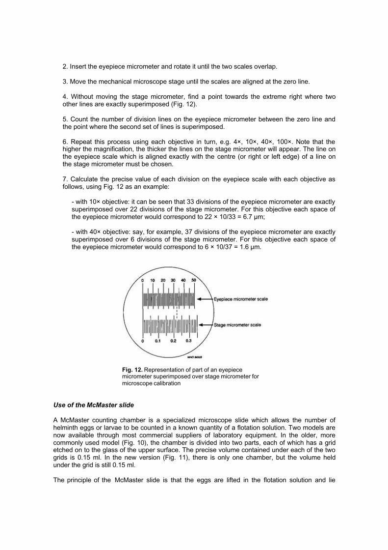
7. Calculate the precise value of each division on the eyepiece scale with each objective asfollows, using Fig. 12 as an example:
- with 10× objective: it can be seen that 33 divisions of the eyepiece micrometer are exactlysuperimposed over 22 divisions of the stage micrometer. For this objective each space ofthe eyepiece micrometer would correspond to 22 × 10/33 = 6.7 µm;
- with 40× objective: say, for example, 37 divisions of the eyepiece micrometer are exactlysuperimposed over 6 divisions of the stage micrometer. For this objective each space ofthe eyepiece micrometer would correspond to 6 × 10/37 = 1.6 µm.
Fig. 12. Representation of part of an eyepiecemicrometer superimposed over stage micrometer formicroscope calibration
Use of the McMaster slide
A McMaster counting chamber is a specialized microscope slide which allows the number ofhelminth eggs or larvae to be counted in a known quantity of a flotation solution. Two models arenow available through most commercial suppliers of laboratory equipment. In the older, morecommonly used model (Fig. 10), the chamber is divided into two parts, each of which has a gridetched on to the glass of the upper surface. The precise volume contained under each of the twogrids is 0.15 ml. In the new version (Fig. 11), there is only one chamber, but the volume heldunder the grid is still 0.15 ml.
The principle of the McMaster slide is that the eggs are lifted in the flotation solution and lie
immediately below the upper glass of the chamber, while the heavier debris settles to the floor. Ifthe microscope is focused on the grid, the eggs will be clearly in focus while the debris is not. Bysearching up and down the grid systematically, the number of eggs in 0.15 ml of the suspensionsolution can be accurately counted.
To fill the two-chambered McMaster slide:
1. Mix the final flotation suspension thoroughly, preferably in a vortex mixer to ensure ahomogeneous mix. Quickly fill a Pasteur pipette and carefully run the solution into onecompartment of the McMaster slide. Fill the whole compartment completely even though it isonly the section under the grid that is to be counted. Work quickly and smoothly at this stageso that eggs do not start to float in the test-tube or in the pipette. Ensure that there are no airbubbles under the grid.
2. Fill the other compartment of the McMaster slide, remembering to rehomogenize thesolution first.
3. Leave the McMaster slide to stand for a few minutes before starting to count, thus ensuringthat all the eggs have floated to the surface and that the debris has been allowed to settle.
4. Count the number of eggs under both grids. If there are large numbers of eggs and someare under the lines, it is usual to count those on two sides of the grid as “in” (e.g. on the topand left hand lines) and discard those under the other two lines (e.g. the bottom and right handlines). This gives a good estimate of the number of eggs present in 0.3 ml.
5. Make at least two (preferably three) counts if there is enough flotation solution, and take amean of the two (or three) counts. Calculate the number of eggs present in the original sampleusing the formula given on page 8 (remember that P = 0.15 if the new single-chambercounting slide is used).
McMaster counting chambers are usually made of glass and can be ordered from most majorsuppliers of scientific equipment. Some companies now produce less expensive (and less fragile)plastic chambers. If these are used, care must be taken to ensure that they do not becomescratched.
Use of centrifuges
Most published methods that involve the use of centrifuges quote centrifuge speed in terms ofrelative centrifugal force. However, in some papers, speed is expressed in revolutions per minute(rpm). To convert rpm to force, the following formula is used:
RCF = r(rpm)2/k
where RCF = relative centrifugal force (g),
r = radius of the centrifuge from the spindle to the centre of the bucket (cm),
k = 89 456.
To convert force to rpm:
)RCF/(rpm rk=
Identification of helminth eggs
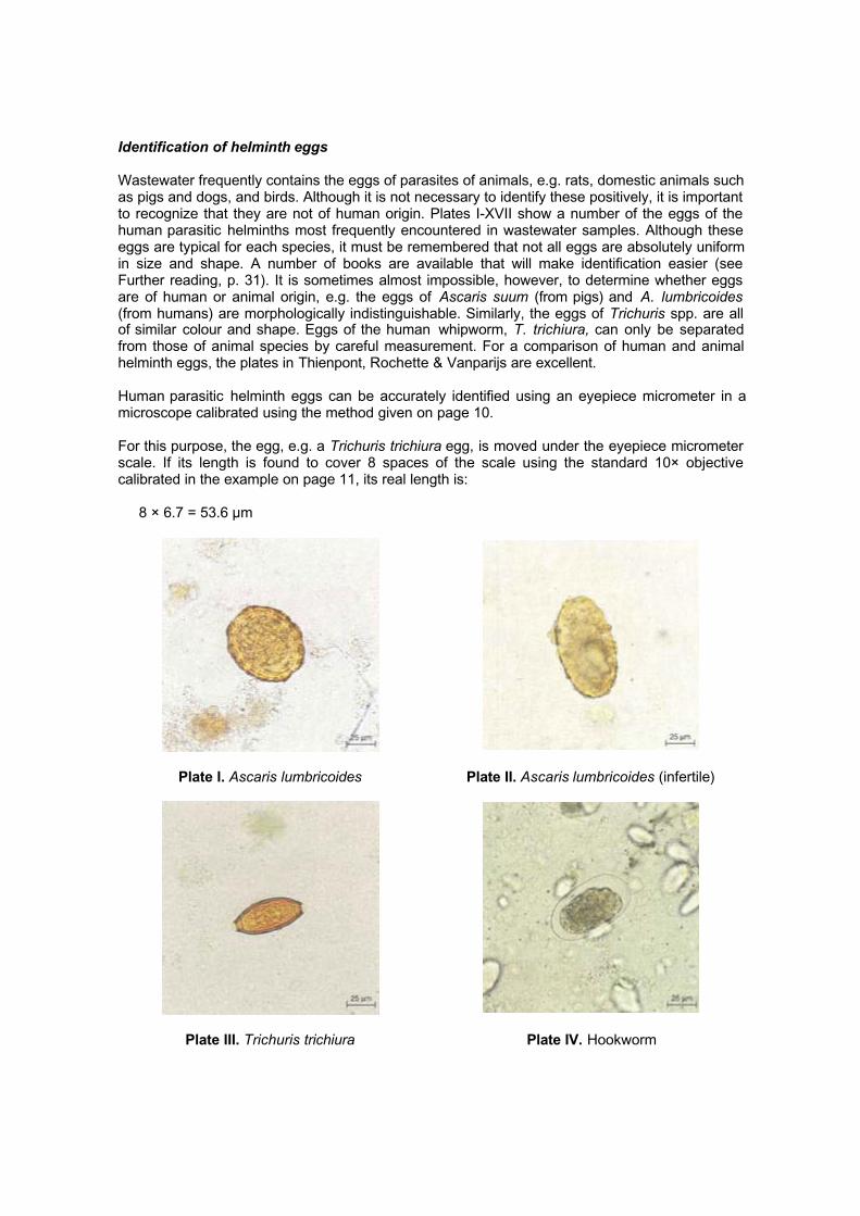
Wastewater frequently contains the eggs of parasites of animals, e.g. rats, domestic animals suchas pigs and dogs, and birds. Although it is not necessary to identify these positively, it is importantto recognize that they are not of human origin. Plates I-XVII show a number of the eggs of thehuman parasitic helminths most frequently encountered in wastewater samples. Although theseeggs are typical for each species, it must be remembered that not all eggs are absolutely uniformin size and shape. A number of books are available that will make identification easier (seeFurther reading, p. 31). It is sometimes almost impossible, however, to determine whether eggsare of human or animal origin, e.g. the eggs of Ascaris suum (from pigs) and A. lumbricoides(from humans) are morphologically indistinguishable. Similarly, the eggs of Trichuris spp. are allof similar colour and shape. Eggs of the human whipworm, T. trichiura, can only be separatedfrom those of animal species by careful measurement. For a comparison of human and animalhelminth eggs, the plates in Thienpont, Rochette & Vanparijs are excellent.
Human parasitic helminth eggs can be accurately identified using an eyepiece micrometer in amicroscope calibrated using the method given on page 10.
For this purpose, the egg, e.g. a Trichuris trichiura egg, is moved under the eyepiece micrometerscale. If its length is found to cover 8 spaces of the scale using the standard 10× objectivecalibrated in the example on page 11, its real length is:
8 × 6.7 = 53.6 µm
Plate I. Ascaris lumbricoides Plate II. Ascaris lumbricoides (infertile)
Plate III. Trichuris trichiura Plate IV. Hookworm
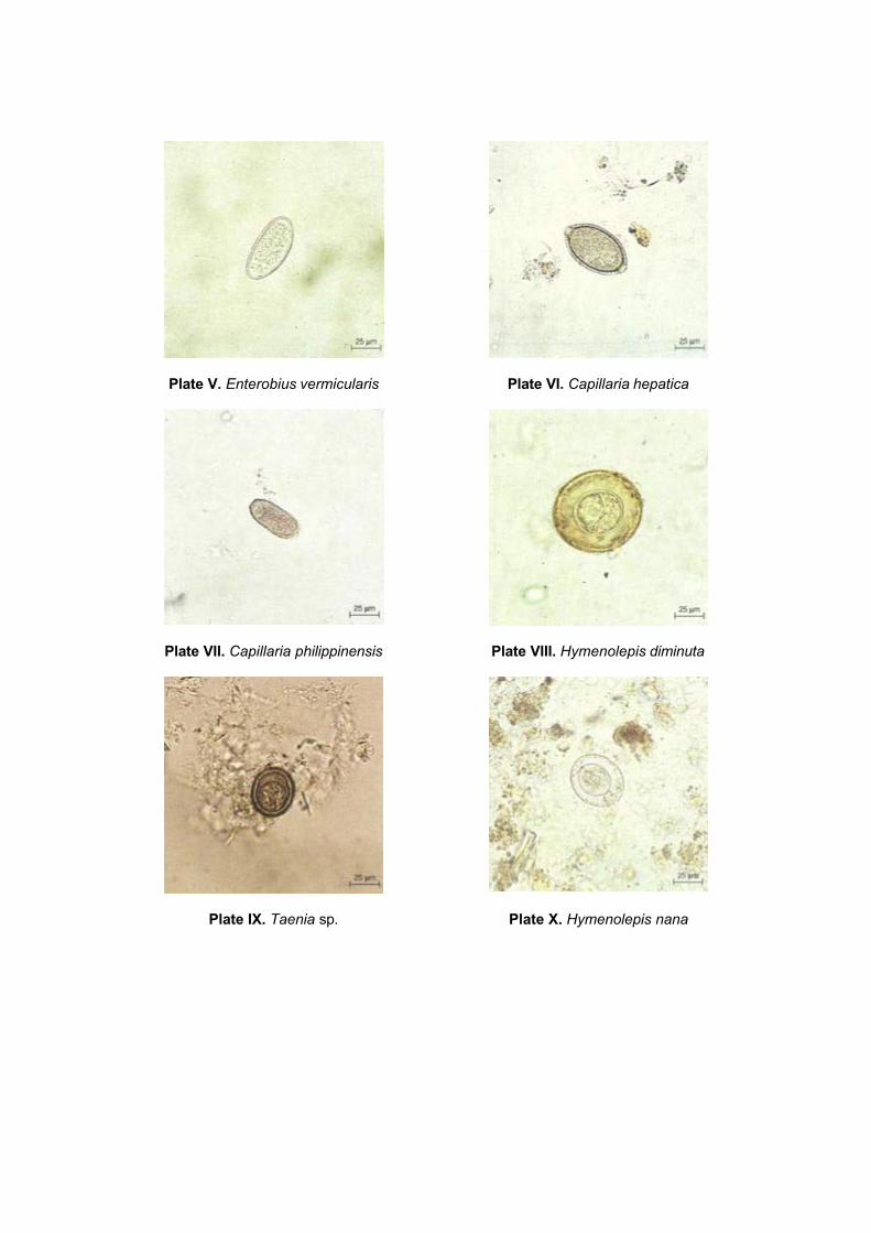
Plate V. Enterobius vermicularis Plate VI. Capillaria hepatica
Plate VII. Capillaria philippinensis Plate VIII. Hymenolepis diminuta
Plate IX. Taenia sp. Plate X. Hymenolepis nana
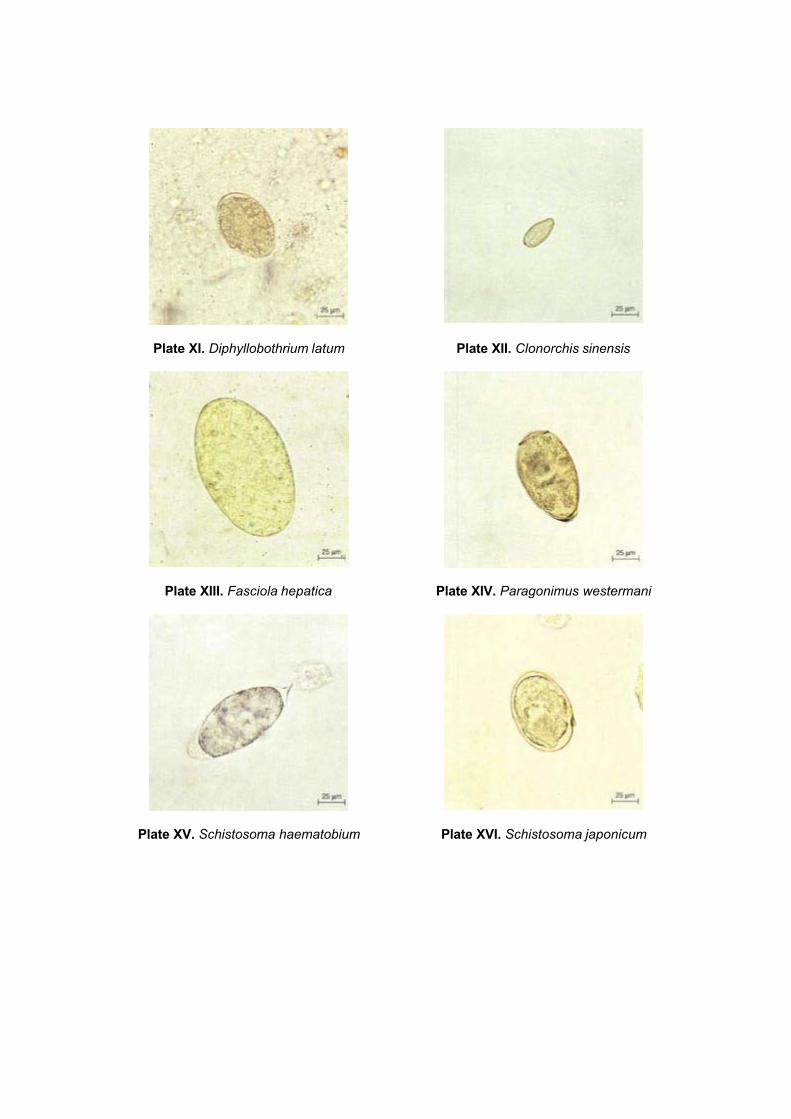
Plate XI. Diphyllobothrium latum Plate XII. Clonorchis sinensis
Plate XIII. Fasciola hepatica Plate XIV. Paragonimus westermani
Plate XV. Schistosoma haematobium Plate XVI. Schistosoma japonicum
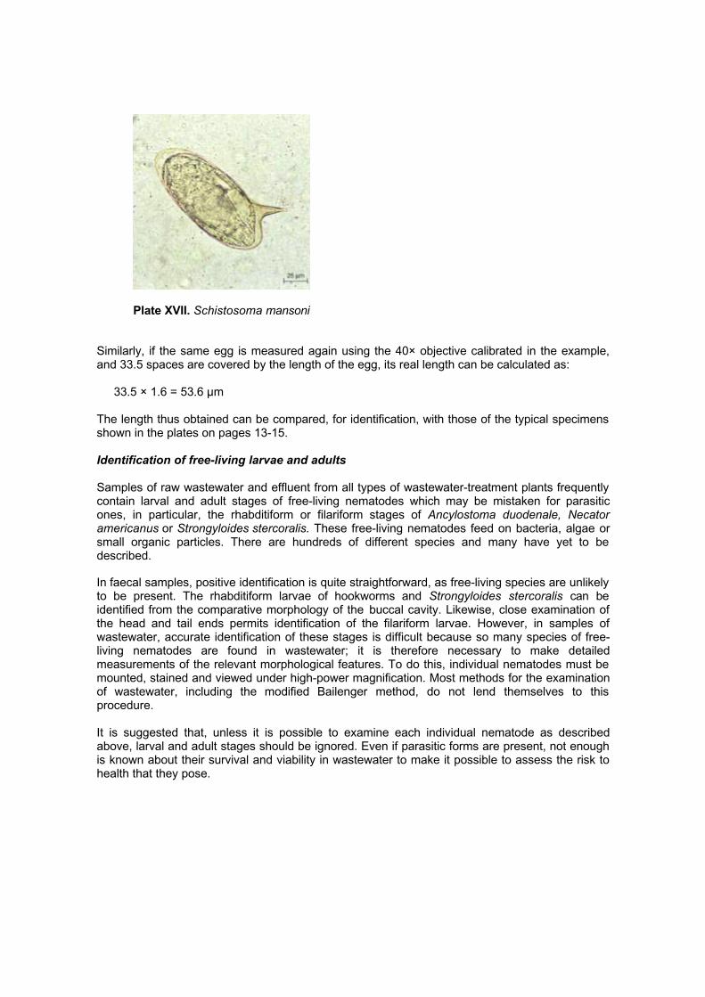
Plate XVII. Schistosoma mansoni
Similarly, if the same egg is measured again using the 40× objective calibrated in the example,and 33.5 spaces are covered by the length of the egg, its real length can be calculated as:
33.5 × 1.6 = 53.6 µm
The length thus obtained can be compared, for identification, with those of the typical specimensshown in the plates on pages 13-15.
Identification of free-living larvae and adults
Samples of raw wastewater and effluent from all types of wastewater-treatment plants frequentlycontain larval and adult stages of free-living nematodes which may be mistaken for parasiticones, in particular, the rhabditiform or filariform stages of Ancylostoma duodenale, Necatoramericanus or Strongyloides stercoralis. These free-living nematodes feed on bacteria, algae orsmall organic particles. There are hundreds of different species and many have yet to bedescribed.
In faecal samples, positive identification is quite straightforward, as free-living species are unlikelyto be present. The rhabditiform larvae of hookworms and Strongyloides stercoralis can beidentified from the comparative morphology of the buccal cavity. Likewise, close examination ofthe head and tail ends permits identification of the filariform larvae. However, in samples ofwastewater, accurate identification of these stages is difficult because so many species of free-living nematodes are found in wastewater; it is therefore necessary to make detailedmeasurements of the relevant morphological features. To do this, individual nematodes must bemounted, stained and viewed under high-power magnification. Most methods for the examinationof wastewater, including the modified Bailenger method, do not lend themselves to thisprocedure.
It is suggested that, unless it is possible to examine each individual nematode as describedabove, larval and adult stages should be ignored. Even if parasitic forms are present, not enoughis known about their survival and viability in wastewater to make it possible to assess the risk tohealth that they pose.
3. Sanitary bacteriology
The numbers of faecal coliform bacteria in wastewater samples are usually counted by: (a) mostprobable number (MPN) methods; or (b) the membrane filtration method. Two MPN methods aredescribed in section 3.1; membrane filtration is described in section 3.2.
3.1 MPN methods
MPN counts are statistical best estimates (hence the name, most probable number) obtained byculturing a number (usually five) of sample volumes and/or dilutions of such samples. Theseestimates are based on the principle of “dilution to extinction”. For example, if a single 1-mlaliquot from each of a series of 1:10 dilutions (see p. 27) is examined and growth occurs at adilution of 10-3 but not at 10-4, the best estimate of the count is 103 bacteria per ml. By increasing,usually to five, the number of 1-ml aliquots examined at each dilution, a better estimate of thecount can be obtained.
In the first of the two MPN methods described here, five 1-ml aliquots of only one dilution areexamined, and so only an approximate faecal coliform MPN count is obtained. This method,which is very simple and inexpensive, is suitable for the routine analysis of treated wastewatersthat comply with the guideline value of no more than 1000 faecal coliforms per 100 ml (see Table1).
In the second MPN method, five 1-ml aliquots of each of three dilutions are examined, so that amuch better estimate of faecal coliform numbers is obtained. This method can be made suitablefor the analysis of wastewater containing any number of faecal coliform bacteria by altering thedilutions examined (see method B, step 10, p. 20).
Equipment and consumables
Consumables
The chemicals listed below for medium A-1 will be needed, together with quarter-strengthRinger’s solution (commercially available in tablet form) or sodium chloride solution (8.5 g NaClper litre of distilled water). Non-absorbent cotton wool is also required.
Medium A-1 (American Public Health Association, 1995) is recommended, as it can be used fordirect incubation at 44°C. It is not commercially available in dehydrated form and must be madeup (see pp. 26-27) to the following formula:
lactose 5 gtryptone 20 gsalicin 0.5 gNaCl 5 gTriton X-100 1 mldistilled water 1 litre
It is dispensed in 5-ml quantities into test-tubes (or screw-capped bottles) each of which containsan inverted Durham tube (this is a very small test-tube). The test-tubes are closed with a plug ofnon-absorbent cotton wool and sterilized (see p. 27). During sterilization, the air in the Durhamtube is expelled and it becomes completely full of medium.
Equipment
The following are required:

- 100-ml screw-capped bottles
- test-tubes (100 mm × 12 mm) or half-ounce (14-ml) screw-capped bottles
- 1-ml serological “blow-out” pipettes
- Bunsen burner
- test-tube rack
- incubator or water-bath
- autoclave or pressure cooker
- balance (± 0.01 g).
Illustrated step-by-step guide
Two MPN methods are described below. Method A is the simpler of the two and is suitable for theroutine monitoring (see section 4) of treated wastewater effluents that contain around 1000 orfewer faecal coliforms per 100 ml.
Method B is more accurate and can also be used for samples containing 1000 or fewer faecalcoliforms per 100 ml, or for those containing many more.
Method A
1. Collect a sample of wastewater effluent in a sterile 100-ml screw-capped bottle.
2. Shake the sample bottle thoroughly, and aseptically withdraw 1 ml using a sterile 1-ml “blow-out” pipette. Transfer this to a sterile test-tube or screw-capped bottle containing 9 ml of quarter-strength Ringer’s solution (or 8.5 g/l NaCl solution) (Fig. 13). Do not pipette by mouth - use apipette suction pump.
3. Shake this 1:10 dilution of the sample thoroughly and, using a single fresh sterile 1-ml pipette,transfer 1 ml to each of five sterile test-tubes or screw-capped bottles containing an invertedDurham tube and 5 ml of medium A-1 (Fig. 14). Label each tube or bottle with a code for thesample, the date and 1:10.
4. Place these five test-tubes or bottles in an incubator or water-bath maintained at 44°C (±0.25°C) (see p. 27).
5. After incubation for 24 h, examine each test-tube or bottle for gas production. (Faecal coliformbacteria produce gas from the lactose in medium A-1, and some of this gas is trapped in theinverted Durham tube.) Count the number of positive tubes or bottles (i.e. those with gasproduction) (Fig. 15) and determine the MPN from Table 2.
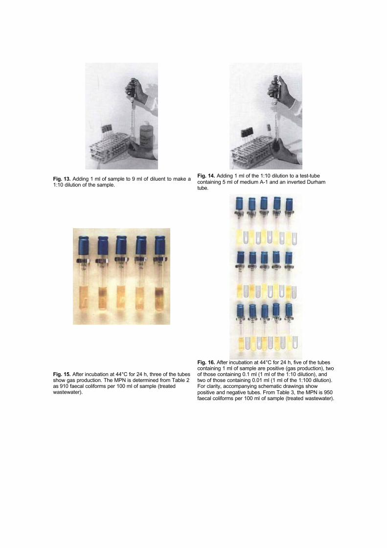
Fig. 13. Adding 1 ml of sample to 9 ml of diluent to make a1:10 dilution of the sample.
Fig. 14. Adding 1 ml of the 1:10 dilution to a test-tubecontaining 5 ml of medium A-1 and an inverted Durhamtube.
Fig. 15. After incubation at 44°C for 24 h, three of the tubesshow gas production. The MPN is determined from Table 2as 910 faecal coliforms per 100 ml of sample (treatedwastewater).
Fig. 16. After incubation at 44°C for 24 h, five of the tubescontaining 1 ml of sample are positive (gas production), twoof those containing 0.1 ml (1 ml of the 1:10 dilution), andtwo of those containing 0.01 ml (1 ml of the 1:100 dilution).For clarity, accompanying schematic drawings showpositive and negative tubes. From Table 3, the MPN is 950faecal coliforms per 100 ml of sample (treated wastewater).
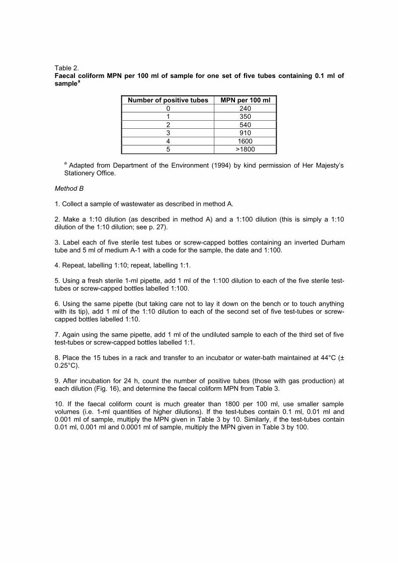
Table 2.Faecal coliform MPN per 100 ml of sample for one set of five tubes containing 0.1 ml ofsamplea
Number of positive tubes MPN per 100 ml0 2401 3502 5403 9104 16005 >1800
a Adapted from Department of the Environment (1994) by kind permission of Her Majesty’sStationery Office.
Method B
1. Collect a sample of wastewater as described in method A.
2. Make a 1:10 dilution (as described in method A) and a 1:100 dilution (this is simply a 1:10dilution of the 1:10 dilution; see p. 27).
3. Label each of five sterile test tubes or screw-capped bottles containing an inverted Durhamtube and 5 ml of medium A-1 with a code for the sample, the date and 1:100.
4. Repeat, labelling 1:10; repeat, labelling 1:1.
5. Using a fresh sterile 1-ml pipette, add 1 ml of the 1:100 dilution to each of the five sterile test-tubes or screw-capped bottles labelled 1:100.
6. Using the same pipette (but taking care not to lay it down on the bench or to touch anythingwith its tip), add 1 ml of the 1:10 dilution to each of the second set of five test-tubes or screw-capped bottles labelled 1:10.
7. Again using the same pipette, add 1 ml of the undiluted sample to each of the third set of fivetest-tubes or screw-capped bottles labelled 1:1.
8. Place the 15 tubes in a rack and transfer to an incubator or water-bath maintained at 44°C (±0.25°C).
9. After incubation for 24 h, count the number of positive tubes (those with gas production) ateach dilution (Fig. 16), and determine the faecal coliform MPN from Table 3.
10. If the faecal coliform count is much greater than 1800 per 100 ml, use smaller samplevolumes (i.e. 1-ml quantities of higher dilutions). If the test-tubes contain 0.1 ml, 0.01 ml and0.001 ml of sample, multiply the MPN given in Table 3 by 10. Similarly, if the test-tubes contain0.01 ml, 0.001 ml and 0.0001 ml of sample, multiply the MPN given in Table 3 by 100.

Table 3.Faecal coliform MPN per 100 ml of sample for three sets of five tubes containing 1 ml, 0.1 ml and0.01 ml of sample respectivelya
Number of positive tubes MPN1 ml 0.1 ml 0.01 ml per 100 ml
0 0 0 00 0 0 200 1 0 201 0 0 201 0 1 401 1 0 401 2 0 502 0 0 402 0 1 502 1 0 502 1 1 702 2 0 702 3 0 1103 0 0 703 0 1 903 1 0 903 1 1 1303 2 0 1303 2 1 1603 3 0 1604 0 0 1104 0 1 1404 1 0 1604 1 1 2004 2 0 2004 2 1 2504 3 0 2504 3 1 3104 4 0 3204 4 1 3805 0 0 2205 0 1 2905 0 2 4105 1 0 3105 1 1 4305 1 2 6005 1 3 8505 2 0 5005 2 1 7005 2 2 9505 2 3 1 2005 3 0 7505 3 1 1 1005 3 2 1 4005 3 3 1 7505 3 4 2 1005 4 0 1 3005 4 1 1 7005 4 2 2 2005 4 3 2 8005 4 4 3 4505 5 0 2 4005 5 1 3 5005 5 2 5 4005 5 3 9 1005 5 4 16 0005 5 5 >18 000
a Adapted from Department of the Environment (1994) by kind permission of Her Majesty’sStationery Office.
3.2 Membrane filtration method
Membrane filtration is a method of obtaining faecal coliform counts by filtering a known volume ofa wastewater sample (or a dilution of it) though a membrane filter. This is a special filter-paperwith a pore size of 0.45 µm, so that all faecal coliform bacteria are retained on it. The membranefilter is then placed on an absorbent pad saturated with a faecal coliform growth medium andincubated. During incubation, each faecal coliform bacterium develops into a visible yellowcolony. After incubation, the yellow colonies are counted, and the count per 100 ml is calculated.
Equipment and consumables
Equipment
The following are required:
- membrane filter forceps- Petri dishes (60-mm diameter glass or disposable plastic)- membrane filtration units (glass or plastic)- 5-ml or 10-ml and 1-ml serological “blow-out” pipettes- pipette suction pump- vacuum pump (electrical, manual or water Venturi pump)- Bunsen burner- incubator- autoclave or pressure cooker- balance (± 0.01 g).
Consumables
These comprise the following:
- membrane filters (0.45 µm pore size, 47 mm diameter)- absorbent pads (47 mm diameter)- membrane lauryl sulfate broth- quarter-strength Ringer’s solution- ethanol.
Membrane lauryl sulfate broth is available commercially in dehydrated form. Alternatively, it maybe made up in accordance with the following formula (Department of the Environment, 1994):
peptone 40 gyeast extract 6 glactose 30 gphenol red (4 g/l aqueous solution) 50 mlsodium lauryl sulfate 1 gdistilled water (pH 7.6 before sterilization) 1 litre
Quarter-strength Ringer’s solution is also available commercially in tablet form; alternatively,sodium chloride solution (8.5 g NaCl in 1 litre of distilled water) may be used.
Illustrated step-by-step guide
The following procedure is suitable for wastewater samples containing 200-2000 faecal coliformsper 100 ml. For samples containing more than 2000 per 100 ml, see step 11 below. Asepticprocedures should be used throughout (see chapter 3.3 Basic laboratory skills).
1. Collect a sample of wastewater in a sterile 100-ml screw-capped bottle.
2. Dip the membrane filter forceps in ethanol, and burn off in the flame of the Bunsen burner.Using the now sterile forceps, transfer a sterile absorbent pad to each of three sterile Petri dishes.
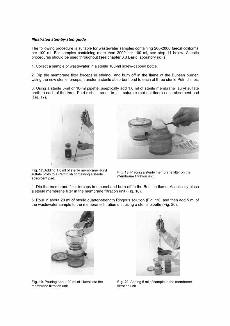
3. Using a sterile 5-ml or 10-ml pipette, aseptically add 1.8 ml of sterile membrane lauryl sulfatebroth to each of the three Petri dishes, so as to just saturate (but not flood) each absorbent pad(Fig. 17).
Fig. 17. Adding 1.8 ml of sterile membrane laurylsulfate broth to a Petri dish containing a sterileabsorbent pad.
Fig. 18. Placing a sterile membrane filter on themembrane filtration unit.
4. Dip the membrane filter forceps in ethanol and burn off in the Bunsen flame. Aseptically placea sterile membrane filter in the membrane filtration unit (Fig. 18).
5. Pour in about 20 ml of sterile quarter-strength Ringer’s solution (Fig. 19), and then add 5 ml ofthe wastewater sample to the membrane filtration unit using a sterile pipette (Fig. 20).
Fig. 19. Pouring about 20 ml of diluent into themembrane filtration unit.
Fig. 20. Adding 5 ml of sample to the membranefiltration unit.
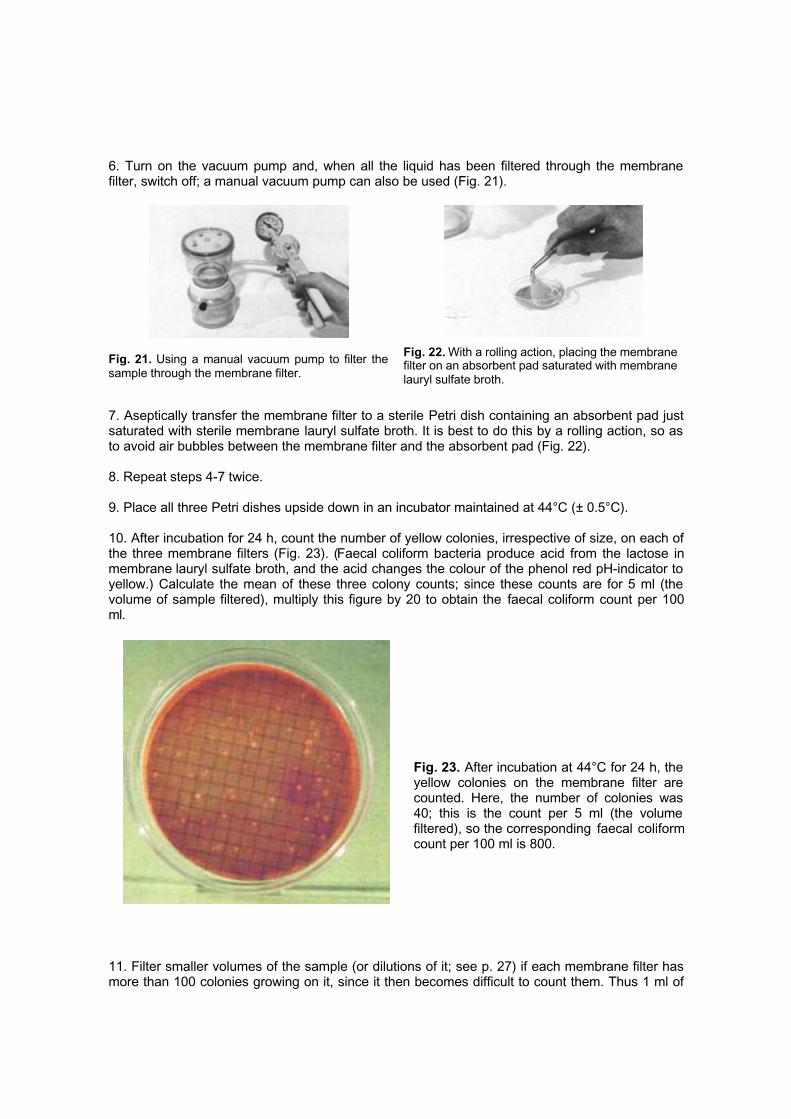
6. Turn on the vacuum pump and, when all the liquid has been filtered through the membranefilter, switch off; a manual vacuum pump can also be used (Fig. 21).
Fig. 21. Using a manual vacuum pump to filter thesample through the membrane filter.
Fig. 22. With a rolling action, placing the membranefilter on an absorbent pad saturated with membranelauryl sulfate broth.
7. Aseptically transfer the membrane filter to a sterile Petri dish containing an absorbent pad justsaturated with sterile membrane lauryl sulfate broth. It is best to do this by a rolling action, so asto avoid air bubbles between the membrane filter and the absorbent pad (Fig. 22).
8. Repeat steps 4-7 twice.
9. Place all three Petri dishes upside down in an incubator maintained at 44°C (± 0.5°C).
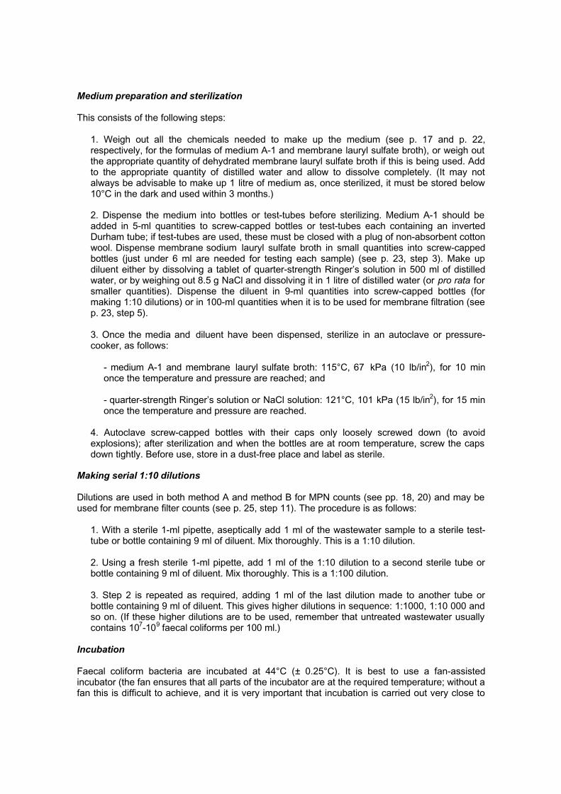
10. After incubation for 24 h, count the number of yellow colonies, irrespective of size, on each ofthe three membrane filters (Fig. 23). (Faecal coliform bacteria produce acid from the lactose inmembrane lauryl sulfate broth, and the acid changes the colour of the phenol red pH-indicator toyellow.) Calculate the mean of these three colony counts; since these counts are for 5 ml (thevolume of sample filtered), multiply this figure by 20 to obtain the faecal coliform count per 100ml.
Fig. 23. After incubation at 44°C for 24 h, theyellow colonies on the membrane filter arecounted. Here, the number of colonies was40; this is the count per 5 ml (the volumefiltered), so the corresponding faecal coliformcount per 100 ml is 800.
11. Filter smaller volumes of the sample (or dilutions of it; see p. 27) if each membrane filter hasmore than 100 colonies growing on it, since it then becomes difficult to count them. Thus 1 ml of

the sample can be used for faecal coliform counts of up to 10 000 per 100 ml, 1 ml of a 1:10dilution for counts up to 100 000 per 100 ml, and so on.
3.3 Basic laboratory skills
Asepsis
Special care has to be taken to ensure that the wastewater sample, or any dilution of it, does notbecome contaminated while it is being examined. In other words, it is necessary to ensure thatonly the faecal coliform bacteria in the sample are being counted, not those which may be on anyglassware or on fingers. All glassware, media and diluent (quarter-strength Ringer’s or saltsolution), and membrane filters and absorbent pads are therefore sterilized before use (seebelow). However, sterilization is only a part of asepsis; in addition, the following standard asepticprocedures are necessary:
1. Wash hands thoroughly before starting work in the laboratory.
2. Ensure that the work is carried out in a dust-free and draught-free part of the laboratory andthat the bench used is clean; swab it down with ethanol just before starting work.
3. Be careful not to touch the top of the bottles containing sterile medium or diluent, or of thoseused for sample collection. Similarly, do not touch the tip or bottom half of sterile pipettes.
4. When opening a bottle (or test-tube) containing sterile medium or diluent, or one containingthe sample, quickly pass the open neck through a Bunsen flame while holding the cap of thebottle (or the cotton wool plug from the test-tube) in the other hand (using only the little fingerfor this - do so by curling it around the cap or plug). Similarly, when withdrawing a pipette fromits sterile wrapping (or container), pass it quickly through the Bunsen flame, and be careful notto touch the pipette tip or allow it to touch anything else; if it does touch something, discard itand use another sterile pipette.
5. Never pipette by mouth. Always use a pipette suction pump.
If nobody in the laboratory has any bacteriological experience, it is best to contact a medicallaboratory technician at a local hospital who will be able to demonstrate aseptic procedures.
Sterilization
Test-tubes containing MPN medium, bottles containing diluent and membrane filtration medium,and membrane filters and absorbent pads are all sterilized in an autoclave or pressure cooker(see p. 27).
Screw-capped bottles used for sample collection are sterilized in an oven at 160°C for 1 h. Duringsterilization, the cap should be slightly unscrewed (to allow air to escape, otherwise the bottle willexplode); after sterilization and when the oven has cooled to room temperature, the bottles areremoved, the caps screwed on tightly and a short length of adhesive tape (preferably markedSTERILE) is placed over the top of each bottle.
Pipettes should also be sterilized in an oven at 160°C for 1 h. Before sterilization, the top of eachpipette should be plugged with non-absorbent cotton wool. They are then wrapped eitherindividually or in a group of up to five in aluminium foil; the tops should be marked (so that theother end is not opened and the tips contaminated).
Medium preparation and sterilization
This consists of the following steps:
1. Weigh out all the chemicals needed to make up the medium (see p. 17 and p. 22,respectively, for the formulas of medium A-1 and membrane lauryl sulfate broth), or weigh outthe appropriate quantity of dehydrated membrane lauryl sulfate broth if this is being used. Addto the appropriate quantity of distilled water and allow to dissolve completely. (It may notalways be advisable to make up 1 litre of medium as, once sterilized, it must be stored below10°C in the dark and used within 3 months.)
2. Dispense the medium into bottles or test-tubes before sterilizing. Medium A-1 should beadded in 5-ml quantities to screw-capped bottles or test-tubes each containing an invertedDurham tube; if test-tubes are used, these must be closed with a plug of non-absorbent cottonwool. Dispense membrane sodium lauryl sulfate broth in small quantities into screw-cappedbottles (just under 6 ml are needed for testing each sample) (see p. 23, step 3). Make updiluent either by dissolving a tablet of quarter-strength Ringer’s solution in 500 ml of distilledwater, or by weighing out 8.5 g NaCl and dissolving it in 1 litre of distilled water (or pro rata forsmaller quantities). Dispense the diluent in 9-ml quantities into screw-capped bottles (formaking 1:10 dilutions) or in 100-ml quantities when it is to be used for membrane filtration (seep. 23, step 5).
3. Once the media and diluent have been dispensed, sterilize in an autoclave or pressure-cooker, as follows:
- medium A-1 and membrane lauryl sulfate broth: 115°C, 67 kPa (10 lb/in2), for 10 minonce the temperature and pressure are reached; and
- quarter-strength Ringer’s solution or NaCl solution: 121°C, 101 kPa (15 lb/in2), for 15 minonce the temperature and pressure are reached.
4. Autoclave screw-capped bottles with their caps only loosely screwed down (to avoidexplosions); after sterilization and when the bottles are at room temperature, screw the capsdown tightly. Before use, store in a dust-free place and label as sterile.
Making serial 1:10 dilutions
Dilutions are used in both method A and method B for MPN counts (see pp. 18, 20) and may beused for membrane filter counts (see p. 25, step 11). The procedure is as follows:
1. With a sterile 1-ml pipette, aseptically add 1 ml of the wastewater sample to a sterile test-tube or bottle containing 9 ml of diluent. Mix thoroughly. This is a 1:10 dilution.
2. Using a fresh sterile 1-ml pipette, add 1 ml of the 1:10 dilution to a second sterile tube orbottle containing 9 ml of diluent. Mix thoroughly. This is a 1:100 dilution.
3. Step 2 is repeated as required, adding 1 ml of the last dilution made to another tube orbottle containing 9 ml of diluent. This gives higher dilutions in sequence: 1:1000, 1:10 000 andso on. (If these higher dilutions are to be used, remember that untreated wastewater usuallycontains 107-109 faecal coliforms per 100 ml.)
Incubation
Faecal coliform bacteria are incubated at 44°C (± 0.25°C). It is best to use a fan-assistedincubator (the fan ensures that all parts of the incubator are at the required temperature; without afan this is difficult to achieve, and it is very important that incubation is carried out very close to

44°C), although a water-bath can be used for the test-tubes in the MPN procedures.
After incubation and examination of the samples, all the test-tubes or screw-capped bottlescontaining medium, and all Petri dishes containing membrane filters and absorbent pads, must besterilized by autoclaving at 121°C for 15 min before disposal to ensure that the billions of bacteriathat have grown during incubation are destroyed.
4. Routine monitoring programmes
The numbers of faecal coliform bacteria in the final effluent of a wastewater treatment plant varymuch less with time (even over a 24-h period) than do the numbers of helminth eggs. Theguideline values for faecal coliforms and helminth eggs given in Table 1 differ markedly (1000faecal coliforms per 100 ml as compared with 1 egg per litre), so routine monitoring programmesfor each are seeking to detect numerical changes of a different order of magnitude. A mean of 2eggs per litre might be a result requiring action, but one of 1002 or 1020 faecal coliforms per 100ml probably would not be (with bacteria the order of magnitude is much more important than theactual number). Because of this difference (1000 per 100 ml versus 1 per litre), and the differentpatterns of removal in wastewater-treatment works, routine monitoring programmes for faecalcoliforms and helminth eggs are somewhat different.
4.1 Faecal coliforms
During the irrigation season, grab samples of the final effluent used for unrestricted crop irrigationshould be taken at least once, and preferably twice, a week and faecal coliform numbersdetermined. The time of day that such samples are collected should be established by conductinga few diurnal studies, as follows:
1. Over a 24-h period, take samples every 3 h (eight samples in all), and determine the faecalcoliform numbers N1, N2, N3, etc. in each.
2. Calculate the geometric mean count for the 24-h period from the formula:
Geometric mean count = (N1 × N2 × N3 × N4 × N5 × N6 × N7 × N8)1/8
3. Determine which of the individual counts is closest to the mean count, and then take theroutine samples at this time (usually between 08:00 and 10:00). As each sample is takenduring the irrigation season, calculate the running geometric mean count; it is this value thatshould not exceed 1000 faecal coliforms per 100 ml.
Running geometric mean = (N1 × N2 × N3 × . . . × Nn)1/n
4.2 Helminth eggs
Time of day
The concentration of human parasitic helminth eggs in raw wastewater varies considerably over24-h periods. If raw wastewater is being monitored, it is important to take 24-h compositesamples, or to take a representative grab sample as described in section 4.1 (the arithmetic,rather than geometric, mean egg count should be used).
Running arithmetic mean = (N1 + N2 + N3 + . . . + Nn)/n
The diurnal variation in the numbers of helminth eggs in effluents from treatment plants(especially waste-stabilization ponds) is less marked.
Frequency of sampling
Frequency of sampling depends on the objectives of the monitoring regime and the method ofwastewater treatment. A diurnal study and an initial period of intense sampling (once or twice aweek) for several weeks, should be carried out at every treatment plant.
Waste-stabilization ponds remove helminth eggs at a fairly constant rate, despite periodic

overloading and changes in flow; this preliminary sampling should therefore give a goodindication of the long-term effluent quality. Egg-removal rates from conventional treatment plantstend to be more variable, and intense monitoring should therefore cover a range of operatingconditions.
After this preliminary sampling, the frequency of sampling will depend on whether the effluent isbeing used for restricted or unrestricted irrigation. If the wastewater is being treated in waste-stabilization ponds and used for unrestricted irrigation (Category A in Table 1), samples need onlybe taken once or twice a month, since the retention time in ponds required to achieve the faecalcoliform guideline value vastly exceeds that for the egg guideline. However, if treatment is byanother process, or the wastewater is going to be used for restricted irrigation (Category B, Table1), samples need to be taken at least once a week. Regular monitoring should always be carriedout while the effluent is being used for irrigation.
Number of samples
If differences over time in the number of eggs per litre are to be determined, several samplesshould be taken on each occasion. A minimum of three is suggested. The actual number ofsamples required will depend on the smallest true difference to be detected and the level ofsignificance desired. This is described in most statistical textbooks (e.g. Sokal & Rohlf, 1981).
References
American Public Health Association (1995) Standard methods for the examination of water andwastewater, 19th ed. Washington, DC.
Ayres RM et al. (1991) Comparison of techniques for the enumeration of human parasitichelminth eggs in treated wastewater. Environmental technology, 12: 617-623.
Bailenger J (1979) Mechanisms of parasitological concentration in coprology and their practicalconsequences. Journal of American medical technology, 41: 65-71.
Bouhoum K, Schwartzbrod J (1989) Quantification of helminth eggs in wastewater. Zentralblattfür Hygiene und Umweltmedizin, 188: 322-330.
Department of the Environment (1994) The microbiology of water 1994 - Part I. Drinking water.London, Her Majesty’s Stationery Office.
Faust EC et al. (1938) A critical study of clinical laboratory technics for the diagnosis of protozoancysts and helminth eggs in feces. American journal of tropical medicine and hygiene, 18: 169-183.
International Reference Centre for Waste Disposal (1985) Health aspects of wastewater andexcreta use in agriculture and aquaculture: the Engelberg report. IRCWD news, No. 23, pp. 11-18.
Janeckso A, Urbanyi L (1931) Méthode d’enrichissement coprologique. Revue générale demédecine vétérinaire, 41: 496-497.
Prost A (1988) Revision of the 1973 WHO guidelines: a WHO Scientific Group proposes revisedhealth guidelines for the use of wastewater. IRCWD news, No. 24/25, p. 11.
Rude RA, Peeler JT, Risty NG (1987) Comparison of diethyl ether and ethyl acetate as extractingagents for recovery of Ascaris spp. and Trichuris spp. eggs. Journal of the Association of OfficialAnalytical Chemists, 70: 1000-1002.
Shuval HI et al. (1986) Wastewater irrigation in developing countries: health effects and technicalsolutions. Washington, DC, The World Bank (Technical Paper No. 51).
Sokal RR, Rohlf FJ (1981) Biometry. New York, W.H. Freeman and Co.
Thienpont E, Rochette F, Vanparijs OFJ (1986) Diagnosing helminthiasis by coprologicalexamination (unpublished document; available on request from Janssen Research Foundation,Turnhoutsebaan 30, 2340 Beerse, Belgium).
World Health Organization (1989) Health guidelines for the use of wastewater in agriculture andaquaculture. Report of a WHO Scientific Group. Geneva (WHO Technical Report Series, No.778).
Further reading
Wastewater reuse
Mara DD, Cairncross S (1989) Guidelines for the safe use of wastewater and excreta inagriculture and aquaculture: measures for public health protection. Geneva, World HealthOrganization.
Sanitary parasitology and bacteriology
Berk SG, Gunderson JH (1993) Wastewater organisms: a color atlas. Boca Raton, FL, LewisPublishers.
Cheeseborough M (1987) Medical laboratory manual for tropical countries, Vols I and II.Sevenoaks, Butterworth.
Jeffry HC, Leach RM (1975) Atlas of medical parasitology and protozoology. Edinburgh, ChurchillLivingstone.
Peters W, Giles HM (1989) A colour atlas of tropical medicine and parasitology. London, WolfePublishing.
World Health Organization (1994) Bench aids for the diagnosis of intestinal parasites. Geneva.
Selected WHO publications of related interest
Bench aids for the diagnosis of intestinal parasites.1994 (20 pages, including 9 colour plates)Sw. fr. 35. -
Laboratory biosafety manual, 2nd ed. • 1993 (144 pages)Sw. fr. 26. -
Parasitic diseases in water resources development. The need for intersectoral negotiation.J.M. Hunter et al. • 1993 (162 pages)Sw. fr. 35. -
A guide to the development of on-site sanitation.R. Franceys, J. Pickford & R. Reed • 1992 (245 pages)Sw. fr. 47. -
Guidelines for the safe use of wastewater and excreta in agriculture and aquaculture.D. Mara & S. Cairncross • 1989 (194 pages)Sw. fr. 35. -
Health guidelines for the use of wastewater in agriculture and aqua-culture. Report of aWHO Scientific Group.WHO Technical Report Series, No. 778 • 1989 (74 pages)Sw. fr. 9. -
Agriculture-health linkages.M. Lipton & E. de Kadt WHO Offset Publication, No. 104 • 1988 (111 pages)Sw. fr. 17. -
Technology for water supply and sanitation in developing countries. Report of a WHOStudy Group.WHO Technical Report Series, No. 742 • 1987 (38 pages)Sw. fr. 7. -
Wastewater stabilization ponds. Principles of planning and practice.WHO Regional Publications, Eastern Mediterranean • 1987 (138 pages)Sw. fr. 25. -
Appropriate technology for the treatment of wastewaters for small rural communities.Report on a WHO meeting.WHO Regional Publications, Europe • 1985 (62 pages)Sw. fr. 8. -
Further information on these and other WHO publications can be obtained fromDistribution and Sales, World Health Organization, 1211 Geneva 27, Switzerland.
Prices in developing countries are 70% of those listed here.

Back cover
The use of wastewater for crop irrigation is becoming increasingly common. Resulting crop yieldsare higher, as wastewater contains nutrients necessary for plant growth: However, there is the
risk that wastewater irrigation may facilitate the transmission of intestinal nematode infections andfaecal bacterial diseases to both consumers and agricultural workers.
This laboratory manual provides a practical, step-by-step guide for the analysis of samples oftreated wastewater to determine whether they comply with the quality guidelines published by
WHO.
The procedures presented here are simple and effective, require a minimum of equipment, andcan be carried out by persons with little or no previous parasitological or microbiological
experience.
The methods include a modified Bailenger method for the enumeration of helminth eggs and twomost probable number (MPN) methods and a membrane filtration method for the enumeration offaecal coliform bacteria. To aid in identification, 17 colour plates show human parasitic helminth
eggs that are frequently encountered in wastewater samples. Guidance on basic laboratory skillsand conducting routine wastewater monitoring programmes is also provided.
Price: Sw. fr. 12.00Price in developing countries: Sw. fr. 8.40
ISBN 92 4 154484 8