analysis of donor site healing following harvesting...
TRANSCRIPT

ANALYSIS OF DONOR SITE HEALINGFOLLOWING HARVESTING OF SPLIT
SKIN GRAFT
Dissertation
Submitted to
THE TAMILNADU Dr. MGR MEDICAL UNIVERSITY
In Partial fulfillment of the requirement for
the award of degree of
M.Ch. DEGREE EXMINATION
BRANCH - PLASTIC SURGERY (BRANCH – III)
THE TAMILNADU
DR. MGR. MEDICAL UNIVERSITYCHENNAI
AUGUST 2012

CERTIFICATE
This is to certify that this dissertation in “ANALYSIS OF
DONORSITE HEALING FOLLOWING HARVESTING OF SPLIT
SKIN GRAFT”, is a genuine work done by Dr.R. ARUN KUMAR
under my guidance during the period of 2009 – 2012. This has been
submitted in partial fulfillment of the award of M.Ch Degree in Plastic
Surgery (Branch – III) by The Tamil Nadu Dr.M.G.R. Medical
University, Chennai.
Prof.V. JAYARAMAN Dr.P.RAMAKRISHNAN.,M.S., MCh., MNAMS., Dip. N.B., Ph.D., FICS., M.D., D.L.O.,
Prof. and Head of the Department, Dean,Department of Burns, Plastic & Kilpauk, Medical College &Reconstructive Surgery, Hospital,Kilpauk Medical College & Hospital, Chennai – 600 010.Chennai – 600 010.

ACKNOWLEDGEMENT
I express my sincere thanks and gratitude to
Dr.P.RAMAKRISHNAN, M.D., D.L.O., Dean, Kilpauk Medical
College and Hospital for permitting me to utilize the clinical materials of
this hospital.
I have great pleasure in thanking my teacher
Prof.V.JAYARAMAN, M.S., M.Ch., MNAMS., Dip. N.B., Ph.D.,
FICS., Professor and Head of the Department, Department of Burns,
Plastic and Reconstructive Surgery, Kilpauk Medical College and
Hospital, Chennai-10, for his valuable support in the conduct of the study
and for his valuable guidance, suggestions and supervision throughout my
career and my period of study. I thank my professor for being helpful in
successfully completing this dissertation.
My sincere thanks to Prof.S.R.VIJAYALAKSHMI, M.S.,
M.Ch., Former Head of the Department for her guidance and suggestions
throughout my study period.
My sincere thanks to Prof.N.RAMESHKUMAR, M.S.,M.Ch.,
Prof.K.GOPALAKRISHNAN, M.S., M.Ch, and Prof.J.JAGAN
MOHAN, M.S.,M.Ch., for their continuous guidance and suggestions
throughout my period of study.

My sincere thanks to all my Assistant Professors who have given
full support by guiding me throughout the period of this study with their
valuable advice.
Finally, I would like to place on record my sincere thanks to all my
patients for their immense cooperation without which this study would
not have been possible.
CONTENTS
Chapters Title Page No.
1. INTRODUCTION 1
2. AIM OF THE STUDY 7
3. REVIEW OF LITERATURE 8
4. MATERIALS & METHODS 45
5. OBSERVATION & RESULTS 50
6. DISCUSSION 57
7. CONCLUSION 67
PROFORMA
BIBLIOGRAPHY
MASTER CHARTS

PROFORMA
Name age/sex
I.p .no
Occupation
Address
Details
Donor site Size
Investigations
Blood haemoglobin
Blood sugar
Blood urea
Serum creatinine
Serum protein
Blood group
Wound swab for culture and sensitivity
Closed dressing
Combination dressing
Date of application
Pain score(48 hrs)
Numerical rating score
Facies rating score

Ambulation- post operative day
Reepithelisation- day
Haaemocoagulase
Collagen sheet size used
Cost
Review of Wound
POST OPERATIVE DAY DRESSING REMARKS
DAY 1
DAY 2
DAY 3
DAY 4
DAY 5
DAY 6
DAY 7
DAY 8
DAY 9
DAY 10
DAY 11
DAY 12
DAY 13
DAY 14
DAY 15
DAY 16
DAY 17

DAY 18
DAY 19
DAY 20
DAY 21
DAY 22
DAY 23
DAY 24
DAY 25
DAY 26
DAY 27
DAY 28
DAY 29
DAY 30
DAY 31
Complications
Group-I
Dressing soakage
Overpadding
Infection
Framycetin application
Group -II
Displacement of collagen
Infection
Date of discharge

ANALYSIS OF DONOR SITE HEALING FOLLOWINGHARVESTING OF SPLIT SKIN GRAFT
ABSTRACT
AIM :To Study
1. The problems encountered during the process of donor site healingfollowing harvesting of split skin grafts
PAINAMBULATIONREEPITHELISATIONDONOR SITE INFECTION
2. Comparative study between Closed and Open dressing(Haemocoagulase + Collagen) for the skin graft donor site and itsinfluence on the above factors associated with donor site healing
MATERIALS AND METHODS:
Closed dressing with sterile Vaseline gauze and Open dressingusing Botroclot with sieved moist collagen were applied to two differentsubsets of 50 donor sites following Split Skin Grafts in each group(TOTAL -100)
INCLUSION CRITERIA:
All donor sites of the Split Skin GraftsAll patients were treated with vitamin and protein supplements andappropriate antibiotics.Blood transfusion to keep Haemoglobin minimum at 10gm%
EXCLUSION CRITERIAS :
Patients with Diabetes mellitus, Hypoproteinemia, Anemia wereexcluded from study.

Large donor site raw areas were excluded from study in bothgroups.
FACTORS ANALYSED:
PainEarly AmbulationRe epitheliation of the donor site wounds after 2 weeksCost factor analysisComplications
PERIOD OF STUDY- NOVEMBER 2009 TO JANUARY 2012
OBSERVATION & RESULTS
PAIN In our study, among the patients the who had the conventional
donor site dressing with meshed Vaseline gauze had a median pain scoreof 5 observed in 23 patients. Minimum pain score in this group was 3noted in 2 patients. Maximum pain score was 6 experienced in 4 patients.Pain score of 4 was noted in 21 patients.
In comparision the Combination Dressing group withHaemocoagulase and Collagen Dressing had different results. Themedian pain score was 2 experienced in 27 patients. Minimum pain scorewas 1 noted in 6 patients. Maximum pain score noted in the collagendressing group was 4 observed in 4 patients. The remaining 13 patientsexperienced a pain score of 3. Pain was drastically reduced in theCollagen Dressing patients.
AMBULATION
Since the pain score was high in the closed dressing subset ofpatients, it limited their mobilization and 22 patients were ambulant onlyon postoperative day 3. Only 10 patients were ambulant on the 2nd postoperative day. The remaining 18 patients ambulated well only on the 4thpostoperative day.

Due to the decreased pain in the collagen dressing for the donorsite patients ambulated early and 27 patients were ambulant from 1 stpostoperative day.
Subsequently 21 patients were ambulant on 2nd post operative day.The remaining 2 patients ambulated on the 3rd postoperative day. Earlyambulation was noted in the in patients with the collagen dressing
RE EPITHELISATION
Donor site reepithelisation was assessed in the closed dressing bythe loosening of the dressings average time taken for the donorsites toreepitheliase was 20 -23 days. Due to complications of donor siteinfection the donor site healing extended to 29 -31 days in about 6patients.
Donor site reepithelisation was noted between 15-17 days in thecollagen dressing group. In 2 patients donor site healing was delayed to21-23 days due to donor site infection which was treated by conservativemethods due to the ease of inspection of donor site and had the advantageof early identification of donor site complications. In 1 patient there wasdisplacement of collagen sheet from the donorsite and it was convertedto closed dressing and excluded from study. The healing time noted inthat patient was 23 days.
COST FACTOR ANALYSIS
The cost for the conventional Closed Dressings was Rs .150inclusive of the over padding needed in review period. The cost factorfor application 25x40 cm Collagen Sheet to the donor site was Rs.450.
DONOR SITE COMPLICATIONS
Infection
Donor site infection was noted in 6 patients of the Closed Dressinggroup. It was managed with conservative dressings with Framycetin.

Donor site dressings on periodic review showing excessive soakage withfoul smell were opened end wound swabs taken
Donor site infection noted in 2 patients in the Collagen Dressinggroup, conservative dressings done to treat with the infection in thesepatients.
Collagen Displacement-
1 patient in the study group had shearing of collagen sheet whenapplied to donor site in the postoperative period. Wound was cleaned andClosed Dressing applied and patient was excluded from the study.
CONCLUSION
Haemocoagulase with Collagen sheet application for donor sitehave the following advantages.
Less pain over the donor site compared to the conventional ClosedDressing with Vaseline gauze.Early ambulation due to reduced pain after the application ofCollagen Sheet to the donor site.Reepithelisation complete for most patients and time ofreepithelisation is similar to the standard expensive dressings forthe donor site.Inspection of the donor site wound can be done easily andcomplications can be recognized early.A Comfortable Dressing due to the decreased bulk of dressing andease to wear the garments over the dressing.Less complications and better outcome compared to theconventional closed dressing.

1
INTRODUCTION
Skin is the largest organ of the human body, representing
approximately 16% of the total body weight. While the functions of
protection and thermoregulation are well recognized, skin also has
important metabolic functions in protein and vitamin D metabolism. The
human body produces the greatest amount of vitamin D in the epidermal
layer of the skin[17]. In addition to providing a physical barrier to
pathogenic organisms, skin functions as an active immune organ with
distinctive antigenic properties that play a significant role with particular
regard to composite tissue allo trans plantation.[30]
Restoration of an intact barrier is of critical importance and may be
achieved in numerous ways, including grafting. Among the indications
for skin grafting are promotion of accelerated healing of burns and other
wounds, reduction of insensible fluid loss, and protection from bacterial
invasion, reduction of scar contracture, enhancement of cosmesis,
Skin grafts are used to cover extensive wound areas or wounds
which may result in scarring. Donor site wounds are often more painful
than the skin graft wound. Skin graft and donor site wounds should be
cared for by a knowledgeable practitioner trained in the care and
management of skin graft and donor site wounds. It is of vital importance
that the patient is aware that in order to heal the original wound a second
wound must be created, which will also produce a scar. The patient
2
should also be warned that the donor site wound may be more
uncomfortable than the graft site wound due to the exposure of sensory
nerve endings (Weber et al, 1995).
Inspite of newer advances split thickness skin grafts(STSG) still
have an important place in many areas of plastic surgery. Though the
technique of skin grafting is more or less standardized the treatment of
the donor site differs greatly and has been a topic of debate. The STSG
donor site usually receives little attention and is often a source of delayed
healing with considerable pain and discomfort to the patient. Thus it is
not uncommon for patients to complain more about the pain at the donor
site than at the site of surgery.
Skin is natural barrier that prevents penetration of pathogens and
escape of interstitial fluid. The harvest of a split thickness skin graft
causes a partial thickness injury and an outflow of blood and protein rich
exudate from the wound. This exudate and coagulated blood combine to
form an eschar which provides a temporary cover to the wound and
underlying regenerating epithelium. However the eschar does not prevent
tissue desiccation and infection at the donor site which can thus convert a
partial thickness injury to a full thickness loss. After the harvest of STSG,
the new epidermis arises from proliferation of the remaining epithelial
cell layer at the donor site periphery and reserve cells in the remaining
hair follicles, sebaceous glands and sweat glands. This is the first phase in

3
the healing of a donor site. The process of cell proliferation is followed
by migration of the cells outward until the wound is reepithelialised.2
Complete re-epithelialisation occurs in 10-14 days, although the rate may
be affected by the thickness of graft taken. (23)
Healing of donor site wounds occurs through reepithelialisation.
Epithelial cells migrate from the remnants of hair follicles, sebaceous and
sweat glands remaining in the reticular dermis of the skin and spread
across the wound bed until full skin integrity is restored. This usually
occurs within 7–10 days, but may take as long as 21 days, depending on
the age and nutritional status of the patient. Wound healing in the elderly
may be speedier if the surgeon uses a small amount of the skin graft and
widely fenestrates it to apply as a dressing to the donor site (Fatah and
Ward,1984). The dual action of the skin graft spreading across the
wound, together with re-epithelialisation from the remains of hair
follicles, sweat and sebaceous glands would speed healing. In the rst 3–
4 days post surgery, the donor site wound produces moderate to heavy
amounts of exudate, depending on the size of the wound area. After this,
exudate levels diminish as re-epithelialisation progresses.
To minimise discomfort for the patient it is vital to use an
appropriate dressing. Removal of an inappropriate dressing can cause a
great deal of pain and may even delay wound healing (European Wound
Management Association[EWMA], 2002). One dressing which could be

4
applied to the donor site and left in situ until the wound is healed would
be ideal. However, this is unlikely due to the variability of patient, skin
texture, wound site, etc. The goal of treating skin graft donor sites is to
promote healing while minimizing the risk of introducing new
complications and pain to an already traumatized patient. Moist wound
healing is not a new idea and providers continue to strive to find the
optimal treatment that provides this ideal, moist environment.
Collagen dressings used are composed of type 1 and type 3 bovine
collagen which is similar to human collagen and thus prevents rejection.
It is commercially available in a sterile pack and is thus easy to use.
Collagen as a donor site dressing has shown that the time to
complete reepithelialisation is comparable with other dressing materials
(28). However it is not possible to assess the true wound healing as the
wound cannot be kept under continuous observation and the mean time to
the first dressing may be longer. Thus many of the donor sites may have
healed long before they are first inspected.
Patients with collagen dressings are found to have only minimal to
moderate pain in the entire post operative period and during the first
dressing. In these patients analgesic requirement is reduced and early
mobilisation can be done. Thus the major advantage of using collagen as
a donor site dressing is decreased pain. The collagen sheet once adherent

5
to the wound has low friction between the wound surface and dressing
and this has made it suitable for awkwardly sited donor sites. Also once
applied it does not require a bulky dressing which would hamper
mobilisation, or require a change of dressing as there is no soakage of the
dressing due to wound exudate.
The collagen provides a scaffolding for epithelial regrowth and
prevents exudation from the raw area. (14,72) After 48 hours the film is
transformed into a stiff sheet which is stable enough to withstand pressure
and shearing of clothes. Thus it protects the donor site from mechanical
trauma and infection and decreased loss of protein in exudate. When
reepithelialisation is completed the overlying film and coagulated blood
separates spontaneously. Thus removal of the dressing is easy and pain
free.
Disadvantages seen with the use of a collagen dressing is the
formation of an haematoma in cases where meticulous haemostasis has
not been achieved. Also infection at the donor site causes a complete
degradation of the film and is associated with significant donor site pain.
Thus donor site pain in patients where collagen dressing is used is highly
suggestive of wound infection. (26,27) The wound infection is usually
limited to the donor area with no associated systemic infection, and it

6
does not convert the donor site to a full thickness loss and once the
wound is redressed it does not affect the time of reepithelialisation.
Thus collagen dressings appear to have a great advantage over
other dressing materials for donor sites especially in terms of a pain free
donor site and thus early mobilisation of the patient and a decreased
morbidity. With its ease of application, with no need for redressing, a
pain free donor site reepithelialisation in the accepted time it attempts to
fulfil the criteria of an ideal donor site dressing.

7
AIM OF THE STUDY
AIM
To Study
1. The problems encountered during the process of donor site healing
following harvesting of split skin grafts
PAIN
AMBULATION
REEPITHELISATION
DONOR SITE INFECTION
2. Comparative study between Closed and Open dressing
(Haemocoagulase + collagen) for the skin graft donor site and its
influence on the above factors associated with donor site healing.
8
REVIEW OF LITERATURE
HISTORY
The history of skin grafts has its beginnings in ancient India, where
Sanskrit texts document skin transplants performed by Hindus in 3000-
2500 BC. Potters and tilemakers of the Koomas caste were reconstructing
noses which had been mutilated as punishment for crimes such as theft
and adultery. Grafts were obtained from buttock skin, which was
reportedly slapped with a wooden paddle until red and congested, and
then cut with a leaf to the appropriate size .
Despite early attempts at plastic and reconstructive surgery,
hundreds of years passed until further work advanced the practice of skin
transplantation. In Italy in1442 AD, Brancas developed a novel technique
of binding the patient's arm to the site of the skin graft . Brancas used skin
from the arm to transplant a slave's nose to his master's nose. He
unfortunately did not receive recognition for his technique of nasal
reconstruction, which was instead credited to his fellow countryman,
Tagliacozzi, over a hundred years later. Tagliacozzi, who is considered to
be the pioneer of modern plastic surgery, publicized Brancas' method of
skin grafting. Although he repaired soldiers' facial battle wounds, the
most common reason for nose deformities at that time was tissue
infection due to syphilis. In 1597, Tagliacozzi published his work in "De

9
curtorum chirurgia per insitionem," and in so doing, transformed plastic
surgery from a trade service to a scientific procedure .
Prior to the 1800's, reports (if skin grafting were mostly anecdotal.
In 1663, the Royal Society of London attempted experimental skin grafts
using a dog . After a few failed attempts at securing the graft, followed by
the escape of their canine subject, research in that area was temporarily
abandoned. In 1731, Garengeot was ridiculed when he reported his
experience of warming in wine and then reattaching a soldier's partially
amputated nose . Although the Italians had been performing skin
transplantation for quite some time, news of India's longstanding method
of skin grafting was only first reported in Europe in 1794 .
The nineteenth century would prove to be the most influential
period with regard to the advancement and acceptance of skin grafting.In
1804 Baronio demonstrated the first successful autograft using the backs
of sheep . By 1823, Bunger achieved the same success with autografts in
human subjects. Attempting to revive the ancient Indian method of
rhinoplasty, Bunger repaired nasal defects using full-thickness skin grafts
from the patient's thigh .
In 1869, the Swiss surgeon Reverdin performed the first allograft
by pinch grafting very thin pieces of epidermis ('epidermic grafts') .
Using this firstsplit-thickness skin graft, Reverdin demonstrated a more
rapid healing of granulating wounds. Two years later, Oilier furthered

10
Reverdin's work and demonstrated a better outcome by using skin grafts
that were not only composed of epidermis, but also contained a portion of
the dermis . These dermoepidermic' grafts effected faster wound healing
with less scarring. In 1871 Pollock introduced the idea of using skin
grafts to treat burn wounds . He donated small pieces of his own skin
which he used in conjunction with a burn victim's skin to cover a large
denuded area. The idea was brilliant and paved the way for one of the
most important modern functions of skin grafts, the treatment of burn
victims. By the end of the century, Wolfe had introduced full- thickness
skin grafts into clinical practice to treat ectropion, and Girdner (14) had
published the first report of skin grafting with human cadaveric skin.
The use of skin grafts revolutionized the care and ultimately the
mortality of burn patients. The evolution of the practice of skin grafting
in the twentieth century has concurrently advanced our understanding of
the biology of wound healing and the immunology of transplant
rejection. Skin grafting continues to be a science in progress.
George Winter is often referenced as a pioneer among wound-care
practitioners because of his work in the early 1960s that proved wounds
re-epithelialize quicker in a moist environment (Winter, 1963). It is
indeed the epithelialization process on which we focus when treating the
skin graft donor site.An old and still practiced strategy is to cover the
wound with petrolatum (paraffin) gauze and allow it to dryout. Drying

11
was often accomplished with the use of hair dryers, heating blankets
(bear huggers), or air drying. The procedure often resulted in pain and
discomfort for the patient, and vigilance was needed to regularly trim
the edges of the dressing as it peeled away from the healing wound. If
not done, the dressing could catch on clothing or linen, causing pain to
the patient, trauma to thewound, and necessitating a repeat of the drying
process. Essentially, the wound was left open to scab, which is
contradictory to the best evidence-based practice of today, that of moist
wound healing.
In recent years, much has been published highlighting the benefits
of moisture-retentive dressings in treating donor sites. Moisture-retentive
dressings that have been used include hydrocolloids, foams, and
transparent thin film dressings, alone or in combination with absorbent
materials such as alginates, hydrofibers or gauze. While hydrocolloids
and foams provide the needed absorbency, they must be removed
whenever wound inspection is required, increasing treatment cost and the
risk of traumatizing the wound. Thin film dressings allow for wound
visualization, but usually fail to contain the drainage for more than 24
hours, even when used secondary to other absorbent dressings(which
also negates the benefit of transparency). The importance of rapid
healing in skin graft donor sites is emphasized by the increasing
number of methods designed to achieve earlierreepithelialization
[17]; however, other unique concerns are associated with the skin
12
graft donor site. In large burns, improved healing may allow for
faster reharvesting, whereas in smaller injuries, hastened
epithelialization may result in less scarring.
Conversely, a secondary infection may either slow the healing
process or ultiimately convert a partial skin-thickness donor site to
a full skin-thickness loss . [70,30]. Thus, size of the donor site,
site selection, skin preparation, graft depth, hemostasis (23), [57],
and choice of dressing become important considerations. All of
these issues have a role in the ultimate healing of a skin graft donor
site and in the incidence of infection.
Accelerated healing at skin graft donor sites has enormous
advantages to patient health. When large burn injuries decrease the
availability of viable donor sites, one option available to surgeons is
subsequent reuse of a donor site after it has completely re-epithelialized.
However, producing a viable split thickness skin graft from a previously
harvested donor site can take as long as 3 weeks.[70].
Once a graft is harvested the dermis lost at donor site is not
replaced. Only reepithelisation occurs and epidermis is formed. The
dermis harvested is a net loss at the donor site. Repeated harvesting at
same donor site cause progressive thinning of dermis at donor
site.Subsequent layers of graft are less elastic. An ideal dressing method
prevents dehydration and infection while facilitating wound healing.

13
Conventional donor-site dressings consist of vaseline gauze and
gauze dressings. While many plastic-surgery units have moved away
from these, other disciplines use skin grafts and still use this type of
dressing. Vaseline gauze dressings have many disadvantages as they are
permeable to bacteria when wet.
They also allow the donor site to dry out and – as well as being
prone to slipping, exposing nerve endings – they adhere to those nerve
endings. Other disadvantages include their bulk and the fact that the
patient cannot bathe.
Biological dressings like collagen are impermeable to bacteria, and
create the most physiological interface between the wound surface and
the environment. Collagen dressings have other advantages over
conventional dressings in terms of ease of application and being natural,
non-immunogenic, non-pyrogenic, hypo-allergenic, and pain-free.
ANATOMY

14
The skin consists of 2 layers . The outer layer, or epidermis, is
derived from ectoderm, and the thicker inner layer, or dermis, is derived
from mesoderm. The epidermis constitutes about 5% of the skin, and the
remaining 95% is dermis.
The skin varies in thickness depending on anatomic location,
gender, and age of the individual. Skin is thickest on the palms and soles
of the feet, while the thinnest skin is found on the eyelids and in the
postauricular region. Male skin is characteristically thicker than female
skin in all anatomic locations. Children have relatively thin skin, but
around age 11 years, the skin progressively thickens. This thickening
continues until the fourth or fifth decade of life, when the skin begins to
thin, primarily due to loss of dermal elastic fibers, epithelial appendages,
and ground substance.
EPIDERMIS
The epidermis, the more external of the two layers, is a stratified
squamous epithelium consisting primarily of keratinocytes in progressive
stages of differentiation from deeper to more superficial layers. The
epidermis has no blood vessels; thus, it must receive nutrients by
diffusion from the underlying dermis through the basement membrane,
which separates the 2 layers.

15
DERMIS
The dermis is a more complex structure. It is composed of 2 layers,
the more superficial papillary dermis and the deeper reticular dermis.
The papillary dermis is thinner, consisting of loose connective tissue that
contains capillaries, elastic fibers, reticular fibers, and some collagen.
The reticular dermis consists of a thicker layer of dense connective tissue
containing larger blood vessels, closely interlaced elastic fibers, and
coarse, branching collagen fibers arranged in layers parallel to the
surface. The reticular layer also contains fibroblasts, mast cells, nerve
endings, lymphatics, and some epidermal appendages. Surrounding the
components of the dermis is the gel-like ground substance composed of
mucopolysaccharides (primarily hyaluronic acid), chondroitin sulfates,
and glycoproteins.
EPITHELIAL CELL SOURCES
Epidermal appendages are important sources of epithelial cells that
re-epithelialize when the overlying epithelium is removed or destroyed in
patients with partial thickness burns, abrasions, or split-thickness skin
graft harvesting. These intradermal epithelial structures, such as
sebaceous glands, sweat glands, and hair follicles, are lined with
epithelial cells with the potential for division and differentiation. They
are found deep within the dermis and in the subcutaneous fat deep to the
dermis.
16
SEBACEOUS GLANDS
Sebaceous glands, or holocrine glands, secrete sebum, which
serves to lubricate the skin and make it more impervious to moisture.
They are found over the entire surface of the body except the palms,
soles, and dorsum of the feet. They are largest and most concentrated in
the face and scalp where they are the site of origin of acne.
SWEAT GLANDS
Sweat glands, or eccrine glands, are found over the entire surface
of the body except the lips, external ear canal, and labia minora. They are
most concentrated in the palms and soles of the feet. The normal function
of the glands is to produce sweat, which cools the body by evaporation.
APOCRINE GLANDS
Apocrine glands are similar in structure but not identical to the
eccrine sweat glands. They are concentrated in the axillae and anogenital
regions. They probably serve a vestigial sexual function because they
produce odor and do not function prior to puberty.
HAIR FOLLICLES
The hair follicle is another important source of epithelial cells, and
many of the other epidermal appendages actually open into the hair
follicle rather than directly onto the skin surface

17
PHYSIOLOGY OF SKIN
Functions of the skin include.
1. Protection
2. Homeostasis
3. Excretion
4. Temperature regulation
5. Vitamin D production
6. Sensory perception
7. Psychosocial function, and
8. Wound healing.
SKIN GRAFTS
Thought to have originated in India more than 2,500 years ago,
skin grafting is the next step on the reconstructive ladder for the closure
of a wound that cannot be closed primarily.
Skin transplanted from one location to another on the same
individual is termed an autogenous graft or autograft. Skin grafts are
classified as either split-thickness or full-thickness, depending on the

18
amount of dermis included in the graft. A partial or split-thickness skin
graft (STSG) contains a variable thickness of dermis, while a full-
thickness skin graft (FTSG) contains the entire dermis. Split-thickness
skin grafts are further categorized as thin (0.005-0.012 in), intermediate
(0.012-0.018 in), or thick (0.018-0.030 in) based on the thickness of graft
harvested.
The thicker the dermal component, the more the characteristics of
normal skin are maintained following grafting. This is because of the
greater collagen content and the larger number of dermal vascular
plexuses and epithelial appendages contained within thicker grafts.
However, thicker grafts require more favorable conditions for survival
because of the greater amount of tissue requiring revascularization. The
choice between full- and split-thickness skin grafting depends on wound
condition, location, and size, as well as aesthetic considerations.
FULL-THICKNESS SKIN GRAFTS
Full-thickness skin grafts are ideal for visible areas of the face that
are inaccessible to local flaps or when local flaps are not indicated. Full-
thickness grafts retain more of the characteristics of normal skin,
including color, texture, and thickness, when compared with split-
thickness grafts. Full-thickness grafts also undergo less contraction while
healing. This is important on the face as well as on the hands and over
mobile joint surfaces. Full-thickness grafts in children are more likely to

19
grow with the individual. However, full-thickness skin grafts are limited
to relatively small, uncontaminated, well-vascularized wounds and thus
do not have as wide a range of application as split-thickness grafts.
Donor sites must be closed primarily or, more rarely, resurfaced with a
split-thickness graft from another site.
SPLIT-THICKNESS SKIN GRAFTS
Split-thickness skin grafts can tolerate less ideal conditions for
survival and have a much broader range of application. They are used to
resurface large wounds, line cavities, resurface mucosal deficits, close
donor sites of flaps, and resurface muscle flaps. They also are used to
achieve temporary closure of wounds created by the removal of lesions
that require pathologic examination prior to definitive reconstruction.
Split-thickness skin graft donor sites heal spontaneously with cells
supplied by the remaining epidermal appendages, and these donor sites
may be reharvested once healing is complete.
Split-thickness grafts also have significant disadvantages that must
be considered.
Split-thickness grafts are more fragile, especially when placed
over areas with little underlying soft tissue bulk for support, and usually
cannot withstand subsequent radiation therapy. They contract more
during healing, do not grow with the individual, and tend to be smoother
and shinier than normal skin because of the absence of skin appendages

20
in the graft. They tend to be abnormally pigmented, either pale or white,
or alternatively, hyperpigmented, particularly in darker-skinned
individuals. Their lack of thickness, abnormally smooth texture, lack of
hair growth, and abnormal pigmentation make these grafts more
functional than cosmetic. When used to resurface large burns of the face,
split-thickness grafts may produce an undesirable masklike appearance.
Finally, the wound created at the donor site from which the graft is
harvested is often more painful than the recipient site to which the graft
is applied.
GRAFT SURVIVAL AND HEALING
The ultimate success of a skin graft, or its "take," depends on
nutrient uptake and vascular ingrowth from the recipient bed, which
occurs in 3 phases. The first phase takes place during the first 24-48
hours. The graft is initially bound to the recipient site through formation
of a fibrin layer and undergoes diffusion of nutrients by capillary action
from the recipient bed by process called PLASMATIC IMBIBITION.
The second phase involves the process of INOSCULATION, in
which the donor and recipient end capillaries are aligned and establish a
vascular network.
REVASCULARIZATION of the graft is accomplished through
those capillaries as well as by in growth of new vessels through
neovascularization in the third and final phase, which is generally

21
complete within 4-7 days. Reinnervation of skin grafts begins
approximately 2-4 weeks after grafting and occurs by ingrowth of nerve
fibers from the recipient bed and surrounding tissue. Sensory return is
greater in full-thickness grafts because they contain a higher content of
neurilemmal sheaths.
Similarly, hair follicles may be transferred with a full-thickness
graft, which allows the graft to demonstrate the hair growth of the donor
site. Split-thickness grafts are usually hairless.
The amount of dermis present in the graft determines the degree of
contraction immediately after harvest from the donor site and following
placement and revascularization in the recipient bed. Freshly harvested
grafts undergo immediate recoil as a result of elastin in the dermis in a
phenomenon termed primary contraction. Therefore, a full-thickness skin
graft contracts more initially following harvest as it contains the dermis in
its entirety. Secondary contraction is likely due to myofibroblast activity
in the wound bed and is defined as the contraction of a healed graft. The
degree of secondary contraction is inversely related to the thickness of
the skin graft.
Accordingly, split-thickness skin grafts contract more than full-
thickness grafts following placement in the recipient bed. For that reason,
full-thickness grafts are preferably used in areas that would be
significantly impacted functionally or aesthetically by scarring or scar

22
contracture, such as the head and neck, hands, genitals, or breast. Current
investigations into methods to reduce initial contraction and subsequent
need for contracture release include early mechanical restraint
immediately following grafting as well as application of topical agents to
delay keratinocyte differentiation or prevent crosslink formation. [57]
DONOR SITE SELECTION
Donor site selection is based on multiple factors, including skin
color, texture, dermal thickness, vascularity, and anticipated donor site
.Split-thickness skin grafts are commonly harvested from the thigh,
buttocks, abdominal wall, or scalp.
The method of harvesting the split-thickness skin graft depends
primarily on the size and thickness needed for coverage of the defect.
Smaller grafts can be taken using a "pinch graft" technique using a
scalpel blade; slightly larger freehand grafts can be obtained with a Weck
blade. Powered dermatomes such as the Zimmer (Zimmer, Inc.,) are most
commonly used to harvest split-thickness skin grafts, as they have a
rapidly oscillating blade that can be set at an adjustable depth and width
for appropriate coverage of the defect.
Lidocaine with epinephrine may be injected subcutaneously at the
donor site prior to harvesting, which aids in reducing blood loss and
providing greater tissue turgor to facilitate graft harvest. The planned

23
harvest site and dermatome can be lubricated with mineral oil, sterile
saline, or Shur-Clens (ConvaTec, Princeton, NJ) to enable easy gliding of
the dermatome over the skin. Epinephrine-soaked gauze may be applied
to the donor site immediately following harvest to achieve hemostasis.
Hemostasis:
Bleeding from a donor site is similar in amount to that of tangential
excision of a fresh, deep dermal burn, i.e., diffuse, puncture, and profuse.
Bleeding from a reused donor is even more profuse and again an analogy
can be made with a tangential excision of a hyperemic wound. Because
blood loss will be substantial, hemostasis at the donor site should be
controlled before pursuing wound excision.
The ideal situation is the use of two teams, one whose role is to
obtain skin grafts and maintain hemostasis. Pressure followed by
application of fine mesh gauze or xeroform gauze, again followed by
pressure (1 to 2 minutes) is usually adequate to control bleeding. As with
the excised wound, topical thrombin or a diluted epinephrine solution can
also be used.
DONOR SITE HEALING:
The split-thickness skin graft donor site epidermis regenerates by
secondary epithelialization from the wound edges and from immigration
of dermal cells originating in the shafts of hair follicles as well as adnexal

24
structures remaining in the dermis. Although the dermis never
regenerates, the same site may be harvested again for subsequent grafts
because only a portion is removed in a split-thickness graft. A skin graft
is typically a thickness of skin comparable in depth to a partial thickness
skin loss, i.e., epidermis and the upper third of the dermis. Typically, the
slice of skin is 0.001 to 0.014 inches thick. A split thickness skin graft
(STSG) of 0.001 inches typically contains the epidermis and upper third
of the dermis, i.e., the papillary dermis. Appendages in the dermis to
allow re- epithelialization in about 14 days. A 0.15 inch thickness graft
usually contains about half of the dermal layer (or more) which includes a
portion of the papillary dermis. Fewer epidermal cells remain and the site
heals much slower, similar to a mid to deep dermal burn.
Protection of remaining epidermal and dermal elements is
essential to allow for proper healing. The most bioactive portion of the
dermis is removed with a STSG, i.e., the papillary dermis. The donor site
healing will depend on when bioactive dermalgrowth enhancing factors
are produced on the surface which can then stimulate re-epithelialization.
Placement of a tissue engineered wound matrix on the donor site will
provide active extracellular matrix components to stimulate healing.
The usual time for re-epithelialization of a donor site of 0.010 inch,
in depth, is about 14 days in a patient 10 - 50 years old and about 21 days
in a toddler or geriatric patient using a typical grease gauze dressing .The

25
donor site is not without impaired cosmesis, however, as(1) hypertrophic
scar formation,(2) Thin Unstable scar or(3) changes in skin pigmentation
can occur upon healing.
In the first 3–4 days postsurgery, the donor site wound produces
moderate to heavyamounts of exudate, depending on the size of the
wound area. After this, exudate levels diminish as re-epithelialisation
progresses.
The healing of donor site wounds can be divided into two phases.
The WET phase is when copious amounts of exudate is produced. An
absorbent dressing such as a foam, alginate or hydro bre dressing can be
used to absorb the excess.
The DRY phase is when the exudate levels fall dramatically and the
wound bed becomes dry. It can be treated with a simple non-adherent
silicone dressing, which can remain undisturbed without adhering to the
wound bed for several days or until the wound has healed. It is in the
patient’s best interests that one dressing is applied and remains in situ
until healing is achieved. Unfortunately if an alginate or hydro bre
dressing is left in situ throughout healing, the dressing is likely to dry out
and possibly adhere to the wound bed (6). Foam dressings draw excess
moisture away and have low adherence to the wound bed so may be
appropriate (Wilkinson,1997). Perhaps the most appropriate dressing is a
simple nonadherent silicone dressing (Platt et al, 1996). During the initial

26
‘wet phase’ this would need padding and the outer dressing renewed
regularly, otherwise the weight of the dressing could cause slippage,
resulting in exposure of the wound and distress to the patient.
COMPLICATIONS:
A number of complications can occur in the donor site. Infection
can occur which can result in deepening and possibly conversion of the
wound to full thickness loss and ulceration.
Infection is usually evident from surrounding cellulitis. Systematic
antibiotics as well as topical antibiotics are required for treatment.
Blistering and continued breakdown are also seen, especially with deep
donors or donors in small children or the elderly. Healing usually occurs
in time. Hyper or hypo-pigmentation may persist for long periods of time
and may be permanent. Hypertrophic scarring is seen especially in dark-
skinned persons and with deep donor sites.
Delayed healing of skin donor sites may be costly and life
threatening, especially in patients with large body-surface area burns.
A donor site dressing should maximize the ability of the wound to heal
without increasing the risk of local infection, systemic infection, or
both. Specifically, the possibility of a secondary infection may either
slow the healing process or ultimately convert the donor site to a full-
thickness wound. A number of materials, ranging from gauze to

27
biological agents, have been investigated for use as donor
sitedressings.
DONOR SITE HEALING AND THE WOUND ENVIRONMENT
Normal wound healing is a series of orchestrated events with
an initiation phase, collagen deposition phase, keratinocyte ingrowth
phase, and maturation phase. The process is dependent on oxygen
delivery to tissue, pH of tissue, and development of a local wound
environment conducive to the cells involved in repair. Growth
factors provided exogenously or by repairing cells have been the
focal point of numerous wound healing investigations,(4,49)1.
Brown and associates [4] investigated epiderma1 growth factor
(EGF) in association with skin graft donor site healing. This work
showed that EGF decreased the time to healing to 7-17 days (mean:
10.9 days) compared with 9-21 days (mean: 12.3 days) for control
donor sites.
Madden et al [42] showed that exudates from wounds
occluded with a hydrocolloid dressing promoted keratinocyte
proliferation.
DONOR SITE HEALING AND BACTERIA
Where healthy tissue exists and bacterial populations are
noninvasive, wound healing proceeds in a normal fashion . In these
cases, bacterial populations may stimulate the inflammatory response
that initiates wound healing. Histologically observed invasion of

28
viable tissue by pathogenic organisms distinguishes invasive wound
sepsis from colonization [71].
Noninvasive bacterial populations may remain over the surface
of the wound without impairing healing below . [59]. The critical
factor in wound healing appears to be the bacterial population in
the wound, as opposed to the population over the surface of the
wound. Bacterial populations vary over different parts of the body.
This fact, plus concern for final cosmetic result, may influence
donor site selection [23]. Preparation of the donor site area before
harvest, as well as careful attention to hemostasis and clot removal
from the bed after harvest, may be important for the control of
microbial populations [57]. Depth of the donor area not only affects
scar formation, but may also have a role in the incidence of
infection [23]. As the depth of the wound increases, healing is
slowed, and the wound becomes more susceptible to bacterial
contamination as the time to healing is prolonged. When
colonization of the wound occurs, there may be enhancement of the
initial inflammatory response caused by skin harvest. If this
inflammatory response persists, the ensuing pathologic finding of
edema and mediator-induced necrosis may predispose the underlying
tissue to invasion.

29
Early after harvest, the inflammatory response in the
surrounding tissue may mask the inflammatory response associated
with bacterial colonization. Hunt [31] showed the cascade of
inflammatory events associated with normal wound healing;
however, the inflammatory response compounded by microorganisms
may be severe and lead to destruction of adjacent tissue [20].
Necrosis of tissue assists microbial invasion and conversion of the
skin graft donor site to a full skin-thickness injury with a reported
incidence of infection as high as 25%[21,30,42,70].
DONOR SITE DRESSINGS AND INFECTION
A donor site dressing should maximize the ability of the
wound to heal without increasing the risk of local or systemic
infection.
Donor site dressings are divided into several categories:
OPEN, SEMI-OPEN, SEMI-OCCLUSIVE, AND OCCLUSIVE.
As early as 1962, Winter . (Winter CD )[76] showed that
moist wounds healed faster than wounds left to dry out. This
observation has led the care of skin graft donor sites away from the
conventional dry gauze dressings toward the semi-occlusive or
occlusive dressings. Although these occlusive dressings provide moist
environments for wound healing, there has been concern that

30
occlusion of wounds would lead to increased infection.(40)1.
However, Hutchinson and McGuckin. (30) , in a review of 29 donor
site studies, showed an infection rate of only 2.7% in 594 occluded
wounds versus an infection rate of 6.4% in 360 conventionally
dressed wounds.
OCCLUSIVE TECHNIQUE
The early occlusive dressings consisted of a fine mesh gauze
covered with an impermeable dressing; these were abandoned in favor of
fine mesh gauze alone because of the potencial for bacterial proliferation
and difficulty in application to many areas, especially those other than
extremities (42).
SEMI-OCCLUSIVE TECHNIQUE.
The group of clear films often referred to as SAM dressings
(synthetic adhesive moisture-vapor-permeable) was introduced for use on
skin graft donor sites . They are also bacteria and liquid impermeable and
so are considered semi-occlusive (23).
While the results of numerous studies have shown these dressings
to promote more rapid and less painful healing, they tend to be labor-
intensive, especially in large donor sites, because of the potential for large
fluid colIections. This problem often requires placement of a drain

31
beneath the dressing at the time of initial application or, altematively,
frequent aspiration or changing of the dressing (76,40).
OPEN TECHNIQUE
The open technique of leaving the wound uncovered is the least
expensive of any dressing, but is quite painful and is associated with
prolonged healing times (72).
SEMI-OPEN TECHNIQUE.
Prior studies of fine mesh gauzes impregnated with various
substances have described their ease of use and low cost, especially for
large donor sites (72). These dressings are semi-Open. There is egress of
fluid and bacteria through the fine mesh; as the dressing dries, fibrin from
the wound bed causes temporary bonding of the dressing to the wound
(30,70).
Split thickness skin graft donor sites have been treated with open or
closed dressings.(59) The open technique of donor dressing has been
long abandoned in favour of the closed method since occlusive dressings
have shown better results with shorter healing time, superior quality of
the regenerated epithelium and more patient comfort. It has also shown
the added advantage of protecting the donor site from desiccation,
mechanical trauma and contamination.(59) A more traditional method is
dressing the donor site with a fine mesh gauze beneath a closed absorbent

32
dressing. The gauze may be dry but is usually impregnated with bismuth,
scarlet red or petroleum jelly. Though the gauze initially provides a moist
environment it gradually becomes desiccated and an eschar forms which
acts as a mechanical barrier and impairs cellular migration. However
these dressings can also become permeable to bacteria if wound exudate
soaks through the entire thickness of the dressing. Furthermore movement
of the donor site dressing produces shearing forces that may cause pain,
dislodge the dressing and impair the migration of epithelial cells. At the
time of removal, the dressing is adherent and liable to damage the fragile
regrown epithelium(17,71).
Studies have shown that a moist environment promotes healing in a
partial thickness skin loss. The use of polyurethane film, a semi
permeable dressing maintains a moist environment allowing diffusion of
oxygen and water vapour while providing a barrier to the passage of
wound exudates. It has claimed to reduce the healing time and donor site
pain. However it has proved difficult to use as wound exudate collects
beneath the film and is liable to leak out( 17,71). Other experiments have
used silicon gel sheets, also a semi permeable dressing with similar
results.
BIOBRANE
Biobrane is a biocomposite of ultrathin semipermeable silicone
membrane bonded to a flexible knitted nylon fabric. The two layers are

33
covalently bonded to porcine collagen peptides, which increase wound
adherence. The flexibility and stretch of Biobrane enable its application
to many different donor site areas; its high water vapor permeability
minimizes fluid collections, and the ability to see through it permits
ongoing evaluation of the wound. A limited number of studies comparing
Biobrane with more conventional donor site dressings have been showing
with mixed results.
DUODERM
Duoderm is an oxygen-impermeable, hydrocolloid dressing, is
being used extensively for treatment of dermal ulcers, burns and minor
abrasions, and as a dressingfor skin graft donar sites. It is composed of an
outer layer of polyurethane foam that is impermeable to oxygen and
water and an inner layer of hydrocolloid polymer complex that is
occlusive and hydrophilic. Its oxygen impermeability has been shown to
promote the rate of epithelialization and collagen synthesis and to
decrease the pH of wound exudate, thus potentially reducing bacterial
counts (23,57,). Because the dressing does not adhere to open wounds, it
neither damages newly formed epithelium nor causes irritation or pain
during dressing changes. The results of studies comparing Duoderm with
conventional fine mesh gauze have confirmed its potencial clinical
usefulness for skin graft donor sites with certain reservations (59,71,14).

34
OMNIDERM
Omniderm is a polyurethane Film , which is transparent,
hydrophilic and highly permeable to water.
XEROFORM
A popular fine mesh gauze, inexpensive,easy to use and associated
with a low infection rates. Results also confirm that reepithelialization of
donor sites covered with xeroform occurs in about ten days. However,
Xeroform was more painful as a dressing than Biobrane or Duoderm .
Patients complain most when the rolled gauze bandage was removed on
the first postoperative day. Coagulum caused the Xeroform to stick to the
gauze and removal was quite painful.
OPSITE
It is a polyurethane dressing. These dressings will provide a seal,
thereby eliminating the risk of external infection as well as diminishing
pain. In addition, these dressings have no pro-healing properties.
TEGADERM
Absorbent Clear Acrylic Dressing is a moisture-retentive,
absorbent dressing which combines the benefits of highly absorbent
dressings such as hydrocolloids, foams, alginates and hydrofibers with
the transparency of thin film dressings.

35
HYDROCOLLOIDS
These promote healing, leaving donor sites soft, pink, supple and
suitable for reharvesting, if necessary, within eight days (Doherty et al,
1986). They are simple to change and cause minimal disruption to new
epithelium. The patient experiences increased comfort and healing rates
and decreased pain. However, hydrocolloids can be costly and time
consuming and require many dressing changes due to leakage, which can
be offensive smelling and distressing for the patient.
CALCIUM ALGINATES
Attwood (1989) suggested these are inexpensive dressings, which
increase haemostasis, comfort, speed of healing and quality of the new
skin. They have been used quite widely for donor sites. They do have
problems with drying out and adhering to the wound surface.
SOFT SILICONE WOUND CONTACT DRESSING (MEPITEL)
This has not been used widely for donor sites, mainly due to cost,
which is significantly more than that for alginates or hydrocolloids.
However, Mepitel is easier to remove and does not shed fibres into the
wound. It has also been found to stop donor-site slippage (Wilkinson,
1997).

36
FOAM DRESSINGS
There is a lack of research in the use of foam dressings to manage
donor sites but their absorbency and comfort suggests they might have a
place in this area.
Wilkinson (1997) supports this and suggests that foams have a low
adherence at the wound interface, can retain significant amounts of
exudate and can be cut to size.
HYDROFIBRE DRESSINGS
Successful use of these dressings (Aquacel) and those impregnated
with silver on donor sites have been reported (Barnea et al, 2004; Perlov
et al 2001).
TISSUE ENGINEERED WOUND MATRIX COVERAGE
The advantage of the use of Wound Matrix dressing is that the
dermis lost with the STSG is replaced with Wound Matrix as it
incorporates. The Tissue Engineered Wound Matrix also contains all the
active proteins and matrix components of dermis which can increase the
rate of re-epithelialization. In addition, the use of a wound matrix results
in immediate wound closure thereby protecting the remaining dermis.
Recent experiments have shown that biological dressings create the
most physiological interface between the wound surface and the

37
environment and permit the body’s reparative and immune system to
function most efficiently.
Experiments have been carried out using porcine xenografts,
amniotic membranes and collagen sheets. However both have shown poor
results. Porcine xenografts showed a large percentage of abnormal
healing due to sub epithelial incorporation and rejection and the amniotic
membrane dressings showed a delayed healing. (58). Collagen sheets
have been used as a donor site dressing which comes close to being
called an ideal donor site dressing.
During the last decade, various new dressing materials developed,
like calcium alginate, hydro-colloid membranes and fine mesh gauze.
These have a disadvantage in that they become permeable to bacteria.
Biological dressings like collagen on the other hand, create the most
physiological interface between the wound surface and environment, and
are impermeable to bacteria(52).
Collagen dressings have other advantages over
* conventional dressings in terms of ease of application and
being natural non-immunogenic, non-pyrogenic, hypo-
allergenic, and pain-free.(41).

38
ROLE OF HAEMOCOAGULASE WITH COLLAGEN SHEET IN
SSG DONOR SITE
In the recent years, patient census has increased four fold.
Rapid relief of pain is essential for early ambulation and discharge
of patients.
The objective was a cost effective measure to aid pain relief and
good reepithelisation of the donor site.
INITIATIVE
Increasing patient census
Need for short hospital stay
Upgraded comfort
Reduction of donor site morbidity
HAEMOCOAGULASE
Haemocoagulase is isolated from bothrops atrox or botrops jarraca
(venoumous snake of South America). It is a C type lectin like protein.It
has Thrombin like action (rapid conversion of fibrinogen to fibrin) and
also has Thromboplastin like enzymatic activity(activates factor x) and
39
reduces capillary bleeding. It is available as a readily usable solution
(botroclot).
Collagen
Proteins are natural polymers and make up almost 15% of the
human body. The building blocks of all proteins are amino acids.
Collagen is the major protein of the extracellular matrix (ECM) and is the
most abundant protein found in mammals, comprising 25% of the total
protein and 70% to 80% of skin (dry weight). Collagen acts as a structural
scaffold in tissues. The central feature of all collagen molecules is their
stiff, triple-stranded helical structure.1 Types I, II, and III are the main
types of collagen found in connective tissue and constitute 90% of all
collagen in the body.
Previously, collagens were thought to function only as a structural
support; however, it is now evident that collagen and collagen-derived
fragments control many cellular functions, including cell shape and
differentiation, migration, and synthesis of a number of proteins.
Findings suggest that cell contact with precise extracellular matrix
molecules influence cell behavior by regulating the quantity and quality
of matrix deposition.
Type I collagen is the most abundant structural component of the
dermal matrix; migrating keratinocytes likely interact with this protein.

40
Collagenase (via formation of gelatin) may aid in dissociating
keratinocytes from collagen-rich matrix and thereby promote efficient
migration over the dermal and provisional matrices. Cellular functions are
regulated by the ECM. The information provided by ECM
macromolecules is processed and transduced into the cells by specialized
cell surface receptors. Evidence demonstrates that the receptors play a
major function in contraction of wounds, migration of epithelial cells,
collagen deposition, and induction of matrix-degrading collagenase.
Although keratinocytes will adhere to denatured collagen (gelatin),
collagenase production is not turned on in response to this substrate.
Keratinocytes have been known to recognize and migrate on Type I
collagen substratum, resulting in enhanced collagenase production.
Collagen plays a key role in each phase of wound healing.
1. Stops bleeding (Hovig et al 1968).
2. Helps in wound debridement by attracting Monocytes
(Postlewaithe and Kang 1976).
3. Provides a matrix for tissue and vascular growth (Kleinman
et al 1981).
4. Attracts fibroblasts and helps in directed migration of cells
(Dunn and Ebendal, 1978).

41
5. Binds with Fibronectin, which promotes cell binding
(Kleinman et al, 1981).
6. Supports growth (Morykwas et al 1989), differentiation and
migration (Emerman and Pitelka,1977) of keratinocytes.
7. Helps in deposition of oriented and organised fibres
(Doillion et al, 1984) which increase the integrity of the
tissue.
The use of collagen dressing has been found to inhibit the action of
metalloproteinases.[ 74] Collagen is a biomaterial that encourages wound
healing through deposition and organization of freshly formed fibres and
granulation tissue in the wound bed thus creating a good environment for
wound healing. [50]
Collagen sheets, when applied to a wound, not only promote
angiogenesis, but also enhance body’s repair mechanisms.[ 52,41] While
acting as a mechanical support these reduce oedema and loss of fluids
from the wound site, along with facilitation of migration of fibroblasts
into the wound and enhancing the metabolic activity of the granulation
tissue.[ 52,48] Moreover, it is easy to apply and has the additional
advantage of stopping bleeding. (10)

42
BIOLOGICAL ADVANTAGES OF COLLAGEN SHEET (28):
Collagen sheets are non-inflammatory
They facilitate migration of fibroblasts and microvascular cells
They help in the synthesis of neodermal collagen matrices
They have low antigenicity
They have minimal biodegradation
They are non-toxic
They help in minimising scarring
PHYSIOLOGICAL ADVANTAGES OF THE COLLAGEN
SHEETS :
They are impermeable to bacterial migration
They modulate fluid flux from the wound
They are elastic, soft, and supple, and therefore fit all contours
They have good tear strength
They have enough strength to be peeled off the wound.

43
The most comprehensive care of donor site wounds was described
by Fowler and Dempsey (1998) who advised:
* Administer analgesia regularly
* Aid pain management by elevation and/or immobilisation of
the donor site area
* Observe and act upon signs of excess bleeding and pain from
infection that is unrelieved by analgesia and pyrexia
* Reassure the patient regarding wound odour which may
cause embarrassment
* Only remove the dressing before the agreed date if it is
contaminated.
* Review the initial primary dressing choice and change to an
antimicrobial dressing if appropriate
* Ensure that the choice of dressing is practical and
appropriate for the patient
* Allow the primary wound contact layer to separate
spontaneously

44
* Classify a donor site as healed only if the primary contact
layer is removed without pain leaving a dry, re-epithelialised
surface
* Ensure the patient has appropriate advice regarding
aftercare.
* Advice for patients about the donor site wound
* Patients need to be reassured that once the wound is healed it
is appropriate for them to take over their own aftercare. The
donor site wound will appear dry, very pink and possibly
itchy when it has recently healed.
* Patients will often be wary because it does not appear the
same as the rest of their skin and will wonder whether this is
normal and whether the wound has healed.
* Patients should be given the following advice:
Although the wound may be itchy it is best not to scratch as the
new skin is fragile and may be broken by scratching. Regular application
of emollients may help. The skin should be washed using a non-perfumed
soap and then patted dry rather than rubbed. (Fowler and Dempsey,
1998).

45
MATERIALS AND METHODS
Conventional closed dressing with Sterile Vaseline gauze and
Combination dressing with Haemocoagulase and sieved Collagen Sheet
are applied to different subsets of patients at the donor site following
harvesting of split skin graft.
Permission was obtained from ethical committee for this study
COMPARISION of Pain at the donor site, Ambulation,
Reepithelisation of donor site wounds after 2 weeks was done.
INVESTIGATION
Blood haemoglobin
Blood sugar and urea
Serum protein and creatinine
Blood group
Wound swab for culture and sensitivity
TREATMENT OPTIONS
1. CLOSED DRESSING with Vaseline impregnated gauze
application over the donorsite.

46
2. OPEN DRESSING with Haemocoagulase and sieved Collagen
Sheet application over the donorsite
INCLUSION CRITERIAS
All patients are given vitamin and protein supplements and
appropriate antibiotics to minimize systemic factors interfering with
healing. Donor site raw areas of 10x 10cm to maximum of 25x 40 cm
was included for both groups
EXCLUSION CRITERIAS
Patients with Diabetes mellitus, Hypoproteinemia, Anemia were
excluded from study.
Donor sites after harvesting of thin split skin graft excluded from
study.
Large donor site raw areas were excluded from study in both
groups.
PERIOD OF STUDY
NOVEMBER 2009 TO JAN 2012’.
Study conducted on two groups of fifty patients. .
GROUP I-control group had conventional closed dressing while

47
GROUP II- the study group had haemocoagulase with collagen
sheet dressing. The study period was November 2009 – January 2012
Closed dressing -50 cases Haemocoagulase with moist collagen sheet -
50 cases.
PROCEDURE
GROUP I- The conventional closed dressing is by sterile vaseline
gauze to the donor site with bulky gamjee pad and bandage
GROUP II- 5 – 10 drops of the haemocoagulase topical solution is
applied to the donor site.
Applied haemocoagulase solution is gently smeared over the donor
site.
Sterile moist collagen sheet is sieved and applied to the donor site
after checking haemostasis.
Sieving the moist collagen sheet helps in the drainage of the
exudate and air bubbles.
The collagen sheet on the donor site is allowed to dry.
Patient is shifted to the post operative ward only after adequate
drying of the collagen sheet. Extra care taken to prevent displacement of
collagen sheet during the shifting maneuvre.

48
POST-OP ASSESSMENT
The pain experienced at the donor site is graded as per the
universally followed pain scores and recorded at 48 hours post op.
Time of starting ambulation of the patient is recorded post
operatively and the day of significant pain free ambulation is noted in
both groups.
Wound reviewed daily in the combination dressing and closed
dressing group for soakage of dressing.
The separation of the collagen sheet from the donorsite and
loosening of the closed dressing is observed for healing of donorsite &
the re epithelisation of the wound is assessed.
ANALYSIS
Pain assessed as per pain score at 48 hrs
Early ambulation assesed
Daily review of the wound
Re epithelisation of wound assesed on day 15, 21
49
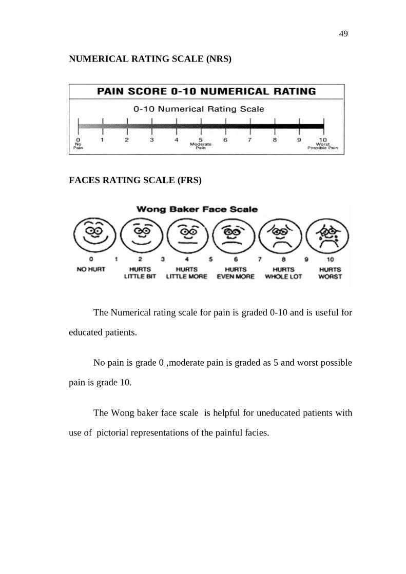
NUMERICAL RATING SCALE (NRS)
FACES RATING SCALE (FRS)
The Numerical rating scale for pain is graded 0-10 and is useful for
educated patients.
No pain is grade 0 ,moderate pain is graded as 5 and worst possible
pain is grade 10.
The Wong baker face scale is helpful for uneducated patients with
use of pictorial representations of the painful facies.

50
OBSERVATION & RESULTS
PAIN
In our study, among the patients the who had the conventional
donor site dressing with meshed Vaseline gauze had a median pain score
of 5 observed in 23 patients. Minimum pain score in this group was 3
noted in 2 patients. maximum pain score was 6 experienced in 4 patients.
Pain score of 4 was noted in 21 patients.
In comparision the combination dressing group with
haemocoagulase and collagen dressing had different results . the median
pain score was 2 experienced in 27 patients. Minimum pain score was 1
noted in 6 patients. Maximum pain score noted in the collagen dressing
group was 4 observed in 4 patients. The remaining 13 patients
experienced a pain score of 3. Pain was drastically reduced in the
collagen dressing patients.
AMBULATION
Since the pain score was high in the closed dressing subset of
patients, it limited their mobilization and 22 patients were ambulant only
on postoperative day 3. Only 10 patients were ambulant on the 2nd post
operative day. The remaining 18 patients ambulated well only on the 4th
postoperative day.

51
Due to the decreased pain in the collagen dressing for the donor
site patients ambulated early and 27 patients were ambulant from 1 st
postoperative day.
Subsequently 21 patients were ambulant on 2nd post operative day.
The remaining 2 patients ambulated on the 3rd postoperative day.Early
ambulation was noted in the in patients with the collagen dressing
RE EPITHELISATION
Donor site reepithelisation was assessed in the closed dressing by
the loosening of the dressings average time taken for the donorsites to
reepitheliase was 20 -23 days. Due to complications of donor site
infection the donor site healing extended to 29 -31 days in about 6
patients.
Donor site reepithelisation was noted between 15-17 days in the
collagen dressing group. In 2 patients donor site healing was delayed to
21-23 days due to donor site infection which was treated by conservative
methods due to the ease of inspection of donor site and had the advantage
of early identification of donor site complications. In 1 patient there was
displacement of collagen sheet from the donorsite and it was converted
to closed dressing and excluded from study. The healing time noted in
that patient was 23 days.

52
COST FACTOR ANALYSIS
The cost for the conventional closed dressings was Rs .150
inclusive of the over padding needed in review period. The cost factor
for application 25x40 cm moist collagen sheet to the donor site was Rs
450.
DONOR SITE COMPLICATIONS
Infection
Donor site infection was noted in 6 patients of the closed dressing
group. It was managed with conservative dressings with framycetin.
donorsite dressings on periodic review showing excessive soakage with
foul smell were opened end wound swabs taken
Donor site infection noted in 2 patients in the collagen dressing
group, conservative e dressings done to treat with the infection in these
patients.
Collagen Displacement-
1 patient in the study group had shearing of collagen sheet when
applied to donor site in the postoperative period. Wound was cleaned and
closed dressing applied and patient was excluded from the study.

53
Donor site dressing problems
DRESSING SOAKAGE AND INFECTION
The donor site dressing soakage was more in the closed dressing
patients and over padding done. This had the disadvantage of increased
bulk of the dressing. Malodour present due to dressing soakage was
promptly managed to rule out donor site infection. The increased bulk of
the dressing due to over padding and the seepage of exudates from
dressing to the garments produced a major discomfort in wearing the
garments in patients with closed dressing. The combination dressing with
hemocoagulase and collagen dressing did not have disadvantage of
soakage of dressing nor bulky dressing. The patients had increased
comfort in wearing the garments which boosted the morale of the
patients and encouraged early ambulation. Though the patients had
initially apprehensions due to the exposed raw area at the donor site,
patients were counseled and in the post operative period direct
visualization of the healing donor sites lessened their anxiety. The
reduced pain, comfort of dressing and advantage to wear the garments
early made patients who were subjected to multiple grafting procedures
and treated with both methods of dressing opt for the combination
dressing in subsequent procedures.

54
OBSERVATIONS
COLLAGENDRESSING
CLOSEDDRESSING
PAIN SCORE 2 5
AMBULANT ON 1st POD 3rd POD
REEPITHELISATION 15-17 DAYS 20-23 DAYS
COST FACTOR RELATIVELYEXPENSIVE
LESS EXPENSIVE
COLLAGEN DRESSING

55
CLOSED DRESSING
COLLAGEN DRESSING
POST OPERATIVE DAY

56
CLOSED DRESSING
Intensity of pain was drastically reduced in the combination
treatment group treated with hemocoagulase and collagen sheet. This
facilitated early ambulation from the very next day.
Complete epithelisation was found in all cases. Eventhough the
procedure was more expensive, it will be cost effective in the longrun as
it helps in higher turnover of patients with better acceptability. Shearing
of collagen sheet on the posterior thigh donor sites is common and may
lead to significant donor site morbidity.
Patients who were subjected to multiple grafting procedures felt
collagen dressing was better and comfortable.
POST OPERATIVE DAY

57
DISCUSSION
Split-thickness skin grafting (STSG) is a frequently used
reconstructive technique but is associated with variations in practice.
Rakel et al (1998) in the review of the literature found a transparent film
to be the best dressing for the care of STSG donor sites. This review of 33
studies found that transparent film was associated with one of the fastest
healing rates, a smooth epithelialized surface, a low infection rate, the
least amount of pain and minimal cost.
Numerous controlled studies in the last 50 years have established
that moist wound healing is the best evidence based practice. Dried
wound tissue is more prone to complications such as infection, scarring,
pain and prolonged healing. The goal of treating skin graft donor sites is
to promote healing while minimizing the risk of introducing new
complications and pain to an already traumatized patient.
Essentially, the wound was left open to scab, which is
contradictory to the best evidence-based practice of today, that of moist
wound healing. In recent years, much has been published highlighting the
benefits of moisture-retentive dressings in treating donor sites.
Moisture-retentive dressings that have been used include
hydrocolloids, foams, and transparent thin film dressings, alone or in
combination with absorbent materials such as alginates, hydrofibers or

58
gauze. While hydrocolloids and foams provide the needed absorbency,
they must be removed whenever wound inspection is required, increasing
treatment cost and the risk of traumatizing the wound. Thin film dressings
allow for wound visualization, but usually fail to contain the drainage for
more than 24 hours, even when used secondary to other absorbent
dressings (which also negates the benefit of transparency).Tegaderm™,
Absorbent Clear Acrylic Dressing is a moisture-retentive, absorbent
dressing which combines the benefits of highly absorbent dressings such
as hydrocolloids, foams, alginates and hydrofibers with the transparency
of thin film dressings. Recent published work indicates that this dressing
provides excellent results with skin donor sites2,3.
As early as 1962, Winter [15] showed that moist wounds
healed faster than wounds left to dry out. This observation has led
the care of skin graft donor sites away from the conventional dry
gauze dressings toward the semi-occlusive or occlusive dressings.
Although these occlusive dressings provide moist environments for
wound healing, there has been concern that occlusion of wounds
would lead to increased infection [16-181. How- ever, Hutchinson
and McGuckin [19], in a review of 29 donor site studies, showed an
infection rate of only 2.7% in 594 occluded wounds versus an
infection rate of 6.4% in 360 conventionally dressed wounds (Figure
1). In a recent study on donor site wound healing, 30 burn patients
with skin graft donor sites were randomized to receive either an

59
occlusive (DUODERM) or conventional gauze dressing on their wounds
[2]. Wounds in both groups were colonized with bacteria; however,
only conventionally dressed wounds became infected (Table I).
Bacteria were pre- sent intraoperatively in 9 of 14 (64%)
hydrocolloid- treated sites and in 5 of 16 (31%) conventionally
treated sites.
Moist wound healing is not a new idea and providers continue to
strive to find the optimal treatment that provides this ideal, moist
environment. George Winter is often referenced as a pioneer among
wound-care practitioners because of his work in the early 1960s that
proved wounds re-epithelialize quicker in a moist environment (Winter,
1963). It is indeed the epithelialization process on which we focus when
treating the skin graft donor site. Historically, the most popular way of
treating skin graft donor sites is the dry open technique using either
scarlet red or petrolatum bismuth dressings. A heat lamp or air blowing
device is then placed over the area to dry out the donor site and form a
scab. This process is commonly perceived as a painful treatment so
investigators sought a pain free dressing that allowed for absorption of
the excessive exudate as well as hemostasis. There are even reports of
placing wax paper over the area thereby protecting the nerve endings
from the pain in an attempt to heal the superficial wound (Weeter, 1952).

60
Utilization of a moisture vapor permeable transparent dressing
yielded freedom from pain, early healing, and less bulky dressings,
however, post-operative leakage from the site required frequent dressing
changes on the fragile tissue (James & Watson, 1975). The aim became
marrying the advantages of painless and moist wound healing with the
concept of absorption. Epinephrine applied to the fresh donor site was
proposed as a solution to excessive sanguinous exudate, but there was
still dressing slippage leaving the donor site exposed (Pontén &
Nordgaard, 1976). In addition, the concern of infection with occlusive
dressings was raised, but Birdsell, Hein, and Lindsay (1979) found no
infections in the 100 patients studied with occlusive dressings. Salisbury,
Bevin, Dingeldein, and Grisham (1979) investigated polyurethane foam
but found that the histologic examination of the pig’s skin showed no
evidence that the foam made a difference in healing time despite the
dressing’s painless removal. The idea of hemostasis was achieved
utilizing an alginate dressing, which also provided the absorption
component yet the dressing still required changing prior to complete
healing, resulting in a painful event (Groves & Lawrence, 1986).
Attwood (1989) continued the use of alginate and demonstrated a
decrease in healing time compared to paraffin gauze dressings.
In the 1990s, the quest continued with the introduction of Biobrane
as a cover over the donor site, yet its cost and rate of infection were too
much to accept when Xeroform (a bismuth petrolatum-impregnated

61
nonstick antimicrobial gauze) continued to be cheap and easy despite the
advantage of reduced pain with Biobrane (Feldman, Rogers, & Karpinski,
1991). The hydrocolloid emerged as a winner because it had a high rate
of absorption, showed fewer infections, and had more rapid healing with
acceptable leakage (Smith, Thomson, Bolton, & Hutchinson,1993). By
1995, Griswold et al. (1995) evaluated a new dressing that was made
from type I bovine collagen. Of no surprise, this dressing indeed healed
faster than Xeroform and was less painful, however, the collagen had to
be inspected and the dressing changed on days 3, 5, and 7 Patients with
collagen dressings are found to have only minimal to moderate pain in the
entire post operative period and during the first dressing. In these patients
analgesic requirement is reduced and early mobilisation can be done.
Thus the major advantage of using collagen as a donor site dressing is
decreased pain.
The collagen sheet once adherent to the wound has low friction
between the wound surface and dressing and this has made it suitable for
awkwardly sited donor sites. (Gupta RL, Jain RK, Kumar M, et al).
Also once applied it does not require a bulky dressing which would
hamper mobilisation, or require a change of dressing as there is no
soakage of the dressing due to wound exudate.
The burn literature continues to examine more donor-site dressings
than any other discipline and Duinslaeger, Verbeken, Vanhalle, and

62
Vanderkelen (1997) evaluated cultured allogeneic keratinocyte sheets.
These sheets accelerated healing compared with transparent dressings
but were quite expensive.
A systematic review in 1998 answered the question of cost but
started with the various options for dressing selection on the donor site.
A review performed by Rakel et al. (1998) found that in the last 40
years the transparent dressing still reigns because of its association with
faster healing rates, smooth epithelialized surface, low infection rates,
less pain, and minimal cost.
Despite the increasing array of products available and this review,
the most commonly used dressing was the calcium alginate even though
it is not rated high in dressing performance (Lyall & Sinclair, 2000). The
most important item from the survey was patient comfort. The gold
standard by which other dressings are now being measured moved away
from the transparent dressing to the calcium alginate but the alginate fell
short in comparison with the silicone-coated polyamide net (Mepitel;
O’Donoghue, O’Sullivan, O’Shaughnessy, & O’Connor, 2000).
Although Mepitel is virtually nonstick and can be left in place for
days at a time, it requires secondary dressings to absorb moisture.
Chitosan is a derivative of shrimp exoskeleton and has the ability to
rapidly clot blood. The chitosan is being impregnated into gauze

63
dressings and used in the field during times of war. This mixture is also
hypoallergenic and has natural anti-bacterial properties. With such
promise, it was only a matter of time that this dressing was evaluated on
the donorsite. Stone, Wright, Clarke, Powell, and Devaraj (2000) were
impressed with the re-epithelialization rates and regeneration of nerves
within the vascular dermis, but the use of chitosan was limited because of
availability. The foray into these often-expensive and limited dressings
prompted some to ask whether the dry open technique was indeed better.
The limitation of this technique due to pain, already detailed, led to a
modification.
The donor site was still left open but under moist conditions
utilizing the application of Moist Exposed Burn Ointment. Although the
full action of the ointment is unknown, Atiyeh, Ghanimeh, Kaddoura,
Ioannovich, Al-Amm (2001) determined that even in their limited
evaluation, there was some promise because of the cosmetic outcome. At
this point, it seems there can be no more new and original ideas in the
arena of donor-site care but a novel topical application must be explored
despite its obvious limitations. Egg membrane as a biological dressing
was evaluated by Yang, Chuang, and Tsay (2003). The dressing indeed
provided pain relief, protection, and healing, and was low cost, yet the
size of the dressing was limited and the feasibility was narrow. Despite
continued examination of foam, alginate, collagen, and even cellulose,
there were not many discoveries but rather the continued confirmation

64
that the alginate and transparent cover were supreme. Then, a new
absorbent transparent dressing hit the wound-care market and was
immediately evaluated by Terrill, Goth, and Bailey (2007) on donor-site
wounds. Terrill et al. found the dressing to be cost comparative and easy
to apply. The donor-site scars with this dressing were less red and itchy,
flatter, and softer. The best attribute of the dressing, of course, was pain-
free healing. The 3M Tegaderm Absorbent Clear Acrylic Dressing is a
moisture-retentive dressing that is absorbent yet transparent. The product
is designed to be changed every 3–5 days per manufacturer guidelines yet
donor sites on average take up to 14 days to heal completely. Even with
the benefit of pain-free dressings, the patient still has to endure two to
three dressing changes until healing has been achieved.
In our study significant pain reduction, early ambulation,
comfortable dressing, similar to the study of Collagen Dressing in The
Management of Donor Site of Split Thickness Skin Grafts (P Halankar*,
D Cunha-Gomes**, C Chaudhari*) good reepithelisation, and early
discharge of the patients with signs of good healing was noted in the
collagen dressing. P Halankar*, D Cunha-Gomes**, C Chaudhari* , at
Bombay Hospital and Medical Research Centre, studied a group of 30
patients were included in this study with 21 males and 9 females ranging
from age 18 to 72 years. All patients required split thickness skin grafts
of approximately 100-250 cm2 in area to provide cover for various
indications . In the group all patients tolerated the collagen dressing well

65
and there was no allergic reaction. In the early postoperative period on
assessment of donor site pain to touch and pressure, 2 patients in the
group had no pain, 23 rated the pain as minimal while 7 patients assessed
the pain as moderate and tolerable. There was no complaint of severe
pain and none of the patients required additional analgesics for donor site
pain. Once the patients were mobilized by the third postoperative day
they were assessed again for pain while walking. In the patient group 21
patients assessed the pain as minimal, 7 assessed the pain as moderate
and 2 as severe. The 2 patients who had severe pain on walking also had
severe pain on touch and pressure and required additional analgesics to
relieve them of the donor site pain. All other patients had no analgesic
requirement for the donor site.
In 2 patients soakage of wound exudate was seen on the fifth and
eighth post operative day respectively. These were the two patients in
whom there was significant donor site pain. In both patients the donor site
dressing was foul smelling and the wound was covered with purulent
discharge with degradation of the collagen sheet. The infection was
limited to the donor site with no evidence of cellulitis of the surrounding
skin or fever. The infected sites were redressed with framycetin
impregnated paraffin gauze. In both patients the wounds were redressed
after forty-eight hours by which time pain had subsided. On the
fourteenth post operative day the donor site dressings were soaked in
saline and easily removed with no pain in 5 patients, while 18 and 7
patients had minimal and moderate pain respectively. None of them

66
complained of severe pain. In the group 24 patients showed 100%
reepithelialisation, 4 showed between 90-100%, while the two with
infected donor sites had < 90% reepithelialisation. In the group 1 patient
had a haematoma beneath the collagen dressing but there was complete
re-epithelialisation beneath the haematoma. On late follow up of 26
patients, 2 showed hypertrophic scarring of the donor site.
In our study the median pain score was 2 in the collagen dressing
compared to the increased pain score of 5 in the conventional closed
dressing group. Patients were ambulant from postoperative day 1 with
the collagen dressing, while patients with the bulky closed dressing were
ambulant from the third postoperative day.
In our study cost factor analysis increased expenditure towards
collagen dressing, but the above advantages help in rapid turnover of
patients with minimal donor site morbidity which is the need of the hour
in recent treatment protocols in management of rawareas. The advantage
of direct review of the donorsite wound in the collagen group helps in
early identification of problems and also manage the wounds properly
and salvage them from becoming full thickness wounds which will
increase donor site morbidity.
Skin graft donor sites in burn patients warrant intense treatment
and optimal wound-management techniques. An ideal dressing method
prevents dehydration and infection while facilitating wound healing.
Dressings should be easy to apply and require minimal care.

67
CONCLUSION
Haemocoagulase with Collagen sheet application for donor site
have the following advantages.
Less pain over the donor site compared to the conventional closed
dressing with Vaseline gauze.
Early ambulation due to reduced pain after the application of
collagen sheet to the donor site.
Reepithelisation complete for most patients and time of
reepithelisation is similar to the standard expensive dressings for
the donor site.
Inspection of the donor site wound can be done easily and
complications can be recognized early.
A comfortable Dressing due to the decreased bulk of dressing and
ease to wear the garments over the dressing.
Less complications and better outcome compared to the
conventional closed dressing.

BIBLIOGRAPHY
1. Albini A, Adelmann-Grill BC. Collagenolytic cleavage products ofcollagen Type I as chemoattractants for human dermal fibroblasts.Eur. J Cell Biol. 1985;36(1):104–107
2. A1arezO, . M., Mertz, P. M., and Eaglestein, W. H. The effect ofocclusivedressings on collagen synthesis and re-epitheli.alization insuperlicial wounds. J. Surg. Res., 1983, 35. 142-148.
3. Altemeier W, Burkerts F, Pruitt B, Sandusky W. Manual onControl of Infection in Surgical Patients, 2nd ed. Philadelphia:JB Lippincott Company, 1976: 29-30.
4. Brown CL, Curtsinger LJ, White M, et nl. Enhancement ofepidermal regeneration of biosynthetic epidermal growthfactor. J Exp Med 1986; 163: 1319-24.
5. Biltz, H., Kiessling, M., and Kreysel, H. W. Comparison ofhydrocolloid dressing and saline gauze in the treatment of skingraít donor sites. In: An Environment for Healing: The Role ofOcclusion. Edited by T. J. Ryan. Pp. 125-128. London: RoyalSociety of Medicine,1985.
6. Brady, S. C., Snelling, C. F. T., and Chow, G. Comparison ofdonor site dressings. Ann. Plast. Surg., 1980, 5: 238-243.
7. Burton JL, Etherington DJ, Peachey RD. Collagen sponge for legulcers. Br J Dermatol. 1978;99(6):681–685
8. Bennett RG. The debatable benefit of occlusive dressings forwounds. Dermatol Surg Oncol 1982; 8: 166-7.

9. Champsaur, A., Amadou, R., Nefzi, A., and Marichy, J. Use ofDuoderm ondonor sites after skin grafting. A comparative studywith Tulle-gras. In: BeyondOcclusion: Wound Care Proceedings.Edited by T. J. Ryan. Pp. 127-131. London: Royal Society ofMedicine, 1988.
10. Chung J, Wang XQ, Lindberg FP, Frazier WA. Thrombospondin-1acts via IAP/CD47 to synergize with collagen in alpha2beta1-mediated platelet activation. Blood 1999;94:642-8.
11. Doillon CJ, Silver FH. Collagen-based wound dressing: effects ofhyaluronic acid and fibronectin on wound healing. Biomaterials.1986;7(1):3 8.
12. Doillon CJ, Whyne CF, Berg RA, Olson RM, Silver FH.Fibroblast-collagen sponge interactions and spatial deposition ofnewly synthesized collagen fibers in vitro and in vivo. ScanElectron Microsc. 1984;(Pt 3):1313–1320
13. Doillon CJ, Silver FH, Olson RM, Kamath CY, Berg RA.Fibroblast and epidermal cell-Type I collagen interactions: cellculture and human studies. Scanning Microsc. 1988;2(2):985–992.
14. Eriksson G, Eklund AE, Kallings LO. The clinical significanceof bacterial growth in venous leg ulcers. Stand J Infect Dis1984; 16: 175-80.
15. Evans AJ. Treatment of burns today. Proc R Sot Med 1971:64: 21-2
16. Falanga V. Classifications for wound bed preparation andstimulation of chronic wounds. Wound Repair Regen.2000;8(5):347–352.

17. Feldman DL. Which dressing for split-thickness skin graftdonor sites? Ann Plast Surg 1991; 27: 288
18. Friedman, G. D., Capozzi, A., and Pennisi, V. R. Care of the splitthickness skin graft donor site. J. Trauma.,1974, 14: 163-167.
19. Freshwater MF, Chi Tsung Su, Hoopes JE. A Comparison ofPolyurethane Foam Dressing and Fine Mesh Gauze in The Healingof Donor Sites. Plastic and Reconstructive Surgery 1976
20. Fantone JC, Ward PA. Role of oxygen-derived free radicalsand metabolites in leukocyte-dependent inflammatory reactions.Am J Path01 1983; 107: 397.
21. Griswold JA, Grube BJ, Engrav LH, et al. Determinants ofdonor site infection in small burn grafts. J Burn Care Rehabil1989; 10: 531-5
22. Griswold, J. A., Grube, B. J., Engrav, L. H., and others.Determinants of donor si te infections in small burn grafts. J. BurnCare Rehabil., 1989, 10: 531-535. Hadjiiski O, Anatassov N.Amniotic membranes for temporary burn coverage. Ann Burns FireDisasters 1996;9:88-92.
23. Greenhalgh DC, Barthel PP, Warden CD. Comparison of backversus thigh donor sites in pediatric patients with burns. JBurn Care Rehabil1993; 14: 21-5.
24. Gruss JS, Jirsch DW. Human amniotic membrane: A versatilewound dressing. Can Med Assoc J 1978;118:1237-46.
25. Ghalambor AA, Mohammad HP, Khodadadi A. The amnioticmembrane: A suitable biological dressing to prevent infection inthermal burns. Med J Islamic Acad Sci 2000;13:115-8.
26. Gupta RL, Jain RK, Kumar M, et al. Fate of Collagen Sheet coverfor artificially created raw areas (an experimental study). Indian JSurgery 1978; 40 (12) : 641-45
27. Gupta RL, Jain RK, Kumar M, et al. Role of collagen sheet coverin Burns. (A Clinical study). Indian J Surgery 1978; 40 (12) : 646-49
28. P Halankar, D Cunha-Gomes, C Chaudhari -Collagen Dressing inThe Management of Donor Site of Split Thickness Skin Grafts
29. Horch RE, Stark GB. Comparison of the effect of a collagendressing and polyurethane dressing on healing of split thicknessskin graft donor sites. Scand J Plast Reconst Surg Hand Surg 1998;32 (4) : 407-13
30. Hutchinson JJ, McGuckin M. Occlusive dressings: a microbio-logic and clinical review. Am J Infect Contr 1990; 18: 257-68
31. Hunt TK. Basic principles of wound healing. J Trauma 1990;30: s122.
32. Hynes RO. Integrins: versatility, modulation, and signaling in celladhesion. Cell. 1992;69(1):11–25.
33. James, J. H., and Watson, A. C. H. The use of Opsite, a vapourpermeable dressing, on skin graft donor sites. Br. J. Plast. Surg.,1975, 28: 107-110.
34. Jeffrey J. Metalloproteinases and tissue turnover. WOUNDS.1995;7(Suppl A):13A–22A.
35. Kilinc H, Sensoz O, Ozdemir R, Unlu RE, Baran C. Whichdressing for split thickness skin graft donor site? Ann Plast Surg2001; 46 (4) : 409-14.

36. Klein CE, Dressel D, Steinmayer T, et al. Integrin alpha 2 beta 1 isupregulated in fibroblasts and highly aggressive melanoma cells inthree-dimensional collagen lattices and mediates the reorganizationof collage I fibrils. J Cells Biol. 1991;115(5):1427–1436.
37. Krieg T. Collagen in the Healing Wound. WOUNDS.1995;7(Suppl A):5A–12A.
38. Kan Gruber D, Gruber C, Seifter E, et al. Acceleration ofwound healing by Staphylococcus aureus. Surg Forum 1981;32: 768
39. Leicht, P., Siim, E., and Sorensen, B. Treatment of donor sites-Duoderm or Omiderm. Burns Incl. Therm. lnj., 1989, 15: 7-10.
40. Laforet EC. Wound dressing or window dressing? Arch Surg1974; 109: 457.
41. Lazovic G, Colic M, Grubor M, Jovanovic M. The application ofcollagen sheet in open wound healing. Ann Burns Fire Disasters2005;18:151
42. Madden, M. R, Finkelstein, J. L., Hefton, J. M., and Yurt, R W.Optimal healing of donor site wounds with hydrocolloid dressings.In: An Environment for Healing: The Role of Occlusion. Edited byT. J. Ryan. Pp. 133-137. London: RoyalSociety ofMedicine, 1985.
43. Madden MR, Nolan E, Finkelstein JL, er al. Comparison of anocclusive and semiocclusive dressing and effect of the woundexudate upon keratinocyte proliferation. J Trauma 1989; 29: 924
44. Madri JA, Marx M. Matrix composition, organization, and solublefactors: modulators of microvascular cell differentiation in vitro.Kidney Int1992;41(3):560–565

45. Mignatti P, Rifkin DB, Welgus HG, Parks WC. Proteinases andtissue remodeling. In: Clark RAF, ed. The Molecular and CellularBiology of Wound Repair. 2nd ed. New York, NY: Plenum Press;1996:427–474
46. Mohammadi1 AA, Riazi1 H, Hasheminasab MJ, Sabet B,Mohammadi MK, Abbasi S, et al. Amniotic membrane dressing v/sconventional topical antibiotic dressing in hospitalized burnpatients. Iranian Red Crescent Med J 2009;11:66-70.
47. Montesano R, Orci L, Vasselli P. In vitro rapid organization ofendothelial cells into capillary-like networks is promoted bycollagen matrices. J Cell Biol. 1983;97(5 Pt 1):1648–1652
48. Motta G, Ratto GB, De Barbieri A, Corte G, Zardi L, Sacco A, etal. Can heterologous collagen enhance the granulation tissuegrowth? An experimental study. Ital J Surg Sci 1983;13:101-8.
49. Nanney LB. Epidermal growth factor-induced effects on woundhealing. [Abstr.] Clin Res 1987; 35: 706A.
50. Nataraj C, Ritter G, Dumas S, Helfer FD, Brunelle J, Sander TW.Extra cellular wound matrices: Novel stabilization and sterilizationmethod for collagen-based biologic wound dressings. Wounds2007;19:148-56.
51. Nagata H, Ueki H, Moriguchi T. Fibronectin: Localization innormal human skin, granulation tissue, hypertrophic scar, maturescar, progressive systemic sclerotic skin, and other fibrosingdermatoses. Arch Dermatol 1985;121:995-9.
52. Park SN, Lee HJ, Lee KH, Suh H. Biological characterization ofEDC-crosslinked collagen-hyaluronic acid matrix in dermal tissuerestoration. Biomaterials 2003;24:1631-41.

53. Palmieri B. Heterologous collagen in wound healing: a clinicalstudy. IntJ Tissue React. 1992;14(Suppl):21–25.
54. Parks WC. The production, role, and regulation of matrixmetalloproteinases in the healing epidermis. WOUNDS.1995;7(Suppl A):23 37
55. Petersen MJ, Woodley DT, Stricklin GP, O’Keefe EJ. Enhancedsynthesis of collagenase by human keratinocytes cultured on TypeI or Type IVcollagen. J Invest Dermatol. 1990;94(3):341–346.
56. Ponten B, Nordgaard JO. The Use of Collagen Film as a Dressingfor Donor Areas in split skin grafting. Scand J Plast Reconstr Surg1976; 10 : 237-40.
57. Porter JM. A comparative investigation of Re epithelialisation ofsplit skin graft donor areas after application of hydrocolloid andalginate dressings. British J Plastic Surgery 1991; 44 : 333-37.
58. Prasad,J. K, Fel1er, l., and Thompson, P. D. A prospectivecontrolled trial of Biobrane versus Scarlet Red on skin graft donorafeas. J. Burn Care Rehabil.,1987,8: 386.
59. Ramakrishnan KM, Jayaraman V. Management of partial-thicknessburn wounds by amniotic membrane: A cost effective treatment indeveloping countries. Burns 1997;23:S33-6.
60. Robson MC, Lea CE, Dalton JB, Heggers JP. Quantitativebacteriology and delayed wound closure. Surg Forum 1968,19:501-2.

61. Richmond, J. D., and Sutherland, A B. A new approach to theproblems encountered with Opsite as a donor site dressing:systemic ). Br. J. Plast.Surg.,1986,39: 516-518.
62. Roberts, L. W., McManus, W. F., Mason, A. D., and Pruitt, B. A.Duoderm in the management of skin graft donor sites. In: SurgicalResearch: Recent Developments. Edited by C. W. Hall.Proceedings of the 1st Annual ScientificSession of the Academy ofSurgical Research. Pp. 55-58. San Antonio, Texas: Pergamon
63. Salisbury, R. E., Wilmore, D. W., Silverstein, P., and, B. A.Biologic dressings for skin graft donor sites. Arch. Surg., 1973,106: 705-706. .Gemberling, R M., Miller, T. A., Caffee, H., andZawacki, B. E. Dressing comparison in the healing of donor sites.J. Trauma., 1976, 16: 812-814
64. Skouge, J. W. Techniques for split-thickness skin grafting. J.Dermatol. Surg.Oncol., 1987, 13: 841- 849
65. Schiro JA, Chan BM, Roswit WT, et al. Integrin alpha 2 beta 1(VLA-2)mediates reorganization and contraction of collagenmatrices by human cells. Cell. 1991;67(2):403–410.
66. Scharffetter-Kochanek K, Klein CE, Heinen G, et al. Migration ofhuman keratinocyte cell line (HaCaT) to interstitial collagen Type Iis mediated by the alpha 2 beta 1-integrin receptor. J InvestDermatol. 1992;98(1):3–11.
67. Subdeck BD, Parks WC, Welgus HG, Pentland AP. Collagen-stimulated induction of keratinocyte collagenase is mediated via
tyrosine kinase and protein kinase C activities. J Biol Chem.1994;269(47):30022–30029.
68. Schultz G, Mast B. Molecular analysis of the environment ofhealing and chronic wounds: cytokines, proteases and growthfactors. WOUNDS.1998;10(6 suppl):1F–9F.
69. Sibbald RG, Williamson D, Orsted HL, et al. Preparing the woundbed debridement, bacterial balance, and moisture balance. OstomyWound Manage. 2000;46(11)14–35.
70. Schultz GS, Sibbald RG, Falanga V, et al. Wound bed preparation:a systematic approach to wound management. Wound RepairRegen.2003;11(Suppl 1):1–28.
71. Smith DJ, Thomson PD, Bolton LL, Hutchinson JJ. Microbiol-ogy and healing of the occluded skin-graft donor site. PlastReconst Surg 1993; 91: 1094-7.
72. Teplitz C, Davis D, Mason AD, Moncrief JA. Pseudomonasburn wound sepsis. I. Pathogenesis of experimental burnwound sepsis. J Surg Res 1964; 4: 200-16.
73. Tenorio A, Jindrak K, Weiner M, et al. Accelerated healing ininfected wounds. Surg Gynecol Obstet 1976; 142: 537-43.
74 Varghese, M. C., Balin, A. K, Carter, M., and Caldwel1, D. Localenvironment of chronic wounds under synthetic dressings. Arch.Dermatol., 1986, 122: 52- 57.

75. Veves A, Sheehan P, Pham HT. A randomized, controlled trial ofpromogran (a collagen/oxidized regenerated cellulose dressing) vsstandard treatment in the management of diabetic foot ulcers. ArchSurg. 2002;137:822-7
76. Weber RS, Hankins P, Limitone E, et al. Split-Thickness SkinGraft Donor Site Management. A Randomised Prospective TrialComparing a Hydrophilic Polyurethane Absorbent Foam Dressingwith a petroleum gauze Dressing. Arch Otolaryngol Head NeckSurg 1995; 121 (10) : 1145-9
77. Winter CD. Formation of the scab and the rate ofepithelialization of superficial wounds in the skin of the youngdomestic pig. Nature 1962; 193: 293-4.
78. Zapata-Sirvent, R, Hansbrough, J. F., Carroll, W., and others.Comparison of Biobrane and Scarlet Red dressings for treatment ofdonor site wounds. Arch.Surg., 1985, 120: 743

CLOSED DRESSING
HAEMOCOAGULASE WITH COLLAGEN SHEET DRESSING

HAEMOCOAGULASE APPLICATION
SIEVED COLLAGEN SHEET

COLLAGEN SHEET APPLIED TO DONOR SITE
DONOR SITE HEALING – COMBINATION DRESSING 1

DONOR SITE HEALING – COMBINATION DRESSING 2
DONOR SITE HEALING – COMBINATION DRESSING 3
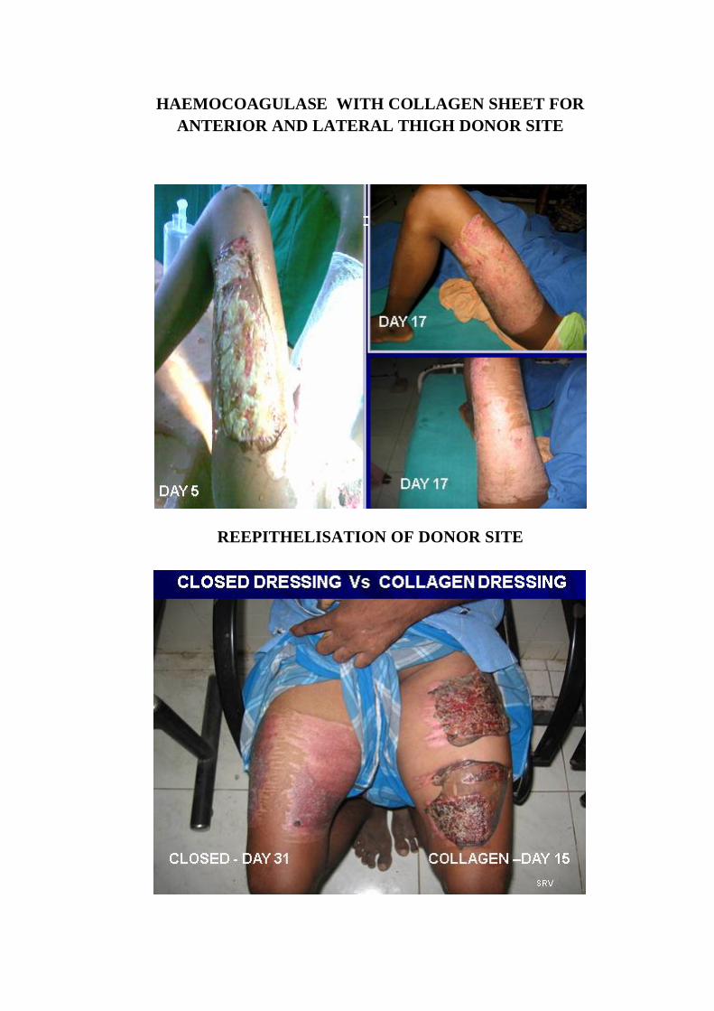
HAEMOCOAGULASE WITH COLLAGEN SHEET FORANTERIOR AND LATERAL THIGH DONOR SITE
REEPITHELISATION OF DONOR SITE

DONOR SITE COMPLICATIONS
DISPLACEMENT OF COLLAGEN SHEET

NAME AGE SEX IP NO CLOSED DONOR SIZE(cm) PAIN AMBULATIONREVIEW
REMARKSREEPITHELISAT
ION SOAKAGE INFECTIONDATE OFSURGERY
DRESSING SITE POD
SUDHARSAN 28 MALE 29230 YES L(T) 25X30 4 3 20 MINIMAL NIL 14/12/09
MOHAN 39 MALE 29236 YES R(T) 25x30 4 3 20 MODERATE NIL 17/12/O9
SAROJA 28 FEMALE 29302 YES L(T) 25X30 6 4 FOUL SMELLING 29 SEVERE STAPH.AUREUS 3/12/2009
MEENAKSHI 29 FEMALE 29315 YES L(T) 15X25 5 2 21 MODERATE NIL 5/12/2009
RAJA 30 MALE 29381 YES L(T) 20X25 5 4 20 MODERATE NIL 17/12/09
PADMAVATHY 32 FEMALE 29350 YES L(T) 25X30 3 3 20 MODERATE NIL 24/12/09
DEVI 36 FEMALE 29354 YES L(T) 25X30 5 3 20 MODERATE NIL 12/12/2009
NATARAJAN 39 MALE 29366 YES L(T) 20x30 4 4 21 MODERATE NIL 16/12/2009
SUGUNA 26 FEMALE 29386 YES R(T) 25x30 5 3 20 MODERATE NIL 12/12/2009
PANNERSELVAM 39 MALE 29402 YES R(T) 20x30 5 3 21 MODERATE NIL 3/1/2010
RAJAKUMARI 27 FEMALE 10171 YES R(L) 10x15 4 2 20 MINIMAL NIL 7/1/2010
ROSELINE 39 FEMALE 10196 YES L(T) 15X25 4 2 20 MINIMAL NIL 21/01/2010
THIRUMAL 40 MALE 10251 YES L(T) 25X30 5 4 FOUL SMELLING 30 MODERATE STAPH.AUREUS 3/2/2010
VENKATESAN 42 MALE 10457 YES L(T) 25X30 4 4 21 MODERATE NIL 22/02/2010
SRIDHAR 27 MALE 11036 YES L(T) 25X30 5 3 21 MODERATE NIL 7/3/2010
SARAVANAN 28 MALE 12781 YES L(T) 25X30 6 4 FOUL SMELLING 30 SEVERE STAPH.AUREUS 15/03/2010
SIVAGAMI 32 FEMALE 12861 YES R(T) 25X30 4 4 20 MODERATE NIL 21/03/2010
SURESH 29 MALE 13741 YES R(T) 25X30 4 3 20 MODERATE NIL 3/4/2010
SARALA 36 FEMALE 14501 YES R(T) 20X25 5 3 20 MINIMAL NIL 14/04/2010
SOUMYA 17 FEMALE 14556 YES L(T) 15x25 5 3 20 MODERATE NIL 18/04/2010
MANI 41 MALE 15771 YES L(T) 20x30 5 2 21 MODERATE NIL 4/5/2010
SHANTHI 28 FEMALE 15892 YES L(T) 20x30 5 3 20 MODERATE NIL 11/5/2010
LAKSHMI 24 FEMALE 16402 YES L(T) 20x30 5 2 20 MODERATE NIL 27/05/2010
DHANALAKSHMI 26 FEMALE 17819 YES L(T) 20x30 4 3 20 MODERATE NIL 6/6/2010
VISWANATHAN 39 MALE 18721 YES R(T) 25X30 5 4 20 MODERATE NIL 10/6/2010
MANIKANDAN 25 MALE 20231 YES L(T) 25X30 3 4 21 MODERATE NIL 17/06/2010
PUSHPARAJ 43 MALE 20239 YES L(T) 25X30 6 4 FOUL SMELLING 31 MODERATE STAPH.AUREUS 15/07/2010
MARIAPPAN 39 MALE 22214 YES R(T) 15x25 4 2 20 MINIMAL NIL 12/8/2010
RAMANAN 37 MALE 22291 YES R(T) 15x25 4 3 20 MODERATE NIL 21/09/2010

MAHALINGAM 35 MALE 22317 YES L(L) 10x15 4 3 20 MINIMAL NIL 2/10/2010
SUSEELA 32 FEMALE 24331 YES R(T) 25X30 5 3 21 MODERATE NIL 17/10/2010
BALAN 30 MALE 25430 YES R(T) 25X30 5 4 FOUL SMELLING 30 MODERATE STAPH.AUREUS 3/11/2010
ASHOK 24 MALE 10871 YES R(T) 20x30 5 3 20 MODERATE NIL 28/11/2010
KUMAR 26 MALE 11782 YES L(T) 20x30 4 4 20 MODERATE NIL 24/12/2010
ARUMUGAM 40 MALE 12001 YES L(T) 20x30 4 3 20 MODERATE NIL 5/1/2011
RAGHU 36 MALE 12585 YES L(T) 20x30 6 4 FOUL SMELLING 30 SEVERE STAPH.AUREUS 22/01/2011
AKILESHWARI 25 FEMALE 13145 YES L(T) 15x25 5 3 20 MODERATE NIL 3/2/2011
GOVINDARAJ 51 MALE 14392 YES L(T) 15x25 5 2 21 MODERATE NIL 9/2/2011
VELUSAMY 47 MALE 14953 YES R(T) 25X30 5 4 20 MODERATE NIL 3/3/2011
NARAYANAN 32 MALE 15745 YES R(T) 25X30 4 4 20 MODERATE NIL 15/03/2011
POWN 34 FEMALE 16709 YES L(T) 20x30 4 4 20 MODERATE NIL 19/03/2011
GOWRI 26 FEMALE 17123 YES R(T) 20x30 4 4 20 MODERATE NIL 3/4/2011
VISHNU 24 MALE 17789 YES R(T) 25X30 4 4 20 MODERATE NIL 6/5/2011
PADMINI 45 FEMALE 19254 YES R(T) 15x25 5 2 21 MODERATE NIL 3/6/2011
SENTHIL 25 MALE 20258 YES R(T) 15x25 5 3 20 MODERATE NIL 17/7/2011
KUMARAN 29 MALE 22354 YES R(T) 15x25 5 4 FOUL SMELLING 29 SEVERE STAPH.AUREUS 21/10/2011
LAKSHMI 22 FEMALE 22514 YES L(T) 15x25 4 2 20 MODERATE NIL 27/10/2011
MURUGAN 35 MALE 10971 YES L(T) 20x30 5 3 21 MODERATE NIL 12/11/2011
SHAKILA 21 FEMALE 1009 YES L(T) 20x30 4 3 20 MODERATE NIL 3/1/2012
MEHRUNISHA 24 FEMALE 1257 YES L(T) 15x25 4 2 20 MINIMAL NIL 21/01/2012

NAME AGE SEX I.P NoDATE OFSURGERY
DONORSITE SIZE(cm)
HAEMOCOAGULASE
COLLAGENSHEET PAIN
AMBULATION
REVIEWREMARKS
REEPITHELISATION INFECTION
VINITHA 16 FEMALE 29230 14/12/2009 L(T) 15X30 YES YES 2 1 15 NIL
SANDHYA 9 FEMALE 29452 18/12/2009 L(T) 25X30 YES YES 2 1 15 NIL
ANNAMALAI 35 MALE 29232 12/12/2009 L(T) 25X30 YES YES 2 1 17 NIL
HIYAKATHULLAH 30 MALE 29515 18/12/2009 L(T) 25X30 YES YES 3 2 15 NIL
SASIKALA 20 FEMALE 11052 27/12/2009 R(T) 25X30 YES YES 2 2 17 NIL
PRIYA 25 FEMALE 11534 6/12/2009 L(T) 25X30 YES YES 4 1 16 NIL
THIRUPURA SUNDARI 65 FEMALE 11896 22/12/2009 L(T) 10X15 YES YES 1 1 17 NIL
LOGANATHAN 29 MALE 12758 3/1/2010 L(L) 15X30 YES YES 2 2 15 NIL
SURESH 25 MALE 13564 12/1/2010 R(T) 15X30 YES YES 2 1 15 NIL
LAKSHMI 32 FEMALE 14011 27/01/2010 R(T) 15X30 YES YES 3 1 17 NIL
DHATCHAYANI 30 FEMALE 14545 6/2/2010 R(T) 25X30 YES YES 2 2 15 NIL
RATHI 23 FEMALE 15149 24/02/2010 R(T) 15X20 YES YES 2 1 15 NIL
SELVI 26 FEMALE 16188 3/3/2010 L(T) 20X30 YES YES 2 1 15 NIL
VANITHA 35 FEMALE 18433 23/03/2010 L(T) 15X20 YES YES 1 1 15 NIL
ANITHA 28 FEMALE 19010 2/4/2010 L(T) 15X20 YES YES 1 2 16 NIL
MANOHAR 49 MALE 19581 17/04/2010 L(T) 25X30 YES YES 3 1 17 NIL
PRABHA 17 MALE 20039 21/04/2010 L(T) 25X30 YES YES 2 1 17 NIL
PARI 40 MALE 21051 4/5/2010 R(T) 25X30 YES YES 4 3 INFECTION+ 20 staph.aureus
GEMINI 25 MALE 22007 15/05/2010 R(T) 25X30 YES YES 3 2 16 NIL
KEERTHIKA 19 FEMALE 22678 19/05/2010 L(T) 20X30 YES YES 3 1 15 NIL
DHANALAKSHMI 55 FEMALE 22912 12/6/2010 L(T) 20X30 YES YES 2 2 16 NIL
BHUVANESHWARI 24 FEMALE 23439 17/06/2010 L(T) 20X30 YES YES 2 1 16 NIL
VIDHYA 23 FEMALE 23931 4/7/2010 L(T) 20X30 YES YES 3 1 16 NIL
NANDHINI 15 FEMALE 23974 17/07/2010 R(L) 25X30 YES YES 3 2 16 NIL
MATHIALAGAN 42 MALE 24117 27/07/2010 L(T) 25X30 YES YES 2 2 17 NIL
JANCY 29 FEMALE 24616 16/08/2010 L(T) 25X30 YES YES 2 1 15 NIL

BALU 25 MALE 24938 23/08/2010 R(T) 15X20 YES YES 1 1 15 NIL
SATHYA 17 FEMALE 12065 11/9/2010 R(T) 15X20 YES YES 2 2 15 NIL
KANNAN 19 MALE 12679 23/09/2010 R(T) 15X20 YES YES 2 1 15 NIL
ILAYARAJA 25 MALE 13119 13/10/2010 R(T) 15X30 YES YES 3 2 17 NIL
THILOTHAMAL 35 FEMALE 13474 11/11/2010 R(T) 15X30 YES YES 2 2 16 NIL
SUMITHRA 26 FEMALE 13675 7/12/2010 L(T) 15X30 YES YES 2 1 17 NIL
KALAI SELVI 24 FEMALE 15153 7/1/2011 L(T) 15X20 YES YES 3 1 15 NIL
VASANTH 24 MALE 16117 3/2/2011 L(T) 15X20 YES YES 2 2 15 NIL
SRIKANTH 25 MALE 16297 27/02/2011 L(T) 15X20 YES YES 2 1 16 NIL
YUVARAJ 21 MALE 16537 7/3/2011 L(T) 20X30 YES YES 2 1 16 NIL
ALEX 30 MALE 17103 15/03/2011 L(T) 20X30 YES YES 2 2 17 NIL
SRINIVASAN 34 MALE 17539 12/4/2011 R(T) 20X30 YES YES 2 1 16 NIL
MUTHU 30 ,ALE 17714 14/05/2011 R(T) 20X30 YES YES 3 2 15 NIL
RAMACHANDRAN 18 MALE 17912 3/6/2011 R(T) 25X30 YES YES 4 3 INFECTION + 23 staph.aureus
SINGARAJ 45 MALE 18215 6/6/2011 R(T) 25X30 YES YES 1 2 15 NIL
MARIMUTHU 45 MALE 18456 16/07/2011 L(T) 25X30 YES YES 3 1 16 NIL
KUMARI 23 FEMALE 18509 28/07/2011 L(T) 15X20 YES YES 3 1 16 NIL
SAKTHIVEL 30 MALE 19004 20/11/2011 L(T) 15X20 YES YES 2 2 16 NIL
SARAVANA KUMAR 26 MALE 20031 24/11/2011 L(T) 15X20 YES YES 2 1 16 NIL
NOORULA KHAN 40 MALE 21237 2/12/2011 R(T) 15X20 YES YES 2 2 17 NIL
SASI KUMAR 24 MALE 22549 7/12/2011 R(T) 20X30 YES YES 4 1 16 NIL
GUNASUNDARI 36 FEMALE 23112 23/12/2011 R(T) 25X30 YES YES 3 2 16 NIL
BASKAR 35 MALE 10975 O2/01/2012 L(T) 15X20 YES YES 1 2 16 NIL
ARUL 30 MALE 1151 6/1/2012 L(T) 15X20 YES YES 2 2 16 NIL