analysing the eosinophil cationic protein - a clue to the
TRANSCRIPT
REVIEW Open Access
Analysing the eosinophil cationic protein - a clueto the function of the eosinophil granulocyteJonas Bystrom1*, Kawa Amin2,3, David Bishop-Bailey1
Abstract
Eosinophil granulocytes reside in respiratory mucosa including lungs, in the gastro-intestinal tract, and inlymphocyte associated organs, the thymus, lymph nodes and the spleen. In parasitic infections, atopic diseasessuch as atopic dermatitis and asthma, the numbers of the circulating eosinophils are frequently elevated. Inconditions such as Hypereosinophilic Syndrome (HES) circulating eosinophil levels are even further raised.Although, eosinophils were identified more than hundred years ago, their roles in homeostasis and in disease stillremain unclear. The most prominent feature of the eosinophils are their large secondary granules, each containingfour basic proteins, the best known being the eosinophil cationic protein (ECP). This protein has been developedas a marker for eosinophilic disease and quantified in biological fluids including serum, bronchoalveolar lavage andnasal secretions. Elevated ECP levels are found in T helper lymphocyte type 2 (atopic) diseases such as allergicasthma and allergic rhinitis but also occasionally in other diseases such as bacterial sinusitis. ECP is a ribonucleasewhich has been attributed with cytotoxic, neurotoxic, fibrosis promoting and immune-regulatory functions. ECPregulates mucosal and immune cells and may directly act against helminth, bacterial and viral infections. The levelsof ECP measured in disease in combination with the catalogue of known functions of the protein and itspolymorphisms presented here will build a foundation for further speculations of the role of ECP, and ultimatelythe role of the eosinophil.
Discovery of the eosinophilsEosinophils were discovered in the blood of humans,frogs, dogs and rabbits in 1879 by Dr. Paul Ehrlich [1].At that time, the German chemical industry was flour-ishing and Ehrlich took advantage of newly developedsynthetic dyes to develop various histological stainingtechniques. The coal tar derived, acidic and bromidecontaining dye eosin identified blood cells containingbright red “alpha-granules” and the cells were namedeosinophilic granulocytes. Due to the acidity of thestaining solution Ehrlich could not at the time say withcertainty that the eosinophilic granules contained pro-tein, though he speculated that if present, protein mightbe denatured by the low pH of the dye [1]. Subsequentlyit was shown that eosin binds highly basic proteinswhich constitute the granules of these cells. Thesecharged proteins are contained in on average twenty
large granules dispersed throughout the cytoplasm ofeach cell, which the eosin stain awards the characteristicred spotted appearance that discriminates eosinophilsfrom other leukocytes [2]. More than a century later thephysiological roles of these granular proteins have yet tobe fully identified.In eosinophil granules pH is maintained at 5.1 by an
ATPase [3] where the basic proteins are packed formingcrystals [2]. The main content of these granules are fourproteins, the major basic protein (MBP) present in theircores, surrounded by a matrix built up of eosinophilperoxidise (EPO), the eosinophil protein X/eosinophilderived neurotoxin (EPX/EDN) and ECP. Vesicotubularstructures within the granules direct a differentialrelease of these proteins [4]. The granule proteins wereall discovered and characterised about one hundredyears after the discovery of the eosinophils [5-8]. ECP isthe best know of the proteins, assessed and used exten-sively as a marker in asthma and other inflammatorydiseases. ECP has been scrutinized in a number of func-tional studies. The aim of this article is to review someof the findings of ECP quantifications in various diseases
* Correspondence: [email protected] Medicine and Therapeutics, William Harvey Research Institute,Bart’s and the London, Queen Mary University of London, CharterhouseSquare, London EC1M 6BQ, UKFull list of author information is available at the end of the article
Bystrom et al. Respiratory Research 2011, 12:10http://respiratory-research.com/content/12/1/10
© 2011 Bystrom et al; licensee BioMed Central Ltd. This is an Open Access article distributed under the terms of the Creative CommonsAttribution License (http://creativecommons.org/licenses/by/2.0), which permits unrestricted use, distribution, and reproduction inany medium, provided the original work is properly cited.

and set those in context of the experiments that havefunctionally analysed the protein. The findings will beused as guidance in a speculation of the biological roleof eosinophil.
ECP is mainly produced during the terminalexpansion of the eosinophils in the bone marrowEosinophil progenitors (EoP’s) in the bone marrow arethe first cell identified exclusively of the eosinophillineages. These EoP’s express the cell surface markersIL-5R+ CD34+ CD38+ IL-3R+ CD45RA-, haematopoieticlineage associated transcription factor GATA-1, ECPmRNA transcripts and have visual characteristics ofearly eosinophilic blast cell [9,10]. Most of the granuleprotein production takes place as EoP’s undergo thefinal stages of maturation [11,12]. ECP is synthesised,transported and stored in the mature secondary granulesat such a high rate as that when the eosinophils areready to leave the bone marrow, they contain 13.5 μgECP/106 cells [13] (Figure 1B). Eosinophils are themajor ECP producing cell while monocytes and myelo-monocytic cell lines produce minute amounts in com-parison [14]. Activated [15] but not resting neutrophilsalso produce some ECP and have the ability to take upfurther ECP from the surrounding environment storingit in their azurophil granules [16,17]. In the myelo-eosi-nophilic cell line HL-60 clone 15, ECP production isdependent on a nuclear factor of activated T-cells(NFAT)-1 binding site in the intron of the ECP gene(denoted RNASE3) [18]. The RNASE3 gene was formedby gene duplication of an ancestral gene about 50
million years ago, the other duplication gene productbeing the eosinophil granule protein EPX/EDN gene(RNASE2). ECP and EPX/EDN are two ribonucleaseswith such a high degree of homology that they areunique to humans and primates and not found in otherspecies. After this gene duplication however, ECP lostpart of its ribonuclease activity, but acquired cytotoxicactivity, whereas EDN/EPX remained a potent ribonu-clease [19].
ECP a cytotoxic ribonucleaseECP has homology to pancreatic ribonuclease and hasthe ability to degrade RNA [20]. The amino acidsequence of ECP has eight cysteine residues spaced allthroughout the peptide establishing the tertiary struc-ture of the protein by the formation of four cysteinedouble bonds. Two catalytic residues, a lysine and a his-tidine, responsible for the RNA degradation have beenidentified, K38 and H128 [20,21] (Figure 2) and theseresidues together with the cysteines are present in allmembers of the pancreatic ribonuclease family [20].Analysis of the crystal structure of ECP verified thisrelationship to these other members of RNase family;namely a b-sheet backbone and three a-helices [22]. Ina grove between two of the alpha helices the catalyticsite for RNA degradation is located, with ECP showinga preference for cleaving poly-U RNA but not double-stranded RNA [23]. ECP consists of a single-chain pep-tide of 133 a.a. containing three sites for N-linked glyco-sylation, a.a.’s 57-59, 65-67 and 92-94 [24] (Figure 2).The glycosylation is composed of sialic acid, galactose
A. Blood, negative control B. Blood positive, ECP
Figure 1 Identification of eosinophil granulocytes in peripheral blood by immunohistochemical detection of ECP. (A) Negative control(omission of primary antibody). Shown are peripheral leukocytes after fixation, incubation with alkaline phosphatase-anti-alkaline phosphatase(APAAP) with fast red substrate and counterstaining with Mayer’s hematoxylin. The characteristic red immune-labelling reaction is absent. (B)Leukocytes are treated as in (A) but with addition of anti-ECP antibody. Peripheral leukocytes are visible but only the eosinophils have beenstained for ECP. Original Magnification (X420).
Bystrom et al. Respiratory Research 2011, 12:10http://respiratory-research.com/content/12/1/10
Page 2 of 20
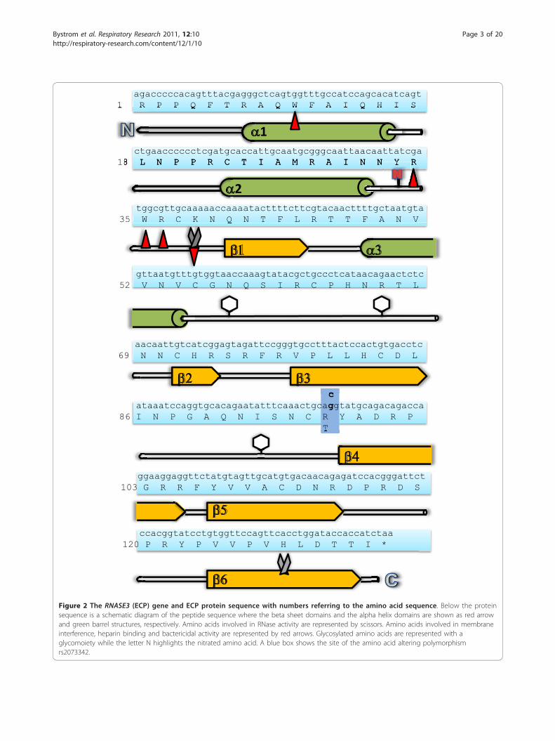
agacccccacagtttacgagggctcagtggtttgccatccagcacatcagt1 R P P Q F T R A Q W F A I Q H I S
ctgaacccccctcgatgcaccattgcaatgcgggcaattaacaattatcga18 L N P P R C T I A M R A I N N Y R
tggcgttgcaaaaaccaaaatacttttcttcgtacaacttttgctaatgta35 W R C K N Q N T F L R T T F A N V
gttaatgtttgtggtaaccaaagtatacgctgccctcataacagaactctc52 V N V C G N Q S I R C P H N R T L
aacaattgtcatcggagtagattccgggtgcctttactccactgtgacctc69 N N C H R S R F R V P L L H C D L
cataaatccaggtgcacagaatatttcaaactgcaggtatgcagacagacca
86 I N P G A Q N I S N C R Y A D R P T
ggaaggaggttctatgtagttgcatgtgacaacagagatccacgggattct103 G R R F Y V V A C D N R D P R D S
ccacggtatcctgtggttccagttcacctggataccaccatctaa 120 P R Y P V V P V H L D T T I *
1
2
Figure 2 The RNASE3 (ECP) gene and ECP protein sequence with numbers referring to the amino acid sequence. Below the proteinsequence is a schematic diagram of the peptide sequence where the beta sheet domains and the alpha helix domains are shown as red arrowand green barrel structures, respectively. Amino acids involved in RNase activity are represented by scissors. Amino acids involved in membraneinterference, heparin binding and bactericidal activity are represented by red arrows. Glycosylated amino acids are represented with aglycomoiety while the letter N highlights the nitrated amino acid. A blue box shows the site of the amino acid altering polymorphismrs2073342.
Bystrom et al. Respiratory Research 2011, 12:10http://respiratory-research.com/content/12/1/10
Page 3 of 20
and acetylglucosamine [25] explaining the variation inits detected size by Western blot of between 16 and 22kDa [26]. Nineteen arginine residues facing the outsideof the protein giving rise to the proteins basicity (pI >11) [27] and possibly also its extraordinary stabilitycompared to other ribonucleases [28]. In the presenceof H2O2 ECP can be nitrated on tyrosine Y33 by EPO.This inflammation-independent nitration occurs duringgranule maturation and was suggested to enhance inter-actions after secretion between several of the otherwiserepulsive, positively charged granule proteins (Figure 2)[29]. ECP has been shown to interact with artificial lipidmembranes [30] and two tryptophan residues, W10 andW35 facing the outside, similar to the present arginine’s,have been associated with this lipid membrane interac-tion [31]. ECP also has RNase independent cytostaticactivity on tumour cells and the tryptophan residuescontribute to this activity [32]. W35 was additionallyfound necessary for killing gram negative and grampositive bacteria [31]. The tryptophan’s also facilitateECP binding to heparin [33,34]. Another study foundthat the residues R34, W35, R36 and K38, all part ofloop 3 (a.a.’s 32-41) contributed to heparin binding andcytotoxicity [35] (Figure 2). Surprisingly, when purifiedfrom granules of circulating cells, large quantities of theprotein were found to lack cytotoxic activity [36]. ECPhas not, like EPX/EDN, been found have alarmin activ-ity, stimulating dendritic cells during Th2 immuneresponses [37], but ECP has the ability to bind lipopoly-saccharide (LPS) and other bacteria cell wall compo-nents [38] which might have a priming influence on theimmune system. The binding of LPS was mainly attribu-ted to a.a.’s 1 to 45 [39]. The 1 to 45 a.a. region wasfound to retain bactericidal activity as well as membranedestabilization activity. One commonly occurring poly-morphism in the gene is leading to the replacement ofan arginine residue with a threonine, R97T [40] (Figure2). The a.a. alteration reduced ECP cytotoxicity to thecell line NCI-H69 assessed by using both recombinantprotein [36] and pools of naive protein variants [41].RNase activity was however not influenced by the R97Talteration. Deglycosylation of the recombinant T97restored the proteins cytotoxicity suggesting that glyco-sylation are responsible for this inhibitory role.
The physiological function of the granulecontained cytotoxic ribonucleaseEosinophils contain a large amount of ECP but the ques-tion is why? What is the function of this protein? There isa constitutive baseline level of the eosinophils in many tis-sues and certain stimuli cause elevated production andinflux of eosinophils in different organs. Moreover levelsof the ECP in tissue and peripheral blood robustly corre-lated with the number of eosinophils present, which might
be indicative that the function of ECP is also key to therole of eosinophils (see table 1). Since the discovery ofECP in 1977 [8] it has been used and evaluated as a bio-marker to assess activity in various inflammatory diseases.This analysis has given indirect information of the proteinsrole in disease. For a comprehensive review of advantagesand pitfalls of the usage of ECP as a biomarker in allergicdisease see ref [42]. Furthermore, a number of in vitro stu-dies have addressed the direct functional activities of theprotein. Detailed following is a comprehensive review ofthese studies with summaries in table 1 and 2. To simplifycomparison the concentrations used have been recalcu-lated to μg/mL using the mean Mw of 19.000 for the nativeprotein (average of 16-22 kDa).
ECP during homeostasis and measured ininflammatory diseasesAt homeostasis the eosinophil contributes 1 - 4 percentof the circulating leukocyte pool. ECP is readily detect-able in blood with plasma levels on the average 3 ug/L(serum 7 μg/L) in healthy individuals which correlateswith circulating eosinophil numbers [43]. ECP in bloodshows a turnover time (t1/2) of 45 min [44], and theplasma protein a2-macroglobulin (a2M) is found to beassociated to the protein, in vitro at a molar ratio of 1.6(ECP/a2M). This interaction is facilitated by proteolyticactivity of cathepsin G or methylamine [45], and concei-vably takes place to facilitate the clearance of ECP [46].When eosinophils encounter adhesion molecules
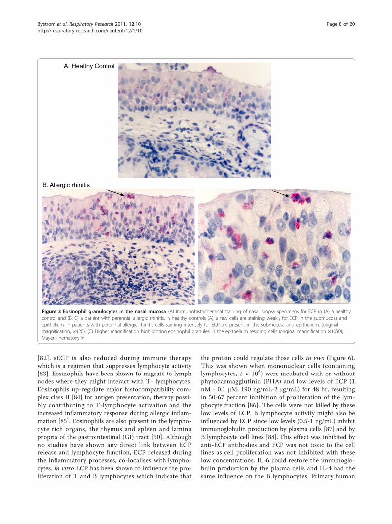
expressed on the endothelial cells of post capillaryvenule wall, the cells adhere and emigrate through thecell layer [47]. Local signals do however drive a lowlevel influx of eosinophils in specific tissues at homeos-tasis. Eosinophils are present in almost all mucosal asso-ciated tissues, nasal mucosa [48] (Figure 3B), lungs [49](Figure 4B), gastrointestinal mucosa [50], the reproduc-tive tract, the uterus [51], breast mucosa of mice [52]and skin [53]. The chemokine eotaxin is responsible forhomeostatic eosinophil influx in the gastrointestinaltract in mice [54] whereas the mechanism of influx inother organs remains unknown. In addition, lympho-cyte-associated tissue: lymph nodes [50], thymus [55]and spleen [50] will have some cells stained red by eosin(see Figure 5).The majority of ECP is released after the cell has left
the circulation [56]. Several types of inflammatory sti-mulation have been shown to cause eosinophil degranu-lation. Interaction with adhesion molecules [57,58],stimulation by leukotriene B4 (LTB4), platelet activatingfactor (PAF) [59], interleukin (IL)-5 [60] immunoglobu-lins and complement factors C5a and C3a [61] all causeECP release. Upon stimulation of eosinophils small var-iants of ECP with sizes 16.1 and 16.3 kDa are released[62]. One line of studies have suggested that during
Bystrom et al. Respiratory Research 2011, 12:10http://respiratory-research.com/content/12/1/10
Page 4 of 20
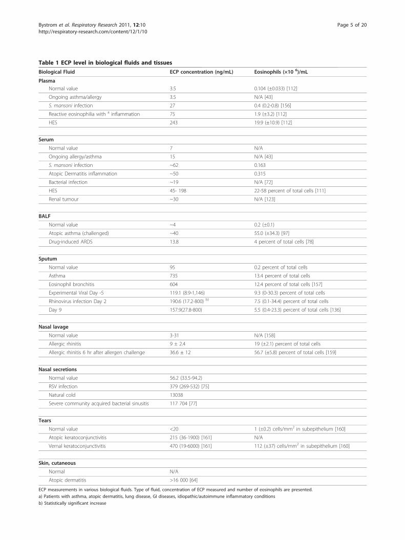
Table 1 ECP level in biological fluids and tissues
Biological Fluid ECP concentration (ng/mL) Eosinophils (×10 6)/mL
PlasmaNormal value 3.5 0.104 (±0.033) [112]
Ongoing asthma/allergy 3.5 N/A [43]
S. mansoni infection 27 0.4 (0.2-0.8) [156]
Reactive eosinophilia with a inflammation 75 1.9 (±3.2) [112]
HES 243 19.9 (±10.9) [112]
Serum
Normal value 7 N/A
Ongoing allergy/asthma 15 N/A [43]
S. mansoni infection ~62 0.163
Atopic Dermatitis inflammation ~50 0.315
Bacterial infection ~19 N/A [72]
HES 45- 198 22-58 percent of total cells [111]
Renal tumour ~30 N/A [123]
BALF
Normal value ~4 0.2 (±0.1)
Atopic asthma (challenged) ~40 55.0 (±34.3) [97]
Drug-induced ARDS 13.8 4 percent of total cells [78]
Sputum
Normal value 95 0.2 percent of total cells
Asthma 735 13.4 percent of total cells
Eosinophil bronchitis 604 12.4 percent of total cells [157]
Experimental Viral Day -5 119.1 (8.9-1,146) 9.3 (0-30.3) percent of total cells
Rhinovirus infection Day 2 190.6 (17.2-800) b) 7.5 (0.1-34.4) percent of total cells
Day 9 157.9(27.8-800) 5.5 (0.4-23.3) percent of total cells [136]
Nasal lavage
Normal value 3-31 N/A [158]
Allergic rhinitis 9 ± 2.4 19 (±2.1) percent of total cells
Allergic rhinitis 6 hr after allergen challenge 36.6 ± 12 56.7 (±5.8) percent of total cells [159]
Nasal secretions
Normal value 56.2 (33.5-94.2)
RSV infection 379 (269-532) [75]
Natural cold 13038
Severe community acquired bacterial sinusitis 117 704 [77]
Tears
Normal value <20 1 (±0.2) cells/mm2 in subepithelium [160]
Atopic keratoconjunctivitis 215 (36-1900) [161] N/A
Vernal keratoconjunctivitis 470 (19-6000) [161] 112 (±37) cells/mm2 in subepithelium [160]
Skin, cutaneous
Normal N/A
Atopic dermatitis >16 000 [64]
ECP measurements in various biological fluids. Type of fluid, concentration of ECP measured and number of eosinophils are presented.
a) Patients with asthma, atopic dermatitis, lung disease, GI diseases, idiopathic/autoimmune inflammatory conditions
b) Statistically significant increase
Bystrom et al. Respiratory Research 2011, 12:10http://respiratory-research.com/content/12/1/10
Page 5 of 20
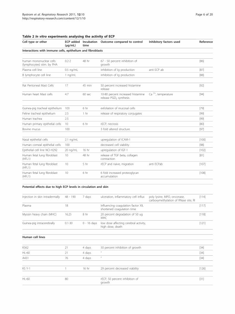
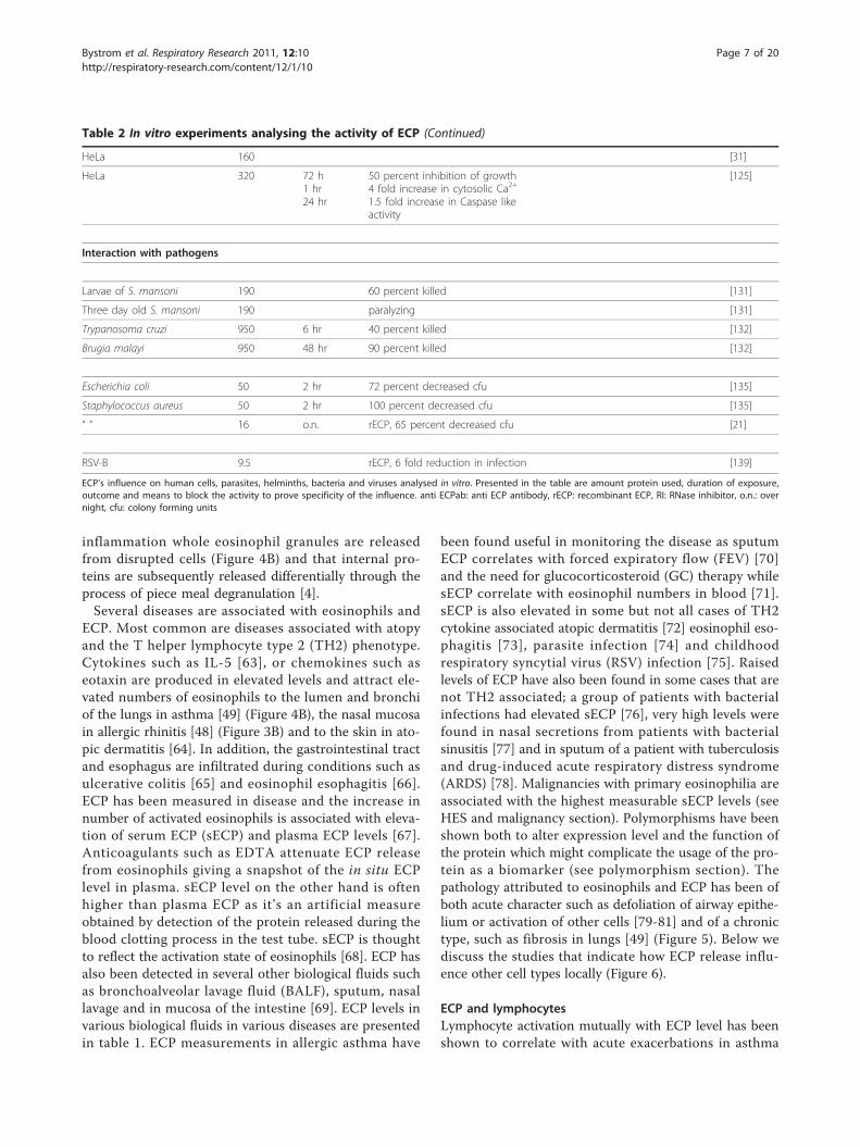
Table 2 In vitro experiments analysing the activity of ECP
Cell type or other ECP added(μg/mL)
Incubationtime
Outcome compared to control Inhibitory factors used Reference
Interactions with immune cells, epithelium and fibroblasts
human mononuclear cells(lymphocytes) stim. by PHA
0.2-2 48 hr 67 - 50 percent inhibition ofgrowth
[86]
Plasma cell line 0.5 ng/mL inhibition of Ig production anti ECP ab [87]
B lymphocyte cell line 1 ng/mL inhibition of Ig production [88]
Rat Peritoneal Mast Cells 17 45 min 50 percent increased histaminerelease
[92]
Human heart Mast cells 4.7 60 sec 10-80 percent increased histaminerelease PGD2 synthesis
Ca 2+, temperature [94]
Guinea-pig tracheal epithelium 103 6 hr exfoliation of mucosal cells [79]
Feline tracheal epithelium 2.5 1 hr release of respiratory conjugates [99]
Human trachea 2.5 [99]
Human primary epithelial cells 10 6 hr rECP, necrosis [80]
Bovine mucus 100 3 fold altered structure [97]
Nasal epithelial cells 2.1 ng/mL upregulation of ICAM-1 [100]
Human corneal epithelial cells 100 decreased cell viability [98]
Epithelial cell line NCI-H292 20 ng/mL 16 hr upregulation of IGF-1 [102]
Human fetal lung fibroblast(HFL1)
10 48 hr release of TGF beta, collagencontraction
[81]
Human fetal lung fibroblast(HFL1)
10 5 hr rECP and naive, migration anti ECPab [107]
Human fetal lung fibroblast(HFL1)
10 6 hr 6 fold increased proteoglycanaccumulation
[108]
Potential effects due to high ECP levels in circulation and skin
Injection in skin intradermally 48 - 190 7 days ulceration, inflammatory cell influx poly lysine, MPO, onconase,carboxymethylation of RNase site, RI
[114]
Plasma 18 Influencing coagulation factor XII,shortened coagulation time
[117]
Myosin heavy chain (MHC) 16.25 8 hr 20 percent degradation of 50 ugMHC
[118]
Guinea-pig intracerebrally 0.1-30 0 - 16 days low dose affecting cerebral activity,high dose, death
[121]
Human cell lines
K562 21 4 days 50 percent inhibition of growth [34]
HL-60 21 4 days “ [34]
A431 76 4 days “ [34]
KS Y-1 1 16 hr 29 percent decreased viability [126]
HL-60 80 rECP, 50 percent inhibition ofgrowth
[31]
Bystrom et al. Respiratory Research 2011, 12:10http://respiratory-research.com/content/12/1/10
Page 6 of 20
inflammation whole eosinophil granules are releasedfrom disrupted cells (Figure 4B) and that internal pro-teins are subsequently released differentially through theprocess of piece meal degranulation [4].Several diseases are associated with eosinophils and
ECP. Most common are diseases associated with atopyand the T helper lymphocyte type 2 (TH2) phenotype.Cytokines such as IL-5 [63], or chemokines such aseotaxin are produced in elevated levels and attract ele-vated numbers of eosinophils to the lumen and bronchiof the lungs in asthma [49] (Figure 4B), the nasal mucosain allergic rhinitis [48] (Figure 3B) and to the skin in ato-pic dermatitis [64]. In addition, the gastrointestinal tractand esophagus are infiltrated during conditions such asulcerative colitis [65] and eosinophil esophagitis [66].ECP has been measured in disease and the increase innumber of activated eosinophils is associated with eleva-tion of serum ECP (sECP) and plasma ECP levels [67].Anticoagulants such as EDTA attenuate ECP releasefrom eosinophils giving a snapshot of the in situ ECPlevel in plasma. sECP level on the other hand is oftenhigher than plasma ECP as it’s an artificial measureobtained by detection of the protein released during theblood clotting process in the test tube. sECP is thoughtto reflect the activation state of eosinophils [68]. ECP hasalso been detected in several other biological fluids suchas bronchoalveolar lavage fluid (BALF), sputum, nasallavage and in mucosa of the intestine [69]. ECP levels invarious biological fluids in various diseases are presentedin table 1. ECP measurements in allergic asthma have
been found useful in monitoring the disease as sputumECP correlates with forced expiratory flow (FEV) [70]and the need for glucocorticosteroid (GC) therapy whilesECP correlate with eosinophil numbers in blood [71].sECP is also elevated in some but not all cases of TH2cytokine associated atopic dermatitis [72] eosinophil eso-phagitis [73], parasite infection [74] and childhoodrespiratory syncytial virus (RSV) infection [75]. Raisedlevels of ECP have also been found in some cases that arenot TH2 associated; a group of patients with bacterialinfections had elevated sECP [76], very high levels werefound in nasal secretions from patients with bacterialsinusitis [77] and in sputum of a patient with tuberculosisand drug-induced acute respiratory distress syndrome(ARDS) [78]. Malignancies with primary eosinophilia areassociated with the highest measurable sECP levels (seeHES and malignancy section). Polymorphisms have beenshown both to alter expression level and the function ofthe protein which might complicate the usage of the pro-tein as a biomarker (see polymorphism section). Thepathology attributed to eosinophils and ECP has been ofboth acute character such as defoliation of airway epithe-lium or activation of other cells [79-81] and of a chronictype, such as fibrosis in lungs [49] (Figure 5). Below wediscuss the studies that indicate how ECP release influ-ence other cell types locally (Figure 6).
ECP and lymphocytesLymphocyte activation mutually with ECP level has beenshown to correlate with acute exacerbations in asthma
Table 2 In vitro experiments analysing the activity of ECP (Continued)
HeLa 160 [31]
HeLa 320 72 h1 hr24 hr
50 percent inhibition of growth4 fold increase in cytosolic Ca2+
1.5 fold increase in Caspase likeactivity
[125]
Interaction with pathogens
Larvae of S. mansoni 190 60 percent killed [131]
Three day old S. mansoni 190 paralyzing [131]
Trypanosoma cruzi 950 6 hr 40 percent killed [132]
Brugia malayi 950 48 hr 90 percent killed [132]
Escherichia coli 50 2 hr 72 percent decreased cfu [135]
Staphylococcus aureus 50 2 hr 100 percent decreased cfu [135]
“ “ 16 o.n. rECP, 65 percent decreased cfu [21]
RSV-B 9.5 rECP, 6 fold reduction in infection [139]
ECP’s influence on human cells, parasites, helminths, bacteria and viruses analysed in vitro. Presented in the table are amount protein used, duration of exposure,outcome and means to block the activity to prove specificity of the influence. anti ECPab: anti ECP antibody, rECP: recombinant ECP, RI: RNase inhibitor, o.n.: overnight, cfu: colony forming units
Bystrom et al. Respiratory Research 2011, 12:10http://respiratory-research.com/content/12/1/10
Page 7 of 20
[82]. sECP is also reduced during immune therapywhich is a regimen that suppresses lymphocyte activity[83]. Eosinophils have been shown to migrate to lymphnodes where they might interact with T- lymphocytes.Eosinophils up-regulate major histocompatibility com-plex class II [84] for antigen presentation, thereby possi-bly contributing to T-lymphocyte activation and theincreased inflammatory response during allergic inflam-mation [85]. Eosinophils are also present in the lympho-cyte rich organs, the thymus and spleen and laminapropria of the gastrointestinal (GI) tract [50]. Althoughno studies have shown any direct link between ECPrelease and lymphocyte function, ECP released duringthe inflammatory processes, co-localises with lympho-cytes. In vitro ECP has been shown to influence the pro-liferation of T and B lymphocytes which indicate that
the protein could regulate those cells in vivo (Figure 6).This was shown when mononuclear cells (containinglymphocytes, 2 × 10⁵) were incubated with or withoutphytohaemagglutinin (PHA) and low levels of ECP (1nM - 0.1 μM, 190 ng/mL-2 μg/mL) for 48 hr, resultingin 50-67 percent inhibition of proliferation of the lym-phocyte fraction [86]. The cells were not killed by theselow levels of ECP. B lymphocyte activity might also beinfluenced by ECP since low levels (0.5-1 ng/mL) inhibitimmunoglobulin production by plasma cells [87] and byB lymphocyte cell lines [88]. This effect was inhibited byanti-ECP antibodies and ECP was not toxic to the celllines as cell proliferation was not inhibited with theselow concentrations. IL-6 could restore the immunoglo-bulin production by the plasma cells and IL-4 had thesame influence on the B lymphocytes. Primary human
A. Healthy Control
B. Allergic rhinitis
Figure 3 Eosinophil granulocytes in the nasal mucosa. (A) Immunohistochemical staining of nasal biopsy specimens for ECP in (A) a healthycontrol and (B, C) a patient with perennial allergic rhinitis. In healthy controls (A), a few cells are staining weakly for ECP in the submucosa andepithelium. In patients with perennial allergic rhinitis cells staining intensely for ECP are present in the submucosa and epithelium. (originalmagnification, ×420). (C) Higher magnification highlighting eosinophil granules in the epithelium residing cells (original magnification ×1050);Mayer’s hematoxylin.
Bystrom et al. Respiratory Research 2011, 12:10http://respiratory-research.com/content/12/1/10
Page 8 of 20

plasma cells and large activated B lymphocytesresponded to ECP in a manner similar to that of the celllines [87]. Thus, ECP might influence the immune sys-tem in that immature lymphocytes are inhibited in theirproliferation by ECP while activated B lymphocytesrespond by decreased immunoglobulin production (seeFigure 6).
ECP and Mast cellsMast cells are found in the skin and in all mucosal tis-sues at homeostasis, and numbers are elevated in asth-matics lungs [49]. Mast cell and eosinophil numbers inmucosa are correlated to bronchial hyperactivity (BHR)[89] and mast cell products and eosinophil MBP but notECP induces BHR [90]. Several lines of evidence suggestthat there is a cross talk between eosinophils and mastcells [91] which to some extent are related to ECPrelease. Mast cells produce and secrete IL-5, PAF andLTB4 known to augment ECP release from eosinophils.Rat peritoneal mast cells on the other hand incubatedwith moderate levels of ECP (0. 9 μM/17 μg/mL) for 45min released 50 percent of their histamine. Histamine isnot released from peripheral basophils by ECP treatment(as by MBP) [92]. However, the release of histaminemay be location specific as no release was observedfrom human skin mast cells treated with up to 200 μg/mL ECP [93]. Histamine and of some tryptase wasthough released from human heart mast cells, purifiedfrom traffic victims or from individuals undergoingheart transplantation, when stimulated with moderatelevels of ECP (2.5 μM; 4.7 μg/mL). Between 10 and 80percent of preformed mediators were released from
these cells and MBP had a similar effect whereas EPX/EDN did not induce any release [94]. This ECP inducedhistamine release occurred within 60 sec of stimulationand was found to be Ca2+-, temperature- and energydependent, and ECP was not toxic to the cells. Anothermast cell product, prostaglandin D2 (PGD2) was synthe-sised de novo by the same amount of ECP added. PGD2
is a chemoattractant for eosinophils and TH2 lympho-cytes, through binding the CRTH2 receptor [95]. There-fore these findings suggest that in some tissue theinteractions between mast cells and eosinophils can beattributed to the positive feedback of ECP release.
ECP and epitheliumECP is detected in nasal mucosa in association withdamaged epithelium [48], in damaged corneal epithe-lium [96] as well as in BALF (at 40 ng/mL, table 1) [97].The function of ECP has been assessed using severalassays in the view of the presence of the eosinophil inthe airways. Both destructive and non-destructive conse-quences have been found when analyzing various con-centrations of the protein in interaction with theepithelium. High levels of ECP (5.4 μM/103 μg/mL)caused exfoliation of guinea-pig mucosal cells after 6 hrincubation with tracheal epithelium [79]. Confluent pri-mary human corneal epithelial cells incubated with 0-100 μg/mL ECP, displayed a concentration-dependentgradual increase in morphological change and with thehighest concentration, 100 μg/mL, being cytotoxic [98].Lower concentration of the ECP (2.5 μg/mL) causedrelease of respiratory glycoconjugates (marker of mucussecretion), with a peak after 1 hr, from feline tracheal
B. Allergic asthmaA. Healthy control
Figure 4 Eosinophil granulocytes in the bronchial mucosa. Sections of bronchial biopsies from (A) a healthy control or (B) an individual withallergic asthma were stained with ECP antibody visualizing eosinophils in the mucosa. The figures show that only a few eosinophils are presentin the tissue of the healthy control, but many eosinophils accumulate in areas of reduced epithelial integrity in a specimen from a patient withallergic asthma. Original magnification ×420; Mayer’s haematoxylin.
Bystrom et al. Respiratory Research 2011, 12:10http://respiratory-research.com/content/12/1/10
Page 9 of 20

explants [99]. The short incubation time and possibilityto repeat the stimulation suggested a non-toxic mechan-ism. MBP, which is almost as basic as ECP, in the sameassay, showed the opposite effect; therefore these effectson mucus secretion are unlikely to be due to electrostaticcharge. ECP at these moderate levels (2.5 μg/mL)
displayed the same effect on human trachea [99]. Howeverhuman primary epithelial cells underwent necrosis athigher levels (10 μg/mL) in another study [80]. ECP hasalso been shown to acting directly on airway mucus invitro. At high levels (100 μg/mL) ECP altered bovinemucus three fold, as measured by a capillary surfactometer
Thymus
Location ofeosinophils
at homeostasis
Lung
G.I. tract
Spleen
Reproductivetract
Lymph nodes
Respiratory mucosaDamaged epithelium (P)Bacterial defence (F)
BronchiEpithelium – exfoliation (P)Mucus - altered (P)
Suggested function (F) or pathology (P) of eosinophils
and released ECP
HeartScarring Fibrosis (P)
LungTissue remodelling (P)Fibrosis (P)Viral defence (F)
EsophagusDamaged epithelium (P)Fibrosis (P)
GI tractHelminth defence (F)Bacterial defence (F)
SkinUlceration (P)
Figure 5 Known anatomical locations of eosinophil granulocytes and suggested activities of released ECP at these sites. On the leftside are eosinophil granulocytes locations at homeostasis shown. On the right side are areas speculated to be affected by increased numbers ofeosinophils and elevated levels of released ECP, in disease (pathology, P) and in physiological defense (function, F). This is a speculation by theauthors of the review.
Bystrom et al. Respiratory Research 2011, 12:10http://respiratory-research.com/content/12/1/10
Page 10 of 20
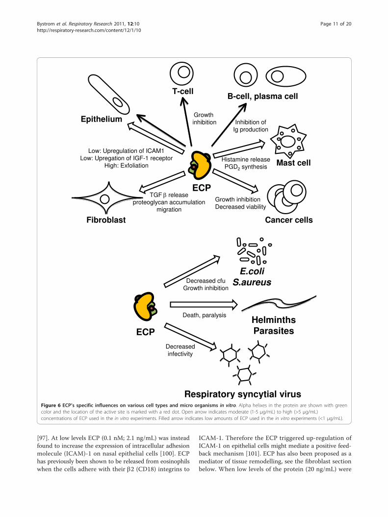
[97]. At low levels ECP (0.1 nM; 2.1 ng/mL) was insteadfound to increase the expression of intracellular adhesionmolecule (ICAM)-1 on nasal epithelial cells [100]. ECPhas previously been shown to be released from eosinophilswhen the cells adhere with their b2 (CD18) integrins to
ICAM-1. Therefore the ECP triggered up-regulation ofICAM-1 on epithelial cells might mediate a positive feed-back mechanism [101]. ECP has also been proposed as amediator of tissue remodelling, see the fibroblast sectionbelow. When low levels of the protein (20 ng/mL) were
Mast cell
Fibroblast
Epithelium
T-cellB-cell, plasma cell
Cancer cells
Growth inhibitionDecreased viability
TGF β releaseproteoglycan accumulation
migration
Low: Upregulation of ICAM1Low: Upregation of IGF-1 receptor
High: Exfoliation
Growth inhibition Inhibition of
Ig production
Histamine releasePGD2 synthesis
ECP
ECP
E.coliS.aureus
HelminthsParasites
Respiratory syncytial virus
Decreased cfuGrowth inhibition
Death, paralysis
Decreasedinfectivity
Figure 6 ECP’s specific influences on various cell types and micro organisms in vitro. Alpha helixes in the protein are shown with greencolor and the location of the active site is marked with a red dot. Open arrow indicates moderate (1-5 μg/mL) to high (>5 μg/mL)concentrations of ECP used in the in vitro experiments. Filled arrow indicates low amounts of ECP used in the in vitro experiments (<1 μg/mL).
Bystrom et al. Respiratory Research 2011, 12:10http://respiratory-research.com/content/12/1/10
Page 11 of 20

used to stimulate the bronchial epithelial cell line NCI-H292 for 16 hr, the insulin growth factor (IGF)-1 receptorwas found to be up-regulated [102]. ECP was speculatedtherefore to be involved in IGF-1-dependent lung tissuerepair processes perhaps present during homeostasis andabnormally amplified during inflammatory conditions.
ECP and FibroblastsThe persistent high number of eosinophils and ECP inthe lungs of allergic asthmatics has led to the suggestionof their participation in the development of chroniclung tissue remodelling. Remodelling has also beenfound in the esophagus of patients with eosinophil eso-phagitis [103] and sECP has been found elevated in onecase [104]. The remodelling in asthmatic lungs is in partcaused by collagen and proteoglycan secretion frominterstitial fibroblasts. Eosinophils have been suggestedto participate in this by secretion of transforminggrowth factor (TGF) beta [105,106] but here is addition-ally described how ECP could influences fibrosis devel-opment. Stimulation of a human fetal lung fibroblastcell line (HFL1) with moderate/high levels of ECP (5-10μg/mL) for 24-48 hr resulted in increased release ofTGF-beta [81]. ECP also augmented fibroblast mediatedcontraction of collagen gel and stimulated migration ofHFL1 fibroblasts which could be blocked with antibo-dies to ECP [107]. In addition, ECP incubated with thefibroblast cell line for 6 hr resulted in a 6-fold increaseof intracellular proteoglycan accumulation [108].
ECP and bronchial smooth muscle cellsBronchial smooth muscles cells are involved during theprogression of asthma development by secretion of cyto-kines as well as remodelling due to proliferation. Eosino-phils have been found located in close proximity withsmooth muscle cells. ECP does not influence smoothmuscle cells by causing BHR [90] but high levels ofECP, similar to used for epithelial cells, appears to becytotoxic, inducing cell death by necrosis in 1 hr. TNFalpha in contrast causes apoptosis of the smooth musclecells [109].
ECP in Hypereosinophilic Syndrome (HES)Conditions where eosinophils are overproduced lead todetrimental effects for the host. One such condition,HES is defined by the presence of more than 1.5 × 10⁶
eosinophils/mL blood during a time period of at least 6months, organ involvement and with no other etiologyidentified. One form of HES, the myeloproliferativeform, is caused by an 800 bp deletion on chromosome 4during the haematopoiesis in the bone marrow, resultingin a fusion between the gene FIP1L1 and the PDGFRAgene [110]. A fusion protein is produced which constitu-tively phosphorylates tyrosine residues leading to
malignant expansion of eosinophils. Another form ofHES is a clonal lymphocytic variant (L-HES) whereaberrant cytokine production by malignant lymphocytescauses HES. For other cases the cause of the overpro-duction of the eosinophils is unknown but HES is asso-ciated with high levels of ECP in plasma and serum, ofup to 0.2 μg/mL [111,112]. It is not know howeverwhether theses high levels of the protein are pathologi-cal. A few in vitro studies might relate to the etiologiesof HES. Eosinophil infiltration of the skin of HESpatients is the most common clinical manifestation[113]. Some of these patients present with erosive andulcerative lesions and ECP was found both depositedand taken up by cells in those lesions [114]. ECP’s abil-ity to cause ulcerations in the skin has been analysed byinjecting the protein intradermally into guinea pig skin,where it was found that the protein can persist there fortwo weeks [64] which is possibly attributed to its highstability [28]. Injections of high levels of ECP (48 and190 μg/mL/2.5 and 10 μM) caused ulcerations whichwere most severe after seven days [114]. Inflammatorycells were found infiltrating the inflamed area and ECPwas found taken up by cells within 48 hr. Injection ofpoly-lysine, other basic granule proteins MBP, EPO andthe basic ribonuclease onconase showed that the severityof the lesions was not directly correlated with level ofbasicity. ECP and EDN were found to be more potent inlesion formations than MBP and EPO. Addition ofRNase inhibitor or obliteration of the RNase activity bycarboxymethylation of the RNase site of ECP reducedthe ulcerations by 60 percent suggesting RNase activityis important, but not wholly responsible for the activity[114]. Some studies have shown that patients with HEShave an slightly elevated risk for thrombosis formationsystemically [115] and in the cardiac ventricle [116].ECP has been shown to shortened the coagulation timefor plasma which was dependent on an interaction withcoagulation factor XII [117]. Eosinophils also infiltratethe endomyocardium of some patients and this has beensuggested to be the cause of development of scaring inthe ventricle [116]. High levels of ECP (16.25 μg/mL)degrade the muscle protein component, the myosinheavy chain in vitro [118] but it is not known whetherECP directly interacts with muscle fibres of the heart.The final stage is endomyocardial fibrosis in which eosi-nophils and ECP have been postulated to participate[119] by their influence on fibroblast function. Althougha rare finding, a few patients with the myeloid form ofHES have been reported to have central nervous system(CNS) manifestation [113,120]. It is not known whetherECP can reach the brain but ECPs effect on the CNShas been assayed by direct intracerebral injection. Gui-nea-pigs injected with ECP, showed with doses of 0.1 μgand up, cerebral symptoms up until the end of the
Bystrom et al. Respiratory Research 2011, 12:10http://respiratory-research.com/content/12/1/10
Page 12 of 20
experiment at day 16 [121]. Purkinje cells in the brainwere decimated in this model, suggesting that the circu-lating ECP could affect the CNS of some HES patients ifthe protein reached the brain.
ECP in malignanciesEosinophils have occasionally been found to infiltratedeveloping tumours and have been suggested to have arole in fighting these malignancies [122]. The involve-ment of the eosinophils have been suggested by thefinding of elevated sECP levels in patients with renaltumours (table 1) [123]. ECP assayed in urine frompatients with urinary bladder tumours showed a twofoldincrease compared to normal’s [124]. The elevated levelssuggest presence of activated eosinophils in somepatients with these malignancies. In the analysis of thepossible involvement of ECP in tumour defence, ECPhas been evaluated in respect of altering proliferation ofvarious cell lines. The cell lines K562 and HL-60 wereincubated with 1.1 uM (21 ug/mL) ECP and the cell lineA431 with 4 μM (76 ug/mL) and this resulted in 50 per-cent inhibition of proliferation after four days. To ana-lyse whether growth inhibition was related to positivecharge or RNase activity, poly-lysine or RNase A wasused with no effect [34]. ECP exists in two forms depen-dent on a polymorphism, R97 and T97. It was foundthat the T97 form had reduced capability to kill K562and NCI-H69 cells [36]. These recombinant (r) ECPswere produced in a baculovirus system and deglycosyla-tion restored the cytotoxic activity.Furthermore, high levels of bacteria expressed rECP
had 50 percent cytostatic effect on HL-60 and HeLacells [31], compared to non-affected controls. ECP wasfound binding the surface of HeLa cells and caused celldeath after 24 hr, accompanied by increases in intracel-lular radical oxygen species (ROS) generation and cas-pase 3-like activity [125]. A mix of ECP and EDNpurified from urine and incubated with the Kaposi’s sar-coma cell line KS Y-1 for 16 hr caused complete celldeath at 0.625 μg/mL while 1 μg recombinant ECP pro-duced in yeast and incubated with the same time spandecreased the viability of the KS cell line by 29 percent.Proteins expressed in yeast lack glycosylation and thepossible implications of this were speculated [126].
ECP as a defence proteinLevels of serum ECP are elevated in TH2 engaging para-sitic and helminth infections and eosinophils have longbeen thought to be a major defence against these types ofinfection. Elevated ECP have also been reported in somecases of bacterial and viral respiratory infections. Giventhat ECP is a cytotoxic ribonuclease, the ability of theprotein to exterminate parasites, bacteria and virus invitro has been extensively investigated (see also Figure 6).
Parasite and helminth infectionsParasitic and helminthic infections drive the immune sys-tem towards TH2 cytokine production and concurrenteosinophilia. Since eosinophil infiltration in infectedorgans and skin is a common finding, eosinophils arethought to have a specific role in parasite killing [127].Although, a challenged theory; the deposition of the cyto-toxic protein ECP could be a mechanism by which theimmune system kills off the intruders. Indeed, the eosi-nophilia in parasitic diseases is associated with elevatedECP in circulation (table 1) [72,128]. ECP is also foundreleased from eosinophils in proximity to parasites inskin and lymph nodes [129,130]. The ability for ECP tokill or paralyse parasites and helminths have been ana-lysed in vitro and high quantities were needed to influ-ence the organisms. Three-hr-old larvae of Schistosomamansoni were incubated with 10 μM (190 μg/mL) ECPand 60 percent were killed. S. mansoni, 3 days of age,were paralysed by the protein [131] while 50 μM (950μg/mL) ECP killed 40 percent of Trypanosoma cruzi by 6hr and 90 percent of Brugia malayi by 48 hr. This cyto-toxicity of ECP to parasites was inhibited by heparin[132] and dextran sulphate, probably by interfering withthe tryptophan and arginine residues as discussed earlier.In addition, heat obliterated the toxic effect of ECP toparasites, highlighting the importance of the conforma-tion of the protein [133]. The RNase activity of ECP wasclearly shown not to be important for parasite toxicity,similar to that observed for EPX/EDN.
ECP in bacterial inflammationEosinophils are found lining and degranulating in both therespiratory and gastrointestinal mucosa [50]. Eosinophilsare generally not thought of as defendants during bacterialinflammation. However sECP has been found elevated inseptic patients [76] and very high levels of ECP in nasalsecretions from patients with normal cold (13 μg/mL) orsevere community acquired rhinosinusitis has beendescribed in one case (11.7 μg/mL, table 1) [77]. More-over, a recent study has shown that eosinophils expelmitochondrial DNA coated with ECP and other granuleproteins which are bactericidal in mice in vivo [134]. Addi-tionally, a few studies have described neutrophils produ-cing ECP [15]. In view of these findings the anti - bacterialproperties of ECP has been evaluated. Bacterial strainschosen for analysis were Escherichia coli (E. coli) and Sta-phylococcus aureus (S. aureus). High levels of ECP (50 μg/mL) decreased the number of colony-forming units (cfu)by 72 percent and close to 100 percent, respectively, forthe two strains after a very short 2 hr of incubation. ECPonly killed E. coli growing in logarithmic phase and actedon both the inner and outer membranes of E. coli [135].Recombinant ECP was also cytotoxicity to S. aureus. Over-night incubation of rECP with the bacteria (16 kDa, 16 ug/
Bystrom et al. Respiratory Research 2011, 12:10http://respiratory-research.com/content/12/1/10
Page 13 of 20
mL/1 uM) left 35 percent of the cfu. rECP in which a.a.’sinvolved in RNase activity had been substituted (K38R andH128D), terminating the RNase activity, had no effect onthe bacterial killing activity [21]. In conclusion therefore,eosinophils and ECP might have a role in bacterialdefence. Due to its stability, it might be feasible to specu-late that ECP over time accumulate in mucus fluids suchas nasal secretions and act as a first line of defence againstbacterial intrusion.
ECP in viral inflammationECP has been found significantly elevated in sputumfrom atopic subjects subjected to experimental rhino-virus infection [136] and in nasal secretions from atopicinfants with respiratory RSV infection (table 1) [75].Eosinophils and ECP are associated with RSV infectionin children’s lungs [137] and RSV can infect, and repli-cate in eosinophils [138]. Recombinant ECP expressedin a baculovirus system was used to evaluate whetherECP can inactivate the B subtype of RSV. ECP (0.5 μM;9.5 μg/mL) incubated with the virus showed a 6-foldreduction of the infectivity of the virus to a human pul-monary epithelial cell line [139]. This antiviral activitywas lower than that found with EPX/EDN (54-foldreduction) [140], but the infectivity was increased byaddition of RNase inhibitor (RI) to both proteins duringincubation. Mixing the two proteins did not mediateany synergistic effects on antiviral activity. RNase A,however [up to 4 mM (76 mg/mL)], did not exert anti-viral activity, suggesting that the RNase site but notactivity is important for inhibition of infectivity.
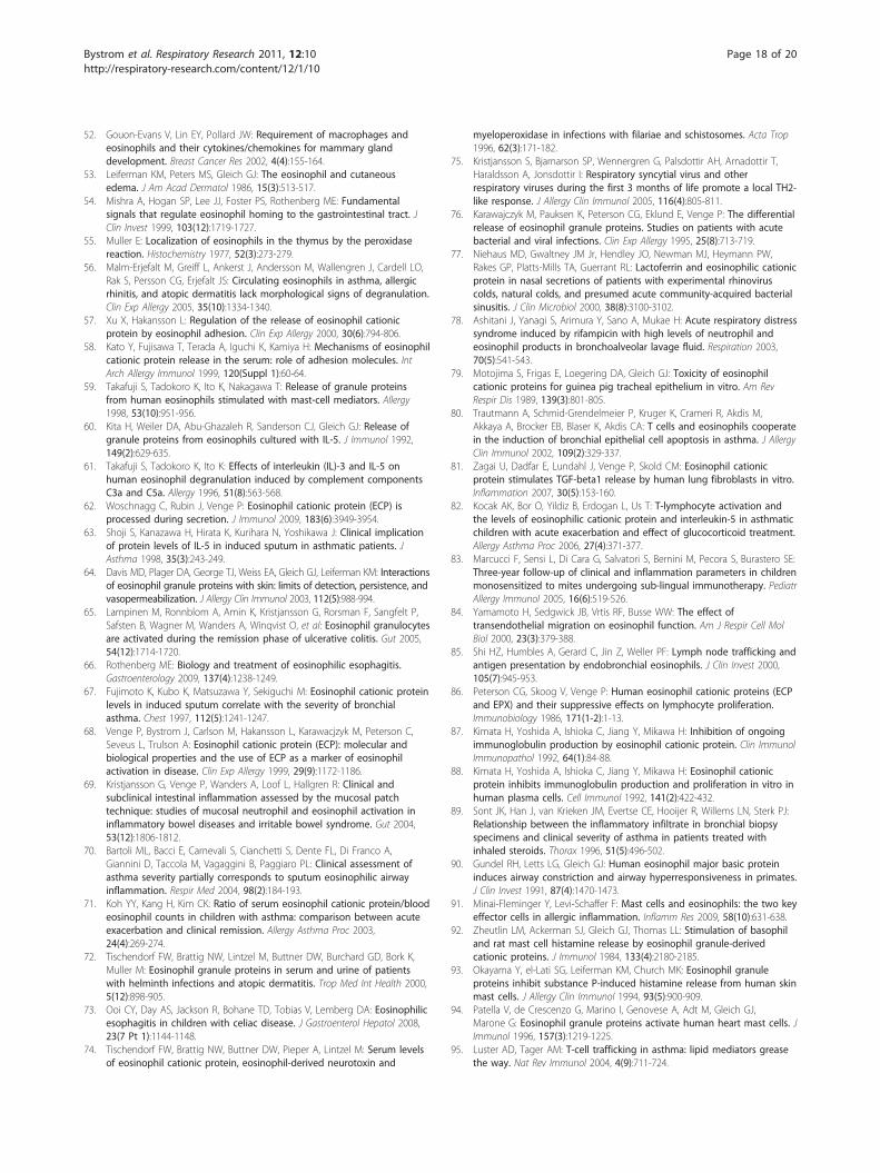
Polymorphisms in the RNASE3 gene andassociation to production and diseaseTable 3 summarizes data from the NCBI entrez nucleo-tide site regarding polymorphisms detected in the ECP
gene. Two polymorphisms are found in the protein cod-ing region, two in intronic regions and two in the 3’untranslated region (UTR). ECP polymorphisms are dif-ferentially distributed according to ethnicity [141]. Twostudies have evaluated polymorphisms in intronic andUTR regions of the ECP gene, and linked them withECP production. One polymorphism rs11575981 (-393T> C) located in the promoter, in an C/EBP binding sitewas associated with decreased ECP level in serum, anddecreased binding of C/EBP alpha [142]. Another poly-morphism, in the 3’UTR, rs2233860 (499G > C or 562G> C) was associated with content of ECP in the eosino-phils [143]. Three studies have analysed whether anypolymorphisms are linked to allergic asthma and allergicrhinitis. The presence of the C allele in the nonsynon-ymous rs2073342G/C (371G > C/434G > C) polymorph-ism in the ECP gene, causing a.a. alteration Y97T, wasfound to be associated with absence of asthma in oneSwedish study [40]. A study of Norwegian and Dutchsubjects instead found that the haplotype C-G-G for thethree polymorphisms rs2233859/rs17792481 (-38C/A),rs2073342 (371G/C/434G/C) and rs2233860 (499G/C/562G/C) being protective [144]. In a third, Koreanstudy, which was the largest, the genotype rs2233860CC(499/562CC) was associated with allergic rhinitis [145].Eosinophils occasionally infiltrate oral squamous cellcarcinoma tumours. A study found a tendency for asso-ciation of the rs2073342G/C C/C (371/434GC/CC) gen-otypes with a poor clinical outcome in patients witheosinophil rich such tumours [146]. As discussed earlier,eosinophils are present during helminth infections. Thers8019343 polymorphism T (1088TT) in the 3’UTR wasexclusively present in the genome of a patient with tro-pical pulmonary eosinophilia [147]. Furthermore a studyhas found the rs2073342 with C (371/434C) polymorph-ism overrepresented in helminth infected Ugandans
Table 3 Polymorphisms associated with the RNASE3 gene
Polymorphism alleles Alternativenames
location, effect
rs2284954 A/G -550A > G promoter
rs11575981 C/T -393T > C promoter, disrupt C/EBP binding site, correlate with s-ECP [142]
rs2233858 C/T intron
rs2233859/rs17792481
A/C -38C > A intron (in a GATA-1 site) a
rs2073342 C/G 371G > C,434G > C
protein coding, Y > G is associated with allergic asthma [40]a, poor outcome in oral squamous cellcarcinoma tumours [146], C over represented in helminth infected Ugandans [148]
rs12147890 A/G protein coding
rs2233860 G/C 3’ UTR,G is correlated to higher intracellular ECP [143], G is associated with allergic rhinitis [145], a
rs8019343 A/T 499G > C,562G > C
3’ UTR T is only present in one patient with helminth infection [147]
Polymorphisms found in the ECP gene and surrounding chromosomal sequence. Listed are Polymorphism i.d.’s, altered bases, alternative names, and types ofassociations
a) C-G-G haplotype associated with allergic rhinitis [144]
Bystrom et al. Respiratory Research 2011, 12:10http://respiratory-research.com/content/12/1/10
Page 14 of 20

[148]. Interestingly, from the -550 polymorphism over astretch of 272 bases to the mRNA transcription startsite, thirteen polymorphism sites are located (NCBIReference Sequence: NC_000014.7, J. Bystrom unpub-lished observation). Similar to the protein coding regionand the 3’UTR, this region is highly homologous to theRNASE2 gene region, with the only differences beingthe sites of the polymorphisms. The replacement base’sfor twelve of the thirteen polymorphisms is to the samebase as in the RNASE2 promoter sequence. This is alsothe case for two of the 3’UTR polymorphisms. Thisfurther highlights the extremely close relationshipbetween RNASE3 (ECP) and RNASE2 (EPX/EDN).
DiscussionECP was first discovered in 1977 and since then, evi-dence has been gathered to understand its roles in phy-siology and pathophysiology. ECP is a peptide of 133 a.a., with the first 40 a.a. necessary for membrane interfer-ing, heparin binding and cytotoxic activity. The heparinbinding ability of ECP might enable the protein to bindproteoglycans on other human cells for possible uptake[34] or heparan sulfate in extracellular matrix for lateruse such as is the case for CXCL10 [149]. In a similarmanner ECP might bind microorganisms peptidoglycansfor uptake and cytotoxicity [32]. The non-synonymouspolymorphism rs2073342 reduces cytotoxicity suggestingan alteration of the three-dimensional structure influen-cing catalytic site elsewhere in the protein. ECP is glyco-sylated, and as recently discovered can be nitrated. Thedevelopment of increasingly sophisticated assays willdetermine whether other modifications, perhaps func-tion associated, are also important in ECP activity.Since the discovery of ECP, assays have been devel-
oped to determine its levels in biological fluids in var-ious diseases (table 1). ECP in serum can reach 0.1 - 0.2μg/mL for HES patients [111] and parasitic diseasesinfected individuals [72] and this is a 30 fold elevationcompared to ECP in serum of healthy individuals. InBALF and nasal lavage from atopic patients the ECPlevels are lower, 0.050 μg/mL but the sample are dilutedduring the collection process. In undiluted tears, sputumand nasal secretions the highest ECP levels have beenfound: 0.5, 0.7 and 10 μg/mL, respectively. The ECPmeasurements correlate with eosinophilic disease buthave been found elevated also in some diseases withoutknown eosinophil involvement [76-78]. The biologicalactivity of ECP has been studied by incubation of theprotein with several different cell types in vitro. Bothhuman cells and pathogens have been assayed analysingdifferent parameters (see table 2). In general, 10 - 20μg/mL and above, result in growth inhibitory anddestructive consequences to mammalian cells, parasitesand bacteria. ECP released in situ in diseases engaging
high levels of eosinophils might reach these destructiveconcentrations (e.g. ECP accumulated in air way mucusof asthmatics, in nasal secretions of some sinusitispatients or released in skin of atopic dermatitis/HESpatients, table 1 and Figure 5). Although it remains tobe proven, there is a possibility that destructive activityto multiple cell types as well as induction of fibrosis ispart of the etiology of disease where ECP levels are ele-vated during prolonged periods, e.g. in HES and hel-minth infection. There is also evidence that neutrophilsare carriers of significant amounts of ECP. Using themurine system, granule proteins have been found asso-ciated with expelled eosinophil mitochondrial DNA andthis DNA/protein complex trapping and killing bacteriain the gut [134]. It is intriguing to speculate whether thehigh levels of ECP present in various human mucosalsecretions would equally be associated with eosinophilmitochondrial DNA and whether such complexes hadthe ability to capture and kill microorganisms.The role of eosinophils in asthma has been under
scrutiny since clinical trials showing that anti-IL-5 ther-apy did not improve the disease symptoms for allergicasthmatics albeit eosinophil numbers were reduced[150]. However, two recent clinical trials have shownthat anti-IL-5 antibodies actually could relieve symp-toms in eosinophil rich, late onset asthma, suggestingthat eosinophils can have a pathogenic role in this dis-ease. In these trials inflammatory exacerbations werereduced when anti-IL-5 antibodies were administrated[151,152]. Earlier studies using diagnostic ECP measure-ments seem to agree with these findings as ECP levelscorrelate with severity of asthma: FEV (sputum ECP)[70], need for GC treatment (sputum ECP) [153] andblood eosinophilia (sECP) [71]. Results from in vitro stu-dies presented in this review may well suggest severalroles for ECP in this type of allergic asthma. The proteinmight act as an inflammatory amplifier by augmentationof release of, for eosinophils chemotactic, PGD2 frommast cells in asthmatic patients. Moreover, proteinreleased in the interstitium might influence fibrosisdevelopment (Figure 3B, 4B and 5). One might speculatethat blocking antibodies to ECP could be a symptomrelieving addition to the already established GC andanti-IL5 therapies used in eosinophil rich asthma andother eosinophilic diseases [113,151,152].Table 2 shows that the level of protein needed to
influence proliferation of lymphocytes and their anti-body production is 1000 times lower than the destruc-tive levels described above, i.e. in the ng/mL range. Inmurine system eosinophils have been ascribed a novelrole in inflammation; the cells enter and contribute tothe well orchestrated process of inflammation resolutionof by release of the pro-resolving lipid protectin D1[154] (for a review see [155]). Whether ECP is released
Bystrom et al. Respiratory Research 2011, 12:10http://respiratory-research.com/content/12/1/10
Page 15 of 20
during this resolution process for the dual role ofsequestering subpopulations of inflammatory lympho-cytes [86] and promoting tissue repair by TGF beta aug-mentation [81] is an intriguing speculation. Eosinophilsare also present at homeostasis at low numbers in lym-phocyte rich organs at various locations but degranu-lated only in the GI tract [50]. A single eosinophilcontains 13 pg ECP. Do eosinophils have a role in main-taining homeostasis and do low levels of ECP also havea role here? EDN, the sister protein has been found toplay an active role during inflammation developmentinfluencing the maturation of DC’s [37]. If EDN is pro-inflammatory, perhaps the two proteins divergencecould be because ECP might have acquired a novel roleas yet unknown role.Finally, analysis of the DNA sequence of the ECP gene
and surrounding regions have unravelled a number ofpolymorphisms. These studies have linked differentpolymorphisms and haplotypes to TH2 diseases, asthma,and allergic rhinitis. The studies have in some casescome to different conclusions but used different patientsand different ethnic groups which might explain the var-iations. Diseases such as allergic asthma are multifac-toral and to determine the role of certainpolymorphisms one might need to look at larger definedgroups to get a clear association. Altered expressionlevels might also influence both destructive functionsand possible homeostatic roles. A careful analysis usingall polymorphisms and corresponding haplotypes andlarge groups of defined populations would more clearlydetermine the role of ECPs genetic make-up, and itspotential functions in physiology and disease.
ConclusionThe eosinophil granulocyte was discovered 130 yearsago but its roles are still being revealed. The most char-acteristic feature of the eosinophil is the large secondarygranules filled with basic proteins. The purpose of theseproteins is still not fully understood. One of the pro-teins, ECP is a highly basic, cytotoxic, heparin bindingribonuclease that seems to need its ribonuclease site butnot activity for its activities. Sensitive assays have beendeveloped for its measurement in biological fluids whichhave contributed to the understanding of the role of theeosinophils in disease. In vitro studies have shown thathigh levels of ECP are necessary for development itsdestructive actions. Diseases engaging high levels ofeosinophils might reach these levels locally in the tissue.At those high levels polymorphisms altering expressionlevel and protein sequence might play a role within cer-tain populations. Whether ECP also has roles at lowerconcentrations, such as the growth inhibitory influenceson lymphocytes found in vitro, remain to be shown within vivo models or clinically. These additional roles for
ECP when discovered, might provide critical answers tothe functions of eosinophil granulocytes and is thereforewell worth waiting for.
AcknowledgementsWe thank Dr. Smita Y Patel for valuable suggestions of the outline of thereview as well as comments on the clinical cases. Professor Per Venge andDr. Helene Rosenberg have provided valuable comments during thedevelopment of this review. We thank the following institutions for kindlyproviding permission to publish results obtained at their sites. Images ofnasal biopsies were obtained from Department of Allergy, Skin and AllergyHospital, University of Helsinki, Finland. Images of bronchial biopsies wereobtained from Department of Respiratory Medicine and Allergology atAkademiska Hospital, University of Uppsala, Sweden and images of bloodsmears was obtained from Department of Clinical Chemistry, AkademiskaHospital, Uppsala, Sweden.Research is funded by the British Heart Foundation (PG/08/070/25464). Thiswork forms part of the research themes contributing to the translationalresearch portfolio of Bart’s and the London Cardiovascular BiomedicalResearch Unit which is supported and funded by the National Institute ofHealth Research.
Author details1Translational Medicine and Therapeutics, William Harvey Research Institute,Bart’s and the London, Queen Mary University of London, CharterhouseSquare, London EC1M 6BQ, UK. 2Respiratory Medicine and Allergology,Department of Medical Science, Uppsala University Hospital, Uppsala,Sweden. 3College of Medicine, Sulaimani University, Sulaimani, Iraq.
Authors’ contributionsJB, DBB and KA have together drafted and completed the manuscript. KAprovided histological images; JB and DBB have provided other figures. Allauthors have read and approved the final version of the manuscript.
Competing interestsThe authors declare that they have no competing interests.
Received: 21 July 2010 Accepted: 14 January 2011Published: 14 January 2011
References1. Ehrlich P: Uber die specifischen Granulation des Blutes. Archiv fur
Anatomie und Physiologie, Physiologische Abteilung 1879.2. Wardlaw AJ, Moqbel R, Kay AB: Eosinophils: biology and role in disease.
Adv Immunol 1995, 60:151-266.3. Kurashima K, Numata M, Yachie A, Sai Y, Ishizaka N, Fujimura M, Matsuda T,
Ohkuma S: The role of vacuolar H(+)-ATPase in the control ofintragranular pH and exocytosis in eosinophils. Lab Invest 1996,75(5):689-698.
4. Neves JS, Weller PF: Functional extracellular eosinophil granules: novelimplications in eosinophil immunobiology. Curr Opin Immunol 2009,21(6):694-699.
5. Gleich GJ, Loegering DA, Maldonado JE: Identification of a major basicprotein in guinea pig eosinophil granules. J Exp Med 1973,137(6):1459-1471.
6. Peterson CG, Venge P: Purification and characterization of a new cationicprotein–eosinophil protein-X (EPX)–from granules of human eosinophils.Immunology 1983, 50(1):19-26.
7. Desser RK, Himmelhoch SR, Evans WH, Januska M, Mage M, Shelton E:Guinea pig heterophil and eosinophil peroxidase. Arch Biochem Biophys1972, 148(2):452-465.
8. Olsson I, Venge P, Spitznagel JK, Lehrer RI: Arginine-rich cationicproteins of human eosinophil granules: comparison of theconstituents of eosinophilic and neutrophilic leukocytes. Lab Invest1977, 36(5):493-500.
9. Iwasaki H, Mizuno S, Mayfield R, Shigematsu H, Arinobu Y, Seed B,Gurish MF, Takatsu K, Akashi K: Identification of eosinophil lineage-committed progenitors in the murine bone marrow. J Exp Med 2005,201(12):1891-1897.
Bystrom et al. Respiratory Research 2011, 12:10http://respiratory-research.com/content/12/1/10
Page 16 of 20

10. Mori Y, Iwasaki H, Kohno K, Yoshimoto G, Kikushige Y, Okeda A, Uike N,Niiro H, Takenaka K, Nagafuji K, et al: Identification of the human eosinophillineage-committed progenitor: revision of phenotypic definition of thehuman common myeloid progenitor. J Exp Med 2009, 206(1):183-193.
11. Egesten A, Calafat J, Weller PF, Knol EF, Janssen H, Walz TM, Olsson I:Localization of granule proteins in human eosinophil bone marrowprogenitors. Int Arch Allergy Immunol 1997, 114(2):130-138.
12. Gruart V, Truong MJ, Plumas J, Zandecki M, Kusnierz JP, Prin L, Vinatier D,Capron A, Capron M: Decreased expression of eosinophil peroxidase andmajor basic protein messenger RNAs during eosinophil maturation.Blood 1992, 79(10):2592-2597.
13. Carlson M, Oberg G, Peterson C, Venge P: Releasability of humanhypereosinophilic eosinophils is related to the density of the cells. Br JHaematol 1994, 86(1):41-47.
14. Bystrom J, Tenno T, Hakansson L, Amin K, Trulson A, Hogbom E, Venge P:Monocytes, but not macrophages, produce the eosinophil cationicprotein. Apmis 2001, 109(7-8):507-516.
15. Monteseirin J, Vega A, Chacon P, Camacho MJ, El Bekay R, Asturias JA,Martinez A, Guardia P, Perez-Cano R, Conde J: Neutrophils as a novelsource of eosinophil cationic protein in IgE-mediated processes. JImmunol 2007, 179(4):2634-2641.
16. Sur S, Glitz DG, Kita H, Kujawa SM, Peterson EA, Weiler DA, Kephart GM,Wagner JM, George TJ, Gleich GJ, et al: Localization of eosinophil-derivedneurotoxin and eosinophil cationic protein in neutrophilic leukocytes. JLeukoc Biol 1998, 63(6):715-722.
17. Bystrom J, Garcia RC, Hakansson L, Karawajczyk M, Moberg L, Soukka J,Venge P: Eosinophil cationic protein is stored in, but not produced by,peripheral blood neutrophils. Clin Exp Allergy 2002, 32(7):1082-1091.
18. Handen JS, Rosenberg HF: Intronic enhancer activity of the eosinophil-derived neurotoxin (RNS2) and eosinophil cationic protein (RNS3) genesis mediated by an NFAT-1 consensus binding sequence. J Biol Chem1997, 272(3):1665-1669.
19. Rosenberg HF, Dyer KD, Tiffany HL, Gonzalez M: Rapid evolution of aunique family of primate ribonuclease genes. Nat Genet 1995,10(2):219-223.
20. Gleich GJ, Loegering DA, Bell MP, Checkel JL, Ackerman SJ, McKean DJ:Biochemical and functional similarities between human eosinophil-derived neurotoxin and eosinophil cationic protein: homology withribonuclease. Proc Natl Acad Sci USA 1986, 83(10):3146-3150.
21. Rosenberg HF: Recombinant human eosinophil cationic protein.Ribonuclease activity is not essential for cytotoxicity. J Biol Chem 1995,270(14):7876-7881.
22. Boix E, Leonidas DD, Nikolovski Z, Nogues MV, Cuchillo CM, Acharya KR:Crystal structure of eosinophil cationic protein at 2.4 A resolution.Biochemistry 1999, 38(51):16794-16801.
23. Sorrentino S, Glitz DG: Ribonuclease activity and substrate preferenceof human eosinophil cationic protein (ECP). FEBS Lett 1991, 288(1-2):23-26.
24. Rosenberg HF, Ackerman SJ, Tenen DG: Human eosinophil cationicprotein. Molecular cloning of a cytotoxin and helminthotoxin withribonuclease activity. J Exp Med 1989, 170(1):163-176.
25. Eriksson J, Woschnagg C, Fernvik E, Venge P: A SELDI-TOF MS study of thegenetic and post-translational molecular heterogeneity of eosinophilcationic protein. J Leukoc Biol 2007, 82(6):1491-1500.
26. Peterson CG, Jornvall H, Venge P: Purification and characterization ofeosinophil cationic protein from normal human eosinophils. Eur JHaematol 1988, 40(5):415-423.
27. Pous J, Mallorqui-Fernandez G, Peracaula R, Terzyan SS, Futami J, Tada H,Yamada H, Seno M, de Llorens R, Gomis-Ruth FX, et al: Three-dimensionalstructure of human RNase 1 delta N7 at 1.9 A resolution. Acta CrystallogrD Biol Crystallogr 2001, 57(Pt 4):498-505.
28. Maeda T, Mahara K, Kitazoe M, Futami J, Takidani A, Kosaka M, Tada H, Seno M,Yamada H: RNase 3 (ECP) is an extraordinarily stable protein among humanpancreatic-type RNases. J Biochem (Tokyo) 2002, 132(5):737-742.
29. Ulrich M, Petre A, Youhnovski N, Promm F, Schirle M, Schumm M, Pero RS,Doyle A, Checkel J, Kita H, et al: Post-translational tyrosine nitration ofeosinophil granule toxins mediated by eosinophil peroxidase. J BiolChem 2008, 283(42):28629-28640.
30. Young JD, Peterson CG, Venge P, Cohn ZA: Mechanism of membranedamage mediated by human eosinophil cationic protein. Nature 1986,321(6070):613-616.
31. Carreras E, Boix E, Rosenberg HF, Cuchillo CM, Nogues MV: Both aromaticand cationic residues contribute to the membrane-lytic and bactericidalactivity of eosinophil cationic protein. Biochemistry 2003,42(22):6636-6644.
32. Carreras E, Boix E, Navarro S, Rosenberg HF, Cuchillo CM, Nogues MV:Surface-exposed amino acids of eosinophil cationic protein play acritical role in the inhibition of mammalian cell proliferation. Mol CellBiochem 2005, 272(1-2):1-7.
33. Fredens K, Dahl R, Venge P: In vitro studies of the interaction betweenheparin and eosinophil cationic protein. Allergy 1991, 46(1):27-29.
34. Maeda T, Kitazoe M, Tada H, de Llorens R, Salomon DS, Ueda M, Yamada H,Seno M: Growth inhibition of mammalian cells by eosinophil cationicprotein. Eur J Biochem 2002, 269(1):307-316.
35. Fan TC, Fang SL, Hwang CS, Hsu CY, Lu XA, Hung SC, Lin SC, Chang MD:Characterization of molecular interactions between eosinophil cationicprotein and heparin. J Biol Chem 2008, 283(37):25468-25474.
36. Trulson A, Bystrom J, Engstrom A, Larsson R, Venge P: The functionalheterogeneity of eosinophil cationic protein is determined by a genepolymorphism and post-translational modifications. Clin Exp Allergy 2007,37(2):208-218.
37. Yang D, Chen Q, Su SB, Zhang P, Kurosaka K, Caspi RR, Michalek SM,Rosenberg HF, Zhang N, Oppenheim JJ: Eosinophil-derived neurotoxinacts as an alarmin to activate the TLR2-MyD88 signal pathway indendritic cells and enhances Th2 immune responses. J Exp Med 2008,205(1):79-90.
38. Torrent M, Navarro S, Moussaoui M, Nogues MV, Boix E: Eosinophil cationicprotein high-affinity binding to bacteria-wall lipopolysaccharides andpeptidoglycans. Biochemistry 2008, 47(11):3544-3555.
39. Torrent M, de la Torre BG, Nogues VM, Andreu D, Boix E: Bactericidal andmembrane disruption activities of the eosinophil cationic protein arelargely retained in an N-terminal fragment. Biochem J 2009,421(3):425-434.
40. Jonsson UB, Bystrom J, Stalenheim G, Venge P: Polymorphism of theeosinophil cationic protein-gene is related to the expression of allergicsymptoms. Clin Exp Allergy 2002, 32(7):1092-1095.
41. Rubin J, Zagai U, Blom K, Trulson A, Engstrom A, Venge P: The coding ECP434(G > C) gene polymorphism determines the cytotoxicity of ECP buthas minor effects on fibroblast-mediated gel contraction and no effecton RNase activity. J Immunol 2009, 183(1):445-451.
42. Koh GC, Shek LP, Goh DY, Van Bever H, Koh DS: Eosinophil cationicprotein: is it useful in asthma? A systematic review. Respir Med 2007,101(4):696-705.
43. Bjork A, Venge P, Peterson CG: Measurements of ECP in serum and theimpact of plasma coagulation. Allergy 2000, 55(5):442-448.
44. Peterson CG, Enander I, Nystrand J, Anderson AS, Nilsson L, Venge P:Radioimmunoassay of human eosinophil cationic protein (ECP) by animproved method. Establishment of normal levels in serum andturnover in vivo. Clin Exp Allergy 1991, 21(5):561-567.
45. Peterson CG, Venge P: Interaction and complex-formation between theeosinophil cationic protein and alpha 2-macroglobulin. Biochem J 1987,245(3):781-787.
46. LaMarre J, Wollenberg GK, Gonias SL, Hayes MA: Cytokine binding andclearance properties of proteinase-activated alpha 2-macroglobulins. LabInvest 1991, 65(1):3-14.
47. Wardlaw AJ: Molecular basis for selective eosinophil trafficking inasthma: A multistep paradigm. J Allergy Clin Immunol 1999,104(5):917-926.
48. Amin K, Rinne J, Haahtela T, Simola M, Peterson CG, Roomans GM,Malmberg H, Venge P, Seveus L: Inflammatory cell and epithelialcharacteristics of perennial allergic and nonallergic rhinitis with asymptom history of 1 to 3 years’ duration. J Allergy Clin Immunol 2001,107(2):249-257.
49. Amin K, Ludviksdottir D, Janson C, Nettelbladt O, Bjornsson E, Roomans GM,Boman G, Seveus L, Venge P: Inflammation and structural changes in theairways of patients with atopic and nonatopic asthma. BHR Group. Am JRespir Crit Care Med 2000, 162(6):2295-2301.
50. Kato M, Kephart GM, Talley NJ, Wagner JM, Sarr MG, Bonno M,McGovern TW, Gleich GJ: Eosinophil infiltration and degranulation innormal human tissue. Anat Rec 1998, 252(3):418-425.
51. Press MF, King WJ: Distribution of peroxidase and granulocytes in thehuman uterus. Lab Invest 1986, 54(2):188-203.
Bystrom et al. Respiratory Research 2011, 12:10http://respiratory-research.com/content/12/1/10
Page 17 of 20
52. Gouon-Evans V, Lin EY, Pollard JW: Requirement of macrophages andeosinophils and their cytokines/chemokines for mammary glanddevelopment. Breast Cancer Res 2002, 4(4):155-164.
53. Leiferman KM, Peters MS, Gleich GJ: The eosinophil and cutaneousedema. J Am Acad Dermatol 1986, 15(3):513-517.
54. Mishra A, Hogan SP, Lee JJ, Foster PS, Rothenberg ME: Fundamentalsignals that regulate eosinophil homing to the gastrointestinal tract. JClin Invest 1999, 103(12):1719-1727.
55. Muller E: Localization of eosinophils in the thymus by the peroxidasereaction. Histochemistry 1977, 52(3):273-279.
56. Malm-Erjefalt M, Greiff L, Ankerst J, Andersson M, Wallengren J, Cardell LO,Rak S, Persson CG, Erjefalt JS: Circulating eosinophils in asthma, allergicrhinitis, and atopic dermatitis lack morphological signs of degranulation.Clin Exp Allergy 2005, 35(10):1334-1340.
57. Xu X, Hakansson L: Regulation of the release of eosinophil cationicprotein by eosinophil adhesion. Clin Exp Allergy 2000, 30(6):794-806.
58. Kato Y, Fujisawa T, Terada A, Iguchi K, Kamiya H: Mechanisms of eosinophilcationic protein release in the serum: role of adhesion molecules. IntArch Allergy Immunol 1999, 120(Suppl 1):60-64.
59. Takafuji S, Tadokoro K, Ito K, Nakagawa T: Release of granule proteinsfrom human eosinophils stimulated with mast-cell mediators. Allergy1998, 53(10):951-956.
60. Kita H, Weiler DA, Abu-Ghazaleh R, Sanderson CJ, Gleich GJ: Release ofgranule proteins from eosinophils cultured with IL-5. J Immunol 1992,149(2):629-635.
61. Takafuji S, Tadokoro K, Ito K: Effects of interleukin (IL)-3 and IL-5 onhuman eosinophil degranulation induced by complement componentsC3a and C5a. Allergy 1996, 51(8):563-568.
62. Woschnagg C, Rubin J, Venge P: Eosinophil cationic protein (ECP) isprocessed during secretion. J Immunol 2009, 183(6):3949-3954.
63. Shoji S, Kanazawa H, Hirata K, Kurihara N, Yoshikawa J: Clinical implicationof protein levels of IL-5 in induced sputum in asthmatic patients. JAsthma 1998, 35(3):243-249.
64. Davis MD, Plager DA, George TJ, Weiss EA, Gleich GJ, Leiferman KM: Interactionsof eosinophil granule proteins with skin: limits of detection, persistence, andvasopermeabilization. J Allergy Clin Immunol 2003, 112(5):988-994.
65. Lampinen M, Ronnblom A, Amin K, Kristjansson G, Rorsman F, Sangfelt P,Safsten B, Wagner M, Wanders A, Winqvist O, et al: Eosinophil granulocytesare activated during the remission phase of ulcerative colitis. Gut 2005,54(12):1714-1720.
66. Rothenberg ME: Biology and treatment of eosinophilic esophagitis.Gastroenterology 2009, 137(4):1238-1249.
67. Fujimoto K, Kubo K, Matsuzawa Y, Sekiguchi M: Eosinophil cationic proteinlevels in induced sputum correlate with the severity of bronchialasthma. Chest 1997, 112(5):1241-1247.
68. Venge P, Bystrom J, Carlson M, Hakansson L, Karawacjzyk M, Peterson C,Seveus L, Trulson A: Eosinophil cationic protein (ECP): molecular andbiological properties and the use of ECP as a marker of eosinophilactivation in disease. Clin Exp Allergy 1999, 29(9):1172-1186.
69. Kristjansson G, Venge P, Wanders A, Loof L, Hallgren R: Clinical andsubclinical intestinal inflammation assessed by the mucosal patchtechnique: studies of mucosal neutrophil and eosinophil activation ininflammatory bowel diseases and irritable bowel syndrome. Gut 2004,53(12):1806-1812.
70. Bartoli ML, Bacci E, Carnevali S, Cianchetti S, Dente FL, Di Franco A,Giannini D, Taccola M, Vagaggini B, Paggiaro PL: Clinical assessment ofasthma severity partially corresponds to sputum eosinophilic airwayinflammation. Respir Med 2004, 98(2):184-193.
71. Koh YY, Kang H, Kim CK: Ratio of serum eosinophil cationic protein/bloodeosinophil counts in children with asthma: comparison between acuteexacerbation and clinical remission. Allergy Asthma Proc 2003,24(4):269-274.
72. Tischendorf FW, Brattig NW, Lintzel M, Buttner DW, Burchard GD, Bork K,Muller M: Eosinophil granule proteins in serum and urine of patientswith helminth infections and atopic dermatitis. Trop Med Int Health 2000,5(12):898-905.
73. Ooi CY, Day AS, Jackson R, Bohane TD, Tobias V, Lemberg DA: Eosinophilicesophagitis in children with celiac disease. J Gastroenterol Hepatol 2008,23(7 Pt 1):1144-1148.
74. Tischendorf FW, Brattig NW, Buttner DW, Pieper A, Lintzel M: Serum levelsof eosinophil cationic protein, eosinophil-derived neurotoxin and
myeloperoxidase in infections with filariae and schistosomes. Acta Trop1996, 62(3):171-182.
75. Kristjansson S, Bjarnarson SP, Wennergren G, Palsdottir AH, Arnadottir T,Haraldsson A, Jonsdottir I: Respiratory syncytial virus and otherrespiratory viruses during the first 3 months of life promote a local TH2-like response. J Allergy Clin Immunol 2005, 116(4):805-811.
76. Karawajczyk M, Pauksen K, Peterson CG, Eklund E, Venge P: The differentialrelease of eosinophil granule proteins. Studies on patients with acutebacterial and viral infections. Clin Exp Allergy 1995, 25(8):713-719.
77. Niehaus MD, Gwaltney JM Jr, Hendley JO, Newman MJ, Heymann PW,Rakes GP, Platts-Mills TA, Guerrant RL: Lactoferrin and eosinophilic cationicprotein in nasal secretions of patients with experimental rhinoviruscolds, natural colds, and presumed acute community-acquired bacterialsinusitis. J Clin Microbiol 2000, 38(8):3100-3102.
78. Ashitani J, Yanagi S, Arimura Y, Sano A, Mukae H: Acute respiratory distresssyndrome induced by rifampicin with high levels of neutrophil andeosinophil products in bronchoalveolar lavage fluid. Respiration 2003,70(5):541-543.
79. Motojima S, Frigas E, Loegering DA, Gleich GJ: Toxicity of eosinophilcationic proteins for guinea pig tracheal epithelium in vitro. Am RevRespir Dis 1989, 139(3):801-805.
80. Trautmann A, Schmid-Grendelmeier P, Kruger K, Crameri R, Akdis M,Akkaya A, Brocker EB, Blaser K, Akdis CA: T cells and eosinophils cooperatein the induction of bronchial epithelial cell apoptosis in asthma. J AllergyClin Immunol 2002, 109(2):329-337.
81. Zagai U, Dadfar E, Lundahl J, Venge P, Skold CM: Eosinophil cationicprotein stimulates TGF-beta1 release by human lung fibroblasts in vitro.Inflammation 2007, 30(5):153-160.
82. Kocak AK, Bor O, Yildiz B, Erdogan L, Us T: T-lymphocyte activation andthe levels of eosinophilic cationic protein and interleukin-5 in asthmaticchildren with acute exacerbation and effect of glucocorticoid treatment.Allergy Asthma Proc 2006, 27(4):371-377.
83. Marcucci F, Sensi L, Di Cara G, Salvatori S, Bernini M, Pecora S, Burastero SE:Three-year follow-up of clinical and inflammation parameters in childrenmonosensitized to mites undergoing sub-lingual immunotherapy. PediatrAllergy Immunol 2005, 16(6):519-526.
84. Yamamoto H, Sedgwick JB, Vrtis RF, Busse WW: The effect oftransendothelial migration on eosinophil function. Am J Respir Cell MolBiol 2000, 23(3):379-388.
85. Shi HZ, Humbles A, Gerard C, Jin Z, Weller PF: Lymph node trafficking andantigen presentation by endobronchial eosinophils. J Clin Invest 2000,105(7):945-953.
86. Peterson CG, Skoog V, Venge P: Human eosinophil cationic proteins (ECPand EPX) and their suppressive effects on lymphocyte proliferation.Immunobiology 1986, 171(1-2):1-13.
87. Kimata H, Yoshida A, Ishioka C, Jiang Y, Mikawa H: Inhibition of ongoingimmunoglobulin production by eosinophil cationic protein. Clin ImmunolImmunopathol 1992, 64(1):84-88.
88. Kimata H, Yoshida A, Ishioka C, Jiang Y, Mikawa H: Eosinophil cationicprotein inhibits immunoglobulin production and proliferation in vitro inhuman plasma cells. Cell Immunol 1992, 141(2):422-432.
89. Sont JK, Han J, van Krieken JM, Evertse CE, Hooijer R, Willems LN, Sterk PJ:Relationship between the inflammatory infiltrate in bronchial biopsyspecimens and clinical severity of asthma in patients treated withinhaled steroids. Thorax 1996, 51(5):496-502.
90. Gundel RH, Letts LG, Gleich GJ: Human eosinophil major basic proteininduces airway constriction and airway hyperresponsiveness in primates.J Clin Invest 1991, 87(4):1470-1473.
91. Minai-Fleminger Y, Levi-Schaffer F: Mast cells and eosinophils: the two keyeffector cells in allergic inflammation. Inflamm Res 2009, 58(10):631-638.
92. Zheutlin LM, Ackerman SJ, Gleich GJ, Thomas LL: Stimulation of basophiland rat mast cell histamine release by eosinophil granule-derivedcationic proteins. J Immunol 1984, 133(4):2180-2185.
93. Okayama Y, el-Lati SG, Leiferman KM, Church MK: Eosinophil granuleproteins inhibit substance P-induced histamine release from human skinmast cells. J Allergy Clin Immunol 1994, 93(5):900-909.
94. Patella V, de Crescenzo G, Marino I, Genovese A, Adt M, Gleich GJ,Marone G: Eosinophil granule proteins activate human heart mast cells. JImmunol 1996, 157(3):1219-1225.
95. Luster AD, Tager AM: T-cell trafficking in asthma: lipid mediators greasethe way. Nat Rev Immunol 2004, 4(9):711-724.
Bystrom et al. Respiratory Research 2011, 12:10http://respiratory-research.com/content/12/1/10
Page 18 of 20
96. Messmer EM, May CA, Stefani FH, Welge-Luessen U, Kampik A: Toxiceosinophil granule protein deposition in corneal ulcerations and scarsassociated with atopic keratoconjunctivitis. Am J Ophthalmol 2002,134(6):816-821.
97. Hohlfeld JM, Schmiedl A, Erpenbeck VJ, Venge P, Krug N: Eosinophilcationic protein alters pulmonary surfactant structure and function inasthma. J Allergy Clin Immunol 2004, 113(3):496-502.
98. Trocme SD, Hallberg CK, Gill KS, Gleich GJ, Tyring SK, Brysk MM: Effects ofeosinophil granule proteins on human corneal epithelial cell viabilityand morphology. Invest Ophthalmol Vis Sci 1997, 38(3):593-599.
99. Lundgren JD, Davey RT Jr, Lundgren B, Mullol J, Marom Z, Logun C,Baraniuk J, Kaliner MA, Shelhamer JH: Eosinophil cationic proteinstimulates and major basic protein inhibits airway mucus secretion. JAllergy Clin Immunol 1991, 87(3):689-698.
100. Altman LC, Ayars GH, Baker C, Luchtel DL: Cytokines and eosinophil-derived cationic proteins upregulate intercellular adhesion molecule-1on human nasal epithelial cells. J Allergy Clin Immunol 1993, 92(4):527-536.
101. Chihara J, Yamamoto T, Kurachi D, Kakazu T, Higashimoto I, Nakajima S:Possible release of eosinophil granule proteins in response to signalingfrom intercellular adhesion molecule-1 and its ligands. Int Arch AllergyImmunol 1995, 108(Suppl 1):52-54.
102. Chihara J, Urayama O, Tsuda A, Kakazu T, Higashimoto I, Yamada H:Eosinophil cationic protein induces insulin-like growth factor I receptorexpression on bronchial epithelial cells. Int Arch Allergy Immunol 1996,111(Suppl 1):43-45.
103. Straumann A, Spichtin HP, Grize L, Bucher KA, Beglinger C, Simon HU:Natural history of primary eosinophilic esophagitis: a follow-up of 30adult patients for up to 11.5 years. Gastroenterology 2003,125(6):1660-1669.
104. Dominguez-Ortega J, Perez-Bedmar J, Rodriguez-Jimenez B, Butron M,Kindelan C, Ledesma A: Eosinophilic esophagitis due to profilin allergy. JInvestig Allergol Clin Immunol 2009, 19(4):338-339.
105. Minshall EM, Leung DY, Martin RJ, Song YL, Cameron L, Ernst P, Hamid Q:Eosinophil-associated TGF-beta1 mRNA expression and airways fibrosisin bronchial asthma. Am J Respir Cell Mol Biol 1997, 17(3):326-333.
106. Cho JY, Miller M, Baek KJ, Han JW, Nayar J, Lee SY, McElwain K, McElwain S,Friedman S, Broide DH: Inhibition of airway remodeling in IL-5-deficientmice. J Clin Invest 2004, 113(4):551-560.
107. Zagai U, Lundahl J, Klominek J, Venge P, Skold CM: Eosinophil cationicprotein stimulates migration of human lung fibroblasts in vitro. Scand JImmunol 2009, 69(4):381-386.
108. Hernnas J, Sarnstrand B, Lindroth P, Peterson CG, Venge P, Malmstrom A:Eosinophil cationic protein alters proteoglycan metabolism in humanlung fibroblast cultures. Eur J Cell Biol 1992, 59(2):352-363.
109. Solarewicz-Madejek K, Basinski TM, Crameri R, Akdis M, Akkaya A, Blaser K,Rabe KF, Akdis CA, Jutel M: T cells and eosinophils in bronchial smoothmuscle cell death in asthma. Clin Exp Allergy 2009, 39(6):845-855.
110. Cools J, DeAngelo DJ, Gotlib J, Stover EH, Legare RD, Cortes J, Kutok J,Clark J, Galinsky I, Griffin JD, et al: A tyrosine kinase created by fusion ofthe PDGFRA and FIP1L1 genes as a therapeutic target of imatinib inidiopathic hypereosinophilic syndrome. N Engl J Med 2003,348(13):1201-1214.
111. Plotz SG, Abeck D, Seitzer U, Hein R, Ring J: UVA1 for hypereosinophilicsyndrome. Acta Derm Venereol 2000, 80(3):221.
112. Park YJ, Oh EJ, Park JW, Kim M, Han K: Plasma eosinophil cationic protein,interleukin-5, and ECP/Eo count ratio in patients with variouseosinophilic diseases. Ann Clin Lab Sci 2006, 36(3):262-266.
113. Ogbogu PU, Bochner BS, Butterfield JH, Gleich GJ, Huss-Marp J, Kahn JE,Leiferman KM, Nutman TB, Pfab F, Ring J, et al: Hypereosinophilicsyndrome: a multicenter, retrospective analysis of clinical characteristicsand response to therapy. J Allergy Clin Immunol 2009, 124(6):1319-1325,e1313..
114. Plager DA, Davis MD, Andrews AG, Coenen MJ, George TJ, Gleich GJ,Leiferman KM: Eosinophil ribonucleases and their cutaneous lesion-forming activity. J Immunol 2009, 183(6):4013-4020.
115. Yamada T, Shinohara K, Katsuki K: A case of idiopathic hypereosinophilicsyndrome complicated with disseminated intravascular coagulation. AmJ Hematol 1998, 59(1):100-101.
116. Leiferman KM, Gleich GJ: Hypereosinophilic syndrome: case presentationand update. J Allergy Clin Immunol 2004, 113(1):50-58.
117. Venge P, Dahl R, Hallgren R: Enhancement of factor XII dependentreactions by eosinophil cationic protein. Thromb Res 1979, 14(4-5):641-649.
118. Sugihara R, Kumamoto T, Ito T, Ueyama H, Toyoshima I, Tsuda T: Humanmuscle protein degradation in vitro by eosinophil cationic protein (ECP).Muscle Nerve 2001, 24(12):1627-1634.
119. Tai PC, Ackerman SJ, Spry CJ, Dunnette S, Olsen EG, Gleich GJ: Deposits ofeosinophil granule proteins in cardiac tissues of patients witheosinophilic endomyocardial disease. Lancet 1987, 1(8534):643-647.
120. Frickhofen N, Marker-Hermann E, Reiter A, Walz C, Jung B, Bauer H,Hochhaus A: Complete molecular remission of chronic eosinophilicleukemia complicated by CNS disease after targeted therapy withimatinib. Ann Hematol 2004, 83(7):477-480.
121. Fredens K, Dahl R, Venge P: The Gordon phenomenon induced by theeosinophil cationic protein and eosinophil protein X. J Allergy ClinImmunol 1982, 70(5):361-366.
122. Lowe D, Jorizzo J, Hutt MS: Tumour-associated eosinophilia: a review. JClin Pathol 1981, 34(12):1343-1348.
123. Trulson A, Nilsson S, Venge P: The eosinophil granule proteins in serum,but not the oxidative metabolism of the blood eosinophils, areincreased in cancer. Br J Haematol 1997, 98(2):312-314.
124. Lose G, Frandsen B: Eosinophil cationic protein in urine in patients withurinary bladder tumors. Urol Res 1989, 17(5):295-297.
125. Navarro S, Aleu J, Jimenez M, Boix E, Cuchillo CM, Nogues MV: Thecytotoxicity of eosinophil cationic protein/ribonuclease 3 on eukaryoticcell lines takes place through its aggregation on the cell membrane. CellMol Life Sci 2008, 65(2):324-337.
126. Al-Owais MM, Hesse L, Talbot SJ, Adams DJ: Human non-pregnancyribonuclease with anti-Kaposi’s sarcoma activity. Cancer Lett 2002,176(2):183-189.
127. Klion AD, Nutman TB: The role of eosinophils in host defense againsthelminth parasites. J Allergy Clin Immunol 2004, 113(1):30-37.
128. Kouriba B, Traore HA, Dabo A, Sangare L, Guindo H, Keita AS, Reimert CM,van Dam GJ, Deelder AM, Doumbo O, et al: Urinary disease in 2 Dogonpopulations with different exposure to Schistosoma haematobiuminfection: progression of bladder and kidney diseases in children andadults. J Infect Dis 2005, 192(12):2152-2159.
129. Gutierrez-Pena EJ, Knab J, Buttner DW: Immunoelectron microscopicevidence for release of eosinophil granule matrix protein ontomicrofilariae of Onchocerca volvulus in the skin after exposure toamocarzine. Parasitol Res 1998, 84(8):607-615.
130. Wildenburg G, Darge K, Knab J, Tischendorf FW, Bonow I, Buttner DW:Lymph nodes of onchocerciasis patients after treatment with ivermectin:reaction of eosinophil granulocytes and their cationic granule proteins.Trop Med Parasitol 1994, 45(2):87-96.
131. McLaren DJ, Peterson CG, Venge P: Schistosoma mansoni: further studiesof the interaction between schistosomula and granulocyte-derivedcationic proteins in vitro. Parasitology 1984, 88(Pt 3):491-503.
132. Hamann KJ, Gleich GJ, Checkel JL, Loegering DA, McCall JW, Barker RL: Invitro killing of microfilariae of Brugia pahangi and Brugia malayi byeosinophil granule proteins. J Immunol 1990, 144(8):3166-3173.
133. Molina HA, Kierszenbaum F, Hamann KJ, Gleich GJ: Toxic effects producedor mediated by human eosinophil granule components onTrypanosoma cruzi. Am J Trop Med Hyg 1988, 38(2):327-334.
134. Yousefi S, Gold JA, Andina N, Lee JJ, Kelly AM, Kozlowski E, Schmid I,Straumann A, Reichenbach J, Gleich GJ, et al: Catapult-like release ofmitochondrial DNA by eosinophils contributes to antibacterial defense.Nat Med 2008, 14(9):949-953.
135. Lehrer RI, Szklarek D, Barton A, Ganz T, Hamann KJ, Gleich GJ: Antibacterialproperties of eosinophil major basic protein and eosinophil cationicprotein. J Immunol 1989, 142(12):4428-4434.
136. Grunberg K, Smits HH, Timmers MC, de Klerk EP, Dolhain RJ, Dick EC,Hiemstra PS, Sterk PJ: Experimental rhinovirus 16 infection. Effects on celldifferentials and soluble markers in sputum in asthmatic subjects. Am JRespir Crit Care Med 1997, 156(2 Pt 1):609-616.
137. Garofalo R, Kimpen JL, Welliver RC, Ogra PL: Eosinophil degranulation inthe respiratory tract during naturally acquired respiratory syncytial virusinfection. J Pediatr 1992, 120(1):28-32.
138. Dyer KD, Percopo CM, Fischer ER, Gabryszewski SJ, Rosenberg HF:Pneumoviruses infect eosinophils and elicit MyD88-dependent release
Bystrom et al. Respiratory Research 2011, 12:10http://respiratory-research.com/content/12/1/10
Page 19 of 20
of chemoattractant cytokines and interleukin-6. Blood 2009,114(13):2649-2656.
139. Domachowske JB, Dyer KD, Adams AG, Leto TL, Rosenberg HF: Eosinophilcationic protein/RNase 3 is another RNase A-family ribonuclease withdirect antiviral activity. Nucleic Acids Res 1998, 26(14):3358-3363.
140. Domachowske JB, Dyer KD, Bonville CA, Rosenberg HF: Recombinanthuman eosinophil-derived neurotoxin/RNase 2 functions as an effectiveantiviral agent against respiratory syncytial virus. J Infect Dis 1998,177(6):1458-1464.
141. Zhang J, Rosenberg HF: Sequence variation at two eosinophil-associatedribonuclease loci in humans. Genetics 2000, 156(4):1949-1958.
142. Noguchi E, Iwama A, Takeda K, Takeda T, Kamioka M, Ichikawa K, Akiba T,Arinami T, Shibasaki M: The promoter polymorphism in the eosinophilcationic protein gene and its influence on the serum eosinophil cationicprotein level. Am J Respir Crit Care Med 2003, 167(2):180-184.
143. Jonsson UB, Bystrom J, Stalenheim G, Venge P: A (G- > C) transversion inthe 3’ UTR of the human ECP (eosinophil cationic protein) genecorrelates to the cellular content of ECP. J Leukoc Biol 2006, 79(4):846-851.
144. Munthe-Kaas MC, Gerritsen J, Carlsen KH, Undlien D, Egeland T,Skinningsrud B, Torres T, Carlsen KL: Eosinophil cationic protein (ECP)polymorphisms and association with asthma, s-ECP levels and relatedphenotypes. Allergy 2007, 62(4):429-436.
145. Kang I, An XH, Oh YK, Lee SH, Jung HM, Chae SC, Lee JH: Identification ofpolymorphisms in the RNase3 gene and the association with allergicrhinitis. Eur Arch Otorhinolaryngol 2010, 267(3):391-395.
146. Pereira MC, Oliveira DT, Olivieri EH, Rogatto SR, Carvalho AL, Landman G,Kowalski LP: The 434(G > C) polymorphism in the eosinophil cationicprotein gene and its association with tissue eosinophilia in oralsquamous cell carcinomas. J Oral Pathol Med 2010, 39(1):56-62.
147. Kim YJ, Kumaraswami V, Choi E, Mu J, Follmann DA, Zimmerman P,Nutman TB: Genetic polymorphisms of eosinophil-derived neurotoxinand eosinophil cationic protein in tropical pulmonary eosinophilia. Am JTrop Med Hyg 2005, 73(1):125-130.
148. Eriksson J, Reimert CM, Kabatereine NB, Kazibwe F, Ireri E, Kadzo H,Eltahir HB, Mohamed AO, Vennervald BJ, Venge P: The 434(G > C)polymorphism within the coding sequence of Eosinophil CationicProtein (ECP) correlates with the natural course of Schistosoma mansoniinfection. Int J Parasitol 2007, 37(12):1359-1366.
149. Luster AD, Greenberg SM, Leder P: The IP-10 chemokine binds to aspecific cell surface heparan sulfate site shared with platelet factor 4and inhibits endothelial cell proliferation. J Exp Med 1995, 182(1):219-231.
150. Leckie MJ, ten Brinke A, Khan J, Diamant Z, O’Connor BJ, Walls CM,Mathur AK, Cowley HC, Chung KF, Djukanovic R, et al: Effects of aninterleukin-5 blocking monoclonal antibody on eosinophils, airwayhyper-responsiveness, and the late asthmatic response. Lancet 2000,356(9248):2144-2148.
151. Nair P, Pizzichini MM, Kjarsgaard M, Inman MD, Efthimiadis A, Pizzichini E,Hargreave FE, O’Byrne PM: Mepolizumab for prednisone-dependentasthma with sputum eosinophilia. N Engl J Med 2009, 360(10):985-993.
152. Haldar P, Brightling CE, Hargadon B, Gupta S, Monteiro W, Sousa A,Marshall RP, Bradding P, Green RH, Wardlaw AJ, et al: Mepolizumab andexacerbations of refractory eosinophilic asthma. N Engl J Med 2009,360(10):973-984.
153. Tarodo de la Fuente P, Romagnoli M, Carlsson L, Godard P, Bousquet J,Chanez P: Eosinophilic inflammation assessed by induced sputum incorticosteroid-dependent asthma. Respir Med 1999, 93(3):183-189.
154. Yamada T, Tani Y, Nakanishi H, Taguchi R, Arita M, Arai H: Eosinophilspromote resolution of acute peritonitis by producing proresolvingmediators in mice. FASEB J .
155. Serhan CN, Brain SD, Buckley CD, Gilroy DW, Haslett C, O’Neill LA,Perretti M, Rossi AG, Wallace JL: Resolution of inflammation: state of theart, definitions and terms. FASEB J 2007, 21(2):325-332.
156. Reimert CM, Fitzsimmons CM, Joseph S, Mwatha JK, Jones FM, Kimani G,Hoffmann KF, Booth M, Kabatereine NB, Dunne DW, et al: Eosinophilactivity in Schistosoma mansoni infections in vivo and in vitro inrelation to plasma cytokine profile pre- and posttreatment withpraziquantel. Clin Vaccine Immunol 2006, 13(5):584-593.
157. Brightling CE, Ward R, Woltmann G, Bradding P, Sheller JR, Dworski R,Pavord ID: Induced sputum inflammatory mediator concentrations ineosinophilic bronchitis and asthma. Am J Respir Crit Care Med 2000, 162(3Pt 1):878-882.
158. Klimek L, Rasp G: Norm values for eosinophil cationic protein in nasalsecretions: influence of specimen collection. Clin Exp Allergy 1999,29(3):367-374.
159. Vancheri C, Mastruzzo C, Armato F, Tomaselli V, Magri S, Pistorio MP,LaMicela M, D’Amico L, Crimi N: Intranasal heparin reduces eosinophilrecruitment after nasal allergen challenge in patients with allergicrhinitis. J Allergy Clin Immunol 2001, 108(5):703-708.
160. Bonini S, Magrini L, Rotiroti G, Lambiase A, Tomassini M, Rumi C: Theeosinophil and the eye. Allergy 1997, 52(34 Suppl):44-47.
161. Montan PG, van Hage-Hamsten M: Eosinophil cationic protein in tears inallergic conjunctivitis. Br J Ophthalmol 1996, 80(6):556-560.
doi:10.1186/1465-9921-12-10Cite this article as: Bystrom et al.: Analysing the eosinophil cationicprotein - a clue to the function of the eosinophil granulocyte.Respiratory Research 2011 12:10.
Submit your next manuscript to BioMed Centraland take full advantage of:
• Convenient online submission
• Thorough peer review
• No space constraints or color figure charges
• Immediate publication on acceptance
• Inclusion in PubMed, CAS, Scopus and Google Scholar
• Research which is freely available for redistribution
Submit your manuscript at www.biomedcentral.com/submit
Bystrom et al. Respiratory Research 2011, 12:10http://respiratory-research.com/content/12/1/10
Page 20 of 20