analgesic effects of nb001 on mouse models of arthralgia
TRANSCRIPT
RESEARCH Open Access
Analgesic effects of NB001 on mousemodels of arthralgiaZhen Tian1†, Dong-sheng Wang2†, Xin-shang Wang1†, Jiao Tian3, Jing Han1, Yan-yan Guo1, Bin Feng1,4,Nan Zhang1, Ming-gao Zhao1* and Shui-bing Liu1*
Abstract
Our previous studies have demonstrated the critical roles of calcium-stimulated adenylyl cyclase 1 (AC1) in the centralnervous system in chronic pain. In the present study, we examined the analgesic effects of NB001, a selective inhibitorof AC1, on animal models of ankle joint arthritis and knee joint arthritis induced by complete Freund’s adjuvantinjection. NB001 treatment had no effect on joint edema, stiffness, and joint destruction. Furthermore, thetreatment failed to attenuate the disease progression of arthritis. However, NB001 treatment (3 mg/kg)significantly weakened joint pain-related behavior in the mouse models of ankle joint arthritis and knee jointarthritis. Results indicated that NB001 exhibited an analgesic effect on the animal models of arthritis but wasnot caused by anti-inflammatory activities.
Keywords: Adenylyl cyclase 1, Arthritis, Pain, NB001, Inflammation
BackgroundArthritis is the most common musculoskeletal diseaseand is characterized by joint inflammation, stiffness,pain, and cartilage damage [1]. Among these symptoms,arthralgia negatively influences the articular functionand quality of life of patients. Chronic arthralgia notonly leads to mobility disorders and physical disabilitybut also causes a substantial amount of emotional and eco-nomic stress [2, 3]. However, the mechanism underlyingarthritic joint pain is still poorly understood. Non-steroidalanti-inflammatory drugs and opioids have been widelyused as therapies in the clinical treatment of arthritic jointpain for decades [4, 5]. However, the long-term use of bothtreatments exhibit non-negligible adverse effects.Arthritic joint pain often occurs when the affected
joint is moved and/or loaded but can also occur at rest[6, 7]. Spontaneous pain (joint pain at rest), movement-evoked pain, thermal hyperalgesia, mechanical allodynia,and joint hyperalgesia are the main features of arthriticpain [8–10]. Arthritic joint pain results in part frominflammation-induced articular nerve sensation and the
activation of peripheral and central neuronal mechanisms[11]. Upon development of inflammation, numerous silentjoint nociceptors develop sensitivity for stimulation; thisrecruitment of fibers significantly increases input into thespinal cord [12, 13]. Ascending spinal nociceptive neuronsshow gradually increasing response to the innocuous andnoxious stimulation exerted on inflamed joints. The gen-eration and maintenance of central sensitization are medi-ated by various transmitter systems in the spinal cord andrelated brain regions [14]. As a key transmitter in the ner-vous system, glutamate, including its receptors, is essentialfor joint nociception; furthermore, NMDA receptor acti-vation is critically involved in central sensitization andjoint inflammatory hypersensitivity [15, 16].Cyclic adenosine monophosphate (cAMP) is a key intra-
cellular second messenger involved in many physiologicalfunctions, such as nociception perception, learning andmemory, emotional fear, and addiction [17–19]. As anenzyme that catalyzes ATP to cAMP, calcium-stimulatedadenylyl cyclase subtype 1 (AC1) is highly expressed inneurons and critically involved in pain-related long-termpotentiation (LTP) in both the spinal cord dorsal horn [20]and the anterior cingulate cortex (ACC) [21]. AC1 signifi-cantly contributes to behavioral sensitization and spinalfacilitation in animal models of pain as a downstream ofglutamate NMDA receptors [22]. AC1 knockout mice have
* Correspondence: [email protected]; [email protected]†Equal contributors1Department of Pharmacology, School of Pharmacy, Fourth Military MedicalUniversity, Xi’an 710032, ChinaFull list of author information is available at the end of the article
© 2015 Tian et al. Open Access This article is distributed under the terms of the Creative Commons Attribution 4.0International License (http://creativecommons.org/licenses/by/4.0/), which permits unrestricted use, distribution, andreproduction in any medium, provided you give appropriate credit to the original author(s) and the source, provide a link tothe Creative Commons license, and indicate if changes were made. The Creative Commons Public Domain Dedication waiver(http://creativecommons.org/publicdomain/zero/1.0/) applies to the data made available in this article, unless otherwise stated.
Tian et al. Molecular Brain (2015) 8:60 DOI 10.1186/s13041-015-0151-9
been known to show hypalgesia to inflammatory pain,muscle pain, and neuropathic pain without evident alter-ations in physiological functions [23–25]. NB001, a select-ive inhibitor of AC1, exhibits analgesic effects on severalanimal models of chronic pain, including neuropathic pain,inflammation pain, and irritable bowel syndrome-inducedvisceral pain [23, 26]. Furthermore, no obvious side effectshave been observed when NB001 is applied in animalmodels [23].The analgesic effects of NB001 on arthritic joint pain
have not been investigated. In this study, we aimed toexamine the analgesic effect of NB001 on the (i) anklejoint pain induced by injecting complete Freund’s adju-vant (CFA) into the hind metatarsal footpad, as well ason the (ii) knee joint pain caused by CFA administrationinto the knee joint cavity. The anti-inflammatory effectsof NB001 on CFA-induced arthritis, including edema,stiffness, and joint damage were also measured.
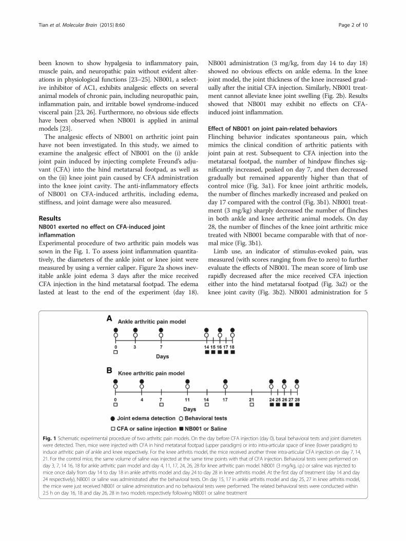
ResultsNB001 exerted no effect on CFA-induced jointinflammationExperimental procedure of two arthritic pain models wassown in the Fig. 1. To assess joint inflammation quantita-tively, the diameters of the ankle joint or knee joint weremeasured by using a vernier caliper. Figure 2a shows inev-itable ankle joint edema 3 days after the mice receivedCFA injection in the hind metatarsal footpad. The edemalasted at least to the end of the experiment (day 18).
NB001 administration (3 mg/kg, from day 14 to day 18)showed no obvious effects on ankle edema. In the kneejoint model, the joint thickness of the knee increased grad-ually after the initial CFA injection. Similarly, NB001 treat-ment cannot alleviate knee joint swelling (Fig. 2b). Resultsshowed that NB001 may exhibit no effects on CFA-induced joint inflammation.
Effect of NB001 on joint pain-related behaviorsFlinching behavior indicates spontaneous pain, whichmimics the clinical condition of arthritic patients withjoint pain at rest. Subsequent to CFA injection into themetatarsal footpad, the number of hindpaw flinches sig-nificantly increased, peaked on day 7, and then decreasedgradually but remained apparently higher than that ofcontrol mice (Fig. 3a1). For knee joint arthritic models,the number of flinches markedly increased and peaked onday 17 compared with the control (Fig. 3b1). NB001 treat-ment (3 mg/kg) sharply decreased the number of flinchesin both ankle and knee arthritic animal models. On day28, the number of flinches of the knee joint arthritic micetreated with NB001 became comparable with that of nor-mal mice (Fig. 3b1).Limb use, an indicator of stimulus-evoked pain, was
measured (with scores ranging from five to zero) to furtherevaluate the effects of NB001. The mean score of limb userapidly decreased after the mice received CFA injectioneither into the hind metatarsal footpad (Fig. 3a2) or theknee joint cavity (Fig. 3b2). NB001 administration for 5
Fig. 1 Schematic experimental procedure of two arthritic pain models. On the day before CFA injection (day 0), basal behavioral tests and joint diameterswere detected. Then, mice were injected with CFA in hind metatarsal footpad (upper paradigm) or into intra-articular space of knee (lower paradigm) toinduce arthritic pain of ankle and knee respectively. For the knee arthritis model, the mice received another three intra-articular CFA injection on day 7, 14,21. For the control mice, the same volume of saline was injected at the same time points with that of CFA injection. Behavioral tests were performed onday 3, 7, 14 16, 18 for ankle arthritic pain model and day 4, 11, 17, 24, 26, 28 for knee arthritic pain model. NB001 (3 mg/kg, i.p.) or saline was injected tomice once daily from day 14 to day 18 in ankle arthritis model and day 24 to day 28 in knee arthritis model. At the first day of treatment (day 14 and day24 respectively), NB001 or saline was administrated after the behavioral tests. On day 15, 17 in ankle arthritis model and day 25, 27 in knee arthritis model,the mice were just received NB001 or saline administration and no behavioral tests were performed. The related behavioral tests were conducted within2.5 h on day 16, 18 and day 26, 28 in two models respectively following NB001 or saline treatment
Tian et al. Molecular Brain (2015) 8:60 Page 2 of 10

days significantly increased the limb use score in mice withankle and knee joint arthritis.Subsequently, limb use during forced ambulation was
detected by using a rotarod test with a similar scale.Figure 3a3, b3 show a remarkable decrease in the aver-age score of forced limb use in both types of arthriticmodels. NB001 administration obviously relieved pain,as reflected by the rotarod test in two animal models.These combined results revealed persistent joint pain inCFA-induced ankle arthritis or knee joint arthritis. Fur-thermore, NB001 showed evident analgesic effects onadjuvant-induced joint pain.
NB001 increased the mechanical thresholdTo further examine the analgesic effect of NB001 on thetypes of pain behavior associated with arthritis, mechan-ical allodynia was detected in both animal models. Themechanical paw withdrawal threshold (MPWT) in ipsi-lateral hindpaw decreased immediately and significantlyin mice injected with CFA into the metatarsal footpad(Fig. 4a). NB001 administration (3 mg/kg from day 14)markedly increased the mechanical threshold on days 16and 18 compared with saline treatment (Fig. 4a). Kneeintra-articular CFA injection gradually decreased MPWTand NB001 obviously attenuated mechanical allodynia(Fig. 4b). No obvious difference was observed in MPWT incontralateral paws among groups in both types of animalmodels (Fig. 4a, b) within the entire experimental period.
NB001 increased the thermal thresholdOn the last day of the experiment (day 18), the thermalpaw withdrawal latency of mouse models of ankle jointarthritis were measured. NB001 treatment (3 mg/kg)showed apparently higher thermal paw withdraw latencythan saline treatment in CFA-injected mice (F(2,21) = 26.555,p = 0.000, Dunnett’s test; Fig. 5a). Furthermore, the thermallatency in contralateral hindpaw was comparable amongthe three groups (F(2,21) = 0.275, p = 0.762, LSD test).
Effect of NB001 on joint hyperalgesiaJoint hyperalgesia was detected by recording the num-bers of mice vocalization when bending and extensionwere applied to the joint. Figure. 5b, c show extremelylow means for vocalization in the control mice. Thenumbers of vocalization in both mouse models of ipsilat-eral joint pain increased sharply in both mouse models(F(2,21) = 123.141, p = 0.000, Dunnett’s test; Fig. 5b andF(2,21) = 171.64, p = 0.000, LSD test; Fig. 5c). NB001 treat-ment (3 mg/kg) significantly decreased the number ofvocalizations in both mouse models of ankle joint painand knee joint pain. The numbers of vocalization amongthe groups in both models of contralateral joint painwere similar (F(2,21) = 0.191, p = 0.828, LSD test; Fig. 5band F(2,21) = 0.653, p = 0.531, LSD test; Fig. 5c).
NB001 exerted no effect on joint stiffnessJoint stiffness was assessed by the bending and extensiontest and was used as an indicator in progressive arthritisdevelopment in adjuvant arthritis models [27]. No restric-tion was set on the full-range movement of joints in anymice on day 0, and the joints of the control mice showedno restriction throughout the whole experimental period(Fig. 6). The joint stiffness score increased sharply andremained at an increased level in the ipsilateral ankle fol-lowing CFA injection (Fig. 6a). This score increased pro-gressively in the ipsilateral knee following CFA injection(Fig. 6b). NB001 treatment (3 mg/kg) showed no evidentinfluence on ankle or knee joint stiffness.
NB001 showed no improvement on arthritis-inducedjoint damageThe histological evaluation of ankle and knee joint tis-sues was performed on day 18 and 28, respectively(Fig. 7). The histological architecture of the ankle jointshowed abnormality in mice after the induction of anklejoint arthritis, as shown by inflammatory cell infiltration,
Fig. 2 The effect of NB001 on joints edema induced by CFA injection. a Ankle diameter reached the maximum on day 3 following CFA injectionand then it remained at a higher level compared with control. NB001 treatment (3 mg/kg, i.p.) did not attenuate the ankle edema. b There was asignificant increase of knee diameter after intra-articular CFA injection. NB001 administration did not alleviate the inflammation. n = 8 mice in eachgroup. *p < 0.05, **p < 0.01 compared to the control group
Tian et al. Molecular Brain (2015) 8:60 Page 3 of 10

synovial hyperplasia, and degeneration of the articularcartilage (Fig. 7b). Intra-articular CFA injection (4times) caused abnormalities in knee joint structures,including pronounced synovial hyperplasia and in-flammation, displacement of the meniscus, thickeningof the joint capsule, and intra-articular fibrin accumu-lation (Fig. 6e). NB001 administration (3 mg/kg) for5 days failed to evidently attenuate CFA-induced jointdamages in both types of arthritic models (Fig.6c, f ).These results demonstrated again that NB001 exertedno anti-inflammatory effects and failed to alleviateinflammation-induced joint damage.
DiscussionArthritis can affect people at any age, and its prevalenceincreases with age. Chronic joint pain, the most debili-tating symptom of arthritis, prompts patients to seekmedical help. However, few effective therapies exert noundesirable side effects [28]. Therefore, new therapiesfor alleviating arthritic joint pain need to be developed.AC1 acts as a key molecule for triggering chronic-
pain-related central plasticity. AC1 is considered indis-pensable for the induction of both spinal and ACC LTP,which is one of the possible cellular mechanisms under-lying chronic pain [21]. Genetically, the knockout of
Fig. 3 NB001 alleviates arthritic pain behaviors. a Data from the ankle joint arthritic model. A1: The number of spontaneous flinches increased significantlyand reached a peak on day 7 following CFA injection and maintained at a higher level as compared with the control. In addition, the score of normal limbuse (A2) and forced ambulation (rotarod) (A3) declined significantly after the CFA injection. b Data from the knee joint arthritic model. B1: Mice exhibited agradually increased spontaneous flinches. The score of normal limb use (B2) and forced ambulation (rotarod) (B3) declined significantly after the CFAinjection. NB001 treatment (3 mg/kg, i.p.) significantly attenuated pain-related behaviors in two types of model (a and b). n= 8 in each group. *p< 0.05,**p< 0.01 compared to the control group; #p< 0.05, ##p< 0.01 compared to the CFA group
Tian et al. Molecular Brain (2015) 8:60 Page 4 of 10

AC1 reduced mice behavioral response to chronic paincaused by peripheral inflammation or nerve injury withthe impaired LTP induction. Nociceptive responses werealso remarkably inhibited in the persistent muscle painmodel of AC1 knockout mice [25]. On the basis of theseresults, inhibiting AC1 activity may be a novel mechan-ism for chronic pain treatment [29]. As the first selectiveinhibitor of AC1, the analgesic effect of NB001 has beeninvestigated in our previous studies. NB001 exhibitedsignificant analgesic effects in the animal models of neuro-pathic pain, which was comparable to those produced bygabapentin [23]. Behavioral allodynia was also inhibited byNB001 in the animal models of inflammatory pain, withhigher doses exerting greater inhibition [20].Alleviating the joint pain of arthritic patients can in-
crease functional recovery and improve quality of life. Inthis study, we investigated the analgesic role of NB001on CFA-induced arthritic pain in two arthritis models.We demonstrated that NB001 produced antinociceptiveeffects in both mouse models of ankle joint pain andknee joint pain. However, NB001 failed to exhibit anti-inflammatory effects, as indicated by no attenuation ofthe related abnormal changes induced by inflammation.Nociceptive articular pain originates locally in the joint
at the site of inflammation or injury. CFA has been widely
used to induce arthritic pain because of its ability to causechronic inflammation in rodents [30] and produce adisease-like state that most closely resembles human arth-ritis [31]. CFA injection leads to T-lymphocytes andmacrophage infiltration, which progress with joint swell-ing and synovial hypertrophy accompanied by significantreduction in joint cavity volume and severe osteolyticlesion [32, 33]. In our experiment, we first examined theeffect of NB001 on CFA-induced joint inflammation.NB001 showed no obvious effects on joint edema causedby CFA injection. Joint stiffness results from long-terminflammation and indicates disease development togetherwith abnormal mobility behavior in arthritic models [27].Patients with arthritis also showed increased joint stiffnessand decreased maximal amplitude of flexion and exten-sion [34]. In the current study, mice exhibited apparentankle or knee joint stiffness indicated by notable restric-tion of full-range joint movement following metatarsal orknee intra-articular CFA injection. Treatment with NB001for 5 days exhibited no influence on the joint stiffnessscore. The effects of NB001 on joint swelling and stiffnessimplied that NB001 had no anti-inflammatory effect andcould not prevent the progressive development of arthritis.This presumption was further demonstrated by the immu-nohistochemical analysis of a joint slice. CFA injection
Fig. 4 NB001 attenuates mechanical allodynia. a Mechanical threshold in the ipsilateral hindpaws decreased in the mice following CFA injectioninto metatarsal footpad, but had no alteration in the contralateral paws. NB001 administration attenuated the mechanical allodynia in the ipsilateralhindpaws. b The mechanical allodynia occurred gradually following knee intra-articular CFA injection and the threshold decreased obviously from day17 after initial CFA injection (three days after the third CFA injection), but had no alteration in the contralateral paws. NB001 administration attenuatedthe mechanical allodynia in the ipsilateral hindpaws. n = 8 mice in each group. *p < 0.05, **p < 0.01 compared to the control group; #p < 0.05, ##p < 0.01compared to the CFA group
Tian et al. Molecular Brain (2015) 8:60 Page 5 of 10

caused abnormal changes in both ankle joint and knee jointas indicated by synovial hypertrophy, inflammatory cell in-filtration, degeneration of the articular cartilage, and thick-ening of the joint capsule. However, NB001 cannotimprove inflammation-induced joint destruction.For arthritis, the analgesic effect must be distinguished
from an anti-inflammatory effect because analgesicsalleviate pain but may not attenuate the progressive
development of the disease [35, 36]. The analgesic ef-fects of NB001 occurred in two types of CFA-inducedarthritic pain model. Spontaneous pain (joint pain atrest) is an important behavioral symptom of arthriticjoint pain, and the number of hindpaw flinches was re-corded as the measure of spontaneous nociceptive behav-ior [6, 37]. CFA injections induced obvious spontaneouspain, as indicated by the increase in the number of hind-paw flinches. NB001 markedly inhibited spontaneous painin our experiment. Furthermore, stimulus-evoked pain be-havior was also examined to measure arthritic pain be-cause it mimics the clinical condition of arthritic patientswhen the joint was moved [37]. Limb use scores duringspontaneous movement and forced ambulation (rotarod)were used as the indexes of stimulus-evoked pain-relatedbehavior according to previous research [38, 39]. NB001administration markedly increased limb use scores in bothmouse models of ankle and knee joint arthritis. These re-sults showed that NB001 treatment inhibited spontaneousmovement and evoked pain in adjuvant-induced arthritis.Thermal hyperalgesia, mechanical allodynia, and joint
hyperalgesia are the main secondary characteristics of arth-ritic pain [40, 41]. Under the condition of chronic arthriticpain, the threshold for nociceptor activation is lowered andthe response to mechanical or thermal stimuli is amplified[42]. In our study, the mice showed significant and quickmechanical allodynia, thermal hypersensitivity, and jointhyperalgesia following CFA-induced ankle arthritis. How-ever, knee intra-articular CFA injection led to a progressivereduction in mechanical threshold, and the mice exhibitedapparent mechanical allodynia on day 17 after the initialCFA injection (3 days after the third CFA injection). Theseeffects may be attributed to the fact that single knee jointintra-articular CFA injection could not adequately causehindpaw mechanical hypersensitivity. NB001 treatment ex-hibited pronounced attenuation of behavioral sensitization,thus validating its analgesic effects on arthritic pain.In conclusion, the results from our current study re-
vealed that NB001 exerted analgesic effects on arthriticjoint pain. Antinociception seemed unlikely to be associ-ated with the anti-inflammatory effect because NB001exhibited no anti-inflammatory action, as indicated bythe lack of attenuation of joint edema, stiffness, and jointstructural destruction caused by CFA. Combined withthe results of previous studies with NB001, NB001 canbe potentially used as a new analgesic.
MethodsAnimalsAll experiments were performed with adult male C57/BL6mice aged from 8 to 10 weeks. Animals were housed understandard laboratory conditions (12 h light/12 h dark,temperature 22–26 °C, humidity 55–60 %) with food andwater provided ad libitum. Animals were allowed to
Fig. 5 Effects of NB001 on thermal and joint hyperalgesia. Thermalhyperalgesia was examined on the last experimental day for anklearthritis mice. a CFA caused thermal hyperalgesia in the ipsilateralhindpaw but not the contralateral hindpaw. NB001 administration(3 mg/kg, i.p.) significantly inhibit thermal hyperalgesia. b Jointhyperalgesia was assessed by the joint flexion test on the lastexperimental day and expressed as the numbers of vocalizationcaused by bending and extension of the joint (five times in eachdirection). The mice exhibited evident ankle joint hyperalgesiafollowing CFA injection in the ipsilateral joint. c The mice exhibitedmarkedly knee joint hyperalgesia following CFA injection in theipsilateral joint. NB001 significantly (3 mg/kg, i.p.) attenuated bothankle (b) and knee joint hyperalgesia (c) indicted by reduction ofvocalized numbers in joint flexion tests. n = 8 in each group.*p < 0.05, **p < 0.01 compared to the control group; ##p < 0.01compared to the CFA group
Tian et al. Molecular Brain (2015) 8:60 Page 6 of 10

Fig. 7 Histological staining of ankle and knee joints. The representative sections of ankle joint from control mice (a) and CFA-injection mice(b). CFA injection induced inflammatory cell infiltration, synovium hyperplasia, degeneration of the articular cartilage of the ankle joint. c NB001 treatment(3 mg/kg, i.p.) did not attenuate CFA-induced ankle joint structural destruction. The representative sections of knee joint from control mice (d) andCFA-injection mice (e). Four times knee intra-articular CFA injection induced abnormal structural changes of the knee joint, including bone, synovium,and meniscus. f NB001 treatment (3 mg/kg, i.p.) did not attenuate CFA-induced knee joint structural destruction
Fig. 6 No effects of NB001 on CFA-induced joint stiffness. Joint stiffness was an indication of progressive disease development of arthritis. It wasreflected by restriction of full range of joint movement. There was obvious restriction on the full-range joint movement in ipsilateral ankle (a) or knee(b) indicted by significant increase of joint stiffness scores. NB001 (3 mg/kg, i.p.) had no effects on joint stiffness. n = 8 in each group. **p < 0.01compared to the control group
Tian et al. Molecular Brain (2015) 8:60 Page 7 of 10

acclimate to laboratory environment for one week prior tothe beginning of the experiment. All experiments wereperformed in accordance with protocols approved byAnimal Care and Use Committee of the FourthMilitary Medical University.
CFA injection and NB001 administrationTo induce arthritic pain of knee joint, CFA (10 μL, in 50 %saline) was injected into the left knee joint cavity of miceusing a 30-gauge 1⁄2-in. needle every 7 days (on day 0, 7,14, and 21) under isoflurane anesthesia [43]. For the anklejoint pain model, 10 μL CFA was injected into the left hindmetatarsal footpad of mice on day 0. The same volumesaline was injected into the left knee joint cavity or hindmetatarsal footpad of the control mice. For the treatment,NB001 (3 mg/kg, in saline) was administrated intraperito-neally once a day from day 24 to day 28 for knee joint painmodels or from day 14 to day 18 for ankle joint painmodels. The first administration of NB001 was performedafter the behavioral tests on day 14 or day 24. At the thirdand last administration of NB001, the behavioral tests werecompleted within 2.5 h after NB001 administration. Thecontrol and model (CFA) groups received intraperitonealinjection of saline at the same time point.
Assessment of paw and knee joint edemaTo quantify the effects of NB001 on the joint inflamma-tion, the diameters across the ankle or knee joints wereassessed using a vernier caliper before and after CFAinjection at the different time points.
Behavioral measurements of arthritic joint painAll behavioral tests were performed during the day timeand the animals were allowed to habituate to the labora-tory room for at least 30 min before the tests. In all cases,behavioral analysis was performed by a trained observerblind to the experimental groups.
Evaluation of spontaneous pain and stimulus-evoked painSpontaneous pain and stimulus-evoked pain are two be-havioral phenotype of arthritic joint pain and always usedas the measurements of arthritis [44]. They reflect theclinical conditions of arthritic patients with joint pain atrest and after joint movement respectively [6, 37]. Hind-paw flinch is one indicator of spontaneous pain. Flincheswere calculated as the number of times mice raised itshindpaw [44]. For the detection of flinching behavior, micewere placed in cylindric plexiglas chambers with theheight of 30 cm and inside diameter of 20 cm for 30 minacclimation. The numbers of spontaneous flinch wererecorded during 2 min. For the assessment of stimulus-evoked pain, the use of normal limb was detected andscored from 5 to 0 during spontaneous movement as previ-ously reported [38, 39], 5 = normal use, 4 = partial limp, but
not pronounced, 3 = pronounced limp, 2 = limp and guard-ing behavior, 1 = partial nonuse of limb in locomotor activ-ity, 0 = totally lack of limb use. In addition, the limb-usewas detected under forced ambulation condition. The micewere placed on a rotarod (Ji Liang Company, Shanghai,China) at a speed of 17 rounds/min for 5 min. Limb-usewas scored: 4 = normal; 3 = limping; 2 = partial non-use ofleft hind limb; 1 = substantial non-use of left hind limb; 0 =non-use of left hind limb.
Mechanical allodynia testMice were put in individual plexiglas cages with a metalmesh floor to acclimate environment for 30 min beforetesting. Mechanical allodynia was detected with a set ofvon Frey filaments (Stoelting) and evaluated by hind-paw responsiveness to different stimulation. Mechanicalpressure from the 1.65 filament (force, 0.008 g) wasused to characterize the threshold stimulus based onthe up-down strategy. The filaments were applied ver-tically to the dorsal surface of the hindpaw with suffi-cient force to cause slight bending for 6 s.Mechanical allodynia was detected five times with aninterval of 10 min within stimulation. Positive re-sponses include licking, biting, flinching, and briskwithdrawal of the hindpaw.
Thermal hyperalgesia testAnimals were placed in individual round containerand allowed to adapt for 30 min before the experi-ment. Thermal hyperalgesia was evaluated by measur-ing the latency of paw withdrawal (PWL) in response to aradiant heat source [45] using a commercially availableplantar analgesia instrument (BME410A, Institute ofBiological Medicine, Academy of Medical Science, China).The heat source was turned off when the mice lifted thefoot and the time from beginning to the ending of heatapplication was defined as the PWL. In order to preventtissue damage resulted from long-time heat application,the heat source would be cut off automatically at 20 s eventhe mice did not lift the foot. The apparatus was modu-lated to give a paw withdrawal latency of approximately10 s in naïve mice. Left paws were tested at 5 min intervalsfor a total of five trials. The mean PWL was obtained fromthe latter three stimuli [46].
Assessment of joint hyperalgesiaThe joint hyperalgesia was measured according to theprevious method [47] with little modification. Bendingand extension (one after the other with a 5 s intervaland five times in each direction) were applied to theankle or knee joint within its limits of range of motion.The numbers of mice vocalized during each stimulus(the bending and extension) were recorded, and score 0
Tian et al. Molecular Brain (2015) 8:60 Page 8 of 10

(no vocalization) or 1 (vocalization) was given dependedon the results. Thus, for each mouse the vocalizationscore ranged from 0 to 10 at each joint [27, 48].
Measurement of joint stiffnessThe joint stiffness scoring was examined according tothe previously reported method [32]. The mice wereheld stably, then the ankle or knee joint was bended andextended (once in each direction). The scores were givenaccording to the following standard: score 2, there wererestrictions of full range of joint movement in bothbending and extension; score 1, there was restriction ofmovement of the joint only in one direction (bending orextension); score 0, there was no restriction in bothdirection [32].
Histopathologic analysisOn the last experimental day, animals were anesthe-tized and sacrificed (day 28 after initial CFA injectioninto knee joint or day 18 after initial CFA injectioninto ankle joint) and the ipsilateral knee joints orankle joints were collected. For collecting knee joints,the legs were cut through both the femur and thetibia. Then the specimens were immerged into 10 %neutral formalin to be post-fixed for 48 h. Subse-quently, the fixed tissue was moved into decalcifyingsolution (4 M formic acid) for 35 days to make thebone completely demineralization. During this period,the decalcifying solution was changed once a week.The specimens were embedded in paraffin wax andcut into 5 μm thick sections, and stained withhematoxylin and eosin for microscopic assessment.
Experimental procedureThe experimental procedure was summarized in Fig. 7.The upper one was experimental time course of ankle arth-ritis model and the lower one was that of knee arthritismodel. All behavioral tests were performed prior to CFAinjection and on day 3, 7, 14, 16, 18 for ankle arthritis oron day 4, 11, 17, 24, 26, 28 for knee arthritis after initialCFA injection. The first administration of NB001 was givenafter behavioral tests on day 14 for ankle joint arthritismodels or on day 24 for knee joint arthritis models. Duringthe treatment period, if there were behavioral measure-ments, they were finished within 2.5 h after NB001 or sa-line administration except it was specially mentioned.
Data analysisData were presented as the mean and standard errors ofthe means (SEM). Statistical analysis of differences be-tween two groups was performed by independent sam-ple, two-tailed t test. Data of multiple groups wereevaluated using one-way analysis of variance (ANOVA)for post hoc comparisons (SPSS 13.0). Data that passed
the homogeneity test were analyzed by the one-wayANOVA least significant difference (LSD) test. Data thatdid not pass the homogeneity test were analyzed by theone-way ANOVA Dunnett’s test. In all cases, p < 0.05was considered statistically significant.
AbbreviationsAC1: Adenylyl cyclase subtype 1; ACC: Anterior cingulate cortex; ACSF: Artificialcerebrospinal fluid; cAMP: Cyclic adenosine monophosphate; CFA: CompleteFreund’s Adjuvant; MPWT: Mechanical paw withdrawal threshold; LTP: Long-termpotentiation.
Competing interestsThe authors declare that they have no competing interests.
Authors’ contributionsMGZ and SBL designed the study; ZT, DSW, XSW, JT, and YYG performed theexperiments. BF and NZ analyzed the data. MGZ and SBL wrote the paper.All authors read and approved the final manuscript.
Authors’ informationZT, YYG, and NZ are Ph.D students in the Department of Pharmacology, Schoolof Pharmacy, Fourth Military Medical University. DSW is the Master student inDepartment of Orthopedics, Jinling Hospital, Clinical School of Nanjing, SecondMilitary Medical University. JT is the Ph.D student in the Department ofPediatrics, Tangdu Hospital, Fourth Military Medical University. BF is a Lecture inthe Department of Pharmacy, School of Stomatology, Fourth Military MedicalUniversity. MGZ is the Professor in Department of Pharmacology, Schoolof Pharmacy, Fourth Military Medical University. SBL is the AssociateProfessor in Department of Pharmacology, School of Pharmacy, FourthMilitary Medical University.
AcknowledgmentsWe thank Dr. Min Zhuo for the gift of NB001. This work was supported bygrants from the National Natural Science Foundation of China (31271126and 31470052 to SBL), (31271144, 81325022, and 2013DFG32650 to MGZ).
Author details1Department of Pharmacology, School of Pharmacy, Fourth Military MedicalUniversity, Xi’an 710032, China. 2Department of Orthopedics, Jinling Hospital,Clinical School of Nanjing, Second Military Medical University, Nanjing210002, China. 3Department of Pediatrics, Tangdu Hospital, Fourth MilitaryMedical University, Xi’an 710038, China. 4Department of Pharmacy, School ofStomatology, Fourth Military Medical University, Xi’an 710032, China.
Received: 24 July 2015 Accepted: 2 October 2015
References1. Hunter DJ, McDougall JJ, Keefe FJ. The symptoms of osteoarthritis and the
genesis of pain. Med Clin North Am. 2009;93(1):83–100. xi.2. Kidd BL. Osteoarthritis and joint pain. Pain. 2006;123(1-2):6–9.3. Woolf AD, Pfleger B. Burden of major musculoskeletal conditions. Bull World
Health Organ. 2003;81(9):646–56.4. Weinblatt ME. Nonsteroidal anti-inflammatory drug toxicity: increased risk in
the elderly. Scand J Rheumatol Suppl. 1991;91:9–17.5. Feinberg SD. Prescribing analgesics. How to improve function and avoid
toxicity when treating chronic pain. Geriatrics. 2000;55(11):9–50.6. Papageorgiou AC, Badley EM. The quality of pain in arthritis: the words
patients use to describe overall pain and pain in individual joints at rest andon movement. J Rheumatol. 1989;16(1):106–12.
7. Wagstaff S, Smith OV, Wood PH. Verbal pain descriptors used by patientswith arthritis. Ann Rheum Dis. 1985;44(4):262–5.
8. Tatsuo MA, Carvalho WM, Silva CV, Miranda AE, Ferreira SH, Francischi JN.Analgesic and antiinflammatory effects of dipyrone in rat adjuvant arthritismodel. Inflammation. 1994;18(4):399–405.
9. Jasmin L, Kohan L, Franssen M, Janni G, Goff JR. The cold plate as a test ofnociceptive behaviors: description and application to the study of chronicneuropathic and inflammatory pain models. Pain. 1998;75(2-3):367–82.
Tian et al. Molecular Brain (2015) 8:60 Page 9 of 10
10. Bertorelli R, Corradini L, Rafiq K, Tupper J, Calo G, Ongini E, et al. Nociceptinand the ORL-1 ligand [Phe1psi (CH2-NH)Gly2]nociceptin(1-13)NH2 exertanti-opioid effects in the Freund’s adjuvant-induced arthritic rat model ofchronic pain. Br J Pharmacol. 1999;128(6):1252–8.
11. Schaible HG, von Banchet GS, Boettger MK, Brauer R, Gajda M, Richter F, et al.The role of proinflammatory cytokines in the generation and maintenance ofjoint pain. Ann N Y Acad Sci. 2010;1193:60–9.
12. Grigg P, Schaible HG, Schmidt RF. Mechanical sensitivity of group III and IVafferents from posterior articular nerve in normal and inflamed cat knee.J Neurophysiol. 1986;55(4):635–43.
13. Schaible HG, Schmidt RF. Time course of mechanosensitivity changes inarticular afferents during a developing experimental arthritis. J Neurophysiol.1988;60(6):2180–95.
14. Schaible HG, Ebersberger A, Von Banchet GS. Mechanisms of pain inarthritis. Ann N Y Acad Sci. 2002;966:343–54.
15. Neugebauer V, Lucke T, Schaible HG. N-methyl-D-aspartate (NMDA) andnon-NMDA receptor antagonists block the hyperexcitability of dorsal hornneurons during development of acute arthritis in rat’s knee joint. JNeurophysiol. 1993;70(4):1365–77.
16. Neugebauer V, Lucke T, Grubb B, Schaible HG. The involvement ofN-methyl-D-aspartate (NMDA) and non-NMDA receptors in theresponsiveness of rat spinal neurons with input from the chronicallyinflamed ankle. Neurosci Lett. 1994;170(2):237–40.
17. Zhuo M. Cortical excitation and chronic pain. Trends Neurosci.2008;31(4):199–207.
18. Kandel ER. The molecular biology of memory storage: a dialogue betweengenes and synapses. Science. 2001;294(5544):1030–8.
19. Xia Z, Storm DR. The role of calmodulin as a signal integrator for synapticplasticity. Nat Rev Neurosci. 2005;6(4):267–76.
20. Wei F, Vadakkan KI, Toyoda H, Wu LJ, Zhao MG, Xu H, et al. Calciumcalmodulin-stimulated adenylyl cyclases contribute to activation ofextracellular signal-regulated kinase in spinal dorsal horn neurons in adultrats and mice. J Neurosci. 2006;26(3):851–61.
21. Liauw J, Wu LJ, Zhuo M. Calcium-stimulated adenylyl cyclases required forlong-term potentiation in the anterior cingulate cortex. J Neurophysiol.2005;94(1):878–82.
22. Zhuo M. Targeting neuronal adenylyl cyclase for the treatment of chronicpain. Drug Discov Today. 2012;17(11-12):573–82.
23. Wang H, Xu H, Wu LJ, Kim SS, Chen T, Koga K, et al. Identification of anadenylyl cyclase inhibitor for treating neuropathic and inflammatory pain.Sci Transl Med. 2011;3(65):65ra3.
24. Wei F, Qiu CS, Kim SJ, Muglia L, Maas JW, Pineda VV, et al. Genetic eliminationof behavioral sensitization in mice lacking calmodulin-stimulated adenylylcyclases. Neuron. 2002;36(4):713–26.
25. Vadakkan KI, Wang H, Ko SW, Zastepa E, Petrovic MJ, Sluka KA, et al. Geneticreduction of chronic muscle pain in mice lacking calcium/calmodulin-stimulatedadenylyl cyclases. Mol Pain. 2006;2:7.
26. Zhang MM, Liu SB, Chen T, Koga K, Zhang T, Li YQ, et al. Effects of NB001and gabapentin on irritable bowel syndrome-induced behavioral anxietyand spontaneous pain. Mol Brain. 2014;7:47.
27. Nagakura Y, Okada M, Kohara A, Kiso T, Toya T, Iwai A, et al. Allodynia andhyperalgesia in adjuvant-induced arthritic rats: time course of progressionand efficacy of analgesics. J Pharmacol Exp Ther. 2003;306(2):490–7.
28. Lanas A, Tornero J, Zamorano JL. Assessment of gastrointestinal andcardiovascular risk in patients with osteoarthritis who require NSAIDs:the LOGICA study. Ann Rheum Dis. 2010;69(8):1453–8.
29. Kim SS, Descalzi G, Zhuo M. Investigation of molecular mechanism ofchronic pain in the anterior cingulate cortex using genetically engineeredmice. Curr Genomics. 2010;11(1):70–6.
30. Hogan Q. Animal pain models. Reg Anesth Pain Med. 2002;27(4):385–401.31. Cook CD, Nickerson MD. Nociceptive sensitivity and opioid antinociception
and antihyperalgesia in Freund’s adjuvant-induced arthritic male and femalerats. J Pharmacol Exp Ther. 2005;313(1):449–59.
32. Butler SH, Godefroy F, Besson JM, Weil-Fugazza J. A limited arthritic modelfor chronic pain studies in the rat. Pain. 1992;48(1):73–81.
33. Romas E, Gillespie MT, Martin TJ. Involvement of receptor activator ofNFkappaB ligand and tumor necrosis factor-alpha in bone destruction inrheumatoid arthritis. Bone. 2002;30(2):340–6.
34. Johnson SR, Archibald A, Davis AM, Badley E, Wright JG, Hawker GA. Is self-reported improvement in osteoarthritis pain and disability reflected inobjective measures? J Rheumatol. 2007;34(1):159–64.
35. Aryaeian N, Shahram F, Djalali M, Eshragian MR, Djazayeri A, Sarrafnejad A,et al. Effect of conjugated linoleic acids, vitamin E and their combination onthe clinical outcome of Iranian adults with active rheumatoid arthritis. Int JRheum Dis. 2009;12(1):20–8.
36. Rossato MF, Hoffmeister C, Tonello R, de Oliveira Ferreira AP, Ferreira J, et al.Anti-inflammatory effects of vitamin E on adjuvant-induced arthritis in rats.Inflammation. 2015;38(2):606–15.
37. Rojkovich B, Gibson T. Day and night pain measurement in rheumatoidarthritis. Ann Rheum Dis. 1998;57(7):434–6.
38. Honore P, Luger NM, Sabino MA, Schwei MJ, Rogers SD, Mach DB, et al.Osteoprotegerin blocks bone cancer-induced skeletal destruction, skeletalpain and pain-related neurochemical reorganization of the spinal cord. NatMed. 2000;6(5):521–8.
39. Luger NM, Sabino MA, Schwei MJ, Mach DB, Pomonis JD, Keyser CP, et al.Efficacy of systemic morphine suggests a fundamental difference in themechanisms that generate bone cancer vs inflammatory pain. Pain.2002;99(3):397–406.
40. Vermeirsch H, Biermans R, Salmon PL, Meert TF. Evaluation of pain behaviorand bone destruction in two arthritic models in guinea pig and rat.Pharmacol Biochem Behav. 2007;87(3):349–59.
41. Kaur S, Bijjem KR, Sharma PL. Anti-inflammatory and antihyperalgesic effectsof the combination of ibuprofen and hemin in adjuvant-induced arthritis inthe Wistar rat. Inflammopharmacology. 2011;19(5):265–72.
42. Krug HE, Frizelle S, McGarraugh P, Mahowald ML, et al. Pain behaviormeasures to quantitate joint pain and response to neurotoxin treatment inmurine models of arthritis. Pain Med. 2009;10(7):1218–28.
43. Gauldie SD, McQueen DS, Clarke CJ, Chessell IP, et al. A robust model ofadjuvant-induced chronic unilateral arthritis in two mouse strains. J NeurosciMethods. 2004;139(2):281–91.
44. Jimenez-Andrade JM, Mantyh PW. Sensory and sympathetic nerve fibersundergo sprouting and neuroma formation in the painful arthritic joint ofgeriatric mice. Arthritis Res Ther. 2012;14(3):R101.
45. Hu J, Wang Z, Guo YY, Zhang XN, Xu ZH, Liu SB, et al. A role ofperiaqueductal grey NR2B-containing NMDA receptor in mediatingpersistent inflammatory pain. Mol Pain. 2009;5:71.
46. Gu X, Zhang J, Ma Z, Wang J, Zhou X, Jin Y, et al. The role of N-methyl-D-aspartate receptor subunit NR2B in spinal cord in cancer pain. Eur J Pain.2010;14(5):496–502.
47. Rupniak NM, Boyce S, Webb JK, Williams AR, Carlson EJ, Hill RG, et al. Effectsof the bradykinin B1 receptor antagonist des-Arg9[Leu8]bradykinin andgenetic disruption of the B2 receptor on nociception in rats and mice.Pain. 1997;71(1):89–97.
48. Kwon YB, Lee JD, Lee HJ, Han HJ, Mar WC, Kang SK, et al. Bee venominjection into an acupuncture point reduces arthritis associated edema andnociceptive responses. Pain. 2001;90(3):271–80.
Submit your next manuscript to BioMed Centraland take full advantage of:
• Convenient online submission
• Thorough peer review
• No space constraints or color figure charges
• Immediate publication on acceptance
• Inclusion in PubMed, CAS, Scopus and Google Scholar
• Research which is freely available for redistribution
Submit your manuscript at www.biomedcentral.com/submit
Tian et al. Molecular Brain (2015) 8:60 Page 10 of 10