an orthodontic-retentive approach
TRANSCRIPT
An Orthodontic-Retentive Approach
During Prosthodontic Rehabilitation in
Cleft Lip and Palate Patients:
A Case Report
MARIO S. RUTOWITSCH, D.D.S.
THALES R. MAGALHAES, D.D.S.
Rio de Janeiro, Brazil
The orthodontic-prosthodontic rehabilitation of an eighteen-year-old girl with a VeauClass III cleft is discussed together with an orthodontic-retentive approach whichminimizes the possibility of relapse during the time required for the insertion of aprosthodontic retentive device.
The biggest challenge in severe orthodontic problems associated with cleft lipand palate is not treatment but retention. After removal of the orthodonticappliances and during the time required for final prosthodontic work, theremovable acrylic prosthesis usually used for temporary retention often does notprevent relapse.
Two suggested solutions for this problem are presented in this case report.
Pre-Treatment Status
An eighteen-year-old girl with a complete unilateral cleft lip and palate came
for treatment to the Hospital of IASEG in Rio de Janeiro. She had not had
surgical closure of the lip until three years of age, of the hard palate until age
seven, and of the soft palate until age 14. Tonsils and adenoids had been removed
when she was six years old. Speech was markedly hypernasal, and there was a
hearing loss in the left ear. There was no history of previous orthodontic
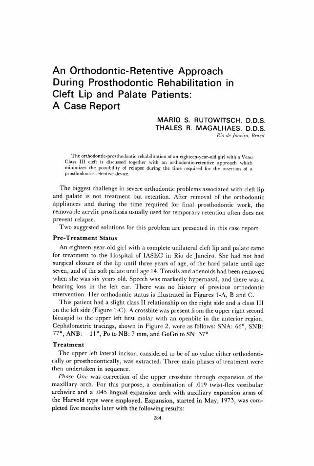
intervention. Her orthodontic status is illustrated in Figures 1-A, B and C.
This patient had a slight class II relationship on the right side and a class III
on the left side (Figure 1-C). A crossbite was present from the upper right second
bicuspid to the upper left first molar with an openbite in the anterior region.
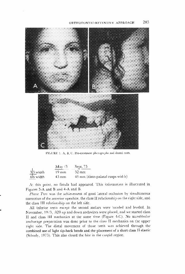
Cephalometric tracings, shown in Figure 2, were as follows: SNA: 66°, SNB:
77°, ANB: -11°, Po to NB: 7 mm, and GoGn to SN: 37°
Treatment
The upper left lateral incisor, considered to be of no value either orthodonti-
cally or prosthodontically, was extracted. Three main phases of treatment were
then undertaken in sequence.
Phase One was correction of the upper crossbite through expansion of the
maxillary arch. For this purpose, a combination of .019 twist-flex vestibular
archwire and a .045 lingual expansion arch with auxiliary expansion arms of
the Harvold type were employed. Expansion, started in May, 1973, was com-
pleted five months later with the following results:
284
ORTHODONTIC-RETENTIVE APPROACH 285
FIGURE 1. A, B, C. Pre-treatment photographs and dental casts.
May 73 Sept. 73
3)3 width 19 mm 32 mm
6/6 width 43 mm 45 mm (disto-palatal cusps width)
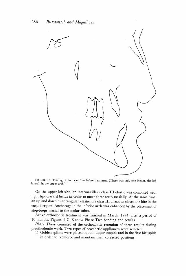
At this point, no fistula had appeared. This information is illustrated in
Figures 3-A and B and 4-A and B.
Phase Two was the achievement of good lateral occlusion by simultaneous
correction of the anterior openbite, the class II relationship on the right side, and
the class III relationship on the left side.
All inferior teeth except the second molars were banded and leveled. In
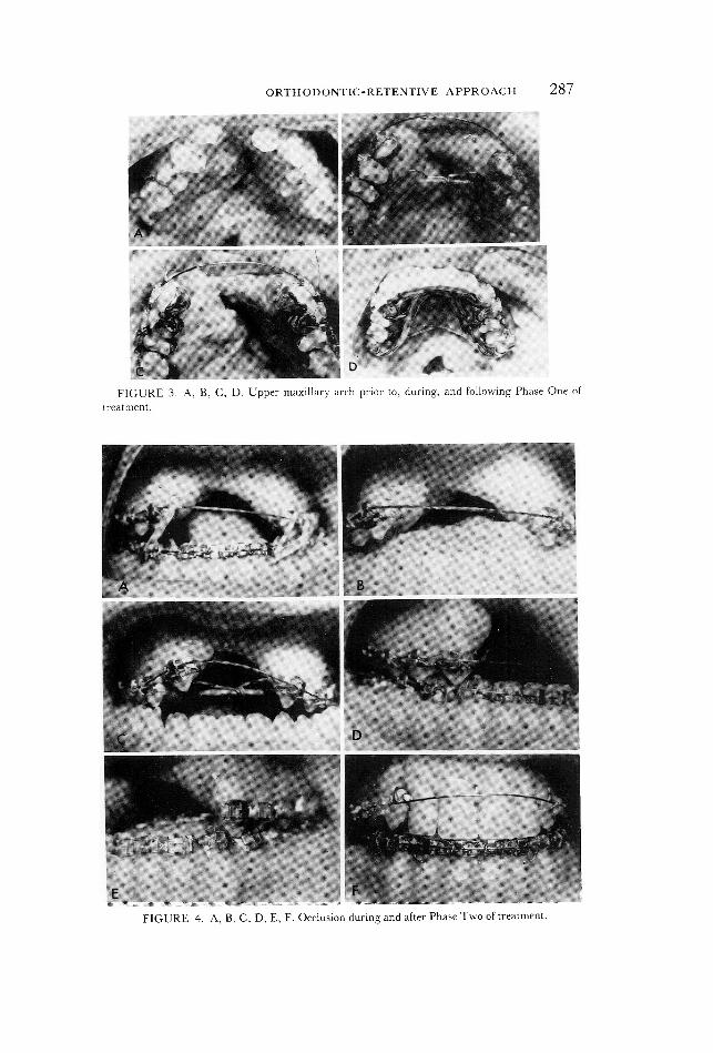
November, 1973, .020 up and down archwires were placed, and we started class
II and class III mechanics at the same time (Figure 4-C). No mandibular
anchorage preparation was done prior to the class II mechanics on the upper
right side. The distal movement of those teeth was achieved through the
combined use of light tip-back bends and the placement of a short class II elastic
(Schudy, 1973). This also closed the bite in the cuspid region.
286 Rutowitsch and Magalhaes -
FIGURE 2. Tracing of the head film before treatment. (There was only one incisor, the left
lateral, in the upper arch.)
On the upper left side, an intermaxillary class III elastic was combined withlight tip-forward bends in order to move these teeth mesially. At the same time,an up and down quadrangular elastic in a class III direction closed the bite in thecuspid region. Anchorage in the inferior arch was enhanced by the placement ofstop-loops mesial to the molar tubes.
Active orthodontic treatment was finished in March, 1974, after a period of10 months. Figures 4-C-E show Phase Two banding and results.Phase Three consisted of the orthodontic retention of these results during
prosthodontic work. Two types of prosthetic appliances were selected:1) Golden splints were placed in both upper cuspids and in the first bicuspids
in order to reenforce and maintain their corrected positions.
ORTHODONTIC-RETENTIVE APPROACH 287
FIGURE 3. A, B, C, D. Upper maxillary arch prior to, during, and following Phase One of
treatment.
FIGURE 4. A, B, C, D, E, F. Occlusion during and after Phase Two of treatment.
288 Rutowitsch and Magalhaes -
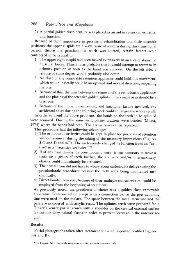
2) A partial golden clasp denture was placed as an aid in retention, esthetics,
and function.
Because of their importance in prosthetic rehabilitation and their unstable
positions, the upper cuspids are always cause of concern during this transitional
period. Before the prosthodontic work was started, certain factors were
considered to be crucial to:
1. The upper right cuspid had been moved extensively in an area of abnormal
muscular forces. Thus, it was probable that it would attempt to revert to its
primary position as soon as the band was removed. On the left side, a
relapse of some degree would probably also occur.
2. No clasp of any removable rétention appliance could hold this movement,
which would logically occur in an upward and inward direction, reopening
the bite.
3. Because of this, the time between the removal of the orthodontic appliances
and the placing of the retentive golden splints in the cuspid area should be a
brief one.
4. Because of the human, mechanical, and functional factors involved, any
accidental delay during the splinting work could endanger the whole result.
In order to avoid the above problems, the bands on the teeth to be splinted
were removed. During the same visit, plastic brackets were bonded (Miura,
1974) where the bands had been. The archwire was then replaced.
This procedure had the following advantages:
1) The orthodontic archwire could be kept in place for purposes of retentionwithout removal during the taking of the necessary impressions (Figures3-C and D and 4-F). The arch merely changed its function from an "ac-
tive" to a "retentive archwire."" *
2) If at any time during the prosthodontic work, it was necessary to move atooth or a group of teeth further, the archwire and/or intermaxillary
elastics could immediately be activated.
3) The dental team did not have to worry about undesirable delays during theprosthodontic procedures because the teeth were being maintained me-
chanically.
4) Direct bonded brackets, because of their multiple characteristics, could be
employed from the beginning of treatment.
As previously noted, the prosthesis of choice was a golden clasp removable
apparatus. Posterior action clasps with a connection bar at the post-damming
line were used on the molars. The space between the metal structure and the
palate was covered with acrylic resin. The splinted teeth were prepared for a
Tinker's veneer partial crown with a shoulder on the cervical external surface
for the auxiliary palatal clasps in order to prevent leverage in the anterior re-
gion.
Results
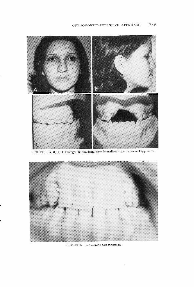
Facial photographs taken after treatment show an improved profile (Figures
- 5-A and B).
* In Figure 3-D, the arch was removed for esthetic reasons only.
ORTHODONTIC-RETENTIVE APPROACH 289
2 . LL
FIGURE 5. A, B, C, D. Photographs and dental casts immediately after removai of appliances.
FIGURE 6. Five months post-treatment.
290 Rutowitsch and Magalhaes |
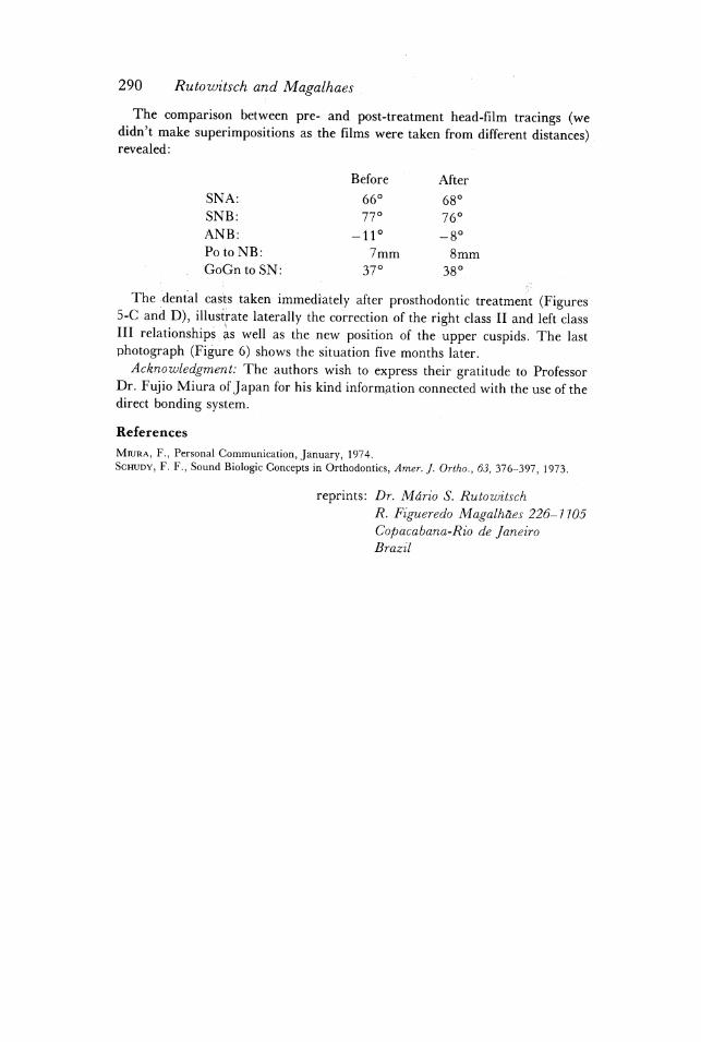
The comparison between pre- and post-treatment head-film tracings (wedidn't make superimpositions as the films were taken from different distances).revealed:
Before _ After
SNA: 66° 68°
SNB: 77° 76°
ANB: -11° -8°
Po to NB: 7mm 8mm
GoGn to SN: 37° 38°
The dental casts taken 1mmed1ately after prosthodontic treatment(Figures5-C andD), lllustrate laterally the correction of the right class II and left classIII relationships as well as the new position of the upper cuspids. The lastphotograph (Figure 6) shows the situation five months later.Acknowledgment: The authors wish to express their gratitude to Professor
Dr. Fujio Miura of Japan for his kind information connected with the use of thedirect bonding system.
References
Mura, F., Personal Communication, January, 1974.ScHupy, F. F., Sound Biologic Concepts in Orthodontics, Amer. J. Ortho., 63, 376-397, 1973.
reprints: Dr. Mdério S. Rutowitsch
R. Figueredo Magalh&es 226-1105
Copacabana-Rio de Janeiro
Brazil