an investigation of a novel anomalous pink feather ... · pdf file152 ©wildfowl &...
TRANSCRIPT

152
© Wildfowl & Wetlands Trust Wildfowl (2011) 61: 152–165
An investigation of a novel anomalous pinkfeather colouration in the Mute Swan Cygnus olor
in Britain and Ireland
MERITA M. O’CONNELL1*, USNA KEATING1, DEIRDRE MCELLIGOTT1, PADDY O’REILLY2,
JULIANNA O’CALLAGHAN2 & JOHN O’HALLORAN1
1School of Biological, Earth and Environmental Science, and Environmental ResearchInstitute, University College Cork, North Mall Campus Cork, Ireland.
2Department of Microbiology, University College Cork, Ireland.*Correspondence author. E-mail: [email protected]
Abstract
Pink feather colouration in normally white adult Mute Swans Cygnus olor is describedhere for the first time. Symmetrical salmon-pink colour was first evident on primaryfeather tips after moult (July–August), spread to secondary and tertiary remiges as theyear progressed, darkened as winter approached, and sometimes developed to a browncolour. Affected feathers tended to become brittle, fragmented and lose their ability torepel water. Surveys made at nine sites in Britain and Ireland between May 2003 andJanuary 2009 found 12–85% of swans with pink coloration. Highest prevalenceoccurred amongst flocks dependent on artificial food on eutrophic water bodies.Feather samples (white and pink), bill swabs and swabs of uropygial oil collected fromswans in the field, and also pink fungus isolated from a bread sample, were cultured andsubjected to high performance liquid chromatography (HPLC), to identify organismsand pigments respectively. Salmon-pink Chrysonilia sitophila fungus colonies developedon agars inoculated with samples from swans at Cork Lough and from the bread sample,but were absent from those inoculated with samples from swans at Lough Aderry andRostellan Lake, where the birds feed mainly on aquatic vegetation. HPLC revealed thatthe dominant pigments in pink feathers were generally consistent with those found inC. sitophila, indicating that C. sitophila is the most likely agent responsible for the pinkcolour on swan plumage. Field experiments implied that C. sitophila was not transferredto the plumage through contact with water; we therefore suggest that C. sitophila isacquired through exposure to contaminated food via the bill and is preened onto theplumage. A layer of environmental contaminants and debris that coats the plumage ofswans inhabiting eutrophic water bodies may provide a substrate for fungal growth.
Key words: Bread mould, carotenoids, Chrysonilia sitophila, Cygnus olor, Mute Swan,pigment, pink feathers, uropygial oil.

Mute Swans with pink plumage 153
© Wildfowl & Wetlands Trust Wildfowl (2011) 61: 152–165
The principle pigments in birds’ plumageare melanins, which produce black, grey andbrown feathers. Carotenoids – naturallyoccurring, organic, fat-soluble pigmentssynthesised by plants, algae, fungi andbacteria, which produce reds, oranges andyellows (Pettingill 1985) – also have beenreported in the plumage of several speciesincluding flamingos (Phoenicopteridae sp.),members of the pelican family (Pelecanidae
sp.) and a number of gulls and terns (Laridae
sp.) (Hudon & Brush 1990; McGraw &Hardy 2006). For instance, the pink hue ofthe Roseate Tern Sterna dougallii is obtainedfrom the carotenoid astaxanthin, stored inthe ovaries of their prey Sand EelsAmmodytes sp. (Hays et al. 2006), pink/redflamingo feathers originate from theapplication of pigmented uropygial oil(Grande et al. 2004), and Greater HornbillsBuceros bucornis similarly produce a yellowuropygial gland secretion that is preened onplumage areas that are used in threatdisplays (Reneerkens & Korsten 2004).Once formed, feathers lack any vascularsupply beneath their outer surface, socannot change colour or form exceptthrough fading, abrasion and staining, orthrough external treatment such as theapplication of pigmented uropygial oil.
Pigmented uropygial oil has not beendocumented in the Anatidae family andthere are no published records of pinkfeather colouration from any swan species,although a rusty brown staining on the headand neck may occur when swans frequentiron-rich waters (Birkhead & Perrins 1986).Unusual pink feathers were noted (but theobservation not published) in 2001 on MuteSwans at Cork Lough, a hyper-eutrophic,
shallow lake in the suburbs of Cork city, inthe south of Ireland (T. Kelly & C. Perrins,pers. comm.). Since then, anomalous pinkfeathering has been reported for swanselsewhere in Ireland, at sites in the UnitedKingdom and in Florida, United States ofAmerica.
This paper describes the prevalence ofpink feathers on Mute Swans Cygnus olor,particularly in Ireland but also at sites in theUK. The factors that influence thedistribution of the colouration on swans’plumage are assessed. For instance, weattempt not only to identify the agentresponsible for producing the pink colourbut to determine its source, as it may beobtained from the uropygial gland, the bill,or the environment. Whether the pigment isderived from microorganisms is alsoassessed. We hypothesise that spores ofbread fungus float on the water surface andattach to feathers of swans when they areswimming and washing.
Methods
Study areas
Cork Lough in the western suburbs of CorkCity, Ireland is a 6 ha hyper-eutrophic,shallow, freshwater lake regularly supportingover 70 Mute Swans (O’Halloran et al. 2002),including immature, non-breeding and post-breeding adults that frequently move tosurrounding sites (Keane & O’Halloran1992). Cork Lough wildfowl are highlydependent on bread and other artificial foodsupplied by the public, as aquatic vegetationis scarce (Irwin & O’Halloran 1997). Fieldobservations, feather samples, bill swabs anduropygial oil samples were collected at Cork

154 Mute Swans with pink plumage
© Wildfowl & Wetlands Trust Wildfowl (2011) 61: 152–165
Lough, and also at Rostellan Lake andLough Aderry, which are located in morerural east County Cork, where swans are lessdependent on bread. There is regularmovement of swans between Cork Loughand these sites (O’Halloran et al. 1995).
Field observations were also madeopportunistically at seven other sites inIreland and the UK. These were located inwest Cork, Wicklow, Dublin, Galway,Antrim, at Windsor in England and at theCosmeston Lakes in Wales.
Field observations
During July 2003–September 2004 and July2008–January 2009, Mute Swans were
attracted with fresh bread at each site, toprovide a clear view of their plumage.Flocks were scanned to quantify the numberof fully white swans and those with pinkcolour on their feathers (Fig. 1). Cygnetswere excluded from surveys as their darkplumage made it too difficult to ascertainwhether or not they had pink feathering.
Focal bird observations were undertakenat Cork Lough, at 10-min intervals per swan,for birds with and without pink plumage.This involved an examination of the extentand distribution of pink feathers on thebody of each bird. A total of 84 focal birdobservations were undertaken: 45 for pinkswans and 39 for non-pink swans. The
Figure 1. Symmetrical pink colour on Mute Swan feathers.
Mute Swans with pink plumage 155
© Wildfowl & Wetlands Trust Wildfowl (2011) 61: 152–165
extent to which there was pink colour on thehead/neck/breast area, the wing feathers,the tail feathers, the flank and the overallbody (i.e. the whole plumage) was recordedon site, and later verified by photographstaken of the individuals.
Microbiology: examination of feathers
Swans with and without the pink featherswere caught at Cork Lough, Lough Aderryand Rostellan Lake under licence from theNational Parks and Wildlife Service betweenJuly and December 2008. White, pink andbrown feathers were clipped from differentparts of the plumage (remiges, retrices,breast, flank and back), using surgical glovesand sterilised scissors, and then placed insterile, zip-lock, labelled bags (Table 1). Forswans with pink feathering, a sample of thepink, white and (if present) brown featherswere taken from each bird, brown feathersbeing associated with the late phase of the
condition. Within two days of collection,pink and non-pink feathers were placedseparately on selective media containingyeast extract glucose chlorophenicol (YGC)agar, tryptic soy agar (TSA) and sabourauddextrose agar (SDA), to promote the growthof fungi, bacteria and yeasts that may occuron the feathers (Table 2). All of the agarswere constituted in accordance with themanufacturer’s instructions.
Agars with feathers were kept at constanttemperature room (20°C) and monitoreddaily. Organisms producing colouredcolonies which showed similarity to the pinkcolour of the pink feathers observed onswans were isolated using a sterile loop. Theloop was then gently inoculated over theagar that best promoted its growth,incubated at 20°C for 1–2 weeks, andmonitored daily.
To investigate whether the isolate coulduse different feather types as a substrate, a
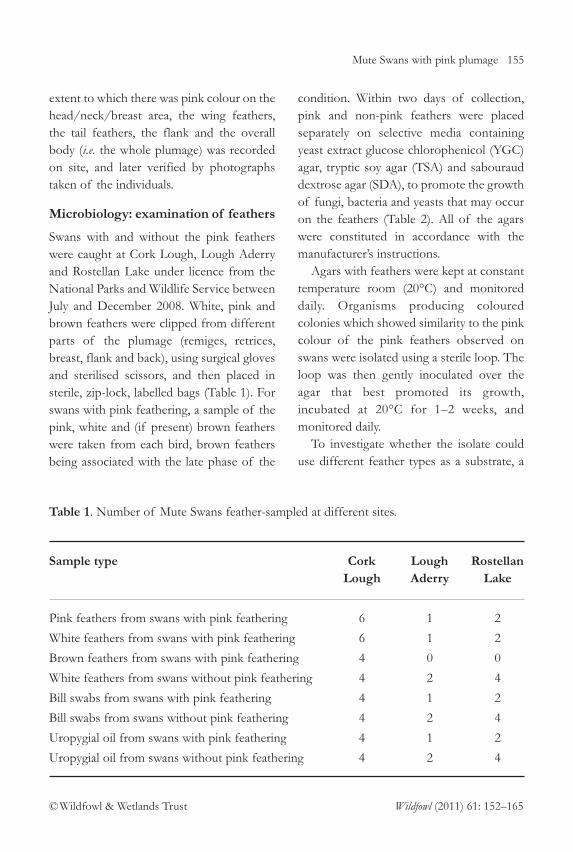
Table 1. Number of Mute Swans feather-sampled at different sites.
Sample type Cork Lough Rostellan Lough Aderry Lake
Pink feathers from swans with pink feathering 6 1 2
White feathers from swans with pink feathering 6 1 2
Brown feathers from swans with pink feathering 4 0 0
White feathers from swans without pink feathering 4 2 4
Bill swabs from swans with pink feathering 4 1 2
Bill swabs from swans without pink feathering 4 2 4
Uropygial oil from swans with pink feathering 4 1 2
Uropygial oil from swans without pink feathering 4 2 4

156 Mute Swans with pink plumage
© Wildfowl & Wetlands Trust Wildfowl (2011) 61: 152–165
number of clean, white feathers wereautoclaved for 15 min at 121°C. Loops of the cultured isolates were inoculated onto the agar that best promoted theirgrowth and the autoclaved feathers weresubsequently placed into these agars usingsterilised forceps. Feathers and isolates
were then monitored over a 6-monthperiod.
Pink fungal colonies resembling thecolour of pink-feathered swans wereobserved by chance on bread before itspackaging had been opened. Isolates fromthis pink bread fungus were placed in YGC
Table 2. Number of YGC agars inoculated with different sample types (e.g. pink feathers,white feathers, bill swabs, uropygial oil swabs) from swans with and without the pink feathercolouration. YGC agars are only included in microbiological analysis of some sample types,as it became evident that no pink colonies were observed in any of the other agars followinginoculation of feathers and bill swabs.
Sample type Cork Lough Rostellan Lough Aderry Lake
No. of YGC agars inoculated with pink feathers from swans with pink feathering* 24 4 8
No. of YGC agars inoculated with white feathers from swans with pink feathering 12 2 4
No. of YGC agars inoculated with brown feathers from swans with pink feathering 8 0 0
No. of YGC agars inoculated with white feathers from swans without pink feathering* 16 8 8
No. of YGC agars inoculated with bill swabs from swans with pink feathering* 12 3 6
No. of YGC agars inoculated with bill swabs from swans without pink feathering* 12 6 8
No. of YGC agars inoculated with uropygial oil from swans with pink feathering 12 3 6
No. of YGC agars inoculated with uropygial oil from swans without pink feathering 12 6 12
No. of YGC agars inoculated which were attached to the experimental station 16 0 0
* = Sample types that were inoculated over same number of TSA and STA agars.

Mute Swans with pink plumage 157
© Wildfowl & Wetlands Trust Wildfowl (2011) 61: 152–165
agar with the autoclaved feathers andincubated at 20°C for six months andmonitored regularly to ascertain whetherthis fungus could use the feather surface as asubstrate.
Microbiology: examination of bill,uropygial oil and bread samples
Bill and uropygial oil swabs were collected inthe field from swans at Cork Lough,Rostellan Lake and Lough Aderry (Table 1).Bill swabs were collected by rubbing a sterilecotton bud along the inside of the swan’sbill. Uropygial oil swabs were obtained byrubbing the wick (a system of feathers thatserve to elongate the uropygial gland) andthe aperture of the uropygial gland withsterilised cotton buds. These cotton budswere then placed in sterile, labelledcontainers. Like the feather samples, swabsfrom the swans’ bills and wicks wereinoculated onto the selective media andincubated at 20°C for 1–2 weeks. Pinkcolonies that grew from these selective agarswere again isolated, using the proceduredescribed above.
Water monitoring
To test the possibility that the pink feathercolouration was caused by staining from anagent in or on the water surface, andwhether different feather types may be more prone to staining than others, anexperimental station involving a system offloats was constructed at Cork Lough.Autoclaved, white, breast, wing and tailfeathers, clipped from swans, were attachedat a level which ensured the feathers were positioned at the water-air interface.Feathers were also positioned under the
floats so that they remained fully submergedunder water. Furthermore, as a control,feathers were positioned above the watersurface. The feathers, deployed on thestation in October 2008, were examinedregularly to see whether they developed pinkcolour. After 6 weeks, feather clippings weretaken using sterilised scissors, inoculatedonto YGC, TSA and SDA agars, thenincubated at 20°C and monitored daily tosee whether they became contaminated byorganisms that produced pink colonies(Table 2).
Microscopy
In the laboratory, white, pink and brownfeathers sampled from swans with andwithout the pink feather colouration were examined and compared under amicroscope. Some pink feathers were left indry sealed bags at room temperature for 12months. These feathers were subsequentlyexamined under a microscope for anystructural changes and colour degradation.Feather samples supplied from two MuteSwans in Orlando, Florida, USA, wheresimilar pink feathers had been observed,were also examined under a microscope tosee whether the same agent was responsible.
Analysis of pigments
Samples of white and pink feathers andsamples of fungus isolated from feathersand bread were analysed using highperformance liquid chromatography (HPLC). The feathers were washed in ethanolfollowed by hexane to remove surface lipids;0.1 mg samples were then mixed with 300 lof 50% potassium hydroxide (w/v) and 2 mlof ethanolic pyrogallol (1%, w/v) in 10 ml

158 Mute Swans with pink plumage
© Wildfowl & Wetlands Trust Wildfowl (2011) 61: 152–165
screw-top Pyrex tubes fitted with teflonlined screw caps. Tubes were kept at 70ºCfor 30 min. When cool, 1ml of water and4ml of hexane were added. Fresh pinkfungus isolated from bill swabs, and an older(four month old) pink fungus isolated frombread, were treated similarly to the feathers.
The tubes were shaken vigorously andcentrifuged at 2,500 rpm for 10 min. Theupper hexane layer was removed and theextraction repeated with 2 ml of hexane.Combined hexane extracts were dried under nitrogen, re-dissolved in 200 µlcetonitile:methanol:dichloromethane (140:40:20) and transferred to a HPLC vial(Buttriss & Diplock 1984).
A HPLC system consisting of an LC-10AD pump, SIL-10A autoinjector, SCL-10A system controller and SPD-6AVdetector set at 450 nm (ShimadzuCorporation, Kyoto, Japan) was used toanalyse the samples. The column systemconsisted of a Spherisorb ODS-2 C18 5µmPEEK guard column (150 × 4.6 mm) and a Spherisorb ODS-2 C18 5µm PEEK guard cartridge (Alltech Associates AppliedScience Ltd), connected to a Vydac201TP54 (250 × 4.6 mm) reversed phaseC18 column (Grace Davison DiscoverySciences, IL, USA). Column temperaturewas maintained at 25°C using an Alltechcolumn water jacket with a thermostaticallycontrolled water bath (Lauda RM6 T; Lauda Dr. R. Wobser GmbH & Co. KG,Lauda-Königshofen, Germany). The mobilephase consisted of acetonitrile, methanol,dichloromethane (75:20:5) with 0.1% BHTand 0.05% triethylamine. The methanolcontained 0.05M ammonium acetate. Theflow rate was 1.5 ml/min. Chromatograms
were recorded using Millenium 32 (version3.05.01) Chromatography Manager Software (Waters Corp. MA, USA). The output wasrecorded as a series of peaks, the retentiontime characterising and hence differentiatingeach pigment that passed through theinstrument, and the area under the peakindicating the quantity of the pigmentpresent. The resulting chromatographs werecompared to see if there were any pigmentsin common between the fungus and thepink feathers.
Data analysis
Kruskal-Wallis tests were used to determineany differences between sites in thepercentage of pink colour recorded ondifferent parts of the body for each swan.All statistical calculations were carried outusing Minitab (Pennsylvania University,USA) and Microsoft Excel 2007 software.
Results
Field observations
Pink feather colouration was observed onthe plumage of swans at all study sites inIreland and the UK (Table 3). At CorkLough, where Mute Swan numbers variedfrom 73–120, the mean proportion of birdswith the pink feather colouration was 85%,suggesting that pink feather colouration iscommon and widespread on swans.
Focal examination of individual swansshowed that the pink feathering occurredacross the birds’ plumage, but wasparticularly evident on the remiges (wingfeathers) and retrices (tail feathers) (Table 4).The percentage of pink coverage recordedon different parts of the body (i.e. the

Mute Swans with pink plumage 159
© Wildfowl & Wetlands Trust Wildfowl (2011) 61: 152–165
head/neck/breast area, the wing feathers, thetail feathers, the flank and the overall body)differed significantly between sites (Kruskal-Wallis tests: Hhead/neck/breast = 35.14, d.f. = 6,P < 0.05; Hremiges = 68.11, d.f. = 6, P <0.05,df=6; Hretrices = 52.45, d.f = 6, P <0.05; Hflank
= 38.48, d.f = 6, P <0.05; Hoverall body = 60.62,d.f. = 5, P <0.05) (Table 4).
Each year, the pink feathering appearedas a salmon-pink colour on the plumagebetween July and August, but darkened overtime, and on some individuals progressed toa browner colour over the winter months.The feathers of these individuals appearedto become brittle and to lose their waterrepellence. In some cases, the plumage ofthese birds became completely water-
logged, which was noted when the birdswere caught by hand.
Inspection of feather samples
All YGC, TSA and SDA agars inoculatedwith pink and white feathers clipped from swans at Cork Lough displayedmicroorganism colonies after a week. Onlysalmon-pink fungus restricted to YGC agarsproduced a colour similar to the pink feathercolouration and was identified as Chrysonilia
sitophila on all 24 YGC agars inoculated with pink feathers sampled from swans atCork Lough. This microorganism was alsoidentified on all 28 YGC agars inoculatedwith white feathers sampled from swans withand without the pink feather colouration at
Table 3. Mean number of Mute Swans present (± s.d.) and the proportion of Mute Swanswith pink feathers across a range of sites in Britain and Ireland. (Site information fromO’Connell 2007.)
Location Site type Mean % number of Population
swans with pink (± s.d.) feathers
Cork Lough Freshwater lake, hyper eutrophic 95 (±9.6) 85
East Cork Freshwater lake, mildly eutrophic 40 (±12.6) 60
West Cork Tidal estuarine, not eutrophic 44 (±14.2) 20
Galway Tidal estuarine, moderately eutrophic 121 (±28.6) 76
Dublin Tidal estuary, moderately eutrophic 75 (±0) 40
Wicklow Tidal estuary, moderately eutrophic 58 (±0) 48
Antrim, N. Ireland Freshwater lake, mildly eutrophic 12 (±0) 92
Windsor, England Freshwater lake, mildly eutrophic 50 (±0) 30
Cosmeston Lakes, Wales Freshwater lake, mildly eutrophic 65 (±0) c. 55

160 Mute Swans with pink plumage
© Wildfowl & Wetlands Trust Wildfowl (2011) 61: 152–165
Cork Lough. This fungus was easilydistinguished by its initial pale pink floccosegrowth and subsequent salmon colour asspore formation occurred. Colonies coveredentire Petri dishes, reaching the lid in tuftsand shedding profuse salmon conidia at therim (Pitt & Hocking 2009). Brown feathersassociated with the advanced phase of thecondition, and sampled from swans in CorkLough in winter, did not yield C. sitophila
after their inoculation to YGC agar. Thefungus isolated from the bread sample wasalso identified as C. sitophila.
All YGC agars inoculated with pink andwhite feathers samples taken from threeswans at Lough Aderry and six swans atRostellan Lake displayed colonies ofmicroorganisms after a period of one week.
Unlike the YGC agars inoculated withfeathers sampled from swans at CorkLough, none of the 34 YGC agarsinoculated with feathers sampled fromswans at Lough Aderry or Rostellan Lakeyielded colonies of C. sitophila, nor any othermicroorganism that appeared pink in colour.
Investigating the source of thecolouration
Examination of uropygial swabs. Swabs ofuropygial oil showed no pink colour in theoil. Moreover, none of the 24 YGC agarsinoculated with uropygial oil swabs takenfrom pink and non-pink swans from CorkLough displayed C. sitophila growth, or anyother pink microorganism that might beassociated with uropygial oil. Similarly, none
Table 4. Mean (± s.e.) % pink colouration on different parts of the body for swans inspectedat a range of sites across Britain and Ireland.
Sites No. of Head/ Remiges Retrices Flank Full body swans neck/ view
observed breast
Cork Lough 84 7.8 ± 11.7 30.6 ± 24.0 29.3 ± 23.6 6.9 ± 10.4 20.8 ± 17.2
East Cork 21 0.8 ± 1.5 2.1 ± 27.0 5.6 ± 9.8 0.2 ± 0.9 3.2 ± 5.3
West Cork 9 0 2.4 ± 40.0 0.5 ± 1.1 0 1.4 ± 2.5
Galway 30 6.9 ± 4.8 34.4 ± 25.0 33.1 ± 24.9 14.3 ± 18.3 23.2 ± 14.4
Limerick 5 0 15 ± 17.2 22.4 ± 30.7 0 *
Dublin 8 0 7.1 ± 10.8 16.9 ± 9.2 0 10.2 ± 9.6
Wicklow 5 1 ± 2.3 12.4 ± 16.1 15 ± 14.6 0.2 ± 0.4 11 ± 11.0
Total 162 2.3 ± 5.5 31.2 ± 28.0 24.9 ± 22.8 2.3 ± 5.2 18.4 ± 16.9
* = no information available.
Mute Swans with pink plumage 161
© Wildfowl & Wetlands Trust Wildfowl (2011) 61: 152–165
of the 27 YGC agars inoculated withuropygial oil swabs taken from swans atLough Aderry and Rostellan Lake showedgrowth by C. sitophila or any other pinkmicroorganism. These findings indicate thatthe uropygial gland was not the source ofthe pink feather colouration.
None of the feathers attached to thestation and left floating in or on the surfaceof Cork Lough became pink over time.Clippings of these feathers inoculated onto16 YGC agar yielded no C. sitophila, nor anyother organism with a colour similar to thaton the pink feathers.
Examination of bill swabs. All agars inoculatedwith bill swabs taken from Cork LoughMute Swans developed colonies ofmicroorganisms after one week. All 24 YGCagars inoculated with bill swabs sampledfrom swans with and without pink feathersat Cork Lough developed growths of C. sitophila. No other pink colouredmicroorganism was identified in these agars.
All agars inoculated with bill swabs takenfrom Mute Swans with and without pinkfeathers at Lough Aderry and Rostellan Lakealso displayed colonies of microorganismafter one week. None of the 23 YGC agarsinoculated with these bill swabs yieldedcolonies of C. sitophila, nor any othermicroorganism that appeared pink in colour.
Detailed feather analysis. Differences betweenwhite feathers and feathers with pinkcolouring were evident under microscopicexamination. Pink feathers sampled fromswans in Florida appeared identical to thosefrom swans at Cork Lough, indicating thatthe Florida swans were affected in the same
way. The pink feathers tended to have largeaccumulations of an unknown material onthem compared with clean, white feathers.These accumulations appeared to clog thebarbules and to deactivate the ability ofbarbules to grip and interlock with adjacentbarbules. It is likely that this unknownmaterial is directly or indirectly responsiblefor the pink feather colour.
C. sitophila smeared onto clean, whitefeathers in the laboratory showed a pinkcolour after one week identical to thatobserved on the plumage of swans seen inthe field. Feathers left for 12 months in thelaboratory, with C. sitophila on their surface,showed no sign of physical degradation.The autoclaved feathers placed in YGC agarinoculated with C. sitophila showed no signsof physical degradation either, despite beingleft within an enclosed petri-dish at roomtemperature for a period of six months. C. sitophila left in petri dishes in a fridge for a period of four months showedconsiderable fading in comparison tofresher, more recently grown C. sitophila.
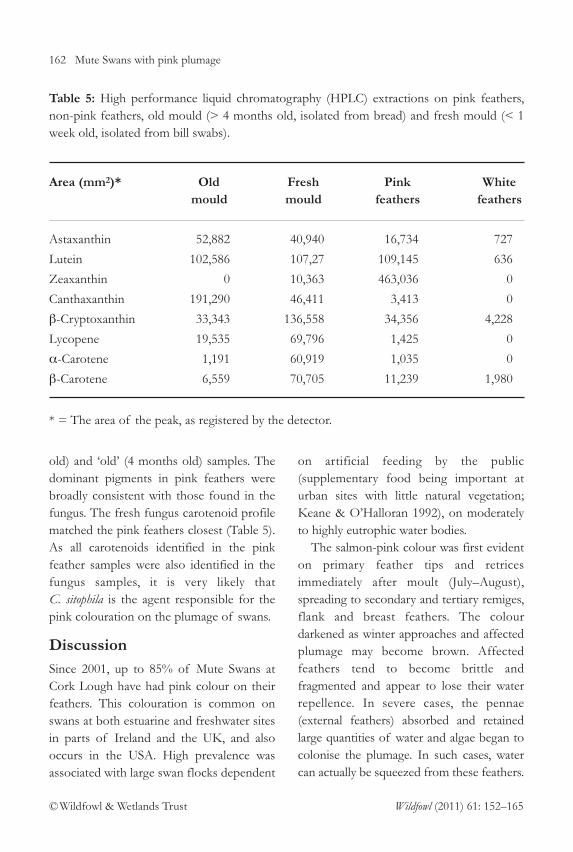
Pigment identification. HPLC extraction forfour pink feathers taken from differentswans at Cork Lough identified severalcarotenoids in the samples (Table 5).Carotenoids were identified by comparisonof retention times with authentic carotenoidstandards. The most common pigments inpink feathers were zeaxanthin, lutein, β-cryptoxanthin, astaxanthin, and β-carotenerespectively (Table 5). Some white feathersalso had low background levels ofcarotenoids (Table 5). These carotenoidswere also present in the fungus samples withsome differences between ‘fresh’ (< 1 week
162 Mute Swans with pink plumage
© Wildfowl & Wetlands Trust Wildfowl (2011) 61: 152–165
old) and ‘old’ (4 months old) samples. Thedominant pigments in pink feathers werebroadly consistent with those found in thefungus. The fresh fungus carotenoid profilematched the pink feathers closest (Table 5).As all carotenoids identified in the pinkfeather samples were also identified in thefungus samples, it is very likely that C. sitophila is the agent responsible for thepink colouration on the plumage of swans.
DiscussionSince 2001, up to 85% of Mute Swans atCork Lough have had pink colour on theirfeathers. This colouration is common onswans at both estuarine and freshwater sitesin parts of Ireland and the UK, and alsooccurs in the USA. High prevalence wasassociated with large swan flocks dependent
on artificial feeding by the public(supplementary food being important aturban sites with little natural vegetation;Keane & O’Halloran 1992), on moderatelyto highly eutrophic water bodies.
The salmon-pink colour was first evidenton primary feather tips and retricesimmediately after moult (July–August),spreading to secondary and tertiary remiges,flank and breast feathers. The colourdarkened as winter approaches and affectedplumage may become brown. Affectedfeathers tend to become brittle andfragmented and appear to lose their waterrepellence. In severe cases, the pennae(external feathers) absorbed and retainedlarge quantities of water and algae began tocolonise the plumage. In such cases, watercan actually be squeezed from these feathers.
Table 5: High performance liquid chromatography (HPLC) extractions on pink feathers,non-pink feathers, old mould (> 4 months old, isolated from bread) and fresh mould (< 1week old, isolated from bill swabs).
Area (mm2)* Old Fresh Pink White mould mould feathers feathers
Astaxanthin 52,882 40,940 16,734 727
Lutein 102,586 107,27 109,145 636
Zeaxanthin 0 10,363 463,036 0
Canthaxanthin 191,290 46,411 3,413 0
β-Cryptoxanthin 33,343 136,558 34,356 4,228
Lycopene 19,535 69,796 1,425 0
α-Carotene 1,191 60,919 1,035 0
β-Carotene 6,559 70,705 11,239 1,980
* = The area of the peak, as registered by the detector.

Mute Swans with pink plumage 163
© Wildfowl & Wetlands Trust Wildfowl (2011) 61: 152–165
The hypothesis that swans with pinkfeathers produce pink coloured uropygial oil was rejected since uropygial oil samples from these swans contained no pink pigment nor gave rise to pinkcoloured colonies of microorganisms afterinoculation onto selective agars. Stainingagents in the water inhabited by swans werealso rejected as a potential source, becauseautoclaved feathers positioned above, onand below the water surface of Cork Loughshowed no pink colouration after six weeks.
The fungus C. sitophila was present on allpink feather samples obtained at CorkLough and HPLC extraction indicated thatcarotenoids present in C. sitophila are alsopresent in pink feathers. Pink feathers wereless prevalent among swans at LoughAderry and Rostellan Lake, where swans areless reliant on bread than those at CorkLough, suggesting that the presence of C. sitophila in the bills of swans from CorkLough could be associated with theiringesting large quantities of bread. Pink C. sitophila fungus was cultured from breadleft in a cupboard for some time and hasbeen commonly reported on bread, pastries,nuts and maize (Pitt & Hocking 2009).Large amounts of “waste” bread areprovided regularly to Cork Lough swans andexposure to C. sitophila contaminated breadlikely explains the prevalence of C. sitophila
in bill swabs taken for swans at the site, fromwhere it could be applied rapidly to feathersduring preening. Alternatively, swans mayinadvertently preen bread material withoutfungus onto the plumage, forming asubstrate for C. sitophila establishment fromcontact with the bills or feathers ofconspecifics affected by the condition, or
through airborne spores landing on thesubstrate.
Swans regularly move between CorkLough, Lough Aderry and Rostellan Lake,potentially explaining the presence of pinkfeathers on some swans at Lough Aderry andRostellan Lake. Swans initially contracting the colour at Cork Lough could transport the fungus elsewhere, as the colour did not disappear from feathers kept in thelaboratory for up to one year. The fungus wasnot active on feathers from swans at LoughAderry of Rostellan Lake; pink feathers fromthese swans yielded no C. sitophila after theirinoculation to selective agars. C. sitophila
maintained at constant room temperature forfour months showed considerable fadingcompared to C. sitophila less than one weekold and did not grow on fresh agars,indicating pigment persistence after the deathof the organisms. This would explain whypink feathers from swans in Lough Aderryand Rostellan Lake did not yield C. sitophila
after inoculation onto YGC agar. Assuming that C. sitophila is the agent
responsible for the pink colouration, it maybe more prevalent on certain feathers,depending on their location on the swan.The condition appeared to be confined topennae (outer feathers), confirmed byinspecting birds in the hand, perhapsbecause of more regular wetting of thesefeathers by the eutrophic waters upon whichthe swans occur.
Fungi have previously been recorded onbirds’ plumage. Beer & Kear (1975) reportedthat Cladosporium herbarum may act as apathogen on the plumage of captiveflamingos, and mentioned that plumage onthe flamingos’ backs may become “dingy

164 Mute Swans with pink plumage
© Wildfowl & Wetlands Trust Wildfowl (2011) 61: 152–165
and frayed” and “wet and miserablelooking”. They also suggested that a birdwashing itself in water contaminated withfood and faeces would result in materialadhering to the feather surface. Thisenriched feather surface would provide asubstrate on which initial fungal growth ispossible. There is some similarity betweenthe fungus C. sitophila, identified on theplumage of swans in the current study, and the fungus C. herbarum described on the plumage of flamingos by Beer & Kear (1975). The symptoms of featherdegradation are similar in both cases and C. sitophila resembles C. herbarum in that it iscommon on organic debris and producesmany airborne spores. The spores of C. sitophila are also heavily pigmented and donot seem to succumb to light either.Furthermore, C. sitophila is opportunistic andcan also grow on weakly nutritive substrates.
Microscopic examination of pink feathersrevealed heavier debris accumulation compared to healthy white feathers. Thisdebris clogged barbules and reduced theirability to grip and interlock with adjacentbarbules. Breakage is likely the result fromgeneral environmental contact and frictionbetween feathers, such as during preening,resulting in water penetration and retentionwithin damaged regions of plumage andenabling algal growth. Subsequent algaldecomposition in winter could explain laterplumage browning. The likelihood of thiswaterlogged environment being unsuitablefor C. sitophila is supported by absence ofthe fungus from brown feathers sampled atCork Lough and inoculated over YGC agar.
In conclusion, it would appear that C. sitophila is cause of the pink colouration
in Mute Swans. C. sitophila does not appearto cause direct feather degradation, butappears to be associated with a contaminantfilm (potentially food and faeces suspendedin eutrophic waters) that adhere to thefeather surface, as a medium for growth.Preening of contaminated feathers couldspread C. sitophila to other parts of theplumage, potentially explaining the presenceof C. sitophila in all samples taken from bills of swans at Cork Lough and thesymmetrical distribution of the colour onthe plumage. More detailed studies arerequired to determine whether pink-feathered swans suffer health or behaviouraleffects that could affect fecundity andsurvival rates compared to white swans, andhence affect management recommendationsfor swans at the sites concerned.
Acknowledgements
This work was supported by the HigherEducation Authority of Ireland. We wish to thank all the technical staff from thevarious departmental laboratories (includingthose from the Chemistry, Microbiologyand Zoology/Ecology Departments) atUniversity College Cork who assistedthroughout this study.
References
Beer, J. & Kear, J. 1975. Fungal infections of theplumage. In J. Kear & N. Duplaix-Hall (eds.),Flamingos. T. & A.D. Poyser, Berkhamsted, UK.
Birkhead, M. & Perrins, C. 1986. The Mute Swan.Croom Helm, London, UK.
Buttriss, J.L. & Diplock, A.T. 1984. Highperformance liquid chromatography methods for vitamin E in tissues. Methods in Enzymology
105: 131–138.

Mute Swans with pink plumage 165
© Wildfowl & Wetlands Trust Wildfowl (2011) 61: 152–165
Hays, H., Hudon, J., Cormons, G., Dicostanzo, J.& Lima, P. 2006. The pink feather blush ofthe Roseate Tern. Waterbirds 29: 296–301.
Hudon, J. & Brush, A.H. 1990. Carotenoidsproduce flush in the elegant tern plumage.The Condor 92: 798–801.
Irwin, S. & O’Halloran, J. 1997. The winteringbehaviour of Coot Fulica atra L. at CorkLough, south-west Ireland. Biology and
Environment: Proceedings of the Royal Irish
Academy 97B (2): 157–162.
Keane, E. & O’ Halloran, J. 1992. The behaviourof a wintering flock of Mute Swans Cygnus
olor in Southern Ireland. Wildfowl 43: 12–19.
McGraw, K.J. & Hardy, L.S. 2006. Astaxanthin isresponsible for the pink plumage flush inFranklin’s and ring-billed gulls. Journal of Field
Ornithology 77: 29–33.
O’Connell, M. 2007. Lead in Mute and WhooperSwans in Ireland: trends and patterns.Unpubl. Ph.D. Thesis, National University ofIreland, Cork, Republic of Ireland.
Pettingill, O. 1985. Ornithology in Laboratory and
Field. Academic Press, London, UK.
Pitt, J.I. & Hocking, A.D. 2009. Fungi and Food
Spoilage. 3rd Edition. New York: Springer.
Photograph: Cork Lough landscape, by Mark Carmody.
Photograph: Mute Swan displaying at Cork Lough, by Mark Carmody.