an investigation into the role of platelet … · and inflammation in coronary artery disease dr...
TRANSCRIPT

1
AN INVESTIGATION INTO THE ROLE OFPLATELET – MONOCYTE INTERACTION
AND INFLAMMATION IN CORONARYARTERY DISEASE
DR BIKASH MAJUMDER
MBBS, MRCP (UK)
Thesis submitted for the degree of MD (Res)
Department of Cardiology and Department of
Haematology, Royal Free Campus
University College London (UCL)
JANUARY 2012

2
DECLARATION
I Bikash Majumder confirm that the work presented in this thesis is my own.Help
and contribution of others to this work are specified in the acknowledgement
section. Where information has been derived from other source, I confirm that
this has been indicated in the thesis .
Bikash Majumder
January 2012

3
ABSTRACT
Introduction: -
Platelet monocyte complex (PMC) formation has been widely reported as a
marker of platelet activation in vascular disease states and several studies have
shown heightened systemic expression of PMC in stable and acute coronary
disease. However, the relationship between intracoronary platelet and
monocyte activation status and local intracoronary inflammation in acute
coronary syndrome (ACS) remains unclear.
Method:-
Fifteen ST elevation myocardial infarction (STEMI), 8 non ST elevation
myocardial infarction (NSTEMI) and 7 stable angina patients were recruited.
PMC, P selectin positive PMC (activated platelet within the complex), tissue
factor (TF) positive PMC (activated monocyte within the complex) were
estimated with flow cytometry from blood samples aspirated from the coronary
artery (distal to the lesion), aorta and right atrium . Plasma CRP, SAA, TNF –
alpha and IL-6 were also measured.
Results:-
In ACS patients no significant transculprit lesion gradient of PMC expression was
observed but significant gradients were found with P-selectin positive PMC (p=

4
0.01) and TF positive PMC expression (p=0.04). Overall median P- selectin
positive PMC expression in ACS patients was significantly higher compared to
stable angina (p= 0.006). Intracoronary P-selectin positive PMC was also found
to be higher in the ACS group compared to stable group (p=0.003). Overall
median CRP (p= 0.001), SAA (p=0.0007) and IL-6 (p= 0.03) levels were
significantly higher in ACS.
In STEMI intracoronary PMC correlated positively with intracoronary TNF-alpha
(r= 0.68, p= 0.03). Positive correlation was also observed between intracoronary
TF positive PMC (% monocyte) with TNF-alpha and IL-6 (r=0.66, p=0.05 &
r=0.71, p= 0.05 respectively).
Conclusion:-
The work outlined in this thesis has demonstrated the importance of platelet and
monocyte activation status of the PMC as a determinant of intracoronary
inflammation. Beyond a local intracoronary role, PMC may contribute to systemic
inflammation through P-selectin expression and local intracoronary inflammation
through increased P-selectin and tissue factor expression.

5
To my wife Jayita,my son Ishaan and my parents.

6
ACKNOWLEDGEMENTS
I would like to thank my supervisor Dr Roby D Rakhit and my co supervisor Dr
Mark W Lowdell for their constant support, guidance and encouragement
throughout my entire research period.
I would also like to thank the following people for their assistance during my
research period.
Dr John G Coghlan for his valuable advice and for his help in recruiting patients.
Dr ManFai Shiu for his constant encouragement, guidance and for his help in
recruiting patients.
Dr John Hurst and Mr Ray Sapsford for their support in performing the ELISA
assays.
Dr Helen Lachmann and Ms Ruth Gallimore for their support in performing the
CRP and SAA assays.
Ms Janet North for helping me in performing flow cytometry.
Ms Colette Smith for providing statistical advice.
I would also like to thank all the staff nurses of the cardiac catheterisation
laboratory and all the technical staffs of the cardiology department of Royal Free
Hospital for their constant support.
I would also like to thank the patients who kindly participated in this study.

7
TABLE OF CONTENTS. PAGE
Title Page............................................................................1
Declaration.......................................................................... 2
Abstract...............................................................................3
Acknowledgement...............................................................6
Table of Contents................................................................7
Abbreviations......................................................................15
List of Figures.....................................................................18
List of Tables.......................................................................20
Publications.........................................................................21
8
CHAPTER 1: INTRODUCTION
1.1INTRODUCTION................................................................................................. 25
1.2 PLATELET – MONOCYTE COMPLEXES......................................................... 25
1.2.1 PATHOPHYSIOLOGIC SIGNIFICANCE OF PLATELET- MONOCYTE COMPLEX
FORMATION .................................................................................................................................................. 25
1.2.2 CLINICAL SIGNIFICANCE OF PMCS IN CORONARY ARTERY DISEASE ............................. 29
1.2.3 PMC FORMATION; SYSTEMIC OR LOCAL INTRACORONARY PHENOMENON? .............. 32
1.2.4 METHODOLOGICAL CONSIDERATIONS OF PMC ESTIMATION WITH FLOW
CYTOMETRY ................................................................................................................................................. 34
1.3 ROLE OF TISSUE FACTOR IN CORONARY ARTERY DISEASE................... 37
1.4 PSELECTIN ....................................................................................................... 39
1.4.1 ROLE OF P SELECTIN IN CORONARY ARTERY DISEASE ...................................................... 39
1.4.2 INFLAMMATION AND SOLUBLE P- SELECTIN ........................................................................... 41
1.5 ROLE OF INFLAMMATION IN THE PATHOGENESIS OF ACUTE
CORONARY SYNDROME....................................................................................... 42
1.5.1 INTERRELATIONSHIP BETWEEN INFLAMMATION AND PMC FORMATION ...................... 42
1.5.2 INFLAMMATION - SYSTEMIC OR INTRACORONARY EVENT?............................................... 46
1.6 INFLAMMATORY MARKERS IN CORONARY ARTERY DISEASE ................ 49
1.6.1 ROLE OF CRP IN CORONARY ARTERY DISEASE ..................................................................... 49
1.6.2 ROLE OF SAA IN CORONARY ARTERY DISEASE ..................................................................... 50
1.6.3 ROLE OF TNF- ALPHA IN CORONARY ARTERY DISEASE ...................................................... 53
1.6.4 ROLE OF IL-6 IN CORONARY ARTERY DISEASE....................................................................... 54
1.7 OBJECTIVES..................................................................................................... 57

9
CHAPTER 2: MATERIAL AND METHODS
2.1 GENERAL METHODS....................................................................................... 60
2.1.1 PATIENTS............................................................................................................................................. 60
2.1.1.1 STEMI PATIENTS ........................................................................................................................ 60
2.1.1.2 NSTEMI PATIENTS ..................................................................................................................... 61
2.1.1.3 STABLE ANGINA PATIENTS ................................................................................................... 61
2.1.2 CORONARY INTERVENTION........................................................................................................... 62
2.2 FLOW CYTOMETRY ......................................................................................... 63
2.2.1 COLLECTION OF BLOOD SAMPLE FOR FLOW CYTOMETRY ................................................ 63
2.2.2 PARAMETERS MEASURED BY FLOW CYTOMETRY ................................................................ 64
2.2.4 FLOW CYTOMETRY TECHNIQUE................................................................................................... 65
2.3 ESTIMATION OF INFLAMMATORY PARAMETERS AND SOLUBLE P
SELECTIN ............................................................................................................... 69
2.3.1 COLLECTION AND PREPARATION OF BLOOD SAMPLES FOR ESTIMATION OF
CRP, SAA, TNF- ALPHA, IL-6 AND SOLUBLE P SELECTIN ............................................................... 69
2.3.2 ELISA..................................................................................................................................................... 70
2.3.2.1 ESTIMATION OF TNF- ALPHA AND IL-6 ............................................................................... 70
2.3.2.2 ESTIMATION OF SOLUBLE P SELECTIN ............................................................................. 72
2.3.3 ESTIMATION OF HIGH SENSITIVE CRP AND SAA ...................................................................... 74

10
CHAPTER 3: PMC METHODOLOGY IMPROVED ACCURACY AND
REPRODUCIBILITY OF ENUMERATION OF PLATELET-MONOCYTE
COMPLEXES THROUGH USE OF DOUBLET DISCRIMINATOR STRATEGY.
3.1 INTRODUCTION................................................................................................ 77
3.2 DOUBLET DISCRIMINATOR STRATEGY........................................................ 78
3.3 PATIENTS, MATERIALS AND METHODS ....................................................... 79
3.3.1 SUBJECT AND BLOOD COLLECTION .......................................................................................... 79
3.3.2 SAMPLE PREPARATION .................................................................................................................. 87
3.4 FLOW CYTOMETRY ......................................................................................... 89
3.5 STATISTICS ...................................................................................................... 92
3.6 RESULTS .......................................................................................................... 92
3.6.1 THE DOUBLET DISCRIMINATOR GATING STRATEGY............................................................. 92
3.6.2 NORMAL PERIPHERAL BLOOD ..................................................................................................... 93
3.6.3 DILUTED SAMPLE.............................................................................................................................. 95
3.6.3.1 EFFECT OF DOUBLET DISCRIMINATOR STRAGEY ON DILUTED SAMPLE .............. 95
3.6.3.2 COMPARISON OF EFFECTIVENESS OF SERIAL DILUTION TECHNIQUE WITH
DOUBLET DISCRIMINATOR STRATEGY IN REDUCING COINCIDENCE................................... 99
3.6.4 REPRODUCIBILITY OF DOUBLET DISCRIMINATOR TECHNIQUE IN PATHOLOGICAL
SAMPLES OF PATIENTS WITH CORONARY ARTERY DISEASE ...................................................101
3.6.5 STABILITY OF SAMPLES ...............................................................................................................103
3.7 DISCUSSION................................................................................................... 105
11
CHAPTER 4: COMPARISON OF PLATELET-MONOCYTE COMPLEX (PMC)
EXPRESSION AND DIFFERENTIAL PLATELET AND MONOCYTE
ACTIVATION STATUS WITHIN THE COMPLEXES IN THE ACUTE
CORONARY SYNDROME AND STABLE ANGINA PATIENTS.
4.1 INTRODUCTION.............................................................................................. 111
4.2 OBJECTIVES................................................................................................... 113
4.3 PATIENTS AND METHODS............................................................................ 113
4.3.1 GROUP 1:-ACS PATIENTS ......................................................................... 113
4.3.2 GROUP 2 – STABLE ANGINA PATIENTS ................................................ 114
4.3.3 EXCLUSION CRITERIA ............................................................................. 114
4.3.4 SAMPLE COLLECTION AND PREPARATION ......................................... 115
4.3.5 FLOW CYTOMETRY.................................................................................. 115
4.4 STATISTICAL ANALYSIS............................................................................... 116
4.5 RESULTS ........................................................................................................ 116
4.6 DISCUSSION................................................................................................... 126
4.7 CONCLUSION ................................................................................................. 127

12
CHAPTER 5: COMPARISON OF SYSTEMIC AND SITE SPECIFIC
INFLAMMATORY BURDEN BETWEEN ACS AND STABLE ANGINA
PATIENTS.
5.1 INTRODUCTION.............................................................................................. 131
5.2 PATIENTS AND METHODS............................................................................ 133
5.2.1 SAMPLE COLLECTION AND PREPARATION ......................................... 133
5.3 OBJECTIVES................................................................................................... 134
5.4 STATISTICAL METHODS ............................................................................... 134
5.5 RESULTS ........................................................................................................ 135
5.6 DISCUSSION................................................................................................... 137
5.7 CONCLUSSION............................................................................................... 138

13
CHAPTER 6: THE RELATIONSHIP BETWEEN TOTAL INTRACORONARY
PMCS, P- SELECTIN, TISSUE FACTOR POSITIVE PMCS AND
INTRACORONARY INFLAMMATION IN ACUTE CORONARY SYNDROME.
6.1 INTRODUCTION.............................................................................................. 141
6.2 OBJECTIVE..................................................................................................... 142
6.3 PATIENTS AND METHODS............................................................................ 143
6.4 STATISTICAL METHODS ............................................................................... 143
6.5 RESULTS ........................................................................................................ 144
6.6 DISCUSSION................................................................................................... 149
6.7 CONCLUSION ................................................................................................. 150

14
CHAPTER 7: SOLUBLE P SELECTIN AND P SELECTIN POSITIVE
PLATELET MONOCYTE COMPLEXES (PMCs) AS DETERMINANTS OF
INFLAMMATION IN PATIENTS WITH CORONARY ARTERY DISEASE.
7.1 INTRODUCTION.............................................................................................. 154
7.2 OBJECTIVES................................................................................................... 155
7.3 PATIENTS AND METHODS............................................................................ 156
7.4 EXCLUSION CRITERIA .................................................................................. 157
7.5 STATISTICAL METHODS ............................................................................... 157
7.6 RESULTS ........................................................................................................ 158
7.7 DISCUSSION................................................................................................... 165
DISCUSSION......................................................................................................... 168
BIBLIOGRAPHY.................................................................................................... 173

15
ABBREVIATIONS
1. PMCs- Platelet Monocyte Complexes
2. ACS- Acute Coronary Syndrome
3. PSGL-1 P Selectin Glycoprotein Ligand- 1
4. RANTES- Regulated on Activation Normal T cell Expressed and Secreted
5. NF- kB – Nuclear Factor Kappa light chain enhancer of activated B cells.
6. IL- Interleukin
7. TNF- Tumour Necrosis Factor
8. CXCL-10- C-X-C Motif Chemokine 10
9. TF- Tissue Factor
10. MCP-1- Monocyte Chemotactic Protein- 1.
11.mAb- Monoclonal Antobody
12. LDL- Low DensityLipoprotein
13. TH1- Type 1 T Helper Cell
14.TH2- Type 2 T Helper Cell
15.ADP- Adenosine di Phosphate
16.TRAP- Thrombin Receptor Activating Peptide
17.CK- Creatine Kinase
18. AMI- Acute Myocardial Infarction
19.PAC- 1 Procaspase Activating Compound 1
20. MPV- Mean Platelet Volume
21. IMR- Index of Microvascular Resistance
22. CFR- Coronary Flow Reserve
23. Mac- 1- Macrophage 1 Antigen
24. s P Selectin- Soluble P Selectin
16
25. hs CRP- High Sensitive C Reactive Protein
26. EDTA- Ethylenediaminetetraacetic Acid
27. EGTA – Ethyleneglycol- Bis- (β – Aminoethylether) Tetraacetate
28. PPACK - D- Phenylalanine- L- propyl-L-arginine chloromethyl ketone
29. CTAD- Citrate Theophylline Adenosine Dipyridamole
30. SAA- Serum Amyloid A
31. PF4- Platelet Factor 4
32. JAM 3- Junctional Adhesion Molecule 3
33. HDL- High Density Lipoprotein
34. CAD- Coronary Artery Disease
35. ERK- Extracellular Signal- Regulated Kinase
36. MAPK- Mitogen Activated Propetin Kinase
37. TIMI- Thrombolysis in Myocardial Infarction
38. NO- Nitirc Oxide
39. VCAM- Vascular Cell Adhesion Molecule
40. ICAM- Intercellular Cell Adhesion Molecule
41. STEMI- ST Elevation Myocardial Infarction.
42. NSTEMI- Non ST Elevation Myocardial Infarction
43. PAI 1- Plasminogen Activator Inhibitor 1
44. Tn T- Troponin T
45. TSP- 1 Thrombospondin 1
46.PPCI- Primary Percutaneous Coronary Intervention.
47.ACT- Activated Clotting Time.
48.PCI- Percutaneous Coronary Intervention.
49.PerCP- Peridinin-Chlorophyll-Protein Complex
17
50. FITC- Fluorescein Isothiocyanate
51.PE- Phycoerythrin.
52. SSC- Sidescatter
53. CV- Coefficient of Variation.
54. F- French
55. IQR- Inter Quartile Range
56. PDGF-Platelet Derived Growth Factor
57. CO- Coronary artery
58. AO- Aorta
59. RA- Right atrium
18
LIST OF FIGURES
CHAPTER 1
Figure 1.1- Interrelationship between Platelet Monocyte and Inflammatory
Parameters .
Figure 1.2– Platelet and Monocyte Interaction Induced Thrombotic and
Inflammatory Changes in Monocyte.
CHAPTER 2
Figure 2.1- Flow Cytometry Dotplots.
Figure 2.2- Standard Curve of TNF- alpha and IL-6 ELISA Analysis.
Figure 2.3- Standard Curve of S P- selectin ELISA Analysis.
CHAPTER 3
Figure 3.1- Flow Cytometry Dotplots for PMC Estimation.
Figure 3.2- Comparison of Frequencies of PMC ‘with’ and ‘without’ ‘ Doublet
Discriminator’ Use.
Figure 3.3- Effect of Serial Dilution in Reducing Coincident Events With and
Without Doublet Discriminator Use.
Fifure 3.4- Effect of Doublet Discriminator Use in Diluted Samples.
Figure 3.5-Effectiveness of Doublet Discriminator Use for estimation of True
PMC Value.
Figure 3.6- Comparison of Effectiveness of Doublet Discrimanator Use and
Serial Dilution in Reducing the Coincident Events.
Figure 3.7- Effect of Doublet Discriminator Use in Pathological Samples.
Figure 3.8 - Stability of Samples Over 24 Hour Period.
CHAPTER 4
Figure 4.1- Transculprit Lesion Gradient of PMC in ACS.

19
Figure 4.2- Site Specific Comparison of P- Selectin Positive PMC Expression in
ACS.
Figure 4.3- Site Specific Comparison of TF Positive PMC in ACS.
CHAPTER 6
Figure 6.1- Correlation of Intracoronary PMC and SAA in ACS.
Figure 6.2- Correlation of Intracoronary PMC and TNF-alpha in STEMI.
Figure 6.3 – Correlation of Intracoronary TF positive PMC with IL-6 and TNF-
alpha in STEMI.
CHAPTER 7
Figure 7.1- Correlation of Overall Median P – Selectin positive PMC with
Inflammatory Parameters in Coronary Artery Disease Patients.
Figure 7.2- Correlation of Overall Median Soluble P-Selectin with Inflammatory
Parameters in Coronary Artery Disease Patients.
Figure 7.3- Correlation of Soluble P –Selectin and P-Selectin Positive PMC in
Coronary Artery Disease.

20
LIST OF TABLES
CHAPTER 3
Table 3.1- General Characteristics and Description of Collected Samples for
PMC Estimation.
CHAPTER 4
Table 4.1- Baseline Demographic and Clinical Characteristics of the Patients
Recruited in the ACS and Stable Angina Group.
Table 4.2- Comparison of Overall Median PMC, P-Selectin Positive PMC and TF
Positive PMC between ACS and Stable Angina Group.
Table 4.3- Site Specific Comparison of PMC, P- Selectin Positive PMC and TF
Positive PMC between ACS and Stable Angina Group.
CHPTER 5
Table 5.1 – Site Specific Comparison of Inflammatory Parameters Between ACS
and Stable Angina Group.
CHAPTER 7
Table 7.1- Baseline Patient and Coronary Artery Characteristics in Coronary
Artery Disease.
Table 7.2- Site Specific Comparison of Inflammatory Parameters, Soluble P-
Selectin, PMC, P-Selectin Positive PMC in Patients with Coronary Artery
Disease.
21
PUBLICATIONS CONTAINING WORK UNDERTAKEN IN THIS THESIS
ABSTRACTS:-
Majumder B, Lowdell M, Lachman H, Gallimore R, Coghlan JG, Shiu M F,
Rakhit RD.Positive Correlation of Intracoronary C- Reactive Protein with
Platelet-Monocyte Complexes and Serum Amyloid A Protein Suggests Local
Interrelationship Between Platelet Activation and Inflammation in the Acute
coronary Syndrome Patients. Circulation, Supplement 2010; 122: A288.
B. Majumder, M. Lowdell, R. Sapsford, J. Hurst, JG. Coghlan, MF. Shiu, R.
Rakhit. Tissue factor expression by platelet bound monocytes correlates with
intracoronary inflammation in ST elevation myocardial infarction patients.
European Heart Journal (2010) 31 (Abstract Supplement), 977.
B. Majumder, M Lowdell, R Sapsford, J Coghlan, M F Shiu, J Hurst, R Rakhit.
Platelet monocyte complex formation is associated with increased intracoronary
P-selectin expression and correlates positively with TNF alpha in ST elevation
myocardial infarction. Cardiovascular Medicine. 2010; 13(5) supplement 18,
p166.
B. Majumder, M. Lowdell, C. Smith, RD. Rakhit. Differential expression of local
versus systemic platelet-monocyte complexes in the circulation of patients with
acute coronary syndrome. European Heart Journal. (2009) 30 (Abstract
Suppliment), 197.
C A Mavroudis, B Majumder. M Lowdell, RD Rakhit. Platelet Monocyte
Aggregates are Determinants of Microvascular Dysfunction during
Percutaneous Coronary Intervention for Stable Angina and Non-ST Elevation
Myocardial Infarction. Heart 2011;97:A20 doi:10.1136/heartjnl-2011-300198.27.

22
SUBMITTED ARTICLES:-
B Majumder; MW Lowdell; JG Coghlan; MFShiu; RD Rakhit. Intracoronary
platelet and monocyte activation status within platelet-monocyte complexes
(PMC) are determinants of inflammation in ST elevation myocardial infarction
(STEMI). Thrombosis and Haemostsasis.
Bikash Majumder, Janet North , Chrysostomos Mavroudis, Roby Rakhit, Mark
W Lowdell. Improved accuracy and reproducibility of enumeration of Platelet-
Monocyte Complexes through use of Doublet Discriminator strategy. Cytometry
B.

23
CHAPTER 1
INTRODUCTION

24
1.1 Introduction.
1.2 Platelet- Monocyte Complexes.
1.2.1 Pathophysiologic significance of platelet- monocyte complex
formation.
1.2.2 Clinical significance of platelet- monocyte complexes in
coronary artery disease.
1.2.3 PMC Formation: systemic or local intracoronary
phenomenon?.
1.2.4 Methodological considerations of PMC estimation with flow
cytometry.
1.3 Role of Tissue Factor in Coronary Artery Disease.
1.4 P-Selectin.
1.4.1 Role of P -Selectin in coronary artery disease.
1.4.2 Inflammation and soluble P -Selectin.
1.5 Role of Inflammation in the Pathogenesis of Acute Coronary Syndrome.
1.5.1 Interrelationship between inflammation and PMC Formation.
1.5.2 Inflammation- systemic or intracoronary event?
1.6 Inflammatory Markers in Coronary Artery Disease.
1.6.1 Role of CRP in coronary artery disease.
1.6.2 Role of SAA in coronary artery disease.
1.6.3 Role of TNF- alpha in coronary artery disease.
1.6.4 Role of IL-6 in coronary artery disease.
1.7 Objectives.

25
1.1INTRODUCTION:-
Traditionally the role of platelets in the pathogenesis of ACS is believed to be in
thrombus formation upon rupture of atherosclerotic plaques. Formation of PMCs
has generated much interest in the recent years in understanding the
pathophysiological process of platelet activation and inflammation in ACS. PMCs
may be the link between thrombogenesis and inflammation. Although PMC
formation is a well known phenomenon in ACS the contribution of platelet and
monocyte interaction to both plaque instability and the progression to ACS is
unknown. It is also unclear whether PMC formation is a systemic phenomenon or
plays a local role in the pathogenesis of local intracoronary inflammation in ACS
patients. How far the formation of PMCs and local inflammatory burden
contribute to microvascular dysfunction and determine myocardial salvage
following percutaneous intervention in the ACS patients are yet to be determined.
1.2 PLATELET – MONOCYTE COMPLEXES : -
1.2.1 PATHOPHYSIOLOGIC SIGNIFICANCE OF PLATELET- MONOCYTE
COMPLEX FORMATION:-
Activated platelets deposit at the sites of plaque rupture (1). Binding of platelets
via P-selectin expressed on the surface of the PSGL-1 may alter leukocyte
recruitment and activation patterns (2-4). The measurement of PMCs may
represent a more robust signal of platelet activation than detection of surface P-

26
selectin on individual platelets because degranulated platelets rapidly lose
surface P-selectin in vivo yet continue to circulate (5).The pathophysiological
significance of PMC formation is unknown. Whether it is just an epiphenomenon
of the inflammatory and thrombotic process occurring within the setting of ACS or
whether the PMC is an effector of disease remains unclear. Neither it is clear
whether PMC formation represents an initial step in the phagocytotic clearance
of spent platelets by monocytic cells. Nevertheless P-selectin on the surface of
activated platelets induces TF from monocytes, and binding of P-selectin to
monocytes in the area of vascular injury may be an initiator of thrombosis (6).
Apart from this, pro-inflammatory cytokines e.g.TNF and IL-1 also partially
regulate the expression of TF on endothelial cells and monocytes (7).
It has been shown that thrombin stimulated platelets induce monocyte cytokine
expression (2). It has also been shown that synthesis of monocyte chemokines
can be regulated by platelet surface P-selectin in conjunction with the platelet
chemokine RANTES (8). It is now known that activated platelets express CD 40
ligand on their surface (9). Binding of CD40L to CD 40 on monocytes leads to
monocyte activation and production of cytokines including IL-6 which is
associated with unstable angina (10, 11) and often associated with increased
CRP (12). In a recent study Bournazas et al. have found that in the absence of
overt activation PSGL-1, P- selectin dependent platelet binding to monocytes
represents a normal physiological process with little impact on the potential of
monocytes to cause vascular injury. Binding of unstimulated platelets does not
affect receptor expression, cytokine production, NF-kB activation, chemotactic
responses, or apoptosis. In contrast they suggest binding of activated platelets
does trigger pro-inflammatory responses in monocytes. They conclude that high

27
levels of P-selectin on the surface of activated platelets or binding of multiple
platelets per monocyte are required to trigger monocyte activation via PSGL-1.
In addition they also suggest that release of a range of cytokines, including IL-1ß,
IL-6 and IL-12 after platelet activation might provide additional signals that lower
the threshold for monocyte responsiveness (13). Monocyte expression of IL-1β
and TNF- alpha are substantially upregulated when monocytes are co-incubated
with thrombin stimulated platelets but not with unstimulated platelets. It has been
shown that prevention of platelet adhesion to monocytes by interfering with the
binding of platelet P-selectin to PSGL-1 reduces inflammation. The extent of
PMC formation is mostly dependent on platelet activation (14) and to a limited
extent to monocyte activation (15).
P-selectin dependent platelet monocyte interaction induces L-selectin shedding
from the monocyte surface and this interaction also increases the monocyte
expression of α4ß1 and αMß2-integrins (16) (17). The presence of RANTES and
CXCL10, deposited by platelets on to the monocytes, augments ß2-integrin
avidity upon PSGL-1 cross linking (18). Monocyte binding to platelets is also
associated with increased pro-inflammatory mediator release and TF expression.
P-Selectin-PSGL-1 interaction is important but not exclusively responsible for this
process. TF expression is found to be reduced by a P-selectin blocking antibody
(19). Monocyte expression of chemokines, induced by thrombin activated
platelets is regulated by NF-kB activity. Ligation of monocyte PSGL-1 together
with RANTES induces NF-kB activity and subsequent secretion of MCP-1
(monocyte chemotactic protein -1), TNF-alpha and IL-8. (8) (20). Weyrich et al
have demonstrated P-selectin on the platelet surface is relatively stable and
sustains the platelet-monocyte contacts for hours. They have demonstrated

28
monocytes in the basal condition do not release MCP-1 whereas when
monocytes are incubated with thrombin stimulated platelets there is significant
increase in MCP-1 release by the monocytes. Thrombin activated platelets show
increased surface expression of P-selectin which suggests P-selectin expression
is required for the platelet-induced monokine secretion. This study also
demonstrates the F(ab’) 2 fragment of blocking mAb to P-selectin, G1,significantly
attenuated MCP- 1 secretion in platelet-monocyte mixtures (8). In summary
platelet monocyte interaction results in monocyte activation which subsequently
renders the monocyte more adhesive, more procoagulant (via TF expression)
and more inflammatory through the production of cytokines such as IL-6 and
TNF- alpha. However, the pathological effect may not only depend on the
formation of PMCs but may be dependent on the activation status of platelets
and monocytes within the complex. It is a possibility that not all the platelets and
monocytes within the complex are equally active. The activation status of
platelets and monocytes within the complexes may have a differential site
specific distribution. Activation status of the platelet and monocytes within the
complex may be higher in the coronary circulation than the peripheral circulation
of patients with coronary artery disease and acute coronary syndrome. P-
selectin expression on the PMC indicates the platelet activation status and TF
expression on the PMC indicates the monocyte activation status within the
complexes. In summary the monocyte undergoes phenotypic changes after
coming into contact with activated platelets resulting in the release of pro-
inflammatory cytokines such as IL-6 ,TNF- alpha and TF which may have
important consequences in the pathogenesis of ACS. These changes may not
occur when monocytes come into contact with unstimulated platelets.

29
1.2.2 CLINICAL SIGNIFICANCE OF PMCS IN CORONARY ARTERYDISEASE:-
Furman et al have shown increased PMC in stable angina patients compared
with control subjects (14). They have studied 19 patients with stable angina and
19 normal control subjects. Anticoagulated peripheral blood samples of patients
with stable angina are also shown to form more PMC than control on stimulation
with 1µmol/litre of ADP or 5µmol/litre of TRAP.
In a separate study Furman et al have also demonstrated higher numbers of
circulating PMCs in patients with AMI compared with unstable angina patients.
They have measured circulating PMCs in 211 consecutive patients with chest
pain (61 patients with AMI diagnosed with greater than 3 times of control CK-MB
levels and 150 patients without AMI). In the patients with AMI PMCs appear in
the peripheral circulation earlier than routine markers of myocardial necrosis
such as CK-MB. Of the 61 patients with AMI, 35 demonstrated normal CK
isoenzyme ratio at the time of presentation but were found to have high
circulating PMCs. They concluded that PMCs may reflect platelet activation
caused by intracoronary plaque disruption, fissuring and erosion (21).
Sarma et al have examined PMCs in 52 patients (12 patients with unstable
angina, 13 patients with AMI and 27 patients with non-cardiac chest pain) and
have reported significantly elevated PMCs in the ACS patients compared to
those with non-cardiac chest pain. Apart from higher total PMC frequencies in
the AMI patients their report also suggests that calcium independent PMC
formation is significantly higher in AMI patients. They have come to a conclusion
that identification and quantification of PMCs in patients with chest pain may
provide key early in vivo evidence of vascular injury responses and offer

30
opportunities for novel therapeutic intervention strategies in the treatment of
acute coronary syndrome (22).
Ray et al have demonstrated increased pre intervention PMC levels are
associated with increased troponin I level following PCI. They have collected
blood samples from 40 patients before percutaneous intervention and 10 minutes
after abciximab administration. Compared to healthy individuals, patients with
coronary artery disease are found to have elevated PMC, P- selectin and PAC- 1
prior to PCI. Increased levels of pre PCI PMC are found to be associated with
increased expression of P-selectin on the platelet surface. Their findings also
reveal that abciximab therapy reduces platelet- monocyte aggregates but have
no effect on platelet surface P- selectin (23). Therefore, increased pre PCI PMC
may be a determinant of PCI related myocardial damage.
Ashman et al have studied stable patients on ambulatory peritoneal dialysis and
haemodialysis patients without any evidence of coronary artery disease. Platelet
expression of CD 62P and PMC formation has been found to be significantly
increased in haemodialysis patients. Increased PMC is associated with reduced
leukocyte PSGL-1 expression irrespective of dialysis modality. Their findings
have also demonstrated higher PMC in dialysis patients is associated with
increased cardiovascular events (24).
No reflow and “slow flow” phenomena following percutaneous coronary
intervention is a problem and often associated with poor outcome. This
phenomenon is observed in 5 to 25% of patients after PCI. There are multiple
factors may be responsible for this phenomenon. The most likely cause is an
acute microcirculatory disturbance due to distal embolisation of thrombus and/or
plaque debris by mechanical intervention. An alternative explanation can be the

31
presence of a pro-inflammatory environment created by plaque disruption; either
spontaneous or secondary to intervention. This process may generate activated
platelet and initiate PMC generation. Increased production of PMCs at the site of
plaque rupture may play an adverse role by activating the monocyte dependent
inflammatory cascade and cytokine release. Ko et al have demonstrated
increased levels of soluble CD40 ligand, IL-6, serotonin, TF and factor VII in the
culprit coronary artery compared to the peripheral blood (25). Kotani et al have
demonstrated no reflow phenomenon in 19.1 % of the patients who underwent a
PCI procedure for acute coronary syndrome. Microscopic analysis of the aspirate
at the time of no reflow has detected plaque elements including foam-cell
macrophages, agitated platelets, cholesterol crystals as well as thrombi in the
debris. Therefore it is probable that no reflow following PCI is not only due to
distal embolisation of thrombi but also to plugging of the coronary
microvasculature with plaque components (26). In Particularly activated platelets
are known to release vasoactive substances and also to enhance PMC
formation. PMC formation in the coronary tree may play a role in thrombus
formation and perpetuation of coagulation and may also be linked with
microvascular dysfunction by mediating ischaemia and reperfusion injury. During
reperfusion PMCs may plug the capillaries in the coronary microcirculation and
monocyte mediated inflammatory substance release and TF expression may
result in the no reflow phenomenon, with loss of coronary vascular flow reserve.
Sezer et al have demonstrated absolute and relative neutrophilia and higher
MPV are independently associated with impaired microvascular perfusion in
patients with anterior myocardial infarction treated with primary PCI. They have
studied 41 patients with anterior wall myocardial infarction successfully treated

32
with primary PCI. They have measured leukocyte count, neutrophil count and
MPV on admission. They have also measured CFR and IMR with the help of a
fibreoptic pressure temperature sensor tipped guidewire in the left anterior
descending artery within 48 hours after the PPCI. Increased neutrophil count and
higher MPV are found to be associated with higher IMR lower CFR and higher
coronary wedge pressure (27). Higher MPV may correspond to an increased
number of both platelet- leukocyte and platelet- platelet aggregates. Therefore
the association of MPV and impaired microvascular perfusion can represent
platelet- leukocyte complex and platelet- platelet aggregates mediated
microvascular injury and endothelial dysfunction in coronary arterioles and
capillaries.
1.2.3 PMC FORMATION; SYSTEMIC OR LOCAL INTRACORONARY
PHENOMENON ?
The presence of increased circulating PMCs have been demonstrated in patients
with stable angina (14) unstable angina (28), AMI (2) and also in the patients
who underwent PCI (29). Botto et al have demonstrated increased leukocyte-
platelet functional interaction at the site of plaque rupture relative to the systemic
circulation (30). They have studied 10 patients with AMI and aspirated
intracoronary blood during PCI and compared leukocyte platelet adhesion index
with peripheral blood. Their findings have demonstrated higher leukocyte-
platelet adhesion index from the samples taken from the coronary occlusion site
compared to peripheral circulation. They have also demonstrated an
upregulation of CD18 adhesion molecule on monocytes and neutrophils in

33
coronary blood compared to peripheral blood. Their study suggests that
increased leukocyte- platelet functional interaction in the blood at the site of
plaque rupture may be one of the pathogenic mechanisms for no reflow in AMI
patients.
In another study Patel et al have studied 39 patients (23 unstable angina and 16
stable angina patients). Trans-coronary gradient of leukocyte platelet aggregates
are calculated from the coronary sinus and aortic root samples; trans-coronary
gradient of platelet and leukocyte activation are also measured. Their findings
have demonstrated 22% increase in neutrophil-platelet aggregates in the
coronary sinus of patients with unstable angina compared to the aortic root
samples and a 92% increment in CD 62 expressing platelets. In stable angina
the increments are found to be 16% and 49% respectively. They have concluded
that a transcoronary gradient of platelet- neutrophil aggregates suggests a local
role of these complexes (31).
Elevated monocyte-platelet interaction at the site of the plaque rupture may play
an adverse role in distal myocardial reperfusion by activating further
inflammation. Increased levels of soluble CD40 ligand, IL-6, serotonin, TF and
factor VII have been demonstrated in the culprit coronary artery compared to
those in peripheral blood (25). These highlight the interaction between
inflammation and thrombotic states at the site of the ruptured plaque. Wang et al.
have shown a positive correlation of PMC with systemic IL-6, s P-selectin and
CRP in stable angina patients suggesting relationship between PMC formation
and systemic inflammation and platelet activation (32).

34
1.2.4 METHODOLOGICAL CONSIDERATIONS OF PMC ESTIMATION WITH
FLOW CYTOMETRY:-
Different flow cytometric methods have been described for the estimation of
PMCs. The high sensitivity of the assay leaves it vulnerable to artefactual in vitro
activation. A number of factors e.g choice of anticoagulation, sample collection
and processing technique potentially affects PMC estimation and frequencies
vary widely amongst published data (33-35).
Unfractionated heparin activates platelets and increase PMC formation by a P-
selectin dependent mechanism. Cation chelation with EDTA and EGTA markedly
reduces in vitro platelet leukocyte interaction (22) . The Direct thrombin inhibitor
PPACK is found to be reliable and does not cause cation chelation or platelet
activation (36). Pearson et al have used CTAD (0.109 M buffered sodium citrate,
15 mM theophylline, 3.7 mM Adenosisne and 0.198 mM dipyridamole) or
sodium citrate (0.106 M) (37) and they have found that CTAD prevents in vitro
platelet activation following venepuncture.
Harding et al have demonstrated that PMCs increase in a time dependent
manner in vitro irrespective of anticoagulant used. The rate of increase in PMCs
appears to be slower in samples anticoagulated with citrate. It is therefore
important to process the samples as soon as possible. Therefore in situations
where there is a possibility of substantial delay prior to immunostaining and
fixation it may be appropriate to use citrate as anticoagulant. They have also
demonstrated following immunostaining and fixation that if the samples are
stored at 40 c they remain stable at 24 hours (36).

35
There are differences of opinion regarding erythrocyte lysis during the processing
of samples. Li et al have suggested that red cell lysis can lead to artificial
increase in platelet leukocyte aggregation(34). On the other hand in abundance
of erythrocyte flow cytometric analysis of PMCs become difficult and
cumbersome. Barnard et al have demonstrated erythrocyte lysis allows efficient
and accurate discrimination of leukocyte subpopulations and flow cytometry can
be performed easily. Red cell lysis along with immediate fixation does not
increase platelet- monocyte aggregation and is less time consuming to perform
(36).
The method of blood collection also affects the PMC aggregation. PMC
frequency increases significantly with time if samples are obtained through an
intravenous cannula where as it does not change with time if samples are
obtained through venepuncture (36) .
A consistent problem of overestimation of the platelet-monocyte complexes
remains due to coincident events (38). Two colour flow cytometry using
monoclonal antibodies specific for monocytes and platelet is widely used for
PMC estimation; platelet-monocyte complexes appear as double positive events.
However double positive events can arise not only from true complexes but also
from non-interacting coinciding platelets and monocytes. As the concentration of
platelets is higher than that of monocytes the chances of having one or multiple
platelets close to but not attached to a monocyte are high and when a non-
conjugated monocyte have a transit through the laser adjacent to (but not
conjugated to) one or more platelets the cells can be detected as one event and
appears as a PMC (39) (40).

36
It is imperative to develop a flow cytometry technique which can address these
issues and is able to exclude the coincidence effectively and is capable of
determining the true conjugates.

37
1.3 ROLE OF TISSUE FACTOR IN CORONARY ARTERY DISEASE:-
TF, formerly known as thromboplastin, is a 47-kDa glycoprotein expressed in
both vascular and nonvascular cells. TF triggers the extrinsic pathway of the
coagulation cascade. The classic view of coagulation implies that upon disruption
of the vessel wall, TF sequestered in the adventitia is exposed to flowing blood
with consequent activation of the coagulation cascade and thrombus formation.
This view has been challenged by demonstration of circulating TF. Palmerini et
al. have demonstrated in vitro that circulating TF plays a pivotal role in thrombus
formation on stents. Monocytes appear to be the main source of TF depositing in
the thrombus. They have also demonstrated that only monocytes attached to
platelets stained positive for TF expression (41). On the contrary Weyrich et al
have found thrombin activated platelets do not elicit TF activity on the monocytes
(8). In the vessel wall TF is expressed in sub-endothelial cells, such as vascular
smooth muscle cells, leading to rapid initiation of coagulation when the vessel is
damaged (42). Endothelial cells and monocytes do not express TF under
physiological conditions. TF expression in monocytes can be induced by
inflammatory stimuli such as CRP or CD40 ligand (43) together with PDGF-BB,
angiotensin II, and oxidized LDL (44),(45) have also been observed to induce TF
in monocytes. Endotoxin is one of the most extensively studied stimuli which is
known to induce TF in monocytes (46) (47).
TH1 but not TH2 cells secrete mainly pro-inflammatory mediators such as TNF-
alpha and interferon,which are involved in macrophage activation (48). Cytokines
derived from TH1 cells as well as cell-to cell contact with TH1 cells induce TF

38
expression in monocytes whereas TH2-derived mediators such as IL-4, IL-10
and IL-13 prevent TH1-induced TF expression (49). TF expression is increased
once monocyte derived macrophages transform into foam cells (50). Infiltration of
the monocyte in the intimal layer and then transformation into macrophages and
foam cells are the hallmark of the inflammatory nature of atherosclerosis (48).
Cytokines such as TNF- alpha and interleukins are released and induce
expression of TF in this inflammatory environment. Enhanced TF expression is
observed in monocytes not only in the early stage but (42) at later stages, TF
expression is also detected in foam cells, endothelial cells, and smooth muscle
cells (42) (51). Increased levels of TF antigen and activity are detected in
atherectomy specimens from patients with unstable angina or myocardial
infarction as compared with those with stable angina (52).
Maier et al have demonstrated in ACS plasma concentration of inflammatory
cytokines such as TNF alpha and interleukins are increased at the site of
coronary artery occlusion to such an extent that TF is induced in vascular cells
(53). In patients with ACS vascular cells as well as circulating leukocytes and
aggregating platelets may be a source of the elevated levels of circulating TF
(54). Exposure of highly pro-coagulant plaque content following plaque rupture in
acute coronary syndrome patients may also contribute to the elevated TF plasma
levels (55). Virmani et al have demonstrated higher plasma TF levels in patients
with unstable angina compared to those with stable angina (56) and elevated TF
plasma levels may even predict future cardiovascular events in patients with
unstable angina(56). Because a substantial number of patients with acute
myocardial infarction have coronary artery thrombi on top of a superficial erosion,
increased TF plasma levels in these patients may also originate from endothelial

39
erosions of atherosclerotic lesions(56). Interestingly, several polymorphisms of
the TF gene are known, and certain data suggest that certain genetic variations
in the TF gene as well as the TF promoter may be associated with a worse
outcome in patients with acute coronary syndrome, possibly through increased
monocyte TF expression(57) (58). Increased TF levels have a negative
prognostic value with regard to the development of restenosis after percutaneous
coronary angioplasty with or without stenting (59).This effect may well be related
to the pro-migratory and pro-proliferative action of TF on vascular smooth muscle
cells, which are known to contribute to the development of restenosis (60) (61)
(62).
Monocytes are one of the principal sources of circulating TF. Human
megakaryocytes neither express TF mRNA nor protein and it has been
hypothesised that growing platelets may take up TF only from other cells.
Indeed, TF containing microparticles, shed from monocytes and possibly
polymorphonuclear leukocytes, are taken up by mature platelets via a CD15 and
P-selectin dependent interaction (63, 64). In addition, activated endothelial cells
release TF containing microparticles, which could potentially be transferred to
platelets as well (65, 66).
1.4 PSELECTIN:-
1.4.1 ROLE OF P SELECTIN IN CORONARY ARTERY DISEASE:-
P- selectin is a member of the selectin family and is localised in the alpha
granule of platelets and the Weibel-Palade bodies of endothelial cells (67). P-

40
selectin is the largest of the selectins with a mass of 140 k Da. P- selectin has an
N- terminal lectin domain, an epidermal growth factor motif, in general nine
regulatory protein repeats, a transmembrane section and a short
intracytoplasmic tail (68). P- selectin on the activated platelets is an aid to
leukocyte or endothelial adhesion, and inhibition with monoclonal antibodies to
P- selectin is able to achieve de-aggregation (69).
Apart from the cellular form of P- selectin soluble P- selectin has also been
identified. Soluble P- selectin lacks the cytosolic/transmembrane domain. Plasma
sP-selectin mostly appears from active cleavage from the cell surface,
presumably by a non specific enzyme or other mediators that arise both in
leukocytes and/or endothelium (70, 71).Though the activated endothelial cell is
also known to be a source of s P- selectin it is believed the majority arises from
platelets (72). It has been suggested that sP-selectin, like its membrane
counterpart has biological activities of its own as it posses the lectin and
epidermal growth factor domains required to bind the physiologically relevant P-
selectin receptor PSGL-1(73). Blood levels of sP- selectin are found to correlate
with the progression of atherosclerosis in humans (74, 75).
Wollard et al have demonstrated s P- selectin is biologically active and induces
neutrophil activation through engagement of PSGL-1, independent of any
contribution from the membrane bound platelet p selectin. They suggest s P-
selectin may promote leukocyte recruitment to the site of vessel wall injury in
patients with vascular disease. Soluble P- selectin binding to PSGL-1 on
leukocytes initiates a signalling cascade to activate Mac-1, enabling it to bind to
activated platelets and/or endothelial cells (76, 77).

41
1.4.2 INFLAMMATION AND SOLUBLE P- SELECTIN:-
Although sP-selectin predominantly originates from activated platelets it is
unclear whether it has any causal relationship in inflammation associated with
coronary artery disease. While IL-6 has been shown to be a stimulator of platelet
function there is no evidence to suggest it results in increased membrane or sP-
selectin (78). Schumacher et al have demonstrated increased levels of IL-6,
TNF- alpha and all soluble adhesion molecules in patients with coronary artery
disease compared to matched controls. Their observations fail to show any
significant multivariate correlations between soluble P- selectin, CRP, IL-6 and
TNF alpha. These observations fail to support the hypothesis that levels of sP-
selectin are responsive to inflammatory cytokines (79). Soluble P- selectin is not
a surrogate marker of platelet cell surface P- selectin. Gurbel et al has shown no
correlation between soluble and cell surface P- selectin in patients with non
cardiac chest pain, unstable angina, acute myocardial infarction and congestive
heart failure (80). They also hypothesise soluble P- selectin reflects antecedent
events (platelet activation) but may not reflect simultaneous platelet activation. It
is not clear whether there is any correlation between platelet activation on the
PMC (P- selectin positive PMC) and sP- selectin levels (80).

42
1.5 ROLE OF INFLAMMATION IN THE PATHOGENESIS OF ACUTE
CORONARY SYNDROME:-
In ACS inflammation plays a significant role in the disease process and
increased levels of inflammatory parameters e.g CRP and SAA (81) have been
associated with adverse outcomes in the patients with ACS (82),(83),(84).
Mulvihill and colleagues have also found that a CRP concentration of < 3mg/l
has a negative predictive value of 97% for a major adverse cardiovascular event
within six months. Conversely a CRP concentration of > 3 mg/l is found to have a
sensitivity of 96% for predicting adverse cardiovascular events, with a specificity
of 52 %. It is not clear whether increased levels of these inflammatory markers
have any causal relationship with the pathogenesis of ACS (85).
1.5.1 INTERRELATIONSHIP BETWEEN INFLAMMATION AND PMC
FORMATION:-
There is an interrelationship between inflammation, platelet activation and
platelet monocyte complex formation. An increased state of inflammation is
paralleled by activation of platelets with phosphorylation of the membrane protein
p-selectin in the platelet alpha granule membrane (86). Upon activation from
TNF- alpha and IL-1 the granule moves to the platelet surface exposing P-
selectin and tethering the platelet with the monocyte via PSGL-1 to form PMCs.
Formation of PMCs is important in regulating monocyte expression and secretion
of TF and inflammatory cytokines. There is evidence to suggest that
inflammation may precede myocardial injury; Hillis and colleagues have found

43
increased expression of CD11b/ CD 18 (Mac-1, CR3) on circulating neutrophils
and monocytes of patients with unstable angina, this appears to occur in some
patients in the absence of any detectable cardiac troponin I (87). Other studies
have indicated that neutrophils and monocyte activation occur in unstable
angina, and this may happen in the coronary circulation (88), (89). An unstable
and subsequently ruptured atherosclerotic coronary plaque with superimposed
thrombosis constitutes the most common, general and pathological background
of the ACS (90).To elucidate the role of inflammation in the pathogenesis of
ACS, many studies have focused on the sensitive and specific serum biomarkers
of inflammation for vulnerable plaques (91).
Upon activation platelets also express CD40L. This transmembrane protein is
structurally related to TNF- alpha. CD40L is rapidly expressed by activated
platelets and induces the expression of chemokines and cell adhesion molecules
by endothelial cells thus provoking cell attraction, activation and migration into
the arterial wall (9, 10). Danenberg et al (92) have reported incubation of human
blood with recombinant human CRP doubles PMC formation. CRP-induced PMC
formation is calcium and P-selectin dependent. CRP modulates PMC both ex
vivo in human blood, and in vivo in mice transgenic to the human CRP gene.
These results further implicate PMCs as an inflammatory-thrombotic link that
may help mediate the pro-thrombotic activity of CRP. CRP increases platelet
activation. As monocytes are recruited at sites of endothelial injury, increased
PMC levels may also result in more activated platelets being recruited to sites of
plaque instability, further increasing the risk of thrombosis.

44
PlateletP selectin
PSGL-1
Monocyte
RANTES
PF4
MCP-1TFTNF- alphaIL-8IL- 1β
TRAPTNFCRPIL-1
Activated Platelet
CD 40 L CD 40
S Pselectin
α granule
Figure 1.1:- Interrelationship between platelet monocyte and inflammatory
parameters Adapted from van Gils JM et al. (93). Upon activation (sometimes by
different inflammatory markers e.g TNF alpha, hs CRP etc) the platelet alpha
granule degranulates and P- selectin is expressed on the activated platelet
surface. Platelets get attached to the monocyte through the PSGL-1. Apart from
this P-selectin/ PSGL-1 mediated conjugation there are several other integrin
dependent mechanisms by which platelet monocyte complexes are formed.
Another important interaction is between CD40 Ligand on the platelet surface
and CD40 on the monocyte. P -selectin from the platelet surface also gets shed
and circulates as sP- selectin. Platelet chemokine RANTES and platelet factor 4
(PF4) also play roles in monocyte activation. Activated monocytes undergo
certain changes and produce different inflammatory cytokines and TF.

45
Platelets
NF-kβ
Monocyte
NF kβ
Thrombosis; TFrelease
Inflammation.TNF-α, IL-8
AdhesionMAC-1
Chemotaxis;MCP-1
Differentiation toMachrophage
Pselectin
PSGL-1MAC-1
Endothelial ligands of MAC-1α IIb β3/fibrinogen, GPIβα or JAM 3
Figure 1.2:- Platelet and monocyte interaction induced thrombotic and
inflammatory changes in the monocyte. Monocytes undergo certain changes
following interaction with platelets becoming more thrombotic, inflammatory and
more adhesive.

46
1.5.2 INFLAMMATION - SYSTEMIC OR INTRACORONARY EVENT?
It remains unclear whether inflammatory markers originate from the rupture of
plaque or represent a systemic process. Inflammatory activity has been
assessed mainly by measuring the levels of CRP, SAA or pro-inflammatory
cytokines in the peripheral blood. Increased local levels of pro-inflammatory
cytokines are known to promote hepatic synthesis of CRP and SAA.
Measurement of inflammatory markers in the peripheral circulation may not be
appropriate for evaluation of local inflammatory activity. Approaches to the
evaluation of local inflammatory markers include use of local perfusate or by
measuring the difference in concentration across the coronary circulation. It has
been reported that the concentration of IL-6, a major inducer of CRP and SAA
synthesis, increases immediately in the coronary sinus after PTCA (94). An
elevated coronary sinus-arterial difference in the level of IL-6 has been reported
in patients with acute coronary syndrome (95). Cusack et al have demonstrated
rise in the levels of IL-6 and TNF alpha between coronary sinus and aortic root
suggestive of intra-myocardial synthesis of these inflammatory markers in
unstable angina. When dividing these patients according to the troponin T levels
the trans-myocardial gradient is mostly associated with the evidence of
myocardial injury (96).Ko et al.(25) have shown increased levels of IL-6,
serotonin, TF, CD40 ligand, factor VII in culprit coronary artery in comparison to
peripheral blood during percutaneous coronary interventions in 18 patients with
acute myocardial infarction. These findings also suggest local intracoronary
production of inflammatory and thrombotic markers in acute myocardial
infarction. Some studies have demonstrated the existence of SAA and CRP in

47
focal atherosclerotic lesions, e.g CRP has been found to co localize with the
terminal complement complex in the intima of early atherosclerotic coronary
arteries (97) and mRNA of CRP has also been detected in both smooth muscle
like cell and thickened intima of atherosclerotic plaques (98). Therefore CRP may
be synthesized in the intima of atherosclerotic coronary arteries for certain period
and may interact directly with atherosclerotic coronary arteries.
Maier et al. (53) have demonstrated increase in levels of IL-6 by > 70% and SAA
by around 10 % in the culprit coronary artery relative to those in the aorta in the
STEMI patients. According to their conclusion this differential expression of
increased inflammatory markers must be related to the arterial wall within the
ruptured plaque or by the blood cells trapped in the occluded coronary artery.
With immunohistochemistry they have also demonstrated co localization of IL-6
with monocytes in the thrombus occluding the culprit coronary artery. Though
SAA is produced predominantly in the liver (99) the increase in local SAA blood
levels in the culprit coronary artery is suggestive of local production of SAA at the
site of occlusion either by cells of the atherosclerotic arterial wall or by the white
blood cells trapped in the thrombus. In line with this interpretation SAA mRNA
and protein have been detected in human atherosclerotic lesions and cultured
arterial smooth muscle cells. (100).
The principal source of CRP and complement components has always been
assumed to be the liver. Up-regulation of CRP after tissue injuries such as acute
myocardial infarction has been attributed to induction of CRP in hepatocytes by
inflammatory cytokines such as interleukin-6 (101). Apart from systemic
response to inflammation it has now been shown that cells within the
atherosclerotic plaque also synthesise CRP (98, 102). Macrophage and smooth

48
muscle like cells in atherosclerotic plaques produce seven times more CRP
mRNA than the liver (98).
Many types of cells including lymphocytes, monocytes, vascular smooth muscle
cells and endothelial cells produce interleukin-6 (IL-6) (103) which is a major
determinant of CRP production in liver (104). Deliargyris et al found that IL-6 is
produced in the coronary circulation of patients who have unstable angina but
not in the patients who have stable angina (95).
Several lines of evidence suggest SAA may play a pathophysiological role in
atherosclerosis. SAA is present in human atherosclerotic lesions (100) and SAA
proteins can be produced by cells of the artery wall.
Increased local levels of pro-inflammatory cytokines are known to promote
hepatic CRP and SAA, but concentrations of these markers in the peripheral
circulation have been found to have no correlation with angiographic severity of
coronary artery disease in patients with stable angina (105, 106). This fact
indicates that measurement of inflammatory markers only in the peripheral blood
samples may not be appropriate for evaluation of local inflammatory activity in
coronary artery. Efficient evaluation of such local inflammatory markers can be
done by the use of local perfusate or by difference in concentrations across
coronary circulation.
49
1.6 INFLAMMATORY MARKERS IN CORONARY ARTERY DISEASE:-
1.6.1 ROLE OF CRP IN CORONARY ARTERY DISEASE:-
CRP stimulates production of TF by mononuclear cells, (107) the main initiator
for blood coagulation. CRP interacts with low density lipoprotein and with
damaged cell membranes (108) and can also activate the complement system
(109). CRP induces PAI -1 expression from the endothelial cells and suppresses
NO release from the endothelial cells (110).
Numerous studies have demonstrated that an elevated CRP in the setting of
unstable angina and non Q wave myocardial infarction is associated with a
worse prognosis (83, 111) (112) (11). Elevated levels of CRP in patients with Q
wave myocardial infarction is reported to be associated with cardiac rupture, left
ventricular aneurysm and cardiac death at one year (113). Tommasi et al have
also reported prognostic value of CRP levels in patients with first acute
myocardial infarction. Only increased CRP levels are independently associated
to the incidence of patients who develop cardiac events (cardiac deaths, new
onset angina, and recurrent myocardial infarction). Importantly this group has not
reported any correlation with CRP levels and extent of rise in cardiac enzymes
(114, 115). Biasscci et al have reported on the prognostic significance of CRP
elevation in patients with unstable angina without myocardial injury (116). In the
setting of percutaneous coronary revascularisation a hyper responsive reaction
of the inflammatory system defined by elevation of CRP, IL-6 and SAA after
angioplasty is considered to be a worse prognostic factor (117). Gaspardone et
al have confirmed this by showing that persistent elevation in CRP 72 hours after

50
coronary artery stenting identified all patients who later suffered adverse
outcome. In contrast no cardiac events occurred in those with normal levels at
one year follow up (118).
CRP levels are a strong predictor of future cardiac events in apparently healthy
men as suggested by MONICA (Monitoring Trends and Determinants in
Cardiovascular Disease Study) study (119). Patients with highest quintile of CRP
levels are shown to have 2.6 fold increased risk of suffering a fatal or nonfatal
myocardial infarction or sudden cardiac death. Similarly in Women’s Health
Study patients with higher baseline hsCRP levels than control subjects are found
to be associated with cardiovascular events and patients with highest baseline
levels are associated with five and seven fold increase in any vascular events
and combined stroke or myocardial infarction respectively (120).
1.6.2 ROLE OF SAA IN CORONARY ARTERY DISEASE:-
Several lines of evidence suggest SAA may play a pathophysiological role in
atherosclerosis. First SAA is found as an apolipoprotein on HDL particles and
may play a role in acute modification of cholesterol transport during physiological
stress. Secondly SAA have been shown to be chemotactic for monocytes (121).
During the acute phase of inflammation, SAA is associated with HDL, during
which it rapidly becomes a predominant apolipoprotein, and acute-phase HDL
may mediate delivery of phospholipids, cholesterol, and cholesterol esters to
regenerating tissue at sites of inflammation, where neutrophil, monocyte and
macrophages are present. Thus SAA may play an important role in the
inflammatory process and reversal of cholesterol transport in the atherosclerotic

51
coronary circulation (122).Transition from chronic stable atherosclerotic coronary
artery disease into an ACS is associated with an increase in inflammatory activity
within the plaque, (123) reflected in an increase in CRP and SAA levels (83).
SAA induces IL-1 and IL-8 in neutrophils and interferon gamma in lymphocytes,
promotes monocyte chemotaxis and adhesion (124),(125, 126), induces matrix-
metalloproteinases and activates NF-kB in monocytoid THP-1 cells (127). SAA
deposits are seen in the atherosclerosis lesions (53) and activated macrophages
express SAA genes (128). SAA also induces TNF in normal neutrophils and
monocytes (129). SAA is also known to be a potent and rapid inducer of TF on
normal monocytes via the NF-Kb, ERK ½ and p38 MAPK pathways (130).
Matsubara et al (131) have reported severity of coronary atherosclerosis is
positively related to the increment of SAA across the coronary circulation. The
TIMI 11A sub-study suggested SAA may provide important prognostic
information with respect to short term mortality among patients presenting with
an acute coronary syndrome without ST elevation. Markedly elevated SAA is
predictive of increased mortality at 14 days even among those with a negative
troponin (132). Liuzzo et al in a study of 31 patients presenting with unstable
angina without evidence of myocardial necrosis have reported more frequent
recurrent angina, myocardial infarction, higher rates of revascularization, death in
those patients who presented with elevated SAA (83) .
Song et al have demonstrated peripheral blood mononuclear cells from patients
with ACS, but not stable angina, express higher basal TF than controls
suggesting a hypercoagulable state in ACS (133). They have also demonstrated
SAA induced higher TF activity in patients with ACS, but not stable angina,
indicating that SAA may contribute to the hypercoagulable state in patients with

52
ACS. In a different study Cai et al have shown excessive SAA synthesis under
inflammatory conditions leads to an imbalance of SAA/HDL plasma
levels(130).The deposition/production of SAA in plaque (53) may represent a
powerful and rapid pro-thrombotic amplification mechanism. Their findings
suggest TF response to SAA differs in ACS patients compared to controls, and
the difference in induced TF between ACS and stable angina is of borderline
significance. SAA, that strongly induces both TF and TNF, may have the capacity
to influence acute events in CAD. Song et al have also demonstrated
upregulation of TNF mRNA in SAA-stimulated peripheral blood mononuclear
cells within 30 min and peaked at 1 h, indicating that TNF is an early response
gene product of SAA. They have reported unlike TF, basal peripheral blood
mononuclear cell-derived TNF levels are similar in patients and controls, and low
dose SAA (1 ng/mL) only induces TNF in cells from ACS patients. Their major
findings is that across the range of doses, SAA induces more TNF in cells from
ACS patients than in stable angina or controls, suggesting SAA may be a pro-
inflammatory amplifier in ACS (133).

53
1.6.3 ROLE OF TNF- ALPHA IN CORONARY ARTERY DISEASE:-
TNF contributes to plaque instability by promoting inflammatory processes and
inducing matrix metalloproteinases in atherosclerotic lesions (134). Elevated
serum levels of TNF in CAD predict coronary events in population studies
(135).Reperfusion induces TNF-alpha expression in the coronary
microcirculation and TNF-alpha can impair endothelium- dependent coronary
flow reserve (136) which may adversely affect outcome. TNF-alpha can cause
depression of cardiac contractility either directly (137) or by induction of inducible
NO synthase (i NOS) in cardiocytes and other cellular constituents within the
heart (138, 139). Upregulation of TNF- alpha is known to mediate and influence a
multitude of interactions resulting in progressive inflammation and plaque
destabilization and prothrombotic tendencies. Administration of TNF alpha
antibody has been demonstrated to rapidly downregulate a spectrum of
cytokines (IL-6) and acute phase proteins e.g amyloid A, haptoglobin and
fibrinogen (140). It is not entirely known whether TNF- alpha is generated locally
in the coronary bed or it is a systemic response. A significant transmyocardial
gradient is expected if TNF- alpha is generated in the coronary circulation of
patients with ACS. In some experimental (141) and clinical (142, 143) studies,
the TNFα concentrations do not differ in paired arterial and coronary sinus blood
samples, suggesting that TNF- alpha is not of cardiac, but of systemic origin.
Aged rats have increased circulating levels of TNFα which are associated with
endothelial dysfunction in coronary arteries (144, 145) and chronic TNFα
inhibition improves flow-mediated dilatation in mesenteric arteries of aged rats
(146).
54
Increased TNFα concentration contributes to vascular dysfunction in peripheral
as well as in the coronary arteries. TNFα induces the rapid expression of cell
adhesion molecules (CAM) such as E-selectin, vascular cell adhesion molecule 1
(VCAM-1) and intercellular cell adhesion molecule 1 (ICAM-1) at the endothelial
surface. These molecules mediate the attachment and transmigration of
leukocytes from the blood stream into the vascular wall (147). In the cellular level
following contact with platelets, activated monocytes produce TNFα which
impacts primarily on the extrinsic pathway of the coagulation cascade with its
central component, i.e. TF (148). TF mediates thrombin formation, leading to
fibrin clot formation and intravascular fibrin deposition. Under pathophysiological
conditions, TF is expressed on the surface of macrophages, neutrophils, and
endothelial cells (149) (150) (81, 151).TNFα also contributes to increased uptake
of LDL into monocytes/macrophages; uptake of oxidized LDL in turn increases
TNFα release, thus initiating a vicious cycle (152).
1.6.4 ROLE OF IL-6 IN CORONARY ARTERY DISEASE:-
IL-6 is one of the most widely studied biomarkers and has been accepted as a
valuable inflammatory marker to identify those at high risk of cardiac events. IL-6
is one of the main triggers of CRP release from the liver. In patients with unstable
angina IL-6 is a strong predictor of risk of serious coronary events (153). Ohtsuka
et al. have reported that circulating IL-6 levels correlate closely with left
ventricular geometric changes during the remodelling process in patients with
reperfused MI (154). Tan et al have demonstrated IL-6 significantly and
independently correlates with the onset of STEMI and cardiac mortality during 24

55
month follow-up suggesting that IL-6 may play a key role in the development of
CAD (155). IL- 6 has also been shown to be elevated in ACS and is associated
with increased risk of in-hospital events (156).IL- 6 is produced by variety of
inflammatory cell types and has been shown to remain elevated up to 4 weeks
after a myocardial infarction. It increases fibrinogen and PAI-1 and promotes
adhesion of neutrophils and monocytes during myocardial reperfusion and
produce a negative inotropic effect on myocardium (157) (137, 158, 159).
Cusack et al have shown during the first 48 h after admission, the levels of IL-6,
CRP and TnT are high in those who subsequently had a major adverse cardiac
events. One week after admission, the prognostic value of IL-6 had been lost
(96). Date et al have studied 35 patients (11 patients with angiographically
proven coronary artery disease and 24 patients with angiographically normal
coronary arteries) and they have demonstrated the amount of IL-6 produced in
the coronary circulation is significantly higher in patients with coronary
atherosclerosis compared with who had normal coronary artery. They have
measured transcoronary IL-6 by measuring the concentration difference between
coronary artery and great cardiac vein. They have also demonstrated a
significant positive correlation between IL-6 and CRP in coronary circulation and
have established a relationship between coronary microvascular resistance and
transcoronary IL-6 production(160). IL-6 is one of the main triggers of C reactive
protein release. In apparently healthy male elevated levels of IL-6 are associated
with increased risk of future myocardial infarction which supports a role of
cytokine mediated inflammation in the early stages of atherogenesis (161).

56
From the above discussion it is clear that there is a wealth of data to support the
hypothesis that PMC formation, subsequent monocyte activation and
inflammation play an important role in the entire process of coronary artery
disease. PMC formation and inflammation are interlinked. As PMC formation can
initiate an inflammatory response, similarly inflammatory cytokines can also
initiate platelet activation and subsequent PMC formation. Formation of PMC is
P- selectin dependent. It is not clear whether PMC formation is a systemic
phenomenon or it is mainly a local intracoronary event which drives the local
intracoronary inflammation and play its part in the pathogenesis of acute
coronary syndrome. It is also not clear whether the platelets and monocytes
within the complex are equally active or they demonstrate differential increased
site specific activation in the coronary circulation of the patients with ACS. It is
also unclear how this activation status can be responsible for driving systemic or
local intracoronary inflammation in patients with CAD and specifically in patients
with ACS. Similarly it is yet to be established what relationship soluble P-
selectin has with activated PMCs and whether there is any relationship with
soluble P- selectin and inflammation in patients with coronary artery disease.
Jurk et al. have investigated the role of leukocyte and platelet activation in
patients with severe asymptomatic and symptomatic carotid artery disease. They
have demonstrated monocytes from symptomatic (acute stroke aetiology) and
asymptomatic patients are highly activated, shown by significantly enhanced
presentation of inflammatory markers CD11b and TSP-1 on the surface. Both are
found to have positive correlation with monocyte-platelet association rate.
Circulating single as well as monocyte-bound platelets from symptomatic

57
patients have shown significantly enhanced surface expression of P-selectin and
TSP-1, whereas platelets from asymptomatic patients are not found to be
significantly activated(162). These results emphasise the importance of
monocyte and platelet activation status during the pathogenesis of
atherothrombotic cerebral ischaemia.
1.7OBJECTIVES:-
1 To develop a flow cytometric method for the accurate estimation of PMC by
effectively reducing the influence of coincidence events.
2 To compare systemic and site specific (coronary circulation, aorta and
systemic venous) PMC, P-selectin and TF expression on the PMCs between
ACS and stable angina patients.
3 To examine the presence of site specific differential activation status of platelet
and monocytes within these complexes.
4 To evaluate the relationship of inflammatory parameters with PMCs and P-
selectin positive PMCs in systemic as well as coronary circulation in ACS
patients.
5 To examine the role of sP- selectin and its correlation with inflammation in
patients with coronary artery disease.

58
CHAPTER 2
MATERIAL & METHODS

59
2.1 General Methods
2.1.1 Patients
2.1.1.1 STEMI patients.
2.1.1.2 NSTEMI patients.
2.1.1.3 Stable patients.
2.1.2 Coronary Intervention.
2.2 Flow Cytometry.
2.2.1 Collection of blood sample for flow cytometry.
2.2.2 Parameters measured by flow cytometry.
2.2.3 Preparation of samples for flow cytometry.
2.2.4 Flow cytometry technique.
2.3 Estimation of Inflammatory Parameters and Soluble P Selectin.
2.3.1 Collectin and preparation of blood samples for estimation of CRP,
SAA, TNF- alpha, IL-6 and soluble P-selectin.
2.3.2 ELISA
2.3.2.1 Estimation of TNF- alpha and IL-6.
2.3.2.2 Estimation of soluble P -Selectin.
2.3.3 Estimation of high sensitive CRP and SAA.
60
2.1 GENERAL METHODS:-
2.1.1 PATIENTS
Fifteen patients with acute STEMI, 8 patients with NSTEMI and 7 patients with
stable angina were recruited for the study.
2.1.1.1 STEMI PATIENTS: -
Fifteen consecutive STEMI patients were recruited following diagnosis based on
the chest pain and characteristic ECG changes of ST elevation in the community
by ambulance paramedics or by medical staff in the emergency department of a
nearby district general hospital. Acute ST elevation myocardial infarction patients
were brought straight to our hospital catheter laboratory for PPCI as a preferred
method of treatment for acute myocardial infarction. These STEMI patients were
consented and recruited before PPCI outside the cardiac catheterisation
laboratory. Ethical permission was granted by the local ethics committee of the
Royal Free Hospital. All the patients were given 300 mg of Aspirin by the
ambulance paramedics and received 600 mg of Clopidogrel on arrival outside
the catheterisation laboratory. Intravenous morphine was administered to
alleviate symptoms of chest pain as necessary. All the patients were given
weight adjusted unfractionated heparin to keep the ACT between 200- 250 sec
before the PPCI.

61
2.1.1.2 NSTEMI PATIENTS: -
Eight NSTEMI patients who were treated with PCI were recruited. NSTEMI was
diagnosed on the basis of a history of cardiac sounding chest pain at rest with or
without acute ECG changes and a 12 hour troponin T value > 0.03 ng/l. On
arrival in our hospital all patients were treated with 300 mg of Aspirin and 600
mg of Clopidogrel and weight adjusted low molecular weight heparin (enoxaparin
1mg/kg twice daily) prior to PCI. All NSTEMI patients were consented and
recruited for the study before scheduled coronary angiography. Angiography was
performed in all cases within 72 hours of onset of chest pain. All the NSTEMI
patients also received weight adjusted unfractionated heparin in the catheter
laboratory before PCI to maintain the ACT between 200 and 250 sec.
2.1.1.3 STABLE ANGINA PATIENTS:-
Elective patients with stable angina pectoris scheduled for day case coronary
angiography and PCI were recruited for the study. All patients had a prior
diagnosis of angina via the chest pain clinic or in the community on the basis of
cardiac sounding chest pain and a positive exercise tolerance test or other non-
invasive test such as stress echocardiography or myocardial scintigraphy. All the
patients had been reviewed by a cardiologists and decision for diagnostic
angiography had been made by the cardiology team. Seven consecutive stable
angina patients who were treated with percutaneous intervention were recruited.
These patients had already received anti - anginal medication. Apart from anti-
anginal medications stable angina patients had also been treated with 75mg of

62
Aspirin a day. Seventy five milligram of Clopidogrel per day was added 7 days
prior to the angiographic procedure as per hospital protocol. All patients were
treated with weight adjusted unfractionated heparin before the intervention to
achieve an ACT between 200 and 250 sec.
2.1.2 CORONARY INTERVENTION:-
Diagnostic coronary angiography was performed via the right femoral artery
route through a 6F sheath following 1% lignocaine local anaesthesia. A 6 F
venous sheath was also inserted via the right femoral vein. Left coronary artery
angiography was performed with a 5F Judkin’s left 4 (JL4) diagnostic catheter
( Cordis ®, internal diameter 0.11 cm) and right coronary angiography was
performed with a 5F Judkin’s right 4 (JR4) diagnostic catheter (5F, Cordis ®,
internal diameter 0.11 cm ) . Through the femoral vein a 5 F pigtail catheter (5F,
Cordis ®, internal diameter 0.11cm) was placed in the right atrium. Following
diagnostic angiography the culprit lesion was identified and weight adjusted
unfractionated heparin was given. Blood samples (10 ml from each site) were
aspirated through the diagnostic 5F catheter with the help of a 10ml syringe from
the ascending aorta and 5F pigtail catheter from right atrium. After identification
of the culprit lesion an appropriate guiding catheter was inserted and lesion
crossed with an angioplasty wire. Once wired an aspiration catheter (Medtronic ®
Export catheter, internal diameter 0.10 cm) was advanced distal to the culprit
lesion and 10 ml blood sample was aspirated through the aspiration catheter with
the help of a syringe. In NSTEMI and stable angina patients if due to technical
reasons passage of export catheter was difficult initially then
63
predilation of the lesion with a balloon was performed in order to pass the export
catheter distal to the lesion. All the samples were collected before patients were
treated with GP IIb/IIIa antagonists. Following collection of samples PCI was
performed according to standard procedure.
Coronary samples were collected to measure the expression of local
intracoronary inflammatory parameters,(CRP, SAA, TNF alpha, IL-6) and
markers of platelet activation e.g s P- selectin, PMCs, P- selectin positive PMC
and TF positive PMC at the site of the culprit lesion. Similar parameters were
measured in aortic and right atrial samples to ascertain the systemic arterial and
systemic venous concentrations of inflammatory markers, soluble and cell
surface platelet activation markers. To evaluate the true systemic values ; overall
median values were calculated by calculating the median values from all the 3
sites rather than relying only on values from one sample from the venous
circulation. Trans-culprit lesion gradient of the above parameters was calculated
by the difference between the values from coronary artery and aorta. Samples
from all the sites were carefully aspirated through catheters of similar internal
diameter in order to maintain similar shear stress during aspiration of samples to
prevent shear stress related differential in vitro platelet activation from different
sites.
2.2 FLOW CYTOMETRY:-
2.2.1 COLLECTION OF BLOOD SAMPLE FOR FLOW CYTOMETRY:-
Ten milliliter blood was collected from each of the 3 sites. Out of this 10 milliliter
4 milliliter was collected into a separate sterile vacutainer tubes containing a
64
combination of 3.2% sodium citrate and EDTA for flow cytometric analysis of
PMC, p-selectin and TF expression on PMC . EDTA was added to sodium citrate
to stop in vitro calcium dependent PMC formation. Samples were immediately
put onto ice and transported to the haematology laboratory for immediate
preparation for flow cytometry.
2.2.2 PARAMETERS MEASURED BY FLOW CYTOMETRY:-
From the samples collected from the coronary artery, aorta and right atrium flow
cytometric analysis was performed to estimate PMCs, p-selectin expression on
these PMCs (p-selectin positive PMCs) and TF expression on these PMCs (TF
positive PMCs).
Overall median PMC, overall median P-selectin positive PMC and overall median
TF positive PMCs were estimated by calculating the median values from all
these 3 sites. P- selectin positive PMC indicates presence of activated platelet
within the complex and TF positive PMC indicates presence of activated
monocytes within the complex. Differential expression of P-selectin and TF
positive PMCs in these 3 sites indicated differential platelet and monocyte
activation status within the complexes in different sites. Difference between aorta
and coronary artery values were also calculated to obtain a transculprit lesion
gradient of the above mentioned parameters.
2.2.3 PREPARATION OF SAMPLES FOR FLOW CYTOMETRY:-
Collected blood samples were immediately transferred to the laboratory and
prepared for flow cytometry. Analysis of all the samples were complete within 2

65
hours of collection. Two round bottom polystyrene tubes (BD Falcon 12 x75 mm
style) were taken. Five microlitre of anti CD61 FITC, anti CD14 PerCp and anti
CD 142 PE were taken in the first tube (tube 1) and in the second tube anti
CD142 PE was replaced by anti CD62P PE (tube-2). One hundred microlitres of
whole blood were aliquoted into each of the tubes. The samples were incubated
at room temperature for 20 minutes. After that erythrocytes were lysed by
addition of 2 ml of easy lyse™ solution (Dako ) (1 in 20 dilution) for 12 minutes
at room temperature. Cells were washed with addition of 1000 microlitres of
FACS flow and centrifuged at 300g for 5 minutes. After that the supernatant was
discarded and the cells resuspended in 500 microlitres of FACS flow for
immediate flow cytometric analysis (FACS Calibur equipped with Cell Quest ®
soft ware – BD Biosciences, Oxford, UK).
2.2.4 FLOW CYTOMETRY TECHNIQUE:-
Cells were acquired on a 2D dot plot arraying CD14 (logarithmic scale abscissa)
and SSC height (linear scale ordinate). The monocyte population was identified
by CD14 expression and distinctive intermediate side scatter height. A minimum
of 5000 monocytes were acquired from each sample. An analysis region (R1 in
Fig 2.1a) was drawn around the monocyte population and cells within R1 plotted
again on a 2D dotplot arraying CD 61 signal width (linear scale abscissa) and
CD14 signal height (logarithmic scale ordinate). To exclude false-positive PMC
arising from co-incident analysis of free platelets and monocytes, a region (R2 in
Fig 2.1b) was drawn around the cells with narrow CD61 width and events falling
within both R1 and R2 subsequently plotted onto another 2D dotplot arraying
66
CD61 FITC signal height (logarithmic scale abscissa ) and CD 142 PE or CD
62P PE (logarithmic scale ordinate) (Fig 2.1 c & d) depending on the presence of
antibody in tube 1 or tube 2. Co- incident cells demonstrate longer time of flight
to pass through the laser and can be distinguished by eliciting signals which lasts
longer than the single complex. This was reflected in the wider width of the
signal. CD14 & CD61 double positive true PMCs were expressed as the
percentage of total monocyte population. P-selectin expression on the
complexes was identified by co-expression of CD62P and TF expression on the
bound monocytes identified by CD 142 PE positivity (figure- 2.1c). The P-selectin
and TF expression on the complex were expressed as percentage of the total
monocyte population as well as a percentage of PMC. The process was
standardised in our laboratory with the percentage of PMCs in the peripheral
circulation of normal healthy individuals was found to be 2.57± 0.31 (CV 13.96±
8.30 %).

67
a b
c d
Figure 2.1- Flow Cytometry Dotplots

68
Figure 2.1 Flowcytometry
a. Monocyte population was identified by CD14 positivity and distinctive
intermediate side scatter. R1 analysis region was drawn around the
monocyte population.
b. Cells within R1 were plotted again on a 2D dotplot arraying CD 61 signal
width (linear scale abscissa) and CD14 signal height (logarithmic scale
abscissa). To exclude false-positive PMC arising from co-incident analysis
of free platelets and monocytes, a region R2 was drawn around the cells
with narrow CD61 width. Cells with wider CD61 width (R3) were identified
as co incidents.
c. Events falling within both R1 and R2 were subsequently plotted onto
another 2D dotplot arraying CD61 FITC signal height (logarithmic scale
abscissa ) and CD 142 PE or CD62 P PE (logarithmic scale ordinate) (Fig
c & d) depending on the presence of antibody in tube 1 or tube 2. Cells
dual positive for CD14 and CD61 were identified as PMCs. PMCs positive
for CD142 were considered as TF positive complex (right upper quadrant,
figure c). Whereas CD14 positive cells which only expressed CD142 (Left
upper quadrant, figure c) are identified as tissue factor expressing free
monocytes. Co expression ofCD62P was identified as P-selectin positive
complexes (right upper quadrant figure- d).

69
2.3 ESTIMATION OF INFLAMMATORY PARAMETERS AND SOLUBLE P
SELECTIN:-
2.3.1 COLLECTION AND PREPARATION OF BLOOD SAMPLES FOR
ESTIMATION OF CRP, SAA, TNF- ALPHA, IL-6 AND SOLUBLE P SELECTIN.
Two vacutainer tubes with 3.2 % sodium citrate were filled with 2.7 ml blood
collected from each site; these samples were used to prepare double spun
platelet poor plasma by centrifuging the samples initially at 800g for 10 minutes
at room temperatur. The supernatant was collected into Eppendorfs and
recentrifuged at 2500g for 5 minutes and 90% of the supernatant plasma was
aspirated and immediately stored at -800 C for later analysis of IL-6 and TNF-
alpha, s P-selectin with ELISA (R&D systems) ; hs CRP and SAA using a high
sensitivity automated microparticle enhanced latex turbidimetric immunoassay
(COBAS MIRA; Roche Diagnostics GmbH).

70
2.3.2 ELISA-
2.3.2.1 ESTIMATION OF TNF- ALPHA AND IL-6:-
TNF-alpha and IL-6 were measured using a quantitative enzyme immunoassay
technique (R&D Systems). Respective monoclonal antibobies specific for TNF-
alpha or IL-6 were precoated onto a microplate. Standards and samples were
pipetted into the wells and any TNF-alpha or IL-6 present was bound by the
immobilized antibody. After washing away any unbound substances, an enzyme-
linked polyclonal antibody specific for TNF-alpha or IL-6 was added to the wells.
Following a wash to remove any unbound antibody- enzyme reagent, a substrate
solution was added to the wells and colour developed in proportion to the amount
of TNF-alpha or IL-6 bound in the initial step. The colour development was
stopped and the intensity of the colour was measured.
A standard curve was constructed by plotting the mean absorbance for each
standard on the y axis against the concentration on the x- axis and a best fit
curve was drawn through the points on the graph. The data was linearized by
plotting the log of TNF-alpha or IL-6 concentrations versus the log of optical
density and the best fit line was determined by regression analysis. The CV of
intraassay precision of this particular TNF alpha kit varied between 4.6% and
5.2% and the CV of interassay precision varied between 5.4% and 7.4%. The
mean minimal detectable dose of TNF-alpha was 1.6 pg/ml. The CV of intra
assay precision of the IL-6 was between 1.6% and 4.2% whereas CV of
interassay precision varied between 3.3% to 6.4%.The minimal detectable dose
of IL-6 was typically less than 0.70 pg/ml.

71
1 10 100 1000 100000.1
1
10
TNF-alpha concentration pg/ml
op
ticald
en
sit
y
a
0 100 200 300 4000.0
0.5
1.0
1.5
2.0
2.5
IL-6 pg/ml
op
tica
lden
sity
b
Figure 2.2- Standard curve for TNF alpha (a) and IL-6 (b). Standard curve was
produced by plotting the log of TNF– alpha (a) and IL-6 (b) concentrations (x
axis) and the log of optical densities (y axis). The best fit line was determined by
regression analysis.

72
2.3.2.2 ESTIMATION OF SOLUBLE P SELECTIN:-
Soluble P- Selectin was estimated as a soluble marker of platelet activation.
Soluble P- selectin was assayed by the quatitative sandwich immunoassay
technique using the R& D Systems kit. In this kit a monoclonal antibody specific
for sP-selectin was precoated into the wells together with a polyclonal antibody
specific for sPselectin which has been conjugated to horseradish peroxidase.
After removal of unbound conjugated antibody, a substrate was added and
colour was developed which was proportional to analyte concentration.
Standard curve was drawn by plotting the mean absorbance for each standard
on the y axis against the concentration on the x axis. For samples the
concentration determined from the standard curve was multiplied by 20 (dilution
factor). The CV of intra assay precision of this kit was between 4.9 % and 5.6 %
and the CV of interassay precion of this test was between 7.9% and
9.9%.Minimal detectable dose of sP- selectin was less than 0.5 ng/ml. Mean
value from healthy individual was 29 ng/ml (± 1sd range 18-40 ng/ml).
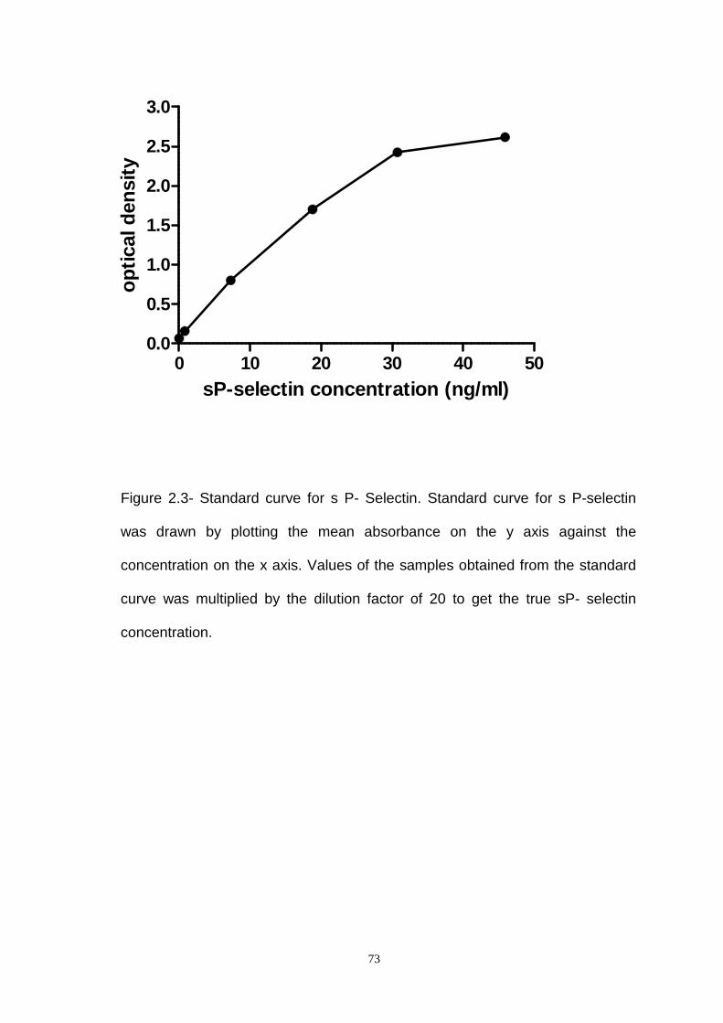
73
Figure 2.3- Standard curve for s P- Selectin. Standard curve for s P-selectin
was drawn by plotting the mean absorbance on the y axis against the
concentration on the x axis. Values of the samples obtained from the standard
curve was multiplied by the dilution factor of 20 to get the true sP- selectin
concentration.
0 10 20 30 40 500.0
0.5
1.0
1.5
2.0
2.5
3.0
sP-selectin concentration (ng/ml)
op
ticald
en
sit
y
74
2.3.3 ESTIMATION OF HIGH SENSITIVE CRP AND SAA:-
CRP was measured in plasma using a high sensitivity automated microparticle
enhanced latex turbidimetric immunoassay (COBAS MIRA; Roche Diagnostics
GmbH). The lower limit of detection was 0.2 mg/l with an interassay CV of 4.2%
at 4 mg/l and 6.3% at 1 mg/l. SAA was measured in plasma by latex
nephelometry (BNII autoanalyser; Dade Behring, Marburg, Germany) (163). The
lower limit of detection was 0.7 mg/l, with an inter-assay CV of 2.6% at15 mg/l
and 3.7% at 80 mg/l. Standardisation of both CRP and SAA assays was based
on the respective WHO International Reference Standards (164),(165).

75
CHAPTER 3: PMC METHODOLOGY
IMPROVED ACCURACY ANDREPRODUCIBILITY OF ENUMERATION OF
PLATELET-MONOCYTE COMPLEXESTHROUGH USE OF DOUBLETDISCRIMINATOR STRATEGY

76
3.1
Introduction
3.2 Doublet Discriminator Strategy
3.3 Patient Material and Methods
3.3.1 Subjects and blood collection
3.3.2 Sample preparation
3.4 Flow Cytometry
3.5 Statistics
3.6 Results
3.6.1 The doublet discriminator gating strategy
3.6.2 Normal peripheral blood
3.6.3 Diluted samples
3.6.3.1 Effect of doublet discriminator strategy on diluted samples
3.6.3.2 Comparison of effectiveness of serial dilution technique with
doublet discriminator strategy in reducing coincidence.
3.6.4 Reproducibility of doublet discriminator technique in pathological
samples of patients with coronary artery disease
3.6.5 Stability of samples
3.7 Discussion

77
3.1 INTRODUCTION:-
The importance of PMC formation as a sensitive marker of platelet activation has
been demonstrated in several clinical conditions including ACS (22, 31, 166-169)
however estimation of PMCs by flow cytometry suffers from several
methodological shortcomings and in vitro artefacts. Modifications of flow
cytometric methods of platelet analysis have been reported including the use of
either washed platelets (170) or whole blood (171). Some investigators have
used red cell lysis for better delineation of platelet and white cell populations
(14).
One of the challenges of platelet monocyte complex estimation is in vitro
activation of the platelets. Mody et al (172) and Kim et al (173) demonstrated that
use of citrate, theophylline, adenosine, dipyridamole (CTAD) anticoagulant
minimises the ex vivo activation and Pearson et al found that CTAD samples are
stable for up to 2 hours post venesection (37). Some investigators have used
whole blood methods to avoid red cell lysis, and centrifugation (24, 171) but the
relative abundance of red cells makes the analysis both time consuming and
insensitive (174). Removal of erythrocytes by cell separation or red cell lysis
facilitates measurements (175, 176) but these procedures might also lead to
significant in vitro artefacts (34).
A consistent problem of overestimation of the platelet-monocyte complexes
remains due to coincident events (177). Two colour flow cytometry using
monoclonal antibodies specific for monocytes and platelet has been widely used
for PMC estimation; platelet-monocyte complexes appearing as double positive
events. However double positive events can originate not only from true
78
complexes but also from non-interacting coinciding platelets and monocytes. As
the concentration of platelets is considerably higher than that of monocytes the
chances of having one or multiple platelets close to but not attached to a
monocyte are high and when a non-conjugated monocyte transits through the
laser adjacent to (but not conjugated to) one or more platelets the cells can be
detected as one event and appear as a PMC (39, 40). To reduce the artefact we
have investigated the use of doublet discriminator (DDM) strategy in estimation
of PMCs in normal peripheral blood, in arterial and femoral venous samples of
patients with CAD and also studied the effect of this strategy on serially diluted
samples and vice versa.
3.2 DOUBLET DISCRIMINATOR STRATEGY:-
The doublet discrimination technique was designed to overcome the problem of
co-incident analysis of doublets of bare nuclei in DNA ploidy experiments (178).
In the analysis of PMC, co-incident platelets and monocytes had a longer time of
flight through the focal point of the laser than true PMC so, using pulse
processed analysis, they elicit signals with a greater fluorescent width than the
single PMC event and a concomitant greater signal area. We hypothesized that
co-incident events may be differentiated from true PMC events by plotting the
cells in a dot plot of signal width and area or signal height and width.

79
3.3 PATIENTS, MATERIALS AND METHODS :-
3.3.1 SUBJECT AND BLOOD COLLECTION
Fourteen normal healthy volunteers aged between 25 and 60 years were
included. Samples were collected from the peripheral vein with 19 G needle and
syringe. Twenty patients with coronary artery disease aged between 56 and 75
years were also included. Arterial samples from different sites (coronary artery
ostium, aorta, femoral artery) were aspirated through the diagnostic coronary
angiogram catheter (5FJudkin’s right or left diagnostic catheter with internal
diameter of 0.11 cm) and venous samples through the femoral venous sheath
(5F) with the help of 5 ml syringe during the angiogram procedures (Table 3.1).
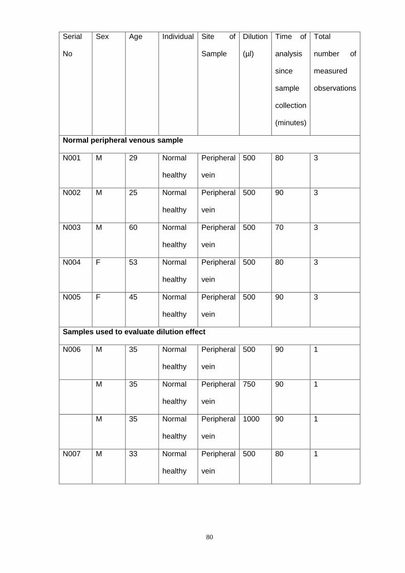
80
Serial
No
Sex Age Individual Site of
Sample
Dilution
(µl)
Time of
analysis
since
sample
collection
(minutes)
Total
number of
measured
observations
Normal peripheral venous sample
N001 M 29 Normal
healthy
Peripheral
vein
500 80 3
N002 M 25 Normal
healthy
Peripheral
vein
500 90 3
N003 M 60 Normal
healthy
Peripheral
vein
500 70 3
N004 F 53 Normal
healthy
Peripheral
vein
500 80 3
N005 F 45 Normal
healthy
Peripheral
vein
500 90 3
Samples used to evaluate dilution effect
N006 M 35 Normal
healthy
Peripheral
vein
500 90 1
M 35 Normal
healthy
Peripheral
vein
750 90 1
M 35 Normal
healthy
Peripheral
vein
1000 90 1
N007 M 33 Normal
healthy
Peripheral
vein
500 80 1
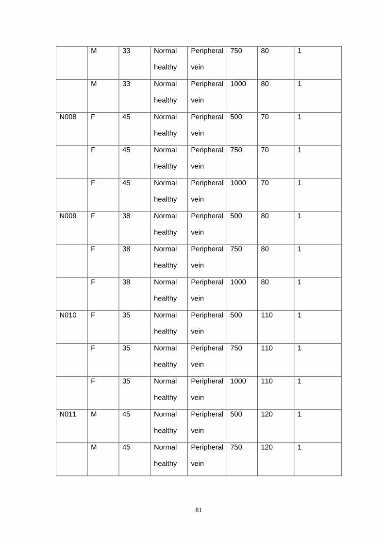
81
M 33 Normal
healthy
Peripheral
vein
750 80 1
M 33 Normal
healthy
Peripheral
vein
1000 80 1
N008 F 45 Normal
healthy
Peripheral
vein
500 70 1
F 45 Normal
healthy
Peripheral
vein
750 70 1
F 45 Normal
healthy
Peripheral
vein
1000 70 1
N009 F 38 Normal
healthy
Peripheral
vein
500 80 1
F 38 Normal
healthy
Peripheral
vein
750 80 1
F 38 Normal
healthy
Peripheral
vein
1000 80 1
N010 F 35 Normal
healthy
Peripheral
vein
500 110 1
F 35 Normal
healthy
Peripheral
vein
750 110 1
F 35 Normal
healthy
Peripheral
vein
1000 110 1
N011 M 45 Normal
healthy
Peripheral
vein
500 120 1
M 45 Normal
healthy
Peripheral
vein
750 120 1

82
M 45 Normal
healthy
Peripheral
vein
1000 120 1
N012 M 40 Normal
healthy
Peripheral
vein
500 110 1
M 40 Normal
healthy
Peripheral
vein
750 110 1
M 40 Normal
healthy
Peripheral
vein
1000 110 1
N013 M 30 Normal
healthy
Peripheral
vein
500 120 1
M 30 Normal
healthy
Peripheral
vein
750 120 1
M 30 Normal
healthy
Peripheral
vein
1000 120 1
N014 F 35 Normal
healthy
Peripheral
vein
500 110 1
F 35 Normal
healthy
Peripheral
vein
750 110 1
F 35 Normal
healthy
Peripheral
vein
1000 110 1
Patients’ sample
N015 F 60 Stable
angina
Femoral
artery
500 90 3
F 60 Stable
Angina
Aorta 500 90 3
F 60 Stable
angina
Coronary
artery
500 90 3
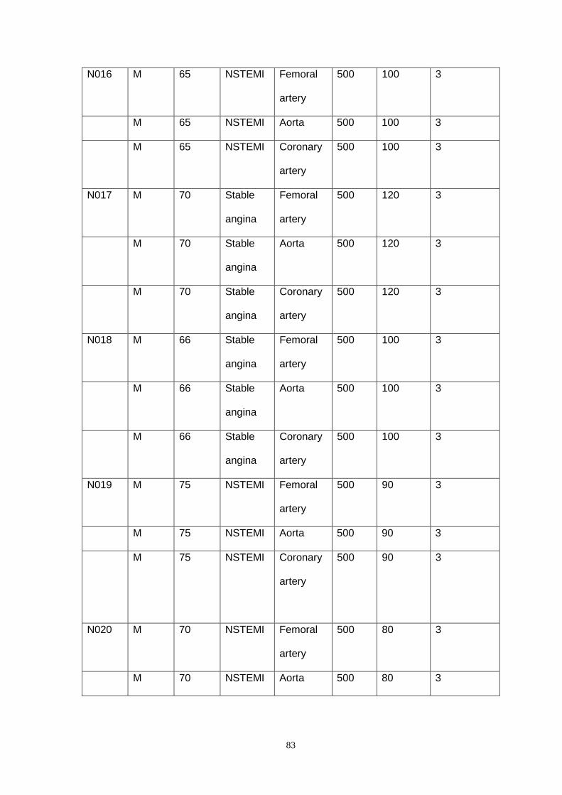
83
N016 M 65 NSTEMI Femoral
artery
500 100 3
M 65 NSTEMI Aorta 500 100 3
M 65 NSTEMI Coronary
artery
500 100 3
N017 M 70 Stable
angina
Femoral
artery
500 120 3
M 70 Stable
angina
Aorta 500 120 3
M 70 Stable
angina
Coronary
artery
500 120 3
N018 M 66 Stable
angina
Femoral
artery
500 100 3
M 66 Stable
angina
Aorta 500 100 3
M 66 Stable
angina
Coronary
artery
500 100 3
N019 M 75 NSTEMI Femoral
artery
500 90 3
M 75 NSTEMI Aorta 500 90 3
M 75 NSTEMI Coronary
artery
500 90 3
N020 M 70 NSTEMI Femoral
artery
500 80 3
M 70 NSTEMI Aorta 500 80 3

84
M 70 NSTEMI Coronary
artery
500 80 3
N021 M 56 Stable
angina
Femoral
artery
500 90 3
M 56 Stable
angina
Aorta 500 90 3
M 56 Stable
angina
Coronary
artery
500 90 3
N022 F 68 Stable
angina
Femoral
artery
500 80 3
F 68 Stable
angina
Aorta 500 80 3
F 68 Stable
angina
Coronary
artery
500 80 3
N023 F 56 NSTEMI Femoral
artery
500 90 3
F 56 NSTEMI Aorta 500 90 3
F 56 NSTEMI Coronary
artery
500 90 3
N024 M 69 Stable
angina
Femoral
artery
500 110 3
M 69 Stable
angina
Aorta 500 110 3
M 69 Stable
angina
Coronary
artery
500 110 3
N025 M 55 Stable
angina
Femoral
artery
500 90 3

85
M 55 Stable
angina
Aorta 500 90 3
M 55 Stable
angina
Coronary
artery
500 90 3
N026 M 74 NSTEMI Femoral
artery
500 80 3
M 74 NSTEMI Aorta 500 80 3
M 74 NSTEMI Coronary
artery
500 80 3
N027 M 70 STEMI Femoral
vein
500 90 3
M 70 STEMI Aorta 500 90 3
M 70 STEMI Coronary 500 90 3
N028 F 72 NSTEMI Femoral
vein
500 80 3
F 72 NSTEMI Aorta 500 80 3
F 72 NSTEMI Coronary
artery
500 80 3
N029 M 65 STEMI Femoral
vein
500 100 3
M 65 STEMI Aorta 500 100 3
M 65 STEMI Coronary
artery
500 100 3
N030 M 72 NSTEMI Femoral
vein
500 90 3
M 72 NSTEMI Aorta 500 90 3

86
Table 3.1 – General descriptions of the collected samples.
M 72 NSTEMI Coronary
artery
500 90 3
N031 F 70 NSTEMI Femoral
vein
500 120 3
F 70 NSTEMI Aorta 500 120 3
F 70 NSTEMI Coronary 500 120 3
N032 M 71 NSTEMI Femoral
vein
500 110 3
M 71 NSTEMI Aorta 500 110 3
M 71 NSTEMI Coronary
artery
500 110 3
N033 F 65 Stable
angina
Femoral
vein
500 120 3
F 65 Stable
angina
Aorta 500 120 3
F 65 Stable
angina
Coronary
artery
500 120 3
N034 M 66 NSTEMI Femoral
vein
500 110 3
M 66 NSTEMI Aorta 500 110 3
M 66 NSTEMI Coronary
artery
500 110 3
87
3.3.2 SAMPLE PREPARATION:-
Protocol 1 - Normal peripheral blood - Peripheral blood samples from 5 healthy
individuals aged between 25 and 60 years were collected from a peripheral vein
with 19 G needle and 5 ml syringe. Blood (2.7 ml) was collected into sodium
citrate (0.109 M/3.2%) anticoagulant (BD vacutainer systems, USA). The
samples were transported to the laboratory within 15 minutes on ice for analysis.
Five microlitres each of anti-CD61 FITC (BD Biosciences, Oxford, UK) and anti-
CD14 PerCP (BD Biosciences, Oxford, UK) monoclonal antibodies were added
to labelled 12mm x 75 mm polystyrene tubes. One hundred microlitres of whole
blood was added to each tube, mixed with the antibodies and incubated for 20
minutes on ice. After labelling, erythrocytes were lysed with the addition of 2ml
BD FACS lysing solution (BD Biosciences, Oxford, UK) (1in 10 dilution) for 10
minutes at room temperature. The samples were washed with 1ml of FACS flow
(BD Biosciences) and then centrifuged at 300g for 5 minutes and the supernatant
was discarded. The cells were resuspended in 0.5ml FACS flow for immediate
flow cytometric analysis. Flow cytometry was performed at low flow rate (not
more than 1000 cells/sec) using a FACS Calibur (BD Biosciences, Oxford, UK)
with Cell Quest Pro ® software. Three thousand gated monocyte events were
acquired and PMCs were expressed as a percentage of the total monocyte
population. All the samples were analysed by flow cytometry within 2 hours of
sample collection.
Protocol 2 - Effect of serial dilution of the samples – Peripheral venous samples
from another 9 healthy individuals were prepared as Protocol 1. After acquisition
the samples were serially diluted to 150% (750µl final volume) and 200% (1ml

88
final volume) in order to physically reduce co-incidence. The diluted samples
were reanalysed by flow cytometry with and without electronic doublet
discrimination (figure 3. 1).
Protocol 3 - Reproducibility in pathological samples - To see the reproducibility
and effectiveness of doublet discriminator strategy in reducing coincident events
in the pathological samples from different sites we included 20 patients (10
NSTEMI, 2 STEMI and 8 Stable angina patients). In STEMI patients samples
were collected during the acute stage while the patients were having PPCI. In
NSTEMI patients samples were collected during their coronary angiogram
procedure within 72 hours of onset of chest pain. In stable angina patients
samples were collected during their elective coronary angiography. Twelve
patients had samples taken from the femoral artery, aorta and coronary artery
ostium and 8 patients had samples from the femoral vein, aorta and coronary
artery ostium. Arterial Samples (2.7 ml from each site) were collected by
aspiration through the Judkin’s 5F right or left diagnostic coronary angiogram
catheter (Cordis) (Internal diameter – 0.11 cm) with the help of 5 ml syringe.
Femoral venous samples were collected from the 5F femoral venous sheath with
the help of 5 ml syringe. Samples were prepared in the same way as peripheral
blood samples from normal donors above and each sample was analysed in
triplicate with the flow cytometry method with DDM on and DDM off (described
below). PMC frequencies were estimated from the average of the triplicate
values.
Protocol 4 - Stability of the samples post labelling - To determine the stability of
the complexes in the samples prepared in the above described method we
prepared 9 samples from 3 patients [2 stable angina patients and 1 NSTEMI

89
patient.] Citrated samples (2.7 ml) were collected from the coronary artery
ostium, aorta and femoral vein. Samples were analysed in triplicate and following
initial flow cytometry analysis the prepared samples were kept in the fridge at
4oC for 24 hours without addition of any further preservatives or fixatives and
were reanalysed in the same manner.
3.4 FLOW CYTOMETRY:-
Cells were acquired on a 2D analysis dot plot arraying CD14 (logarithmic scale
abscissa) and side scatter (SSC) height (linear scale ordinate) (Figure 3. 1 A)
and the region was drawn (R1) around the monocyte population identified by
their distinctive CD14 positivity and intermediate SSC height. Cells in the R1
were gated (G1) and plotted on a 2D dotplot arraying CD61 FITC (linear
abscissa) vs SSC height (Figure 3.1B). Cells with the narrow CD61-width were
magnified on the dot plot C to select events with the narrowest fluorescence
width as putative “singlet” events. All CD14+ events with higher CD61 width were
gated together as putative “multiplets”. Cells both positive for CD61 and CD14
with narrowest CD61 width were considered as true PMCs (subsequently
described as “DDM on”). Cells both positive for CD61 and CD14 with wider CD61
width were hypothesised to be coincident events. PMC frequency data with
‘DDM off’ were determined for all CD14 positive cells.
After validation of the “DDM on” and “DDM off” criteria future experiments
estimated the frequencies of PMCs with and without doublet discriminator use.
Values were measured in triplicate and the averages were taken to estimate the

90
PMC frequency. Co efficient of variation (CV) was calculated to ascertain the
reproducibility.
Experimental design: Using the above flow cytometry technique we conducted
series of experiments to ascertain the effectiveness and reproducibility of doublet
discriminator strategy in different situations. Cells with wider FL1 width were
considered as coincidents and excluded from the analysis. Cells double positive
for CD14 and CD61 with narrow FL1 width were considered as true PMCs.

91
Figure.3. 1. Flow cytometry:- Monocytes were identified with CD14 positivity and
distinctive intermediate sidescatter height. Region (R1) was drawn around the
monocyte populations (A). Monocytes were plotted on a 2D dotplot arraying
CD61-height (abscissa) vs SSC (ordinate) (B) and the CD61+ve events plotted
on a CD61-width vs SSC height array. The plot was zoomed to expand the CD61
width data array (C). Regions were set around the “singlet” population and the
“multiplets”. PMC were calculated as the percentage of CD61+ cells within the
CD14+ population either with inclusion of the multiplets (described as DDM ”off”
results) or after exclusion of the multiplets (described as DDM “on” results).
CD14 PerCP
SS
C-H
eig
ht
100
101
102
103
104
0
256
512
768
1024
Monocytes
CD61 FITC
SS
C-H
eig
ht
100
101
102
103
104
0
256
512
768
1024
CD61+
CD61- Width
SS
C-H
eig
ht
0 6 13 19 250
256
512
768
1024
Multiplets
Singlets
A B
C

92
3.5 STATISTICS:-
Data were represented as mean and standard deviation.The paired t test was
used to compare the PMC frequencies with DDM on and DDM off and also to
compare the baseline PMC values with 24 hour PMC values. One way ANOVA
with Bonferroni’s multiple comparison was used to compare the PMC
frequencies between different dilutions. To evaluate the relationship of PMC
values with DDM on and DDM off we used linear regression analysis. A two
tailed p value <0.05 was considered to be significant .Statistical analysis was
performed using the Graphpad prism® software (version 5).
3.6 RESULTS:-
3.6.1 THE DOUBLET DISCRIMINATOR GATING STRATEGY:-
Support for the hypothesis that analysis of the CD61 fluoresence width signal
could discriminate between true PMC and co-incident events of free monocytes
and free platelets was provided by the exemplar gating strategy in figure 3. 1.
The singlet events as determined by CD61 width could be clearly distinguished
from signals with a wider distribution of CD61 fluorescence.

93
3.6.2 NORMAL PERIPHERAL BLOOD:-
The frequency of platelet-monocyte complexes within the circulating monocytes
in the peripheral blood of 5 healthy normal individuals was found to be 6.27% ±
1.77 (mean ± sd) without the use of doublet discrimination strategy. This
frequency fell significantly to 2.57% ± 0.99 using the doublet discriminator
strategy (p=0.02). (CV 13.96 ± 8.30 %) (Fig:3. 2).

94
DDM OFF DDM ON
0
5
10
P=0.02%
pm
c
Figure. 3.2-Comparison of frequency of PMCs in the peripheral blood of normal
healthy individuals (n=5) with (DDM on) and without (DDM off) the use of doublet
discriminator (DDM) technique. DDM use effectively reduced the co incident
events hence lower values compared to DDM off.

95
3.6.3 DILUTED SAMPLE:-
3.6.3.1 EFFECT OF DOUBLET DISCRIMINATOR STRAGEY ON DILUTED
SAMPLE:-
Peripheral venous samples from 9 different individuals were prepared as
described above and the cells were resuspended in FACS flow with serial
dilution of 500, 750 and 1000 microlitres before analysis. The PMC frequencies
(%) without the doublet discriminator use (DDM off) were calculated as 16.44 ±
4.09, 12.21± 3.44, and 9.67± 2.91 respectively confirming the ability of dilution to
reduce coincident acquisition of free platelets and monocytes in the PMC gate.
However, the additional use of doublet discrimination (DDM on) reduced the
PMC values in the same samples to 6.46± 1.65, 4.52± 1.27 and 4.05± 1.19
respectively; all of which were significantly lower (p<0.05) than the matched
“DDM Off” results. PMC values were consistently lower using the doublet
discriminator strategy (Figure: 3.3,3.4). The diluted samples tested without DDM
showed significant differences between dilutions and the co-efficient of variance
remained constant despite dilution. In contrast, the 750µl and 1000µl samples
when analysed with doublet discrimination showed no significant difference,
suggesting that the “true” value of PMC had been determined and that no co-
incidence was occurring but it is unlikely that doublet discrimination alone is able
to remove the artefact of co-incidence (Figure 3.5).

96
0 250 500 750 1000125015000
5
10
15
20
25
30
DDM off
DDM on
Serial dilution in microlitre
%p
mc
Figure 3. 3- Effect of serial dilution in reducing coincident events with and without
doublet discriminator use Serial dilution reduced the frequency of PMCs as
dilution reduced coincident acquisition of free platelets and monocytes in the
PMC gate. However, the additional use of doublet discrimination reduced the
PMC values in the same samples even further.

97
0 5 10 15 20 250
5
10
15
20
25
y=0.25 to
8.96 while
x=0, R2=0.42,
P= 00.0003
DDM ON
DD
MO
FF
Figure 3. 4. - Effect of doublet discriminator mode use in diluted samples.
Without the use of doublet discriminator (DDM off) the PMC frequency was
significantly overestimated even in the samples with serial dilution. Serial dilution
alone was not sufficient to reduce the coincident events effectively. Use of
doublet discriminator (DDM on) reduced the coincident events in the diluted
samples even further.

98
500
750
1000
0
2
4
6
8
10500
750
1000
p<0.05
p-ns
p<0.05
dilution (microlitre)
PM
C%
Mo
no
cyte
Figure 3. 5. Effectiveness of doublet discriminator use for estimation of true PMC
value. Doublet discriminator use did not fully abolish the effect of serial dilution
but no significant difference in PMC values between 750 µl and 1000 µl dilution
suggests with the use of DDM true value of PMC has been achieved (n=9).

99
3.6.3.2 COMPARISON OF EFFECTIVENESS OF SERIAL DILUTION
TECHNIQUE WITH DOUBLET DISCRIMINATOR STRATEGY IN REDUCING
COINCIDENCE.
To determine the efficiency of doublet discrimination in reducing the coincidence
events compared to the sample dilution strategy we estimated the difference of
average percentage of PMCs in the 500µl dilution with DDM off and DDM on and
compared with the result of estimated difference in PMC frequency between the
500 and 750 microlitre and 500 and 1000 microlitre dilutions. The average
reduction in percentage of PMC frequency by dilution alone was 4.23% (± 2.19)
and 6.78% (± 2.68) in the 750 and 1000 microlitre samples respectively. In
contrast, the use of doublet discrimination on the samples with 500µl dilution
reduced the PMC frequency by an average of 9.98% (± 3.54). These results
suggested that serial dilution does lower co-incidence but it is a less efficient
strategy than doublet discrimination strategy (Figure 3.6).

100
Del
ta50
0DDM
Off
-500
DDM
on
Del
ta10
00-5
00DDM
Off
Del
ta75
0-50
0DDM
Off
5
10
15
p= <0.05
p=ns
p=ns%
pm
c
Figure 3.6- Comparison of effectiveness of doublet discriminator use and serial
dilution in reducing the coincident events. Average reduction in coincident events
were higher in 1000 microlitre dilution compared to the 750 microlitre dilution as
higher dilution reduced the chance of detection of coincident events but use of
doublet discriminator strategy alone without any further dilution of samples
reduced the coincident events more effectively. This proved the doublet
discriminator strategy was more effective than serial dilution in reducing the
coincident events.

101
3.6.4 REPRODUCIBILITY OF DOUBLET DISCRIMINATOR TECHNIQUE IN
PATHOLOGICAL SAMPLES OF PATIENTS WITH CORONARY ARTERY
DISEASE.
PMC analysis using the doublet discriminator strategy was performed with 60
samples collected from the 20 patients with coronary artery disease during
coronary angiography. This confirmed its value in significant reduction in
coincidence events. PMC frequency within the monocyte population was
16.04% (± 11.26) without the use of doublet discriminator strategy which reduced
to 7.66% (± 5.18) using doublet discrimination (p<0.01).

102
0 5 10 15 20 25 30 35 40 45 50 55 6005
1015202530354045505560
DDM ON
DD
MO
FF
y=1.29 to 4.99 while
x=0, p<0.0001
Figure 3. 7. Effect of doublet discriminator use in pathological samples. Linear
regression analysis showed doublet discriminator use (DDM on) effectively
reduced the co incident events in samples from patients with coronary artery
disease in the same manner as it did in normal individuals (n= 20).

103
3.6.5 STABILITY OF SAMPLES:-
Prepared samples were stable even after 24 hrs while kept at 40C. In these 3
patients the baseline and 24 hrs average PMC frequency (%) were 15.11± 9.83
and 14.03± 9.44 (p=0.22) respectively (DDM off). Those values with DDM on
were 8.84 ± 6.04 and 9.40 ± 6.13. (p=0.36) (Figures 3. 8a & 3. 8b).

104
Bas
elin
e
24hrs
05
1015202530354045505560
%p
mc
ns
a b
Figure 3.8 a &b. Stability of samples over 24 hour period. Comparison of the
PMC values at baseline and reanalysis of the same samples after 24 hours with
DDM off and DDM on respectively. Following preparation and initial flow
cytometry assessment samples were kept in the fridge at 4 0 C without addition
of any further preservatives or fixatives. Samples were reanalyzed after 24 hours
in the similar manner. No significant difference was noted between the baseline
and 24 hour mean PMC values with DDM off , p= 0.22 and with DDM on,
p=0 .36 (n=3).
Bas
elin
e24
hr
05
1015202530354045505560
ns%
pm
c
105
3.7 DISCUSSION:-
In peripheral circulation platelets are found both as free entities and in complexes
with monocytes, granulocytes and even erythrocytes and it has been presumed
that this affected their activation and function in vivo. The frequency of platelet-
monocyte complexes appears to have a role in coronary disease and stroke (22,
31, 168) (179). Erythrocyte-platelet complexes have recently been found at the
centre of atherosclerotic plaques by scanning electron microscopy in an
experimental murine model (180). The potential importance of complexed
platelets in normal and pathological thrombosis is clear but flow cytometric
assays for these complexes are poorly standardised.
Different methods of platelet monocyte complex estimation are described in the
literature. Many concerns have been raised regarding in vitro platelet activation
and artefactual platelet-monocyte complex formation resulting in overestimation
of platelet-monocyte complexes. It has been reported that the choice of
anticoagulant, sample preparation e.g. fixation, erythrocyte lysis, centrifugation,
washing technique etc might influence the estimated frequency of platelet-
monocyte complexes. So whole blood techniques are now preferred, most of
which exclude fixation, erythrocyte lysis and centrifugation steps in order to
minimise the in vitro platelet activation and artefactual platelet-monocyte complex
formation. Sample dilution and flow rate also influence PMC estimation. So in
different studies the frequency (%) of PMC in the normal peripheral venous
sample varies widely from 3.72± 1.39 (13) to 12.3± 3.3 (24, 32, 36).This wide
variation could also be due to the fact that coincident events were probably not
considered during estimation of PMCs.

106
The potential influence of coincident events has remained one of the major
concerns that needed to be addressed. In order to exclude the coincident events
Bihari et al used a subtraction method where they divided the blood samples into
two parts one part was stained for platelets and granulocytes, the other part was
stained only for platelets and fluorescent beads were mixed with it. Then
measuring the platelet-granulocyte and platelet-bead double positivity at the
same dilution (platelet concentrations needed to be the same) the exact amount
of platelet-granulocyte complexes could be calculated by subtraction method
(177). Another way of reducing coincidence was to use higher dilution (200 or
400 fold). Coincidence was found to be negligible resulting in very low (1-5%)
frequencies of complexes. It should be taken into account that dilution might
result in the dissociation of the complexes, therefore, analysis of highly diluted
samples did not allow the authors to draw conclusions with respect to the
undiluted samples in terms of its complex content (166, 169). A method
independent of dilution for exclusion of coincidence was thus needed.
In this study we confirmed the importance of coincidence events in the
overestimation of the frequency platelet:monocyte complexes in clinical samples.
The use of doublet discrimination to overcome co-incidence was not new but did
not appear to have been used in studies of platelet:leukocyte complexes
previously. Many publications in this field have ignored the problem of co-
incidence entirely whist others have used extremely diluted samples and/or low
sample flow rate to reduce its impact on precision (179).Whilst effective this very
significantly extended sample acquisition time which severely hindered its use in
busy routine or research laboratories or settings where a high throughput is
required.

107
This strategy was reproducible and similarly effective in reducing the co-incident
events in the peripheral venous samples of normal individuals, in the diluted
samples and in the arterial, femoral venous, aortic and coronary artery samples
of the patients with coronary artery disease.
In our study for the first time we demonstrated the doublet discriminator strategy
could be used to exclude the coincident events to a great extent thus improving
the accuracy of estimation and reducing intra-assay variability. As predicted, we
found that the percentage of PMCs within the monocyte population in the
peripheral venous circulation of normal individual was 2.57± 0.31 which was
much lower than some of the previous published data (32, 36) but comparable to
the data published by Ashman et al.(24). In samples from other 9 individuals in
which we looked at the use of dilution to exclude co-incidence the PMC
frequencies were found to be higher as they were taken at different times of the
day and they were not age matched. Dilution of the samples reduced the PMC
frequency. This could be due to the fact that dilution further reduced the chance
of co-incidence as the probability of presence of cells in the detection volume
was lower with serial dilution. Alternatively, the additional dilution could have led
to spontaneous dissociation of cells. We did demonstrate that sample dilution
alone could not exclude the co-incident events completely. Using the doublet
discriminator strategy we managed to reduce the co-incident events much more
effectively. Doublet discriminator strategy proved effective but not completely
independent of the dilution effect. The effect of dilution, still present with the
doublet discriminator strategy was probably due to the fact that with increasing
dilution there was a possibility of spontaneous dissociation of platelet-monocyte
complexes resulting in lowering of true complex estimation.

108
We have demonstrated that use of a doublet discriminator strategy effectively
reduces the co-incident events in the samples of the patients with coronary artery
disease taken from different sites (femoral vein, femoral artery, aorta and
coronary artery) and can eliminate co-incidence if used with moderate sample
dilution. This study also demonstrated that prepared samples remain stable with
respect to PMC complex content even after 24 hrs at 4o C; an important
consideration for analysis of samples in busy laboratories where flow cytometer
access is at a premium.

109
CHAPTER 4
COMPARISON OF PLATELET-MONOCYTE COMPLEX
(PMC) EXPRESSION AND DIFFERENTIAL PLATELET
AND MONOCYTE ACTIVATION STATUS WITHIN THE
COMPLEXES IN THE ACUTE CORONARY SYNDROME
AND STABLE ANGINA PATIENTS

110
4.1Introduction.
4.2 Objectives.
4.3 Patients and Methods
4.3.1 Group 1- Acute coronary syndrome patients (ACS).
4.3.2 Group 2- Stable angina patients.
4.3.3 Exclusion criteria.
4.3.4 Sample collection and preparation.
4.3.5 Flow cytometry.
4.4 Statistical analysis.
4.5 Results.
4.6 Discussion.
4.7 Conclusion.

111
4.1 INTRODUCTION:-
Formation of PMCs has already been demonstrated in the ACS. The role of
PMCs in the pathogenesis of ACS remains unclear. It is not clear whether
platelet activation and PMC formation is a systemic response or whether there is
a direct role for intracoronary PMC formation in the pathophysiology of acute
myocardial infarction via monocyte activation, TF expression and inflammation.
Therefore, PMCs may be a link between platelet mediated thrombogenesis and
monocyte mediated inflammation in ACS. Following phosphorylation of the
membrane protein p-selectin in the platelet alpha granule (86) the granule moves
to the platelet surface exposing P-selectin and tethering the platelet with the
monocyte via PSGL-1 to form PMCs. In addition, other integrin dependant
mechanisms may be involved in the formation of PMCs (104) . Binding of
platelets to monocytes has been shown to regulate various monocyte actions.
Platelets play a significant role in supplying cholesterol to monocytes assisting
monocytes to mature into lipid laden macrophages (181),(182). Furthermore, the
binding of thrombin stimulated platelets induces monocyte cytokine expression
(2) and monocyte chemokine synthesis may have been regulated by platelet
surface p selectin in conjunction with the platelet chemokine RANTES (8). It is
shown that activated platelets express CD40 ligand (CD40L) on their surface (9).
Binding of CD40L to CD40 on monocytes results in monocyte activation and
production of cytokines including IL-6 (10) which often parallels elevation of CRP
(12) seen in ACS (183). A further hypothesis is that binding of activated platelets
might alter monocyte activation status resulting in an increase in TF expression
from the monocyte component acting as a trigger for the initiation of a cascade of

112
thrombogenesis and the release of proinflammatory cytokines, including IL-6, IL-
1β and IL-12. Therefore, it is likely that, following platelet activation, a cascade of
different processes occur in parallel to augment local inflammation and
thrombogenesis. However, binding with unstimulated platelets might not be
associated with a similar cellular response (13). Therefore, in the absence of
platelet activation PMC formation might just represent a physiological
phenomenon. Depending upon the level of activation, the platelets within PMCs
might actually be responsible for triggering a pro-inflammatory response in
monocytes. P-selectin expression on the PMCs is indicative of activated platelets
on these complexes and TF expression on these complexes demonstrates the
presence of activated monocytes within the complexes.
In the context of ACS it is unclear whether PMC formation is a systemic
phenomenon or a local intracoronary event. Presuming PMCs are mainly formed
at the site of plaque disruption at the culprit lesion site a significant transculprit
lesion gradient (gradient between distal coronary and aorta) might be expected
and PMC expression would be highest in the coronary circulation compared to
the other systemic vascular compartments (aorta and right atrium).
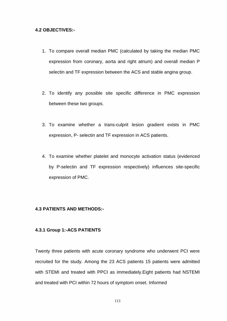
113
4.2 OBJECTIVES:-
1. To compare overall median PMC (calculated by taking the median PMC
expression from coronary, aorta and right atrium) and overall median P
selectin and TF expression between the ACS and stable angina group.
2. To identify any possible site specific difference in PMC expression
between these two groups.
3. To examine whether a trans-culprit lesion gradient exists in PMC
expression, P- selectin and TF expression in ACS patients.
4. To examine whether platelet and monocyte activation status (evidenced
by P-selectin and TF expression respectively) influences site-specific
expression of PMC.
4.3 PATIENTS AND METHODS:-
4.3.1 Group 1:-ACS PATIENTS
Twenty three patients with acute coronary syndrome who underwent PCI were
recruited for the study. Among the 23 ACS patients 15 patients were admitted
with STEMI and treated with PPCI as immediately.Eight patients had NSTEMI
and treated with PCI within 72 hours of symptom onset. Informed

114
consent was obtained before the procedure. Ethical approval for the study was
granted from the local research and ethics committee. All patients were given
600mg of Clopidogrel and 300mg of Aspirin before the procedure as per protocol
for performing PCI in our institution. Prior to PCI patients also received weight
adjusted unfractionated heparin to maintain ACT between 200-250 sec.
4.3.2 GROUP 2 – STABLE ANGINA PATIENTS:-
Seven patients with stable angina were also recruited prior to elective PCI.
Patients were already taking 75 mg of Aspirin and 75mg of Clopidogrel for 7 days
prior to the procedure according to the protocol for performing elective PCI in our
hospital. Prior to PCI they also received weight adjusted unfractionated heparin
to maintain the ACT between 200-250 sec.
4.3.3 EXCLUSION CRITERIA:-
Patients with renal failure, prior coronary artery bypass grafts and who had
already received Glycoprotein iib/iiia antagonists prior to sampling were
excluded.

115
4.3.4 SAMPLE COLLECTION AND PREPARATION:-
Samples were aspirated from the coronary artery distal to the culprit lesion (using
a coronary aspiration, Medtronic ® Export catheter , internal diameter 0.10 cm),
aorta using a Judkin’s diagnostic catheter (5F, Cordis ®, internal diameter 0.11
cm) and from the right atrium (using a pigtail cather (5F, Cordis ®, internal
diameter 0.11 cm). It has been demonstrated before that aspiration of blood
through the Export catheter does not induce artefactual platelet activation (184).
Four milliliters of blood from each site was collected into 3 separate sterile
vacutainer tubes containing a combination of 3.2% sodium citrate and EDTA for
flow cytometric analysis of PMC, p-selectin and TF expression . EDTA was
added to sodium citrate to stop in vitro calcium dependent PMC formation .
Samples were prepared for flow cytometry analysis as described in chapter 2.
4.3.5 FLOW CYTOMETRY:-
PMC, P -Selectin positive PMC and TF positive PMCs from all the 3 sites were
estimated with flow cytometry as described in chapter 2. Overall median values
were calculated by taking median values from the three sample sites and site
specific values were (aorta, right atrium and coronary) calculated separately.

116
4.4 STATISTICAL ANALYSIS:-
Data were presented as median and interquartile range. To compare variables
between the two groups non-parametric Mann-Whitney test was used. To
compare variables within the same group for calculation of transculprit lesion
gradients paired t test was used. A Two-tailed p value < 0.05 was considered
statistically significant. All calculations were performed using graphpad prism®
(version 5) software.
4.5 RESULTS:-
Twenty three ACS patients (15 STEMI and 8 NSTEMI) and 7 stable angina
patients were recruited for the study. The baseline demoraphic and clinical
charcteristics of the patients are described in table 4.1.

117
ACS (n= 23) Stable (n= 7)Age median (IQR), years 60 (48 - 69) 70 (58-75)
Male 18 6Female 5 1
Risk Factors
History of hypertension 8 4History of smoking 11 5Diabetes 3 0Dyslipidaemia 9 5
Angiographiccharacteristics
1 vessel disease 12 42 vessel disease 8 13 vessel disease 3 2
Culprit vessel
Left Anterior Descending 6 4Left Circumflex 8 0Right Coronary Artery 9 3
Time of coronarysampling since theonset of pain
STEMI [Median (IQR) ]hours
4 hours and 50 minutes(1 hour and 45 minutes-6 hours)
NSTEMI [Median (IQR)]hours
66 (46-72) -
Table 4.1 -Baseline demographic and clinical characteristics of the patientsrecruited.

118
There was no significant difference (median ; IQR) in overall median PMC
expression (% monocyte) or overall median TF positive PMCs (%PMC) between
ACS and stable angina groups. However, overall median P- selectin positive
PMC (% PMC) was significantly higher in the ACS group compared to the stable
angina group (p= 0.006). See Table 4.2.

119
ACS median (IQR) Stable Angina median (IQR) p
Overall median PMC(%monocyte)
9.98 (7.17- 18.02) 10.18 (6.22- 15.28) 0.43
Overall median P-selectin + PMC (%PMC)
19.34 (14.05-32.20)
13.98 (7.93- 16.85) 0.006
Overall median TF+PMC (%PMC)
4.79 (3.09-6.14) 5.25 (3.16-8.69) 0.48
Table 4.2- Comparison of Overall Median PMC, P-Selectin Positive PMC and TF
Positive PMC between ACS and Stable Angina Group. There was no significant
difference in overall median PMC and overall median TF positive PMCs between
the ACS and stable angina group but the overall median P- selectin positive PMC
expression was significantly higher in the ACS group compared to the stable
angina group. This finding suggested a higher percentage of activated platelets
within the PMCs in the ACS patients compared with stable angina patients.
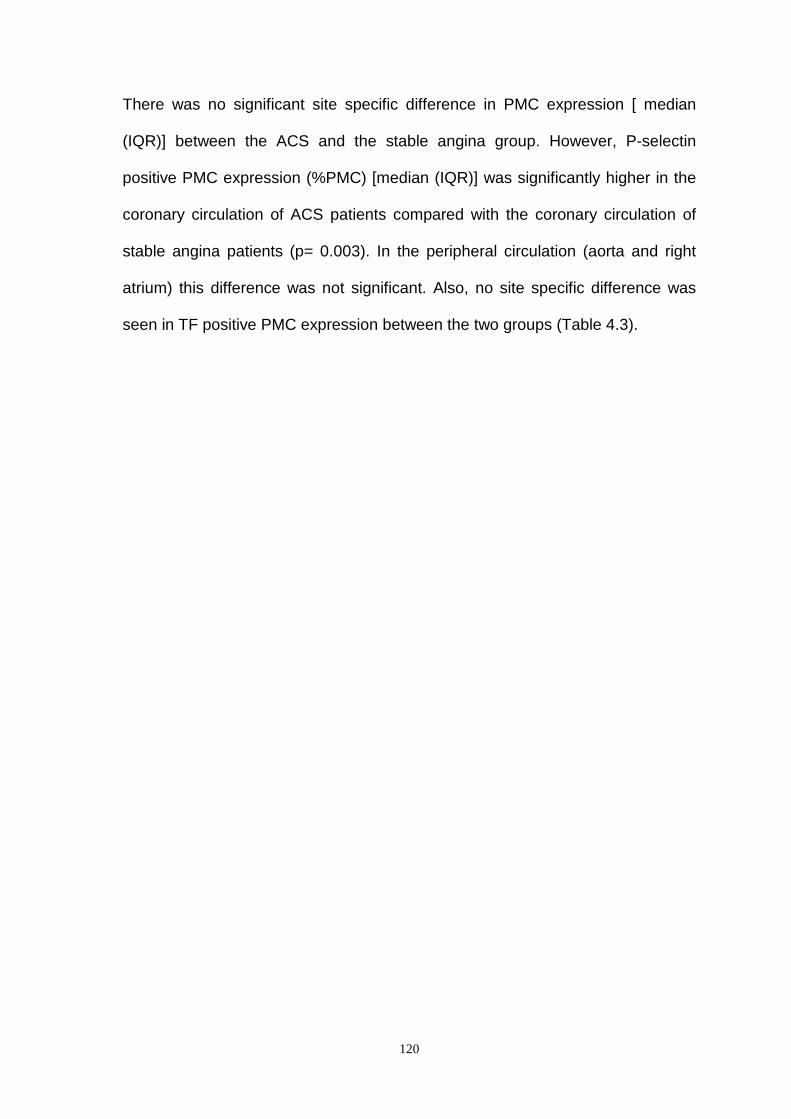
120
There was no significant site specific difference in PMC expression [ median
(IQR)] between the ACS and the stable angina group. However, P-selectin
positive PMC expression (%PMC) [median (IQR)] was significantly higher in the
coronary circulation of ACS patients compared with the coronary circulation of
stable angina patients (p= 0.003). In the peripheral circulation (aorta and right
atrium) this difference was not significant. Also, no site specific difference was
seen in TF positive PMC expression between the two groups (Table 4.3).

121
ACS (n = 23) STABLE (n=7) p
PMC(%Monocytes)
CO 12.4 (7.3-17.3) 8.8 (6.3-16.75) 0.41
AO 9.6 (6.4-17.9 ) 8.2 (5.13-1.34 0.45
RA 12.4(8.7-18.26) 12.0 (8.7-17.9) 0.29
P-Selectinpositive PMCs(%PMC)
CO 29.05 (21.27-55.03) 9.16 (7.4-15.6) 0.003
AO 15.99(11.97-22.11) 13.3 (7.97-24.18) 0.54
RA 15.57(10.89-23.00) 15.98(10.76-16.89) 0.71
TF Positive PMC(%PMC)
CO 4.9 ( 3.3-8.6) 4.5 (4.0-7.7) 0.69
AO 3.6 ( 2.5- 4.9) 6.3 (3.4-9.8) 0.09
RA 5.2 (2.5- 6.6) 3.2 (2.1- 8.5) 0.90
Table-4.3 Site specific comparison between the ACS and stable angina group:-
PMC expression was non-significantly higher in all the compartments in the ACS
group. Intracoronary P- selectin positive PMC expression was significantly higher
in the ACS group compared with the stable angina patients suggesting increased
intracoronary expression of activated platelets within the complexes in ACS
patients.

122
In ACS patients although there was no significant transculprit lesion gradient
(p=0.35) seen in total PMC expression (Figure 4.1) a significant transculprit
lesion gradient in P-selectin positive (p= 0.01) (Figue 4. 2) and TF positive PMC
expression (p=0.04) (Figure 4. 3) was seen. In the stable angina patients there
were no significant transculprit lesion gradient in total PMC, P-selectin positive
and TF positive PMC expressions (data not shown).
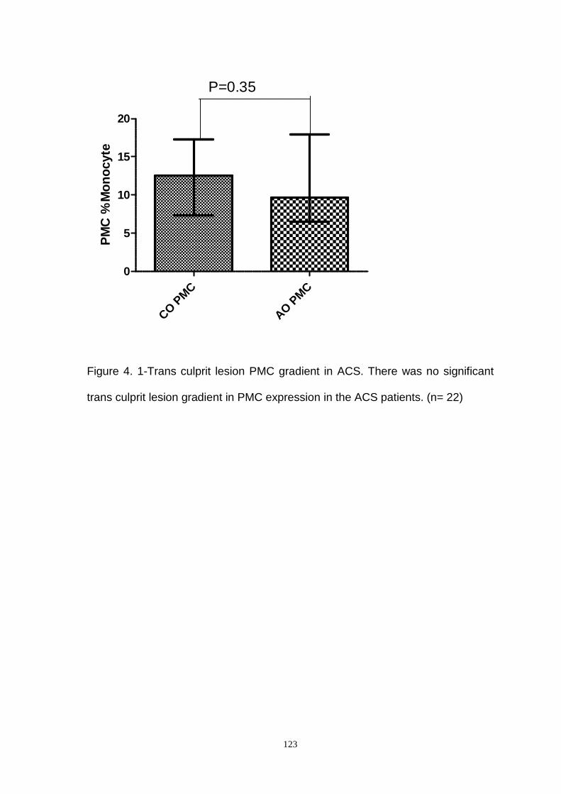
123
CO
PMC
AO
PMC
0
5
10
15
20
P=0.35P
MC
%M
on
ocyte
Figure 4. 1-Trans culprit lesion PMC gradient in ACS. There was no significant
trans culprit lesion gradient in PMC expression in the ACS patients. (n= 22)
124
CO
Psele
ctin
+PM
C
AO
Pse
lect
in+P
MC
RA
Psele
ctin
+PM
C
0
20
40
60
p= 0.01
p= 0.95
%P
MC
Figure 4. 2- Site specific comparison of P- selectin positive PMC expression in
the ACS patients showing significant transculprit lesion gradient.(n= 14)

125
CO
TF
AO
TF
RA
TF
0
2
4
6
8
10
p= 0.04
p= 0.36%
pm
c
Figure- 4.3 Site specific comparison of TF positive PMCs in ACS.TF positive
PMC expression had significant trans culprit lesion gradient in the ACS patients.
This finding suggested heightened intracoronary monocyte activation within the
PMC in the ACS patients.

126
4.6 DISCUSSION:-
Monocyte and macrophages are known to produce cytokines, extracellular matrix
molecules, enzymes and other mediators which might play a central role in
inflammatory disease including atherosclerosis (185). Antibody inhibition studies
indicate that monocytes bind platelets primarily via PSGL-1 and P-selectin. The
extent of monocyte-platelet interaction via PSGL-1 could lead to the development
of a proatherogenic monocyte phenotype (22). Furman et al (21) first showed
that circulating PMCs might be an early marker of acute myocardial infarction. As
expected, our data showed increased site specific PMC expresson in all the 3
sites in ACS patients compared with stable angina patients but the difference
was not statistically significant. This is either due to the limited number of
patients recruited or the acknowledgement that overall PMC formation per se is
most likely a non-specific systemic phenomenon in the context of coronary artery
(vascular) disease. These findings did not implicate a significant
pathophysiological role for overall PMC formation per se in acute coronary
syndrome. Bournazos et al suggested that, in the absence of platelet activation,
PSGL-1 mediated platelet adhesion to circulating monocytes represents a
physiological process with little impact on cell physiology. In contrast high levels
of P- selectin on the surface of activated platelets or binding of multiple platelets
per monocyte were required to trigger monocyte activation via PSGL-1 (13). Our
in vivo findings in human subjects showed that the percentage of PMCs which
expressed P-selectin on their surface (P-selectin positive PMC) was significantly
higher in the ACS group. Our findings suggested that although PMC formation
appeared to be more of a general phenomenon it was platelet activation status
within the complex (expressed by P- selectin positivity) which distinguished ACS

127
and stable angina patients. Therefore, we concluded that not all the complexes
were equally active. In ACS patients activated platelets expressing P-selectin
were higher compared with the stable angina group. This difference was most
evident in the coronary circulation of ACS patients. These findings support the
findings of Bournazos et al. However, unexpectedly monocyte activation status
within the complexes (TF positive PMCs) did not significantly differ between the
ACS and stable angina groups. In the ACS group a significant transculprit lesion
gradient of both P-selectin and TF positive PMC suggested heightened
intracoronary platelet and monocyte activation status within the complex at the
site of plaque disruption. This provided clear support to the hypothesis that only
activated PMC are of pathophysiological importance in ACS whereas overall
PMC is merely a marker of generalised vascular inflammation. The role of
heightened intracoronary platelet and monocyte activated PMC in contributing to
microvascular injury requires further investigation.
4.7 CONCLUSION:-
Increased expression of PMCs was seen in ACS and stable angina patients
compared with normal healthy individuals. No significant difference existed in
total PMC expression between ACS and stable angina patients suggesting that
PMC formation more likely a physiological marker of generalised vascular
inflammation in coronary artery disease but not specifically implicated in the
pathogenesis of ACS. However, activated PMC (with either activation of the
platelet or monocyte portions) are increased in ACS patients and are more likely
to be implicated in the pathogenesis of ACS. This hypothesis is supported by the

128
observation of a clear transculprit gradient for activated PMC in ACS patients.
This study provides key insights into the role of platelet and monocyte
interactions in coronary artery disease.
129
CHAPTER 5
COMPARISON OF SYSTEMIC AND SITE SPECIFIC
INFLAMMATORY BURDEN BETWEEN ACS AND
STABLE ANGINA PATIENTS.

130
5.1 Introduction.
5.2 Patients and Methods.
5.2.1 Sample collection and preparation.
5.3 Objectives.
5.4 Statistical methods.
5.5 Results.
5.6 Discussion.
5.7 Conclusion.

131
5.1 INTRODUCTION:-
In ACS inflammation has been found to play a significant role in the disease
process. Mulvihill and colleagues have reported that a CRP concentration of <
3mg/l is associated with a negative predictive value of 97% for major adverse
cardiovascular events within six months. Conversely a CRP concentration of > 3
mg/l has shown to have a sensitivity of 96% for predicting adverse
cardiovascular events, with a specificity of 52 % (82). It is not clear whether
increased levels of these inflammatory markers have any causal relationship with
the pathogenesis of ACS (85). Using the principle of Mendelian randomisation
CRP genetic variants as proxies a recent study by C Reactive Protein Coronary
Heart Disease Genetics Collaboration (CCGC) group has shown that genetically
raised concentrations of CRP are unrelated to conventional risk factors and risk
of coronary heart disease. This group have also suggested that CRP
concentration is unlikely to have even a modest causal role in coronary heart
disease. Furthermore, this group did not find any material associations between
CRP genetic variants and fibrinogen, interleukin 6, and leukocyte count. These
results suggest that C reactive protein concentration is unlikely to be causally
relevant (186). However other inflammatory markers like SAA, TNF alpha, IL-6
may have some relationship with the pathogenesis of acute coronary syndrome.
It remains unclear whether inflammatory markers originate from the site of
plaque rupture or represent a systemic process. Measurement of inflammatory
markers in the peripheral circulation may not be appropriate for evaluation of
local inflammatory activity. Accurate evaluation of local inflammatory markers
can be done by use of either local perfusate or by measuring the difference in
concentration across the coronary circulation. An elevated coronary sinus-arterial
132
difference in the levels of IL-6 has been reported in patients with ACS (95).
Deliargyris et al have described that IL-6 is produced in the coronary circulation
of patients who have unstable angina but not in the patients who have stable
angina (95).
Maier et al. (53) have demonstrated increase in levels of IL-6 by > 70% and SAA
by around 10 % in the culprit coronary artery relative to those in the aorta in the
STEMI patients. According to their conclusion this differential expression of
increased inflammatory markers must be related to the arterial wall within the
ruptured plaque or by the blood cells trapped in the occluded coronary artery.
Though SAA is known to be produced predominantly in the liver (99) the
increase in local SAA blood levels in the culprit coronary artery can suggest local
production of SAA at the site of occlusion either by cells of the atheroscelorotic
arterial wall or by white blood cells trapped in the thrombus. In line with this
interpretation SAA mRNA and protein have been detected in human
atherosclerotic lesions and cultured arterial smooth muscle cells (100).
Although In general it is believed that inflammation is a systemic effect of acute
coronary syndrome and inflammatory markers are mostly synthesised in the
liver, nevertheless the evidence above supports the idea of a local intracoronary
synthesis of inflammatory markers. In accordance with these findings it is
expected there should be a significant transculprit lesion gradient of inflammatory
marker expression in the ACS patient and inflammatory burden will be highest at
the coronary circulation distal to the culprit lesion. We also expect inflammatory
burden will be higher in the ACS patients compared with the stable patients.

133
5.2 PATIENTS AND METHODS:-
In order to prove the above hypothesis blood samples were collected from 23
ACS patients (15 STEMI, 8 NSTEMI) and 7 stable angina patients. STEMI
patients were recruited immediately after their admission in hospital just before
the PPCI. NSTEMI patients were recruited within 72 hours of onset of chest pain
before their percutaneous interventions. Stable angina patients were recruited
before their elective percutaneous coronary intervention. Patients’ baseline
characteristics and drug treatments have already been described in the previous
chapter. Samples were collected from aorta, coronary artery (distal to the culprit
lesion) and right atrium. Rather than having a single venous sample we chose to
measure overall median values by taking the median concentrations from all the
3 sites to ascertain the systemic inflammatory burden.
5.2.1 SAMPLE COLLECTION AND PREPARATION:-
Samples were aspirated from the coronary artery distal to the culprit lesion (using
a coronary aspiration, Medtronic ® Export catheter , internal diameter 0.10 cm),
aorta (using a Judkin’s diagnostic catheter (5F, Cordis ®, internal diameter 0.11
cm) and from the right atrium (using a pigtail cather (5F, Cordis ®, internal
diameter 0.11 cm). Blood samples (2.7ml) collected from each site were placed
into 3.2% sodium citrate vacutainer tubes and the samples were used to prepare
double spun platelet poor plasma by centrifuging the samples initially at 800g for
10 minutes at room temperature then supernatant was collected into eppendorfs
and was recentrifuged at 2500g for 5 minutes and 90% of the supernatant
134
plasma was aspirated and immediately stored at -800 C for later analysis of IL-6
and TNF-alpha with ELISA (R&D systems) ; hs CRP and SAA protein using a
high sensitivity automated microparticle enhanced latex turbidimetric
immunoassay (COBAS MIRA; Roche Diagnostics GmbH).
5.3 OBJECTIVES:-
To compare the overall median values and site specific values of different
inflammatory markers to ascertain the difference of inflammatory burden
between the ACS and stable angina patients.
To examine the presence of differential expression of inflammatory parameters
e.g CRP, SAA, TNF-alpha and IL-6 between the culprit coronary artery and
peripheral circulation in the ACS group.
5.4 STATISTICAL METHODS:-
Data were presented as median (interquartile range). For comparison of
continuous variables within a group p values were calculated by using a non
parametric (Kruskal-Wallis) test. To compare variables between the two groups
the nonparametric Mann-Whitney test was used. Correlation analysis was
performed with Spearman’s correlation analysis. A two-tailed p values < 0.05
was considered statistically significant. All calculations were performed using
graphpad prism® (version 5) software.

135
5.5 RESULTS:-
The overall median CRP and SAA and IL- 6 concentrations were significantly
higher in the ACS group compared with the stable angina group [CRP median
(IQR) mg/l 2.9 (0.8- 6.4) vs 0.80 (0.70-1.8); p= 0.001. SAA median (IQR) mg/l 4.5
(2.4-12.8) vs 2.5 (1.4-2.7); p= 0.0007 and IL-6 median (IQR) pg/ml 5.9 (2.9-10.9)
vs 1.7 (1.1-5.6) p= 0.03]. However, the difference was not significant in overall
median TNF- alpha concentrations between these two groups [TNF- alpha
median (IQR) pg/ml 25.4 (16.6- 39.2) vs 31.9 (21.8- 37.9) p= 0.29]. IL-6
concentration was below lower detection level of 0.7 pg/ml in the coronary artery
and aorta of 3 patients and in right atrium of 5 patients with stable angina .
In the ACS group intracoronary CRP and SAA concentrations [median (IQR)]
were found to be lower [ 2.4 (0.8-5.9) mg/l and 3.7 (1.8-12.7) mg/l ] compared
with the aorta [3.0 (1.07-6.5) mg/l and 5.3 (2.6-14.0) mg/l ] and right atrium [3.0
(1.07-6.5) mg/l and 5.5 (2.5-13.6) mg/l ] although the differences were not
statistically significant (p= 0.76 and p= 0.81 respectively).No significant site
specific difference in the TNF-alpha and IL-6 concentrations were noted in the
ACS patients (p= 0.59 and p= 0.18 respectively).In the stable angina group there
were no significant site specific difference in the inflammatory parameters either.

136
SAA (mg/ l) ACS Stable p
CO 3.7 (1.8-12.7) 2.4 (1.1-2.7) 0.07
AO 5.3 (2.6-14.0) 2.6 (1.4-3.0) 0.04
RA 5.5 (2.5-13.6 2.5 (1.6-2.8) 0.04
CRP ( mg/ l)
CO 2.4 (0.8-5.9) 0.8 (0.5-1.5) 0.09
AO 3.0 (1.07-6.5) 0.8 (0.4-2.3) 0.07
RA 3.0 (1.07-6.5) 0.8 (0.7-2.1) 0.03
TNF-alpha(pg/ml)
CO 24.24 (14.61- 33.42) 25.95 (13.84-36.88) 0.79
AO 26.36 (13.98-45.14) 31.85 (21.58- 39.40) 0.42
RA 29.89 (19.38- 52.30) 35.52 (26.33- 46.10) 0.81
Table -5.1 Site specific comparison of inflammatory parameters between the ACS
and Stable angina group - CRP and SAA concentrations were higher in the ACS
group compared to the stable angina group in all the sites.

137
5.6 DISCUSSION:-
In our study we have demonstrated the concentrations of CRP and SAA are
higher in the ACS patients compared to the stable angina patients. These
findings suggest the inflammatory burden in the ACS patients is higher than the
stable angina patients. Findings of below lower detection level of IL-6 in different
sites of the stable angina patients also emphasise the fact of lower inflammatory
burden in the stable disease. Deliargyris et al have demonstrated that IL-6 is
produced in the coronary circulation of patients of unstable angina but not in the
patients who have stable angina (95). Though traditionally CRP and SAA
productions by the liver are regarded as a systemic response to inflammation it
has now been shown that cells within the atherosclerotic plaque also synthesise
CRP (95, 99-101). Macrophage and smooth muscle like cells in atherosclerotic
plaques have been shown to produce seven times more CRP mRNA than the
liver (100). SAA is shown to be chemotactic for monocytes (121) and SAA
proteins are shown to be produced by cells of the artery wall. Transition from
chronic stable atherosclerotic CAD into an acute coronary syndrome has been
found to be associated with an increase in inflammatory activity within the
plaque, reflected in an increase in C- reactive protein and SAA levels. (83, 123).
These findings raise the question whether these pro inflammatory cytokines play
any pathogenic local role within the coronary artery of the ACS patients. If that is
the case we expect concentrations of inflammatory markers will be highest in the
coronary artery compared to the aorta and right atrium. In contrary to the
expectation the levels of CRP and SAA are found to be slightly low in the
coronary circulation of the ACS patients compared to the peripheral circulation.

138
In absence of any increased coronary artery specific differential expression in the
different inflammatory cytokine markers we conclude inflammation is a systemic
response. Maier et al (53) also reported low intracoronary CRP values in acute
myocardial infarction patients. This could be due to the fact CRP must have
undergone partial catabolism in the vessel wall while it is trapped. Previous
studies have demonstrated CRP protein in the coronary plaques (97, 187).
5.7 CONCLUSSION:-
We conclude inflammatory burden is higher in the ACS patients compared to the
stable angina patients. It has been suggested in the previous studies that apart
from being a systemic response inflammation can be a local intracoronary
phenomenon in the pathogenesis of acute coronary syndrome.Increased levels
of intracoronary inflammatory parameters compared to the peripheral circulation
has also supported this hypothesis in the past. In contrary to the expectation our
findings have not shown any significant transcoronary gradient of CRP, SAA,
TNF- alpha and IL-6 levels. This suggests inflammation is more of a systemic
phenomenon rather than a local intracoronary event. Surprisingly enough our
findings of slightly lower CRP and SAA concentrations in the coronary circulation
compared to the aorta and right atrium of the ACS patient can be due to the fact
CRP and SAA may have undergone partial catabolism in the vessel wall as
postulated by Maier et al (53).

139
CHAPTER 6
THE RELATIONSHIP BETWEEN TOTALINTRACORONARY PMCs, P- SELECTIN, TISSUE
FACTOR POSITIVE PMCs AND INTRACORONARYINFLAMMATION IN ACUTE CORONARY SYNDROME

140
6.1
Introduction
6.2 Objective.
6.3 Patients and Methods.
6.4 Statistical methods.
6.5 Results.
6.6 Discussion.
6.7Conclusion
141
6.1 INTRODUCTION:-
In ACS inflammation plays a significant role in the disease process and
increased levels of inflammatory parameters e.g CRP and SAA have been
associated with adverse outcomes (82-84). There is evidence that an increased
state of inflammation is paralleled by activation of platelets with phosphorylation
of the membrane protein P-selectin in the platelet alpha granule membrane (86).
The granule moves to the platelet surface exposing P-selectin and tethering the
platelet with the monocyte via P-selectin glycoprotein ligand-1 (PSGL-1) to form
platelet-monocyte complexes. In addition, other integrin dependant mechanisms
may be involved in the formation of PMCs (188).
Another hypothesis is that binding of activated platelets may alter monocyte
activation status resulting in an increase in TF expression from the monocyte
component acting as a trigger for the initiation of a cascade of thrombogenesis
and the release of proinflammatory cytokines, including IL-6, IL-1β and IL-12. It
has been shown activated platelets express CD 40 ligand on their surface (9).
Binding of CD40L to CD 40 on monocytes leads to monocyte activation and
production of cytokines including IL-6 which is associated with unstable angina
(10, 11) and is often associated with increased CRP.
CRP also stimulates production of TF by mononuclear cells, (107) the main
initiator for blood coagulation. CRP interacts with low density lipoprotein and with
damaged cell membranes (108) and can also activate the complement system
(109). CRP induces PAI -1 expression from the endothelial cells and suppresses
NO release from the endothelial cells (110).
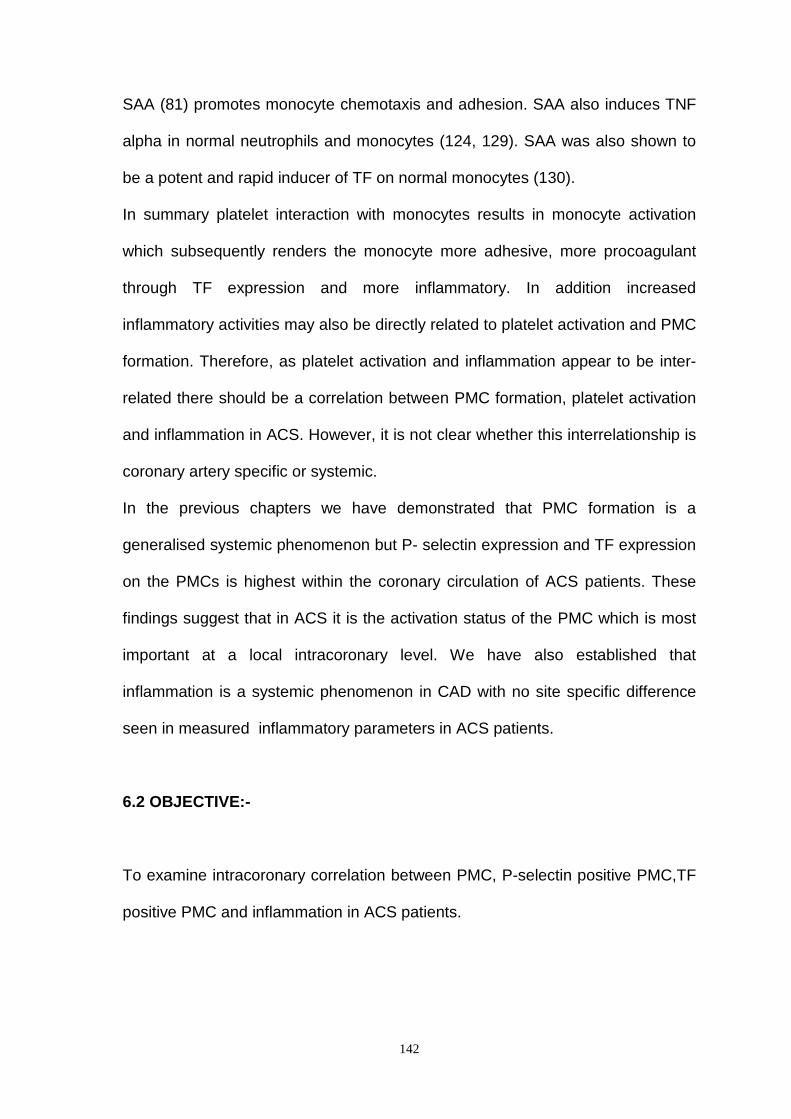
142
SAA (81) promotes monocyte chemotaxis and adhesion. SAA also induces TNF
alpha in normal neutrophils and monocytes (124, 129). SAA was also shown to
be a potent and rapid inducer of TF on normal monocytes (130).
In summary platelet interaction with monocytes results in monocyte activation
which subsequently renders the monocyte more adhesive, more procoagulant
through TF expression and more inflammatory. In addition increased
inflammatory activities may also be directly related to platelet activation and PMC
formation. Therefore, as platelet activation and inflammation appear to be inter-
related there should be a correlation between PMC formation, platelet activation
and inflammation in ACS. However, it is not clear whether this interrelationship is
coronary artery specific or systemic.
In the previous chapters we have demonstrated that PMC formation is a
generalised systemic phenomenon but P- selectin expression and TF expression
on the PMCs is highest within the coronary circulation of ACS patients. These
findings suggest that in ACS it is the activation status of the PMC which is most
important at a local intracoronary level. We have also established that
inflammation is a systemic phenomenon in CAD with no site specific difference
seen in measured inflammatory parameters in ACS patients.
6.2 OBJECTIVE:-
To examine intracoronary correlation between PMC, P-selectin positive PMC,TF
positive PMC and inflammation in ACS patients.

143
6.3 PATIENTS AND METHODS:-
Twenty three patients with acute ACS (15 STEMI and 8 NSTEMI) who had
undergone percutaneous coronary intervention (PCI) were recruited for the
study. Patients‘ details, recruitment process and characteristics have already
been described in the previous chapters. Sample collection, flow cytometry and
ELISA methods have also been described in the previous chapter.
6.4 STATISTICAL METHODS:-
Data were presented as median (Interquartile range). Correlation analysis was
performed with Spearman’s correlation analysis. Two-tailed p values < 0.05 was
considered statistically significant. All calculations were performed using
graphpad prism® (version 5) software.

144
6.5 RESULTS:-
CORRELATION OF INTRACORONARY PMC, P- SELECTIN POSITIVE PMC
AND INFLAMMATION IN ACS PATIENTS:-
A significant positive correlation was found between intracoronary PMC and
TNF-alpha (r = 0.54, p= 0.02) and a non-significant positive correlation with
intracoronary CRP (r= 0.32, p=0.14), SAA (r= 0.32, p=0.14) and IL-6 (r= 0.47, p=
0.06).
Intracoronary P-selectin positive PMCs also demonstrated a non significant
positive correlation with TNF –alpha (p= 0.53, r= 0.09), CRP (r= 0.16, p= 0.56)
and SAA (r=0.12, p=0.66) similarly TF positive PMCs have shown a non
significant positive correlation with IL-6 (r= 0.21, p= 0.53) and TNF- alpha
(r=0.23, p= 0.48).
Ten patients in the ACS group were found to have intracoronary SAA
concentrations ≥ 4.5 mg/l (≥ overall median value). In this subgroup of patients
with relatively higher intracoronary inflammatory burden there was a significant
correlation between intracoronary SAA and intracoronary PMC expression (r=
0.75, p= 0.01) (figure -6.1). This correlation was not observed in the peripheral
circulation of the ACS patients. (data not shown ).

145
Figure –6. 1. Correlation of intracoronary PMC and SAA in ACS. In ACS patients
with higher intracoronary inflammatory burden SAA (≥ 4.5 mg/l) there was
significant correlation of intracoronary PMC and SAA.
0 50 100 1500
5
10
15
20
25
Coronary SAA (mg/l)
PM
C%
Mo
no
cyte
r= 0.75, p= 0.01

146
CORRELATION OF INTRACORONARY PMCs AND INFLAMMATORY
MARKERS IN THE STEMI SUBGROUP.
In the STEMI subgroup intracoronary PMC expression demonstrated a
significant positive correlation with intracoronary TNF-alpha (r= 0.68, p= 0.03)
(figure-6.2). TF positive PMCs (% monocytes) also showed a positive correlation
with intracoronary TNF-alpha and IL-6 (r=0.66, p=0.05 & r=0.71, p= 0.05
respectively) (Figure-6.3). In the peripheral circulation no significant correlations
were observed between PMC expression, P-selectin and TF factor expression
and inflammatory markers (data not shown).

147
Figure –6.2.Correlation of intracoronary PMC and TNF- alpha in STEMI. In
STEMI patients intracoronary PMC has significant positive correlation with
intracoronary TNF- alpha.
0 10 20 30 400
20
40
60
80
CO PMC STEMI (% Monocyte)
TN
F-
(pg
/ml)
r=0.68, p=0.03

148
Figure-6.3 Correlation of intracoronary TF positive PMC with TNF- alpha and IL-6
in STEMI.Intracoronary TF positive PMC showed positive correlations with
intracoronary IL-6 (n= 8) and TNF- alpha (n=9).
0 1 2 3 40
20
40
60
80
% Monocyte
TN
F-
(pg
/ml)
r= 0.66, p= 0.05
0 1 2 3 40
10
20
30
40
50
IL-6
pg
/ml
r= 0.71, p=0.05
% Monocyte

149
6.6 DISCUSSION:-
The finding of a significant positive correlation with intracoronary PMC and TNF
alpha overall in the ACS patients and in the STEMI subgroup emphasises the
local relationship between PMC formation and inflammation in the acute stage of
the disease. Similarly, positive correlation of intracoronary TF positive PMC with
TNF alpha and IL-6 in the STEMI patients provides a further possible
mechanistic insight into the role of activated monocytes within the PMC in the
pathogenesis of intracoronary inflammation and thrombosis. Unstimulated
monocytes are not proinflammatory but within activated monocytes there is
significant upregulation in the expression of inflammatory parameters. TF
positive complexes indicate presence of monocyte activation within these
complexes. It has previously been shown binding of CD40L on platelets to CD 40
on monocytes leads to monocyte activation and production of cytokines including
IL-6 which is associated with unstable angina (10),(11). Monocyte expression of
chemokines is also shown to be induced by thrombin activated platelets and is
regulated by NF-kB activity. Ligation of monocyte PSGL-1 together with RANTES
induces NF-kB activity and subsequent secretion of MCP-1 , TNF-alpha and IL-8
(8, 20). Therefore, from the summary of in vitro studies it is clear that upon
activation monocytes secrete different inflammatory cytokines including TNF
alpha and IL-6. Our in vivo study in patients with acute STEMI supports these in
vitro study findings.
In the ACS patients intracoronary PMC has shown positive correlation with CRP,
SAA, IL-6 andTNF- alpha suggesting a local intracoronary relationship between
PMC and inflammatory mediators. The correlation is statistically significant only
with TNF- alpha. This finding suggest TNF alpha may be a more coronary artery

150
specific marker dependent on PMC formation. It has been shown before that
TNF- alpha is a central mediator of inflammation and is localised in
atherosclerotic lesions. And may contribute to triggering, progression and rupture
of plaque (189-191). It has been reported previously that TNF- alpha can cause
depression of cardiac contractility either directly (137) or by induction of inducible
NO synthase (Inos) in cardiocytes and other cellular constituents within the heart
(138).
Correlation of intracoronary SAA with PMC in the subgroup of patients with
higher inflammatory burden (SAA≥ 4.5 mg/l) further emphasises the local
intracoronary inter-relationship between PMC formation and inflammation.
Dotsenko et al. in a previous study demonstrated an association between
elevated CRP and increased PMC in patients with atherosclerotic patients
suggesting a relationship between inflammation and PMC formation (192).
Positive correlation of PMC and TF expression with intracoronary TNF alpha
suggests PMC may play a contributory role through heightened TF expression in
the pathogenesis of intracoronary inflammation in the STEMI patients. Celi et al
demonstrated platelet surface P-selectin induces the expression of TF on
monocytes and promotes fibrin deposition within the growing thrombus at the site
of the vascular injury (6),(193).
6.7 CONCLUSION:-
This study demonstrates a local intracoronary interrelationship between PMC
formation and inflammation in ACS patients. TNF- alpha is the most significant
intracoronary inflammatory marker related to PMC formation and monocyte

151
activation within the PMC; particularly in STEMI patients in the hyperacute
stages of intracoronary inflammation. In STEMI patients PMC may play a
significant local role through TF expression in determining TNF-alpha and IL-6
production.

152
CH APTER 7
SOLUBLE P SELECTIN AND P SELECTIN POSITIVE
PLATELET MONOCYTE COMPLEXES (PMCs) AS
DETERMINANTS OF INFLAMMATION IN PATIENTS WITH
CORONARY ARTERY DISEASE.

153
7.1
Introduction.
7.2 Objectives.
7.3 Patients and Methods.
7.4 Exclusion criteria.
7.5 Statistical Method.
7.6 Results.
7.7Discussion.

154
7.1 INTRODUCTION:-
Heightened level of inflammation is a well recognised phenomenon in patients
with CAD. Increased CRP has been implicated as an adverse prognostic marker
in patients with coronary artery disease (82-84). Formation of PMCs can be a link
between platelet mediated thrombogenesis and monocyte mediated
inflammation but the mechanism by which PMCs contribute to intracoronary
inflammation is not clearly established. One hypothesis is that binding of
activated platelets may alter monocyte activation status resulting in an increase
in TF expression from the monocyte component acting as a trigger for the
initiation of a cascade of thrombogenesis and the release of proinflammatory
cytokines, including IL-6, IL-1β and IL-12. However, binding with unstimulated
platelets may not be associated with a similar cellular response (13). Therefore,
in the absence of platelet activation PMC formation may just represent a
physiological phenomenon. Depending upon the level of activation, the platelets
within PMCs may actually be responsible for triggering a pro-inflammatory
response in monocytes. On the other hand there is evidence that an increased
inflammatory state is paralleled by activation of platelets with phosphorylation of
the membrane protein p-selectin in the platelet alpha granule membrane
(86).Then the granule moves to the platelet surface exposing P-selectin and
tethering the platelet with the monocyte via PSGL-1 to form PMC. Apart from cell
surface expression a variant of P-selectin can also be detected in the plasma (s
p- selectin) leading to presumption that they are secreted, shed or cleaved from
the cell surface however, the changes in soluble levels in the plasma may not
reflect levels at the cell membrane (194). Mere PMC formation may not have any

155
significant relationship with inflammation but platelet activation status within this
complexes (P-selectin positive PMC) and sP-selectin may have some
relationship with inflammation in coronary artery disease. P-selectin has been
implicated at the cellular level as a causative factor for inflammation as it
contributes to leukocyte rolling and triggers TF release from the acitvated
monocytes (195). So far no direct correlation with inflammatory biomarkers such
as CRP, SAA and TNF alpha have been demonstrated in vivo in human subjects
with CAD.
There have been some suggestions that CRP and SAA may play a local
intracoronary role in the pathogenesis of ACS (53, 98, 102, 196). If these
inflammatory markers are responsible for local intracoronary inflammation we
expect their concentrations will be higher in the coronary circulation compared to
the peripheral circulation. However, data largely implicates these biomarkers are
a systemic response synthesised in response to coronary artery disease (117,
197, 198).
7.2 OBJECTIVES:-
To investigate whether a relationship exists between P-selectin expression on
the PMCs and s P-selectin levels.
To examine whether any differential expression exists between expression of
inflammatory biomarkers in the coronary circulation compared with the peripheral
circulation.

156
To investigate the relationship between different inflammatory markers e.g CRP,
SAA and TNF- alpha and P-selectin expression on the PMCs and soluble plasma
P-selectin.
7.3 PATIENTS AND METHODS:-
Thirty patients with coronary artery disease were recruited for the study. Consent
was obtained from 15 patients with STEMI immediately after their admission to
hospital for PPCI, 8 patients with NSTEMI within 72 hours of their symptom onset
and 7 patients with stable angina just before their elective percutaneous coronary
intervention (PCI). Prior ethical approval was obtained from the local research
and ethics committee. Patients with STEMI and NSTEMI were loaded with 600
mg of clopidogrel and 300 mg of Aspirin prior to the percutaneous procedure.
Stable angina patients had already received 75 mg of Aspirin and 75mg of
Clopidogrel for 7 days prior to the procedure according to the protocol for
performing elective PCI in our hospital. Prior to PCI all the patients were treated
with weight adjusted unfractionated heparin to maintain the ACT between 200-
250 seconds.
Samples were collected from the coronary artery distal to the lesion, aorta and
right atrium as previously described in chapter 2. PMC estimation and P-selectin
expression on the PMCs were estimated by flow cytometry as described in
chapter 2. PMCs were expressed as % monocyte and P- selectin positive PMCs
were expressed as % monocyte as well as % of PMC.

157
High sensitive CRP, SAA,TNF-alpha and s-Pselectin were estimated from double
spun platelet poor plasma prepared from collected blood samples from all the 3
sites as described in chapter 2.
Coronary samples distal to the lesion were taken to estimate the local
intracoronary concentrations of soluble P-selectin, inflammatory markers, total
PMC and P-selectin positive PMC expression. Aortic and right atrial samples
were taken to estimate similar parameters in the arterial and venous circulation
respectively. Overall median values were measured to estimate the systemic
expression of PMC, P-selectin positive PMC, CRP, SAA and TNF- alpha. Overall
median values were calculated by calculating the median concentrations from all
the 3 sites rather than having a single value from a venous sample.
7.4 EXCLUSION CRITERIA:--
Patients with renal failure, prior coronary artery bypass grafts and who have
already received Glycoprotein IIb/IIIa antagonists prior to sampling were
excluded.
7.5 STATISTICAL METHODS:-
Data were presented as median and inter-quartile range. Inflammatory
parameters, PMC, P-selectin positive PMC in the coronary circulation, aorta and
right atrium were compared using the non parametric Kruskal- Wallis one way
analysis of variance test. Correlation analysis was performed by using
spearman’s correlation analysis. Two tailed p value < 0.05 was considered
significant.

158
7.6 RESULTS:-
Patient demographics and coronary characteristics are described in table 7.1. No
significant site specific difference in PMC expression was seen. Similarly there
was no significant site specific difference in CRP, TNF alpha, SAA and s P-
selectin concentration (table-7.2). However, percentage of PMC expressing P-
selectin on their surface (P-selectin positive PMC) was significantly higher in the
coronary circulation compared with the aorta and right atrium (p= 0.02) (Table-
7.2)

159
Table-7.1 Baseline patient and coronary characteristics.
Age median (IQR), years 61(51-72)
Male 24
Female 6
Risk Factors
History of hypertension 12
History of smoking 16
Diabetes 3
Dyslipidaemia 14
Angiographic
characteristics
1 vessel disease 16
2 vessel disease 9
3 vessel disease 5
Culprit vessel
Left Anterior Descending 10
Left Circumflex 8
Right Coronary Artery 12

160
Table 7. 2:- Site specific concentrations of inflammatory markers, s P- selectin,
PMC and P-selectin positive PMC in patients with coronary artery disease
(n=30). Percentage of P- selectin positive PMC (%PMC) was significantly higher
in the coronary circulation compared with aorta and right atrium.
Aorta
Median (IQR)
Right Atrium
Median (IQR)
CoronaryArtery
Median (IQR)
p
CRP mg/l 1.7 (0.8- 5.5) 1.9 (0.8- 5.8) 1.6(0.72- 4.85) 0.77
SAA mg/l 3.8(2.05- 9.35) 3.6 (2.4-8.8) 2.8 (1.5- 9.0) 0.77
TNF–alpha pg/ml 27.30 (14.25-
43.11)
34.86 (19.38-
45.81)
23.06 (13.24-
35.57)
0.27
sP -selectin ng/ml 21.90 (14.90-
29.80)
23.40 (14.20-
39.00)
16.80 (13.00-
25.20)
0.49
P-Selectin positive
PMC (% monocyte)
1.98 (0.98- 3.06) 1.62 (0.89-
3.34)
3.08 (1.77- 7.85) 0.12
P-selectin positive
PMC (%PMC)
15.37 (9.88-19.18) 15.98 (10.96-
19.76)
24.17 (17.95-
43.37)
0.02

161
Overall median PMC [% monocyte ; median (IQR)] was 9.98 (7.13- 17.46)% and
overall median P selectin positive PMC [% PMC median (IQR)] was 16.89 (11.14
– 29.76) %; % monocyte median (IQR) was 2.07 (1.15- 3.77)%.]
Overall median P selectin positive PMC (% monocyte) showed a significant
positive correlation with overall median CRP (r= 0.37, p=0.005), overall median
SAA (r=0.38, p=0.003) and a non significant positive correlation with overall
median TNF- alpha (r= 0.26, p= 0.09) (Figure- 7.1)
Similarly, overall median s P-selectin showed a significant positive correlation
with overall median CRP, SAA and TNF-alpha (Figure-7. 2)
In addition there was a significant positive correlation between overall median P-
selectin positive PMC (%monocyte) and s P-Selectin. (Figure-7. 3)

162
0 5 10 15 20 250
20
40
60 r=0.38, p=0.003
P selectin positive PMC (% Monocyte)
SA
Am
g/l
(a) (b)
0 5 10 15 20 250
20
40
60
80r= 0.26, p=0.09
P selectin positive PMC (% Monocyte)
TN
F-
(pg
/ml)
(c)
Figure 7.1- Correlation of overall median P-Selectin positive PMC with
inflammatory parameters in CAD Patients Overall median P- selectin positive
PMC (%monocyte) has significant positive correlation with overall median CRP
(a), SAA (b) and non-significant positive correlation with overall median TNF-
alpha (c).
0 5 10 15 20 250
10
20
30
40r= 0.37, p=0.005
P selectin positive PMC (% Monocyte)
CR
Pm
g/l
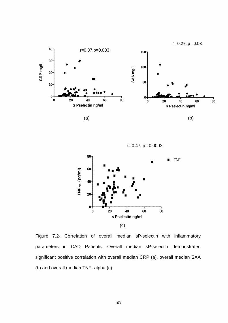
163
0 20 40 60 800
10
20
30
40 r=0.37,p=0.003
S Pselectin ng/ml
CR
Pm
g/l
(a) (b)
0 20 40 60 800
20
40
60
80TNF
r= 0.47, p= 0.0002
s Pselectin ng/ml
TN
F-
(pg
/ml)
(c)
Figure 7.2- Correlation of overall median sP-selectin with inflammatory
parameters in CAD Patients. Overall median sP-selectin demonstrated
significant positive correlation with overall median CRP (a), overall median SAA
(b) and overall median TNF- alpha (c).
0 20 40 60 800
50
100
150
r= 0.27, p= 0.03
s Pselectin ng/ml
SA
Am
g/l
164
0 5 10 15 20 250
20
40
60
80
r= 0.34, p= 0.03
Pselectin positive PMC (% monocyte)
sp
sele
cti
nn
g/l
Figure - 7.3 Correlation of P- selectin positive PMC and s P-selectin. P-selectin
positive PMC (% monocyte) and sP-selectin showed a significant positive
correlation.

165
7.7 DISCUSSION:-
Although traditionally CRP and SAA production by the liver are regarded as a
systemic response to inflammation it has now been shown that cells within the
atherosclerotic plaque also synthesise CRP (98, 102, 196). Macrophage and
smooth muscle - like cells in atherosclerotic plaques are shown to produce seven
times more CRP mRNA than the liver (98). SAA has also been shown to be
chemotactic for monocytes (121). It has also been shown that SAA proteins are
produced by cells of the artery wall. The transition from chronic stable
atherosclerotic coronary artery disease into an acute coronary syndrome has
been found to be associated with an increase in inflammatory activity within the
plaque, reflected in an increase in C- reactive protein and SAA levels (83) (123).
These findings raise the question whether these pro inflammatory cytokines play
any pathogenic local role within the coronary artery of the ACS patients.
Therefore, if inflammation is a local intracoronary event one would expect local
intracoronary levels of inflammatory markers to be higher in the coronary
circulation compared to the peripheral circulation. Maier et al. (53) demonstrated
increase in levels of IL-6 by > 70% and SAA by around 10 % in the culprit
coronary artery relative to those in the aorta in the STEMI patients. Our study
does not support the hypothesis that inflammation is primarily an intracoronary
event as there is no differential increased expression of intracoronary
inflammatory parameters in the coronary compared with systemic circulation.
Interestingly, contrary to what one might have expected, CRP, SAA and TNF
alpha concentrations are non-significantly lower in the coronary circulation
compared to the peripheral circulation. Decreased intracoronary CRP values in

166
acute myocardial infarction patients have been reported by Maier et al (53) as
described in the chapter 5. This could be due to the fact CRP must have
undergone partial catabolism in the vessel wall while it is trapped. CRP protein
within coronary plaques have been reported in previous studies (97), (187).
Upon activation from inflammatory cytokines, mainly TNF- alpha, cell surface
expression of P-selectin is enhanced. This process is vital in the early phase of
inflammation mediating leukocyte recruitment and transient leukocyte to
endothelial interaction (tethering and rolling phase) (199, 200) (201). P- selectin
is expressed in platelets , stored in membrane alpha granules. Upon activation, it
is translocated to the surface of the platelets (202, 203).
P- selectin sheds from the surface of platelets and circulate in the soluble form
as well. P –selectin, either on the surface of activated platelets or as a soluble
form in plasma, may play a significant role in thrombosis, inflammation and
thrombosis- mediated inflammation events. One hypothesis is that binding of
activated platelets alters monocyte activation status resulting in an increase in
TF expression from the monocyte component acting as a trigger for the initiation
of a cascade of thrombogenesis and the release of proinflammatory cytokines
promotes fibrin deposition within the growing thrombus at the site of the vascular
injury (193),(204, 205). It is not clear how P-selectin, either on the cell surface or
in the soluble form, contributes to inflammation as previous data have not
established a correlation between P- selectin and inflammatory parameters.
Schmacher et al. measured IL-6, CRP, TNF alpha alongside the slouble
adhesion molecules but they did not find any significant correlations between s
P- selectin and CRP, TNF- alpha or IL-6 (79),(80). To our knowledge our study is
the first of its kind where we have clearly demonstrated significant positive

167
correlations of sP- selectin with CRP, SAA and TNF- alpha. We have also
demonstrated a significant positive correlation of P-selectin positive PMCs (%
monocyte) with the CRP and SAA. These findings establish the relationship of s
P- selectin with inflammation in CAD. Similarly relationship of P-selectin positive
PMC (% monocyte) with inflammatory parameters emphasises the fact that
monocytes bound within the complex with P selectin positive platelets may play a
significant role in the systemic inflammatory response associated with ACS. Also
in this study we have demonstrated a significant correlation between P-selectin
positive PMC and sP- selectin. We conclude P -selectin plays a significant role in
the inflammatory process in coronary artery disease both in the soluble form and
by influencing monocytes within the platelet monocyte complexes. P-selectin has
been linked with an enhanced inflammatory response, restenosis and increased
risk of cardiovascular events and may be a predictor of future thrombotic events
(74). Though this study does not definitely establish the causal relationship of P-
selectin and inflammation nevertheless it emphasises the importance of platelet
activation, P- selectin expression and inflammation in coronary artery disease. P-
selectin may be a future therapeutic target to modulate the inflammatory
response in coronary artery disease.

168
CHAPTER 8
DISCUSSION
169
Over the last decade PMCs have generated considerable interest in
understanding the pathophysiology of ACS. PMCs have been implicated as a link
between platelet mediated thrombogenesis and monocyte mediated
inflammation. The exact mechanism through which PMCs contribute to the
pathogenesis of ACS is yet to be understood. Similarly it is not clear whether
PMC formation is a systemic phenomenon or a local intracoronary phenomenon
which augments the intracoronary inflammatory response in patients with ACS.
There are some suggestions that PMC formation may be merely a physiological
phenomenon without much pathological significance (13). On the other hand
existing flow cytometry methods have not taken into consideration the possibility
of coincident events which may result in over estimation of PMCs. Similarly, the
exact role of inflammation in the pathogenesis of ACS is also controversial. Data
suggests that inflammatory markers can be generated locally in the coronary
artery of the patients with acute coronary syndrome (25). With this background in
mind the first aim of this thesis was to establish a flow cytometric method which
can exclude the coincident events carefully so that the true PMCs can be
estimated accurately. The aim of further investigations in this study was to first
evaluate whether PMC formation and inflammation represented systemic or local
phenomena. Also, to establish the relationship between PMC, and inflammation
and to find out whether any local intracoronary relationship existed between PMC
and inflammation in the ACS patients.
In chapter 3 we have described the flow cytometric methodology using a double
discriminator technique previously designed to overcome the problem of co-
incident analysis of doublets of bare nuclei in DNA ploidy experiments. With this
technique we have successfully excluded coincident platelets and monocytes as

170
they have a longer time of flight through the focal point of the laser. Using pulse
processed analysis, they elicit signals with a greater fluorescent width than the
single cell and a concomitant greater signal area. Using this technique we have
successfully excluded the coincident events with wider fluorescent width and
managed to accurately evaluate true PMC expression.
In the chapter 4 we have demonstrated that overall PMC is higher in all vascular
compartments of ACS patients compared with stable angina patients. However,
no differential expression of heightened PMC expression was obvious within the
intracoronary compartment compared with venous or aortic, suggestin a general
systemic phenomenon.We have demonstrated that activated PMC (complexes
expressing P-selectin or TF, suggestive of activated platelets and monocytes
within the complexes respectively) have been found to be significantly increased
with a significant trans - culprit lesion gradient demonstrated. We have also
demonstrated P selectin positive PMC expression in the coronary circulation is
significantly higher in ACS compared with stable angina patients. These findings
suggest differential site specific platelet and monocyte activation status within the
complex may be more important in the pathogenesis of ACS rather than overall
PMC formation.
In chapter 5 we have demonstrated that inflammatory burden is higher in ACS
patients compared with stable angina patients but we have not found any
significant trans-coronary gradient in inflammatory parameters to suggest that
inflammation is a local intracoronary event. We conclude inflammation is a
systemic phenomenon in coronary artery disease.
In Chapter 6 we have described the local intracoronary relationship of PMC and
inflammation in ACS patients. We have found positive correlation with

171
intracoronary TNF alpha with PMC and TF positive PMC .This findings suggest
local monocyte activation on the PMC may be the trigger for local intracoronary
inflammation.
In chapter 7 we have emphasised the role of P selectin and systemic
inflammation in patients with coronary artery disease in general both in the
soluble form and on the cell surface of PMC. In this chapter we have also
demonstrated the significant positive correlation between soluble and PMC
surface P-selectin.
Our study is an observational study and although it does not establish a causal
relationship between PMC formation and inflammation in ACS patients.
Nevertheless our data importantly establishes the fact mere PMC formation may
not have much pathological significance but instead represents a general
systemic marker of inflammation. However, it is the platelet and monocyte
activation status within the complex which appears directly related to
inflammation in patients with ACS. This provides an important pathophysiological
insight into patelet-monocyte interaction in coronary disease. Our data also
support the hypothesis that PMC may contribute to systemic inflammation
through P-selectin expression and local intracoronary inflammation through
increased P-selectin and TF expression.
Future Work-
This study provides some mechanistic insight about the role of P-selectin and TF
expression on the PMC in relation to systemic as well as local intracoronary
inflammation in ACS patients. The activated PMC (expressing P- selectin or TF)
may represent a novel therapeutic targets in abrogating the intracoronary
inflammatory burden in ACS. This in turn by reducing intracoronary

172
thrombogenesis and cytokine-mediated microcellular damage may represent a
strategy to reduce microvascular injury and aid myocardial salvage.

173
BIBLIOGRAPHY

174
1. Fitzgerald DJ, Roy L, Catella F, FitzGerald GA. Platelet activation in
unstable coronary disease. N Engl J Med. 1986;315(16):983-9.
2. Neumann FJ, Marx N, Gawaz M, Brand K, Ott I, Rokitta C, et al. Induction
of cytokine expression in leukocytes by binding of thrombin-stimulated
platelets. Circulation. 1997;95(10):2387-94.
3. Theilmeier G, Lenaerts T, Remacle C, Collen D, Vermylen J, Hoylaerts
MF. Circulating activated platelets assist THP-1 monocytoid/endothelial
cell interaction under shear stress. Blood. 1999;94(8):2725-34.
4. Merhi Y, Provost P, Guidoin R, Latour JG. Importance of platelets in
neutrophil adhesion and vasoconstriction after deep carotid arterial injury
by angioplasty in pigs. Arterioscler Thromb Vasc Biol. 1997;17(6):1185-
91.
5. Michelson AD, Barnard MR, Hechtman HB, MacGregor H, Connolly RJ,
Loscalzo J, et al. In vivo tracking of platelets: circulating degranulated
platelets rapidly lose surface P-selectin but continue to circulate and
function. Proceedings of the National Academy of Sciences of the United
States of America. 1996;93(21):11877-82.
6. Celi A, Pellegrini G, Lorenzet R, De Blasi A, Ready N, Furie BC, et al. P-
selectin induces the expression of tissue factor on monocytes.
Proceedings of the National Academy of Sciences of the United States of
America. 1994;91(19):8767-71.
7. Shebuski RJ, Kilgore KS. Role of inflammatory mediators in
thrombogenesis. J Pharmacol Exp Ther. 2002;300(3):729-35.

175
8. Weyrich AS, Elstad MR, McEver RP, McIntyre TM, Moore KL, Morrissey
JH, et al. Activated platelets signal chemokine synthesis by human
monocytes. J Clin Invest. 1996;97(6):1525-34.
9. Henn V, Slupsky JR, Grafe M, Anagnostopoulos I, Forster R, Muller-
Berghaus G, et al. CD40 ligand on activated platelets triggers an
inflammatory reaction of endothelial cells. Nature. 1998;391(6667):591-4.
10. Alderson MR, Armitage RJ, Tough TW, Strockbine L, Fanslow WC,
Spriggs MK. CD40 expression by human monocytes: regulation by
cytokines and activation of monocytes by the ligand for CD40. J Exp Med.
1993;178(2):669-74.
11. Biasucci LM, Vitelli A, Liuzzo G, Altamura S, Caligiuri G, Monaco C, et al.
Elevated levels of interleukin-6 in unstable angina. Circulation.
1996;94(5):874-7.
12. Bataille R, Klein B. C-reactive protein levels as a direct indicator of
interleukin-6 levels in humans in vivo. Arthritis and rheumatism.
1992;35(8):982-4.
13. Bournazos S, Rennie J, Hart SP, Fox KA, Dransfield I. Monocyte
functional responsiveness after PSGL-1-mediated platelet adhesion is
dependent on platelet activation status. Arterioscler Thromb Vasc Biol.
2008;28(8):1491-8.
14. Furman MI, Benoit SE, Barnard MR, Valeri CR, Borbone ML, Becker RC,
et al. Increased platelet reactivity and circulating monocyte-platelet
aggregates in patients with stable coronary artery disease. J Am Coll
Cardiol. 1998;31(2):352-8.
176
15. Li N, Hu H, Lindqvist M, Wikstrom-Jonsson E, Goodall AH, Hjemdahl P.
Platelet-leukocyte cross talk in whole blood. Arterioscler Thromb Vasc
Biol. 2000;20(12):2702-8.
16. da Costa Martins PA, van Gils JM, Mol A, Hordijk PL, Zwaginga JJ.
Platelet binding to monocytes increases the adhesive properties of
monocytes by up-regulating the expression and functionality of beta1 and
beta2 integrins. J Leukoc Biol. 2006;79(3):499-507.
17. Yago T, Tsukuda M, Minami M. P-selectin binding promotes the adhesion
of monocytes to VCAM-1 under flow conditions. J Immunol.
1999;163(1):367-73.
18. Atarashi K, Hirata T, Matsumoto M, Kanemitsu N, Miyasaka M. Rolling of
Th1 cells via P-selectin glycoprotein ligand-1 stimulates LFA-1-mediated
cell binding to ICAM-1. J Immunol. 2005;174(3):1424-32.
19. Lindmark E, Tenno T, Siegbahn A. Role of platelet P-selectin and CD40
ligand in the induction of monocytic tissue factor expression. Arterioscler
Thromb Vasc Biol. 2000;20(10):2322-8.
20. Weyrich AS, McIntyre TM, McEver RP, Prescott SM, Zimmerman GA.
Monocyte tethering by P-selectin regulates monocyte chemotactic protein-
1 and tumor necrosis factor-alpha secretion. Signal integration and NF-
kappa B translocation. J Clin Invest. 1995;95(5):2297-303.
21. Furman MI, Barnard MR, Krueger LA, Fox ML, Shilale EA, Lessard DM, et
al. Circulating monocyte-platelet aggregates are an early marker of acute
myocardial infarction. J Am Coll Cardiol. 2001;38(4):1002-6.
177
22. Sarma J, Laan CA, Alam S, Jha A, Fox KA, Dransfield I. Increased
platelet binding to circulating monocytes in acute coronary syndromes.
Circulation. 2002;105(18):2166-71.
23. Ray MJ, Walters DL, Bett JN, Cameron J, Wood P, Aroney CN. Platelet-
monocyte aggregates predict troponin rise after percutaneous coronary
intervention and are inhibited by Abciximab. Int J Cardiol.
2005;101(2):249-55.
24. Ashman N, Macey MG, Fan SL, Azam U, Yaqoob MM. Increased platelet-
monocyte aggregates and cardiovascular disease in end-stage renal
failure patients. Nephrol Dial Transplant. 2003;18(10):2088-96.
25. Ko YG, Jung JH, Park S, Choi E, Joung B, Hwang KC, et al. Inflammatory
and vasoactive factors in the aspirate from the culprit coronary artery of
patients with acute myocardial infarction. Int J Cardiol. 2006;112(1):66-71.
26. Kotani J, Nanto S, Mintz GS, Kitakaze M, Ohara T, Morozumi T, et al.
Plaque gruel of atheromatous coronary lesion may contribute to the no-
reflow phenomenon in patients with acute coronary syndrome. Circulation.
2002;106(13):1672-7.
27. Sezer M, Okcular I, Goren T, Oflaz H, Nisanci Y, Umman B, et al.
Association of haematological indices with the degree of microvascular
injury in patients with acute anterior wall myocardial infarction treated with
primary percutaneous coronary intervention. Heart. 2007;93(3):313-8.
28. Ott I, Neumann FJ, Gawaz M, Schmitt M, Schomig A. Increased
neutrophil-platelet adhesion in patients with unstable angina. Circulation.
1996;94(6):1239-46.

178
29. Mickelson JK, Lakkis NM, Villarreal-Levy G, Hughes BJ, Smith CW.
Leukocyte activation with platelet adhesion after coronary angioplasty: a
mechanism for recurrent disease? J Am Coll Cardiol. 1996;28(2):345-53.
30. Botto N, Sbrana S, Trianni G, Andreassi MG, Ravani M, Rizza A, et al. An
increased platelet-leukocytes interaction at the culprit site of coronary
artery occlusion in acute myocardial infarction: a pathogenic role for "no-
reflow" phenomenon? Int J Cardiol. 2007;117(1):123-30.
31. Patel PB, Pfau SE, Cleman MW, Brennan JJ, Howes C, Remetz M, et al.
Comparison of coronary artery specific leukocyte-platelet conjugate
formation in unstable versus stable angina pectoris. Am J Cardiol.
2004;93(4):410-3.
32. Wang J, Zhang S, Jin Y, Qin G, Yu L, Zhang J. Elevated levels of platelet-
monocyte aggregates and related circulating biomarkers in patients with
acute coronary syndrome. Int J Cardiol. 2007;115(3):361-5.
33. Hagberg IA, Lyberg T. Evaluation of circulating platelet-leukocyte
conjugates: a sensitive flow cytometric assay well suited for clinical
studies. Platelets. 2000;11(3):151-60.
34. Li N, Goodall AH, Hjemdahl P. A sensitive flow cytometric assay for
circulating platelet-leucocyte aggregates. Br J Haematol. 1997;99(4):808-
16.
35. Barnard MR, Krueger LA, Frelinger AL, 3rd, Furman MI, Michelson AD.
Whole blood analysis of leukocyte-platelet aggregates. Curr Protoc
Cytom. 2003;Chapter 6:Unit 6 15.
36. Harding SA, Din JN, Sarma J, Jessop A, Weatherall M, Fox KA, et al.
Flow cytometric analysis of circulating platelet-monocyte aggregates in

179
whole blood: methodological considerations. Thromb Haemost.
2007;98(2):451-6.
37. Pearson L, Thom J, Adams M, Oostryck R, Krueger R, Yong G, et al. A
rapid flow cytometric technique for the detection of platelet-monocyte
complexes, activated platelets and platelet-derived microparticles. Int J
Lab Hematol. 2009;31(4):430-9.
38. Bihari P, Fent J, Hamar J, Furesz J, Lakatos S. An easy-to-use practical
method to measure coincidence in the flow cytometer--the case of
platelet-granulocyte complex determination. J Biochem Biophys Methods.
2008;70(6):1080-5.
39. Snow C. Flow cytometer electronics. Cytometry A. 2004;57(2):63-9.
40. Keij JF, van Rotterdam A, Groenewegen AC, Stokdijk W, Visser JW.
Coincidence in high-speed flow cytometry: models and measurements.
Cytometry. 1991;12(5):398-404.
41. Palmerini T, Coller BS, Cervi V, Tomasi L, Marzocchi A, Marrozzini C, et
al. Monocyte-derived tissue factor contributes to stent thrombosis in an in
vitro system. J Am Coll Cardiol. 2004;44(8):1570-7.
42. Wilcox JN, Smith KM, Schwartz SM, Gordon D. Localization of tissue
factor in the normal vessel wall and in the atherosclerotic plaque. Proc
Natl Acad Sci U S A. 1989;86(8):2839-43.
43. Mach F, Schonbeck U, Bonnefoy JY, Pober JS, Libby P. Activation of
monocyte/macrophage functions related to acute atheroma complication
by ligation of CD40: induction of collagenase, stromelysin, and tissue
factor. Circulation. 1997;96(2):396-9.

180
44. He M, He X, Xie Q, Chen F, He S. Angiotensin II induces the expression
of tissue factor and its mechanism in human monocytes. Thromb Res.
2006;117(5):579-90.
45. Wada H, Kaneko T, Wakita Y, Minamikawa K, Nagaya S, Tamaki S, et al.
Effect of lipoproteins on tissue factor activity and PAI-II antigen in human
monocytes and macrophages. Int J Cardiol. 1994;47(1 Suppl):S21-5.
46. Guha M, Mackman N. The phosphatidylinositol 3-kinase-Akt pathway
limits lipopolysaccharide activation of signaling pathways and expression
of inflammatory mediators in human monocytic cells. J Biol Chem.
2002;277(35):32124-32.
47. Mackman N. Regulation of the tissue factor gene. Thrombosis and
haemostasis. 1997;78(1):747-54.
48. Hansson GK. Inflammation, atherosclerosis, and coronary artery disease.
N Engl J Med. 2005;352(16):1685-95.
49. Del Prete G, De Carli M, Lammel RM, D'Elios MM, Daniel KC, Giusti B, et
al. Th1 and Th2 T-helper cells exert opposite regulatory effects on
procoagulant activity and tissue factor production by human monocytes.
Blood. 1995;86(1):250-7.
50. Meisel SR, Xu XP, Edgington TS, Dimayuga P, Kaul S, Lee S, et al.
Differentiation of adherent human monocytes into macrophages markedly
enhances tissue factor protein expression and procoagulant activity.
Atherosclerosis. 2002;161(1):35-43.
51. Thiruvikraman SV, Guha A, Roboz J, Taubman MB, Nemerson Y, Fallon
JT. In situ localization of tissue factor in human atherosclerotic plaques by

181
binding of digoxigenin-labeled factors VIIa and X. Lab Invest.
1996;75(4):451-61.
52. Annex BH, Denning SM, Channon KM, Sketch MH, Jr., Stack RS,
Morrissey JH, et al. Differential expression of tissue factor protein in
directional atherectomy specimens from patients with stable and unstable
coronary syndromes. Circulation. 1995;91(3):619-22.
53. Maier W, Altwegg LA, Corti R, Gay S, Hersberger M, Maly FE, et al.
Inflammatory markers at the site of ruptured plaque in acute myocardial
infarction: locally increased interleukin-6 and serum amyloid A but
decreased C-reactive protein. Circulation. 2005;111(11):1355-61.
54. Suefuji H, Ogawa H, Yasue H, Kaikita K, Soejima H, Motoyama T, et al.
Increased plasma tissue factor levels in acute myocardial infarction. Am
Heart J. 1997;134(2 Pt 1):253-9.
55. Mallat Z, Benamer H, Hugel B, Benessiano J, Steg PG, Freyssinet JM, et
al. Elevated levels of shed membrane microparticles with procoagulant
potential in the peripheral circulating blood of patients with acute coronary
syndromes. Circulation. 2000;101(8):841-3.
56. Virmani R, Kolodgie FD, Burke AP, Farb A, Schwartz SM. Lessons from
sudden coronary death: a comprehensive morphological classification
scheme for atherosclerotic lesions. Arterioscler Thromb Vasc Biol.
2000;20(5):1262-75.
57. Malarstig A, Tenno T, Johnston N, Lagerqvist B, Axelsson T, Syvanen AC,
et al. Genetic variations in the tissue factor gene are associated with
clinical outcome in acute coronary syndrome and expression levels in

182
human monocytes. Arteriosclerosis, thrombosis, and vascular biology.
2005;25(12):2667-72.
58. Ott I, Koch W, von Beckerath N, de Waha R, Malawaniec A, Mehilli J, et
al. Tissue factor promotor polymorphism -603 A/G is associated with
myocardial infarction. Atherosclerosis. 2004;177(1):189-91.
59. Tutar E, Ozcan M, Kilickap M, Gulec S, Aras O, Pamir G, et al. Elevated
whole-blood tissue factor procoagulant activity as a marker of restenosis
after percutaneous transluminal coronary angioplasty and stent
implantation. Circulation. 2003;108(13):1581-4.
60. Sato Y, Asada Y, Marutsuka K, Hatakeyama K, Sumiyoshi A. Tissue
factor induces migration of cultured aortic smooth muscle cells. Thromb
Haemost. 1996;75(3):389-92.
61. Cirillo P, Cali G, Golino P, Calabro P, Forte L, De Rosa S, et al. Tissue
factor binding of activated factor VII triggers smooth muscle cell
proliferation via extracellular signal-regulated kinase activation.
Circulation. 2004;109(23):2911-6.
62. Pyo RT, Sato Y, Mackman N, Taubman MB. Mice deficient in tissue factor
demonstrate attenuated intimal hyperplasia in response to vascular injury
and decreased smooth muscle cell migration. Thromb Haemost.
2004;92(3):451-8.
63. Falati S, Liu Q, Gross P, Merrill-Skoloff G, Chou J, Vandendries E, et al.
Accumulation of tissue factor into developing thrombi in vivo is dependent
upon microparticle P-selectin glycoprotein ligand 1 and platelet P-selectin.
J Exp Med. 2003;197(11):1585-98.

183
64. Rauch U, Bonderman D, Bohrmann B, Badimon JJ, Himber J, Riederer
MA, et al. Transfer of tissue factor from leukocytes to platelets is mediated
by CD15 and tissue factor. Blood. 2000;96(1):170-5.
65. Aras O, Shet A, Bach RR, Hysjulien JL, Slungaard A, Hebbel RP, et al.
Induction of microparticle- and cell-associated intravascular tissue factor
in human endotoxemia. Blood. 2004;103(12):4545-53.
66. Shet AS, Aras O, Gupta K, Hass MJ, Rausch DJ, Saba N, et al. Sickle
blood contains tissue factor-positive microparticles derived from
endothelial cells and monocytes. Blood. 2003;102(7):2678-83.
67. Woollard KJ. Soluble bio-markers in vascular disease: much more than
gauges of disease? Clin Exp Pharmacol Physiol. 2005;32(4):233-40.
68. Johnston GI, Cook RG, McEver RP. Cloning of GMP-140, a granule
membrane protein of platelets and endothelium: sequence similarity to
proteins involved in cell adhesion and inflammation. Cell.
1989;56(6):1033-44.
69. Merten M, Thiagarajan P. P-selectin expression on platelets determines
size and stability of platelet aggregates. Circulation. 2000;102(16):1931-6.
70. Ishiwata N, Takio K, Katayama M, Watanabe K, Titani K, Ikeda Y, et al.
Alternatively spliced isoform of P-selectin is present in vivo as a soluble
molecule. J Biol Chem. 1994;269(38):23708-15.
71. Fox JE. Shedding of adhesion receptors from the surface of activated
platelets. Blood coagulation & fibrinolysis : an international journal in
haemostasis and thrombosis. 1994;5(2):291-304.

184
72. Fijnheer R, Frijns CJ, Korteweg J, Rommes H, Peters JH, Sixma JJ, et al.
The origin of P-selectin as a circulating plasma protein. Thromb Haemost.
1997;77(6):1081-5.
73. Mehta P, Patel KD, Laue TM, Erickson HP, McEver RP. Soluble
monomeric P-selectin containing only the lectin and epidermal growth
factor domains binds to P-selectin glycoprotein ligand-1 on leukocytes.
Blood. 1997;90(6):2381-9.
74. Ridker PM, Buring JE, Rifai N. Soluble P-selectin and the risk of future
cardiovascular events. Circulation. 2001;103(4):491-5.
75. Blann AD, McCollum CN. Increased soluble P-selectin in peripheral artery
disease: a new marker for the progression of atherosclerosis. Thromb
Haemost. 1998;80(6):1031-2.
76. Woollard KJ, Kling D, Kulkarni S, Dart AM, Jackson S, Chin-Dusting J.
Raised plasma soluble P-selectin in peripheral arterial occlusive disease
enhances leukocyte adhesion. Circulation research. 2006;98(1):149-56.
77. Galkina E, Ley K. Double jeopardy: how soluble P-selectin activates
leukocytes in peripheral arterial occlusive disease. Circ Res.
2006;98(1):12-4.
78. Burstein SA. Cytokines, platelet production and hemostasis. Platelets.
1997;8(2-3):93-104.
79. Schumacher A, Seljeflot I, Sommervoll L, Christensen B, Otterstad JE,
Arnesen H. Increased levels of markers of vascular inflammation in
patients with coronary heart disease. Scand J Clin Lab Invest.
2002;62(1):59-68.

185
80. Gurbel PA, Kereiakes DJ, Serebruany VL. Soluble P-selectin is not a
surrogate marker for platelet P-selectin: evidence from a multicenter chest
pain study group. J Thromb Thrombolysis. 2000;10(1):15-22.
81. Saadi S, Holzknecht RA, Patte CP, Stern DM, Platt JL. Complement-
mediated regulation of tissue factor activity in endothelium. J Exp Med.
1995;182(6):1807-14.
82. Mulvihill NT, Foley JB, Murphy RT, Curtin R, Crean PA, Walsh M. Risk
stratification in unstable angina and non-Q wave myocardial infarction
using soluble cell adhesion molecules. Heart. 2001;85(6):623-7.
83. Liuzzo G, Biasucci LM, Gallimore JR, Grillo RL, Rebuzzi AG, Pepys MB,
et al. The prognostic value of C-reactive protein and serum amyloid a
protein in severe unstable angina. N Engl J Med. 1994;331(7):417-24.
84. Monaco C, Rossi E, Milazzo D, Citterio F, Ginnetti F, D'Onofrio G, et al.
Persistent systemic inflammation in unstable angina is largely unrelated to
the atherothrombotic burden. J Am Coll Cardiol. 2005;45(2):238-43.
85. Lagrand WK, Visser CA, Hermens WT, Niessen HW, Verheugt FW,
Wolbink GJ, et al. C-reactive protein as a cardiovascular risk factor: more
than an epiphenomenon? Circulation. 1999;100(1):96-102.
86. Furie B, Furie BC, Flaumenhaft R. A journey with platelet P-selectin: the
molecular basis of granule secretion, signalling and cell adhesion. Thromb
Haemost. 2001;86(1):214-21.
87. Hillis GS, Dalsey WC, Terregino CA, Daskal I, Nangione A. Altered CD18
leucocyte integrin expression and adhesive function in patients with an
acute coronary syndrome. Heart. 2001;85(6):702-4.

186
88. Mazzone A, De Servi S, Ricevuti G, Mazzucchelli I, Fossati G, Pasotti D,
et al. Increased expression of neutrophil and monocyte adhesion
molecules in unstable coronary artery disease. Circulation.
1993;88(2):358-63.
89. de Servi S, Mazzone A, Ricevuti G, Mazzucchelli I, Fossati G, Angoli L, et
al. Expression of neutrophil and monocyte CD11B/CD18 adhesion
molecules at different sites of the coronary tree in unstable angina
pectoris. Am J Cardiol. 1996;78(5):564-8.
90. Schroeder AP, Falk E. Pathophysiology and inflammatory aspects of
plaque rupture. Cardiol Clin. 1996;14(2):211-20.
91. Fichtlscherer S, Heeschen C, Zeiher AM. Inflammatory markers and
coronary artery disease. Curr Opin Pharmacol. 2004;4(2):124-31.
92. Danenberg HD, Kantak N, Grad E, Swaminathan RV, Lotan C, Edelman
ER. C-reactive protein promotes monocyte-platelet aggregation: an
additional link to the inflammatory-thrombotic intricacy. Eur J Haematol.
2007;78(3):246-52.
93. van Gils JM, Zwaginga JJ, Hordijk PL. Molecular and functional
interactions among monocytes, platelets, and endothelial cells and their
relevance for cardiovascular diseases. J Leukoc Biol. 2009;85(2):195-204.
94. Hojo Y, Ikeda U, Katsuki T, Mizuno O, Fukazawa H, Kurosaki K, et al.
Interleukin 6 expression in coronary circulation after coronary angioplasty
as a risk factor for restenosis. Heart. 2000;84(1):83-7.
95. Deliargyris EN, Raymond RJ, Theoharides TC, Boucher WS, Tate DA,
Dehmer GJ. Sites of interleukin-6 release in patients with acute coronary
187
syndromes and in patients with congestive heart failure. Am J Cardiol.
2000;86(9):913-8.
96. Cusack MR, Marber MS, Lambiase PD, Bucknall CA, Redwood SR.
Systemic inflammation in unstable angina is the result of myocardial
necrosis. J Am Coll Cardiol. 2002;39(12):1917-23.
97. Torzewski J, Torzewski M, Bowyer DE, Frohlich M, Koenig W,
Waltenberger J, et al. C-reactive protein frequently colocalizes with the
terminal complement complex in the intima of early atherosclerotic lesions
of human coronary arteries. Arterioscler Thromb Vasc Biol.
1998;18(9):1386-92.
98. Yasojima K, Schwab C, McGeer EG, McGeer PL. Generation of C-
reactive protein and complement components in atherosclerotic plaques.
Am J Pathol. 2001;158(3):1039-51.
99. Smith JW, McDonald TL. Production of serum amyloid A and C-reactive
protein by HepG2 cells stimulated with combinations of cytokines or
monocyte conditioned media: the effects of prednisolone. Clin Exp
Immunol. 1992;90(2):293-9.
100. Meek RL, Urieli-Shoval S, Benditt EP. Expression of apolipoprotein serum
amyloid A mRNA in human atherosclerotic lesions and cultured vascular
cells: implications for serum amyloid A function. Proc Natl Acad Sci U S A.
1994;91(8):3186-90.
101. Gabay C, Kushner I. Acute-phase proteins and other systemic responses
to inflammation. N Engl J Med. 1999;340(6):448-54.

188
102. Calabro P, Willerson JT, Yeh ET. Inflammatory cytokines stimulated C-
reactive protein production by human coronary artery smooth muscle
cells. Circulation. 2003;108(16):1930-2.
103. Ikeda U, Ito T, Shimada K. Interleukin-6 and acute coronary syndrome.
Clin Cardiol. 2001;24(11):701-4.
104. Heinrich PC, Castell JV, Andus T. Interleukin-6 and the acute phase
response. Biochem J. 1990;265(3):621-36.
105. Hoffmeister A, Rothenbacher D, Bazner U, Frohlich M, Brenner H,
Hombach V, et al. Role of novel markers of inflammation in patients with
stable coronary heart disease. Am J Cardiol. 2001;87(3):262-6.
106. Rifai N, Joubran R, Yu H, Asmi M, Jouma M. Inflammatory markers in
men with angiographically documented coronary heart disease. Clin
Chem. 1999;45(11):1967-73.
107. Cermak J, Key NS, Bach RR, Balla J, Jacob HS, Vercellotti GM. C-
reactive protein induces human peripheral blood monocytes to synthesize
tissue factor. Blood. 1993;82(2):513-20.
108. Pepys MB, Rowe IF, Baltz ML. C-reactive protein: binding to lipids and
lipoproteins. Int Rev Exp Pathol. 1985;27:83-111.
109. Volanakis JE, Narkates AJ. Interaction of C-reactive protein with artificial
phosphatidylcholine bilayers and complement. J Immunol.
1981;126(5):1820-5.
110. Verma S, Wang CH, Li SH, Dumont AS, Fedak PW, Badiwala MV, et al. A
self-fulfilling prophecy: C-reactive protein attenuates nitric oxide
production and inhibits angiogenesis. Circulation. 2002;106(8):913-9.

189
111. Toss H, Lindahl B, Siegbahn A, Wallentin L. Prognostic influence of
increased fibrinogen and C-reactive protein levels in unstable coronary
artery disease. FRISC Study Group. Fragmin during Instability in Coronary
Artery Disease. Circulation. 1997;96(12):4204-10.
112. Haverkate F, Thompson SG, Pyke SD, Gallimore JR, Pepys MB.
Production of C-reactive protein and risk of coronary events in stable and
unstable angina. European Concerted Action on Thrombosis and
Disabilities Angina Pectoris Study Group. Lancet. 1997;349(9050):462-6.
113. Anzai T, Yoshikawa T, Shiraki H, Asakura Y, Akaishi M, Mitamura H, et al.
C-reactive protein as a predictor of infarct expansion and cardiac rupture
after a first Q-wave acute myocardial infarction. Circulation.
1997;96(3):778-84.
114. Tommasi S, Carluccio E, Bentivoglio M, Buccolieri M, Mariotti M, Politano
M, et al. C-reactive protein as a marker for cardiac ischemic events in the
year after a first, uncomplicated myocardial infarction. Am J Cardiol.
1999;83(12):1595-9.
115. Robbins M, Topol EJ. Inflammation in acute coronary syndromes. Cleve
Clin J Med. 2002;69 Suppl 2:SII130-42.
116. Biasucci LM, Liuzzo G, Grillo RL, Caligiuri G, Rebuzzi AG, Buffon A, et al.
Elevated levels of C-reactive protein at discharge in patients with unstable
angina predict recurrent instability. Circulation. 1999;99(7):855-60.
117. Liuzzo G, Buffon A, Biasucci LM, Gallimore JR, Caligiuri G, Vitelli A, et al.
Enhanced inflammatory response to coronary angioplasty in patients with
severe unstable angina. Circulation. 1998;98(22):2370-6.

190
118. Gaspardone A, Crea F, Versaci F, Tomai F, Pellegrino A, Chiariello L, et
al. Predictive value of C-reactive protein after successful coronary-artery
stenting in patients with stable angina. The American journal of cardiology.
1998;82(4):515-8.
119. Koenig W, Sund M, Frohlich M, Fischer HG, Lowel H, Doring A, et al. C-
Reactive protein, a sensitive marker of inflammation, predicts future risk of
coronary heart disease in initially healthy middle-aged men: results from
the MONICA (Monitoring Trends and Determinants in Cardiovascular
Disease) Augsburg Cohort Study, 1984 to 1992. Circulation.
1999;99(2):237-42.
120. Ridker PM, Buring JE, Shih J, Matias M, Hennekens CH. Prospective
study of C-reactive protein and the risk of future cardiovascular events
among apparently healthy women. Circulation. 1998;98(8):731-3.
121. Badolato R, Wang JM, Murphy WJ, Lloyd AR, Michiel DF, Bausserman
LL, et al. Serum amyloid A is a chemoattractant: induction of migration,
adhesion, and tissue infiltration of monocytes and polymorphonuclear
leukocytes. J Exp Med. 1994;180(1):203-9.
122. Artl A, Marsche G, Lestavel S, Sattler W, Malle E. Role of serum amyloid
A during metabolism of acute-phase HDL by macrophages. Arterioscler
Thromb Vasc Biol. 2000;20(3):763-72.
123. van der Wal AC, Becker AE, van der Loos CM, Das PK. Site of intimal
rupture or erosion of thrombosed coronary atherosclerotic plaques is
characterized by an inflammatory process irrespective of the dominant
plaque morphology. Circulation. 1994;89(1):36-44.

191
124. Furlaneto CJ, Campa A. A novel function of serum amyloid A: a potent
stimulus for the release of tumor necrosis factor-alpha, interleukin-1beta,
and interleukin-8 by human blood neutrophil. Biochem Biophys Res
Commun. 2000;268(2):405-8.
125. Yavin EJ, Preciado-Patt L, Rosen O, Yaron M, Suessmuth RD,
Levartowsky D, et al. Serum amyloid A-derived peptides, present in
human rheumatic synovial fluids, induce the secretion of interferon-
gamma by human CD(4)(+) T-lymphocytes. FEBS Lett. 2000;472(2-
3):259-62.
126. Badolato R, Wang JM, Stornello SL, Ponzi AN, Duse M, Musso T. Serum
amyloid A is an activator of PMN antimicrobial functions: induction of
degranulation, phagocytosis, and enhancement of anti-Candida activity. J
Leukoc Biol. 2000;67(3):381-6.
127. Lee HY, Kim MK, Park KS, Bae YH, Yun J, Park JI, et al. Serum amyloid
A stimulates matrix-metalloproteinase-9 upregulation via formyl peptide
receptor like-1-mediated signaling in human monocytic cells. Biochem
Biophys Res Commun. 2005;330(3):989-98.
128. Ray BK, Chatterjee S, Ray A. Mechanism of minimally modified LDL-
mediated induction of serum amyloid A gene in monocyte/macrophage
cells. DNA Cell Biol. 1999;18(1):65-73.
129. Lee HY, Kim MK, Park KS, Shin EH, Jo SH, Kim SD, et al. Serum amyloid
A induces contrary immune responses via formyl peptide receptor-like 1 in
human monocytes. Mol Pharmacol. 2006;70(1):241-8.

192
130. Cai H, Song C, Endoh I, Goyette J, Jessup W, Freedman SB, et al. Serum
amyloid A induces monocyte tissue factor. J Immunol. 2007;178(3):1852-
60.
131. Matsubara T, Ishibashi T, Hori T, Ozaki K, Mezaki T, Tsuchida K, et al.
Association between coronary endothelial dysfunction and local
inflammation of atherosclerotic coronary arteries. Mol Cell Biochem.
2003;249(1-2):67-73.
132. Morrow DA, Rifai N, Antman EM, Weiner DL, McCabe CH, Cannon CP, et
al. Serum amyloid A predicts early mortality in acute coronary syndromes:
A TIMI 11A substudy. J Am Coll Cardiol. 2000;35(2):358-62.
133. Song C, Shen Y, Yamen E, Hsu K, Yan W, Witting PK, et al. Serum
amyloid A may potentiate prothrombotic and proinflammatory events in
acute coronary syndromes. Atherosclerosis. 2009;202(2):596-604.
134. Galis ZS, Muszynski M, Sukhova GK, Simon-Morrissey E, Libby P.
Enhanced expression of vascular matrix metalloproteinases induced in
vitro by cytokines and in regions of human atherosclerotic lesions. Ann N
Y Acad Sci. 1995;748:501-7.
135. Tuomisto K, Jousilahti P, Sundvall J, Pajunen P, Salomaa V. C-reactive
protein, interleukin-6 and tumor necrosis factor alpha as predictors of
incident coronary and cardiovascular events and total mortality. A
population-based, prospective study. Thromb Haemost. 2006;95(3):511-8.
136. Lefer AM, Tsao P, Aoki N, Palladino MA, Jr. Mediation of cardioprotection
by transforming growth factor-beta. Science. 1990;249(4964):61-4.

193
137. Finkel MS, Oddis CV, Jacob TD, Watkins SC, Hattler BG, Simmons RL.
Negative inotropic effects of cytokines on the heart mediated by nitric
oxide. Science. 1992;257(5068):387-9.
138. Kelly RA, Balligand JL, Smith TW. Nitric oxide and cardiac function. Circ
Res. 1996;79(3):363-80.
139. Kelly RA, Smith TW. Cytokines and cardiac contractile function.
Circulation. 1997;95(4):778-81.
140. Charles P, Elliott MJ, Davis D, Potter A, Kalden JR, Antoni C, et al.
Regulation of cytokines, cytokine inhibitors, and acute-phase proteins
following anti-TNF-alpha therapy in rheumatoid arthritis. Journal of
immunology. 1999;163(3):1521-8.
141. Recchia FA, Bernstein RD, Sehgal PB, Ferreri NR, Hintze TH. Cytokines
are not a requisite part of the pathophysiology leading to cardiac
decompensation. Proc Soc Exp Biol Med. 2000;223(1):47-52.
142. Munger MA, Johnson B, Amber IJ, Callahan KS, Gilbert EM. Circulating
concentrations of proinflammatory cytokines in mild or moderate heart
failure secondary to ischemic or idiopathic dilated cardiomyopathy. Am J
Cardiol. 1996;77(9):723-7.
143. Petretta M, Condorelli GL, Spinelli L, Scopacasa F, de Caterina M, Leosco
D, et al. Circulating levels of cytokines and their site of production in
patients with mild to severe chronic heart failure. Am Heart J.
2000;140(6):E28.
144. Csiszar A, Ungvari Z, Koller A, Edwards JG, Kaley G. Proinflammatory
phenotype of coronary arteries promotes endothelial apoptosis in aging.
Physiol Genomics. 2004;17(1):21-30.

194
145. Csiszar A, Labinskyy N, Smith K, Rivera A, Orosz Z, Ungvari Z.
Vasculoprotective effects of anti-tumor necrosis factor-alpha treatment in
aging. Am J Pathol. 2007;170(1):388-98.
146. Arenas IA, Xu Y, Davidge ST. Age-associated impairment in
vasorelaxation to fluid shear stress in the female vasculature is improved
by TNF-alpha antagonism. Am J Physiol Heart Circ Physiol.
2006;290(3):H1259-63.
147. Chandrasekharan UM, Siemionow M, Unsal M, Yang L, Poptic E, Bohn J,
et al. Tumor necrosis factor alpha (TNF-alpha) receptor-II is required for
TNF-alpha-induced leukocyte-endothelial interaction in vivo. Blood.
2007;109(5):1938-44.
148. Gebhard C, Stampfli SF, Gebhard CE, Akhmedov A, Breitenstein A,
Camici GG, et al. Guggulsterone, an anti-inflammatory phytosterol, inhibits
tissue factor and arterial thrombosis. Basic Res Cardiol. 2009;104(3):285-
94.
149. Eisenreich A, Bogdanov VY, Zakrzewicz A, Pries A, Antoniak S, Poller W,
et al. Cdc2-like kinases and DNA topoisomerase I regulate alternative
splicing of tissue factor in human endothelial cells. Circ Res.
2009;104(5):589-99.
150. Khan MM, Liu Y, Khan ME, Gilman ML, Khan ST, Bromberg M, et al.
Upregulation of tissue factor in monocytes by cleaved high molecular
weight kininogen is dependent on TNF-alpha and IL-1beta. Am J Physiol
Heart Circ Physiol;298(2):H652-8.
151. Taylor FB, Jr., He SE, Chang AC, Box J, Ferrell G, Lee D, et al. Infusion
of phospholipid vesicles amplifies the local thrombotic response to TNF

195
and anti-protein C into a consumptive response. Thromb Haemost.
1996;75(4):578-84.
152. Kleinbongard P, Heusch G, Schulz R. TNFalpha in atherosclerosis,
myocardial ischemia/reperfusion and heart failure. Pharmacol
Ther;127(3):295-314.
153. Koukkunen H, Penttila K, Kemppainen A, Halinen M, Penttila I, Rantanen
T, et al. C-reactive protein, fibrinogen, interleukin-6 and tumour necrosis
factor-alpha in the prognostic classification of unstable angina pectoris.
Ann Med. 2001;33(1):37-47.
154. Ohtsuka T, Hamada M, Inoue K, Ohshima K, Sujzuki J, Matsunaka T, et
al. Relation of circulating interleukin-6 to left ventricular remodeling in
patients with reperfused anterior myocardial infarction. Clin Cardiol.
2004;27(7):417-20.
155. Tan J, Hua Q, Gao J, Fan ZX. Clinical implications of elevated serum
interleukin-6, soluble CD40 ligand, metalloproteinase-9, and tissue
inhibitor of metalloproteinase-1 in patients with acute ST-segment
elevation myocardial infarction. Clin Cardiol. 2008;31(9):413-8.
156. Biasucci LM, Liuzzo G, Fantuzzi G, Caligiuri G, Rebuzzi AG, Ginnetti F, et
al. Increasing levels of interleukin (IL)-1Ra and IL-6 during the first 2 days
of hospitalization in unstable angina are associated with increased risk of
in-hospital coronary events. Circulation. 1999;99(16):2079-84.
157. Miyao Y, Yasue H, Ogawa H, Misumi I, Masuda T, Sakamoto T, et al.
Elevated plasma interleukin-6 levels in patients with acute myocardial
infarction. American heart journal. 1993;126(6):1299-304.
196
158. Bevilacqua MP, Schleef RR, Gimbrone MA, Jr., Loskutoff DJ. Regulation
of the fibrinolytic system of cultured human vascular endothelium by
interleukin 1. The Journal of clinical investigation. 1986;78(2):587-91.
159. Youker K, Smith CW, Anderson DC, Miller D, Michael LH, Rossen RD, et
al. Neutrophil adherence to isolated adult cardiac myocytes. Induction by
cardiac lymph collected during ischemia and reperfusion. The Journal of
clinical investigation. 1992;89(2):602-9.
160. Date H, Imamura T, Sumi T, Ishikawa T, Kawagoe J, Onitsuka H, et al.
Effects of interleukin-6 produced in coronary circulation on production of
C-reactive protein and coronary microvascular resistance. Am J Cardiol.
2005;95(7):849-52.
161. Ridker PM, Rifai N, Stampfer MJ, Hennekens CH. Plasma concentration
of interleukin-6 and the risk of future myocardial infarction among
apparently healthy men. Circulation. 2000;101(15):1767-72.
162. Jurk K, Ritter MA, Schriek C, Van Aken H, Droste DW, Ringelstein EB, et
al. Activated monocytes capture platelets for heterotypic association in
patients with severe carotid artery stenosis. Thromb
Haemost;103(6):1193-202.
163. Ledue TB, Weiner DL, Sipe JD, Poulin SE, Collins MF, Rifai N. Analytical
evaluation of particle-enhanced immunonephelometric assays for C-
reactive protein, serum amyloid A and mannose-binding protein in human
serum. Ann Clin Biochem. 1998;35 ( Pt 6):745-53.
164. WHO Expert Committee on Biological Standardization 48th Report. WHO
Technical Report Series 889. Geneva; 1999:18-9.

197
165. Poole S, Walker D, Gaines Das RE, Gallimore JR, Pepys MB. The first
international standard for serum amyloid A protein (SAA). Evaluation in an
international collaborative study. J Immunol Methods. 1998;214(1-2):1-10.
166. Bunescu A, Widman J, Lenkei R, Menyes P, Levin K, Egberg N. Increases
in circulating levels of monocyte-platelet and neutrophil-platelet complexes
following hip arthroplasty. Clinical science. 2002;102(3):279-86.
167. Gawaz M, Bogner C. Changes in platelet membrane glycoproteins and
platelet-leukocyte interaction during hemodialysis. Clin Investig.
1994;72(6):424-9.
168. Gawaz M, Reininger A, Neumann FJ. Platelet function and platelet-
leukocyte adhesion in symptomatic coronary heart disease. Effects of
intravenous magnesium. Thromb Res. 1996;83(5):341-9.
169. Jensen MK, de Nully Brown P, Lund BV, Nielsen OJ, Hasselbalch HC.
Increased circulating platelet-leukocyte aggregates in myeloproliferative
disorders is correlated to previous thrombosis, platelet activation and
platelet count. Eur J Haematol. 2001;66(3):143-51.
170. Wehmeier A, Tschope D, Esser J, Menzel C, Nieuwenhuis HK, Schneider
W. Circulating activated platelets in myeloproliferative disorders. Thromb
Res. 1991;61(3):271-8.
171. Shattil SJ, Cunningham M, Hoxie JA. Detection of activated platelets in
whole blood using activation-dependent monoclonal antibodies and flow
cytometry. Blood. 1987;70(1):307-15.
172. Mody M, Lazarus AH, Semple JW, Freedman J. Preanalytical
requirements for flow cytometric evaluation of platelet activation: choice of
anticoagulant. Transfus Med. 1999;9(2):147-54.

198
173. Kim HK, Song KS, Lee ES, Lee YJ, Park YS, Lee KR, et al. Optimized
flow cytometric assay for the measurement of platelet microparticles in
plasma: pre-analytic and analytic considerations. Blood Coagul
Fibrinolysis. 2002;13(5):393-7.
174. Li N, Goodall AH, Hjemdahl P. Efficient flow cytometric assay for platelet-
leukocyte aggregates in whole blood using fluorescence signal triggering.
Cytometry. 1999;35(2):154-61.
175. Gawaz MP, Mujais SK, Schmidt B, Gurland HJ. Platelet-leukocyte
aggregation during hemodialysis. Kidney Int. 1994;46(2):489-95.
176. Rinder HM, Bonan JL, Rinder CS, Ault KA, Smith BR. Activated and
unactivated platelet adhesion to monocytes and neutrophils. Blood.
1991;78(7):1760-9.
177. Bihari P, Fent J, Hamar J, Furesz J, Lakatos S. An easy-to-use practical
method to measure coincidence in the flow cytometer--the case of
platelet-granulocyte complex determination. Journal of biochemical and
biophysical methods. 2008;70(6):1080-5.
178. Wersto RP, Chrest FJ, Leary JF, Morris C, Stetler-Stevenson MA,
Gabrielson E. Doublet discrimination in DNA cell-cycle analysis.
Cytometry. 2001;46(5):296-306.
179. McCabe DJ, Harrison P, Mackie IJ, Sidhu PS, Purdy G, Lawrie AS, et al.
Platelet degranulation and monocyte-platelet complex formation are
increased in the acute and convalescent phases after ischaemic stroke or
transient ischaemic attack. Br J Haematol. 2004;125(6):777-87.

199
180. Barr j BJ, Meurice M, Motto D. Scanning electron microscopy study of
endothelial injury and thrombus formation in WT and VWF deficient mice.
Blood. 2009;114(22):3062.
181. Mendelsohn ME, Loscalzo J. Role of platelets in cholesteryl ester
formation by U-937 cells. The Journal of clinical investigation.
1988;81(1):62-8.
182. Munro JM, Cotran RS. The pathogenesis of atherosclerosis:
atherogenesis and inflammation. Lab Invest. 1988;58(3):249-61.
183. Berk BC, Weintraub WS, Alexander RW. Elevation of C-reactive protein in
"active" coronary artery disease. Am J Cardiol. 1990;65(3):168-72.
184. Yong AS, Pennings GJ, Chung T, Brieger D, Lowe HC, Kritharides L.
Intravascular blood sampling using the export catheter does not induce
artifactual platelet activation. J Invasive Cardiol. 2009;21(4):159-61.
185. Ross R. Atherosclerosis--an inflammatory disease. N Engl J Med.
1999;340(2):115-26.
186. Wensley F, Gao P, Burgess S, Kaptoge S, Di Angelantonio E, Shah T, et
al. Association between C reactive protein and coronary heart disease:
mendelian randomisation analysis based on individual participant data.
Bmj;342:d548.
187. Torzewski M, Rist C, Mortensen RF, Zwaka TP, Bienek M, Waltenberger
J, et al. C-reactive protein in the arterial intima: role of C-reactive protein
receptor-dependent monocyte recruitment in atherogenesis. Arterioscler
Thromb Vasc Biol. 2000;20(9):2094-9.
188. Brown KK, Henson PM, Maclouf J, Moyle M, Ely JA, Worthen GS.
Neutrophil-platelet adhesion: relative roles of platelet P-selectin and

200
neutrophil beta2 (DC18) integrins. American journal of respiratory cell and
molecular biology. 1998;18(1):100-10.
189. Song HY, Regnier CH, Kirschning CJ, Goeddel DV, Rothe M. Tumor
necrosis factor (TNF)-mediated kinase cascades: bifurcation of nuclear
factor-kappaB and c-jun N-terminal kinase (JNK/SAPK) pathways at TNF
receptor-associated factor 2. Proceedings of the National Academy of
Sciences of the United States of America. 1997;94(18):9792-6.
190. Boesten LS, Zadelaar AS, van Nieuwkoop A, Gijbels MJ, de Winther MP,
Havekes LM, et al. Tumor necrosis factor-alpha promotes atherosclerotic
lesion progression in APOE*3-Leiden transgenic mice. Cardiovascular
research. 2005;66(1):179-85.
191. Rizzello V, Liuzzo G, Brugaletta S, Rebuzzi A, Biasucci LM, Crea F.
Modulation of CD4(+)CD28null T lymphocytes by tumor necrosis factor-
alpha blockade in patients with unstable angina. Circulation.
2006;113(19):2272-7.
192. Dotsenko O, Chaturvedi N, Thom SA, Wright AR, Mayet J, Shore A, et al.
Platelet and leukocyte activation, atherosclerosis and inflammation in
European and South Asian men. Journal of thrombosis and haemostasis :
JTH. 2007;5(10):2036-42.
193. Palabrica T, Lobb R, Furie BC, Aronovitz M, Benjamin C, Hsu YM, et al.
Leukocyte accumulation promoting fibrin deposition is mediated in vivo by
P-selectin on adherent platelets. Nature. 1992;359(6398):848-51.
194. Blann AD, Nadar SK, Lip GY. The adhesion molecule P-selectin and
cardiovascular disease. Eur Heart J. 2003;24(24):2166-79.

201
195. Larsen E, Celi A, Gilbert GE, Furie BC, Erban JK, Bonfanti R, et al.
PADGEM protein: a receptor that mediates the interaction of activated
platelets with neutrophils and monocytes. Cell. 1989;59(2):305-12.
196. Jabs WJ, Theissing E, Nitschke M, Bechtel JF, Duchrow M, Mohamed S,
et al. Local generation of C-reactive protein in diseased coronary artery
venous bypass grafts and normal vascular tissue. Circulation.
2003;108(12):1428-31.
197. Kinlay S, Schwartz GG, Olsson AG, Rifai N, Leslie SJ, Sasiela WJ, et al.
High-dose atorvastatin enhances the decline in inflammatory markers in
patients with acute coronary syndromes in the MIRACL study. Circulation.
2003;108(13):1560-6.
198. Liuzzo G, Baisucci LM, Gallimore JR, Caligiuri G, Buffon A, Rebuzzi AG,
et al. Enhanced inflammatory response in patients with preinfarction
unstable angina. J Am Coll Cardiol. 1999;34(6):1696-703.
199. Novo S, Basili S, Tantillo R, Falco A, Davi V, Novo G, et al. Soluble
CD40L and cardiovascular risk in asymptomatic low-grade carotid
stenosis. Stroke. 2005;36(3):673-5.
200. Schonbeck U, Varo N, Libby P, Buring J, Ridker PM. Soluble CD40L and
cardiovascular risk in women. Circulation. 2001;104(19):2266-8.
201. Mo VY, De Lemos JA. Individualizing therapy in acute coronary
syndromes: using a multiple biomarker approach for diagnosis, risk
stratification, and guidance of therapy. Current cardiology reports.
2004;6(4):273-8.

202
202. Stenberg PE, McEver RP, Shuman MA, Jacques YV, Bainton DF. A
platelet alpha-granule membrane protein (GMP-140) is expressed on the
plasma membrane after activation. J Cell Biol. 1985;101(3):880-6.
203. Berman CL, Yeo EL, Wencel-Drake JD, Furie BC, Ginsberg MH, Furie B.
A platelet alpha granule membrane protein that is associated with the
plasma membrane after activation. Characterization and subcellular
localization of platelet activation-dependent granule-external membrane
protein. J Clin Invest. 1986;78(1):130-7.
204. Ley K, Bullard DC, Arbones ML, Bosse R, Vestweber D, Tedder TF, et al.
Sequential contribution of L- and P-selectin to leukocyte rolling in vivo. J
Exp Med. 1995;181(2):669-75.
205. Subramaniam M, Saffaripour S, Watson SR, Mayadas TN, Hynes RO,
Wagner DD. Reduced recruitment of inflammatory cells in a contact
hypersensitivity response in P-selectin-deficient mice. The Journal of
experimental medicine. 1995;181(6):2277-82.