an introduction to crispr technology for genome activation and
TRANSCRIPT

Topic Introduction
An Introduction to CRISPR Technology for Genome Activationand Repression in Mammalian Cells
Dan Du1 and Lei S. Qi1,2,3,4
1Department of Bioengineering, Stanford University, Stanford, California 94305; 2Department of Chemical andSystems Biology, Stanford University, Stanford, California 94305; 3ChEM-H; Stanford University, Stanford,California 94305
CRISPR interference/activation (CRISPRi/a) technology provides a simple and efficient approach fortargeted repression or activation of gene expression in the mammalian genome. It is highly flexible andprogrammable, using an RNA-guided nuclease-deficient Cas9 (dCas9) protein fused with transcrip-tional regulators for targeting specific genes to effect their regulation. Multiple studies have shown howthis method is an effective way to achieve efficient and specific transcriptional repression or activationof single or multiple genes. Sustained transcriptional modulation can be obtained by stable expressionof CRISPR components, which enables directed reprogramming of cell fate. Here, we introduce thebasics of CRISPRi/a technology for genome repression or activation.
BACKGROUND
Targeted genome activation or repression is an important approach for engineering complex cellularfunctions, reprogramming cell fate and for diseasemodeling. In the past, RNA interference (RNAi) hasbeen used as a major method for silencing the expression of genes in mammalian cells. RNAi usesbase-pairing between small RNAs and mRNAs for triggering degradation of target transcripts (Changet al. 2006). Protein-based tools such as zinc-fingers and transcription-activator-like effectors (TALEs)also provide customizable tools for site-specific perturbation of gene expression when fused to tran-scriptional activators or repressors (Kabadi and Gersbach 2014). However, these techniques havelimited usefulness when compared with the emerging CRISPR technology owing to either high off-target effects (in the case of RNAi) or the difficulty experienced in their construction and delivery intocells (in the case of zinc fingers and TALEs). In contrast, the CRISPR technology offers a moreefficient, robust, multiplexable, and designable approach for genome-wide activation or repression(Gilbert et al. 2013, 2014; Qi et al. 2013; Tanenbaum et al. 2014; Zalatan et al. 2015).
The CRISPR system for gene activation or repression has been repurposed from natural type IICRISPR systems in bacteria. We have named this CRISPR technology for gene regulation as “CRISPRinterference” (CRISPRi for repression) or “CRISPR activation” (CRISPRa for activation). BothCRISPRi and CRISPRa use a catalytically inactive form of the Cas9 protein, termed dCas9, fused withtranscriptional repressors and activators, respectively. Targeting of dCas9 to the genome is dictatedby a single guide RNA (sgRNA) containing a designed 20-nucleotide sequence complementary to theDNA target, which is adjacent to a short DNA motif, termed the protospacer-adjacent motif(PAM; Fig. 1). Different homologs of Cas9 recognize different PAM sequences. For example, Strepto-
4Correspondence: [email protected]
© 2016 Cold Spring Harbor Laboratory PressCite this introduction as Cold Spring Harb Protoc; doi:10.1101/pdb.top086835
1
Cold Spring Harbor Laboratory Press at STANFORD UNIV on January 27, 2016 - Published by http://cshprotocols.cshlp.org/Downloaded from
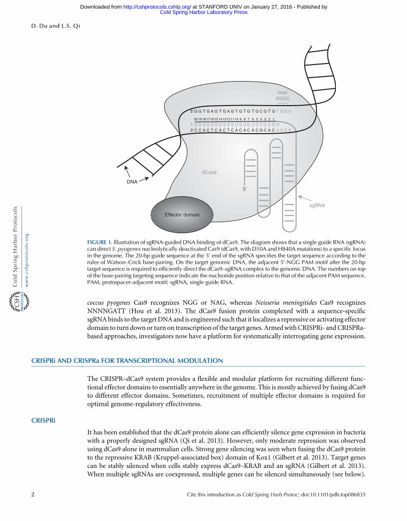
coccus pyogenes Cas9 recognizes NGG or NAG, whereas Neisseria meningitides Cas9 recognizesNNNNGATT (Hou et al. 2013). The dCas9 fusion protein complexed with a sequence-specificsgRNAbinds to the targetDNAand is engineered such that it localizes a repressive or activating effectordomain to turn downor turn on transcription of the target genes. ArmedwithCRISPRi- andCRISPRa-based approaches, investigators now have a platform for systematically interrogating gene expression.
CRISPRi AND CRISPRa FOR TRANSCRIPTIONAL MODULATION
The CRISPR–dCas9 system provides a flexible and modular platform for recruiting different func-tional effector domains to essentially anywhere in the genome. This is mostly achieved by fusing dCas9to different effector domains. Sometimes, recruitment of multiple effector domains is required foroptimal genome-regulatory effectiveness.
CRISPRi
It has been established that the dCas9 protein alone can efficiently silence gene expression in bacteriawith a properly designed sgRNA (Qi et al. 2013). However, only moderate repression was observedusing dCas9 alone in mammalian cells. Strong gene silencing was seen when fusing the dCas9 proteinto the repressive KRAB (Kruppel-associated box) domain of Kox1 (Gilbert et al. 2013). Target genescan be stably silenced when cells stably express dCas9–KRAB and an sgRNA (Gilbert et al. 2013).When multiple sgRNAs are coexpressed, multiple genes can be silenced simultaneously (see below).
Effector domain
dCas9
sgRNA
3′5′ 3′
5′
20 19181716151413121110 9 8 7 6 5 4 3 2 1
5′ G G G G G G G G G GCAA T T T T T 3′G GG T
PAM(NGG)
T
CG G G G G G G G G G GCU U U U U U
C AA A
C T C A C T C A C A C A C G C A AC C C
DNA3′
FIGURE 1. Illustration of sgRNA-guided DNA binding of dCas9. The diagram shows that a single guide RNA (sgRNA)can direct S. pyogenes nucleolytically deactivated Cas9 (dCas9, with D10A and H840A mutations) to a specific locusin the genome. The 20-bp guide sequence at the 5′ end of the sgRNA specifies the target sequence according to therules of Watson–Crick base-pairing. On the target genomic DNA, the adjacent 5′-NGG PAM motif after the 20-bptarget sequence is required to efficiently direct the dCas9–sgRNA complex to the genomic DNA. The numbers on topof the base-pairing targeting sequence indicate the nucleotide position relative to that of the adjacent PAM sequence.PAM, protospacer-adjacent motif; sgRNA, single guide RNA.
2 Cite this introduction as Cold Spring Harb Protoc; doi:10.1101/pdb.top086835
D. Du and L.S. Qi
Cold Spring Harbor Laboratory Press at STANFORD UNIV on January 27, 2016 - Published by http://cshprotocols.cshlp.org/Downloaded from
Effective targeting sites of CRISPRi include enhancers, proximal promoters, and the coding regiondownstream from the transcription start site (TSS) of a gene (Gilbert et al. 2013; Kearns et al. 2014).
CRISPRa
It has been shown that the dCas9 fusion with a transcription activator VP64 can activate a reportergene relatively effectively (Cheng et al. 2013; Gilbert et al. 2013; Maeder et al. 2013; Mali et al. 2013;Perez-Pinera et al. 2013; Chakraborty et al. 2014; Kearns et al. 2014; Chavez et al. 2015). However,direct fusion of dCas9 to VP64 results in only very mild activation of endogenous target genes. Forbetter activation of transcription, several systems have been developed. For example, fusing VP64 toboth the amino and carboxyl terminus of dCas9 or fusing 10 copies of VP16 to dCas9 in each caseenhanced activation (Cheng et al. 2013; Chakraborty et al. 2014). Chavez and colleagues generated aVP64–p65–Rta tripartite activator with dCas9 and showed that this construct enabled efficient en-dogenous gene activation (Chavez et al. 2015). All of these methods increased transcription activationof the target genes compared with that of the dCas9–VP64 system.
Additionally, a series of systems for indirect fusions of effector domains to CRISPR–Cas9 havebeen developed. For example, Konermann and colleagues appended two MS2 bacteriophage coat-protein-binding RNA motifs to two sgRNA stem loops. They coexpressed an MS2-activator (MS2–p65–HSF1) fusion protein together with dCas9–VP64 and modified sgRNA and observed moreefficient transcription activation compared with dCas9–VP64 (Konermann et al. 2015). Tanenbaumand colleagues have developed a “SunTag” scaffold protein that can specifically recruit multiple copiesof single-chain variable fragment (scFv), an artificial antibody fusion protein. When fusing scFv to aVP64 activator, multiple transcriptional activators can be recruited by dCas9–SunTag to the targetDNA for very strong activation of endogenous genes (Gilbert et al. 2014; Tanenbaum et al. 2014).
Notably, CRISPRi and CRISPRa have low off-target effects. Using RNA-seq to assay the tran-scriptome, it has been shown that CRISPRi and CRISPRa can specifically modulate gene expressionwhile inducing minimal off-target effects (Cheng et al. 2013; Gilbert et al. 2013, 2014; Perez-Pineraet al. 2013; Konermann et al. 2015).
We provide a working protocol for designing, cloning, and using sgRNAs for effective geneactivation and repression in mammalian cells in Protocol: CRISPR Technology for Genome Activa-tion and Repression in Mammalian Cells (Du and Qi 2016).
Modulation of Multiple Genes Using CRISPRi and CRISPRa
Multiple genes can be simultaneously activated or repressed by co-deliveringmultiple cognate sgRNAs,thus providing a powerful platform for analyzing the interaction of multiple genes (Cheng et al. 2013;Gilbert et al. 2013;Qi et al. 2013;Chavez et al. 2015;Konermann et al. 2015). To simultaneously activateand repress multiple genes in the same cell, scaffold RNAs (scRNAs) have been engineered by fusingsgRNAs to orthogonal protein-binding bacteriophage RNAs such asMS2, PP7, andCom (Zalatan et al.2015). It has been shown that co-delivery ofMCP–VP64 andCOM–KRABwith a dCas9 protein allowssimultaneous activation of CXCR4 (a chemokine receptor) and repression of B4GAL4NT1 (encodingβ-1,4-N-acetyl-galactosaminyl transferase) in the same cell (Zalatan et al. 2015). Thus, engineeredscRNAs provide a versatile platform for multigene modulation for recruiting diverse effectors todifferent genomic loci.
Repression and Activation of Noncoding RNA Genes
In addition to protein-coding genes, CRISPRi can be harnessed to repress transcription of longnoncoding RNAs (lncRNAs). For example, strong knockdown (>80%) of five tested lncRNAs(H19, MALAT1, NEAT1, TERC, and XIST) has been observed in human myelogenous leukemiaK562 cells (Gilbert et al. 2014). These results showed that CRISPRi was able to repress lncRNAexpression effectively, enabling further functional analysis of these noncoding genes. Konermannand colleagues have also shown that CRISPRa can activate long intergenic noncoding RNAs (linc-RNAs) such as TINCR, PCAT, and HOTTIP (Konermann et al. 2015).
Cite this introduction as Cold Spring Harb Protoc; doi:10.1101/pdb.top086835 3
CRISPR for Genome Regulation in Mammalian Cells
Cold Spring Harbor Laboratory Press at STANFORD UNIV on January 27, 2016 - Published by http://cshprotocols.cshlp.org/Downloaded from
Application of CRISPRi and CRISPRa for Reprogramming of Cell Fate
Expression of exogenous transcription factors has been used as a major approach for directed cellreprogramming (Ladewig et al. 2013). Now, CRISPR-based gene regulation provides a novel ap-proach. For example, Kearns and colleagues showed that CRISPRi could modulate the differentiationof human pluripotent stem cells by using an sgRNA to repress the OCT4 gene (Kearns et al. 2014).Furthermore, Chakraborty and colleagues showed that CRISPRa could induce the transdifferentiationof mouse embryonic fibroblasts into skeletal myocytes by activating transcription of the endogenousMyod1 gene (Chakraborty et al. 2014). Finally, when paired with sgRNAs targeting the OCT4 pro-moter, dCas9–VP192 has been used to replace the requirement for exogenous OCT4 overexpressionin a methodology for reprogramming human-induced pluripotent stem cells (Balboa et al. 2015).
In summary, CRISPRi and CRISPRa offer powerful approaches for repression and activation ofendogenous genes, which is useful for studying gene functions, rewiring genetic networks, andreprogramming cell fates.
ACKNOWLEDGMENTS
We thank Antonia Dominguez, Marie La Russa, and Yanxia Liu for critical comments on the man-uscript. The authors acknowledge support from the California Institute for Quantitative BiomedicalResearch (QB3), National Institutes of Health Office of The Director (OD), and National Institute ofDental and Craniofacial Research (NIDCR). This work was supported by National Institutes of HealthDirector’s Early Independence Award (grant OD017887 L.S.Q.).
REFERENCES
Balboa D, Weltner J, Eurola S, Trokovic R, Wartiovaara K, Otonkoski T.2015. Conditionally stabilized dCas9 activator for controlling gene ex-pression in human cell reprogramming and differentiation. Stem CellReports 5: 448–459.
Chakraborty S, Ji H, Kabadi AM, Gersbach CA, Christoforou N, Leong KW.2014. A CRISPR/Cas9-based system for reprogramming cell lineagespecification. Stem Cell Reports 3: 940–947.
Chang K, Elledge SJ, Hannon GJ. 2006. Lessons from nature: MicroRNA-based shRNA libraries. Nat Methods 3: 707–714.
Chavez A, Scheiman J, Vora S, Pruitt BW, Tuttle M, PR Iyer E, Lin S, Kiani S,Guzman CD, Wiegand DJ, et al. 2015. Highly efficient Cas9-mediatedtranscriptional programming. Nat Methods 12: 326–328.
Cheng AW, Wang H, Yang H, Shi L, Katz Y, Theunissen TW, Rangarajan S,Shivalila CS, Dadon DB, Jaenisch R. 2013. Multiplexed activation ofendogenous genes by CRISPR-on, an RNA-guided transcriptional ac-tivator system. Cell Res 23: 1163–1171.
Du D, Qi LS. 2016. CRISPR technology for genome activation and repres-sion in mammalian cells. Cold Spring Harb Protoc doi: 10.1101/pdb.prot090175.
Gilbert LA, Larson MH, Morsut L, Liu Z, Brar GA, Torres SE, Stern-Ginossar N, Brandman O, Whitehead EH, Doudna JA, et al. 2013.CRISPR-mediated modular RNA-guided regulation of transcriptionin eukaryotes. Cell 154: 442–451.
Gilbert LA, Horlbeck MA, Adamson B, Villalta JE, Chen Y, Whitehead EH,Guimaraes C, Panning B, Ploegh HL, Bassik MC, et al. 2014. Genome-scale CRISPR-mediated control of gene repression and activation. Cell159: 647–661.
Hou Z, Zhang Y, Propson NE, Howden SE, Chu LF, Sontheimer EJ,Thomson JA. 2013. Efficient genome engineering in human pluripotentstem cells using Cas9 from Neisseria meningitidis. Proc Natl Acad Sci110: 15644–15649.
Kabadi AM, Gersbach CA. 2014. Engineering synthetic TALE and CRISPR/Cas9 transcription factors for regulating gene expression. Methods 69:188–197.
Kearns NA, Genga RM, Enuameh MS, Garber M, Wolfe SA, Maehr R.2014. Cas9 effector-mediated regulation of transcription and differ-entiation in human pluripotent stem cells. Development 141: 219–223.
Konermann S, Brigham MD, Trevino AE, Joung J, Abudayyeh OO, BarcenaC, Hsu PD, Habib N, Gootenberg JS, Nishimasu H, et al. 2015.Genome-scale transcriptional activation by an engineered CRISPR-Cas9 complex. Nature 517: 583–588.
Ladewig J, Koch P, Brustle O. 2013. LevelingWaddington: The emergence ofdirect programming and the loss of cell fate hierarchies. Nat Rev MolCell Biol 14: 225–236.
Maeder ML, Linder SJ, Cascio VM, Fu Y, Ho QH, Joung JK. 2013. CRISPRRNA-guided activation of endogenous human genes. Nat Methods 10:977–979.
Mali P, Aach J, Stranges PB, Esvelt KM, Moosburner M, Kosuri S, Yang L,Church GM. 2013. CAS9 transcriptional activators for target specificityscreening and paired nickases for cooperative genome engineering. NatBiotechnol 31: 833–838.
Perez-Pinera P, Kocak DD, Vockley CM, Adler AF, Kabadi AM, Polstein LR,Thakore PI, Glass KA, Ousterout DG, Leong KW, et al. 2013. RNA-guided gene activation by CRISPR-Cas9-based transcription factors.Nat Methods 10: 973–976.
Qi LS, Larson MH, Gilbert LA, Doudna JA, Weissman JS, Arkin AP,Lim WA. 2013. Repurposing CRISPR as an RNA-guided platformfor sequence-specific control of gene expression. Cell 152: 1173–1183.
TanenbaumME, Gilbert LA, Qi LS, Weissman JS, Vale RD. 2014. A protein-tagging system for signal amplification in gene expression and fluores-cence imaging. Cell 159: 635–646.
Zalatan JG, LeeME, Almeida R, Gilbert LA,Whitehead EH, La RussaM, TsaiJC, Weissman JS, Dueber JE, Qi LS, et al. 2015. Engineering complexsynthetic transcriptional programs with CRISPR RNA scaffolds. Cell160: 339–350.
4 Cite this introduction as Cold Spring Harb Protoc; doi:10.1101/pdb.top086835
D. Du and L.S. Qi
Cold Spring Harbor Laboratory Press at STANFORD UNIV on January 27, 2016 - Published by http://cshprotocols.cshlp.org/Downloaded from
doi: 10.1101/pdb.top086835Cold Spring Harb Protoc; Dan Du and Lei S. Qi in Mammalian CellsAn Introduction to CRISPR Technology for Genome Activation and Repression
ServiceEmail Alerting click here.Receive free email alerts when new articles cite this article -
CategoriesSubject Cold Spring Harbor Protocols.Browse articles on similar topics from
(1013 articles)Molecular Biology, general (69 articles)Expression of Cloned Genes
(114 articles)Analysis of Gene Expression, general (57 articles)Analysis of Gene Expression in Cultured Cells
(160 articles)Analysis of Gene Expression
http://cshprotocols.cshlp.org/subscriptions go to: Cold Spring Harbor Protocols To subscribe to
© 2016 Cold Spring Harbor Laboratory Press
Cold Spring Harbor Laboratory Press at STANFORD UNIV on January 27, 2016 - Published by http://cshprotocols.cshlp.org/Downloaded from
Protocol
CRISPR Technology for Genome Activation and Repressionin Mammalian Cells
Dan Du1 and Lei S. Qi1,2,3,4
1Department of Bioengineering, Stanford University, Stanford, California 94305; 2Department of Chemical andSystems Biology, Stanford University, Stanford, California 94305; 3ChEM-H; Stanford University, Stanford,California 94305
Targeted modulation of transcription is necessary for understanding complex gene networks and hasgreat potential for medical and industrial applications. CRISPR is emerging as a powerful system fortargeted genome activation and repression, in addition to its use in genome editing. This protocoldescribes how to design, construct, and experimentally validate the function of sequence-specific singleguide RNAs (sgRNAs) for sequence-specific repression (CRISPRi) or activation (CRISPRa) of tran-scription in mammalian cells. In this technology, the CRISPR-associated protein Cas9 is catalyticallydeactivated (dCas9) to provide a general platform for RNA-guided DNA targeting of any locus in thegenome. Fusion of dCas9 to effector domains with distinct regulatory functions enables stable andefficient transcriptional repression or activation in mammalian cells. Delivery of multiple sgRNAsfurther enables activation or repression of multiple genes. By using scaffold RNAs (scRNAs), differenteffectors can be recruited to different genes for simultaneous activation of some and repression ofothers. The CRISPRi and CRISPRa methods provide powerful tools for sequence-specific control ofgene expression on a genome-wide scale to aid understanding gene functions and for engineeringgenetic regulatory systems.
MATERIALS
It is essential that you consult the appropriate Material Safety Data Sheets and your institution’s EnvironmentalHealth and Safety Office for proper handling of equipment and hazardous materials used in this protocol.
RECIPES: Please see the end of this protocol for recipes indicated by <R>. Additional recipes can be found online athttp://cshprotocols.cshlp.org/site/recipes.
Reagents
Chemically competent Escherichia coli cells (e.g., One Shot TOP10 Cells from Life Technologies)dCas9 expression vector(s) appropriate for experiment
• CRISPR activation (CRISPRa) dCas9–SunTag expression vectorsTwo constructs are required: a lentiviral vector containing an SV40-promoter-driven dCas9 fusion betweendCas9, 2X nuclear localization signal (NLS), 10X GCN4, and a P2A-tagBFP (Addgene 60903) and alentiviral vector containing an SV40-promoter-driven fusion protein between the single chain variablefragment (scFv) for GCN4, a superfolder (sf) GFP, VP64, and 1X NLS (Addgene 60904).
• CRISPR interference (CRISPRi) dCas9–KRAB expression vectorThis comprises a lentiviral vector containing a spleen focus-forming virus SFFV-promoter-driven dCas9fused to 2X NLS, a tagBFP and a KRAB domain (Addgene 46911).
4Correspondence: [email protected]
© 2016 Cold Spring Harbor Laboratory PressCite this protocol as Cold Spring Harb Protoc; doi:10.1101/pdb.prot090175
40
Cold Spring Harbor Laboratory Press at Stanford University Libraries on January 27, 2016 - Published by http://cshprotocols.cshlp.org/Downloaded from
dNTPs (10 mM)Double-distilled water (ddH2O), sterile and nuclease-freeDulbecco’s modified Eagle’s medium (DMEM), high-glucose (Life Technologies 11965-092)Fetal bovine serum (FBS)Gel electrophoresis reagents
Agarose gels (1%, w/v)
DNA ladder
Ethidium bromide
Tris-acetate-EDTA (TAE) buffer (50×) <R>
HEK293T cells (ATCC CRL-11268)HEK293T cells (or other cells derived from HEK293T cells) are required for lentivirus production in Steps 26–31.In addition, they are used here as an example of target cells in Steps 32–36. Other target cells may be used asappropriate for the experiment.
In-Fusion HD Cloning Kit (Clontech 011614)iQ SYBR Green Supermix (Bio-Rad 170-8880)iScript cDNA Synthesis Kit (Bio-Rad 170-8890)Lentiviral packaging plasmids pCMV-dR8.91 and pMD2.G (Addgene 12259)
At the time of this writing, pCMV-dR8.91 is not avialable from Addgene. Alternatively, a lower version of theplasmid is available from Addgene (pCMV-dR8.2; Addgene 8455). Its use will not affect this protocol.
Lysogeny broth (LB) with carbenicillin (liquid medium and agar plates) <R>Mirus TransIT-LT1 Transfection Reagent (Mirus MIR 2300)Opti-MEM Reduced-Serum Medium (Life Technologies 31985-062)Penicillin-Streptomycin (100×), presterilized (Life Technologies 15070-063)Phusion High-Fidelity Polymerase and 5× Phusion HF Buffer (New England BioLabs M0536L)Polybrene (optional; see Step 33)Primers
• PCR primers, one of which (sgRNA-F) contains the gene-specific sgRNA target sequence
Forward primer (sgRNA-F): 5′-CCCTTGGAGAACCACCTTGTTGGN(19)GTTTAAGAGCTATGCTGGAAACAGCA-3′
Reverse primer (sgRNA-R): 5′-GATCCTAGTACTCGAGAAAAAAAGCACCGACTCGGTGCCAC-3′
For sgRNA target sequence selection, see Steps 1–3.
• Sequencing primer: 5′-GAGGCTTAATGTGCGATAAAAGA-3′
This primer binds to the mouse U6 promoter and is used to confirm the generation of sgRNA expressionconstructs in Step 21.
• Target gene-specific primers for qRT-PCR (see Step 40)
QIAGEN Plasmid Midi Kit (QIAGEN 12143)It is important to use an endotoxin-free midiprep kit when purifying plasmid DNA for better transfection efficiencyinto mammalian cells.
QIAprep Spin Miniprep Kit (QIAGEN 27106)QIAquick Gel Extraction Kit (QIAGEN 28706)QIAquick PCR Purification Kit (QIAGEN 28106)Restriction enzymes BstXI, XhoI, and DpnIRNeasy Plus Mini Kit (QIAGEN 74134)Single guide RNA (sgRNA) expression vector
This comprises a lentiviral vector containing the mouse U6 promoter driving sgRNA expression (Addgene51024). It also contains an expression cassette consisting of a cytomegalovirus (CMV) promoter, a puromycin-resistance gene cassette, and an mCherry gene for selection purposes.
Trypsin-EDTA (0.05%) (e.g., Life Technologies 25300-054)
Cite this protocol as Cold Spring Harb Protoc; doi:10.1101/pdb.prot090175 41
CRISPRa and CRISPRi in Mammalian Cells
Cold Spring Harbor Laboratory Press at Stanford University Libraries on January 27, 2016 - Published by http://cshprotocols.cshlp.org/Downloaded from
Equipment
Access to sequencing facility (see Step 21)BD FACSAria II Cell Sorter (BD Biosciences) equipped with lasers and filters for detecting mCherry,EGFP, and tagBFP
CFX96 Real-Time PCR Detection System (Bio-Rad 185-5195)CO2 incubator at 37˚C and 5% CO2 for mammalian cell cultureComputer with Internet-connected web browserConical tubesDigital gel-imaging systemErlenmeyer flasks (250 mL)Gel electrophoresis systemGlass tubes (25-mm)Incubators at 37˚C for growing bacteria (one standard and one capable of shaking at 200 rpm)MicrocentrifugeMicrocentrifuge tubesMicroplate for qRT-PCRNanoDrop 8000 UV-Vis Spectrophotometer (Thermo Scientific)PCR tubes (0.2-mL)Six-well tissue-culture platesSyringe filter (0.45-µm), sterileSyringe, sterileThermocycler
METHOD
We have implemented a computational tool, termed CRISPR-ERA (“editing, repression, and activation”) for automat-ed design of sgRNAs for given mammalian organisms, such as mouse, rat, and human (Liu et al. 2015). The CRISPR-ERA algorithm aligns the designed sgRNA to the whole genome and reports potential off-target sites as defined bypossession of fewer than three mismatches. The tool is freely available at http://CRISPR-ERA.stanford.edu. If usingCRISPR-ERA, skip Steps 1–5.
Selection of sgRNA Targets in the Genome
1. Determine the DNA sequence of the target gene using an available genome database—forexample, the UCSC genome browser (Kent et al. 2002).
2. Obtain annotation information of the target gene, including the location of the transcriptionstart site (TSS).
3. Search for patterns of GN(19)NGG around the TSS, wherein GN(19) is the binding site of thesgRNA and NGG is the protospacer adjacent motif (PAM), which is required for efficient DNAbinding of Streptococcus pyogenes Cas9.
Our sgRNA expression construct uses a mouse U6 promoter, which requires a G at the very 5′ end foreffective transcription. Therefore, we search for GN(19) as the binding site of the sgRNA. If another promoteris used, it is likely that the first nucleotide will be different.
The recommended window of the target DNA is −50 to +300 bp relative to the TSS for CRISPR interference(CRISPRi) for gene repression, or −400 to −50 bp for CRISPR activation (CRISPRa). Usually, multiplesgRNA-binding sites within the target window of the gene need to be tested to define the most efficienttargeting site for repression or activation.
Many mammalian genes possess transcript isoforms with different TSSs. In this case, different sgRNAs needto be designed for each transcript. Currently, there is no direct evidence that the activities of CRISPRi andCRISPRa are sensitive to the DNA strand or GC content (Gilbert et al. 2014).
42 Cite this protocol as Cold Spring Harb Protoc; doi:10.1101/pdb.prot090175
D. Du and L.S. Qi
Cold Spring Harbor Laboratory Press at Stanford University Libraries on January 27, 2016 - Published by http://cshprotocols.cshlp.org/Downloaded from
Design of sgRNA Sequences
4. Ensure that the base-pairing sequence on the sgRNA is the reverse complement of the GN(19)
sequence identified in Step 3.
5. Analyze the specificity of the target sequence in the genome use the basic local alignment searchtool (BLAST; http://blast.ncbi.nlm.nih.gov) (Bhagwat et al. 2012).
The BLAST algorithm enables the specificity of sgRNA targeting in the genome to be analyzed when notusing the CRISPR-ERA tool.
6. Generate the full-length sgRNA by appending GN(19) 3′ to the rest of the optimized sgRNA
sequence (5′-GN(19)GUUUAAGAGCUAUGCUGGAAACAGCATAGCAAGUUUAAAUAAGGCUAGUCCGUUAUCAACUUGAAAAAGUGGCACCGAGUCGGUGCUUUUUUU-3′) (Chenet al. 2013).
7. Confirm that the GN(19) target sequence does not contain any transcription termination sequencefor the U6 promoter (Paul et al. 2002).
Preparation of sgRNA Expression Constructs
8. Generate the sgRNA backbone by digesting the empty sgRNA expression vector withrestriction enzymes BstXI and XhoI for 4–16 h at 37˚C according to the manufacturer’sinstructions.
9. Separate the digested sgRNA backbone products by electrophoresis through a 1% (w/v) agarosegel in 1× TAE buffer. Stain the gel with ethidium bromide, and visualize the bands using a digitalgel imaging system. Compare the bands to those of a proper DNA ladder, and confirm that theband representing the sgRNA backbone DNA is ~8 kb.
10. Gel-purify the sgRNA backbone DNA using a QIAquick Gel Extraction Kit according to themanufacturer’s instructions. Store the DNA at −20˚C until use in Step 15.
11. Perform PCR as follows, using primers that contain the 20-nt target sequences identified inSteps 1–3.
i. Assemble the following reaction (volumes shown are for one reaction) in a 0.2-mL PCRtube on ice.
0.5 µL Empty sgRNA expression vector (undigested) as template (100 ng/µL)2.5 µL Forward primer (sgRNA-F) (10 µM)2.5 µL Reverse primer (sgRNA-R) (10 µM)2 µL dNTPs (10 mM)0.5 µL Phusion High-Fidelity Polymerase (2 U/µL)10 µL Phusion HF Buffer (5×)32 µL Nuclease-free water50 µL Total volume
ii. Perform PCR with the following cycling conditions:
1 cycle 98˚C 30 sec25 cycles 98˚C 10 sec
62˚C 30 sec72˚C 10 sec
1 cycle 72˚C 5 min1 cycle 4˚C Forever
12. Confirm that the PCRs successfully amplified a ~150-bp DNA product by separating 5 µL of thePCR products on a 1% agarose gel as in Step 9.
Cite this protocol as Cold Spring Harb Protoc; doi:10.1101/pdb.prot090175 43
CRISPRa and CRISPRi in Mammalian Cells
Cold Spring Harbor Laboratory Press at Stanford University Libraries on January 27, 2016 - Published by http://cshprotocols.cshlp.org/Downloaded from

13. Add 1 µL of DpnI (20 U/µL) into each PCR and then incubate for 1 h at 37˚C.Treatment of DpnI will digest the PCR templates.
14. Purify the PCRs using a QIAquick PCR Purification Kit by following the manufacturer’s instruc-tions. Store the DNA at −20˚C until use in Step 15.
15. Measure the concentrations of the purified sgRNA backbone DNA (from Step 10) and PCRfragments (from Step 14) using a NanoDrop UV-Vis 8000 Spectrophotometer.
16. Ligate the PCR fragments to the sgRNA backbone DNA using an In-Fusion HD Cloning Kit.
i. Assemble the cloning reaction.
1 µL In-Fusion HD Enzyme Premix (5×)50 ng Linearized sgRNA backbone DNA (from Step 10)25 ng Purified PCR fragments (from Step 14)x µL ddH2O5 µL Total volume
ii. Incubate the reaction for 15 min at 50˚C using a thermocycler.
iii. Place on ice for 5 min. Store at −20˚C until use in Step 17.
17. Transform chemically competent E. coli cells with the products of the ligation reactions. Followthe manufacturer’s instructions for the E. coli cells. Spread transformed E. coli cells onto LB agarplates supplemented with 100 µg/mL carbenicillin. Incubate the plates overnight in a 37˚Cincubator.
18. Transfer single colonies into 25-mm glass tubes containing 5 mL of LB medium supplementedwith 100 µg/mL carbenicillin. For each colony, use a sterile pipette tip to touch the colony, andthen swirl the tip in the LB medium to dissolve the colony. Incubate overnight in a 37˚C shakingincubator, swirling at 200 rpm.
19. Transfer 0.5 mL of bacterial culture into a 250-mL Erlenmeyer flask containing 50 mL of LBmedium with 100 µg/mL carbenicillin. Incubate overnight in a 37˚C shaking incubator, swirlingat 200 rpm.
20. Extract the plasmid DNA from the remaining 4.5 mL of bacterial culture using a QIAprep SpinMiniprep Kit according to the manufacturer’s instructions.
21. Send the extracted plasmid DNA for sequencing with the sequencing primer.
22. After the plasmid is verified by sequencing, extract DNA from the 50-mL bacterial culture usinga QIAGEN Plasmid Midi Kit according to the manufacturer’s instructions. Store the DNAat −20˚C until use in Step 27.
Preparation of dCas9 Expression Vectors
23. Transform chemically competent E. coli cells with the dCas9 expression vectors appropriatefor the experiment (CRISPRi or CRISPRa). Spread transformed E. coli cells onto LB agarplates supplemented with 100 µg/mL carbenicillin. Incubate the plates overnight in a 37˚Cincubator.
24. Transfer a single colony into 50 mL of LB medium supplemented with 100 µg/mL carbenicillin.Incubate overnight in a 37˚C shaking incubator, swirling at 200 rpm.
25. Extract DNA using a QIAGEN Plasmid Midi Kit according to the manufacturer’s instructions.Store the DNA at −20˚C until use in Step 27.
Packaging of dCas9 and sgRNA Expression Constructs into Lentiviral Particles
If more lentiviruses are required, scale up the cell numbers, DNA amounts, and transfection reagent volumesused here.
44 Cite this protocol as Cold Spring Harb Protoc; doi:10.1101/pdb.prot090175
D. Du and L.S. Qi
Cold Spring Harbor Laboratory Press at Stanford University Libraries on January 27, 2016 - Published by http://cshprotocols.cshlp.org/Downloaded from
26. On the day before transfection, seed a six-well tissue-culture plate with 2–3 × 10⁵ HEK293T cellsin 2 mL of high-glucose DMEM containing 10% (v/v) FBS per well. Incubate overnight at 37˚Cand 5% CO2.
HEK293T cells can be maintained in regular high-glucose DMEM medium supplemented with 10% (v/v)FBS, 100 U/mL streptomycin, and 100 µg/mL penicillin and regularly passaged using 0.05% (w/v) trypsin–EDTA. However, antibiotic-free DMEM is required during transfection and virus collection to achieve betterefficiency.
27. Twenty-four hours after plating the cells, prepare the transfection complexes as follows.
i. Combine the following DNA samples.
1.32 µg pCMV-dR8.91 (lentiviral packaging plasmid)165 ng pMD2.G (lentiviral packaging plasmid)1.51 µg dCas9 or sgRNA expression construct
Nontargeting sgRNA vector or dCas9 without fusion vector can be used in parallel as a negative control.
ii. Add this 3-µg DNA mixture into 250 µL of Opti-MEM Reduced-SerumMedium in a micro-centrifuge tube. Mix well by pipetting up and down.
iii. Add 7.5 µL of Mirus TransIT-LT1 Transfection Reagent into the same tube. Mix well bypipetting up and down.
iv. Allow transfection complexes to form for 30 min at room temperature.
28. Remove 250 µL of medium from each well in the six-well plate.
29. Add the ~250-µL mixture from Step 27.iv into one well in the six-well plate. Mix well by rockingthe plate gently back and forth. Incubate for 24 h at 37˚C and 5% CO2.
Cells will begin producing viruses 24 h after transfection.
30. After the 24-h incubation, replace the transfection medium with 2.5 mL of fresh DMEM with10% FBS.
If the target cells to be infected have any additional medium requirements, replace the transfection mediumwith 2.5 mL of special growth medium for the target cells.
31. Use a sterile syringe to harvest the viral supernatant 24–48 h after medium replacement.Filter the medium through a 0.45-µm syringe filter into a conical tube to avoid transferringHEK293T cells.
The total volume will be ~2 mL after filtering. Lentiviral particles can be stored for up to 1 wk at 4˚C, or snap-frozen in liquid nitrogen and stored for several months at −80˚C. However, we recommend using thelentiviruses immediately after collection.
Transduction of Target Cells with dCas9 and sgRNA Lentiviral Particles
In the following, the use of HEK293T cells is given as an example. For other types of cells, modify the procedure (e.g.,cell number and growth medium) as appropriate.
32. Sixteen hours before transduction, seed a six-well tissue-culture plate with 1.5–2 × 10⁵ HEK293Tcells in 2 mL of high-glucose DMEM supplemented with 10% FBS per well. Incubate at 37˚Cand 5% CO2.
33. Replace the medium with 1 mL of DMEM containing 10% FBS and 1 mL of filtered viralsupernatant. Incubate overnight at 37˚C and 5% CO2.
Depending on the virus titration, the viral supernatant can be diluted with growth medium for the targetcells. Polybrene can be used to promote the infection efficiency with proper concentration; however, it istoxic for some types of cells, including HEK293T cells.
34. Replace the viral supernatant with 2 mL of fresh DMEM with 10% FBS, and incubate for 48 hat 37˚C and 5% CO2.
Cite this protocol as Cold Spring Harb Protoc; doi:10.1101/pdb.prot090175 45
CRISPRa and CRISPRi in Mammalian Cells
Cold Spring Harbor Laboratory Press at Stanford University Libraries on January 27, 2016 - Published by http://cshprotocols.cshlp.org/Downloaded from
Cells usually will express dCas9 protein 48 h after addition of lentiviruses. However, for repression exper-iments, we suggest collecting cells at least 72 h after infection to minimize the interference by preexistingtarget gene mRNA. If necessary, split the cells when they reach 80%–90% confluence before sorting.
35. Use a BD FACSAria II Cell Sorter to collect the cells.
• For the CRISPRi system, collect cells positive for both blue fluorescent protein (BFP) andmCherry.
The BFP-positive cells should express dCas9 protein, and mCherry-positive cells should express sgRNA.
• For the CRISPRa (dCas9–Suntag) system, collect cells that are positive for BFP, mCherry,and GFP.
The GFP-positive cells should express scFv-sfGFP-VP64 fusion protein.
36. Incubate the collected cells at 37˚C and 5% CO2.After the cells are grown up, analyze the expression levels of target genes by qRT-PCR as described inSteps 37–40.
Quantification of the Effects of CRISPRi or CRISPRa on GeneExpression in Target Cells
37. Extract total RNA from the cells infected using an RNeasy Plus Mini Kit according to themanufacturer’s instructions.
Typically, 0.5–1 × 10⁶ cells (50%–80% confluence of cells in one well in a six-well plate) are sufficient fortotal RNA extraction.
38. Measure the concentrations of the total RNA samples using a NanoDrop spectrophotometer.
39. Synthesize cDNA using an iScript cDNA Synthesis Kit.
i. Set up the cDNA synthesis reaction.
4 µL iScript reaction mix (5×)1 µL iScript reverse transcriptase1 µg Total RNA templatex µL Nuclease-free water20 µL Total volume
ii. Incubate the reaction as follows (e.g., using a thermocycler):
1 cycle 25˚C 5 min1 cycle 42˚C 30 min1 cycle 85˚C 5 min1 cycle 4˚C Forever
iii. Store the DNA at −20˚C until use in Step 40.i.
40. Analyze the cDNA levels of target genes using a standard qRT-PCR protocol.
i. Set up the PCR in a microplate using the iQ SYBR Green Supermix according to themanufacturer’s instructions.
10 µL iQ SYBR Green Supermix (2×)1.2 µL Forward primer (target gene-specific; 5 µM)1.2 µL Reverse primer (target gene-specific; 5 µM)0.25 µL Template (cDNA from Step 39)7.35 µL Nuclease-free water20 µL Total volume
The amount of template cDNA can be scaled up or down according to the expression levels of the targetgenes in the cells. Housekeeping genes—for example, GAPDH, encoding glyceraldehyde-3-phosphatedehydrogenase—should be used as references.
46 Cite this protocol as Cold Spring Harb Protoc; doi:10.1101/pdb.prot090175
D. Du and L.S. Qi
Cold Spring Harbor Laboratory Press at Stanford University Libraries on January 27, 2016 - Published by http://cshprotocols.cshlp.org/Downloaded from

ii. Run the following real-time PCR profile in a CFX96 Real-Time PCR Detection System.
1 cycle 95˚C 2–3 min39 cycles 95˚C 10–15 sec
55˚C–60˚C 15–30 sec72˚C 30 sec
Melt curve (optional) 55˚C–95˚C (in 0.5˚C increments) 10–30 sec
iii. Analyze the qRT-PCR data by standard methods to obtain the relative transcriptional expres-sion levels of the target genes regulated by CRISPRi/a.
We use the 2–ΔΔCt method to obtain the relative mRNA expression level of the CRISPRi orCRISPRa sample vs. a control sample, where ΔΔCt = ΔCt(CRISPRi/a sample)–ΔCt(control sample,e.g., nontargeting sgRNA sample), and where ΔCt(sample) = Ct(any sample)–Ct(endogenous house-keeping gene).
DISCUSSION
To date, several tools have been developed to functionally interrogate gene expression. RNAi has beenshown to disrupt gene expression by triggering the degradation of target mRNAs (Chang et al. 2006).However, the technique is somewhat limited in its application owing to off-target effects and throughbeing restricted to cytosolic target mRNAs (Jackson et al. 2003; Adamson et al. 2012; Sigoillot et al.2012). Protein-based tools are difficult to be designed, cloned, and delivered into target cells. Thecomplex programming and limited targeting sites also restrict the application of zinc fingers and toolsbased on transcription-activator-like effectors (TALEs) for perturbing the expression of multiplegenes. Loss-of-function approaches based on genome editing, such as CRISPR–Cas9, cause irrevers-ible frameshift disruptions, cytotoxic double-stranded DNA breaks, and in-frame insertion–deletions(indels) arising from error-prone DNA repair. These could limit the ability of the CRISPR techniqueto completely abolish the function of genes and noncoding RNAs (Huang et al. 1996; Jackson 2002;Koike-Yusa et al. 2014; Shalem et al. 2014; Wang et al. 2014).
In contrast, RNA-guided DNA targeting of the dCas9 protein to a specific locus provides aprogrammable platform to modulate genome status while generating minimal off-target effects.Fusion of different effector domains to dCas9 enables transcriptional repression (CRISPRi) or acti-vation (CRISPRa) of specific target genes. CRISPRi and CRISPRa enable inducible and reversiblemodulation of specific endogenous gene expression within an intact biological system. The modula-tion of the transcription of single or multiple genes can be specifically achieved by delivery of multiplesgRNAs (Gilbert et al. 2013, 2014; Qi et al. 2013; Tanenbaum et al. 2014; Zalatan et al. 2015). By usingscRNA, transcriptional activation or repression of different target genes can be achieved simultane-ously in the same cell (Zalatan et al. 2015). Recently, CRISPRa has been used to effectively activateexpression of target genes in plants and flies (Lin et al. 2015; Lowder et al. 2015). Furthermore,Kleinstiver and colleagues have modified S. pyogenes Cas9 (spCas9) to recognize alternative PAMsequences (other than NGG) by using a selection-based approach in bacterial cells (Kleinstiver et al.2015). This provides researchers an expanded targetable sequence space in the genome for usingCRISPR–dCas9. Thus, owing to its simplicity and flexibility, CRISPRi or CRISPRa can facilitategenome-scale perturbation of gene expression (Gilbert et al. 2014; Konermann et al. 2015).
However, the detailed mechanism underlying how CRISPRi and CRISPRa components interactwith local transcriptional machinery and epigenetic factors is not well established. We usually designthree to five sgRNAs for each target transcript and choose the best one for functional analysis. Thereason why some of the designed sgRNAs have no function and why the efficiency of differentdesigned sgRNAs varies is not clear. Knowledge of the mechanism would assist the efficiency ofdesigning functional sgRNAs. Moreover, the spCas9 protein, which is widely used for transcriptionalmodulation, is a large molecule that is difficult to clone and package with the necessary regulatoryelements into a size-restricted virus, such as the adeno-associated virus (AAV) that has been generally
Cite this protocol as Cold Spring Harb Protoc; doi:10.1101/pdb.prot090175 47
CRISPRa and CRISPRi in Mammalian Cells
Cold Spring Harbor Laboratory Press at Stanford University Libraries on January 27, 2016 - Published by http://cshprotocols.cshlp.org/Downloaded from
used for gene therapy. In contrast, the smaller ortholog Staphylococcus aureus Cas9 (saCas9) has beenshown to edit the targets efficiently and to be compatible with the AAV system, which has also beenengineered as a transcriptional activating system (SAM) (Nishimasu et al. 2015; Ran et al. 2015).Currently, the gene-regulatory tools based on the S. aureus dCas9 are being developed for more-efficient transcriptional repression and activation. Thus, in summary, CRISPRi and CRISPRa basedon different species of Cas9 or its homologs provide a versatile platform tomanipulate and interrogategene expression systematically.
RECIPES
Lysogeny Broth (LB) with Carbenicillin
Reagent Quantity
Agar (for plates only) 20 gNaCl 10 gTryptone 10 gYeast extract 5 g
Prepare the above-listed ingredients in 1 L of deionized water. Adjust the pH to 7.0 with5 N NaOH. Autoclave for 20 min at 15 psi (1.05 kg/cm2). Cool to �60˚C and addcarbenicillin (final concentration 100 µg/mL). Pour the medium into Petri dishes(�25 mL per 100-mm plate). Store the LB plates at 4˚C; they will keep for at least 4 mo.
Tris-Acetate-EDTA (TAE) Buffer (50×)
Reagent Final concentration (1×)
Tris base 40 mM
EDTA 2 mM
Acetic acid 20 mM
Adjust to pH 8.5 and dilute to 1× with Milli-Q H2O before use.
REFERENCES
Adamson B, Smogorzewska A, Sigoillot FD, King RW, Elledge SJ. 2012. Agenome-wide homologous recombination screen identifies the RNA-binding protein RBMX as a component of the DNA-damage response.Nat Cell Biol 14: 318–328.
Bhagwat M, Young L, Robison R.R. 2012. Using BLAT to find sequencesimilarity in closely related genomes. Curr Protoc BioinformaticsChapter 10: Unit 10.18.
Chang K, Elledge SJ, Hannon GJ. 2006. Lessons from Nature: MicroRNA-based shRNA libraries. Nat Methods 3: 707–714.
Chen B, Gilbert LA, Cimini BA, Schnitzbauer J, Zhang W, Li GW, Park J,Blackburn EH, Weissman JS, Qi LS, et al. 2013. Dynamic imaging ofgenomic loci in living human cells by an optimized CRISPR/Cas system.Cell 155: 1479–1491.
Gilbert LA, Larson MH, Morsut L, Liu Z, Brar GA, Torres SE, Stern-Ginos-sar N, Brandman O, Whitehead EH, Doudna JA, et al. 2013. CRISPR-mediated modular RNA-guided regulation of transcription in eukary-otes. Cell 154: 442–451.
Gilbert LA, Horlbeck MA, Adamson B, Villalta JE, Chen Y, Whitehead EH,Guimaraes C, Panning B, Ploegh HL, Bassik MC, et al. 2014. Genome-scale CRISPR-mediated control of gene repression and activation. Cell159: 647–661.
Huang LC, Clarkin KC, Wahl GM. 1996. Sensitivity and selectivity of theDNA damage sensor responsible for activating p53-dependent G1arrest. Proc Natl Acad Sci 93: 4827–4832.
Jackson SP. 2002. Sensing and repairing DNA double-strand breaks. Carci-nogenesis 23: 687–696.
Jackson AL, Bartz SR, Schelter J, Kobayashi SV, Burchard J, Mao M, Li B,Cavet G, Linsley PS. 2003. Expression profiling reveals off-target generegulation by RNAi. Nat Biotechnol 21: 635–637.
Kent WJ, Sugnet CW, Furey TS, Roskin KM, Pringle TH, Zahler AM,Haussler D. 2002. The human genome browser at UCSC. Genome Res12: 996–1006.
Kleinstiver BP, Prew MS, Tsai SQ, Topkar VV, Nguyen NT, Zheng Z, Gon-zales AP, Li Z, Peterson RT, Yeh JR, et al. 2015. Engineered CRISPR-Cas9 nucleases with altered PAM specificities. Nature 523: 481–485.
Koike-Yusa H, Li Y, Tan EP, Velasco-Herrera Mdel C, Yusa K. 2014.Genome-wide recessive genetic screening in mammalian cells with alentiviral CRISPR-guide RNA library. Nat Biotechnol 32: 267–273.
Konermann S, Brigham MD, Trevino AE, Joung J, Abudayyeh OO, BarcenaC, Hsu PD, Habib N, Gootenberg JS, Nishimasu H, et al. 2015.Genome-scale transcriptional activation by an engineered CRISPR-Cas9 complex. Nature 517: 583–588.
Lin S, Ewen-Campen B, Ni X, Housden BE, Perrimon N. 2015. In vivotranscriptional activation using CRISPR-Cas9 in Drosophila. Genetics201: 433–442.
Liu H, Wei Z, Dominguez A, Li Y, Wang X, Qi LS. 2015. CRISPR-ERA: Acomprehensive disign tool for CRISPR-mediated gene editing, repres-sion, and activation. Bioinformatics 31: 3676–3678.
Lowder LG, Zhang D, Baltes NJ, Paul JW III, Tang X, Zheng X, Voytas DF,Hsieh TF, Zhang Y, Qi Y. 2015. A CRISPR/Cas9 toolbox for multi-plexed plant genome editing and transcriptional regulation. PlantPhysiol 169: 971–985.
48 Cite this protocol as Cold Spring Harb Protoc; doi:10.1101/pdb.prot090175
D. Du and L.S. Qi
Cold Spring Harbor Laboratory Press at Stanford University Libraries on January 27, 2016 - Published by http://cshprotocols.cshlp.org/Downloaded from
Nishimasu H, Cong L, Yan WX, Ran FA, Zetsche B, Li Y, Kurabayashi A,Ishitani R, Zhang F, Nureki O. 2015. Crystal structure of Staphylococcusaureus Cas9. Cell 162: 1113–1126.
Paul CP, Good PD, Winer I, Engelke DR. 2002. Effective expression of smallinterfering RNA in human cells. Nat Biotechnol 20: 505–508.
Qi LS, Larson MH, Gilbert LA, Doudna JA, Weissman JS, Arkin AP,Lim WA. 2013. Repurposing CRISPR as an RNA-guided platformfor sequence-specific control of gene expression. Cell 152: 1173–1183.
Ran FA, Cong L, Yan WX, Scott DA, Gootenberg JS, Kriz AJ, Zetsche B,Shalem O, Wu X, Makarova KS, et al. 2015. In vivo genome editingusing Staphylococcus aureus Cas9. Nature 520: 186–191.
Shalem O, Sanjana NE, Hartenian E, Shi X, Scott DA, Mikkelsen TS,Heckl D, Ebert BL, Root DE, Doench JG, et al. 2014. Genome-scale
CRISPR-Cas9 knockout screening in human cells. Science 343: 84–87.
Sigoillot FD, Lyman S, Huckins JF, Adamson B, Chung E, Quattrochi B,King RW. 2012. A bioinformatics method identifies prominent off-targeted transcripts in RNAi screens. Nat Methods 9: 363–366.
TanenbaumME, Gilbert LA, Qi LS, Weissman JS, Vale RD. 2014. A protein-tagging system for signal amplification in gene expression and fluores-cence imaging. Cell 159: 635–646.
Wang T, Wei JJ, Sabatini DM, Lander ES. 2014. Genetic screens in humancells using the CRISPR-Cas9 system. Science 343: 80–84.
Zalatan JG, LeeME, Almeida R, Gilbert LA,Whitehead EH, La RussaM, TsaiJC, Weissman JS, Dueber JE, Qi LS, et al. 2015. Engineering complexsynthetic transcriptional programs with CRISPR RNA scaffolds. Cell160: 339–350.
Cite this protocol as Cold Spring Harb Protoc; doi:10.1101/pdb.prot090175 49
CRISPRa and CRISPRi in Mammalian Cells
Cold Spring Harbor Laboratory Press at Stanford University Libraries on January 27, 2016 - Published by http://cshprotocols.cshlp.org/Downloaded from
doi: 10.1101/pdb.prot090175Cold Spring Harb Protoc; Dan Du and Lei S. Qi CRISPR Technology for Genome Activation and Repression in Mammalian Cells
ServiceEmail Alerting click here.Receive free email alerts when new articles cite this article -
CategoriesSubject Cold Spring Harbor Protocols.Browse articles on similar topics from
(1013 articles)Molecular Biology, general (69 articles)Expression of Cloned Genes
(114 articles)Analysis of Gene Expression, general (57 articles)Analysis of Gene Expression in Cultured Cells
(160 articles)Analysis of Gene Expression
http://cshprotocols.cshlp.org/subscriptions go to: Cold Spring Harbor Protocols To subscribe to
© 2016 Cold Spring Harbor Laboratory Press
Cold Spring Harbor Laboratory Press at Stanford University Libraries on January 27, 2016 - Published by http://cshprotocols.cshlp.org/Downloaded from