an international journal of inorganic chemistry · · 2013-04-18either organic or metal ions. ......
TRANSCRIPT
1477-9226(2008)3O;1-H
Number 30 | 14 August 2008 | Pages 3909–4056
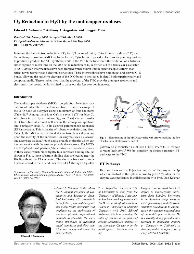
PERSPECTIVESolomon et al.O2 Reduction to H2O by the multicopper oxidases
PERSPECTIVEChisholm and McIndoeCharged ligands for catalyst immobilisation and analysis
ISSN 1477-9226
www.rsc.org/dalton
An international journal of inorganic chemistry
PERSPECTIVE www.rsc.org/dalton | Dalton Transactions
O2 Reduction to H2O by the multicopper oxidases
Edward I. Solomon,* Anthony J. Augustine and Jungjoo Yoon
Received 16th January 2008, Accepted 28th March 2008First published as an Advance Article on the web 7th May 2008DOI: 10.1039/b800799c
In nature the four electron reduction of O2 to H2O is carried out by Cytochrome c oxidase (CcO) andthe multicopper oxidases (MCOs). In the former, Cytochrome c provides electrons for pumping protonsto produce a gradient for ATP synthesis, while in the MCOs the function is the oxidation of substrates,either organic or metal ions. In the MCOs the reduction of O2 is carried out at a trinuclear Cu cluster(TNC). Oxygen intermediates have been trapped which exhibit unique spectroscopic features thatreflect novel geometric and electronic structures. These intermediates have both intact and cleaved O–Obonds, allowing the reductive cleavage of the O–O bond to be studied in detail both experimentally andcomputationally. These studies show that the topology of the TNC provides a unique geometric andelectronic structure particularly suited to carry out this key reaction in nature.
Introduction
The multicopper oxidases (MCOs) couple four 1-electron oxi-dations of substrate to the four electron reductive cleavage ofthe O–O bond of dioxygen using a minimum of four Cu atoms(Table 1).1,2 Among these four Cu’s is a type 1 (T1) or blue Cusite, characterized by an intense SCys → Cu(II) charge transfer(CT) transition at around 600 nm in the absorption spectrumand a uniquely small A‖ in its electron paramagnetic resonance(EPR) spectrum. This is the site of substrate oxidation, and fromTable 1, the MCOs can be divided into two classes dependingupon the identity of the substrate. For enzymes such as laccase3
and ascorbate oxidase,4 redox active organic molecules which caninteract weakly with the enzyme provide the electrons. For MCOslike Fet3p5 and ceruloplasmin,6 the substrate is a metal ion (ferrousin these cases) which binds tightly to a substrate binding site. Asshown in Fig. 1, these substrate binding sites are located near theHis ligands of the T1 Cu center. The electron from substrate isfirst transferred to the T1 and then over >13 A through a Cys–His
Department of Chemistry, Stanford University, Stanford, California, 94305,USA. E-mail: [email protected].; Fax: +1 (650) 725-0259;Tel: +1 (650) 723-9104
Edward I. Solomon
Edward I. Solomon is the Mon-roe E. Spaght Professor of Hu-manities and Science at Stan-ford University. His research isin the fields of physical-inorganicand bioinorganic chemistry withemphasis on the application ofspectroscopic and computationalmethods to elucidate the elec-tronic structures of transitionmetal complexes and their con-tributions to physical propertiesand reactivity.
T. J. Augustine received a B.S.in Chemistry in 2003 from theUniversity of Illinois. Since thenhe has been working towards hisPh.D. as a Stanford GraduateFellow in Chemistry at StanfordUniversity with Prof. EdwardSolomon. He is researching theroles of residues in the first andsecond coordination spheres ofthe trinuclear Cu cluster in themulticopper oxidases in reactiv-ity.
Jungjoo Yoon received his Ph.D.degree in bio-inorganic chem-istry from Stanford Universityin the Solomon group, where heused spectroscopy and electronicstructure calculations to charac-terize the oxygen intermediatesof the multicopper oxidases. Heis currently doing post-doctoralresearch in molecular biologyat University of California atBerkeley under the supervision ofProf. Michael Marletta.
Fig. 1 The structure of the MCO active site with arrows marking the flowof substrates, electrons (e−), and O2.
pathway to a trinuclear Cu cluster (TNC) where O2 is reducedto water (vide infra).7 We first consider the electron transfer (ET)pathways to the TNC.
ET Pathways
Here we focus on the Fe(II) binding site of the enzyme Fet3p,which is involved in the uptake of iron by yeast.8 (Studies on thisenzyme were performed in collaboration with Prof. Dan Kosman
This journal is © The Royal Society of Chemistry 2008 Dalton Trans., 2008, 3921–3932 | 3921
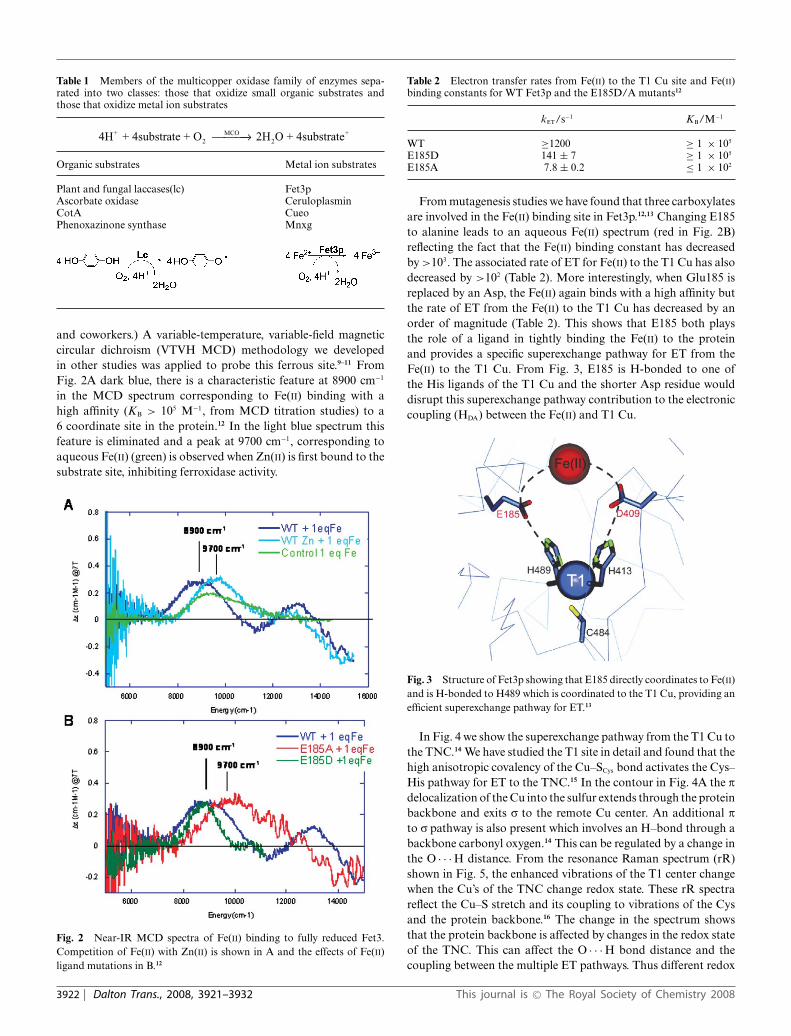
Table 1 Members of the multicopper oxidase family of enzymes sepa-rated into two classes: those that oxidize small organic substrates andthose that oxidize metal ion substrates
4H + 4substrate + O 2H O + 4substrate+2
MCO2
+⎯ →⎯⎯
Organic substrates Metal ion substrates
Plant and fungal laccases(lc) Fet3pAscorbate oxidase CeruloplasminCotA CueoPhenoxazinone synthase Mnxg
and coworkers.) A variable-temperature, variable-field magneticcircular dichroism (VTVH MCD) methodology we developedin other studies was applied to probe this ferrous site.9–11 FromFig. 2A dark blue, there is a characteristic feature at 8900 cm−1
in the MCD spectrum corresponding to Fe(II) binding with ahigh affinity (KB > 105 M−1, from MCD titration studies) to a6 coordinate site in the protein.12 In the light blue spectrum thisfeature is eliminated and a peak at 9700 cm−1, corresponding toaqueous Fe(II) (green) is observed when Zn(II) is first bound to thesubstrate site, inhibiting ferroxidase activity.
Fig. 2 Near-IR MCD spectra of Fe(II) binding to fully reduced Fet3.Competition of Fe(II) with Zn(II) is shown in A and the effects of Fe(II)ligand mutations in B.12
Table 2 Electron transfer rates from Fe(II) to the T1 Cu site and Fe(II)binding constants for WT Fet3p and the E185D/A mutants12
kET/s−1 KB/M−1
WT ≥1200 ≥ 1 × 10⁵
E185D 141 ± 7 ≥ 1 × 10⁵
E185A 7.8 ± 0.2 ≤ 1 × 10²
From mutagenesis studies we have found that three carboxylatesare involved in the Fe(II) binding site in Fet3p.12,13 Changing E185to alanine leads to an aqueous Fe(II) spectrum (red in Fig. 2B)reflecting the fact that the Fe(II) binding constant has decreasedby >103. The associated rate of ET for Fe(II) to the T1 Cu has alsodecreased by >102 (Table 2). More interestingly, when Glu185 isreplaced by an Asp, the Fe(II) again binds with a high affinity butthe rate of ET from the Fe(II) to the T1 Cu has decreased by anorder of magnitude (Table 2). This shows that E185 both playsthe role of a ligand in tightly binding the Fe(II) to the proteinand provides a specific superexchange pathway for ET from theFe(II) to the T1 Cu. From Fig. 3, E185 is H-bonded to one ofthe His ligands of the T1 Cu and the shorter Asp residue woulddisrupt this superexchange pathway contribution to the electroniccoupling (HDA) between the Fe(II) and T1 Cu.
Fig. 3 Structure of Fet3p showing that E185 directly coordinates to Fe(II)and is H-bonded to H489 which is coordinated to the T1 Cu, providing anefficient superexchange pathway for ET.13
In Fig. 4 we show the superexchange pathway from the T1 Cu tothe TNC.14 We have studied the T1 site in detail and found that thehigh anisotropic covalency of the Cu–SCys bond activates the Cys–His pathway for ET to the TNC.15 In the contour in Fig. 4A the pdelocalization of the Cu into the sulfur extends through the proteinbackbone and exits r to the remote Cu center. An additional pto r pathway is also present which involves an H–bond through abackbone carbonyl oxygen.14 This can be regulated by a change inthe O · · · H distance. From the resonance Raman spectrum (rR)shown in Fig. 5, the enhanced vibrations of the T1 center changewhen the Cu’s of the TNC change redox state. These rR spectrareflect the Cu–S stretch and its coupling to vibrations of the Cysand the protein backbone.16 The change in the spectrum showsthat the protein backbone is affected by changes in the redox stateof the TNC. This can affect the O · · · H bond distance and thecoupling between the multiple ET pathways. Thus different redox
3922 | Dalton Trans., 2008, 3921–3932 This journal is © The Royal Society of Chemistry 2008
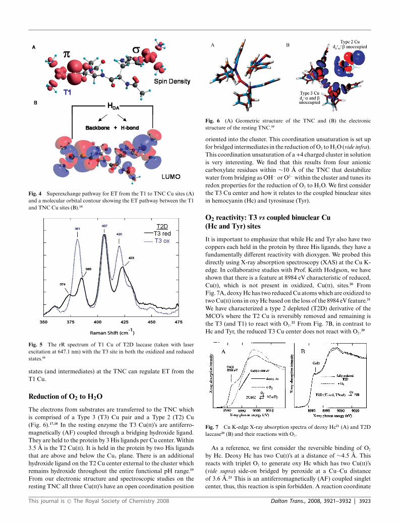
Fig. 4 Superexchange pathway for ET from the T1 to TNC Cu sites (A)and a molecular orbital contour showing the ET pathway between the T1and TNC Cu sites (B).14
Fig. 5 The rR spectrum of T1 Cu of T2D laccase (taken with laserexcitation at 647.1 nm) with the T3 site in both the oxidized and reducedstates.16
states (and intermediates) at the TNC can regulate ET from theT1 Cu.
Reduction of O2 to H2O
The electrons from substrates are transferred to the TNC whichis comprised of a Type 3 (T3) Cu pair and a Type 2 (T2) Cu(Fig. 6).17,18 In the resting enzyme the T3 Cu(II)’s are antiferro-magnetically (AF) coupled through a bridging hydroxide ligand.They are held to the protein by 3 His ligands per Cu center. Within3.5 A is the T2 Cu(II). It is held in the protein by two His ligandsthat are above and below the Cu3 plane. There is an additionalhydroxide ligand on the T2 Cu center external to the cluster whichremains hydroxide throughout the entire functional pH range.19
From our electronic structure and spectroscopic studies on theresting TNC all three Cu(II)’s have an open coordination position
Fig. 6 (A) Geometric structure of the TNC and (B) the electronicstructure of the resting TNC.19
oriented into the cluster. This coordination unsaturation is set upfor bridged intermediates in the reduction of O2 to H2O (vide infra).This coordination unsaturation of a +4 charged cluster in solutionis very interesting. We find that this results from four anioniccarboxylate residues within ∼10 A of the TNC that destabilizewater from bridging as OH− or O2− within the cluster and tunes itsredox properties for the reduction of O2 to H2O. We first considerthe T3 Cu center and how it relates to the coupled binuclear sitesin hemocyanin (Hc) and tyrosinase (Tyr).
O2 reactivity: T3 vs coupled binuclear Cu(Hc and Tyr) sites
It is important to emphasize that while Hc and Tyr also have twocoppers each held in the protein by three His ligands, they have afundamentally different reactivity with dioxygen. We probed thisdirectly using X-ray absorption spectroscopy (XAS) at the Cu K-edge. In collaborative studies with Prof. Keith Hodgson, we haveshown that there is a feature at 8984 eV characteristic of reduced,Cu(I), which is not present in oxidized, Cu(II), sites.20 FromFig. 7A, deoxy Hc has two reduced Cu atoms which are oxidized totwo Cu(II) ions in oxy Hc based on the loss of the 8984 eV feature.21
We have characterized a type 2 depleted (T2D) derivative of theMCO’s where the T2 Cu is reversibly removed and remaining isthe T3 (and T1) to react with O2.22 From Fig. 7B, in contrast toHc and Tyr, the reduced T3 Cu center does not react with O2.20
Fig. 7 Cu K-edge X-ray absorption spectra of deoxy Hc21 (A) and T2Dlaccase20 (B) and their reactions with O2.
As a reference, we first consider the reversible binding of O2
by Hc. Deoxy Hc has two Cu(I)’s at a distance of ∼4.5 A. Thisreacts with triplet O2 to generate oxy Hc which has two Cu(II)’s(vide supra) side-on bridged by peroxide at a Cu–Cu distanceof 3.6 A.23 This is an antiferromagnetically (AF) coupled singletcenter, thus, this reaction is spin forbidden. A reaction coordinate
This journal is © The Royal Society of Chemistry 2008 Dalton Trans., 2008, 3921–3932 | 3923

was generated by systematically varying the distance of theperoxide above the molecular plane and optimizing the rest of thestructure.25 From Fig. 8A, left to right, the structure first butterfliesthen goes to an asymmetric end-on/side-on bridged structureand then end-on/end-on bridged in the reversible loss of O2.These structures maximize metal–ligand overlap with increasingdistances of the oxygen from the copper. From Fig. 8B, proceedingalong this coordinate the peroxide becomes less negative andthe Cu’s less positive indicating that charge is transferred fromperoxide to the two Cu(II)’s. Importantly, the charge on both Cu’schanges at the same rate even in the asymmetric bridged structure(dashed) indicating that O2 binding involves the simultaneoustransfer of two electrons.
Fig. 8C accounts for the change in spin and total energy alongthe reaction coordinate. Oxy Hc has a singlet ground state dueto AF coupling of the two Cu(II)’s through the p* orbital ofthe l-g2:g2 peroxide in the molecular plane. As we proceed alongthe coordinate the structure becomes butterflied and each Cu(II)
interacts with a different p* orbital on the peroxide. This involvesorthogonal magnetic orbitals producing a triplet ground state forthe butterflied structure. The peroxide can then directly transferone electron of the same spin to each Cu leading to triplet dioxygenwhich is further energetically stabilized by single center exchange.24
Importantly, O2 binding to Hc is found to be exothermic by3 kcal mol−1.25 This is contrasted to O2 binding to the deoxyT3 center of T2D laccase in Fig. 9, where O2 binding is foundto be uphill by 6 kcal mol−1. As shown in Fig. 10 the origin ofthis 10 kcal mol−1 destabilization of O2 binding relative to Hcreflects the relative stabilization of the deoxy T3 structure in theMCO protein environment. The deoxy potential energy surfaces inFig. 10 were obtained by geometry optimizing the reduced Hc andT3 sites with their respective protein constraints imposed on thedeoxy structures. From Fig. 10, the deoxy T3 center (red) is 7 kcalmol−1 lower in energy than deoxy Hc (blue) and has an equilibriumCu(I)–Cu(I) distance of 6.5 A, in contrast to the 4.2 A optimizeddistance of deoxy Hc. This decrease in energy of the deoxy T3
Fig. 8 Reaction coordinate of O2 binding to Hc viewed along the O–O bond (top) and then perpendicular to the initial Cu2O2 plane (bottom) (A) andplots of the charge transfer (B) and singlet/triplet intersystem crossing energy (C) along the reaction coordinate. R:d(X–X) is the distance between thecenter of the O–O and Cu–Cu vectors. R:d(X–X) values less than ∼0.6 and greater than ∼0.6 represents symmetric and asymmetric O2 coordination,respectively.24
3924 | Dalton Trans., 2008, 3921–3932 This journal is © The Royal Society of Chemistry 2008

Fig. 9 Energy of O2 binding to the T3 Cu site with S = 0 in blue andS = 1 in red.25
Fig. 10 Potential energy surfaces for deoxy-Hc and deoxy-T3 sites as afunction of Cu–Cu distance (dashed lines show the energy of electrostaticinteraction between the two Cu(I) centers).25
center dominantly reflects the decrease in electrostatic repulsionof the two Cu(I)’s in the low dielectric of the protein (dashed blue)and an additional smaller contribution due to a decrease in sterricinteractions of the three His ligands between Cu centers which areeclipsed in the T3 center and staggered in Hc (difference betweendashed red and dashed blue). Thus the lack of O2 reactivity of thedeoxy T3 site reflects its electrostatic and structural stabilizationat its long 6.5 A Cu–Cu distance. From Fig. 11 the large structuraldifferences between the coupled binuclear site in Hc and the T3 sitein the MCO’s relate to very different structural constraints in thetwo protein environments. The binuclear cuprous site of deoxy Hcis kept at its 4.2 A Cu–Cu distance due to the constraint associated
Fig. 11 Comparison of the constrained structures of deoxy-Hc (PDBcode 1JS8) and deoxy-T3 (PDB code 1GYC).
with two His ligands, one on each Cu, each deriving from adifferent helix bundle held together by a salt bridge. Alternatively,in the T3 Cu center the two Cu(I)’s are kept at an electrostaticallystable distance of 6.5 A by two sets of two His ligands, where eachset is from an H–X–H bridging motif that is on a loop extendingfrom a b sheet, leaving the Cu(I)’s relatively unconstrained.
In summary, deoxy Hc is electrostatically destabilized to reactwith O2 to form oxy Hc and this can be cooperatively regulated bychanging the Cu(I)–Cu(I) distance in tensed and relaxed proteinquaternary structures,26 while the reduced T3 site in the MCOs iselectrostatically stable and does not react with O2 when the T2 Cuis not present.
O2 Reactivity: the trinuclear Cu cluster (TNC)
The native enzyme with its intact TNC reduced and a reducedT1 reacts with O2 to generate the native intermediate (NI).27,28
(Scheme 1, top). Derivatives of the native enzyme have beenprepared where the T1 is either eliminated29,30 (replacement of theCys ligand of the T1 with a Ser to generate a type 1 depletedor T1D form) or replaced by a redox innocent mercuric ion(the T1Hg derivative).31,32 These have valid TNCs which, whenreduced, react with O2 with essentially the same rate constantas the native enzyme. (kNat = 1.7 × 10⁶ M−1s−1, kT1Hg = 2.2 ×10⁶ M−1s−1)33 This reaction generates a species with at least oneless electron transferred to O2 which we have, in fact, determinedto be a peroxide intermediate (PI).34 (Scheme 1, bottom) Therates of formation show that PI is kinetically competent to bea precursor to NI and indeed we have found that PI decays toNI. The conversion of PI to NI is very fast for the native enzymeas it involves two electron reduction of peroxide, but is ∼106 slowerfor the T1D/T1Hg derivatives where the electron from the T1 isnot available.35 We will first consider new results on PI, then thedefinition of the unique spectral features, and the electronic andgeometric structure of NI. Using the decreased rate of the PI → NIdecay in the T1D/T1Hg derivatives, we can then experimentallyand computationally evaluate the reductive cleavage of the O–Obond.
Scheme 1 Schematic of the O2 reactivity of both native and T1D/T1Hgforms of the MCOs and the cleavage of the O–O bond.
Peroxide intermediate (PI)
In earlier studies we used isotope-ratio mass spectrometry (IRMS)and the ligand field (LF) transitions (from circular dichroism) to
This journal is © The Royal Society of Chemistry 2008 Dalton Trans., 2008, 3921–3932 | 3925
find that the reaction of reduced T1D/T1Hg with O2 produced aperoxide bound to two Cu(II) ions. From EPR, low temperature(LT) MCD, and SQUID magnetic susceptibility the Cu(II)’s are AFcoupled and therefore bridged.34 There was no pH dependence orkinetic isotope effect (KIE) on the reaction of O2 with the reducedTNC; therefore, a proton is not involved in the formation of PI.35
From a combination of spectroscopic studies on the peroxideadduct (PA) of the TNC36 (where all Cu’s are oxidized withperoxide bound), QM/MM studies of PI and PA,37 and EXAFSstudies we found that peroxide formed an internal bridge betweenthe T2 and T3 Cu centers.
Importantly, from Fig. 12, the peroxide to Cu(II) charge transfer(CT) spectrum of PI is very different from that of oxy Hc indicatinga very different geometric and electronic structure. However, if O2
is computationally added to the reduced TNC and optimized,a side-on bridged structure is obtained (Fig. 13, left) which, asdescribed earlier, is energetically unfavorable and not consistentwith the absorption spectrum in Fig. 12. Importantly, if an Asp,present in the crystal structure (D94 in Fet3p) near the T2 Cu, isincluded in the calculation a structure consistent with the spectrumis obtained with the peroxide binding g2 to the T3 CuB (nearer tothe Asp) and g1 to each of the other two Cu’s (Fig. 13, right).38
This computational result is consistent with experimental resultswhere the D94A and D94N mutants do not react with O2, whilereplacing D with a negatively charged E residue allows the O2
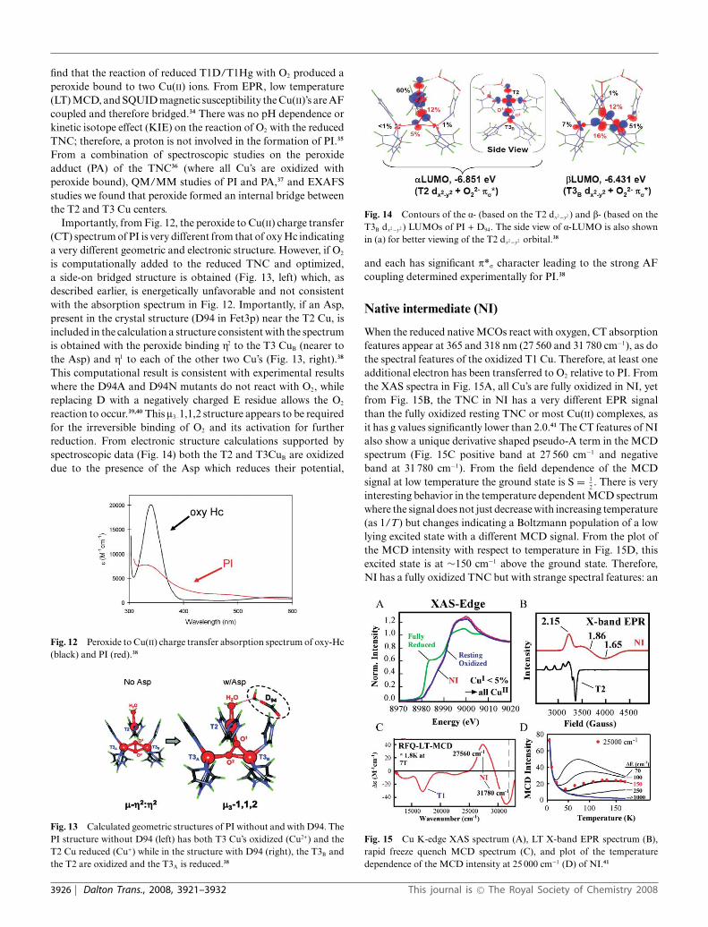
reaction to occur.39,40 This l3–1,1,2 structure appears to be requiredfor the irreversible binding of O2 and its activation for furtherreduction. From electronic structure calculations supported byspectroscopic data (Fig. 14) both the T2 and T3CuB are oxidizeddue to the presence of the Asp which reduces their potential,
Fig. 12 Peroxide to Cu(II) charge transfer absorption spectrum of oxy-Hc(black) and PI (red).38
Fig. 13 Calculated geometric structures of PI without and with D94. ThePI structure without D94 (left) has both T3 Cu’s oxidized (Cu2+) and theT2 Cu reduced (Cu+) while in the structure with D94 (right), the T3B andthe T2 are oxidized and the T3A is reduced.38
Fig. 14 Contours of the a- (based on the T2 dx2−y2 ) and b- (based on theT3B dx2−y2 ) LUMOs of PI + D94. The side view of a-LUMO is also shownin (a) for better viewing of the T2 dx2−y2 orbital.38
and each has significant p*r character leading to the strong AFcoupling determined experimentally for PI.38
Native intermediate (NI)
When the reduced native MCOs react with oxygen, CT absorptionfeatures appear at 365 and 318 nm (27 560 and 31 780 cm−1), as dothe spectral features of the oxidized T1 Cu. Therefore, at least oneadditional electron has been transferred to O2 relative to PI. Fromthe XAS spectra in Fig. 15A, all Cu’s are fully oxidized in NI, yetfrom Fig. 15B, the TNC in NI has a very different EPR signalthan the fully oxidized resting TNC or most Cu(II) complexes, asit has g values significantly lower than 2.0.41 The CT features of NIalso show a unique derivative shaped pseudo-A term in the MCDspectrum (Fig. 15C positive band at 27 560 cm−1 and negativeband at 31 780 cm−1). From the field dependence of the MCDsignal at low temperature the ground state is S = 1
2. There is very
interesting behavior in the temperature dependent MCD spectrumwhere the signal does not just decrease with increasing temperature(as 1/T) but changes indicating a Boltzmann population of a lowlying excited state with a different MCD signal. From the plot ofthe MCD intensity with respect to temperature in Fig. 15D, thisexcited state is at ∼150 cm−1 above the ground state. Therefore,NI has a fully oxidized TNC but with strange spectral features: an
Fig. 15 Cu K-edge XAS spectrum (A), LT X-band EPR spectrum (B),rapid freeze quench MCD spectrum (C), and plot of the temperaturedependence of the MCD intensity at 25 000 cm−1 (D) of NI.41
3926 | Dalton Trans., 2008, 3921–3932 This journal is © The Royal Society of Chemistry 2008
S = 1/2 ground state with g values below 2.0, a low lying excitedstate at ∼150 cm−1, and an intense pseudo-A term in the CT regionof the MCD spectrum.41 We consider the geometric and electronicstructural origin of the unique spectral features of NI below.
Origin of the low lying excited state: spin frustration
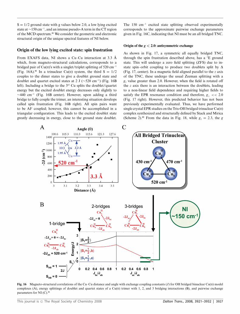
From EXAFS data, NI shows a Cu–Cu interaction at 3.3 Awhich, from magneto-structural calculations, corresponds to abridged pair of Cu(II)’s with a singlet/triplet splitting of 520 cm−1
(Fig. 16A).41 In a trinuclear Cu(II) system, the third S = 1/2couples to the dimer states to give a doublet ground state anddoublet and quartet excited states at 2 J (∼520 cm−1) (Fig. 16Bleft). Including a bridge to the 3rd Cu splits the doublet/quartetenergy but the excited doublet energy decreases only slightly to∼440 cm−1 (Fig. 16B center). However, upon adding a thirdbridge to fully couple the trimer, an interesting situation developscalled spin frustration (Fig. 16B right). All spin pairs wantto be AF coupled; however, this cannot be accomplished in atriangular configuration. This leads to the excited doublet stategreatly decreasing in energy, close to the ground state doublet.
The 150 cm−1 excited state splitting observed experimentallycorresponds to the approximate pairwise exchange parametersgiven in Fig. 16C, indicating that NI must be an all bridged TNC.
Origin of the g < 2.0: antisymmetric exchange
As shown in Fig. 17, a symmetric all equally bridged TNC,through the spin frustration described above, has a 2E groundstate. This will undergo a zero field splitting (ZFS) due to in-state spin–orbit coupling to produce two doublets split by D(Fig. 17, center). In a magnetic field aligned parallel to the z axisof the TNC, these undergo the usual Zeeman splitting with ag‖ value greater than 2.0. However, when the field is rotated offthe z axis there is an interaction between the doublets, leadingto a non-linear field dependence and requiring higher fields tosatisfy the EPR resonance condition and therefore, g⊥ << 2.0(Fig. 17 right). However, this predicted behavior has not beenpreviously experimentally evaluated. Thus, we have performedsingle crystal EPR studies on the Tris-OH bridged trinuclear Cu(II)complex synthesized and structurally defined by Stack and Mirica(Scheme 2).42 From the data in Fig. 18, while g‖ = 2.3, the g
Fig. 16 Magneto-structural correlations of the Cu–Cu distance and angle with exchange coupling constants (J) for OH bridged binuclear Cu(II) modelcomplexes (A), energy splittings of doublet and quartet states of a Cu(II) trimer with 1, 2, and 3 bridging interactions (B), and pairwise exchangeparameters for NI (C).41
This journal is © The Royal Society of Chemistry 2008 Dalton Trans., 2008, 3921–3932 | 3927

Fig. 17 Ground state splitting of spin-frustrated Cu(II) trimer due to antisymmetric exchange.42
Scheme 2 Structures of the Tris-OH (A) and l3-oxo (B) trinuclear Cu(II)model complexes.43
Fig. 18 Single crystal X-band EPR spectrum of Tris-OH bridgedtrinuclear Cu(II) complex as it is rotated in the magnetic field (0◦ =magnetic field aligned along the molecular z-axis).42
value greatly decreases as the field is oriented off the z axis goingdown to an unprecedented g ∼ 1.2 before it becomes too broadto observe.43 This experimentally confirms that the ZFS of the 2Eground state of this complex derives from antisymmetric exchange.This requires good ground-to-ground state exchange coupling,spin–orbit coupling (SOC) of the ground to an excited state onone Cu(II), and good exchange coupling of this excited state to theground state on an adjacent Cu(II). From the contours in Fig. 19,
Fig. 19 Antisymmetric exchange mechanism based on orbital contours ofthe ground and ligand field excited state of the Tris-OH bridged trinuclearCu(II) complex.43
the hydroxide bridges of the Tris-OH structure provide goodexchange coupling between ground dx2−y2 orbitals on adjacentCu(II)’s and good exchange coupling of the spin orbit coupleddxy excited state with the dx2−y2 ground state of adjacent Cu(II)’s.Importantly, these studies show that the low g values of NIlogically derive from its all bridged structure. This leads to twopossible structures for NI, either with three OH bridges (two fromO2 reduction and one from H2O) or a l3-oxo bridge (from O2
reduction) at the center of the cluster.
Origin of MCD pseudo-A term: excited state spin–orbit coupling
Model complexes of both structures exist (Scheme 2), and bothexhibit pseudo-A terms in their l2-hydroxo and l3-oxo CT regions.The Tris-OH complex has the positive component of the MCDspectrum higher in energy (Fig. 20A) while the l3-oxo model hasits negative component higher in energy (Fig. 20B) as is also thecase for NI (Fig. 20C, which is an expanded version of the highenergy region of Fig. 15C). These pseudo-A terms require twoperpendicular CT transitions that can SOC in a third, mutuallyperpendicular direction. This spin–orbit coupling is dominatelya single center, one electron operator. For both complexes theCT transitions are in the trinuclear Cu (x,y) plane. As shown inFig. 21A, for the Tris-OH structure the CT transitions involve two
Fig. 20 MCD spectra of the Tris-OH bridged (A) and l3-oxo bridged (B) trinuclear Cu(II) complexes, and NI (C).43 (Assignments of bands to specificCu’s in C are based on their temperature dependence in ref. 41.)
3928 | Dalton Trans., 2008, 3921–3932 This journal is © The Royal Society of Chemistry 2008
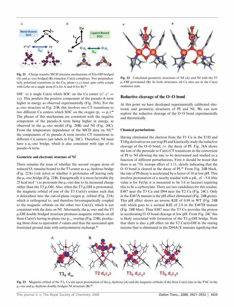
Fig. 21 Charge transfer MCD intensity mechanisms of Tris-OH bridged(A) and l3-oxo bridged (B) trinuclear Cu(II) complexes. Two perpendicu-larly polarized transitions in the Cu3 plane (x,y) must spin–orbit couplewith LzSz on a single atom (Cu for A and O for B).43
OH− to a single Cu(II) which SOC on the Cu center (x2–y2 ↔xy). This predicts the positive component of the pseudo-A termhigher in energy as observed experimentally (Fig. 20A). For thel3-oxo structure in Fig. 21B, this involves oxo CT transitions totwo different Cu centers which SOC on the oxygen (px ↔ py).44
The phases of this mechanism are consistent with the negativecomponent of the pseudo-A term being higher in energy, asobserved in the l3-oxo model (Fig. 20B) and NI (Fig. 20C).From the temperature dependence of the MCD data on NI,41
the components of its pseudo-A term involve CT transitions todifferent Cu centers (see labels in Fig. 20C). Therefore, NI musthave a l3-oxo bridge, which is also consistent with sign of itspseudo-A term.
Geometric and electronic structure of NI
There remains the issue of whether the second oxygen atom ofreduced O2 remains bound to the T3 center as a l2-hydroxo bridge(Fig. 22A) (vide infra) or whether it protonates off leaving onlythe l3-oxo bridge (Fig. 22B). Energetically it is more favorable (by25 kcal mol−1) to protonate the l3-oxo due to its increased chargerather than the T3 l2OH. Also, when the T3 l2OH is protonated,the magnetic orbital of one of the T3 Cu(II)’s rotates such thatit delocalizes into the out-of-plane oxygen pz orbital (Fig. 23A)which is orthogonal to, and therefore ferromagnetically coupledto the magnetic orbitals on the other two Cu(II)’s, which is notconsistent with the data on NI. Alternately, the l3-oxo and the T3l2OH doubly bridged structure produces magnetic orbitals on allthree Cu(II)’s having in-plane oxo px,y overlap (Fig. 23B), produc-ing three close to equivalent J values and thus the associated spinfrustrated ground state with antisymmetric exchange.45
Fig. 22 Calculated geometric structures of NI (A) and NI with the T3l2-OH protonated (B). In both structures, all Cu sites are in the Cu(II)oxidation state.
Reductive cleavage of the O–O bond
At this point we have developed experimentally calibrated elec-tronic and geometric structures of PI and NI. We can nowexplore the reductive cleavage of the O–O bond experimentallyand theoretically.
Chemical perturbations
Having eliminated the electron from the T1 Cu in the T1D andT1Hg derivatives we can trap PI and kinetically study the reductivecleavage of the O–O bond, i.e. the decay of PI. Fig. 24A showsthe loss of the peroxide to Cu(II) CT transitions in the conversionof PI to NI allowing the rate to be determined and studied as afunction of different perturbations. First it should be noted thatthere is an 18O2 isotope effect of 1.11, clearly indicating that theO–O bond is cleaved in the decay of PI.35 From Fig. 24B black,the rate of PI decay is accelerated by a factor of 10 at low pH. Thisinvolves protonation of a nearby residue with a pKa of ∼5.0 (thisvalue is for Fet3p, it is measured to be 5.6 in laccase) requiringthis to be a carboxylate. There are two candidates for this residue,E487 near the T3 Cu and D94 near the T2 Cu (Fig. 24C). Onlyin the E487A mutant is the pH effect eliminated (Fig. 24B green).This pH effect shows an inverse KIE of 0.89 in WT (Fig. 24Bred) which goes to a normal KIE of 2.0 in the E487D mutant(Fig. 24B blue). Thus E487 near the T3 Cu provides the protonin accelerating O–O bond cleavage at low pH. From Fig. 24C thisis likely associated with formation of the T3 l2OH bridge. Notethat there is also a pH effect on the T2 Cu(II)-OH in the restingenzyme that is eliminated in the D94A/E mutants signifying that
Fig. 23 Magnetic orbital of the T3A Cu site upon protonation of the l2-hydroxo (A) and the magnetic orbitals of the three Cu(II) sites in the TNC in thel3-oxo and l2-hydroxo doubly bridged NI structure (B).46
This journal is © The Royal Society of Chemistry 2008 Dalton Trans., 2008, 3921–3932 | 3929
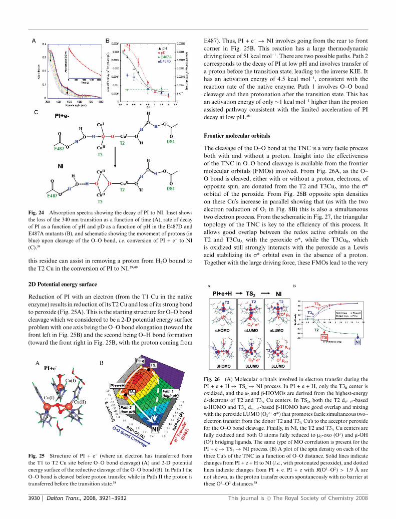
Fig. 24 Absorption spectra showing the decay of PI to NI. Inset showsthe loss of the 340 nm transition as a function of time (A), rate of decayof PI as a function of pH and pD as a function of pH in the E487D andE487A mutants (B), and schematic showing the movement of protons (inblue) upon cleavage of the O–O bond, i.e. conversion of PI + e− to NI(C).39
this residue can assist in removing a proton from H2O bound tothe T2 Cu in the conversion of PI to NI.39,40
2D Potential energy surface
Reduction of PI with an electron (from the T1 Cu in the nativeenzyme) results in reduction of its T2 Cu and loss of its strong bondto peroxide (Fig. 25A). This is the starting structure for O–O bondcleavage which we considered to be a 2-D potential energy surfaceproblem with one axis being the O–O bond elongation (toward thefront left in Fig. 25B) and the second being O–H bond formation(toward the front right in Fig. 25B, with the proton coming from
Fig. 25 Structure of PI + e− (where an electron has transferred fromthe T1 to T2 Cu site before O–O bond cleavage) (A) and 2-D potentialenergy surface of the reductive cleavage of the O–O bond (B). In Path I theO–O bond is cleaved before proton transfer, while in Path II the proton istransferred before the transition state.38
E487). Thus, PI + e− → NI involves going from the rear to frontcorner in Fig. 25B. This reaction has a large thermodynamicdriving force of 51 kcal mol−1. There are two possible paths. Path 2corresponds to the decay of PI at low pH and involves transfer ofa proton before the transition state, leading to the inverse KIE. Ithas an activation energy of 4.5 kcal mol−1, consistent with thereaction rate of the native enzyme. Path 1 involves O–O bondcleavage and then protonation after the transition state. This hasan activation energy of only ∼1 kcal mol−1 higher than the protonassisted pathway consistent with the limited acceleration of PIdecay at low pH.38
Frontier molecular orbitals
The cleavage of the O–O bond at the TNC is a very facile processboth with and without a proton. Insight into the effectivenessof the TNC in O–O bond cleavage is available from the frontiermolecular orbitals (FMOs) involved. From Fig. 26A, as the O–O bond is cleaved, either with or without a proton, electrons, ofopposite spin, are donated from the T2 and T3CuA into the r*orbital of the peroxide. From Fig. 26B opposite spin densitieson these Cu’s increase in parallel showing that (as with the twoelectron reduction of O2 in Fig. 8B) this is also a simultaneoustwo electron process. From the schematic in Fig. 27, the triangulartopology of the TNC is key to the efficiency of this process. Itallows good overlap between the redox active orbitals on theT2 and T3CuA with the peroxide r*, while the T3CuB, whichis oxidized still strongly interacts with the peroxide as a Lewisacid stabilizing its r* orbital even in the absence of a proton.Together with the large driving force, these FMOs lead to the very
Fig. 26 (A) Molecular orbitals involved in electron transfer during thePI + e + H → TS2 → NI process. In PI + e + H, only the T3B center isoxidized, and the a- and b-HOMOs are derived from the highest-energyd-electrons of T2 and T3A Cu centers. In TS2, both the T2 dx2−y2 -baseda-HOMO and T3A dx2−y2 -based b-HOMO have good overlap and mixingwith the peroxide LUMO (O2
2− r*) that promotes facile simultaneous two–electron transfer from the donor T2 and T3A Cu’s to the acceptor peroxidefor the O–O bond cleavage. Finally, in NI, the T2 and T3A Cu centers arefully oxidized and both O atoms fully reduced to l3-oxo (O1) and l-OH(O2) bridging ligands. The same type of MO correlation is present for thePI + e → TS1 → NI process. (B) A plot of the spin density on each of thethree Cu’s of the TNC as a function of O–O distance. Solid lines indicatechanges from PI + e + H to NI (i.e., with protonated peroxide), and dottedlines indicate changes from PI + e. PI + e with R(O1–O2) > 1.9 A arenot shown, as the proton transfer occurs spontaneously with no barrier atthese O1–O2 distances.38
3930 | Dalton Trans., 2008, 3921–3932 This journal is © The Royal Society of Chemistry 2008

Fig. 27 Schematic of triangular topology of the TNC and depiction ofthe orbitals relevant in O–O cleavage. Note that T2 and T3A transfer twoelectrons into the peroxide r* orbital and T3B is oxidized and acts as aLewis acid, similar to a proton, in stabilizing the r* orbital.38
low barrier for the two electron reductive cleavage of the peroxideO–O bond.38
NI Conversion to resting: interconversion of the twofully oxidized forms of the trinuclear cluster
The NI and resting sites have very different spectral features dueto the fully bridged structure of NI. From isotope studies, oneoxygen from O2 reduction ends up bound to the T2 Cu(II) of theresting enzyme.34,45,46 Thus, there must be a rearrangement of thel3-oxo bridge from inside to outside the cluster (Fig. 28A). Thisinvolves protons, the first to the l3O and a second to the T3 l2OH,which breaks this bridge and allows the exchange of one oxygenfrom O2 into solvent. The internal l3-hydroxo no longer binds toT3CuB and can rotate from inside to outside the cluster throughthe T3CuA-T2 Cu edge. From Fig. 28B this rotation removes oneproton from the H2O at T3CuB to reform the T3 l2OH bridgeand as the T2-T3CuA bridging H2O rotates out of the cluster itdonates one proton to the terminal OH at the T2 Cu, weakeningthis bond and allowing its substitution by the rotated OH fromO2 reduction.47 This process is calculated to have a barrier of∼ 8.5 kcal mol−1 which is reasonable given that the experimentalbarrier for the conversion of NI to resting is 8.8–13.9 kcal mol−1.48
Importantly, this rate of conversion (k = 0.05 s−1)27 is ordersof magnitude slower than the turnover rate of the enzyme (k =350 s−1).49 Therefore, NI is actually the catalytically relevant fullyoxidized form of the MCOs.
Fig. 28 Schematic showing the conversion of NI to resting marking theposition of the oxygen atoms from dioxygen in red (A) and geometricstructures and energies for the doubly protonated form of NI rotating thel3-OH from inside to outside the TNC through the T3 CuA-T2 Cu edge(B).46
Molecular mechanism of O2 reduction to H2O
The above detailed chemical, spectroscopic, and electronic struc-ture studies have led to the mechanism of the MCO’s shown inFig. 29. The reaction of the fully reduced enzyme with O2 occursin two 2 electron steps, with the second being fast, so it is effectivelya four electron process. The first step is rate determining and drivenby the presence of an anionic Asp residue near the T2 Cu. The lowbarrier of the fast second step reflects the large driving force fortwo electron reduction of peroxide combined with the triangulartopology of the TNC. Importantly, NI is a fully oxidized allbridged structure and is the catalytically relevant fully oxidizedform of the enzyme. Our thoughts are that the l3-oxo bridgeprovides superexchange pathways for rapid ET (proton coupled)to all three Cu’s of the TNC, and we are currently experimentallytrapping intermediates in the rapid reduction of NI.
Fig. 29 Mechanism of O2 reduction to water by the MCOs. Red arrows indicate the steps that take place in the catalytic cycle of the MCOs. Blackarrows indicate steps that can be experimentally observed but are not part of the catalytic cycle. The dashed arrows at the right indicate the transfer of anelectron from the T1 Cu to the T2 Cu to create PI + e−, that occurs in going from PI to NI but is not experimentally observed in the wild type enzyme.
This journal is © The Royal Society of Chemistry 2008 Dalton Trans., 2008, 3921–3932 | 3931
Acknowledgements
We thank collaborators and past students for their contributionsto this field. This research was funded by the National Instituteof Health (DK31450). A.J.A. is supported by a John StaufferStanford Graduate Fellowship and J.Y. by a Franklin VeatchMemorial Fellowship.
References
1 E. I. Solomon, U. M. Sundaram and T. E. Machonkin, Chem. Rev.,1996, 96, 2563–2605.
2 A. Messerschmidt, R. Ladenstein, R. Huber, M. Bolognesi, L.Avigliano, R. Petruzzelli, A. Rossi and A. Finazzi-Agro, J. Mol. Biol.,1992, 224, 179–205.
3 K. Nitta, K. Kataoka and T. Sakurai, J. Inorg. Biochem., 2002, 91,125–131.
4 A. Messerschmidt, A. Rossi, R. Ladenstein, R. Huber, M. Bolognesi,G. Guiseppina, A. Marchesini, R. Petruzzelli and A. Finazzi-Agro,J. Mol. Biol., 1989, 206, 513–529.
5 A. B. Taylor, C. S. Stoj, L. Ziegler, D. J. Kosman and P. J. Hart, Proc.Natl. Acad. Sci. U. S. A., 2005, 102, 15459–15464.
6 I. Zaitseva, V. Zaitsev, G. Card, K. Moshkov, B. Bax, A. Ralph and P.Lindley, JBIC, J. Biol. Inorg. Chem., 1996, 1, 15–23.
7 E. I. Solomon, P. Chen, M. Metz, S.-K. Lee and A. E. Palmer, Angew.Chem., Int. Ed., 2001, 40, 4570–4590.
8 D. de Silva, C. C. Askwith, D. Eide and J. Kaplan, J. Biol. Chem., 1995,270, 1098–1101.
9 E. I. Solomon, E. G. Pavel, K. E. Loeb and C. Campochiaro, Coord.Chem. Rev., 1995, 144, 369–460.
10 E. G. Pavel, N. Kitajima and E. I. Solomon, J. Am. Chem. Soc., 1998,120, 3949–3962.
11 E. I. Solomon, T. C. Brunold, M. I. Davis, J. N. Kemsley, S.-K. Lee, N.Lehnert, F. Neese, A. J. Skulan, Y.-S. Yang and J. Zhou, Chem. Rev.,2000, 100, 235–349.
12 L. Quintanar, M. Gebhard, T.-P. Wang, D. J. Kosman and E. I.Solomon, J. Am. Chem. Soc., 2004, 126, 6579–6589.
13 C. Stoj, A. J. Augustine, L. Zeigler, E. I. Solomon and D. J. Kosman,Biochemistry, 2006, 45, 12741–12749.
14 E. Solomon, I. Inorg. Chem., 2006, 45, 8012–8025.15 M. D. Lowery, J. A. Guckert, M. S. Gebhard and E. I. Solomon, J. Am.
Chem. Soc., 1993, 115, 3012.16 A. J. Augustine, M. E. Kragh, R. Sarangi, S. Fujii, B. D. Liboiron,
C. S. Stoj, D. J. Kosman, K. O. Hodgson, B. Hedman, E. I. Solomon,Biochemistry. In Press.
17 M. D. Allendorf, D. J. Spira and E. I. Solomon, Proc. Natl. Acad. Sci.U. S. A., 1985, 82, 3063–3067.
18 D. J. Spira-Solomon, M. D. Allendorf and E. I. Solomon, J. Am. Chem.Soc., 1986, 108, 5318–5328.
19 L. Quintanar, J. Yoon, C. Aznar, A. E. Palmer, K. Andersson, D. Brittand E. I. Solomon, J. Am. Chem. Soc., 2005, 127, 13832–13845.
20 L.-S. Kau, D. J. Spira-Solomon, J. E. Penner-Hahn, K. O. Hodgsonand E. I. Solomon, J. Am. Chem. Soc., 1987, 109, 6433.
21 C. D. LuBien, M. E. Winkler, T. J. Thamann, R. A. Scott, M. S. Co,K. O. Hodgson and E. I. Solomon, J. Am. Chem. Soc., 1981, 103, 7014.
22 M. T. Graziani, L. Morpurgo, G. Rotilio and B. Mondovi, FEBS Lett.,1976, 70, 87.
23 K. A. Magnus, B. Hazes, H Ton-That, C. Bonaventura, J. Bonaventuraand W. G. J. Hol, Proteins: Struct., Funct., Genet., 1994, 19, 302.
24 M. Metz and E. I. Solomon, J. Am. Chem. Soc., 2001, 123, 4938–4950.25 J. Yoon, E. I. Solomon,in preparation.26 M. Brouwer, C. Bonaventura and J. Bonaventura, Biochemistry, 1978,
17, 2148–2154.27 L.-E. Andreasson and B. Reinhammar, Biochim. Biophys. Acta, 1976,
445, 579–597.28 L.-E. Andreasson, R. Branden and B. Reinhammar, Biochim. Biophys.
Acta, 1976, 438, 370–379.29 N. J. Blackburn, M. Ralle, R. Hassett and D. J. Kosman, Biochemistry,
2000, 39, 2316–2324.30 A. E. Palmer, L. Quintanar, S. Severance, T.-P. Wang, D. J. Kosman
and E. I. Solomon, Biochemistry, 2002, 41, 6438–6448.31 M. M. Morie-Bebel, M. C. Morris, J. L. Menzie and D. R. McMillin,
J. Am. Chem. Soc., 1984, 106, 3677.32 J. L. Cole, P. A. Clark and E. I. Solomon, J. Am. Chem. Soc., 1990, 112,
9534–9548.33 J. L. Cole, G. O. Tan, E. K. Yang, K. O. Hodgson and E. I. Solomon,
J. Am. Chem. Soc., 1990, 112, 2243.34 W. Shin, U. M. Sundaram, J. L. Cole, H. H. Zhang, B. Hedman, K. O.
Hodgson and E. I. Solomon, J. Am. Chem. Soc., 1996, 118, 3202–3215.35 A. E. Palmer, S.-K. Lee and E. I. Solomon, J. Am. Chem. Soc., 2001,
123, 6591–6599.36 U. M. Sundaram, H. H. Zhang, B. Hedman, K. O. Hodgson and E. I.
Solomon, J. Am. Chem. Soc., 1997, 119, 12525–12540.37 L. Rulislek, E. I. Solomon and U. Ryde, Inorg. Chem., 2005, 44, 5612–
5628.38 J. Yoon and E. I. Solomon, J. Am. Chem. Soc., 2007, 129, 13127–13136.39 A. J. Augustine, L. Quintanar, C. S. Stoj, D. J. Kosman and E. I.
Solomon, J. Am. Chem. Soc., 2007, 129, 13118–13126.40 L. Quintanar, C. Stoj, T.-P. Wang, D. J. Kosman and E. I. Solomon,
Biochemistry, 2005, 44, 6081–6091.41 S. K. Lee, S. D. George, W. E. Antholine, B. Hedman, K. O. Hodgson
and E. I. Solomon, J. Am. Chem. Soc., 2002, 124, 6180–6193.42 L. M. Mirica and T. D.P. Stack, Inorg. Chem., 2005, 44, 2131–2133.43 J. Yoon, L. M. Mirica, T. D. P. Stack and E. I. Solomon, J. Am. Chem.
Soc., 2004, 126, 12586–12595.44 J. Yoon, L. M. Mirica, T. D. P. Stack and E. I. Solomon, J. Am. Chem.
Soc., 2005, 127, 13680–13693.45 R. Branden and J. Deinum, FEBS Lett., 1977, 73, 144–146.46 R. Branden, J. Deinum and M. Coleman, FEBS Lett., 1978, 89, 180–
182.47 J. Yoon, B. D. Liboiron, R. Sarangi, K. O. Hodgson, B. Hedman and
E. I. Solomon, Proc. Natl. Acad. Sci. U. S. A., 2007, 104, 13609–13614.48 H. W. Huang, G. Zoppellaro and T. Sakurai, J. Biol. Chem., 1999, 274,
32718–32724.49 L. C. Petersen and H. Degn, Biochim. Biophys. Acta, 1978, 526, 85–92.
3932 | Dalton Trans., 2008, 3921–3932 This journal is © The Royal Society of Chemistry 2008