an intact peripheral nerve preparation for monitoring the activity of single, periosteal afferent...
TRANSCRIPT
A
atrao©
K
1
tdHcai(fttsmdct2dio
0d
Journal of Neuroscience Methods 156 (2006) 140–144
An intact peripheral nerve preparation for monitoring the activityof single, periosteal afferent nerve fibres
David A. Mahns, Jason J. Ivanusic, Vineet Sahai, Mark J. Rowe ∗Department of Physiology and Pharmacology, School of Medical Sciences, The University of New South Wales, Sydney, NSW 2052, Australia
Received 9 November 2005; received in revised form 17 February 2006; accepted 17 February 2006
bstract
A preparation is described in which it is possible to selectively activate and monitor the activity of the individual periosteal afferent nerve fibresrising from the humerus bone of the cat. The nerve is a fine branch of the median nerve that accompanies the small artery and vein that enter
he nutrient foramen of the humerus. By freeing this fine nerve from nearby tissue over a length of ∼1–2 cm and placing it over a silver hookecording electrode, it becomes possible to identify and monitor electrophysiologically, the impulse activity of individual periosteal afferent fibresctivated by focal mechanical stimulation of the periosteum. With this preparation it will be possible to examine the central actions and securityf transmission at central synaptic targets for single, small-diameter afferent fibres arising from bone.2006 Elsevier B.V. All rights reserved.
nerve
tMttfific22ia(ocHiw
p
eywords: Bone afferent nerves; Bone pain; Periosteal nociceptors; Peripheral
. Introduction
Most studies on the processing of sensory information withinhe central nervous system (CNS) have been based upon inputserived from indeterminate numbers of sensory nerve fibres.owever, for certain studies of sensory transmission and pro-
essing it can be essential to monitor and define the exact numbernd nature of the recruited afferent fibres. One of the earliestnstances of this was achieved in the study by McIntyre et al.1967) in which they were able to selectively activate, and recordrom, single Pacinian corpuscle (PC)-related afferent fibres ofhe intact interosseous nerve in the cat hindlimb. We have usedhis preparation in a paired-recording paradigm for monitoringingle PC fibre activity while recording simultaneously with aicroelectrode from the PC fibres’ central target neurons in the
orsal column nuclei (DCN), in quantitative studies of the effi-acy of transmission between single PC fibres and their DCNarget neurones (Ferrington et al., 1986, 1987a,b; Rowe, 1990,002). We have also extended this methodological approach to
evelop other peripheral nerve preparations that retain continu-ty with the CNS and permit selective activation and monitoringf the activity of single sensory fibres of other tactile or kinaes-∗ Corresponding author. Tel.: +61 2 9385 1054; fax: +61 2 9385 1059.E-mail address: [email protected] (M.J. Rowe).
tfipbtfm
165-0270/$ – see front matter © 2006 Elsevier B.V. All rights reserved.oi:10.1016/j.jneumeth.2006.02.019
; Sensory nerve fibres
hetic classes (e.g. Coleman et al., 1998; Mackie et al., 1995;ackie and Rowe, 1997). This has now allowed us to quantita-
ively analyse central synaptic transmission characteristics forhese fibre classes, which include slowly adapting type I and IIbres (SAI and SAII fibres), Hair Follicle Afferent fibres (HFAbres), and kinaesthetic afferent fibres of both joint and mus-le origin (Coleman et al., 2003a,b; Gynther et al., 1995; Rowe,002; Rowe et al., 2004; Vickery et al., 1994; Zachariah et al.,001). Other approaches for examining the central actions ofndividual afferent fibres have been based on monitoring thectivity of fine dorsal root filaments that are left in continuitye.g. Kirkwood and Sears, 1982; Tracey and Walmsley, 1984),r upon intracellular stimulation of single dorsal root ganglionells (e.g., Brown et al., 1987; De Koninck and Henry, 1994).owever, a major limitation with the latter procedures is that it
s not possible to verify the selectivity of single fibre activationhen natural stimulation is applied at the periphery.In the present study we report a further peripheral nerve
reparation that represents an important advance, as it permitshe selective activation and monitoring of individual sensorybres of fine diameter that may be nociceptive in function. Theeripheral nerve we have identified for this purpose is the fine
ranch of the median nerve that supplies the humerus bone ofhe cat forearm. We have already established that this nerve isree of large fibre (Group I or II) components and exhibits a uni-odal fibre distribution of Group III fibres (1–7 �m in range of
roscie
dtefiatfio
2
2h
Sbwmabufifbbwcpfa
enuaepawtatRtfiaoadtl
3
3
mt
Fpp0c1s
D.A. Mahns et al. / Journal of Neu
iameters, with a median value of ∼2 �m), with approximatelywice that number of unmyelinated (Group IV) fibres (Ivanusict al., 2006). In the present report, we show that these individual,ne-diameter bone-associated afferent fibres can be selectivelyctivated and monitored with high signal-to-noise ratio fromhe intact nerve. The preparation therefore should prove idealor analysing central transmission characteristics for identified,ndividual fine afferent fibres, free of any concurrent activationf large fibre input to the central nervous system.
. Methods
.1. Single fibre recording from the intact nerve to theumerus
Experiments, which were approved by the University of Newouth Wales Animal Care and Ethics Committee (approval num-er C2/148), were performed in adult cats anaesthetised initiallyith chloralose (70 mg/kg, i.p.). Full surgical anaesthesia wasaintained throughout with supplementary (i.v.) doses of this
gent. The medial aspect of the humerus bone was exposedy reflecting the overlying biceps and triceps muscles of thepper arm. This procedure exposed the median nerve and ane branch of this nerve that extends to the humerus bone andorms, near the bone, a close association with branches of therachial artery and vein. The nerve to the humerus undergoesranching near the bone to supply the periosteum and then,ith the vessels, enters the nutrient foramen and medullary
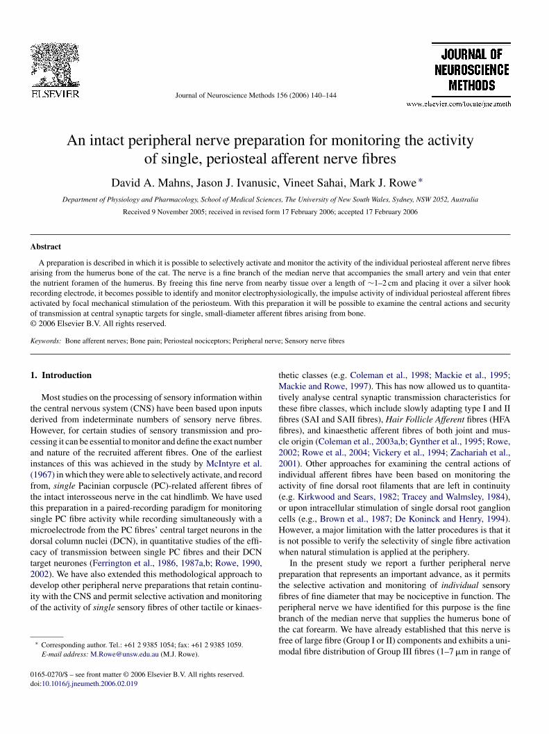
avity of the humerus (Fig. 1A). In preparation for electro-hysiological recording, the nerve was carefully teased awayrom its associated blood vessels over a distance of 1–2 cmnd placed over a silver hook electrode. A second, indifferent
ttci
ig. 1. Experimental arrangement for recording from single periosteal afferent nerveeripheral nerve innervating the periosteum and medullary cavity of the humerus boneeriosteum and the black oval areas indicate the receptive fields of eight individual fib.50 mN to 15.7 mN, that were required to generate responses for these eight fibres. (Bomponent of a 1 mm step indentation lasting 2 s. (C) Impulse traces showing the resp0 Hz, 20 Hz, 50 Hz and 100 Hz (amplitude 300 �m). Step indentations and sinusoidatimulator.
nce Methods 156 (2006) 140–144 141
lectrode was attached to nearby subcutaneous tissue. Both theerve and periosteal tissue were protected from dessication bysing skin flaps to create a paraffin-filled pool that was heldt 34–36 ◦C. Recorded action potentials were fed to a differ-ntial amplifier (1000-fold gain) and filter unit (typical bandass ∼10 Hz–10 kHz). This impulse activity was displayed onn oscilloscope, passed to an audio-amplifier and speaker, andas stored on both a modified video cassette recorder (Vet-
er, model 420 G) and a laboratory computer after sampling onn analogue-to-digital converter (CED-1401-Powerlab). Recep-ive fields (RFs) were mapped using calibrated von Frey hairs.eproducible vibro-mechanical stimuli were applied normal to
he periosteal surface to the most sensitive point of the afferentbre’s receptive field, using a 200 �m diameter probe driven byfeedback-controlled mechanical stimulator of the kind used inur earlier studies (Ferrington et al., 1986, 1987a,b; Coleman etl., 1998, 2003a,b). Stimuli consisted of maintained (1.5–2.0 s inuration) forms of mechanical displacement (Fig. 1B) at ampli-udes of up to 300 �m, or trains of sinusoidal vibration usuallyasting 1 s and at frequencies up to 100 Hz (Figs. 1C and 2).
. Results
.1. Nerve supply to the humerus bone
As the path taken by the nerve after separating from the parentedian nerve is a perivascular one that does not involve passage
hrough muscle or other nearby tissue, it is free of any con-
aminating, larger-diameter nerve fibres associated with muscle,endons or joints (Fig. 1A; and Ivanusic et al., 2006). This wasonfirmed in the present study by the absence of any responsesn electrophysiological recordings from the nerve in associationfibres in the intact nerve to the humerus. (A) Schematic representation of theand the arrangement for recording (Rec.). The grey shaded area represents theres. The adjacent numbers indicate the minimal von Frey forces, ranging from) Impulse activity recorded from a single afferent fibre responsive to the onset
onses of another afferent fibre to focal vibration applied to the periosteal RF atl vibration were applied to the periosteum using a servo-controlled mechanical

142 D.A. Mahns et al. / Journal of Neuroscience Methods 156 (2006) 140–144
F indiv( re to(
wo
3p
effiamntaaepitpialwmwf(ohtdocutv
tco2aittasefutoa3Gc
3f
snaseeprF
ig. 2. (A) Schematic representation of the receptive field (grey shading) of anpositions a–e). (B) Impulse traces showing the response of the same sensory fiba–e).
ith mechanical probing of muscle and tendons in the vicinityf the humerus.
.2. Receptive field and threshold characteristics of singleeriosteal afferent fibres of the humerus
Mechanical stimuli applied in close proximity to the nutri-nt foramen of the humerus usually generated multiunit activityrom which it was difficult to selectively activate individualbres. It remains uncertain the extent to which this multiunitctivity was attributable to the activity of afferent fibres ter-inating within the medullary cavity of the bone, around the
utrient foramen itself, or within the loosely-attached connectiveissue associated with the small artery and vein as they approachnd enter the foramen. However, when mechanical stimuli werepplied a short distance (∼2–3 mm) beyond the foramen, andlsewhere over the broader expanse of the periosteum, it wasossible to selectively activate with calibrated von Frey hairsndividual afferent fibres that displayed circumscribed and punc-ate receptive fields which are schematically represented on theeriosteum in Fig. 1A. A total of 15 individual fibres were stud-ed in terms of RF characteristics or vibro-mechanical sensitivitynd responsiveness. The individual RFs were all of a single-ocus and were usually of an approximately oval configurationhich, in area, ranged from 2 mm2 to 42 mm2 (18 ± 14 mm2,ean ± S.D.) for the 12 fibres whose RF areas were quantifiedith suprathreshold von Frey hairs or fine probes. The threshold
orces for activation were between 0.5 mN and 15.7 mN forces4.9 ± 4.3 mN force, mean ± S.D.) for the 15 fibres. The sizef RFs was little affected by the use of either higher von Freyair forces or rigid, needle-like stimulus probes, suggesting thathe mechanosensitivity of each fibre was confined to a well-emarcated region of distribution of the fine axonal terminalsf the recorded fibre. As individual periosteal afferent fibres
ould usually be selectively activated with the use of fine stim-lus probes, it appears that there is rather limited overlap of theerminal RFs of individual fibres, and therefore, not a high inner-ation density of the humerus periosteum, at least for fibres offitac
idual periosteal afferent fibre and the positioning of the mechanical stimulatora 1 s train of 50 Hz (300 �m amplitude) vibration at the five indicated locations
he mechanosensitive type that we have been able to activate andharacterise. These we believe to be Group III fibres based first,n our earlier fibre spectrum analysis of the nerve (Ivanusic et al.,006), second, the impulse rates that they displayed (see below)nd, third, some estimates that we made of conduction veloc-ty which fell within the Group III range. However, we believehat other fibres of lesser mechanical sensitivity, also innervatehe periosteum, based on the observation that, in regions thatppeared insensitive to direct mechanical probing, it was pos-ible to selectively activate individual fibres by applying focallectrical stimuli (0.1–10 mA, 2 ms pulse duration) to the sur-ace of the periosteum with a concentric bipolar electrode. Basedpon the latency of recorded responses, and the distance betweenhe stimulus site on the periosteum and the recording electroden the nerve, the calculated conduction velocities for four fibresctivated in this way with electrical stimulation were all below0 m/s, with three fibres in the range 2–30 m/s, consistent with aroup III classification, and one fibre with a value below 2 m/s,
onsistent with a Group IV classification.
.3. Signal-to-noise ratio for individual afferents recordedrom the intact humerus nerve
Mechanically sensitive periosteal afferent fibres displayedpike activity, recorded from the intact nerve, with signal-to-oise ratios in excess of five-to-one, and often around 10:1,s shown in Fig. 1C. All 15 fibres displayed a pure dynamicensitivity, as step indentation of the periosteum, by means ofither hand-held probes or servo-controlled mechanical stimuli,licited responses only in association with the dynamic com-onents of the stimulus as illustrated by the burst of impulsesecorded from one fibre to the onset of the step indentation inig. 1B.
The impulse traces of Fig. 1C were obtained from a single
bre in response to focal vibromechanical stimuli delivered in 1 srains to the most sensitive point of the periosteal RF by means of200 �m diameter circular probe at the four indicated frequen-ies of 10–100 Hz. These stimuli were delivered by a feedback-

roscie
cwtlt
oFeptbR
4
bw1ecFisIendsanfgciba
pganspttdfitioestlfi
ctttitfetip
A
RR
R
B
C
C
C
D
F
F
F
G
G
I
J
K
K
D.A. Mahns et al. / Journal of Neu
ontrolled mechanical stimulator and, for the Fig. 1C responses,ere applied at an amplitude of 300 �m which elicited a one-
o-one response in which the fibre responded in a regular, phaseocked pattern with an action potential on each cycle of the vibra-ory waveform.
The dynamic mechanosensitivity and circumscribed naturef the RFs for individual periosteal afferents is illustrated inig. 2 where 50 Hz (300 �m) vibratory stimulation was highlyffective at the field centre (position a), generating a regular 1:1attern of response, and was quite effective near the boundary ofhe RF, at position e, but was without effect on the fibre at sites, c and d, each 1 mm from the edge of the identified periostealF.
. Discussion
Previous electrophysiological studies of the nerve supply toone have revealed mechanosensitive afferent fibres associatedith, for example, the mandible and tibia (Sakada and Aida,971; Sakada and Maeda, 1967; Tokunaga, 1967). However, inach case, large diameter fibres from closely associated mus-le or interosseous tissues are present in the nerve filaments.urthermore, it has not been possible at these locations to exam-
ne electrophysiologically the periosteal afferents with the nerveupply remaining in continuity with the central nervous system.n contrast, the nerve to the humerus bone is free of large diam-ter (Groups I and II) fibres, whether from the bone itself orearby tissues, and therefore constitutes a source of pure, fine-iameter (Groups III and IV fibres) input to the central nervousystem (Ivanusic et al., 2006). Although some of the periostealfferents in our sample appeared to have relatively low mech-osensitive thresholds, these fibres may still be nociceptive inunction as the stimuli were delivered with von Frey hairs thatenerate quite high pressures or torsional forces at the point ofontact of the tip on the periosteum. It is perhaps relevant to thisssue that similar or even lower activation force thresholds haveeen reported for dural afferents that were presumed to subservenociceptive function (Levy and Strassman, 2002, 2004).
Although small-diameter afferent fibres from the dura, toothulp or cornea may be monitored in dorsal root or trigeminalanglion recordings (e.g., Levy and Strassman, 2002; Gallar etl., 1993; Jyvasjarvi and Kniffki, 1987; Kenshalo, 1960), it isot possible with these preparations to verify the selectivity ofingle fibre activation when natural stimulation is applied to theeriphery. In contrast, the present peripheral nerve preparationhat supplies the humerus bone has a concatenation of attributeshat offers perhaps unique advantages for the study of small-iameter, presumed-nociceptive inputs to the CNS. These arise,rst from the pure fine-fibre composition of the nerve. Second,he whole nerve is sufficiently fine in calibre that one can mon-tor, with a hook electrode under the intact nerve, the responsef any active mechanosensitive periosteal afferent fibres. Third,ach mechanosensitive periosteal afferent examined has had a
ignal-to-noise ratio in recordings from the intact humerus nervehat exceeds 5:1, creating a clear discontinuity between the noiseevel on the recording trace and the spike height for any activatedbre. Fourth, individual mechanosensitive periosteal afferentsL
nce Methods 156 (2006) 140–144 143
an be selectively activated by focal stimulation because ofheir relatively low innervation density. Furthermore, with mul-iple mechanical stimulators, one can potentially recruit one,wo, three, or more fibres, each of which could be identifiedn the whole nerve recording, enabling systematic quantita-ive analysis, for the first time, of transmission characteristicsor single identified, fine-diameter, presumed-nociceptive affer-nts at central synaptic target sites, together with analysis ofhe integrative processing of inputs from defined numbers ofdentified afferents of this type within the central nociceptiveathways.
cknowledgements
This work was supported by the National Health and Medicalesearch Council of Australia. The technical assistance of C.iordan and D. Sarno is acknowledged.
eferences
rown AG, Koerber HR, Noble R. Excitatory actions of single impulses insingle hair follicle afferent fibres on spinocervical tract neurones in thecat. J Physiol (Lond) 1987;382:291–312.
oleman GT, Zhang HQ, Mackie PD, Rowe MJ. An intact peripheralnerve preparation for examining the central actions of single kinaes-thetic afferent fibres arising in the wrist joint. Prim Sens Neuron 1998;3:61–70.
oleman GT, Mahns DA, Zhang HQ, Rowe MJ. Impulse propagation overtactile and kinaesthetic sensory axons to central target neurones of thecuneate nucleus in cat. J Physiol (Lond) 2003a;550:553–62.
oleman GT, Zhang HQ, Rowe MJ. Transmission security for single kines-thetic afferent fibers of joint origin and their target cuneate neurons inthe cat. J Neurosci 2003b;23:2980–92.
e Koninck Y, Henry JL. Prolonged GABAA-mediated inhibition followingsingle hair afferent input to single spinal dorsal horn neurones in cats. JPhysiol (Lond) 1994;476:89–100.
errington DG, Rowe MJ, Tarvin RP. High gain transmission of sin-gle impulses through dorsal column nuclei of the cat. Neurosci Lett1986;65:277–82.
errington DG, Rowe MJ, Tarvin RP. Actions of single sensory fibres oncat dorsal column nuclei neurones: vibratory signalling in a one-to-onelinkage. J Physiol (Lond) 1987a;386:293–309.
errington DG, Rowe MJ, Tarvin RP. Integrative processing of vibratoryinformation in cat dorsal column nuclei neurones driven by identifiedsensory fibres. J Physiol (Lond) 1987b;386:311–31.
allar J, Pozo MA, Tuckett RP, Belmonte C. Response of sensory units withunmyelinated fibres to mechanical, thermal and chemical stimulation ofthe cat’s cornea. J Physiol (Lond) 1993;468:609–22.
ynther BD, Vickery RM, Rowe MJ. Transmission characteristics for the 1:1linkage between slowly adapting type II fibers and their cuneate targetneurons in cat. Exp Brain Res 1995;105:67–75.
vanusic J, Mahns DA, Sahai V, Rowe MJ. Absence of large diameter sensoryfibres in a nerve to the cat humerus. J Anat 2006;208:251–5.
yvasjarvi E, Kniffki KD. Cold stimulation of teeth: a comparison betweenthe responses of cat intradental A delta and C fibres and human sensation.J Physiol (Lond) 1987;391:193–207.
enshalo DR. Comparison of thermal sensitivity of the forehead, lip, con-junctiva and cornea. J Appl Physiol 1960;15:987–91.
irkwood PA, Sears TA. Excitatory post-synaptic potentials from single mus-
cle spindle afferents in external intercostal motoneurones of the cat. JPhysiol (Lond) 1982;322:287–314.evy D, Strassman AM. Mechanical response properties of A and C pri-mary afferent neurons innervating the rat intracranial dura. J Neurophysiol2002;88:3021–31.

1 roscie
L
M
M
M
R
R
R
S
S
T
T
V
44 D.A. Mahns et al. / Journal of Neu
evy D, Strassman AM. Modulation of dural nociceptor mechanosensitiv-ity by the nitric oxide-cyclic GMP signaling cascade. J Neurophysiol2004;92:766–72.
ackie PD, Zhang HQ, Schmidt RF, Rowe MJ. An intact nerve preparationfor monitoring inputs from single joint afferent fibres. J Neurosci Methods1995;56:31–5.
ackie PD, Rowe MJ. An intact peripheral nerve preparation for monitoringinputs from single muscle afferent fibres. Exp Brain Res 1997;113:186–8.
cIntyre AK, Holman ME, Veale JL. Cortical responses to impulsesfrom single Pacinian corpuscles in the cat’s hind limb. Exp Brain Res1967;4:243–55.
owe MJ. Impulse patterning in central neurons for vibrotactile coding. In:Rowe MJ, Aitkin LM, editors. Information processing in mammalianauditory and tactile systems. New York: Wiley-Liss; 1990. p. 111–25.
owe MJ. Synaptic transmission between single tactile and kinaestheticsensory nerve fibres and their central target neurons. Behav Brain Res2002;135:197–212.
owe MJ, Mahns DA, Sahai V. The capacity of tactile systems for thedetection and discrimination of sensory events. In: Ballesteros S, Heller
Z
nce Methods 156 (2006) 140–144
M, editors. Touch, blindness and neuroscience. Madrid: UNED; 2004. p.221–34.
akada S, Aida H. Localization and shape of Golgi-Mazzoni corpusclesin the facial bones’ periosteum of the cat. Bull Tokyo Dent Coll1971;12:235–53.
akada S, Maeda K. Characteristics of innervation and nerve ending in cat’smandibular periosteum. Bull Tokyo Dent Coll 1967;8:77–94.
racey DJ, Walmsley B. Synaptic input from identified muscle afferents toneurones of the dorsal spinocerebellar tract in the cat. J Physiol (Lond)1984;350:599–614.
okunaga J. The innervation of the diaphysis of the cat tibia. J Anat1967;101:125–36.
ickery RM, Gynther BD, Rowe MJ. Synaptic transmission between singleslowly adapting type I fibres and their cuneate target neurones in cat. J
Physiol (Lond) 1994;474:379–92.achariah MK, Coleman GT, Mahns DA, Zhang HQ, Rowe MJ. Trans-mission security for single, hair follicle-related tactile afferent fibersand their target cuneate neurons in cat. J Neurophysiol 2001;86:900–11.