an injectable mechanically robust hydrogel of kappa
TRANSCRIPT

Contents lists available at ScienceDirect
Carbohydrate Polymers
journal homepage: www.elsevier.com/locate/carbpol
An injectable mechanically robust hydrogel of Kappa-carrageenan-dopamine functionalized graphene oxide for promoting cell growth
Hamidreza Mokhtari, Mahshid Kharaziha⁎, Fathallah Karimzadeh, Shima TavakoliDepartment of Materials Engineering, Isfahan University of Technology, Isfahan 84156-83111, Iran
A R T I C L E I N F O
Keywords:Shear thinningSelf-healingNanohybrid hydrogelsKappa-carrageenanGraphene oxideDopamine
A B S T R A C T
An injectable nanohybrid hydrogel with robust mechanical properties was developed based on Methacrylate-Kappa-carrageenan (KaMA)-dopamine functionalized graphene oxide (GOPD) for soft tissue engineering. KaMA-GOPD hydrogels revealed shear-thinning behavior and injectability through interaction of active catechol groupsof dopamine with other moieties in the structure of hydrogels. In addition, these interactions promoted me-chanical properties of hydrogels, depending on the GOPD content. Noticeably, encapsulation of 20wt.% GOPDsignificantly enhanced compressive strength (8-folds) and toughness (6-folds) of KaMA. Furthermore, the hybridhydrogel consisting of 20 wt.% GOPD significantly reduced energy loss from 70% (at KaMA) to about 61%, aftera two-cycle compression test, while significantly enhanced recovery of the KaMA structure. Reinforcing theKaMA with 20wt.% GOPD resulted in enhanced fibroblast proliferation (2.5-times) and spreading (5.7 times)after 5 days of culture. Based on these findings, KaMA-GOPD hydrogel could be used for cell delivery through theinjection process and applied as a suitable bio-ink for 3D-bioproiting process.
1. Introduction
Recently, soft tissue engineering has been recognized as an inter-section point among engineering, medicine, and biology to repair da-mages of soft tissue (Stratton et al., 2018). Current medical therapy isusually invasive surgery such as tumor resection and large traumaticinjuries (Rnjak-Kovacina et al., 2015). To overcome the limitations ofthe current therapies, soft tissue engineering has been developed as anew strategy for restoring diseased soft tissues and organs, pointingrepair of volume loss (Rnjak-Kovacina et al., 2015; Yuksel, Choo,Wettergreen, & Liebschner, 2005). In this field, various types of bio-materials with appropriate properties have been utilized in order toheal and regenerate the damaged tissue. However, this approach isusually accompanied with invasive surgery for pasting biomaterials-based constructs, facing with side-effects such as post-operativebleeding, infection and damage to other nearby organs (Keller,Tahilramani, Flores-Gonzalez, Mahmood, & Haas, 2016; Pittet,Montandon, & Pittet, 2005). In this respect, injectable biomaterials arepopular platforms to precisely fill the damaged area and facilitate tissueregeneration. Between them, thanks to unique properties of injectablehydrogels, consisting of high-water content, permeability to solublefactors such as oxygen, good biocompatibility, tunable biodegradabilityand ability to encapsulate cell and bio-macromolecules, they are
extensively applied for soft tissue regeneration (Vunjak-Novakovicet al., 2010; Yu & Ding, 2008). Moreover, injectable hydrogels revealexcellent features that give them ability to closely mimic the naturalliving tissues and turned the hydrogels to unique scaffolds with aminimally invasive injection method (Kretlow, Klouda, & Mikos, 2007;Loebel, Rodell, Chen, & Burdick, 2017). Between various types of in-jectable hydrogels, shear-thinning injectable ones are capable ofshielding encapsulated cells from high shear forces (Guvendiren, Lu, &Burdick, 2012). Shear-thinning hydrogels exhibit viscous flow undershear stress. When the stress is released, they recover depending on thetime during relaxation (Guvendiren et al., 2012). This strange responseattracts researcher to provide an alternative strategy for injectablehydrogel application and thereby improving outcome of cell-basedtherapeutics (Yang et al., 2012; Yang, Zhang, Yue, & Khademhosseini,2017). Researchers have utilized different polymers for making in-jectable hydrogels that can be extruded and molded to the desiredshapes (Diba, Pape et al., 2017; Diba, Wang, Kodger, Parsa, &Leeuwenburgh, 2017; Wang, Wang, Lu, Detamore, & Berkland, 2010).Kappa-carrageenan (κ-CA) is one of the biopolymers with great prop-erties making it a suitable candidate for tissue engineering applications(Mihaila et al., 2013).
Kappa-carrageenan (κ-CA) extracted from red seaweeds possessesester sulfate content of about 25–30% (Jiao, Yu, Zhang, & Ewart, 2011;
https://doi.org/10.1016/j.carbpol.2019.03.030Received 5 February 2019; Received in revised form 10 March 2019; Accepted 10 March 2019
⁎ Corresponding author.E-mail address: [email protected] (M. Kharaziha).
Carbohydrate Polymers 214 (2019) 234–249
Available online 12 March 20190144-8617/ © 2019 Elsevier Ltd. All rights reserved.
T

Necas & Bartosikova, 2013). κ-CA has been proposed as a potentialcandidate for tissue engineering applications, owing to its gelationproperties, mechanical strength and resembling chondroitin-4-sulphateand dermatan sulphate, which are major components of native extra-cellular matrices called glycosaminoglycan (GAGs) (Campo, Kawano,Silva, & Carvalho, 2009; Popa, Caridade, Mano, Reis, & Gomes, 2015).Moreover, the ionic crosslinking process of κ-CA using KCl could pro-vide hydrogels with strong ionic interactions between sulfate groupsand ions. Although ionic crosslinking process promoted mechanicalstrength and stiffness, it changes κ-CA to a brittle hydrogel (Thakuret al., 2016). Recently, Mihaila et al. (Mihaila et al., 2013) developedphoto-crosslinkable κ-CA via methacrylate κ-CA (KaMA) and foundthat, the mixture of physical and chemical crosslinking procedure im-proved mechanical properties and promoting integrity of KaMA, whilepermitting maintenance of viable encapsulated cells. Recently, variousresearches have shown the successful generation of nanocompositehydrogels based on κ-CA hydrogels consisting of silicate (Cross, Shah,Palani, Peak, & Gaharwar, 2018; Lokhande et al., 2018; Thakur et al.,2016), whitlockite (Yegappan et al., 2019) and metal oxide (Daniel-da-Silva et al., 2012; Thanusha et al., 2018). These nanocomposite hy-drogels could improve mechanical properties of κ-CA and enhance thephysiological stability. However, according to our knowledge, shear-thinning behavior of κ-CA- graphene oxide hydrogels have never beeninvestigated.
Lately, graphene based materials have revealed great potentials forbiomedical applications, due to their biocompatibility and uniqueelectrical, mechanical and thermal properties (Chung et al., 2013; Paulet al., 2014; Shin et al., 2014). Graphene is a two-dimensional (2D)material composed of sp2 hybridized carbon atoms with a single atomlayer. The specific structure of graphene with high specific area andinteraction between graphene as a reinforcement and polymer’s chainsmakes it appropriate for development of nanocomposites (Chung et al.,2013). Between various derivations of graphene, graphene oxide (GO)consists of various functional groups such as hydroxyl, carboxyl andepoxy which make it a good reinforcing additive for various polymersand hydrogels applied for tissue engineering (Liao, Qu, Chu, Zhang, &Qian, 2015; Paul et al., 2014; Zhang et al., 2011). Del Giudice and Shen(2017) found that incorporation of special amounts of GO to watercreated shear-thinning behavior for Newtonian fluid like water, owingto the functional groups on GO surface. Recently, GO was also appliedas an reinforcing agent for hydrogels to improve mechanical properties(Das et al., 2013; Paul et al., 2014) and develop Newtonian solution byinteraction of sacrificing electrostatic band between GO and polymericstructure (Kim & Lee, 2014). However, results showed that the inter-action between GO nanosheets and hydrogel matrix was slightly weak(Kim & Lee, 2014; Paul et al., 2014). In this regard, polydopamine (PD)has been introduced as a mussel-inspired adhesive material which hasbeen widely applied to develop robust and significant hydrogels (Hanet al., 2017; Lee, Dellatore, Miller, & Messersmith, 2007).
PD is usually prepared via the oxidation of the catechol groups ofdopamine (DA) by oxygen or oxidant reagents such as FeCl3 and NaOI4.Moreover, the thickness of PD can be adjusted by changing the time ofcross-linking or oxidizing of the catechol groups. It could provide anadvantage to obtain ultrathin PD film on the surface of nanomaterials toachieve excellent binding kinetics (Luo, Jiang, & Liu, 2013). PD hasrecently been applied as a thin layer on the surface of GO nanosheets inorder to achieve the well-compatibility in polymeric structure. More-over, the adhered PD can act as the anchor to graft the secondaryfunctional biopolymers by the thiols, imino and amines which conse-quently leads to development of innovative hydrogels with admireproperties (Cheng et al., 2012; Cheng et al., 2013; Xing et al., 2017).However, according to our knowledge, the role of PD-functionalized GOto develop shear thinning hydrogel has never been investigated.
Herein, we developed a novel injectable shear-thinning and me-chanically robust hydrogel based on κ-CA-GO for soft tissue en-gineering. In this respect, dual-crosslinkable KaMA was primary
synthesized to create photo- crosslinkable hydrogels emphasizing theiruse in the context of tissue engineering. Subsequently, KaMA-GO hybridhydrogels, consisting of various amounts of GO nanosheets (1, 4, 10 and20wt.%) were developed. In order to provide a robust and shear-thinning hydrogel, GO nanosheets were primarily functionalized with athin layer of PD (GOPD). It is expected that the proposed shear-thinningand mechanically robust hydrogel can be used for soft tissue en-gineering.
2. Materials and methods
2.1. Materials
Kappa-Carrageenan (κ-CA, sulfated plant polysaccharide), a linearpolysaccharide constructed of repeated 1,3-linked β-D-galactopyranoseand 1,4-linked 3,6-anhydro-D-galactopyranose units (14.57 wt.% K+,5.50 wt.% Ca2+ tested by ICP, MW=3×10⁵ g/mol) was purchasedfrom Sigma-Aldrich. The 3,6-AnGal/Gal ratio was investigated about1:1.03 (Navarro & Stortz, 2003). Moreover, methacrylic anhydride(MA) (C8H10O3) (Sigma-Aldrich, 64100), photoinitiator (PI) Irgacure D-2959 (2-Hydroxy-4′-(2-hydroxyethoxy)-2-methylpropiophenone)(C12H16O4) (Sigma-Aldrich, 410896), potassium chloride (KCl, Merck)and sodium hydroxide (NaOH, Merck Co) were purchased to synthesizedual-crosslinkable KaMA. Dialysis membrane (Mol. Wt. cut-off˜12–14 kDa) was obtained from Betagen Co, Mashhad, Iran. Grapheneoxide (GO) powder (purity > 99%, thickness: 3.4–7 nm, 6–10 layers,Nanosany Co, Mashhad, Iran), methanol (CH3OH, Merck Co), aceticacid (CH3COOH, Merck Co), dopamine (DA) (C8H11NO2, Merck Co),ammonium persulfate (APS, Merck Co), trisaminomethane (Tris, MerckCo), hydrochloric acid (HCl, Merck Co) were provided to synthesizepolydopamine (PD) coated GO (GOPD). Moreover, double distilledwater (DDW) was used at all experiments.
2.2. Synthesis of Kappa-carrageenan methacrylate (KaMA)
Methacrylation of κ-CA was carried out by reacting κ-CA with MA,fallowing the recent study, conducted by Mihaila et al. (2013). Thechemical interaction is provided in Supplementary Fig. S1. Briefly, inorder to reach homogeneous 1 wt.% solution, κ-CA was fully dissolvedin 100ml DDW at 50 °C. Consequently, 4.2 ml MA was added to solu-tion and allowed to react for 6 h at 50 °C. To adjust the pH (˜8.0), 5MNaOH solution was added drop-wise. After 6 h, the resulting κ-CA so-lution was dialyzed against DDW using 12–14 kDa cutoff cellulosedialysis membrane for 7 days at 4 °C to remove excess of unreacted MAas well as by-products formed during the reaction. Refined KaMA so-lution were frozen at−80 °C and then lyophilized. Until further use, thelyophilized KaMA was stored at −20 °C, while it was protected fromlight.
2.3. Synthesis of dopamine functionalized GO (GOPD) nanoparticles
Dopamine functionalized GO (GOPD) nanoparticles were preparedby the self-polymerization of dopamine as a coating layer on the GOnanoparticles based on previous studies (Tan, Liu, & Ren, 2017). Afterdispersion of 20mg GO nanosheets in 50ml DDW for 30min, 3mg APS,25mg DA and 5ml Tris-HCl solution (200mM, pH 8.5) were added toit, consequently. The resulted suspension was endured a polymerizationreaction at room temperature for 2 h to synthesize GOPD nanoparticles.The resulting GOPD nanoparticles was removed, and sufficiently rinsedwith a methanol: acetic acid solution (volume ratio= 9:1) using cen-trifugation at 3500 rpm for 10min. Finally, the remained precipitatewas dried under vacuum for 24 h.
2.4. Synthesize of KaMA-GOPD hybrid hydrogel
KaMA-GOPD hybrid hydrogels consisting of various concentrations
H. Mokhtari, et al. Carbohydrate Polymers 214 (2019) 234–249
235

of GOPD (1, 4, 10 and 20wt.%) were prepared. Primarily, prepolymerof freeze dried KaMA solution in DDW water with concentrations of1.5 wt.% was prepared at 80 °C. Consequently, specific amounts ofGOPD were added. Afterward, 0.5 wt.% PI solution in acetone wasadded to KaMA-GOPD solution and let it to fully mixed, while theyprotected from light. Consequently, 2 ml of KaMA-GOPD solution wasadded into disc-shaped polystyrene molds (10mm diameter and 20mmheight) followed by gently adding a 5 wt.% KCl solution to initiatephysical crosslinking. After 20min, the samples were removed from themolds and washed in DDW in order to remove the salt residues. Finally,ion-crosslinked KaMA-GOPD hydrogels were chemically crosslinked by180-sec irradiation of UV-A (1.69 mW/cm2). Depending on the con-centrations of GOPD nanoparticles (1, 4, 10 and 20wt.%) in the hybridhydrogels, the samples were named K-G1, K-G4, K-G10, K-G20, re-spectively. KaMA hydrogel was also similarly fabricated as the control.In this regard, after preparation of 1.5 wt.% KaMA solution in DDWwater at 80 °C, 0.5 wt.% PI solution in acetone was added to it.Consequently, the two-step process of ionic crosslinking in 5 wt.% KClsolution followed by chemical crosslinking in 180-sec irradiation of UV-A was performed.
2.5. Characterization of KaMA-GOPD hybrid hydrogel
The microstructure of hydrogels was evaluated using a ScanningElectron Microscopy (SEM, Philips, XL30). The samples were drownedin liquid nitrogen, followed by freeze drying for 24 h. Before imaging,the samples were sputter coated with a thin layer of gold using aHummer 6.2 sputter. Moreover, the pore size distribution of hydrogels(n=30) was achieved using ImageJ software, based on the SEMimages. Moreover, the thickness (n=3) and surface topography ofGOPD were investigated using atomic force microscopy (AFM, Veeco,CP II, PCResearch, USA). After preparation of GO and GOPD suspen-sions in DDW (0.1mg/ml) using an ultrasonic oscillator (20 kHz, 50W)for 30min, they were coated on a Si wafer using a spin-coater (ModernTechnology Development Institute, Iran) at the optimized rate of500 rpm for 5min. In order to study the surface modification of GOnanosheets with dopamine, X-ray diffraction (XRD, X’Pert Pro X-raydiffractometer, Phillips, Netherlands), Fourier transform infraredspectroscopy (Tensor27, Bruker, Germany) and Raman spectroscopy(Takram P50C0R10, 532 nm, Iran) were performed. Furthermore, theabsorption spectra were measured using a UV–vis spectrophotometer(EU-2800DS, Onlab, China)
The chemical modification of κ-CA was determined by H NMRspectroscopy (Bruker Avance II, 500MHz). In this regard, H NMRspectra of κ-CA and KaMA were collected in D2O at 25 °C, at a frequencyof 500MHz on a Varian INOVA NMR spectrometer with a single axisgradient inverse probe. Moreover, the chemical composition of the re-sultant KaMA-GOPD hybrid hydrogels was verified by Fourier trans-form infrared spectroscopy (Tensor27, Bruker, Germany), in the rangeof 500–4000 cm−1.
2.6. In vitro physiological stability evaluation of KaMA-GOPD hybridhydrogel
To study the effect of GOPD content on the equilibrium watercontent (EWC) and stability of the hydrogels, KaMA-GOPD hydrogels(n=3) consisting of various amounts of GOPD nanoparticles werelyophilized, weighted (W1) and, subsequently, soaked in phosphatebuffer solution (PBS) at 37 °C for 1 h. It needs to mention that PBS wasprepared according to previous protocols (Popuri, Harris-Logie, Lino,Cadogan, & Lee, 2014). After being wiped off to remove the excess li-quid from the samples surface, the wet hydrogels were weighted (W2)and the EWC was measured according to Eq. (1) (Mihaila et al., 2013):
=−
×EWC W WW
( ) 1002 1
2 (1)
To study the degradation rate of hydrogel, the samples (n= 3) werelyophilized and weighed (W1), prior to incubation in 37 °C. After 3, 24,48 and 72 h of incubation, the specimens were lyophilized again andwere consequently weighed (W2). Finally, the weight loss of hybridhydrogels was calculated according to Eq. (2) (Lokhande et al., 2018):
=−
×Weight loss W WW
(%) ( ) 1001 2
1 (2)
2.7. Rheological properties of KaMA-GOPD hybrid hydrogel
All rheological measurements were carried out on a MCR 502(Anton Paar, Germany) rheometer equipped with 25mm parallel plategeometry at a gap of 500 μm. Primarily, flow test was achieved to assessthe viscosity of polymer solution while shear rate was modulated from0.01–100 s−1 at 37 °C. Subsequently, oscillatory stress sweep was ap-plied between 0.1–1000 Pa at 37 °C and at a frequency of 0.1 Hz. Allthese tests were performed for both KaMA and KaMA-GOPD (K-G4),pre-polymer solution and crosslinked hydrogels, to investigate the ef-fect of GOPD nanoparticles on rheological and injectability of pre-polymer solution, and viscoelastic behavior of crosslinked hydrogels.
2.8. Compressive mechanical analysis of KaMA-GOPD hybrid hydrogel
Mechanical properties of KaMA-GOPD hydrogels were determinedusing a tensile tester (Hounsfield H25KS, United Kingdom) with a loadcell capacity of 500 N. Before compressive mechanical testing, the cy-linders of specimens with diameter of 10mm and thickness of 20mm(n=5) were dually crosslinked, according to the previous protocol.The samples were compressed with a strain rate of 1mm/min up to60% strain. Compressive modulus and strength (at 60% strain) werecalculated for each composition. Compressive modulus was defined asthe slope of the linear region of the strain/stress curve, correspondingto 5–15% strain. Furthermore, cyclic compressive tests were performedat a rate of 0.5mm/min until 60% of strain, as 1st complete loading-unloading cycle. Then, the specimens were put in PBS solution for 2 hand 2nd cycle was applied. The energy lost during the cycle was alsocalculated by determining the area between the loading and unloadingcycle of curves. Finally, the sample’s recovery was estimated based oncomparison between 1st and 2nd loading curves, based on Mahalia et al.researches (Mihaila et al., 2013). Finally, in some hydrogels, work-hardening energy was determined by measuring area between 1st and2nd loading curves, after 2nd loading curve crossed 1st loading curve.
2.9. Cell culture
In order to evaluate the role of GOPD nanoparticles on the cellularbehavior of KaMA hydrogel, L929 fibroblast cell line, purchased fromRoyan Institute of Iran, was applied. First, disk-like hydrogels with10mm diameter and thickness of 1mm were prepared. Before cellseeding, the specimens were washed with phosphate buffer saline (PBS,Bioidea, Iran), sterilized for 30min in 70% (v/v) ethanol and 2 h underUV light. Fibroblast cells were cultured in Dulbecco’s Modified EagleMedium (DMEM-low, Bioidea, Iran) containing 10%(v/v) fetal bovineserum (FBS, Bioidea, Iran) and 1%(v/v) streptomycin/penicillin(Bioidea, Iran) in incubator at 37 °C containing 5% CO2. Consequently,fibroblast cells with a density of 104 cells/well were seeded on thehydrogels and tissue culture plate (TCP) (control). Finally, the cell-seeded hydrogels were incubated at 37 °C under 5% CO2 for 5 days,while culture medium was changed after 3 days.
2.9.1. Cell viability studyTo evaluate the viability of fibroblasts seeded on hybrid hydrogels,
after 1, 3 and 5 days, calcein-AM/ ethidium homodimer (EthD-III) live/dead assay (Biotium, UK) was performed. After rinsing the cell-culturedsamples, 100 μl of live/dead solution consisting of 2 μM calcein AM and
H. Mokhtari, et al. Carbohydrate Polymers 214 (2019) 234–249
236

4 μM ethidium homodimer was added to the samples to cover the cellmonolayer, and incubated for 1 h at 37 °C. The samples were then im-aged using an inverted fluorescence microscope (Nikon TE2000-U,Japan). Finally, the cell viability was determined using Image J soft-ware by the number of live cells (green cells) divided by total cellnumber (green and red stain) (n= 3).
2.9.2. Immunostaining for cell cytoskeletal organizationIn order to assess the role of GOPD on the cytoskeletal organization
(F-actin) of fibroblast seeded on the various hydrogels, DAPI/phalloidinstaining was performed. The cell-seeded samples were fixed using4%(v/v) paraformaldehyde (Sigma Aldrich Co, USA) for 2 h. Then, thecells were permeabilized in 0.1%(v/v) Triton X-100 (Sigma Aldrich Co,USA) for 5min, fallowing incubation with 2%(v/v) bovine serum al-bumin (Sigma Aldrich Co, USA) in PBS for 1 h in order to decreasenonspecific background staining. Therefore, after triple rinsing withPBS, fibroblast cells were incubated with 1:1000 dilution of 40, 6-dia-midino-2-phenyl indole dihydrochloride (DAPI, sigma-Aldrich,Germany) in PBS for 5min to stain cell’s nuclei. Subsequently, the actinfilaments were stained with 1:40 dilution of rhodamine phalloidin(cytoskeleton, USA) solution for 30min. Finally, the stained sampleswere studied using a fluorescence microscope (Nikon TE 2000-U, Nikoninstruments Inc., USA). Moreover, to quantify cell proliferation, cellarea of each sample (n= 3), were assessed.
2.9.3. Cell survival studyThe relative cell proliferation was investigated using MTT assay,
performed according to the manufacturer protocol (Sigma Aldrich Co,USA). At the specific time points (1, 3 and 5 days), the culture mediumwas discarded and 300 μl MTT solution (0.5 gr/ml) was added to thecell-seeded samples and control. After 3 h incubation at 37 °C under 5%CO2, the dark blue formazan crystals were dissolved in DMSO (Merck,Germany) and were kept for 30min at 37 °C. Subsequently, 100 μldissolved formazan solution of each sample was moved to 96-well plateand the optical density (OD) of each well was measured with a mi-croplate reader (Biotek Instruments, China) against DMSO (blank) at awavelength of 490 nm. The relative cell survival (% control) was cal-culated based on the following Eq. (3) (Mokhtari, Ghasemi, Kharaziha,Karimzadeh, & Alihosseini, 2018):
−
−
X XX X
Relative cell survival (% control): Sample b
c b (3)
Where XSample, Xb and Xc were absorbance of the sample, blank (DMSO)and control (TCP), respectively.
2.10. Statistical analysis
The data in this study was analyzed using one-way ANOVA ana-lyses. To determine a statistical significance between groups,Tukey–Kramer post-hoc test using GraphPad Prism Software (V.6) wasapplied and P-value < 0.05 was defined as statistically significant.
3. Results and discussion
3.1. Characterization of GOPD nanosheets
In this study, we offered a robust and shear-thinning KaMA-GOPDhydrogel based on muscle-inspired adhesion mechanism as well asnanohybrid formation strategy for soft tissue engineering. This nano-hybrid hydrogel was prepared via a two-step process: synthesize ofGOPD nanosheets and consequently nanohybrid formation. Accordingto Fig. 1A, dopamine was firstly intercalated in to the GO nanosheetswith free catechol and amino groups and in-situ oxidized in the alkalineenvironment leading to the formation of a thin PD layer. Amino is thefunctional group that attached the PD layers to GO surfaces. This in-teraction led to enhanced number of catechol groups on the
nanoparticles allowed the anchor-like PD to connect with other che-mical structures via hydrogen bonding, π–π stacking interactions,physical entanglement, chemical crosslinking and even non-covalentbonding which might lead to superior mechanical and physical char-acteristics (Gaharwar et al., 2010; Phua et al., 2012; Xu, Yang, Neoh,Kang, & Fu, 2010). In addition, PD layer could significantly affect themorphology and size distribution of GO nanoparticles. SEM and AFMtechniques were used to study the surface topography of both GO andGOPD nanoparticles. According to the SEM images of GO and GOPD(Fig. 1B), the GO surface was completely covered by a thin layer ofcoating, after PD coating. The GO nanosheets consisted of a smoothsurface, while GOPD became rough after PD coating, which was similarto other results (Su et al., 2018). Moreover, the agglomeration of GOnanosheets originated from π-π-stacking interactions was significantlyreduced. Besides, nanoparticles were characterized by AFM (Fig. 1Cand D) in order to evaluate the thickness of nanoparticle’s sheets, beforeand after PD coating process. The results revealed the enhanced surfaceroughness, confirming the SEM images on the GO covered PD layer.Furthermore, the average thickness of GO nanosheets was estimatedabout 11.7 nm (Fig. 1C), due to the functional groups on the GO sur-faces, which was much thicker than pristine graphene (Yamaguchi, Eda,Mattevi, Kim, & Chhowalla, 2010). That was also a big number for GOthickness, showing that nanoparticles were agglomerated and not fullydispersed in water. Instead, the average thickness of GOPD was 24 nm(Fig. 1C), showing enhanced thickness compared to GO nanosheets(about 12 nm), demonstrating that PD layer was successfully added toGO. This result was similarly reported in other researches (Hwang,Kang, Ruoff, Shin, & Park, 2014; Mu et al., 2015).
In addition, oxidization of DA to PD could reduce GO without em-ployment of toxic reductants such as hydrazine which are not bio-compatible. According to Fig. 1E, the color of the GO solution (0.1 mg/ml) was changed from brownish to dark grey, demonstrating the suc-cessful reduction of GO, that was similar to other researches (Jing, Mi,Napiwocki, Peng, & Turng, 2017; Liu et al., 2012). The successful sur-face functionalization of GO using PD and reduction of GO was studiedby UV–vis spectroscopy. Fig. 1F shows the UV–vis spectra of GO andGOPD dispersed in DDW. The GO spectrum consisted of a specific ab-sorption peak at 224 nm due to the π→π* transition of aromatic C]Cbonds, as similarly reported previously (Xu et al., 2017). After func-tionalization with PD, this peak shifted to 294 nm, confirming thesuccessful reduction of GO during polymerization of DA to modifyGOPD (Jing et al., 2016). Moreover, we observed that absorbance ofGOPD in the visible range increased compared to GO. Fu, Lai, Jia, andYu (2014) reached the same results, and suggested that the dispersionof functionalized-GO did not show any perceptible precipitation, im-plying that the PD layer was a great stabilizer to prevent the stacking ofthe reduced graphene sheets. FTIR spectrum of GOPD also similarlyconfirmed the reduction of GO after coating with PD (Fig. 1G).
The FTIR spectrum of dopamine consisted of a CeC bond at826 cm−1, CeH bond at 1291 cm−1, CeC bond of the aromatic ring at1510 cm−1 and a bending vibration of the amine group at 1605 cm−1.Moreover, the stretching vibrations of CeH revealed at 3040 cm−1,while the stretching vibration of the hydroxyl group of catechol andstretching of the amine group was observed at 3320 cm−1 and3340 cm−1, respectively (Fu et al., 2014; Liu et al., 2012; Ma, Niu,Zhang, & Cai, 2011; Zhu et al., 2017). The spectrum of GO nanosheetsconsisted of multiple peaks in the range of 900 to 1500 cm−1, whichcould be assigned to the various functional groups, such as CeO at1060 cm−1, CeOeC at 1250 cm−1, CeOH (stretching) at 1398 cm−1,C]O (stretching vibration) at 1620 cm−1 and C]O in carboxylic acidat 1730 cm−1 (Fu et al., 2014; Liu et al., 2012; Marcano et al., 2010).After modification of GOPD, the intensity of GO’s peaks decreased,corresponded to reducing the amount of oxygen containing groups atthe surface of GO. Our results confirmed the successful reduction of theGO nanosheets after dopamine functionalization. In this regard, thepeak belonging to the carboxyl group in GOPD and the peak intensity
H. Mokhtari, et al. Carbohydrate Polymers 214 (2019) 234–249
237

between 900 and 1400 cm−1 decreased, significantly. Additionally, thespecific absorbance bands of dopamine were disappeared in GOPDconfirming the effective polymerization of dopamine. Furthermore, thebroad bands at the range of 900 to 1800 cm−1 with no distinguishablepeaks, representing the highly complex structure of PD was also ob-served, as similarly reported in other researches (Xiong et al., 2014;Zangmeister, Morris, & Tarlov, 2013). Additionally, FTIR spectrum ofthe GOPD exhibited new peaks at 1358 cm-1 and 1504 cm−1, due to thestretching vibration of CeNeC of indole ring and the NeH bendingmode of aromatic secondary amine in GOPD, respectively. These peaksconfirmed the successful polymerization of dopamine, which was con-sistent with other researches (Centeno & Shamir, 2008). Additionally,the presence of the board peak of OeH group in the GO nanosheets at3450 cm−1 was often due to absorbed humidity which could not beavoided. Based on our result, DA have been used as a reducing agent, asthe oxygen groups on the surface of GO were chemically reduced duringthe polymerization process. To further assess the reduction of GO andinteraction between PD and GO, XRD patterns were studied (Fig. 1H).Dopamine consisted of a low intensity and board pick between2θ=13–38° due to its amorphous structure. In another word, XRDpattern of GO consisted of the characteristic peaks of GO centered at2θ=10.5° and 42.4° corresponding to the d-spacing of 0.85 and0.23 nm, respectively, due to the formation of hydroxyl, epoxy andcarboxyl groups, respectively. In contrast to GO, the GOPD revealed thesmall and broad peak at 2θ=9.7° with d-spacing of 0.92 nm, indicatingthat most of the oxygen functional groups of GO was efficiently at-tacked by the polymerization reaction of dopamine and GO nanosheets(Xu et al., 2010). Moreover, it could be concluded that the incorpora-tion of dopamine onto the GO nanosheets and in-situ polymerizationenlarged the d-spacing of GO nanosheets. The other peak at around2θ=21.5° was corresponded to the d-spacing of 0.44 nm, belonging tothe partial reduction of GO by polymerization of dopamine (Fu et al.,
2014). Moreover, it could be due to the attached dopamine on the GOnanosheets, since the same broad peak was detected in the XRD patternof pure dopamine.
Raman spectroscopy was also applied to characterize the GOPDnanoparticle, particularly for distinguishing ordered and disorderedcrystal structures of carbon (Fig. 1I). The peak at around 1597 cm−1 (Gband) was corresponded to the vibration of the sp2-bonded carbonatoms in a two-dimensional hexagonal lattice, while a peak at about1325 cm−1 (D band) was an indication of disorder in the Raman of theGO. The disorder in the GO nanosheets was originated from defectsassociated with vacancies, grain boundaries and amorphous carbonspecies (Ferrari et al., 2006; Schönfelder et al., 2007). Our results de-monstrated that, the D band of GOPD remained unchanged, whencompared to that of GO, while the G band of GOPD was shifted to1585 cm−1, confirming the successful reduction of GO to GOPD (Zhenget al., 2012). Furthermore, the intensity ratio of the D to G band (ID/IG)was calculated, reflecting the graphitization degree of carbonaceousmaterials and the defect density (Zhao et al., 2010). Our findingsshowed that ID/IG of GO was about 0.95 which enhanced to 0.97, due toa decrease in the average size of the sp2domains upon reduction of GO(Fang et al., 2017). It could also be due to the formation of defects bythe elimination of oxide functional groups attached to the GO surface(Eigler, Dotzer, & Hirsch, 2012). After functionalization with PD, the Gband was also broadened and shifted to 1601 cm−1, whereas the in-tensity of the D band at 1325 cm−1 increased, substantially. These re-sults confirmed the semi-reduction of GO and successful polymerizationof dopamine leading to an effective development of GOPD. Ye et al.(2014) also found these changes, because of a decrease in the size of thein-plane sp2 domains and a partially ordered crystal structure of gra-phene.
Fig. 1. Characterization of GOPD: A) The schematic representation of the formation of GOPD nanosheets. B) SEM images, of GO and GOPD nanoparticles. AFM imageand height profile of C) GO and D) GOPD. E) The representative suspensions of GO and GOPD in water. The change in the color of suspension from brown to dark greyconfirmed the reduction of GO after formation of GOPD. F) UV–vis absorption spectra of GO and GOPD. G) FTIR spectra and H) XRD patterns of dopamine, GO andGOPD. I) Raman spectra of GO and GOPD.
H. Mokhtari, et al. Carbohydrate Polymers 214 (2019) 234–249
238

3.2. Characterization of nanohybrid KaMA-GOPD hydrogel
After synthesize of GOPD, methacrylation process was completelyaccomplished (Supplementary Fig. S1). Subsequently nanohybridKaMA-GOPD hydrogels were developed, according to Fig. 2. KaMA is aphotocrosslinkable polymer which structurally mimic glycosami-noglycan component of native ECM. UV-crosslinkable KaMA has re-cently been proposed for tissue engineering applications (Mihaila et al.,2013; Thakur et al., 2016). However, we incorporated GOPD withinKaMA hydrogel in order to develop shear-thinning and robust hydrogelfor soft tissue engineering. It was expected that the chemical interactionbetween catechol groups of PD with other moieties on KaMA might beprovided leading to improvement of the KaMA hydrogel properties.
At first, the successful methacrylation process of κ-CA was in-vestigated using H NMR spectroscopy (Fig. 3A). H NMR spectrum of κ-CA spectrum consisted of the following chemical shifts: H NMRδ=4.97 (H1), δ=3.95–4.06 (H4), δ=3.73–3.80 (H3), δ=3.62 (H5,H6) and δ=3.42 (H2), which were in agreement with other researches(Abad et al., 2011; Campo et al., 2009; Mobarak, Ramli, Ahmad, &Rahman, 2012). After methacrylation process, H NMR spectrum ofKaMA consisted of one peak corresponding to the methyl group anddouble-peaks to vinyl group of the methacrylate group at δ=1.9–2 and5.5–6 ppm, respectively; this result confirming the methacrylation of κ-CA. Consequently, nanohybrid hydrogels containing various amounts ofGOPD were developed, via two-step ionic and subsequently UV cross-linking process. When KCl was injected, the coil-helix conformationaltransition occurred rapidly (less than 1min) leading to the formation ofa robust hydrogel. Subsequently, in order to improve mechanicalproperties, chemically crosslinking was performed via UV-crosslinkingprocess. Fig. 3B represents the image of nanohybrid hydrogels con-sisting of various amounts of GOPD. The color of samples changed fromwhite (KaMA) to completely black (K-G20) as a result of GOPD in-corporation.
ATR-FTIR spectroscopy was applied in order to investigate the for-mation of nanohybrid hydrogels of KaMA-GOPD (Fig. 3C). The spec-trum of κ-CA consisted of some main characteristic peaks including asharp vibration of a sulfated group CeOeS bond, vibration of CeC,stretching O]S]O and stretching vibration of O]S]O at 820, 925,1010 and 1210 cm−1, respectively (Lokhande et al., 2018; Mihaila
et al., 2013; Thakur et al., 2016). The validity of methacrylation pro-cedure of κ-CA at KaMA was further confirmed by the main peaks ofCeC at 1530 cm−1, C]C at 1620 cm−1 and carbonyl (C]O) at1730 cm−1 which were not appeared in κ-CA. Moreover, the sulphategroups of κ-CA was shifted slightly in the KaMA spectrum at 1215 cm−1
(O]S]O antisymmetric vibration) and 1023 cm−1 (O]S]O sym-metric bond), respectively. This result showed that the essential sul-phate group of κ-CA, playing an important role in ionic crosslinking,was not affected by the methacrylation reaction conditions.
The presence of GOPD within the dual crosslinked KaMA hydrogelwas also proved by FTIR spectroscopy (Fig. 3C). With increasingamounts of GOPD in KaMA hydrogel, a new peak at 1358 cm−1, due tothe stretching vibration of CeNeC of indole ring, could be detected.Furthermore, the intensity of the peaks at 1530 cm−1 (related to CeCband) and 1730 cm−1 (related to C]O band) were slightly decreasedfrom K-G1 to K-G20. Moreover, the C]C absorbance band at1620 cm−1 in K-G20 was disappeared confirming that nanoparticlesaffected the C]C band by interaction with polymer.
In order to investigate the effect of GOPD, nanohybrid hydrogelswere also studied by XRD analysis (Fig. 3D). XRD pattern of κ-CAconsisted of one board peak at 2θ=19.6° which was not changed aftermethacrylation procedure. Moreover, XRD pattern of KaMA exhibitedfive special peaks at 2θ=28.5°, 39.8°, 50.1°, 58.1° and 74.2° corre-sponded to the KCl (JCPDS Card number 41-1476), while all nanohy-brid hydrogels consisted of these five peaks, without any comparabledifference in their intensities. Moreover, the board peak was appearedin the XRD patterns of all K-G series at 2θ=22°, which its intensityenhanced with increasing GOPD content up to 20 wt.%. This main peakcould be improved by overlapping both peaks related to KaMA andpeak around 2θ=21.5° related to GOPD.
In order to investigate the effect of GOPD on structure and pore sizeof KaMA hydrogel, nanohybrid hydrogels were also studied by SEMimages (Fig. 4A). All samples revealed highly porous networks withinterconnected pores. This interconnected porous network could pro-mote the nutrient and waste product transportation and enable efficientcell survival and further proliferation. However, GOPD changed themicrostructure of KaMA hydrogel and made the pores bigger. Notice-ably, according to Fig. 4B, the average pore size of KaMA(41.3 ± 5.5 μm) enhanced to 117.0 ± 17.0 μm at K-G20 hydrogel.
Fig. 2. Schematic outlining the design strategy to synthesize nanohybrid KaMA-GOPD hydrogels.
H. Mokhtari, et al. Carbohydrate Polymers 214 (2019) 234–249
239

There was a significant change in pore size between KaMA and all of K-G series, showing that incorporation of GOPD in KaMA resulted information of bigger pores. In addition, GOPD nanoparticles with irre-gular convex-like morphology were distributed in the pore walls of allhybrid hydrogels, as indicated by red arrows. By increasing amountGOPD, these particles transformed to filamentous shape which con-nected the polymeric wall to each other. High magnification image ofK-G20 hydrogel (Fig. 4C) revealed these GOPD nanoparticles whichstrongly interacted with KaMA structure. In addition, ionic cubes wereentrapped between these nanoparticles due to the interaction betweencatechol group of GOPD and K+ ions which may affect the degradationand mechanical properties of nanohybrid hydrogels.
3.3. In vitro swelling and degradation properties of KaMA-GOPD hybridhydrogel
One of the advantages of using hydrogels in soft tissue engineeringis their water absorption capability and degradation in biological en-vironment. The swelling ratio and weight loss of the nanohybrid hy-drogels are depicted in Fig. 5A,B. Results demonstrated that the pre-sences of GOPD in the hydrogel structures significantly enhanced theswelling ratio of hydrogels (Fig. 5A). Noticeably, swelling ratio of K-G20 hydrogel was 98.8 ± 0.6% which was greater than that of KaMA(95.4 ± 0.2%) which might be due to the hydrophilic nature of GOPD(Jing et al., 2017). GOPD consisted of high density of catechol groupswith two hydroxyl groups which facilitated water adsorption duringsoaking in PBS (Hwang et al., 2014). Moreover, according to the SEMimages, pore size of hydrogels enhanced with increasing GOPD contentleading to absorption of more water, which confirmed these results. Inanother study, Huanga et al. (2012) similarly reported that incorpora-tion of graphene-based nanoparticles in hydrogel matrix could promotewater uptake.
In order to investigate degradation behavior of hydrogels consistingof different concentrations of GOPD, weight loss of hydrogels during a72-h period was considered (Fig. 5B). Results indicated that in-corporation of GOPD within the KaMA hydrogel reduced the weightloss and degradation rate. KaMA hydrogel revealed a significant weightloss during the first 3 h, and finally, 43 ± 3wt.% of KaMA was
degraded during 72 h. However, increasing the amounts of GOPD dra-matically promoted the stability of the nanohybrid hydrogels in biolo-gical environment. Noticeably, K-G20 revealed less than 9 ± 2wt.%weight loss after 72 h immersing in PBS solution. Our findings revealedthe interesting fact that, with increasing GOPD content, water holdingcapability improved, while significantly promoted physiological stabi-lity of KaMA- hydrogels. The less stabilization of the KaMA networkcould be likely due to the exchange of K+ ions entrapped within thenetwork leading to the weakening of the network. Similar results werereported for various ionically crosslinkable hydrogels such as alginate(Kuo & Ma, 2008), κ-CA (Mihaila, Popa, Reis, Marques, & Gomes, 2014)and gellan gum (Coutinho et al., 2010), In this research, incorporationof GOPD resulted in reduced degradation rate due to the strong inter-action between K+ ions and free catechol groups of PD which resultedin preventing from ion exchange. Similar result was reported for thecombination of chitosan with alginate and formation of an electrolytichydrogel with the ability to protect from ion exchange (Colinet, Dulong,Mocanu, Picton, & Le Cerf, 2010). Moreover, based on Jing et al. study(Jing et al., 2017), the presence of GOPD nanoparticles in hydrogelmatrix eventuated in a denser and stronger crosslinking leading to re-duced weight loss of hybrid hydrogels.
3.4. Rheological properties of KaMA-GOPD hybrid hydrogel
In order to evaluate the shear-thinning properties of the KaMA-GOPD hydrogels, the rheological properties of K-G4 compared to KaMAhydrogel were investigated. According to Fig. 5C, incorporation ofGOPD changed the viscosity of pre-polymer solution which might bedue to the electrostatic interactions between catechol group of GOPDand KaMA matrix. It also could be realized that structure stability,moldability and mechanical properties of K-G4 hydrogel were muchbetter than those of KaMA after crosslinking process. In addition, K-G4sample exposed to stress could restore its first shape, after stress re-leasing. Finally, the interaction between the catechol groups of GOPDwith KaMA enhanced the viscoelastic behavior of cross-linked andoverwhelming elastic-like of K-G4 compared to KaMA.
In order to study the effect of GOPD on the shear-thinning char-acteristics of pre-polymer solution and viscoelastic behavior of
Fig. 3. A) H NMR spectra of κ-CA and KaMA, B) Molded quintet hydrogel, C) ATR-FTIR spectra of κ-CA, GOPD and D) XRD patterns of κ-CA, GOPD and quintethydrogels.
H. Mokhtari, et al. Carbohydrate Polymers 214 (2019) 234–249
240

Fig. 4. A) SEM images of the KaMA, K-G1, K-G4, K-G10 and K-G20 hydrogels. B) The average pore size of hydrogels as a function of GOPD content (n=30) (*:P < 0.05). C) SEM image of K-G20 at higher magnification (8000×). (For interpretation of the references to colour in the text, the reader is referred to the webversion of this article.)
H. Mokhtari, et al. Carbohydrate Polymers 214 (2019) 234–249
241

crosslinked hydrogels, the viscosity of KaMA and K-G4 pre-polymersolution (marked as S) and crosslinked hydrogel (marked as H) wasmeasured at different shear rates (0.01–100 l/s). Fig. 5D revealed that,at the pre-polymer stage, the viscosity decreased with increasing shearrate for both KaMA and K-G4 compositions. However, incorporation ofGOPD in KaMA solution (K-G4 (S)) dramatically improved its shear-thinning behavior. In this regard, when the shear rate increased from10−2 to 102 1/s, the viscosity of K-G4 (S) decreased from 1.2× 10⁴ to1.3×10−1 mPa s. However, the viscosity of KaMA only reduced from9.7×102 to 1.2× 10° mPa s, in this shear rate range. KaMA-GOPDhydrogel made homogenous structure, though interaction between ca-techol and other moieties of KaMA (Liang, Zhen, Chen, & Reith, 2009).In this procedure, KaMA selectively attracted the nanoparticles andexhibited dramatic shear-thinning and rapid self-healing behaviorwhich could be promising for tissue engineering application to shieldthe encapsulated cells from shear mechanical forces (Loebel et al.,2017), Thakur et al. (2016) similarly developed nanosilicate reinforced
methacrylate κ-CA and found that the pre-polymer solution showed aviscosity change from 106 to 102mPa s, when the shear rate increasedfrom 10−2 to 102 1/s. In other work, Lokhande et al. (2018) made 1% κ-CA-2% nanosilicate hydrogel and revealed a decrease in viscosity from105 to 102 mPa.s, when the shear rate increased from 10−1 to 102 1/s. Itseems that in the case of GOPD based nanohybrid hydrogels, electro-static interactions destroyed, and network broke up, during ruinousshear rate.
Moreover, the elastomeric behavior of KaMA and K-G4 hydrogelswas studied. Our results demonstrated that K-G4 hydrogel (K-G4 (H))revealed greater viscosity than KaMA, at all shear rates. Moreover, theviscosity of the nanohybrid hydrogel reduced slightly with increasingshear rate, confirming the viscoelastic behavior of GOPD incorporatedhydrogels. There are some similar results showing considerable inter-action between catechol group and K+ ions in hydrogels, leading toelastomeric behavior enhancement (Liang et al., 2009). Therefore, su-perior elastomeric properties of K-G4 compared to KaMA could be due
Fig. 5. Physiological stability and rheological properties of KaMA-GOPD hydrogels: A) The equilibrium water content of nanohybrid hydrogels after 1-h soaking inPBS solution at 37 °C (*: P < 0.05). B) The weight loss of nanohybrid hydrogels during 72 h-soaking in PBS. C) The effect of GOPD incorporation on the prepolymersolution’s viscosity, gelation and viscoelastic behavior of dual-crosslinked hydrogels. D) The viscosity changes of KaMA and K-G4 at both pre-polymer solution(marked with S) and crosslinked hydrogel (marked with H) stages as a function of shear rate (0.01–100 1/s). E) Storage modulus (G′) and loss modulus (G″) of KaMA,and K-G4 at both pre-polymer solution (marked with S) and crosslinked hydrogel (marked with S) stages as a function of oscillatory shear stress (0.1–1000 Pa).
H. Mokhtari, et al. Carbohydrate Polymers 214 (2019) 234–249
242

to the catechol group/K+ interactions.The oscillatory shear experiments were performed to analyze
rheological behavior of pre-polymer solution and the viscoelasticproperties of the hydrogels (Fig. 5E). The storage (G′) and loss (G″)modulus are parameters helping to comprehend the viscoelastic prop-erties of hydrogels. In addition, loss factor defined as the ratio betweenthe loss and storage modulus (tan δ=G″/G′) was similarly measured. Itneeds to mention that in a loss factor < 1 (G′ > G″), hydrogels reveala solid or elastic-like behavior, while a loss factor > 1 (G′ < G″) re-sulted in a viscous behavior in hydrogels. In a constant frequency, theseparameters measured, while stress swept from 0.1–1000 Pa. With in-creasing the stress, tan δ enhanced for all samples, until they reached acritical point (G″=G′), that represented a change in the viscoelasticbehavior of hydrogels to a liquid-like. This point was highlighted bycircles in Fig. 5E. This phenomenon happened, when the crosslinkingconnection destroyed and consequently the polymeric network of hy-drogels was collapsed. At the pre-polymer stage, K-G4 exhibited ahigher difference between G′ and G″ than KaMA, showing that K-G4had lower tan δ in the same shear stress, in the initial stage. For in-stance, in shear stress of 1 Pa, tan δ of K-G4 and KaMA was estimatedabout 0.40 and 0.93, respectively, revealing the solid-like behavior ofK-G4 sample compared to KaMA. Furthermore, in shear stress of100 Pa, tan δ of K-G4 and KaMA was calculated about 1.97 and 1.38,respectively, confirming the liquid-like behavior of K-G4 in highershear rates. Moreover, it could be found that at both solid-like and li-quid-like zones, K-G4 revealed greater shear-thinning behavior thanKaMA. Moreover, while the critical point of K-G4 happened at 46.3 Pa(Circle No. IV), it was occurred at 2.7 Pa (Circle No. III) for KaMA,showing higher stability of nanohybrid hydrogel than KaMA in thesame range of shear stress. At the hydrogel state of samples (marked asH), each G′ and G′ revealed a linear viscoelastic region (0.1–83.4 Pa forK-G4 and 0.1–5.1 Pa for KaMA) representing the stability of hydrogelsfor both specimens, as similarly reported in previous researches(Mihaila et al., 2013; Yu et al., 2015). Moreover, it was obvious that K-G4 exhibited a longer linear viscoelastic region, showing a strongerstructure, reinforced by GOPD which could be deformed at higherstresses. At low stresses for both KaMA and K-G4, the G′ value washigher than G″. For example, at 1 Pa, tan δ of K-G4 and KaMA was 0.11and 0.23, respectively, confirming higher elastic–like behavior of K-G4at a low stress. At higher stress (for instance, at 100 Pa), tan δ of K-G4and KaMA was estimated about 0.17 and 1.1, respectively. It was in-teresting that KaMA went to liquid-like region and its polymericstructure was collapsed at this shear stress, while K-G4 still resistedagainst failure and preserved its own structure. In addition, the criticalpoint of KaMA (Circle No. II) was at 98.7 Pa, while it was at about401.4 Pa for K-G4 (Circle No. I). Therefore, it was found that GOPDnanoparticles postponed this point to a higher stress, showing promotedelastic-like behavior of K-G4. In other research, Yu et al. (2015) foundout that incorporation of GO to gum resulted in delayed in the criticalpoint to near 11 Pa. In addition, Mihaila et al. (2013) revealed thatmethacrylation of κ-CA and its chemical crosslinking could promote itscritical point to about 250 Pa, while we reached it to higher shearstressed with incorporation of GOPD.
3.5. Compressive mechanical analysis of KaMA-GOPD hybrid hydrogel
The mechanical properties of nanohybrid hydrogels were char-acterized by applying compression stress-strain test on the dual-cross-linked hydrogels. Fig. 6A revealed that K-G4 hydrogel could preserve itsintegrity under 80% strain, while KaMA specimen collapsed and couldnot find its previous state. This behavior was quantitatively evaluatedusing stress-strain test (Fig. 6B). The comprehensive stress-strain curveswere plotted using disk-like samples with diameter of 20mm andheight of 10 mm, up to 60% strain. The plots between 0–20 % strainwere also presented with higher magnification. These curves consistedof both elastic and plastic regions, which were dramatically affected by
the addition of nanoparticles. Based on the stress-strain curves, thecompressive strength, compressive modulus and toughness were eval-uated and data are presented in Fig. 6C–E, respectively. In order toinvestigate the role of dual cross-linking process on the mechanicalproperties of hydrogels, ionic-crosslinked hydrogels were similarly ex-posed to the mechanical test and their results are represented in Sup-plementary Fig. S2. Results revealed that addition of 1 wt.% GOPD toKaMA structure (K-G1 sample) considerably promoted (5.5-folds) thecompressive strength of dual-crosslinked KaMA hydrogel from20.9 ± 2.9 kPa to more than 116.3 ± 9.2 kPa (P < 0.05). However,the only significant difference in the compressive strength of K-G serieswas detected between K-G1 and K-G20 (1.5-folds) (P < 0.05). Ac-cording to Supplementary Fig. S2A, the mechanical strength of ioniccrosslinked hydrogels increased gradually from 9.3 ± 3.7 kPa (atKaMA) to 167.3 ± 5.2 kPa (at K-20). Moreover, according to Fig. S2,compared to ionic crosslinked hydrogels, the mechanical strength ofdual crosslinked K-G1 and K-G4 hydrogels was significantly improved(P < 0.05), confirming the role of chemical crosslinking process topromote mechanical strength. Endorsed mechanical strength of nano-hybrid hydrogels might be due to the strong interaction between thecatechol groups on the surface of GOPD with other moieties of KaMAand ions, which enhanced with increasing GOPD content. However,incorporation of high amounts of GOPD (e.g. 20 wt.% (K-G20)) led toweakening the methacrylation process noticeably, due to the engage-ment of methacrylate groups of KaMA with catechol groups of GOPD.Consequently, the difference between mechanical strength of sampleswas not significant (P > 0.05). Moreover, compressive modulus ofdual crosslinked hydrogels was significantly enhanced with increasingGOPD content (P < 0.05) (Fig. 6D). For instance, the compressivemodulus enriched form 12.1 ± 2.2 kPa to 64.7 ± 7.4 kPa for K-G4(5.4-folds). However, incorporation of 20 wt.% GOPD (K-G20 sample)resulted in reduced compressive modulus to 55.4 ± 5.6 kPa. It wasremarkable that by increasing amount of GOPD, the compressivemodulus of ionic-crosslinked hydrogels increased gradually (Supple-mentary Fig. S2B). These data revealed that at lower amounts of GOPDcontent, the strong interaction between GOPD and KaMA hydrogel re-sulted in transferring the stress to nanoparticle resulted in enhancedcompressive modulus. However, at higher GOPD content, the formationof chemical crosslinking bridges between methacrylate groups of KaMAwas prohibited by GOPD leading to reduced compressive modulus fromK-G4 to K-G20. Finally, we observed that incorporation of GOPD upon1wt.% (K-G1) in the dual-crosslinked hydrogels resulted in the sig-nificantly enhanced toughness from 4.0 ± 0.2 kJ/m3 (for KaMA) to20.1 ± 0.4 kJ/m3 (K-G1) (5-folds) (P < 0.05), while there was nospecific difference between K-G series (P > 0.05) (Fig. 6E). Our find-ings revealed that the maximum toughness was obtained at K-G10sample (25.3 ± 0.9 kJ/m3). According to Fig. S2C, the effects of GOPDon the toughness of ionic-crosslinked hydrogels gradually enhancedfrom KaMA (2.3 ± 0.1 kJ/m3) to K-G20 (18.2 ± 1.6 kJ/m3). In addi-tion, the toughness of dual-crosslinked K-G1- K-G10 was considerablyhigher than that of ionic-crosslinked hydrogels. It could be concludedthat chemical crosslinking of the KaMA hydrogel resulted in sig-nificantly enhanced mechanical toughness, when GOPD enhanced to1 wt.%.
In order to study the recovery properties, KaMA-GOPD hydrogelswere exposed to the two- cycles of stress-relaxation process. All speci-mens were compressed until 60% strain with 0.5 mm/min strain rate.Consequently, the specimens were unloaded and the stress was mea-sured. After 2 h soaking in PBS solution, the hydrogels were exposed toone more loading and unloading process. The stress-strain curves of 1st
and 2nd cycle and recovery curves are presented in Fig. 7A. Accordingto the stress-strain curves at two cycles, the energy loss at 1st and 2nd
cycles were determined by calculating the area between loading andunloading cycles of each curve (Fig. 7B). Our results showed that in-creasing amount of GOPD concentration improved the surface areaunder stress-strain curve at 1st cycle. Generally, two factors affect the
H. Mokhtari, et al. Carbohydrate Polymers 214 (2019) 234–249
243

Fig. 6. Mechanical properties of KaMA-GOPD hydrogel: A) Qualitative pictures showing the elastomeric behavior of hydrogels under compressed pressure. B) Stress-strain curves of KaMA, K-G1, K-G4, K-G10 and K-G20 hydrogels. C) Compressive strength (at 60% strain), D) compressive modulus and E) toughness of dual-crosslinked hydrogels (*: P < 0.05).
Fig. 7. Compressive properties of KaMA-GOPD hydrogels though cyclic compression test: A) Cyclic stress-strain curves of hydrogels. Hydrogels were compresseduntil 60% strain, preserved in PBS solution for 2 h and, consequently, compressed for the second cycle. Finally, recovery was evaluated by comparison of area under1st and 2nd loading curves though the cycle test. B) Energy loss of hydrogels after 1st and 2nd cycles calculated by determining the area between the loading andunloading curves (*: P < 0.05). C) Recovery and work-hardening energy of hydrogels though the cyclic test (*: P < 0.05). For some hydrogels, work-hardeningenergy were determined by measuring area between 1st and 2nd loading curves. D) The schematic showing the self-healing properties of K-G4 hydrogels. (Forinterpretation of the references to colour in the text, the reader is referred to the web version of this article.)
H. Mokhtari, et al. Carbohydrate Polymers 214 (2019) 234–249
244

area under stress-strain curves: compressive strength and the strain atthe end of unloading cycle. According to Fig. 7B, the ending strain of 1st
cycle for KaMA, K-G1 to K-G20 was 58.84, 56.05, 54.31, 52.34 and49.70%, respectively, showing that the ability of hydrogel matrix torestore initial state after burden catastrophic strain enhanced with in-creasing GOPD content. KaMA network could absorb energy for me-chanical test, while it could not give it back, at the moment. It seemsthat the interaction between GOPD and KaMA could transfer part of theforce to nanoparticle. This phenomenon was due to the effective role ofcatechol groups of GOPD which created sacrificing bonds with othermoieties and K+ and postponed the destruction of hydrogel leading tobetter regaining the initial state (Fig. 5C).
For 2nd cycle, after soaking in PBS solution, some of hydrogels suchas KaMA absorbed water while others such as K-G10 and K-G20 at-tempted to self-heal by recreating the broken sacrificed bond.Consequently, the initial strain for KaMA, K-G1 to K-G20 in 2nd cyclewas 54.09, 51.16, 46.78, 39.31 and 30.40%, respectively. It could befound that none of nanocomposite hydrogels found its initial shape.Between various samples, pure KaMA hydrogel revealed the weakshape recovery. Moreover, incorporation of GOPD improved shape re-covery ability of KaMA structure. By increasing amounts of GOPDcontent, more water was absorbed in the hydrogels leading to accel-eration of self-healing rate, recreating secondary bond and also re-gaining the initial structure. Energy loss, the area between loading andunloading, was also evaluated for 1st and 2nd cycles (Fig. 7B). For the 1st
cycle, energy loss of nanohybrid hydrogels significantly enhanced from2.8 ± 0.10 kJ/m3 (for KaMA) to 21.1 ± 0.53 kJ/m3 at K-G10 sample.According to our previous results, addition of nanoparticles to theKaMA network resulted in enhanced toughness, leading to improve-ment of absorbed energy volume. By comparing the energy loss ofKaMA (2.8 ± 0.10 kJ/m) with toughness (4.0 ± 0.15 kJ/m3), it couldbe concluded that 70% of absorbed energy lost in this cycle. Samecalculation for K-G20 revealed that, 61% of energy lost. Therefore, ourresults revealed that incorporation of GOPD not only increased abilityof hydrogel’s network to absorb more energy upon deformation, butalso promoted the elastic-like behavior of structure to return the en-ergy. Moreover, at K-G1, K-G4 and K-G10 samples, the energy loss of2nd cycle was significantly less than that of 1st cycle. It could be due tolow energy absorption at the second cycle leading to less energy loss.However, K-G20 hydrogel revealed different behavior. At this sample,better shape recovery of the deformed hydrogel structure resulted in thedecrease in the initial strain point of the second cycle to less than 30%.Subsequently, compared to 1st cycle, the energy absorption and losssignificantly increased. Moreover, based on both cycle curves of eachspecimen, some of specimen could recover their own strength, even ifthey could not recover their initial shape. Strength recovery was eval-uated by comparison of area under 1st and 2nd cycles (Fig. 7C). Resultedshowed that the strength recovery was significantly enhanced withincreasing amount of GOPD nanoparticles, from 19% (at KaMA) to 93%(at K-G20). It might be due to the role of catechol groups to absorbmore water and assist the polymeric structure to recover its own me-chanical properties.
Moreover, our results revealed that the compressive strength ofhydrogels (KaMA, K-G1, K-G4, K-G10 and K-G20) in the 2nd cycle was20.9, 120.78, 150.2, 190.7 and 232.6 kPa, respectively. It could beconcluded that increasing GOPD content resulted in enhanced strengthin 2nd cycle compared to that of 1st one which we named them as thework-hardening phenomenon. Noticeably, while the strength of K-G20sample at 1st cycle was estimated 171.3 ± 8.3 kPa (at 60% strain), itwas enhanced to 232.6 ± 5.9 kPa at 2nd cycle. Based on these results,work-hardening energy were determined by measuring area between1st and 2nd loading curve (Fig. 7D). Our results revealed that work-hardening energy enhanced with increasing the amounts of GOPDcontent from 0.13 ± 0.03 kJ/m3 (for K-G1) to 13.9 ± 0.67 kJ/m3 (forK-G20). It could be due to the interaction between catechol groups ofGOPD and K+ ions, in order to assist the structure to self-heal,
efficiently. Catechol interaction with other moieties not only supportedbroken bonds in hydrogel structure to recreated in PBS solution, butalso the strong interaction of catechol group with K+ led to the for-mation of a strengthen interpreting network. In other research, Mihailaet al. (2013) found that increasing methacrylation degree of metha-crylate κ-CA resulted in increasing energy loss. Furthermore, they foundthat by increasing degree of methacrylation, recovery of hydrogels wasenhanced. However, they did not find any sign of work-hardening.Therefore, we concluded that this phenomenon was truly based on theinteraction between GOPD and K+ or other moieties. In another re-search, Thakur et al. (2016) found out the addition of nanosilica to themethacrylate κ-CA resulted in mechanically stiff and flexible network,which reduced energy loss which was different with the behavior ofGOPD. The schematic presenting in Fig. 7D reveals two K-G4 hydrogelswith two different red and blue colors. After 1 h setting of these hy-drogels together in PBS solution, some bonds were created in the in-terface between two hydrogels showing its integration. After cutting theinterface, we found that the interface’s color changed to purple con-firming the integration of these hydrogels. This fact showed that in PBSsolution, nanohybrid hydrogels could recreate the broken bonds andintra-structures could heal its own network, which confirmed previousresults.
In order to evaluate the behavior of K-G10 and K-G20 in 2nd cycle,the cross-section images of hydrogels after 1st cycle loading and 2-himmersing in PBS solution was investigated (Fig. 8). Lower magnifi-cation images (30×) revealed that 3D porous structure of K-G10transformed to the deformed and aligned structure due to the cata-strophic strain. It needs to mention that especially KaMA hydrogelslightly lost their integrity and consequently destroyed. Initial re-sistance for postponing the destruction under severe strain, and sub-sequently recovery of K-G10 in PBS was due to catechol groups ofpolydopamine. According to higher magnification images (2000×),GOPD nanoparticles with convex morphology completely covered thepore walls of KaMA as a reinforcement (red arrows), which supportedKaMA to tolerate higher stress. Furthermore, the catechol groups ofGOPD could interact with other moieties leading to the formation ofbridges between the pore walls and improved strength of structure.
Moreover, SEM images revealed that compared to K-G10, K-G20revealed less deformation and could better recover its structure.Moreover, red arrows revealed GOPD nanoparticles with filamentousshapes strongly interacted with wall surfaces. Furthermore, the bridgesthat connected the walls were increased and also were bigger with moreinteractions with polymeric structure, helping to create more connectednetwork, leading to improved stability.
3.6. Cell culture
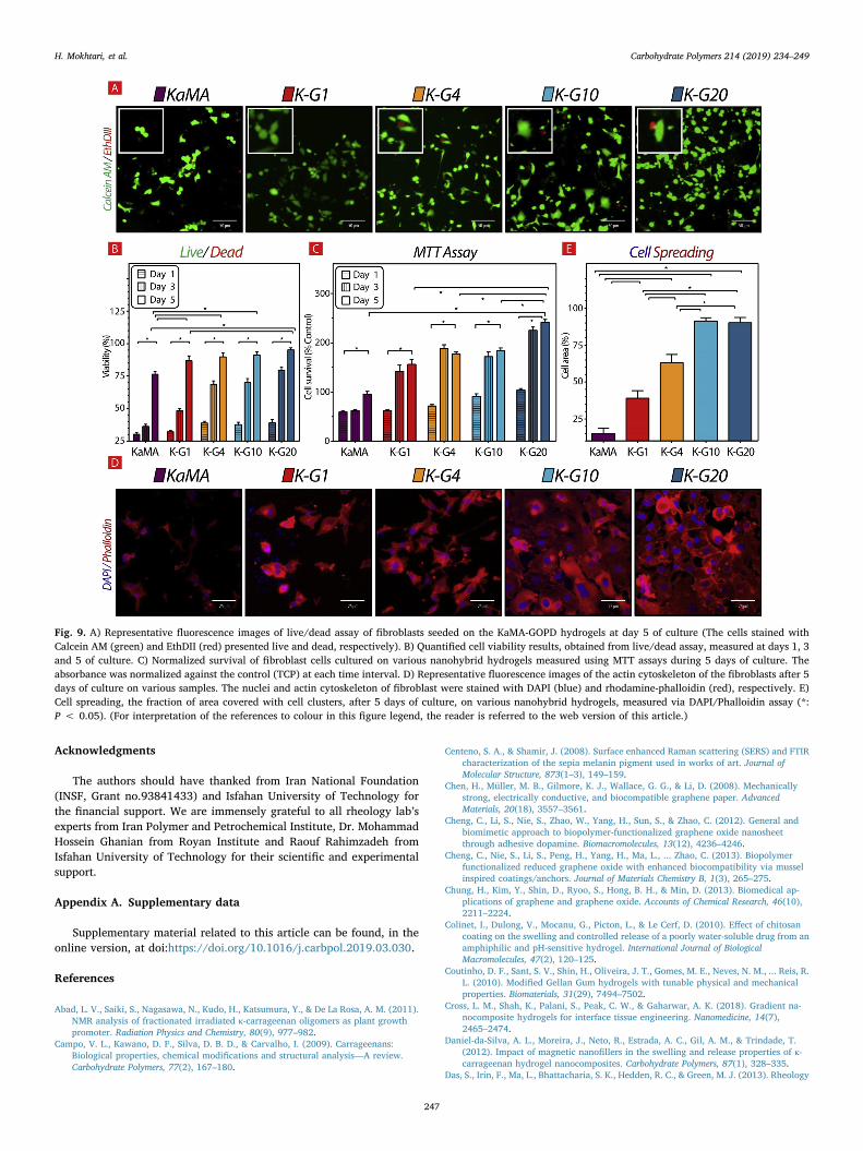
In order to evaluate the role of GOPD incorporation on the biolo-gical properties of KaMA, fibroblasts were seeded on the samples andcell proliferation and spreading on various samples were investigated.At first, cell viability on various nanohybrid hydrogels was determinedby using Live/dead assay. Fig. 9A shows that after 5 days of culture, byincreasing the amounts of GOPD content, the number of live cells en-hanced intensely. At the specific time points, the viability of cells wasevaluated by comparing the number of live cells to all cells. Cell via-bility on various samples is presented in Fig. 9B. The significant as-cending viability during 5 days of culture showed that KaMA-GOPDnanohybrid hydrogels were biocompatible with fibroblast cells. In ad-dition, our results revealed that, after 5 days of culture, the viability offibroblasts enhanced from 76.3 ± 2.1%–86.7 ± 3.7%, when the hy-drogel substrate was changed from KaMA to K-G1. In addition, wefound that increasing amounts of GOPD content upon 20wt.% (K-G20sample) significantly improved cell viability (95.2 ± 1.6%) comparedto K-G1 sample. It might be related to the positive role of both GO andPD in GOPD nanoparticles. According to previous results, GO na-nosheets could modulate the surface chemistry and stiffness of
H. Mokhtari, et al. Carbohydrate Polymers 214 (2019) 234–249
245

substrates leading to promotion of cell function (Halim, Luo, Ju, &Song, 2018; Wan, Frydrych, & Chen, 2011). Chen, Müller, Gilmore,Wallace, and Li (2008) also found out that graphene and its derivativessuch as GO could promote the adhesion and proliferation of fibroblastcells. In a similar research, Kang et al. (2015) observed considerablyimprovement in the cell function with incorporation of GO to thepolymeric structure due to promoted stiffness of soft environment. Inaddition, Zhang et al. (2011) observed, incorporation of optimized GOinto polymeric hydrogels improved its mechanical properties and pro-moted cell viability. In another word, Tsai et al. (Tsai, Chen, Chien,Kuo, & Wang, 2011) showed that surface modification of nanoparticleswith PD could result in faster proliferation of cells due to promotedimmobilization of serum adhesive proteins such as fibronectin on thesurface. Cell proliferation was also investigated using MTT assay during5 days of culture (Fig. 9C). Results showed significant improvement ofcell survival, during 5 days of culture. Moreover, addition of nano-particle to KaMA hydrogel resulted in significantly enhanced cell sur-vival (1.5-folds), from 95.4 ± 5.4% control (for KaMA) to156.0 ± 9.9% control (for K-G1). In addition, with increasing amountsof GOPD in K-G series, survival of cells noticeably improved. Re-markably, after 5 days of culture, cell survival on the K-G20 hydrogelwas estimated 241.8 ± 5.5%control, confirming live/dead assay re-sult. Furthermore, we studied cytoskeletal organization (F-actin) of fi-broblast cells on the nanohybrid hydrogels, after 5 days of culture(Fig. 9D). Actin filament staining showed that the cytoskeletal organi-zation of the cells were considerably affected by the hydrogel’s struc-ture. While low density of cells was spread on the KaMA hydrogel, byincreasing amounts GOPD content, the cells started to expand theircytoskeletal and spread over a larger area. After spreading, cells tried toform their pseudopodia on K-G series (especially K-G10 and KG-20),leading to development of bridges connections between fibroblast cellson the nanohybrid hydrogels. Previous researches showed that thisbehavior was related to PD, which facilitated protein adsorption andcell adhesion (Ku, Ryu, Hong, Lee, & Park, 2010; Shin, Lee, & Shin,2011). Liu et al. (2015) found out similar cellular behavior on zirconiacoated PD. To evaluate the role of substrate on the cell spreading, cellarea on various hydrogels was measured (Fig. 9E). Our results revealedthat the fraction of surface hydrogel covered with fibroblasts
significantly improved (5.7 folds) from 15.7 ± 3.7% (at KaMA) to90.2 ± 3.5% (at K-G20), showing the effective role of GOPD on cellspreading. KaMA hydrogels showed the lowest cell attachment andspreading, due to lack of cell binding domains on KaMA surface.
According to previous results, Cell’s fate could be controlled bymany chemical and physical factors of environment, named cell niche(Halim et al., 2018; Tong, Jiang, Zhu, & Yang, 2016). Between them,mechanical signals and especially stiffness played crucial role to controlcell function (Jiang et al., 2016). Our results revealed that incorpora-tion of GOPD to structure of KaMA hydrogels resulted in improvedmechanical properties leading to enhanced cell adhesion and spreading.Moreover, presence of catechol groups of PD on the GO surface couldeffectively increase the adhesion sites to attract proteins inside culturemedia through electrostatically interaction leading to improved cellattachment and spreading.
4. Conclusion
In this research, we engineered an injectable shear-thinning andmechanically robust hydrogel based on Kappa-carrageenan (KaMA)-dopamine functionalized graphene oxide (GOPD) for soft tissue en-gineering. This hydrogel was developed using dual-crosslinking me-chanism, which transmuted this hydrogel to unique biomaterials withsuperior properties. The presence of GOPD nanoparticle in the KaMAhydrogels allowed it to inject efficiently by enhancing shear-thinningbehavior of KaMA. Furthermore, compressive strength and toughness ofhydrogel were improved dramatically, giving the hydrogel potential toresist high shear stress, while showing self-healing behavior by re-creating secondary bond. Moreover, the addition of GOPD nanoparticleto KaMA matrix caused recovery properties originating from corpora-tion of catechol group of polydopamine with other moieties.Additionally, biocompatibility of this nanohybrid hydrogel was no-ticeably promoted with increasing GOPD content. Accordingly, KaMA-GOPD hybrid hydrogel could be a desirable choice for soft tissue en-gineering and 3D bioprinting applications.
Fig. 8. SEM images of the K-G10 and K-G20 hydrogels after 1st cycle compression until 60% strain and immersing in PBS solution for 2 h at two different magni-fications.
H. Mokhtari, et al. Carbohydrate Polymers 214 (2019) 234–249
246
Acknowledgments
The authors should have thanked from Iran National Foundation(INSF, Grant no.93841433) and Isfahan University of Technology forthe financial support. We are immensely grateful to all rheology lab’sexperts from Iran Polymer and Petrochemical Institute, Dr. MohammadHossein Ghanian from Royan Institute and Raouf Rahimzadeh fromIsfahan University of Technology for their scientific and experimentalsupport.
Appendix A. Supplementary data
Supplementary material related to this article can be found, in theonline version, at doi:https://doi.org/10.1016/j.carbpol.2019.03.030.
References
Abad, L. V., Saiki, S., Nagasawa, N., Kudo, H., Katsumura, Y., & De La Rosa, A. M. (2011).NMR analysis of fractionated irradiated κ-carrageenan oligomers as plant growthpromoter. Radiation Physics and Chemistry, 80(9), 977–982.
Campo, V. L., Kawano, D. F., Silva, D. B. D., & Carvalho, I. (2009). Carrageenans:Biological properties, chemical modifications and structural analysis—A review.Carbohydrate Polymers, 77(2), 167–180.
Centeno, S. A., & Shamir, J. (2008). Surface enhanced Raman scattering (SERS) and FTIRcharacterization of the sepia melanin pigment used in works of art. Journal ofMolecular Structure, 873(1–3), 149–159.
Chen, H., Müller, M. B., Gilmore, K. J., Wallace, G. G., & Li, D. (2008). Mechanicallystrong, electrically conductive, and biocompatible graphene paper. AdvancedMaterials, 20(18), 3557–3561.
Cheng, C., Li, S., Nie, S., Zhao, W., Yang, H., Sun, S., & Zhao, C. (2012). General andbiomimetic approach to biopolymer-functionalized graphene oxide nanosheetthrough adhesive dopamine. Biomacromolecules, 13(12), 4236–4246.
Cheng, C., Nie, S., Li, S., Peng, H., Yang, H., Ma, L., ... Zhao, C. (2013). Biopolymerfunctionalized reduced graphene oxide with enhanced biocompatibility via musselinspired coatings/anchors. Journal of Materials Chemistry B, 1(3), 265–275.
Chung, H., Kim, Y., Shin, D., Ryoo, S., Hong, B. H., & Min, D. (2013). Biomedical ap-plications of graphene and graphene oxide. Accounts of Chemical Research, 46(10),2211–2224.
Colinet, I., Dulong, V., Mocanu, G., Picton, L., & Le Cerf, D. (2010). Effect of chitosancoating on the swelling and controlled release of a poorly water-soluble drug from anamphiphilic and pH-sensitive hydrogel. International Journal of BiologicalMacromolecules, 47(2), 120–125.
Coutinho, D. F., Sant, S. V., Shin, H., Oliveira, J. T., Gomes, M. E., Neves, N. M., ... Reis, R.L. (2010). Modified Gellan Gum hydrogels with tunable physical and mechanicalproperties. Biomaterials, 31(29), 7494–7502.
Cross, L. M., Shah, K., Palani, S., Peak, C. W., & Gaharwar, A. K. (2018). Gradient na-nocomposite hydrogels for interface tissue engineering. Nanomedicine, 14(7),2465–2474.
Daniel-da-Silva, A. L., Moreira, J., Neto, R., Estrada, A. C., Gil, A. M., & Trindade, T.(2012). Impact of magnetic nanofillers in the swelling and release properties of κ-carrageenan hydrogel nanocomposites. Carbohydrate Polymers, 87(1), 328–335.
Das, S., Irin, F., Ma, L., Bhattacharia, S. K., Hedden, R. C., & Green, M. J. (2013). Rheology
Fig. 9. A) Representative fluorescence images of live/dead assay of fibroblasts seeded on the KaMA-GOPD hydrogels at day 5 of culture (The cells stained withCalcein AM (green) and EthDII (red) presented live and dead, respectively). B) Quantified cell viability results, obtained from live/dead assay, measured at days 1, 3and 5 of culture. C) Normalized survival of fibroblast cells cultured on various nanohybrid hydrogels measured using MTT assays during 5 days of culture. Theabsorbance was normalized against the control (TCP) at each time interval. D) Representative fluorescence images of the actin cytoskeleton of the fibroblasts after 5days of culture on various samples. The nuclei and actin cytoskeleton of fibroblast were stained with DAPI (blue) and rhodamine-phalloidin (red), respectively. E)Cell spreading, the fraction of area covered with cell clusters, after 5 days of culture, on various nanohybrid hydrogels, measured via DAPI/Phalloidin assay (*:P < 0.05). (For interpretation of the references to colour in this figure legend, the reader is referred to the web version of this article.)
H. Mokhtari, et al. Carbohydrate Polymers 214 (2019) 234–249
247

and morphology of pristine graphene/polyacrylamide gel. ACS Applied Materials &Interfaces, 5(17), 8633–8640.
Del Giudice, F., & Shen, A. Q. (2017). Shear rheology of graphene oxide dispersions.Chemical Engineering, 16, 23–30.
Diba, M., Pape, B., Klymov, A., Zhang, Y., Song, J., Lowik, D., ... Leeuwenburgh, S. C. G.(2017). Nanostructured raspberry-like gelatin microspheres for local delivery ofmultiple biomolecules. Acta Biomaterialia, 58, 67–79.
Diba, M., Wang, H., Kodger, T. E., Parsa, S., & Leeuwenburgh, S. C. (2017). Highly elasticand self-healing composite colloidal gels. Advanced Materials, 29(11).
Eigler, S., Dotzer, C., & Hirsch, A. (2012). Visualization of defect densities in reducedgraphene oxide. Carbon, 50(10), 3666–3673.
Fang, S., Huang, D., Lv, R., Bai, Y., Huang, Z.-H., Gu, J., & Kang, F. (2017). Three-di-mensional reduced graphene oxide powder for efficient microwave absorption in theS-band (2–4 GHz). RSC Advances, 7(41), 25773–25779.
Ferrari, A. C., Meyer, J. C., Scardaci, V., Casiraghi, C., Lazzeri, M., Mauri, F., ... Geim, A.K. (2006). Raman spectrum of graphene and graphene layers. Physical Review Letters,97(18), 187401.
Fu, L., Lai, G., Jia, B., & Yu, A. (2014). Preparation and electrocatalytic properties ofpolydopamine functionalized reduced graphene oxide-silver nanocomposites.Electrocatalysis, 6(1), 72–76.
Gaharwar, A. K., Schexnailder, P., Kaul, V., Akkus, O., Zakharov, D., Seifert, S., &Schmidt, G. (2010). Highly extensible bio-nanocomposite films with direction-de-pendent properties. Advance Functional Material, 20, 429–436.
Guvendiren, M., Lu, H. D., & Burdick, J. A. (2012). Shear-thinning hydrogels for bio-medical applications. Soft Matter, 8(2), 260–272.
Halim, A., Luo, Q., Ju, Y., & Song, G. (2018). A mini review focused on the recent ap-plications of graphene oxide in stem cell growth and differentiation. Nanomaterials(Basel), 8(9).
Han, L., Lu, X., Liu, K., Wang, K., Fang, L., Weng, L. T., ... Li, Z. (2017). Mussel-inspiredadhesive and tough hydrogel based on nanoclay confined dopamine polymerization.ACS Nano, 11(3), 2561–2574.
Huanga, Y., Zenga, M., Rena, J., Wanga, J., Fana, L., & Xu, Q. (2012). Preparation andswelling properties of graphene oxidepoly(acrylic acid-co-acrylamide) super-absor-bent hydrogel nanocomposites. Colloids and Surfaces A: Physicochemical andEngineering Aspects, 401, 97–106.
Hwang, S., Kang, D., Ruoff, R. S., Shin, H. S., & Park, Y. (2014). Poly(vinyl alcohol)reinforced and toughened with poly(dopamine)-treated graphene oxide, and its usefor humidity sensing. American Chemical Society, 8(7), 6739–6747.
Jiang, L., Sun, Z., Chen, X., Li, J., Xu, Y., Zu, Y., ... Yang, C. (2016). Cells sensing me-chanical cues: Stiffness influences the lifetime of cell-extracellular matrix interactionsby affecting the loading rate. ACS Nano, 10(1), 207–217.
Jiao, G., Yu, G., Zhang, J., & Ewart, H. (2011). Chemical structures and bioactivities ofsulfated polysaccharides from marine algae. Marine Drugs, 9(2), 196–223.
Jing, Y., Yuan, X., Yuan, Q., He, K., Liu, Y., Lu, P., ... Li, G. (2016). Determination ofnicotine in tobacco products based on mussel-inspired reduced graphene oxide-sup-ported gold nanoparticles. Scientific Reports, 6, 29230.
Jing, X., Mi, H.-Y., Napiwocki, B. N., Peng, X.-F., & Turng, L.-S. (2017). Mussel-inspiredelectroactive chitosan/graphene oxide composite hydrogel with rapid self-healingand recovery behavior for tissue engineering. Carbon, 125, 557–570.
Kang, S., Park, J. B., Lee, T.-J., Ryu, S., Bhang, S. H., La, W.-G., ... Kim, B.-S. (2015).Covalent conjugation of mechanically stiff graphene oxide flakes to three-dimen-sional collagen scaffolds for osteogenic differentiation of human mesenchymal stemcells. Carbon, 83, 162–172.
Keller, D. S., Tahilramani, R. N., Flores-Gonzalez, J. R., Mahmood, A., & Haas, E. M.(2016). Transanal minimally invasive surgery: Review of indications and outcomesfrom 75 consecutive patients. Journal of the American College of Surgeons, 222(5),814–822.
Kim, J. E., & Lee, H. S. (2014). Oscillatory shear induced gelation of graphene-poly(vinylalcohol) composite hydrogels and rheological premonitor of ultra-light aerogels.Polymer, 55, 287–294.
Kretlow, J. D., Klouda, L., & Mikos, A. G. (2007). Injectable matrices and scaffolds fordrug delivery in tissue engineering. Advanced Drug Delivery Reviews, 59(4–5),263–273.
Ku, S. H., Ryu, J., Hong, S. K., Lee, H., & Park, C. B. (2010). General functionalizationroute for cell adhesion on non-wetting surfaces. Biomaterials, 31(9), 2535–2541.
Kuo, C. K., & Ma, P. X. (2008). Maintaining dimensions and mechanical properties ofionically crosslinked alginate hydrogel scaffolds in vitro. Journal of BiomedicalMaterials Research Part A, 84(4), 899–907.
Lee, H., Dellatore, S. M., Miller, W. M., & Messersmith, P. B. (2007). Mussel-inspiredsurface chemistry for multifunctional coatings. Science, 318, 426–430.
Liang, Y. J., Zhen, J., Chen, N., & Reith, M. E. (2009). Interaction of catechol and non-catechol substrates with externally or internally facing dopamine transporters.Journal of Neurochemistry, 109(4), 981–994.
Liao, J., Qu, Y., Chu, B., Zhang, X., & Qian, Z. (2015). Biodegradable CSMA/PECA/gra-phene porous hybrid scaffold for cartilage tissue engineering. Scientific Reports, 5,9879.
Liu, H., Xi, P., Xie, G., Shi, Y., Hou, F., Huang, L., ... Wang, J. (2012). Simultaneousreduction and surface functionalization of graphene oxide for hydroxyapatite mi-neralization. The Journal of Physical Chemistry C, 116(5), 3334–3341.
Liu, M., Zhou, J., Yang, Y., Zheng, M., Yang, J., & Tan, J. (2015). Surface modification ofzirconia with polydopamine to enhance fibroblast response and decrease bacterialactivity in vitro: A potential technique for soft tissue engineering applications.Colloids and Surfaces B: Biointerfaces, 136, 74–83.
Loebel, C., Rodell, C. B., Chen, M. H., & Burdick, J. A. (2017). Shear-thinning and self-healing hydrogels as injectable therapeutics and for 3D-printing. Nature Protocols,12(8), 1521–1541.
Lokhande, G., Carrow, J. K., Thakur, T., Xavier, J. R., Parani, M., Bayless, K. J., &Gaharwar, A. K. (2018). Nanoengineered injectable hydrogels for wound healingapplication. Acta Biomaterialia, 70, 35–47.
Luo, J., Jiang, S., & Liu, X. (2013). Efficient one-pot synthesis of mussel-inspired mole-cularly imprinted polymer coated graphene for protein-specific recognition and fastseparation. The Journal of Physical Chemistry C, 117(36), 18448–18456.
Ma, Y., Niu, H., Zhang, X., & Cai, Y. (2011). One-step synthesis of silver/dopamine na-noparticles and visual detection of melamine in raw milk. Analyst, 136(20),4192–4196.
Marcano, D. C., Kosynkin, D. V., Berlin, J. M., Sinitskii, A., Sun, Z., Slesarev, A., ... Tour, J.M. (2010). Improved synthesis of graphene oxide. ACS Nano, 4(8), 4806–4814.
Mihaila, S. M., Gaharwar, A. K., Reis, R. L., Marques, A. P., Gomes, M. E., &Khademhosseini, A. (2013). Photocrosslinkable kappa-carrageenan hydrogels fortissue engineering applications. Advanced Healthcare Materials, 2(6), 895–907.
Mihaila, S. M., Popa, E. G., Reis, R. L., Marques, A. P., & Gomes, M. E. (2014). Fabricationof endothelial cell-laden carrageenan microfibers for microvascularized bone tissueengineering applications. Biomacromolecules, 15(8), 2849–2860.
Mobarak, N. N., Ramli, N., Ahmad, A., & Rahman, M. Y. A. (2012). Chemical interactionand conductivity of carboxymethyl κ-carrageenan based green polymer electrolyte.Solid State Ionics, 224, 51–57.
Mokhtari, H., Ghasemi, Z., Kharaziha, M., Karimzadeh, F., & Alihosseini, F. (2018).Chitosan-58S bioactive glass nanocomposite coatings on TiO2 nanotube: Structuraland biological properties. Applied Surface Science, 441, 138–149.
Mu, J., Hou, C., Wang, H., Li, Y., Zhang, Q., & Zhu, M. (2015). Origami-inspired activegraphene-based paper for programmable instant self-folding walking devices. ScienceAdvances, 1(10).
Navarro, D. A., & Stortz, C. A. (2003). Determination of the configuration of 3,6-anhy-drogalactose and cyclizable α-galactose 6-sulfate units in red seaweed galactans.Carbohydrate Research, 338(20), 2111–2118.
Necas, J., & Bartosikova, L. (2013). Carrageenan a review. Veterinarni Medicina, 58(4),187–205.
Paul, A., Hasan, A., Kindi, H. A., Gaharwar, A. K., Rao, V. T., Nikkhah, M., ...Khademhosseini, A. (2014). Injectable graphene oxide hydrogel-based angiogenicgene delivery system for vasculogenesis and cardiac repair. American ChemicalSociety, 8(8), 8050–8062.
Phua, S. L., Yang, L., Toh, C. L., Huang, S., Tsakadze, Z., Lau, S. K., ... Lu, X. (2012).Reinforcement of polyether polyurethane with dopamine-modified clay: The role ofinterfacial hydrogen bonding. ACS Applied Materials & Interfaces, 4(9), 4571–4578.
Pittet, B., Montandon, D., & Pittet, D. (2005). Infection in breast implants. The LancetInfectious Diseases, 5(2), 94–106.
Popa, E. G., Caridade, S. G., Mano, J. F., Reis, R. L., & Gomes, M. E. (2015). Chondrogenicpotential of injectable kappa-carrageenan hydrogel with encapsulated adipose stemcells for cartilage tissue-engineering applications. Journal of Tissue Engineering andRegenerative Medicine, 9(5), 550–563.
Popuri, S. R., Harris-Logie, A., Lino, K. H., Cadogan, E. I., & Lee, C.-H. (2014). Evaluationof antibacterial activity and characterization of synthesized biodegradable copoly-mers. Polymer-Plastics Technology and Engineering, 53(15), 1625–1635.
Rnjak-Kovacina, J., Wray, L. S., Burke, K. A., Torregrosa, T., Golinski, J. M., Huang, W., &Kaplan, D. L. (2015). Lyophilized silk sponges: A versatile biomaterial platform forsoft tissue engineering. ACS Biomaterials Science & Engineering, 1(4), 260–270.
Schönfelder, R., Rümmeli, M. H., Gruner, W., Löffler, M., Acker, J., Hoffmann, V., ...Pichler, T. (2007). Purification-induced sidewall functionalization of magneticallypure single-walled carbon nanotubes. Nanotechnology, 18(37), 375601.
Shin, S. R., Aghaei-Ghareh-Bolagh, B., Gao, X., Nikkhah, M., Jung, S. M., Dolatshahi-Pirouz, A., ... Khademhosseini, A. (2014). Layer-by-layer assembly of 3D tissueconstructs with functionalized graphene. Advanced Functional Materials, 24(39),6136–6144.
Shin, Y. M., Lee, Y. B., & Shin, H. (2011). Time-dependent mussel-inspired functionali-zation of poly(L-lactide-co-varepsilon-caprolactone) substrates for tunable cell beha-viors. Colloids and Surfaces B: Biointerfaces, 87(1), 79–87.
Stratton, S., Manoukian, O. S., Patel, R., Wentworth, A., Rudraiah, S., & Kumbar, S. G.(2018). Polymeric 3D printed structures for soft-tissue engineering. Journal of AppliedPolymer Science, 135(24).
Su, Z., Wang, H., Tian, K., Huang, W., Guo, Y., He, J., & Tian, X. (2018). Multifunctionalanisotropic flexible cycloaliphatic epoxy resin nanocomposites reinforced by alignedgraphite flake with non-covalent biomimetic. Composites Part A: Applied Science andManufacturing, 109, 472–480.
Tan, F., Liu, M., & Ren, S. (2017). Preparation of polydopamine-coated graphene oxide/Fe3O4 imprinted nanoparticles for selective removal of fluoroquinolone antibiotics inwater. Scientific Reports, 7(1), 5735.
Thakur, A., Jaiswal, M. K., Peak, C. W., Carrow, J. K., Gentry, J., Dolatshahi-Pirouz, A., &Gaharwar, A. K. (2016). Injectable shear-thinning nanoengineered hydrogels for stemcell delivery. Nanoscale, 8(24), 12362–12372.
Thanusha, A. V., Dinda, A. K., & Koul, V. (2018). Evaluation of nano hydrogel compositebased on gelatin/HA/CS suffused with asiatic acid/ZnO and CuO nanoparticles forsecond degree burns. Materials Science & Engineering C: Materials for BiologicalApplications, 89, 378–386.
Tong, X., Jiang, J., Zhu, D., & Yang, F. (2016). Hydrogels with dual gradients of me-chanical and biochemical cues for deciphering cell-niche interactions. ACSBiomaterials Science & Engineering, 2(5), 845–852.
Tsai, W. B., Chen, W. T., Chien, H. W., Kuo, W. H., & Wang, M. J. (2011). Poly(dopamine)coating of scaffolds for articular cartilage tissue engineering. Acta Biomaterialia,7(12), 4187–4194.
Vunjak-Novakovic, G., Tandon, N., Godier, A., Maidhof, R., Marsano, A., Martens, T. P., &Radisic, M. (2010). Challenges in cardiac tissue engineering. Tissue Engineering: PartB, 16(2).
H. Mokhtari, et al. Carbohydrate Polymers 214 (2019) 234–249
248

Wan, C., Frydrych, M., & Chen, B. (2011). Strong and bioactive gelatin–graphene oxidenanocomposites. Soft Matter, 7(13), 6159.
Wang, Q., Wang, J., Lu, Q., Detamore, M. S., & Berkland, C. (2010). Injectable PLGAbased colloidal gels for zero-order dexamethasone release in cranial defects.Biomaterials, 31(18), 4980–4986.
Xing, R., Wang, W., Jiao, T., Ma, K., Zhang, Q., Hong, W., ... Peng, Q. (2017). Bioinspiredpolydopamine sheathed nanofibers containing carboxylate graphene oxide nanosheetfor high-efficient dyes scavenger. ACS Sustainable Chemistry & Engineering, 5(6),4948–4956.
Xiong, S., Wang, Y., Yu, J., Chen, L., Zhu, J., & Hu, Z. (2014). Polydopamine particles fornext-generation multifunctional biocomposites. Journal of Materials Chemistry A,2(20), 7578–7587.
Xu, L. Q., Yang, W. J., Neoh, K., Kang, E., & Fu, G. D. (2010). Dopamine-induced re-duction and functionalization of graphene oxide nanosheets. Macromolecules, 43,8336–8339.
Xu, F., Xie, S., Cao, R., Feng, Y. N., Ren, C., & Wang, L. (2017). Prepare poly-dopaminecoated graphene@silver nanohybrid for improved surface enhanced Raman scat-tering detection of dyes. Sensors and Actuators B: Chemical, 243, 609–616.
Yamaguchi, H., Eda, G., Mattevi, C., Kim, H., & Chhowalla, M. (2010). Highly uniform300mm wafer-scale deposition of single and multilayered chemically derived gra-phene thin films. ACS Nano, 4(1), 524–528.
Yang, B., Zhang, Y., Zhang, X., Tao, L., Lia, S., & Wei, W. (2012). Facilely prepared in-expensive and biocompatible self-healing hydrogel: A new. Polymer Chemistry, 3(12),3235–3238.
Yang, J., Zhang, Y. S., Yue, K., & Khademhosseini, A. (2017). Cell-laden hydrogels forosteochondral and cartilage tissue engineering. Acta Biomaterialia, 57, 1–25.
Ye, W., Shi, X., Su, J., Chen, Y., Fu, J., Zhao, X., ... Xue, D. (2014). One-step reduction andfunctionalization protocol to synthesize polydopamine wrapping Ag/graphene hybridfor efficient oxidation of hydroquinone to benzoquinone. Applied Catalysis B:
Environmental, 160–161, 400–407.Yegappan, R., Selvaprithiviraj, V., Amirthalingam, S., Mohandas, A., Hwang, N. S., &
Jayakumar, R. (2019). Injectable angiogenic and osteogenic carrageenan nano-composite hydrogel for bone tissue engineering. International Journal of BiologicalMacromolecules, 122, 320–328.
Yu, L., & Ding, J. (2008). Injectable hydrogels as unique biomedical materials. ChemicalSociety Reviews, 37(8), 1473–1481.
Yu, M., Song, A., Xu, G., Xin, X., Shen, J., Zhang, H., & Song, Z. (2015). 3D welangum–graphene oxide composite hydrogels with efficient dye adsorption capacity.RSC Advances, 5(92), 75589–75599.
Yuksel, E., Choo, J., Wettergreen, M., & Liebschner, M. (2005). Challenges in soft tissueengineering, seminars in plastic surgery, Vol. 19, 333 Seventh Avenue, New York, NY10001, USA: Thieme Medical Publishers, Inc261–270.
Zangmeister, R. A., Morris, T. A., & Tarlov, M. J. (2013). Characterization of poly-dopamine thin films deposited at short times by autoxidation of dopamine. Langmuir,29(27), 8619–8628.
Zhang, L., Wang, Z., Xu, C., Li, Y., Gao, J., Wang, W., & Liu, Y. (2011). High strengthgraphene oxide/polyvinyl alcohol composite hydrogels. Journal of MaterialsChemistry, 21(28), 10399.
Zhao, W., Fang, M., Wu, F., Wu, H., Wang, L., & Chen, G. (2010). Preparation of grapheneby exfoliation of graphite using wet ball milling. Journal of Materials Chemistry,20(28), 5817.
Zheng, Q., Zhang, B., Lin, X., Shen, X., Yousefi, N., Huang, Z.-D., ... Kim, J.-K. (2012).Highly transparent and conducting ultralarge graphene oxide/single-walled carbonnanotube hybrid films produced by Langmuir–Blodgett assembly. Journal of MaterialsChemistry, 22(48), 25072.
Zhu, W., Chen, T., Li, Y., Lei, J., Chen, X., Yao, W., & Duan, T. (2017). High performancesof artificial nacre-like graphene oxide-carrageenan bio-nanocomposite films.Materials (Basel), 10(5).
H. Mokhtari, et al. Carbohydrate Polymers 214 (2019) 234–249
249