an evaluation of contaminated complete feed as a vehicle ...€¦ · an evaluation of contaminated...
TRANSCRIPT

Dee et al. BMC Veterinary Research 2014, 10:176http://www.biomedcentral.com/1746-6148/10/176
RESEARCH ARTICLE Open Access
An evaluation of contaminated complete feed asa vehicle for porcine epidemic diarrhea virusinfection of naïve pigs following consumption vianatural feeding behavior: proof of conceptScott Dee1*, Travis Clement2, Adam Schelkopf1, Joel Nerem1, David Knudsen2, Jane Christopher-Hennings2
and Eric Nelson2
Abstract
Background: Since its initial detection in May 2013, porcine epidemic diarrhea virus (PEDV) has spread rapidlythroughout the US swine industry. Initially, contaminated feed was proposed as a risk factor for PEDV; however,data were not available to support this theory. Here we provide proof of concept of this risk by describing a novelmeans for recovering PEDV-contaminated complete feed material from commercial swine sites and conducting anin vivo experiment to prove its infectivity.
Results: For on-farm detection of PEDV RNA in feed, paint rollers were used to collect material from at-risk feedbins from 3 clinically affected breeding herds. This material was tested by PCR and determined to be positive forPEDV-RNA (Ct = 19.50-22.20 range). To test infectivity, this material was pooled (Ct = 20.65) and a Treatment groupof 3-week old PEDV-naïve piglets were allowed to consume it via natural feeding behavior. For the purpose of aPositive control, piglets were allowed to ingest feed spiked with stock PEDV (Ct = 18.23) while the negative controlgroup received PEDV-free feed. Clinical signs of PEDV infection (vomiting and diarrhea) and viral shedding wereobserved in both the Positive control and Treatment group’ post-consumption with virus and microscopic lesionsdetected in intestinal samples No evidence of infection was observed in the Negative controls.
Conclusions: These data provide proof of concept that contaminated complete feed can serve as a vehicle for PEDVinfection of naïve pigs using natural feeding behavior.
Keywords: Porcine, Epidemic, Diarrhea, Virus, PEDV, Complete, Feed, Bioassay, Transmission
BackgroundPorcine epidemic diarrhea virus (PEDV) is an envelopedsingle-stranded positive sense RNA virus belonging tothe Order Nidovirales, the family Coronaviridae and thegenus Alphacoronavirus (Saif et al. [1]). Following detec-tion in the US swine population during May, 2013, thevirus spread rapidly across the country and 6317 casesof Porcine Epidemic Diarrhea (PED) have been con-firmed in 29 states as of May 3, 2014 [2,3]. While littleinformation is known regarding the routes of PEDV
* Correspondence: [email protected] Applied Research, Pipestone Veterinary Services, Pipestone, MN,USAFull list of author information is available at the end of the article
© 2014 Dee et al.; licensee BioMed Central LtdCommons Attribution License (http://creativecreproduction in any medium, provided the orDedication waiver (http://creativecommons.orunless otherwise stated.
transmission between herds, potential risk factors includeinfected pigs, contaminated transport and PEDV-positiveaerosols [4-6]. Recently, contaminated feedstuffs havebeen proposed as a route of PEDV transmission to naïvepigs but its current status is unclear [7]. While an initialreport from the Canadian Food Inspection Agency indi-cated that consumption of PEDV-positive porcine bloodplasma caused disease in pigs, a follow-up study could notdemonstrate that the feed pellets (complete feed) contain-ing the blood plasma in question were capable of causingdisease [8,9]. Despite this lack of evidence, dietary modifi-cations to enhance the biosecurity of feed have beenrecommended to reduce this perceived risk [10]. As moredata regarding the risk of PEDV transmission via complete
. This is an Open Access article distributed under the terms of the Creativeommons.org/licenses/by/4.0), which permits unrestricted use, distribution, andiginal work is properly credited. The Creative Commons Public Domaing/publicdomain/zero/1.0/) applies to the data made available in this article,

Dee et al. BMC Veterinary Research 2014, 10:176 Page 2 of 9http://www.biomedcentral.com/1746-6148/10/176
feed are needed, we conducted a study to test the risk ofPEDV-contaminated complete feed using a novel on-farmsampling method for virus detection in feed along with anin vivo experiment (swine bioassay) using at-risk feedmaterial. The study was based on the hypothesis that con-taminated complete feed can serve as a vehicle for PEDVinfection of naïve swine.
MethodsClinical historyDuring the period of January 9–13, 2014, clinical PorcineEpidemic Diarrhea was diagnosed in 3 breeding herdsfollowing acute outbreaks of anorexia, diarrhea andvomiting in isolated groups of sows. These herds werepart of an organized system of commercial pork produc-tion; Farm A (4973 sows) was located in NW Iowa,while Farms B and C, 3390 sows and 3016 sows respect-ively, were located in SW Minnesota. All 3 herds empha-sized strict biosecurity, using protocols previously validatedto reduce the risk of PRRSV infection [11,12]. Once a diag-nosis of PEDV was confirmed, an investigation of each sitewas conducted to identify possible routes of viral entry.During the investigation, a consistent observation commonto all 3 herds was noted. Specifically, from January 6–9,2014, all 3 farms experienced an unexpected feed outagewhich required an “emergency” delivery. The emergencydelivery had been deposited into a designated external stor-age bin which sourced feed to a distinct subpopulation ofthe herd. Following consumption of said feed, clinical signsbecame apparent only in the animals that had consumedthis feed, i.e. no other signs were noted in other animalsconsuming other feed from other bins.Based on this history, information regarding dates
corresponding to recent feed deliveries, the location ofthe associated storage bin, the period of time betweendelivery of feed and clinical signs, the location of indexcases in each farm, mill source and whether porcine by-products were present in feed was collected during theinvestigation. In addition, all transport-related activities,diagnostic data pertaining to recent genetic introduc-tions, and records of personnel and supply entry to eachfarm were reviewed. Finally, as Farms A and B were airfiltered, an evaluation of filter integrity and inspectionfor the potential of air bypass (entry of non-filtered airthrough improperly sealed fans, etc.) was conducted.
Feed samplingBecause of the potential link to feed, it was planned tosample the designated bin on each farm to determinewhether PEDV could be detected in “at-risk feed”, whichwas defined as feed consumed by the index population.Unfortunately, across all 3 sites, the majority of feed de-fined as “at-risk” had been consumed, leaving the desig-nated bins nearly (or completely) empty. However, upon
inspection of the bin lumen it was observed that clustersof feed material (feed particles and feed dust) wereadhered to the interior walls. To access this material, amodification of a published method for sampling con-taminated transport for PEDV RNA was devised [5].Specifically, synthetic woven paint roller pads, 23 cm inlength, 0.95 cm nap length (Sherwin Williams, ClevelandOH, USA) were attached to 3.6 m extension poles to ac-cess the cylindrical surface area of interior bin walls atmultiple heights. To minimize environmental contamin-ation of the roller prior to placement within the bin in-terior, a 4.4 L plastic bag (Ziploc, SC Johnson & Son Inc,Racine, WI, USA) covered the roller during ascension ofthe bin ladder. Following insertion of the roller into thebin lumen, the bag was removed and the roller wasdrawn across the inner walls, forcing large quantities ofthe adhered feed material to attach to the pad. Inaddition, if stored feed was present in a bin, the pad wasdrawn across the top layer to collect more material.Upon completion of sampling, the bag was replaced overthe roller and the entire sampling apparatus was re-moved from the bin. Once on the ground, 200 mL of7.2% phosphate buffered saline was poured into the bag,immersing the pad and promoting absorption of liquid.Using manual pressure, liquid was then forced from thepad into the bag and a 10 mL aliquot was decanted intoa 15 mL plastic Falcon tube (Becton Dickenson, FranklinLakes, NJ, USA) for diagnostic testing.In addition to the sampling of the “at risk feed bin” on
each farm, an “on-farm control bin” was also sampled.Control bins were located within 10 m of the “at-riskbin” but had not received a recent feed delivery and ani-mals consuming feed from these bins were not clinicallyaffected. Finally, to insure that the method did not gen-erate false positive results, 8 feed bins across 4 PEDVnegative farms were also sampled. All samples weretested for the presence of PEDV RNA using a RT-PCRat the South Dakota State University Animal Disease Re-search and Diagnostic Laboratory (SDSU ADRDL). Asample with a Cycle threshold (Ct) of less than 38 wasconsidered PEDV positive.
Swine bioassay facilities and source of animalsThe swine bioassay component of this study was con-ducted in Biosafety Level 2+ rooms at the Animal Re-source Wing (ARW) at SDSU. All procedures involvinganimals throughout the study were performed under theguidance and approval of the SDSU Institutional AnimalCare and Use Committee. Animals (n = 11, three-weekold piglets) were sourced from a PEDV-naïve herd andwere tested on arrival to the ARW via blood samplingand collection of rectal swabs from each pig. Prior toanimal arrival, all rooms (walls, ceilings, floors anddrains) were monitored for the presence of PEDV by
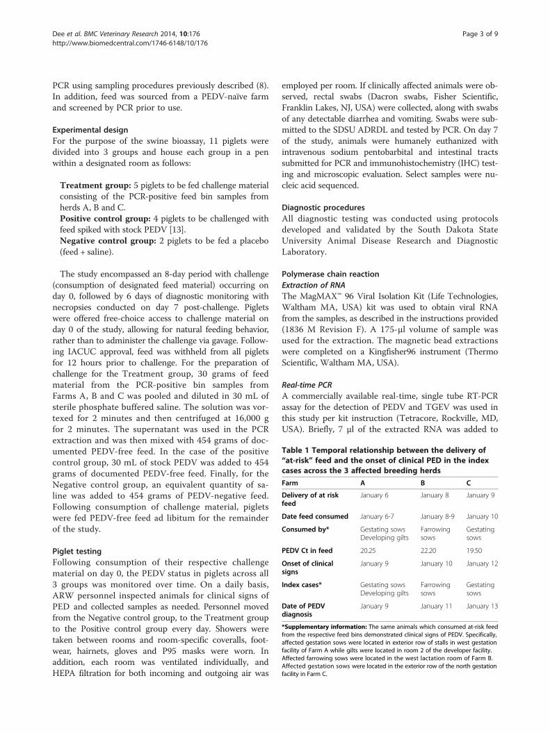
Table 1 Temporal relationship between the delivery of“at-risk” feed and the onset of clinical PED in the indexcases across the 3 affected breeding herds
Farm A B C
Delivery of at riskfeed
January 6 January 8 January 9
Date feed consumed January 6-7 January 8-9 January 10
Consumed by* Gestating sowsDeveloping gilts
Farrowingsows
Gestatingsows
PEDV Ct in feed 20.25 22.20 19.50
Onset of clinicalsigns
January 9 January 10 January 12
Index cases* Gestating sowsDeveloping gilts
Farrowingsows
Gestatingsows
Date of PEDVdiagnosis
January 9 January 11 January 13
*Supplementary information: The same animals which consumed at-risk feedfrom the respective feed bins demonstrated clinical signs of PEDV. Specifically,affected gestation sows were located in exterior row of stalls in west gestationfacility of Farm A while gilts were located in room 2 of the developer facility.Affected farrowing sows were located in the west lactation room of Farm B.Affected gestation sows were located in the exterior row of the north gestationfacility in Farm C.
Dee et al. BMC Veterinary Research 2014, 10:176 Page 3 of 9http://www.biomedcentral.com/1746-6148/10/176
PCR using sampling procedures previously described (8).In addition, feed was sourced from a PEDV-naïve farmand screened by PCR prior to use.
Experimental designFor the purpose of the swine bioassay, 11 piglets weredivided into 3 groups and house each group in a penwithin a designated room as follows:
Treatment group: 5 piglets to be fed challenge materialconsisting of the PCR-positive feed bin samples fromherds A, B and C.Positive control group: 4 piglets to be challenged withfeed spiked with stock PEDV [13].Negative control group: 2 piglets to be fed a placebo(feed + saline).
The study encompassed an 8-day period with challenge(consumption of designated feed material) occurring onday 0, followed by 6 days of diagnostic monitoring withnecropsies conducted on day 7 post-challenge. Pigletswere offered free-choice access to challenge material onday 0 of the study, allowing for natural feeding behavior,rather than to administer the challenge via gavage. Follow-ing IACUC approval, feed was withheld from all pigletsfor 12 hours prior to challenge. For the preparation ofchallenge for the Treatment group, 30 grams of feedmaterial from the PCR-positive bin samples fromFarms A, B and C was pooled and diluted in 30 mL ofsterile phosphate buffered saline. The solution was vor-texed for 2 minutes and then centrifuged at 16,000 gfor 2 minutes. The supernatant was used in the PCRextraction and was then mixed with 454 grams of doc-umented PEDV-free feed. In the case of the positivecontrol group, 30 mL of stock PEDV was added to 454grams of documented PEDV-free feed. Finally, for theNegative control group, an equivalent quantity of sa-line was added to 454 grams of PEDV-negative feed.Following consumption of challenge material, pigletswere fed PEDV-free feed ad libitum for the remainderof the study.
Piglet testingFollowing consumption of their respective challengematerial on day 0, the PEDV status in piglets across all3 groups was monitored over time. On a daily basis,ARW personnel inspected animals for clinical signs ofPED and collected samples as needed. Personnel movedfrom the Negative control group, to the Treatment groupto the Positive control group every day. Showers weretaken between rooms and room-specific coveralls, foot-wear, hairnets, gloves and P95 masks were worn. Inaddition, each room was ventilated individually, andHEPA filtration for both incoming and outgoing air was
employed per room. If clinically affected animals were ob-served, rectal swabs (Dacron swabs, Fisher Scientific,Franklin Lakes, NJ, USA) were collected, along with swabsof any detectable diarrhea and vomiting. Swabs were sub-mitted to the SDSU ADRDL and tested by PCR. On day 7of the study, animals were humanely euthanized withintravenous sodium pentobarbital and intestinal tractssubmitted for PCR and immunohistochemistry (IHC) test-ing and microscopic evaluation. Select samples were nu-cleic acid sequenced.
Diagnostic proceduresAll diagnostic testing was conducted using protocolsdeveloped and validated by the South Dakota StateUniversity Animal Disease Research and DiagnosticLaboratory.
Polymerase chain reactionExtraction of RNAThe MagMAX™ 96 Viral Isolation Kit (Life Technologies,Waltham MA, USA) kit was used to obtain viral RNAfrom the samples, as described in the instructions provided(1836 M Revision F). A 175-μl volume of sample wasused for the extraction. The magnetic bead extractionswere completed on a Kingfisher96 instrument (ThermoScientific, Waltham MA, USA).
Real-time PCRA commercially available real-time, single tube RT-PCRassay for the detection of PEDV and TGEV was used inthis study per kit instruction (Tetracore, Rockville, MD,USA). Briefly, 7 μl of the extracted RNA was added to

Table 2 Summary of clinical signs and diagnostic data across the 3 groups of pigs involved in the swine bioassay
Treatments Positive controls Negative controls
Date DPI Rectal Ct Clinical signs Rectal Ct Clinical signs Rectal Ct Clinical signs
1/17 0 neg neg neg neg neg neg
1/18 1 neg neg neg neg neg neg
1/19 2 neg neg 29.63 Vomit 32.21 neg neg
1/20 3 neg neg 16.06/32.21* neg neg neg
1/21 4 34.09 Diarrhea 18.94 15.48/29.63* Diarrhea 23.19 neg neg
1/22 5 28.89 Diarrhea 16.23 15.79 neg neg neg
1/23 6 15.01/18.94* Vomit 14.59 16.94 neg neg neg
1/24 Nx 5/5 pigs PCR/IHC (+) 4/4 pigs PCR/IHC (+) 2/2 pigs PCR/IHC (−)
Nx = Necropsy, small intestinal tracts.DPI = Days post-ingestion of at-risk or spiked feed.V/D = vomiting & diarrhea.* = 2 pigs detected as positive on rectal swabs.
Figure 1 PED in treatment group. Depicted in this figure are clinically affected piglets from the Treatment group on day 6 post-ingestion ofPEDV-contaminated feed. Piglets demonstrated loss of condition, rough hair coats, along with clinical signs of PED (diarrhea and vomiting). Evidenceof diarrhea is visible on the pen wall behind the pigs. Prior to necropsy, rectal swabs from all 5 piglets were PEDV-positive as detected by PCR.
Dee et al. BMC Veterinary Research 2014, 10:176 Page 4 of 9http://www.biomedcentral.com/1746-6148/10/176

Dee et al. BMC Veterinary Research 2014, 10:176 Page 5 of 9http://www.biomedcentral.com/1746-6148/10/176
18 μl of the master mix. The one-step real-time RT-PCRamplification conditions started with 15 minutes at 48°C,followed by 2 minutes at 95°C. The final cycles consistedof 5 seconds at 95°C and then 40 seconds at 60°C (datacollection step). The program was run for 38 cycles (Cycletime) and the FAM detector was used for PEDV and theTAMRA detector was used for TGEV. Positive and nega-tive controls were included on each run. All amplificationwas completed on the ABI7500 instrumentation (Austin,TX, USA).
PEDV stock virus propagationA cell-culture adapted variant of PEDV was used for in-oculation of Positive controls. For PEDV propagation,Vero 76 cells (ATCC CRL-1587) were maintained inMEM plus 10% fetal bovine serum and antibiotics.Three-day old confluent monolayers of Vero 76 cells in150 cm2 flasks were washed 3 times with serum freeminimum essential media (MEM) prior to inoculation.Monolayers were infected at ~0.1 moi of PEDV in MEMcontaining 2.5ug/ml TPCK-treated trypsin, incubated at
Figure 2 Diarrhea in the treatment group pen. This figure illustrates fecpiglets. This image was taken on day 6 post-ingestion of PEDV-positive fee
37°C for approximately 48 hrs until obvious CPE was ap-parent. Flasks were frozen at −80°C until needed.
PEDV S1 SequencingFor select samples, it was planned to conduct nucleicacid sequencing of the PEDV S1 gene. Specifically, frag-ments of the S1 domain of the spike gene were amplifiedfrom extracted RNA. Primers 1: (5′- ATGARGTCTTTAAYYTACTTCTGG-3′), 2: (5′-CATCCTCACCWGCACTAGTAAC-3′), 3: (5′- GTTGTGCTATGCAATATGTTTAY-3′), 4: (5′-TGAAATTAATTGTGACAGCATC-3′),5: (5′ -TTGTCATCACCAAGTAYGGTG -3′), 6: (5′- CTAAAAGACAGGTAATCATTAACAG- 3′), 7: (5′- CTGTGTTGACACTAGACAATTTAC- 3′), 8: (5′- CATACTAAAGTTGGTGGGAATAC- 3′) were designed to annealto conserved genomic regions. Incorporation of degener-ate bases maximizes the ability of the PCR to amplify gen-etically divergent PEDV variants between the US and UKstrains. Primer pair 1 and 2 obtained a PCR product sizeof 670 bp, primer pair 3 and 4 obtained a PCR productsize of 678 bp, primer pair 5 and 6 obtained a PCR
al staining on the wall of the pen housing the Treatment groupd.
Dee et al. BMC Veterinary Research 2014, 10:176 Page 6 of 9http://www.biomedcentral.com/1746-6148/10/176
product size of 565 bp and primer pair 7 and 8 obtained aPCR product size of 745 bp. Fragments were assembledsing the Vector NTI Software (Life Technologies,Waltham, MA, USA) for a complete S1 domain. QIAGENOne-Step Master Mix (Valencia, CA, USA) was used perkit instructions with an annealing temperature of 58°C for30 seconds.
ImmunohistochemistryImmunohistochemistry slides were prepared using thestandard SDSU ADRDL IHC procedure, with the followingmodification being the use of PEDV Monoclonal antibody,of mouse ascites origin, courtesy of Steve Lawson, SDSU,at a 1:1000 dilution.
ResultsClinical history and feed samplingDuring the on-farm inspection, no obvious breaches inany of the biosecurity protocols were detected across all3 herds. No evidence of viral entry through geneticintroduction, personnel error, contaminated transport orsupplies were noted upon review of on-farm documenta-tion. Finally, no breaches in the air filtration system(filter integrity, air bypass etc.) were noted on Farms Aand B. In regards to feed, all herds received feed from
Figure 3 Diarrhea on floor below the positive control group pen. Follcontrol group developed clinical signs of PED 2 days post-ingestion. This pthese piglets.
different mills and diets contained corn, soybean meal,vitamins and trace minerals. No porcine by-productswere included in any diet. Upon review of the history,a temporal relationship between the delivery of at-riskfeed and the onset of clinical signs in index cases wasobserved (Table 1). Across all 3 herds, clinical signswere observed within 2 days post-delivery of at-riskfeed. In addition, index cases were isolated to isolatedareas of each farm which only received at-risk feed.Specifically, Farm A cases were located in the exteriorrow of stalls in the east gestation room and in giltshoused in a single room in the developer facility. ForFarm B, index cases were located in the west farrowingroom while the exterior row of stalls in the north ges-tation room housed index cases for Farms C. Assess-ment of feed material in the at-risk bins across the 3affected sites indicated the presence of PEDV RNAwith Ct values ranging from 19.50-22.20 (Table 1). In con-trast, all samples from on-farm control bins and samplesfrom bins on PEDV-negative sites were PCR-negative.
Swine bioassayThe in vivo phase of the study was conducted fromJanuary 17–24 and results are summarized in Table 2.Prior to initiation of the in vivo phase of the study, all
owing ingestion of feed spiked with stock PEDV, piglets in the Positivehoto illustrates watery diarrhea observed below the pen floor housing

Dee et al. BMC Veterinary Research 2014, 10:176 Page 7 of 9http://www.biomedcentral.com/1746-6148/10/176
samples from incoming piglets, ARW facilities and feedwere PCR negative. In regards to the Treatment group,the pooling of feed material from Farms A, B and C re-sulted in challenge material having a Ct value of 20.65.Following challenge, clinical signs of diarrhea were ob-served in the index piglet in the Treatment group on day4 post-ingestion. PEDV-RNA was detected in a rectalswab from this animal, along with swabs collected fromdiarrhea in the pen. This piglet continued to shedthrough the remainder of the study period and 1 otherpiglet displayed clinical signs of diarrhea and vomitingon day 6 post-ingestion (Figures 1, 2 and 3). Followingeuthanasia, rectal swabs and intestinal tract samplesfrom all 5 pigs were positive by PCR and IHC (Figure 4).In addition, microscopic evaluation of small intestinaltissues indicated re-epithelialization with diffuse villousblunting and fusion was noted (Figure 5). In the Positivecontrol group, the Ct of the stock virus used to spike itsrespective challenge feed was 18.23. Following consump-tion, shedding and clinical signs were observed in theindex piglet on day 2 post-consumption with subsequent
Figure 4 Presence of PEDV antigen in a jejunal section from a treatmpresence of PEDV antigen in multiple infected enterocytes following applic
evidence of viral shedding to 2 other piglets on days 3and 4. Similar to the Treatment group, all rectal swabsamples and intestinal tract samples were PCR and IHC-positive at necropsy, along with evidence of microscopiclesions. In contrast, clinical signs, viral shedding orPEDV-positive intestinal tract samples were not ob-served in the Negative control group.S1 PEDV sequencing was completed on PCR-positive
feed challenge material from both the Positive controlgroup and the Treatment group, as well as an intestinalhomogenate from the index piglet in the Treatmentgroup. The sequencing results of the intestine and theTreatment feed material were similar, but different fromthe Positive control sample, indicating intestinal infec-tion from consumption of the feed material.
DiscussionThe purpose of this paper was to provide proof of con-cept that complete feed that was contaminated withPEDV could act as a vehicle for infection of naïve pigs.To accomplish this goal, we developed a novel means of
ent group piglet. This photomicrograph (400x) illustrates theation of immunohistochemical staining.

Figure 5 Histopathological changes caused by PEDV. This photomicrograph (400x) of a jejunal section from a Treatment group pigletdemonstrates lesions secondary to PEDV infection, i.e. degeneration of enterocytes as indicated by the presence of re-epithelialization, along withvillous blunting and fusion.
Dee et al. BMC Veterinary Research 2014, 10:176 Page 8 of 9http://www.biomedcentral.com/1746-6148/10/176
sampling at-risk feed material under controlled fieldconditions and conducted a swine bioassay to demon-strate transmission under controlled experimental condi-tions. Under the conditions of this study, we successfullydetected PEDV RNA in complete feed material across all3 sites and proved its infectivity using natural feedingbehavior, a novel finding not yet reported. These resultswere strengthened through the inclusion of on-farmcontrol feed bins and bins from naïve farms and the useof a Negative control group and natural feeding behaviorduring the in vivo phase. The sequencing data also sug-gest that the intestinal infection in the Treatment groupresulted from ingestion of pooled feed material from the3 affected herds.An acknowledged limitation of the study was that the
in vivo study was not designed to estimate the frequencyof feed-related PEDV infections. These results werebased on very small populations of pigs and cannot beextrapolated to today’s commercial farm conditions.
However, the ability to complete a successful swine bio-assay using a small number of animals cannot be ig-nored and the fact that our at-risk feed samples werecollected from large commercial production sites addsto the credibility of this first attempt to investigate thisrisk factor. It was interesting to note the pattern of shed-ding (as detected by rectal swabs) in the Treatment andPositive control groups. Despite the fact we employedsmall numbers of animals, shedding was first detected inan index case and spread occurred throughout the groupof piglets and differences were observed between the 2groups. This suggests that when small groups areemployed and/or field samples used for inoculation, thebioassay period may need to be extended to avoid therisk of false negative results.In closing, this is the first publication providing proof of
concept of the risk of PEDV-contaminated complete feedto naïve pigs. It was interesting to note that since neitherfeed source contained animal by-products, suggesting that

Dee et al. BMC Veterinary Research 2014, 10:176 Page 9 of 9http://www.biomedcentral.com/1746-6148/10/176
contamination may have occurred post-processing, how-ever, this was not proven. Further studies should focus onunderstanding the possibility of post-processing contam-ination as well as evaluating the ability of interventionstrategies, i.e. the application of heat and pressure throughthe pelleting process and/or the inclusion of select feedadditives which may have anti-viral effects, for reductionof this risk. Finally, it is the authors hope that the resultsfrom this study will assist in uniting the North Americanveterinary profession with the feed and swine industries aswe collectively move forward in reducing the threat of thisdevastating transboundary disease.
ConclusionsThese data provide the initial proof of concept that con-taminated complete feed can serve as a vehicle for PEDVinfection of naïve pigs. Information from this studyshould be used to justify the need for further researchtowards the mitigation of said risk.
Availability of supporting dataThe data set(s) supporting the results of this article isincluded within the article.
AbbreviationsPEDV: Porcine epidemic diarrhea virus; PED: Porcine epidemic diarrhea;Ct: Cycle threshold; PCR: Polymerase chain reaction; ARW: Animal resourcewing; FAM: Fluorescein; TAMRA: Carboxytetramethylrhodamin; MEM: Minimalessential media.
Competing interestsThe authors declare that they have no competing interests.
Authors’ contributionsSD: Developed study, co-wrote paper. TC: Conducted molecular diagnostics,co-wrote paper. AS: Participated in on-farm collection. JN: Participated inon-farm collection. DK: Conducted pathological assessment of tissues anddeveloped Figures 4–5. JCH: Provided critical review and revising ofpaper. EN: Provided virological expertise at the laboratory level and co-wrotepaper. All authors read and approved the final manuscript.
AcknowledgementsThe authors would like to recognize Dr. Michele Mucciante and the AnimalResource Wing team for their significant contributions to the success of thisstudy. Funding for this study was provided by Pipestone Applied Research
Author details1Pipestone Applied Research, Pipestone Veterinary Services, Pipestone, MN,USA. 2Animal Disease Research and Diagnostic Laboratory, South DakotaState University, Brookings, SD, USA.
Received: 16 May 2014 Accepted: 28 July 2014Published: 5 August 2014
References1. Saif LJ, Pensaert MB, Sestak K, Yeo S, Jung K: Coronaviruses. In Diseases of
Swine. 10th edition. Edited by Zimmerman JJ, Karriker LA, Ramierez A,Schwartz KJ, Stevenson GW. Ames: Wiley and Sons; 2012:501–524.
2. Chen Q, Ganwu L, Stasko J, Thomas JT, Stensland WR, Pillatzki AE, GaugerPC, Schwartz KJ, Madson D, Yoon KJ, Stevenson GW, Burrough ER, HarmonKM, Main RG, Zhang J: Isolation and characterization of Porcine EpidemicDiarrhea Viruses associated with the 2013 disease outbreak amongswine in the United States. J Clin Microbiol 2014, 52:234–243.
3. USDA APHIS VS NVSL NAHLN UMN Swine Health Monitoring Report: PorcineEpidemic Diarrhea Virus Reporting; 2014.
4. Van Reeth K, Pensaert M: Prevalence of infections with enzooticrespiratory and enteric virus in feeder pigs entering fattening units.Vet Rec 1994, 135:594–597.
5. Lowe J, Gauger P, Harmon K, Zhang J, Connor J, Yeske P, Loula T, Levis I,Dufrense L, Main R: Role of transportation in spread of porcine epidemicdiarrhea virus infection, United States. Emerg Infect Dis 2014, 20(5):1–5.http://dx.doi.org/10.3201/10.3201/eid2005.131628.
6. Goede D, Robbins R, Dufrense L, Engle M, Morrison R: Detection of PorcineEpidemic Diarrhea Virus in air Samples at Varying Distances to EpidemicFarms in Oklahoma. In Proceedings of the 40th Allen D. Leman SwineConference; 2013.
7. American Association of Swine Veterinarians and USDA Center forEpidemiology and Animal Health: PED Epidemiologic Survey. 2013.http://www.aasv.org/aasv%20website/Resources/Diseases/PED/AbbreviatedResultsPEDSurvey20130803.pdf.
8. Canadian Food Inspection Agency: Statement on Porcine Epidemic DiarrheaVirus in Feed; 2014.
9. Canadian Food Inspection Agency: Investigation into Feed as a PossibleSource of Porcine Epidemic Diarrhea (PED); 2014.
10. AASV e-letter: Kansas State University applied swine nutrition teamprovides nursery diet options in response to PEDV concerns. 2014.http://www.aasv.org/news/story.php?id=7065.
11. Pitkin AN, Deen J, Dee SA: Use of a production region model to assessthe airborne spread of porcine reproductive and respiratory syndromevirus. Vet Microbiol 2009, 136:1–7.
12. Dee SA, Otake S, Deen J: Use of a production region model to assess theefficacy of various air filtration systems for preventing the airbornetransmission of porcine reproductive and respiratory syndrome virusand Mycoplasma hyopneumoniae. Results of a 2-year study. Virus Res2010, 154:177–184.
13. Nelson J, Okda F, Parmar R, Singrey A, Lawson S, Liu X, Christopher-Hennings J,Nelson E: Environmental Stability of a Cell Culture Adapted U.S. Isolate of PEDV23rd IPVS Congress, Mexico. 2014:94.
doi:10.1186/s12917-014-0176-9Cite this article as: Dee et al.: An evaluation of contaminated completefeed as a vehicle for porcine epidemic diarrhea virus infection of naïvepigs following consumption via natural feeding behavior: proof ofconcept. BMC Veterinary Research 2014 10:176.
Submit your next manuscript to BioMed Centraland take full advantage of:
• Convenient online submission
• Thorough peer review
• No space constraints or color figure charges
• Immediate publication on acceptance
• Inclusion in PubMed, CAS, Scopus and Google Scholar
• Research which is freely available for redistribution
Submit your manuscript at www.biomedcentral.com/submit