an evaluation of apical root resorption after orthodontic...
TRANSCRIPT
AN EVALUATION OF EXTERNAL APICAL ROOT RESORPTION AFTER
ORTHODONTIC TREATMENT
Elizabeth Thomas
A research report submitted to the Faculty of Health Sciences, University of the
Witwatersrand, Johannesburg, in partial fulfillment of the requirements for the degree of
Master of Science in Dentistry.
Johannesburg 2011
ii
DECLARATION
I, Elizabeth Thomas, declare that this research report is my own work. It is being submitted for
the degree of Master of Science in Dentistry at the University of the Witwatersrand,
Johannesburg. It has not been submitted before for any degree or examination at this or any
other university.
.....................................................
E. Thomas
…20th…… day of ……October………, 2011

iii
This work is dedicated to my father, Prof. C. T. Mathew, one of the first
Orthodontists in Kerala, India, and the founder-Director of the Calicut Dental College.
iv
PUBLICATIONS AND PRESENTATIONS
Conference paper
Thomas, E. (2010). An evaluation of external apical root resorption after orthodontic
treatment. Presented at the 45th Indian Orthodontic Conference, 17th-19th December,
Mangalore, India.
v
ABSTRACT
Root resorption is a common problem encountered in all branches of dentistry but is more
commonly seen in cases that had been treated orthodontically. Orthodontists are constantly
improving materials and techniques to reduce undesirable side effects like root resorption.
Therefore in this retrospective study the primary objective was to compare the amount of root
resorption observed after active orthodontic treatment with three different appliance systems
namely, Tip Edge, Modified Edgewise and Damon. The sample consisted of pre and post-
treatment cephalograms of sixty eight cases that were treated in three different groups (i.e.,
techniques). Root resorption of the maxillary central incisor was assessed from pre- and post-
treatment lateral cephalograms using two schemes. In the first method overall tooth length
(Black, 1902) from the incisal edge to the apex was measured on both pre and post-treatment
lateral cephalograms and root resorption was recorded as an actual millimetre loss of tooth
length. Percentage shortening per tooth was also recorded.
The results were subjected to various statistical analyses. There was a significant upward
linear trend (p=0.022) for root resorption from Group 1 (Tip Edge) to Group 3 (Damon).
Statistical modeling illustrated that only baseline length (pre-treatment incisor length) was a
significant confounder. Gender, race, age and treatment time did not have a significant
influence on the amount of root resorption seen after orthodontic treatment. In the final
analysis after having adjusted for baseline length it was found that there were no significant
differences (p=0.133; ANCOVA) in the degree of root resorption observed after the active
phase of orthodontic treatment between groups. Similarly the percentage of root resorption

vi
calculated did not differ significantly between groups (p=0.067). The result was also
confirmed by following a non-parametric approach by doing an analysis of covariance
(ANCOVA) in which data was allocated a rank value.
In the second method root resorption was visually evaluated by using the five grade ordinal
scale of Levander and Malmgren (1988). It was found that majority of cases in the sample
came under Grade1 and Grade 2 category of root resorption. The upward linear trend between
actual measurements and visual measurements was found to be statistically significant
(p=0.0183).
vii
ACKNOWLEDGEMENTS
I wish to express my sincere gratitude to my supervisors Dr. R. Chamda and Professor W.G.
Evans, for the valuable guidance, assistance and constant encouragement offered to me in the
preparation and execution of this research report.
I am also grateful to Professor Piet Becker, Medical Research Council, Pretoria for his expert
help in the statistical analyses contained in this report.
I wish to thank Dr. R. Chamda, Dr. M. Wertheimer and Dr C.Lesar for granting me access to
their respective offices, staff and records for data collection. This research would not have
been possible without their kindness.
I would also like to thank my colleagues Dr N. Green Thompson and Dr A. Morar for their
constructive criticisms.
The assistance of my son George Thomas in copying all the data compiled from the three
groups is gratefully acknowledged.
Finally, I would like to acknowledge my husband, Thomas Joseph, who has been a constant
pillar of strength and support to me, especially during the execution and submission of this
report.

viii
TABLE OF CONTENTS
Page
DECLARATION ii
DEDICATION iii
PUBLICATIONS AND PRESENTATIONS iv
ABSTRACT v
ACKNOWLEDGEMENTS vii
TABLE OF CONTENTS viii
LIST OF FIGURES x
LIST OF TABLES xi
CHAPTER 1: INTRODUCTION 1
CHAPTER 2: REVIEW OF LITERATURE 3
CHAPTER 3: MATERIALS AND METHODS 11
3.1 Sample selection 11
3.2 Calibration of X-rays 12
3.3 Method of measuring 15
3.3.1 Method one 15
3.3.2 Method two 18
3.4 Statistics 19
3.4.1 Error of the method: method one 19
3.4.2 Statistical analysis for method one 19
3.4.3 Error of the method: method two 20
3.4.4 Statistical analysis for method two 20
3.4.5 Comparative statistics 20
ix
CHAPTER 4: RESULTS 21
4.1 Descriptive statistics of the sample 21
4.2 Error of the method 24
4.2.1 Results of intra and inter-examiner tests for method one 24
4.2.2 Results of intra examiner tests for method two 24
4.3 Data on assessment of resorption 25
4.4 Comparative statistics between groups 26
4.5 Results of ANOVA method one 26
4.6 Results for method two 27
4.7 Comparative statistics (method one vs. method two) 28
CHAPTER 5: DISCUSSION 29
5.1 Limitations and difficulties encountered in this study 40
CHAPTER 6: CONCLUSIONS 42
APPENDICES 43
APPENDIX A: Listing of individual results 43
APPENDIX B: Assessment of root resorption using the scale of Levander and
Malmgren 46
REFERENCES 47
ETHICS CLEARANCE CERTIFICATE 57
x
LIST OF FIGURES
Figure 3.1 Set up used for calibration of radiographs (two views) 13
Figure 3.2 Measurement of tooth length 16
Figure 3.3 Illustration of various root apices seen in post-treatment radiographs 17
Figure 3.4 Illustration of the five ordinal grades of root resorption 19
Figure 4.1 Linear prediction of the mean percentage of root resorption 26
xi
LIST OF TABLES
Table 4.1 Mean age distribution by groups 21
Table 4.2 Duration of treatment in each group 22
Table 4.3 Gender distribution by group, frequency (%) 22
Table 4.4 Race distribution by group 23
Table 4.5 New racial sample distribution by groups 23
Table 4.6 Intra -class correlation 24
Table 4.7 Mean pre-treatment incisor length by groups 25
Table 4.8 Mean post-treatment incisor length by groups 25
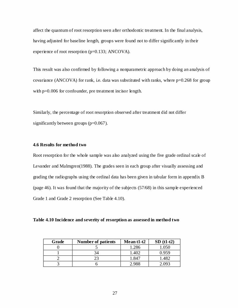
Table 4.9 Mean root resorption (t1-t2) seen in each group 25
Table 4.10 Incidence and severity of resorption as assessed in method two 27
1
CHAPTER 1: INTRODUCTION
External root resorption is a multifactorial problem experienced in all disciplines of dentistry
but most commonly in cases receiving orthodontic treatment. Root resorption was first
reported by Bates as far back as 1856 (Philips, 1955). Although Ottolengui in 1914 was the
first clinician to report apical root loss after orthodontic treatment, the finding was received
with little trepidation by the orthodontic profession. However, thirteen years later, Albert
Ketcham’s dramatic evaluations of apical root loss, derived from a radiographic survey of 385
treated cases in his own practice, forced the discipline to sit up and take note of this
complication. A spate of investigations on both experimental animals and human patients
followed (Rudolph, 1940; Philips, 1955; Rygh, 1977). These studies dealt with the histological
and clinical aspects and with factors affecting apical root loss. At present it is known that the
aetiologic factors are complex and multifactorial (Brezniak and Wasserstein, 1993b).
Root resorption is undesirable as it can affect the long term viability of the dentition. A
considerable reduction in the root length of the affected teeth results in an unfavorable crown-
root ratio, making them less suitable as abutments in prosthetic restorations. An apical root
loss of three millimetre is equivalent to one millimetre of crestal bone loss, so periodontitis
occurring in teeth affected by root resorption will progress more rapidly to a critical alveolar
bone level when compared with teeth unaffected by root resorption.
Ketcham (1929) also made a less well- known discovery, namely that different appliance
systems influence the degree of root resorption. Many general dentists and dental specialists in
other disciplines believe that root resorption is avoidable and blame the orthodontist involved

2
when it occurs during orthodontic treatment. It is therefore important to identify whether
different orthodontic appliance systems are associated with an increased risk of root
resorption. In recent years the difference in risk occasioned by different appliance systems has
been re-evaluated, but other contributing factors have not been as thoroughly investigated.
The purpose of this study is to examine the influence of variables such as different types of
mechanotherapy, age, gender and ethnicity together with duration of treatment time on root
resorption.
3
CHAPTER 2: REVIEW OF LITERATURE
External apical root resorption (EARR) can be defined as a shortening or blunting of the root
apex (Malmgren and Levander, 2004). Rudolph (1940) noted that it typically attacks the root
tip and travels coronally, creating a “shed roof” effect to the root.
Andreason (1988) classified root resorption into three main categories, namely surface
resorption, inflammatory resorption and replacement resorption.
Surface resorption is a self- limiting process usually involving small localized areas. These
undergo spontaneous repair by cellular reaction from adjacent areas of the periodontal
ligament.
Inflammatory resorption occurs when the initial root resorption has reached the dentinal
tubules of an infected necrotic pulpal tissue.
Replacement resorption produces ankylosis of a tooth as in this instance the resorbed root
substance is replaced by bone, firmly linking tooth and socket.
Tronstad (1988) believes that inflammatory resorption is related to the presence of
multinucleated cells that colonize the denuded cemental surface. He characterizes two kinds of
inflammatory resorption: Transient inflammatory resorption occurs when the etiological
factors occur for a short period of time and have minimal effect. This defect usually goes
undetected radiographically and is repaired by a cementum-like tissue. Progressive
inflammatory resorption occurs when the etiological effect or stimulus continues over a long
period of time. The severely denuded root areas attract hard- tissue resorbing cells.
4
Gradually there is complete destruction of the local root structure and replacement resorption
with ankylosis of the tooth occurs.
Mechanically induced tooth movement, the foundation of orthodontic treatment, typically
produces some external apical root resorption. Radiographic studies have confirmed this
(Philips, 1955; Ten Hoeve and Mulie, 1976; Linge and Linge, 1991; Mirabella and Artun
1995; Blake, Woodside and Pharoah 1995; Sameshima and Asgarifar 2001). Replacement
resorption, however, is rarely seen in association with orthodontic mechanotherapy (Brezniak
and Wasserstein, 1993a).
According to Brezniak and Wasserstein (2002a), orthodontic root resorption is a distinct
pathologic process and differs from other types of root resorption. Orthodontic force
applications produce a clinical picture that includes all the characteristics of inflammation
namely rubor (redness), calor (heat), tumor (swelling), dolor (pain) and to some extent functio
laesa (inhibited function). The inflammation that occurs is the main culprit underlying the root
resorption process. Brezniak and Wasserstein (2002a) suggested a new term: Orthodontically
Induced Inflammatory Root Resorption (OIIRR). Patient- related and treatment- related
factors are responsible for the onset and progression of this root resorption which results from
a sterile necrosis of the periodontal ligament. In orthodontically induced inflammatory root
resorption the injury results from the pressure applied to the root during tooth movement.
Ischaemic necrosis or hyalinization of the periodontal ligament occurs on the pressure side or
area.
5
Three stages can be described in the hyaline zone namely, degeneration, elimination of
destroyed products, and re-establishment. Studies in experimental animals conducted by
Brudvik and Rygh (1994) confirmed that OIIRR occurs as a part of the hyaline zone
elimination process. Macrophage- like and multinucleated cells are activated by biochemical
signals coming from the sterile necrotic tissue, the result of orthodontic force application.
Macrophages are scavenger cells of haematopoietic origin and their role is to eliminate
necrotic tissue. The initial elimination process takes place at the periphery of the hyaline zone,
where blood supply to the periodontal ligament still exists or is even increased. During
removal of the hyaline zone, the nearby outer surface of the root can be affected. In severe
cases the outer root surface layers, including cementoblasts and the pre-cementum layers can
be damaged. The activity of the macrophage- like and multinucleated cells continues until the
hyaline tissue has been completely removed and /or the force level decreases. Resorption
lacunae that are formed on the root surfaces involved help to increase the root surface area and
thereby decrease the pressure exerted through force application. The decompression that
occurs allows the process to reverse and the cementum to be repaired.
Force application of an intermittent nature results in lesser root resorption than does
application of a continuous force. Intermittent forces permit intervals with an absence of
mechanical stress and consequently, allow the resorbed cementum to heal. This in turn helps
to prevent further resorption (Brezniak and Wasserstein, 2002a; Pizzo, Licata, Giuglia and
Guilianna, 2007).
If the orthodontic force is applied for a long time, the multinucleated cells develop
odontoclastic- like morphology and functions and begin a complete cementum resorption,
6
denuding mineralized dentin areas. These cells produce a tridimensional, non-reversible and
radio-graphically evident root resorption. This process is usually located in the apical third of
the root.
Faltin, Faltin, Sander and Arana-Chavez (2001) in their investigations regarding orthodontic
tooth movement with application of continuous intrusive forces using super- elastic wires in
humans found that the mineralized surface of the cementum in the apical part is resorbed the
most. The changes are directly proportional to the magnitude of the continuous force applied.
Brezniak and Wasserstein (2002a) classified OIIRR into three degrees according to the
severity of affliction:
Cemental or surface resorption with remodeling
In this process only the outer layers of cementum are resorbed. Later on these layers are
completely regenerated or remodelled.
Dentinal resorption with repair (deep resorption)
In this process the cementum and outer layers of dentine are resorbed. Repairs usually take
place with cementum-like material. The final shape of the root after repair may or may not
resemble the original form
Circumferential apical root resorption
In this process complete resorption of the apical portion of the hard tissue components of
the root occurs and the root is shortened.
7
This study will examine orthodontically induced external root resorption which has occurred
at the apices of the maxillary central incisor roots after active orthodontic therapy.
For the majority of orthodontic patients the biggest motivation for seeking orthodontic
treatment is a desire to improve dental and facial esthetics. In Class II Division I
malocclusions, the retraction of upper teeth results in a dropping back of the upper lip which
may improve the relative protrusiveness of the profile ( Looi and Mills, 1986). Hence
retraction of maxillary anterior teeth is an essential step in such orthodontic treatment. Rudee
(1964) measured associated linear changes in incisor position and upper lip protrusion. He
found that three millimetre retraction of upper anterior teeth results in one millimetre
retraction of the lips. Garner (1974) found the ratio to be even higher in black males (3.6:1).
Thus we see that the upper incisors have to undergo considerable movement to bring about
associated and desirable soft tissue profile changes. However, when treating any malocclusion
an important objective of the orthodontist is to create or maintain esthetic harmony consistent
with a functional occlusion. Under these stringent conditions, when examining the
vulnerability of different teeth to root resorption during orthodontic movement, it will be
found that the tendencies differ. The maxillary incisors are the teeth most affected by root
resorption. In general the extent of movement demanded of these teeth is usually greater due
to the nature of the malocclusion, function and to the exigencies of esthetics.
The fact that incisors are single rooted with spindly, cone shaped roots may also be a
predisposing factor to external apical root resorption. The root structure and its relationship to
bone and periodontal membrane tend to transfer the effect of the forces mainly to the apex.
The apical third of a root is covered with cellular cementum and its integrity is dependent on
8
the vascularity of the area. This makes it more fragile and readily injured by heavy forces.
Hence the peri-apical portion is more prone to root resorption than the rest of the root (Harris,
2000). It is often stated that if there is no evidence of apical root resorption in the maxillary
and mandibular incisors, then significant apical resorption is less likely to occur in other teeth
(Copeland and Green, 1986; Goldson and Hendrikson, 1975; Sjolien and Zachrisson, 1973).
Root resorption is a complex process with a multifactorial etiology. It has been postulated that
the risk of orthodontically –associated root resorption will vary according to the type of
appliance or technique used. The pin and tube and ribbon and bracket appliances produced
more resorption than other labial appliances which utilized tipping movements (Ketcham,
1927). Beck and Harris (1994) found no difference in the degree of root resorption in cases
treated with the Begg and Tweed techniques. Apical root resorption of central incisors was
significantly higher in cases treated using the edgewise technique than in the straight wire
group (Mavragani, Vergari, Selliseth, Boe and Wisth, 2000).
Is there a technique or force system that can reduce or prevent external apical root resorption?
No previous study could satisfactorily answer this question (Brezniak and Wasserstein,
2002b).
Nevertheless, orthodontists continue to seek improved materials and techniques which may
minimize undesirable treatment side effects like apical root resorption. Nickel- titanium wire
was first used in orthodontics in 1971. Twenty two years later new super-elastic nickel-
titanium wires that deliver light and continuous forces with large amounts of activation and for
longer periods of time were developed. Concurrent with the evolution of wires, bracket
9
systems were also improved with the objective of reducing the magnitude of force applied to
the teeth during orthodontic treatment. Viazis (1995) introduced triangular brackets with an
increased interslot distance, considerably decreasing the forces delivered by the new
generation nickel–titanium wires. He called the technique the Bioefficient Therapy. Janson,
Canto, Martin, Henriques and De Freitas (1999) compared apical root resorption after
orthodontic treatment with Bioefficient Therapy with that recorded in cases trea ted with the
Edgewise Straight Wire system and the Simplified Standard Edgewise Technique. They found
less root resorption in cases treated with Bioefficient Therapy. It was suggested that the
bracket design, use of heat activated and super-elastic wires as well as limiting wires to
smaller rectangular stainless steel arches in a 0.22 x 0.028 inch slot during incisor retraction
and in finishing stages may have contributed to the observed decrease.
The Differential Straight Arch technique developed by Kesling (1994) utilizes the Tip Edge
bracket, designed to provide all the benefits and advantages of differential tooth movement
plus predetermined degrees of final crown tip and torque. Damon (2004) developed a
technique that sets out to match each phase of treatment with the natural force systems of
normal growth and development. The Damon technique is a low force, low friction system
where passive self- ligating brackets and high technology wires are carefully selected with the
intention of keeping the force applied on teeth in the optimal force zone. Pandis, Nasika,
Polychronopoulou and Eliades (2008) found no statistically significant difference for the
incidence of root resorption between cases treated with conventional Edgewise and with
passive self- ligating brackets. They found, conversely, that a trend indicating more root
resorption for the self- ligating system was evident, although the differences did not reach
10
significance (p=0.06). They stressed, however, that further investigations are needed to
validate this claim.
The suggestion that different appliance systems can alter the risk of root resorption is not new
but has not been widely tested among contemporary treatment modalities (Pandis et al, 2008).
To date a thorough search of literature has revealed no study which compares the extent of
root resorption recorded during treatment with the Tip Edge appliance with that demonstrated
in cases treated using a self- ligating bracket system or a pre-adjusted modified edgewise
appliance system.
In this context, the purpose of this retrospective study is to:
1) Assess the incidence and severity of apical root resorption recorded in association
with orthodontic treatment undertaken with Tip Edge, Damon and Modified Edgewise
techniques
2) Evaluate other possible contributing factors such as age, gender, ethnicity and duration
of treatment time.
3) The null hypothesis states that there will be no difference in the extent of apical root
resorption seen after orthodontic treatment undertaken with Tip Edge, Damon and
Modified Edgewise techniques*.
*An edgewise technique practised in the Department of Orthodontics, University Of Witwatersrand, based on the philosophy of Crockatt and Holdaway (1971) with certain modifications incorporated in to the prescription and procedure
11
CHAPTER 3: MATERIALS AND METHODS
The application for ethical clearance for this study was submitted to the Human Research
Ethics Committee (Medical), University of the Witwatersrand, Johannesburg on 28.02.2008
and was approved unconditionally (Clearance certificate – M090827).
3.1 Sample Selection:
Pre- and post- treatment lateral cephalometric radiographs of sixty eight cases have been used
in this study, comprising twenty six cases from a Tip Edge group (group 1); twenty three cases
from a Modified Edgewise group (group 2) and nineteen cases from a Damon group (group 3).
In each of these clinical settings the cephalograms had been taken by the same operator, on the
same machine, at the same object – film distance.
A total of one hundred and thirty six digital radiographs were therefore included.
Cases fulfilling the following criteria were consecutively selected, irrespective of whether
treatment had included extractions or not:
a) Subjects had a Class II skeletal malocclusion as indicated by an original ANB angle of
four to eight degrees, and had been treated in the permanent dentition
b) Each case had cephalograms taken before and after the end of active treatment.
c) The overbite was 30%-70% as measured on the cephalogram.
d) The overjet on the cephalogram ranged from two-nine millimetres when measured
from the incisal edge of the upper incisor to the labial surface of the lower incisor.
12
e) Root formation of the maxillary central incisors had been completed before treatment
was initiated.
f) There was no history of trauma to the permanent maxillary incisors which were also
caries- free, and none had received endodontic treatment
g) None of the subjects showed any apparent genetic or developmental defect of the root.
h) The radiographs were of good quality with reasonable distinction of structures.
The age at start of treatment, gender, total active treatment time and ethnicity were recorded
for each case.
Each set of records was assessed to define the degree and severity of external apical root
resorption (EARR) which may have occurred on the most procumbent maxillary inc isor. Root
resorption was assessed by comparing measurements recorded on the pre-treatment and post-
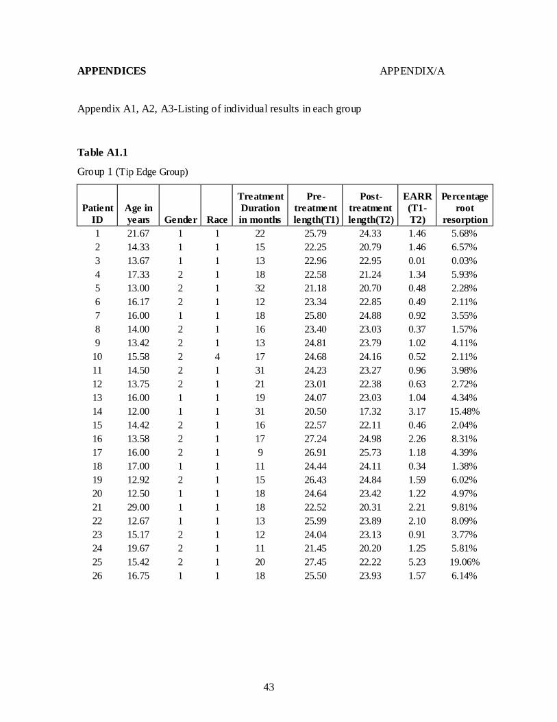
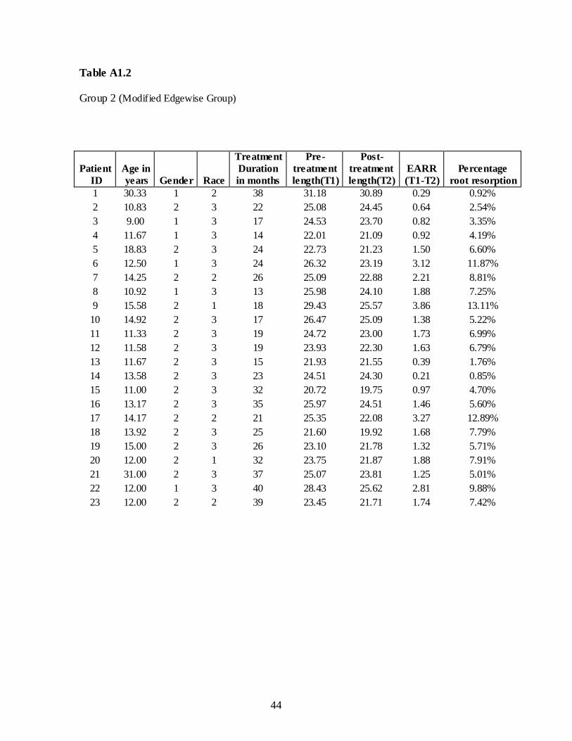
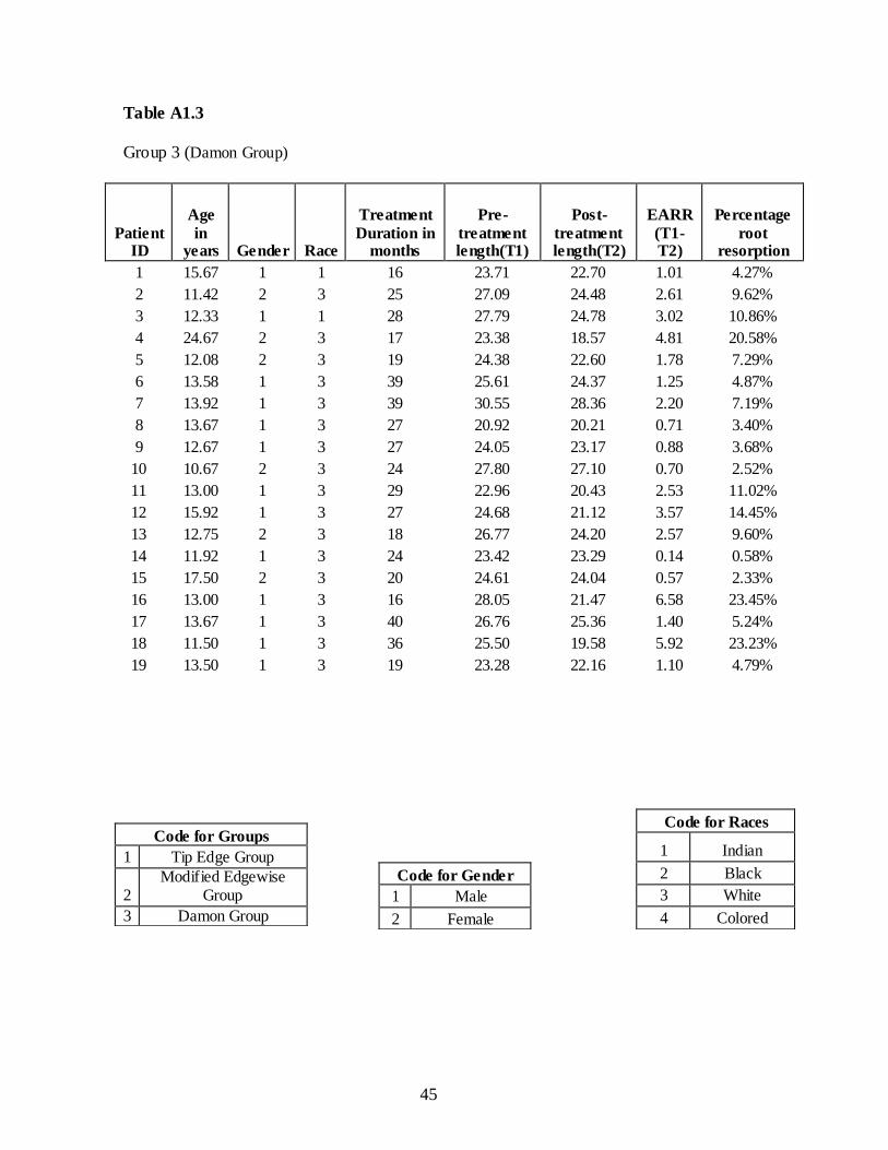
treatment lateral cephalograms. A listing of individual results in each group is given in
Appendix A (page 43).
The maxillary central incisor was used as the reference test tooth because:
a) This tooth has been shown to have the greatest frequency of external apical root
resorption (Copeland and Green, 1986; Goldson and Hendrikson, 1975)
b) It may be readily visualized on lateral cephalograms (Parker and Harris, 1998)
3.2 Calibration of x-rays
A standard method of calibration was devised to compensate for the disparate effects of
magnification produced by the three different radiology units. A 12mm thick Perspex sheet of
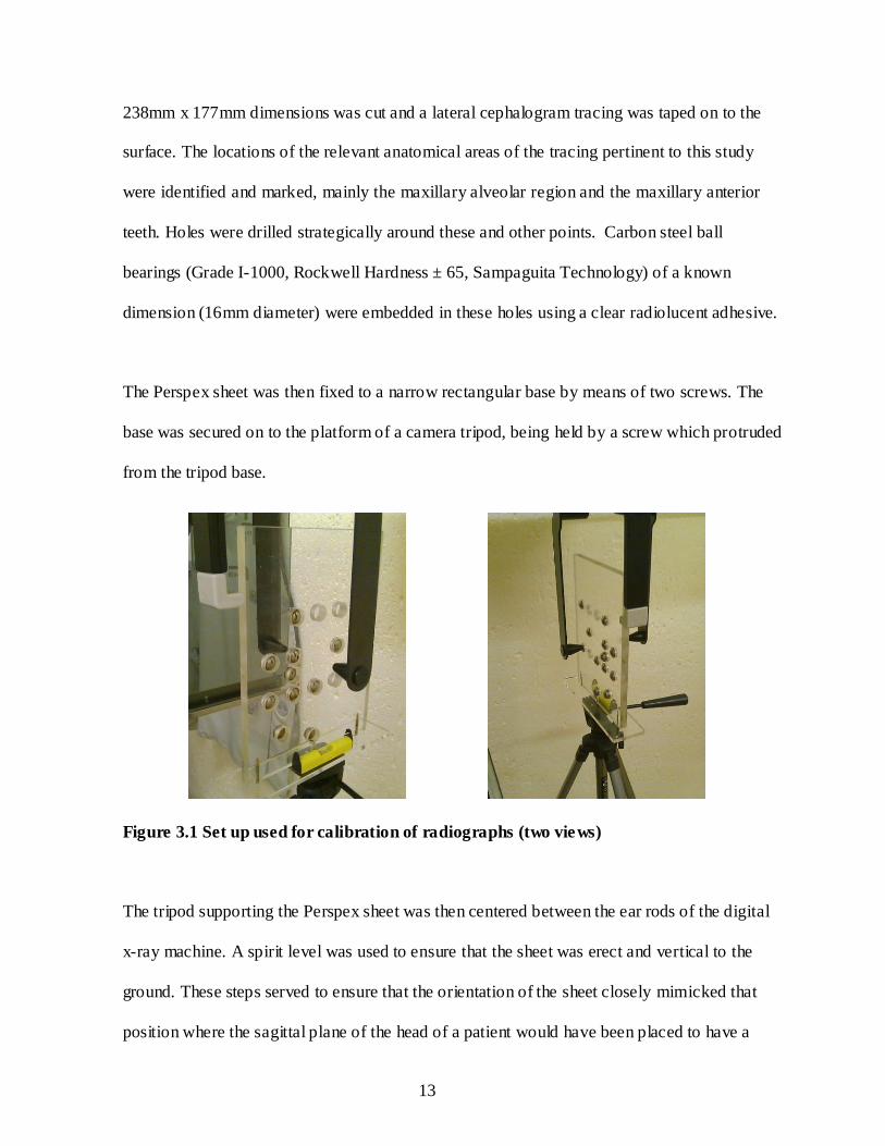
13
238mm x 177mm dimensions was cut and a lateral cephalogram tracing was taped on to the
surface. The locations of the relevant anatomical areas of the tracing pertinent to this study
were identified and marked, mainly the maxillary alveolar region and the maxillary anterior
teeth. Holes were drilled strategically around these and other points. Carbon steel ball
bearings (Grade I-1000, Rockwell Hardness ± 65, Sampaguita Technology) of a known
dimension (16mm diameter) were embedded in these holes using a clear radiolucent adhesive.
The Perspex sheet was then fixed to a narrow rectangular base by means of two screws. The
base was secured on to the platform of a camera tripod, being held by a screw which protruded
from the tripod base.
Figure 3.1 Set up used for calibration of radiographs (two views)
The tripod supporting the Perspex sheet was then centered between the ear rods of the digital
x-ray machine. A spirit level was used to ensure that the sheet was erect and vertical to the
ground. These steps served to ensure that the orientation of the sheet closely mimicked that
position where the sagittal plane of the head of a patient would have been placed to have a
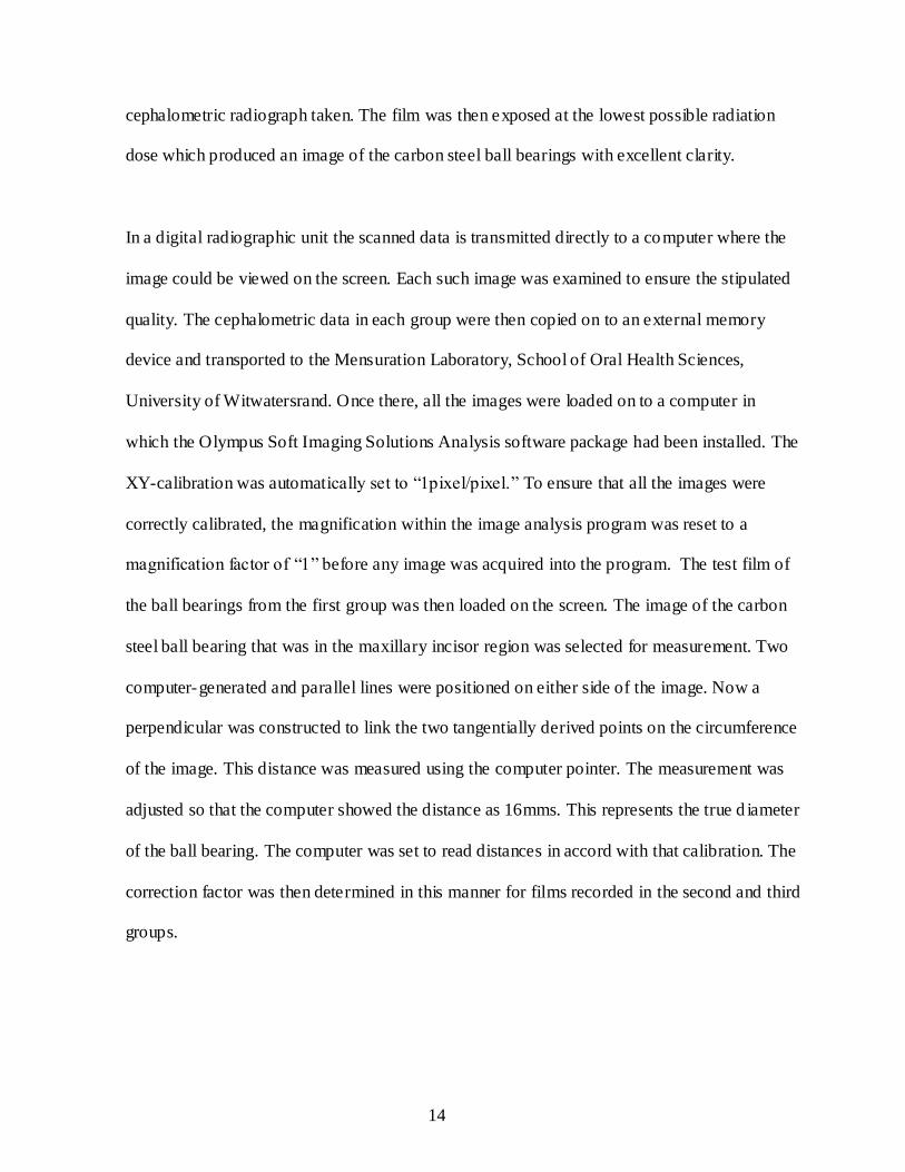
14
cephalometric radiograph taken. The film was then exposed at the lowest possible radiation
dose which produced an image of the carbon steel ball bearings with excellent clarity.
In a digital radiographic unit the scanned data is transmitted directly to a computer where the
image could be viewed on the screen. Each such image was examined to ensure the stipulated
quality. The cephalometric data in each group were then copied on to an external memory
device and transported to the Mensuration Laboratory, School of Oral Health Sciences,
University of Witwatersrand. Once there, all the images were loaded on to a computer in
which the Olympus Soft Imaging Solutions Analysis software package had been installed. The
XY-calibration was automatically set to “1pixel/pixel.” To ensure that all the images were
correctly calibrated, the magnification within the image analysis program was reset to a
magnification factor of “1” before any image was acquired into the program. The test film of
the ball bearings from the first group was then loaded on the screen. The image of the carbon
steel ball bearing that was in the maxillary incisor region was selected for measurement. Two
computer-generated and parallel lines were positioned on either side of the image. Now a
perpendicular was constructed to link the two tangentially derived points on the circumference
of the image. This distance was measured using the computer pointer. The measurement was
adjusted so that the computer showed the distance as 16mms. This represents the true d iameter
of the ball bearing. The computer was set to read distances in accord with that calibration. The
correction factor was then determined in this manner for films recorded in the second and third
groups.
15
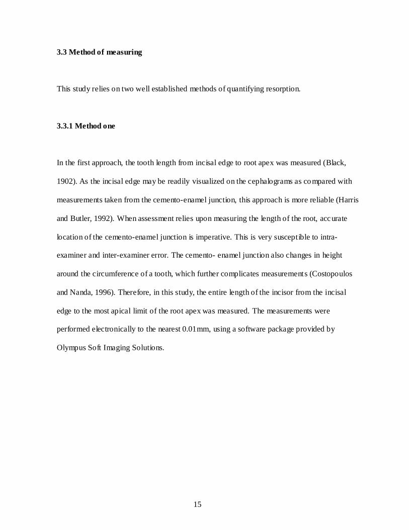
3.3 Method of measuring
This study relies on two well established methods of quantifying resorption.
3.3.1 Method one
In the first approach, the tooth length from incisal edge to root apex was measured (Black,
1902). As the incisal edge may be readily visualized on the cephalograms as compared with
measurements taken from the cemento-enamel junction, this approach is more reliable (Harris
and Butler, 1992). When assessment relies upon measuring the length of the root, accurate
location of the cemento-enamel junction is imperative. This is very susceptible to intra-
examiner and inter-examiner error. The cemento- enamel junction also changes in height
around the circumference of a tooth, which further complicates measurements (Costopoulos
and Nanda, 1996). Therefore, in this study, the entire length of the incisor from the incisal
edge to the most apical limit of the root apex was measured. The measurements were
performed electronically to the nearest 0.01mm, using a software package provided by
Olympus Soft Imaging Solutions.
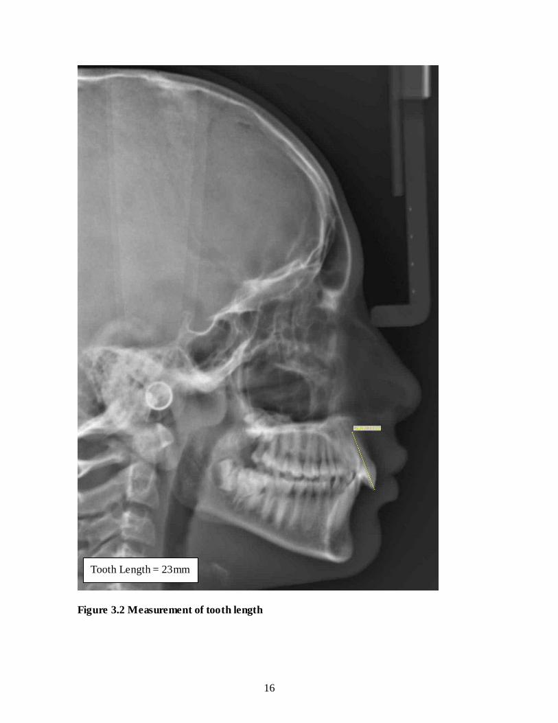
16
Figure 3.2 Measurement of tooth length
Tooth Length = 23mm
17
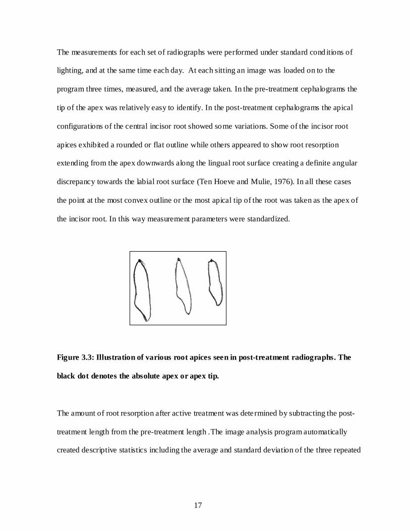
The measurements for each set of radiographs were performed under standard cond itions of
lighting, and at the same time each day. At each sitting an image was loaded on to the
program three times, measured, and the average taken. In the pre-treatment cephalograms the
tip of the apex was relatively easy to identify. In the post-treatment cephalograms the apical
configurations of the central incisor root showed some variations. Some of the incisor root
apices exhibited a rounded or flat outline while others appeared to show root resorption
extending from the apex downwards along the lingual root surface creating a definite angular
discrepancy towards the labial root surface (Ten Hoeve and Mulie, 1976). In all these cases
the point at the most convex outline or the most apical tip of the root was taken as the apex of
the incisor root. In this way measurement parameters were standardized.
Figure 3.3: Illustration of various root apices seen in post-treatment radiographs. The
black dot denotes the absolute apex or apex tip.
The amount of root resorption after active treatment was determined by subtracting the post-
treatment length from the pre-treatment length .The image analysis program automatically
created descriptive statistics including the average and standard deviation of the three repeated
18
measurements. These descriptive statistical data together with the raw data were then exported
on to Microsoft Excel spreadsheets and tabulated for further statistical analysis.
External Apical root resorption (EARR) = T1 - T2
Where T1 = Tooth length before treatment
T2 = Tooth length after treatment.
Root resorption was recorded as actual millimeter loss of tooth length. Percentage shortening
for each tooth was also calculated:
Percentage Resorption per tooth = EARR x 100 T1
3.3.2 Method two
In the second method the occurrence and degree of external root resorption at the end of the
active phase of orthodontic treatment was assessed using the five grade ord inal scale of
Levander and Malmgren (1988)
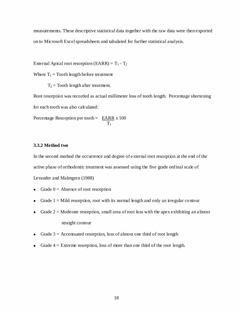
Grade 0 = Absence of root resorption
Grade 1 = Mild resorption, root with its normal length and only an irregular contour
Grade 2 = Moderate resorption, small area of root loss with the apex exhibiting an a lmost
straight contour
Grade 3 = Accentuated resorption, loss of almost one third of root length
Grade 4 = Extreme resorption, loss of more than one third of the root length.
19
Figure 3.4 Illustration of the five ordinal grades of root resorption
Each set of pre- and post- treatment cephalograms were analysed visually and the ordinal
grade of root resorption were noted.
3.4 Statistics
3.4.1 Error of the method: method one
To test the error involved in the measuring technique, ten cephalograms were selected
randomly and the measurements repeated ten times under the same conditions over ten
consecutive days (10x10=100 measurements). The results were subjected to a statistical
evaluation of intra-class correlation. The inter-operator error was assessed by comparing the
results recorded by the investigator with those recorded by an experienced orthodontist under
the same conditions on a random sample of fifteen cases. Statistical analysis was done to test
the inter-class coefficient.
3.4.2 Statistical analysis: method one
The incidence and severity of apical root resorption occurring after active treatment by three
different orthodontic techniques were compared statistically. Statistical significance was set at
20
P< 0.05. The influence of various confounders like technique, pre-treatment incisor length,
age, gender, race and duration of treatment were also explored statistically. One or more of
these confounders in various permutations were included in an analysis of covariance
(ANCOVA).This gave rise to the final analysis where it was necessary to adjust for baseline
length (pre-treatment incisor length) only. A nonparametric approach by doing an analysis of
covariance for ranks was also done.
3.4.3 Error of the method: method two
Intra -operator error was assessed by randomly selecting fifteen cases and grading them again.
3.4.4 Statistical analysis: method two
Visual assessment of pre- and post-treatment cephalograms to grade the observed root
resorption observed showed that the majority of cases in the study sample experienced Grade
1 and Grade 2 resorption. There was also a significant linear trend (p=0.018) over grades 0-3.
3.4.5 Comparative statistics
Statistical analysis was also done to discover whether there was any association between
actual millimeter loss of tooth length recorded and the visual assessment of grades, using a one
way ANOVA analysis.
21
CHAPTER 4: RESULTS
The primary concern of this study is to compare the incidence and severity of apical root
resorption occurring on the upper incisor during the course of class II correction by three
different orthodontic techniques. A listing of individual results recorded for each technique is
given in Appendix A (page 43).
4. 1 Descriptive statistics of the sample
The following table illustrates in each group, the mean age of the subjects and the standard
deviation of the same variable (age). Groups do not differ significantly with respect to this
variable (p=0.349)
Table 4.1: Mean age distribution by groups
* Note: group 1= Tip Edge, group 2= Modified Edgewise, group 3= Damon
Table 4.2 demonstrates the means and standard deviations of the variable “duration of
treatment” in each group. The average treatment times in each group as well as the standard
deviations also have been recorded. It can be seen that the longest mean treatment time is in
group three and the shortest mean treatment time is recorded in group one. Groups differ
significantly with respect to treatment time (p=0.0004). Treatment duration in group 2 and
group 3 were significantly longer than group 1.
Group Mean SD
Group 1 15.635 3.510
Group 2 14.402 5.528
Group 3 13.965 3.100
22
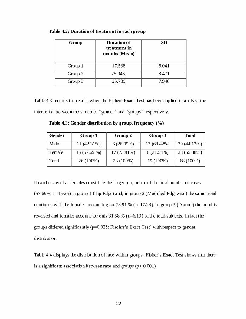
Table 4.2: Duration of treatment in each group
Group Duration of
treatment in
months (Mean)
SD
Group 1 17.538 6.041
Group 2 25.043. 8.471
Group 3 25.789 7.948
Table 4.3 records the results when the Fishers Exact Test has been applied to analyze the
interaction between the variables “gender” and “groups” respectively.
Table 4.3: Gender distribution by group, frequency (%)
It can be seen that females constitute the larger proportion of the total number of cases
(57.69%, n=15/26) in group 1 (Tip Edge) and, in group 2 (Modified Edgewise) the same trend
continues with the females accounting for 73.91 % (n=17/23). In group 3 (Damon) the trend is
reversed and females account for only 31.58 % (n=6/19) of the total subjects. In fact the
groups differed significantly (p=0.025; Fischer’s Exact Test) with respect to gender
distribution.
Table 4.4 displays the distribution of race within groups. Fisher’s Exact Test shows that there
is a significant association between race and groups (p< 0.001).
Gender Group 1 Group 2 Group 3 Total
Male 11 (42.31%) 6 (26.09%) 13 (68.42%) 30 (44.12%)
Female 15 (57.69 %) 17 (73.91%) 6 (31.58%) 38 (55.88%)
Total 26 (100%) 23 (100%) 19 (100%) 68 (100%)
23
Table 4.4: Race distribution by group
Race Group 1 Group 2 Group 3 Total
Indian (Code 1) 25 (96.15%) 2 (8.70%) 2 (10.53%) 29 (42.65%)
Black (Code 2) 0 (0.00%) 4 (17.39%) 0 (0.00%) 4 (5.88%)
White (Code 3) 0 (0.00%) 17 (73.91%) 17 (89.47%) 34 (50.00%)
Coloured (Code 4) 1 (3.85%) 0 (0.00%) 0 (0.00%) 1 (1.47%)
Total 26 (100%) 23 (100%) 19 (100%) 68 (100%)
The following codes were used to identify the racial background of the patients in the study:
code 1=Indian code 2=Black code 3=White code 4=Coloured.
Table 4.4 shows that the total number of white patients across groups is thirty-four (50%).
Indians account for twenty-nine cases (42.65%) while Black and Coloured races account for
four (5.885%) and one (1.47%) respectively. Since the total number of Blacks (4) and
Coloured (1) was small it was decided on statistical advice to combine the Indians, Blacks and
the Coloured people together for statistical purposes and then to compare this newly formed
sample group (code 5) with the Whites (code 3) by groups.
Table 4.5: New racial sample distribution by groups
Code Group 1 Group 2 Group 3 Total
5 26 (100%) 6 (26.09%) 2 (10.53%) 34 (50.00%)
3 0 (0.00%) 17 (73.91%) 17 (89.47%) 34 (50.00%)
Total 26 (100%) 23 (100%) 19 (100%) 68 (100%)
Groups differ significantly (p<0.001; Fisher’s Exact Test) with respect to race distribution.
Group 1 (Tip Edge group) differs from the other two groups in that the sample drawn from it
did not include any white patients.
24
4.2 Error of the method
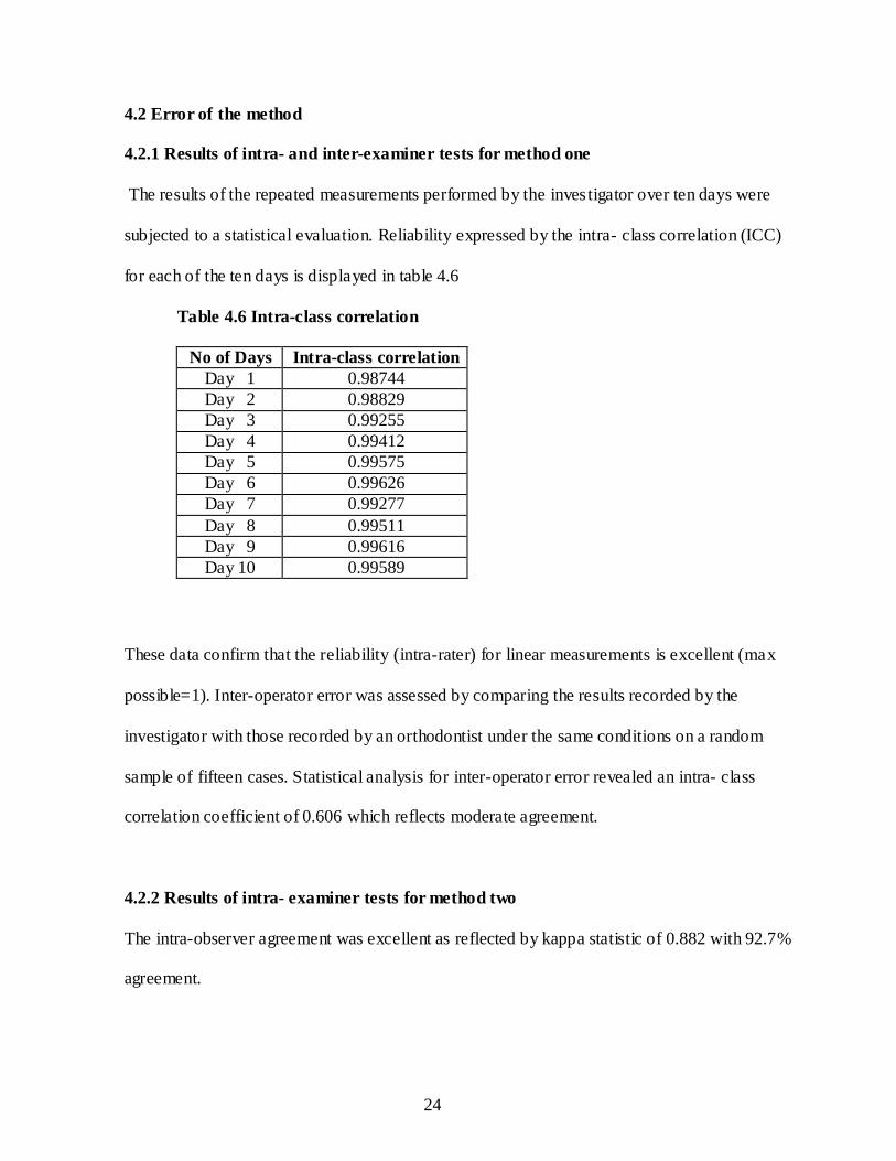
4.2.1 Results of intra- and inter-examiner tests for method one
The results of the repeated measurements performed by the investigator over ten days were
subjected to a statistical evaluation. Reliability expressed by the intra- class correlation (ICC)
for each of the ten days is displayed in table 4.6
Table 4.6 Intra-class correlation
No of Days Intra-class correlation
Day 1 0.98744
Day 2 0.98829
Day 3 0.99255
Day 4 0.99412
Day 5 0.99575
Day 6 0.99626
Day 7 0.99277
Day 8 0.99511
Day 9 0.99616
Day 10 0.99589
These data confirm that the reliability (intra-rater) for linear measurements is excellent (max
possible=1). Inter-operator error was assessed by comparing the results recorded by the
investigator with those recorded by an orthodontist under the same conditions on a random
sample of fifteen cases. Statistical analysis for inter-operator error revealed an intra- class
correlation coefficient of 0.606 which reflects moderate agreement.
4.2.2 Results of intra- examiner tests for method two
The intra-observer agreement was excellent as reflected by kappa statistic of 0.882 with 92.7%
agreement.
25
4.3 Data on assessment of resorption
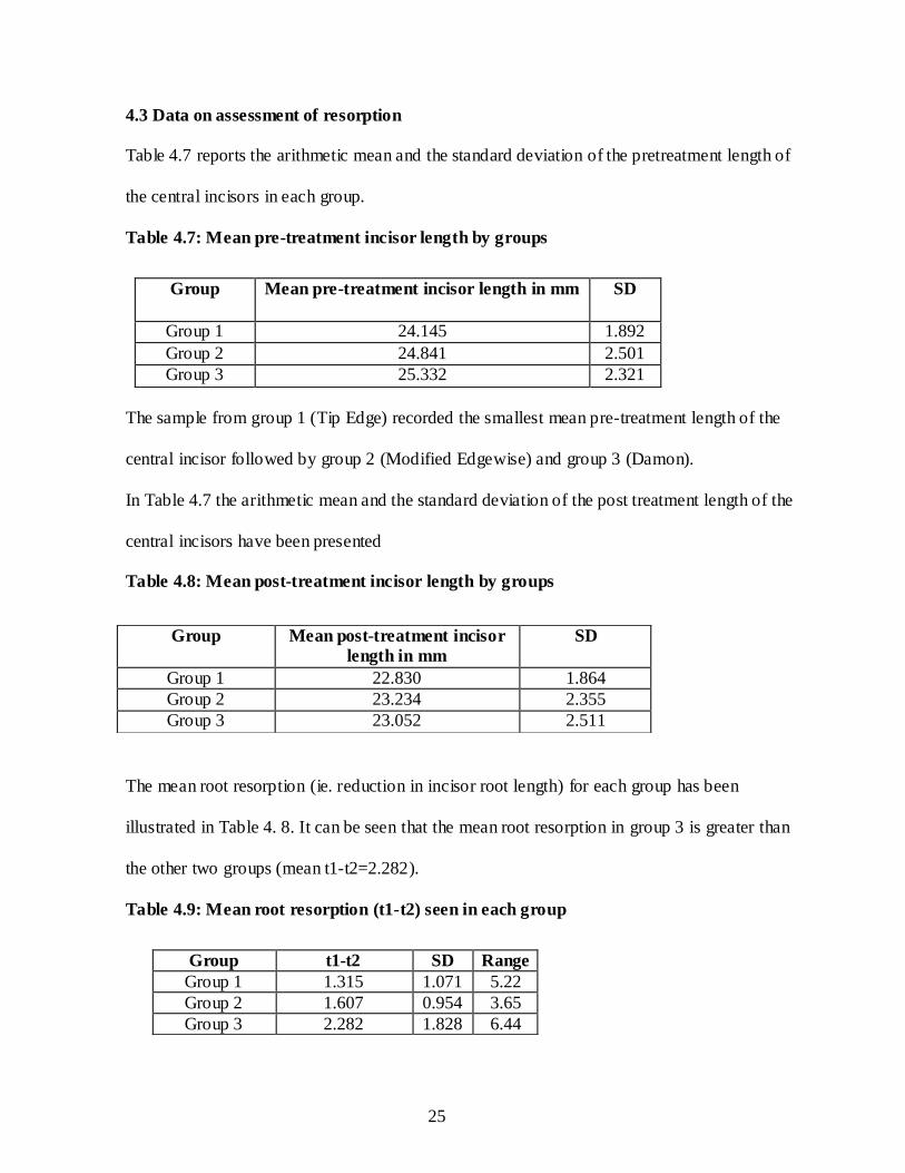
Table 4.7 reports the arithmetic mean and the standard deviation of the pretreatment length of
the central incisors in each group.
Table 4.7: Mean pre-treatment incisor length by groups
The sample from group 1 (Tip Edge) recorded the smallest mean pre-treatment length of the
central incisor followed by group 2 (Modified Edgewise) and group 3 (Damon).
In Table 4.7 the arithmetic mean and the standard deviation of the post treatment length of the
central incisors have been presented
Table 4.8: Mean post-treatment incisor length by groups
The mean root resorption (ie. reduction in incisor root length) for each group has been
illustrated in Table 4. 8. It can be seen that the mean root resorption in group 3 is greater than
the other two groups (mean t1-t2=2.282).
Table 4.9: Mean root resorption (t1-t2) seen in each group
Group Mean pre-treatment incisor length in mm SD
Group 1 24.145 1.892
Group 2 24.841 2.501
Group 3 25.332 2.321
Group Mean post-treatment incisor
length in mm
SD
Group 1 22.830 1.864
Group 2 23.234 2.355
Group 3 23.052 2.511
Group t1-t2 SD Range
Group 1 1.315 1.071 5.22
Group 2 1.607 0.954 3.65
Group 3 2.282 1.828 6.44
26
4.4 Comparative statistics between groups
When comparing groups with respect to mean root resorption a significant difference was
found (p = 0.051; group 1 = 1.32 vs. group 2 = 1.61 vs. group 3 = 2.28), in particular the
amount of root resorption seen for group 3 is significantly higher than that for group 1.
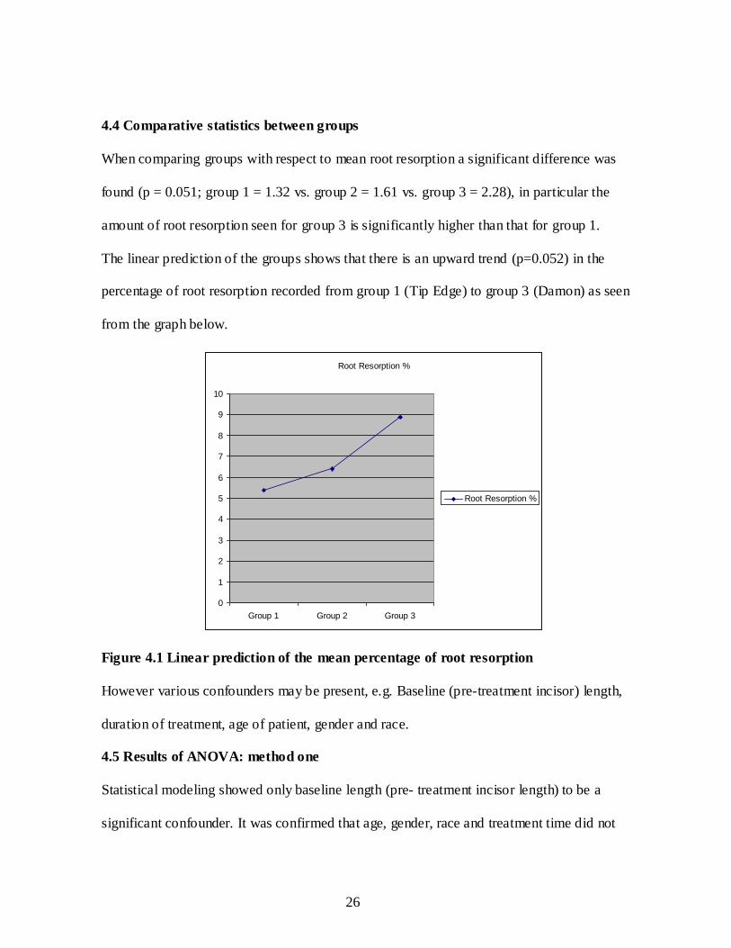
The linear prediction of the groups shows that there is an upward trend (p=0.052) in the
percentage of root resorption recorded from group 1 (Tip Edge) to group 3 (Damon) as seen
from the graph below.
Root Resorption %
0
1
2
3
4
5
6
7
8
9
10
Group 1 Group 2 Group 3
Root Resorption %
Figure 4.1 Linear prediction of the mean percentage of root resorption
However various confounders may be present, e.g. Baseline (pre-treatment incisor) length,
duration of treatment, age of patient, gender and race.
4.5 Results of ANOVA: method one
Statistical modeling showed only baseline length (pre- treatment incisor length) to be a
significant confounder. It was confirmed that age, gender, race and treatment time did not
27
affect the quantum of root resorption seen after orthodontic treatment. In the final analysis,
having adjusted for baseline length, groups were found not to differ significantly in their
experience of root resorption (p=0.133; ANCOVA).
This result was also confirmed by following a nonparametric approach by doing an analysis of
covariance (ANCOVA) for rank, i.e. data was substituted with ranks, where p=0.268 for group
with p=0.006 for confounder, pre treatment incisor length.
Similarly, the percentage of root resorption observed after treatment did not differ
significantly between groups (p=0.067).
4.6 Results for method two
Root resorption for the whole sample was also analyzed using the five grade ordinal scale of
Levander and Malmgren(1988). The grades seen in each group after visually assessing and
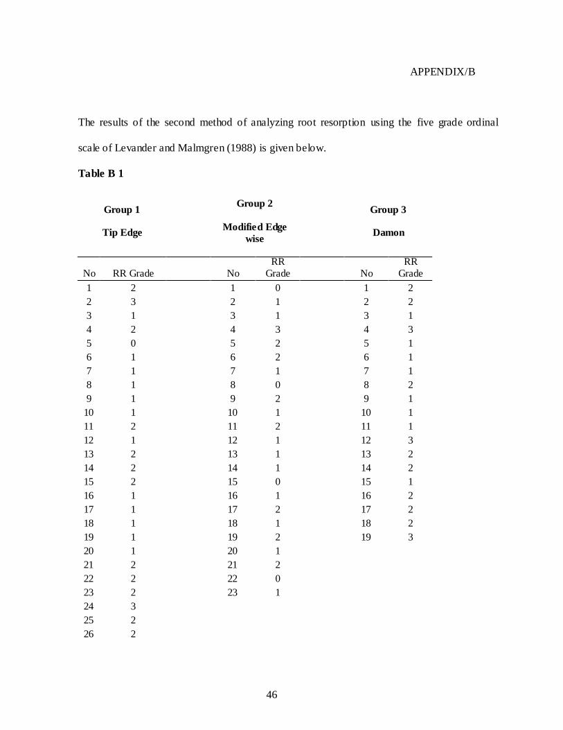
grading the radiographs using the ordinal data has been given in tabular form in appendix B
(page 46). It was found that the majority of the subjects (57/68) in this sample experienced
Grade 1 and Grade 2 resorption (See Table 4.10).
Table 4.10 Incidence and severity of resorption as assessed in method two
Grade Number of patients Mean t1-t2 SD (t1-t2)
0 5 1.286 1.050
1 34 1.402 0.959
2 23 1.847 1.482
3 6 2.988 2.093
28
4.7 Comparative statistics (method one vs. method two)
This was done to test whether there was any agreement between visual grading and assessment
of root resorption using actual measurements. The grades differed significantly with respect to
the amount of mean resorption (p=0.041). The ordinal data grades and the objective grades
(actual millimeter loss of tooth length) followed the same direction, i.e., as the grade increased
the amount of tooth material loss recorded also increased.
The statistics demonstrate a clear consensus between the increasing damage observed visually
and the reduced tooth lengths as measured on the cephalograms (p=0.018).
29
CHAPTER 5: DISCUSSION
The primary purpose of this retrospective study is to assess the occurrence of apical root
resorption experienced in association with orthodontic treatment undertaken with Tip Edge,
Damon and Modified Edgewise techniques. Secondly, amongst the many factors possibly
implicated in apical root resorption to specifically evaluate contributing factors such as:
Age
Gender
Ethnicity
Duration of treatment time.
The null hypothesis was that there would be no difference in the extent of apical root
resorption seen after orthodontic treatment for class II correction undertaken with Tip Edge,
Damon or Modified Edgewise techniques.
In this study of root resorption in a sample of cases treated by three different modalities of
orthodontic technique, it was found that resorption was a common, although not an
overwhelming complication. Quantification of the extent of resorption is more challenging
than may first be considered. Although various other records (clinical, histologic, biologic
markers) are available to assess the pre- and post-treatment status of the tooth roots,
radiographs remain the most popular tool. Radiology also offers a wide variety of formats -
periapical films, panoramic films, lateral cephalograms, digital radiographs and computerised
tomography. But there are limitations. In the first place resorption occurs not only at the very
30
apex but may also attack the surface of the root at any point in its circumference. Secondly
radiological assessment for comparative purposes must also be accurately repeatable.
Sameshima and Asgarifar (2001) compared the efficacy of periapical and panoramic films in
the assessment of apical root resorption. They recommended the use of periapical films.
However, these views may suffer projection errors and are not readily reproducible. This can
be overcome by using the long-cone periapical paralleling technique (Brezniak and
Wasserstein, 1993b), which reduces distortion and superimposition errors when compared
with panoramic radiographs and lateral cephalograms. Long- cone periapical radiographs are
not routinely amongst the standard records for orthodontic patients in private practices in
South Africa and hence the method has not been available for this retrospective study.
According to Pandis et al (2008) root resorption can be underestimated due to the inherent
inability of panoramic radiographs to show loss of tooth structure in a facial direction.
Computerised tomography on the other extreme is highly sensitive to site specific evaluation
of root resorption (mesial, distal, buccal or lingual). The main drawbacks of this excellent
diagnostic technology are its high cost and the need for special equipment (Krishnan, 2005).
The sensitivity of digital radiographs, according to Levander, Bajka and Malmgren (1998),
was comparable to or better than conventional film- based radiographs. In addition the
radiation exposure is much less than that delivered by conventional x-rays.
In this retrospective study digital lateral cephalograms were used as they are an integral part of
standard diagnostic records in private practices in South Africa. These films also provide a
convenient image of the specific tooth (central incisor) selected for assessment of root
resorption. Several authors have also used lateral cephalograms for assessment of resorption
31
due to the high degree of reproducibility the technique offers (Costopoulos and Nanda, 1996;
Harris, Kineret and Tolley, 1997; Horiuchi, Hotokezaka and Kobayashi, 1998).
.
Different teeth have different tendencies to root resorption. It is claimed that if there is no
evidence of apical root resorption in maxillary and mandibular incisors then significant apical
resorption in other teeth is less likely to occur (Copeland and Green, 1986; Sjolien and
Zachrisson, 1973). In the maxillary dentition, the incisors are the teeth most affected by root
resorption (Brezniak and Wasserstein, 1993a). Upper central incisors showed more root
resorption than the upper lateral incisors (Janson et al, 1999). In general the extent of
movement experienced by these teeth is usually greater due to the demands of esthetics, nature
of malocclusion and function. In addition Oppenheim (1942) suggested that the morphology
of roots of the incisors served as a catalyst in tooth resorption. The conical shapes of these
roots contribute to a rapid diminution in the cross-sectional area towards the apex. Hence axial
components of force exert relatively more force per unit area probably leading to a higher
degree of resorption at the root apex.
Two methods have been generally used to quantify resorption namely: visually assessed
grades of resorption (ordinal scale data) and measurements with calipers or some computer
based software program. In this study both methods have been used to quantify resorption. In
the visually assessed method, the scoring criteria of Levander and Malmgren (1988) have
provided the grades (method two). Previously this ordinal scale of Levander and Malmgren
(1988) has been used almost exclusively to assess root resorption evident on inspection of
periapicals and orthopantomograms. In the current study it was used to assess root resorption
as demonstrated on lateral cephalograms, in accord with the protocol followed by Harris and
32
Butler (1992). Lateral cephalograms reveal resorption on the apical, facial and lingual surfaces
of the incisor root as opposed to periapicals and orthopantomograms which reveal only the
apical, mesial and distal surfaces. Maxillary incisor teeth generally experience more resorption
on the lingual surface, especially during torquing. In the post-treatment cephalograms some of
the incisor root apices exhibited a rounded or flat outline while others appeared to show root
resorption extending from the apex downwards along the lingual root surface creating a
definite angular discrepancy towards the labial root surface (Ten Hoeve and Mulie, 1976).
This effect tends to be masked in the anterio-posterior view shown in panoramic radiographs.
Hence it is believed that visual assessment of the images of the incisors on cephalograms will
offer a more accurate grading of root resorption levels as opposed to panoramic films. Direct
measurement of the teeth on the radiographs offers an attractive alternate approach. S tatistical
evaluation tested the extent of agreement in this study between visual measurements and
actual measurements and demonstrated a significant association (p=0.018) between the
methods.
The literature review suggests that in the quantitative assessment of root resorption, either the
tooth length (incisal edge to root apex) or root length (cemento-enamel junction to root apex)
should be measured on pre-treatment and post-treatment radiographs and the differences
compared to evaluate any root resorption which has occurred. In this study, the tooth length
was measured as the distance from the incisal edge to the apex of the root (Black, 1902). The
most apical limit of the root was taken as the absolute apex of the tooth especially in cases that
exhibited an angular or jagged apical outline. This took into account any viable living root
material and the periodontal support system encompassing it that was present at the apical
limit. Since the right and left incisor teeth are usually moved in tandem it was assumed that the
33
differences arising from a right or left side overlap of central incisor teeth for pre- and post-
treatment cephalograms would be minimal. The image of the most procumbent central incisor
was measured on the pre-treatment and post-treatment radiographs. The images were
magnified by a zoom factor of 100% to facilitate the evaluation of the apex. Any pre-treatment
x-ray that showed incomplete root formation or an indistinct apex had been discarded from the
study at the outset. The measurements were performed to the nearest 0.01mm, using a
software package produced by Olympus Soft Imaging Solutions GmbH.
Statistically significant differences between the means were found when the data from the
three groups (Tip Edge, Modified Edgewise, Damon) were compared at a p value of 0.051.
The differences were especially significant between group 1 (Tip Edge) and group 3 (Damon).
The mean percentage root resorption between groups was also calculated. The lowest extent
was in group 1 (Tip Edge) followed by group 2 (Modified Edgewise) and group 3 (Damon),
which differed significantly (p=0.006) from group 1 (Tip Edge). However various
confounders were present, e.g. baseline (pre-treatment) incisor length, duration of treatment,
age of patient, gender and race.
In agreement with Mirabella and Artun (1995) and Sameshima and Sinclair, (2001a) a positive
association was found between initial tooth length and the amount of root resorption observed.
Their studies report that longer roots are more prone to resorption than shorter roots. A simple
explanation might be that longer roots require greater displacement than shorter roots to
produce an equal amount of torque expression.
34
Parker and Harris (1998) confirmed a definite sexual dimorphism for the length of the
maxillary central incisor. On an average the central inc isor was found to be longer by one
millimetre in male subjects. Group 3 (Damon) recorded the longest pre-treatment incisor
length in this study although the data was not statistically significant. This might be due to the
fact that the percentage of males in the group 3 (68.42%) was significantly higher (p=0.025;
Fischer’s Exact Test) than in either group 1 (42.31%) or group 2 (26.09%). However, when
the data was adjusted for pre-treatment incisor length there was no significant difference
(p=0.516) between genders with respect to the amount of root resorption. These findings are in
agreement with other workers who found no significant differences in root resorption between
male and female patients (McFadden, Engstrom, Engstrom, Anholm (1989); Mirabella and
Artun (1995); Sameshima and Sinclair (2001a).
The four subjects with the greatest amount of root resorption (>3.5mm) in group 3 were of
Caucasian origin. The relationship between ethnicity and root resorption after orthodontic
treatment was evaluated by Sameshima and Sinclair (2001a). They found that Asian patients
had significantly less resorption than did White or Hispanic patients. This might explain the
upward linear trend of the incidence of root resorption seen from group 1 (Tip Edge) with a
dominant Indian population (96.15%) to group 2 (Modified Edgewise) with 73.91% of White
patients to group 3 (Damon) with 89.94% of White patients. This may have accounted for the
low mean resorption percentage (5.394%) seen in group 1 when compared with group 3
(8.893%). However race was not shown to be a significant confounder to the mean resorption
seen after orthodontic treatment in this study although it must be acknowledged that sample
sizes may be too small to draw firm conclusions.
35
The length of treatment time and root resorption has been shown in many studies to have a
positive correlation (McFadden et al, 1989; Brezniak and Wasserstein, 1993b; Sameshima and
Sinclair, 2001b). In particular there is a significant association between longer treatment
duration and increased root resorption of maxillary incisors (Sameshima and Sinclair, 2001b).
Other studies report that treatment duration does not affect the amount of root resorption seen
after orthodontic treatment (Beck and Harris, 1994; Mirabella and Artun, 1995). Although
treatment duration is shorter (17.54 months) in group 1 (Tip Edge) when compared with 25.04
months in group 2 (Modified Edgewise) and 25.79 months in group 3 (Damon), statistically
significant correlation was not found between treatment time and the extent of root resorption
in the current study. The overall correlation between these factors was 0.136 (p=0.270).The
correlation coefficient for these two factors in group 1(Tip Edge) was 0.223 (p=0.274), in
group 2 (Modified Edgewise) was 0.002 (p=0.993) and in group 3 (Damon) was 0.061
(p=0.803) respectively. Likewise, Dermaut and De Munck (1986) and Levander and
Malmgren (1988) found weak and non-significant correlations between treatment time and
root resorption.
The risk of resorption seems to be independent of age once root formation is complete.
However, adults are speculated to be at a higher risk for root resorption as the rate of alveolar
turnover is slower in adults than in children and young adolescents. Tooth movement is also
slower in adults (Harris, 2000). Yet, the age of the patient and the incidence of root resorption
were reported to be poorly correlated in a study conducted by Beck and Harris (1994). No
significant association was found between the patient age and the amount of root resorption
36
observed in the three groups in the present study. This might be due to the fact that root
formation was complete for all the patients at the outset of treatment in this sample.
The effect of age is quite different when dealing with children in the mixed dentition. Children
treated before their roots are completely formed exhibited less active root resorption, than did
their older counterparts. It has been suggested that in fact orthodontic treatment actually
slowed and reduced the root growth leaving the roots with relatively shorter final root lengths
(Linge and Linge, 1983; Ogaard, 1988).
Therefore, it may be safely concluded that the results of the present study demonstrate that
gender, race, treatment time and age do not have a discernible influence on the amount of root
resorption seen after orthodontic treatment in the samples investigated. Following a statistical
modeling only baseline length (pre-treatment incisor length) was a significant confounder.
Once having adjusted for baseline length, and using an analysis of covariance, groups were
found to not differ significantly (p=0.133), a result confirmed when a nonparametric approach
was followed. It was also seen that the groups do not differ significantly (p=0.067) with regard
to the percentage of root resorption seen after orthodontic treatment.
However, irrespective of the technique employed, every case that had undergone orthodontic
treatment in this study exhibited some root resorption. In group 1 actual tooth material loss
ranged from 0.01 to 5.23mm. In group 2 it ranged from 0.21mm to 3.86mm and in group 3
from 0.57 mm to 6.58mm. In all three groups some cases exhibited a higher degree of
resorption, possibly attributable to individual susceptibility and/or genetic influence. A study
conducted by Al-Qawasmi, Hartsfield, Everett, Flury, Liu, Foroud, Macri and Roberts (2003)
37
found that orthodontic patients homozygous for the IL-IB allele-1 polymorphism have a 5.6-
fold increase in external apical root resorption over non IL-IB homozygous patients. They
concluded that this gene could account for up to 15% of maxillary incisor root resorption. This
possibility was not explored in the current study.
What is the long term prognosis of cases where root resorption has been identified? This
question is often posed to orthodontists by general dentists and other dental specialists when
post-orthodontic patients approach them for restorative rehabilitation. It is accepted that any
root resorption ceases after orthodontic treatment has been stopped. Over a period of time the
root surface becomes smoother due to the deposition of reparative cellular cementum. The root
length does not shorten any further (Krishnan, 2005). Parker (1997) conducted a long term
follow up on a case that had exhibited root resorption. He found that even after twenty five
years the severely resorbed maxillary incisors were still present and functioning well. Kokich
(2008) believes that the need for a permanent lingual splint for teeth with moderate to severe
resorption is dictated by the presence of para- functional habits, crown mobility and need for
any restorations. In cases that demonstrated moderate to severe root resorption orthodontic re-
treatment did not produce any further resorption. Kokich (2008) suggests that the presence of
reparative cellular cementum may have played a protective role in inhibiting further root
resorption during retreatment.
There was an upward trend (p=0.052) in the expression of root resorption from group 1 (Tip
Edge) to group 3 (Damon). A similar trend indicating more external apical root resorption
(EARR) for Damon 2 systems when compared with conventional Edgewise systems was
reported by Pandis et al in 2008. Since the data did not reach significance (p=0.06) it was
38
concluded that no difference should be expected for root resorption between cases treated by
conventional Edgewise and Damon appliances.
Light forces are the key to the Damon system. It advocates the use of low forces generated by
small dimension “high tech” arch wires to bring about tooth movement. In contrast, the use of
light and constant forces has been linked with higher degrees of root resorption. Weiland
(2003) explained that the mode of persistent activation of nickel titanium wires originating
from their increased work range relative to stainless steel wires might be responsible for the
significantly larger resorption seen when these wires are used in treatment of malocclusions.
Stainless steel wires generate a rapidly declining force during de-activation whereas super
elastic wires deliver a constant force over an extended portion of the deactivation range
(Miura, Mogi, Ohura and Hamanaka, 1986). The type and level of force can also influence the
development of root resorption in orthodontic patients. Reitan (1985) advocated the use of
intermittent forces to prevent the development of root resorption. The pause in treatment with
intermittent forces where little or no force is applied allows the resorbed cementum some time
to heal. Discontinuous force application seems to be a more favorable mode of force delivery
in achieving tooth movement as it causes less vertical root resorption (Kocaaga, Canyurek,
Acar and Erverdi, 1997). In this context, it could be postulated that the temporal
characteristics of force application to teeth are more important than force magnitude per se in
modulating root resorption.
Damon (1998) believes that tooth movement is more efficient when the teeth are allowed to
move individually and that passive self- ligating brackets offer more freedom for each tooth to
move to their individual natural positions even though they are still interconnected. The arch
39
wire is never actively tied in to the bracket slot. The final position of the teeth in the arch is
determined by interplay between the oral musculature and the periodontal forces and not by
heavy orthodontic forces. Does this make the teeth more susceptible to resorption by jiggling
forces from normal masticatory functions and speech as they are not firmly held in place by
heavy stabilizing stainless steel wires? Does this in turn prevent the natural repair process
inherent in every biological organ? This speculation is, however, almost contradictory to the
effect super elastic wires have on tissues. Proffit and Fields (2000) also advocate caution when
using the new light force rectangular wires in the initial phase of treatment. Faltin et al (2001)
advocated a reduction in continuous force magnitude to preserve the integrity of the tooth and
surrounding tissues.
Further investigations are required to expound the differences in root resorption observed with
different techniques that use different force systems and materials. The current study revealed
no significant difference in the root resorption seen after orthodontic treatment utilizing the
Tip Edge, Modified Edgewise and the Damon technique. It could not, however, statistically
pinpoint the causative factor for the upward trend in root resorption that was seen from group
1(Tip Edge) to group 3 (Damon). It might be speculated that the longer treatment time, racial
character of the patients and gender distribution may have resulted in group 3 (Damon) having
the highest degree of root resorption recorded.
Individual susceptibility or a heritable component to root resorption might be the final piece
that throws some light into the root resorption puzzle. More research is needed in this regard.
Detailed investigations that explore the connection between root resorption and treatment
techniques by studying the effect of variables like arch wire sequence, time spent in super
40
elastic wires, method of force application (continuous or intermittent), type of elastics used
(intra vs inter), type of bracket and ligation employed in each technique will help us to
understand which technique is kinder to the tissues involved.
5.1 Limitations and difficulties encountered in this study
There were several limitations to this study.
1. The retrospective nature of the study.
2. Lack of availability of peri-apical radiographs limited the control of variables exposed
using the long cone paralleling technique.
3. This investigation was limited to the assessment of apical resorption on the maxillary
incisors only. Although these teeth do in general show a higher degree of root resorption,
inferences on the overall severity of root resorption with different appliance systems would
require examination of the entire dentition.
4. It was assumed that differences arising from an overlap of right and left sides in
measurement of the central incisor teeth would be negligible.
5. Sometimes it was difficult to locate the apex accurately on the radiograph due to
superimposition of the images of the alveolar structures or the adjacent teeth. The
measurements were repeated three times and the average calculated in an attempt to reduce
any inconsistencies.
6. It was assumed that the incisal edge of the incisor in each case was not subject to the
effects of attrition or incisal wear and that the change in tooth length between pre and post
treatment cephalograms was entirely due to the effects of root resorption.

41
7. In grading the teeth using the ordinal scale of Levander and Malmgren (1988) it was
sometimes difficult to assess the amount of resorption due to the change in inclination of
the incisors in the post -treatment radiographs as well as due to the change in an anterior-
posterior direction. However, it was seen that the two methods of quantifying resorption
used in this study were comparable.
42
CHAPTER 6: CONCLUSIONS
The current study explored the variations in root resorption seen after orthodontic
treatment with Tip Edge, Modified Edgewise and Damon systems.
1. All cases exhibited some amount of resorption after orthodontic treatment illustrating
the fact that root resorption is a common phenomenon in this discipline.
2. The majority of patients (84%) in the overall sample exhibited Grade 1 or Grade 2
resorption and only a few patients were affected severely.
3. Tabulation of the results demonstrated considerable individual variation in the amount
of root resorption observed in all three groups. Individual variation in the biologic
response to orthodontic forces and/ or a genetic component may be the predisposing
factor responsible for the results seen.
4. There was a significant upward linear trend (p=0.022) from group 1 (Tip Edge) to
group 3 (Damon) in the raw data.
5. Statistical analysis revealed that age, gender and race of the patients involved in this
study and the duration of treatment did not influence the degree of root resorption
observed after treatment.
6. Pre- treatment incisor length was the only significant confounding variable in this
sample.
7. Once the baseline pre-treatment incisor length was adjusted this study could not find
any significant difference in the degree of root resorption observed after treatment
utilizing the three different techniques.
43
APPENDICES APPENDIX/A
Appendix A1, A2, A3-Listing of individual results in each group
Table A1.1
Group 1 (Tip Edge Group)
Patient
ID
Age in
years Gender Race
Treatment
Duration
in months
Pre-
treatment
length(T1)
Post-
treatment
length(T2)
EARR
(T1-
T2)
Percentage
root
resorption
1 21.67 1 1 22 25.79 24.33 1.46 5.68%
2 14.33 1 1 15 22.25 20.79 1.46 6.57%
3 13.67 1 1 13 22.96 22.95 0.01 0.03%
4 17.33 2 1 18 22.58 21.24 1.34 5.93%
5 13.00 2 1 32 21.18 20.70 0.48 2.28%
6 16.17 2 1 12 23.34 22.85 0.49 2.11%
7 16.00 1 1 18 25.80 24.88 0.92 3.55%
8 14.00 2 1 16 23.40 23.03 0.37 1.57%
9 13.42 2 1 13 24.81 23.79 1.02 4.11%
10 15.58 2 4 17 24.68 24.16 0.52 2.11%
11 14.50 2 1 31 24.23 23.27 0.96 3.98%
12 13.75 2 1 21 23.01 22.38 0.63 2.72%
13 16.00 1 1 19 24.07 23.03 1.04 4.34%
14 12.00 1 1 31 20.50 17.32 3.17 15.48%
15 14.42 2 1 16 22.57 22.11 0.46 2.04%
16 13.58 2 1 17 27.24 24.98 2.26 8.31%
17 16.00 2 1 9 26.91 25.73 1.18 4.39%
18 17.00 1 1 11 24.44 24.11 0.34 1.38%
19 12.92 2 1 15 26.43 24.84 1.59 6.02%
20 12.50 1 1 18 24.64 23.42 1.22 4.97%
21 29.00 1 1 18 22.52 20.31 2.21 9.81%
22 12.67 1 1 13 25.99 23.89 2.10 8.09%
23 15.17 2 1 12 24.04 23.13 0.91 3.77%
24 19.67 2 1 11 21.45 20.20 1.25 5.81%
25 15.42 2 1 20 27.45 22.22 5.23 19.06%
26 16.75 1 1 18 25.50 23.93 1.57 6.14%
44
Table A1.2
Group 2 (Modified Edgewise Group)
Patient
ID Age in
years Gender Race
Treatment
Duration
in months
Pre-
treatment
length(T1)
Post-
treatment
length(T2) EARR
(T1-T2) Percentage
root resorption
1 30.33 1 2 38 31.18 30.89 0.29 0.92%
2 10.83 2 3 22 25.08 24.45 0.64 2.54%
3 9.00 1 3 17 24.53 23.70 0.82 3.35%
4 11.67 1 3 14 22.01 21.09 0.92 4.19%
5 18.83 2 3 24 22.73 21.23 1.50 6.60%
6 12.50 1 3 24 26.32 23.19 3.12 11.87%
7 14.25 2 2 26 25.09 22.88 2.21 8.81%
8 10.92 1 3 13 25.98 24.10 1.88 7.25%
9 15.58 2 1 18 29.43 25.57 3.86 13.11%
10 14.92 2 3 17 26.47 25.09 1.38 5.22%
11 11.33 2 3 19 24.72 23.00 1.73 6.99%
12 11.58 2 3 19 23.93 22.30 1.63 6.79%
13 11.67 2 3 15 21.93 21.55 0.39 1.76%
14 13.58 2 3 23 24.51 24.30 0.21 0.85%
15 11.00 2 3 32 20.72 19.75 0.97 4.70%
16 13.17 2 3 35 25.97 24.51 1.46 5.60%
17 14.17 2 2 21 25.35 22.08 3.27 12.89%
18 13.92 2 3 25 21.60 19.92 1.68 7.79%
19 15.00 2 3 26 23.10 21.78 1.32 5.71%
20 12.00 2 1 32 23.75 21.87 1.88 7.91%
21 31.00 2 3 37 25.07 23.81 1.25 5.01%
22 12.00 1 3 40 28.43 25.62 2.81 9.88%
23 12.00 2 2 39 23.45 21.71 1.74 7.42%
45
Table A1.3
Group 3 (Damon Group)
Patient ID
Age
in years Gender Race
Treatment
Duration in months
Pre-
treatment length(T1)
Post-
treatment length(T2)
EARR
(T1-T2)
Percentage
root resorption
1 15.67 1 1 16 23.71 22.70 1.01 4.27%
2 11.42 2 3 25 27.09 24.48 2.61 9.62%
3 12.33 1 1 28 27.79 24.78 3.02 10.86%
4 24.67 2 3 17 23.38 18.57 4.81 20.58%
5 12.08 2 3 19 24.38 22.60 1.78 7.29%
6 13.58 1 3 39 25.61 24.37 1.25 4.87%
7 13.92 1 3 39 30.55 28.36 2.20 7.19%
8 13.67 1 3 27 20.92 20.21 0.71 3.40%
9 12.67 1 3 27 24.05 23.17 0.88 3.68%
10 10.67 2 3 24 27.80 27.10 0.70 2.52%
11 13.00 1 3 29 22.96 20.43 2.53 11.02%
12 15.92 1 3 27 24.68 21.12 3.57 14.45%
13 12.75 2 3 18 26.77 24.20 2.57 9.60%
14 11.92 1 3 24 23.42 23.29 0.14 0.58%
15 17.50 2 3 20 24.61 24.04 0.57 2.33%
16 13.00 1 3 16 28.05 21.47 6.58 23.45%
17 13.67 1 3 40 26.76 25.36 1.40 5.24%
18 11.50 1 3 36 25.50 19.58 5.92 23.23%
19 13.50 1 3 19 23.28 22.16 1.10 4.79%
Code for Gender
1 Male
2 Female
Code for Groups
1 Tip Edge Group
2 Modified Edgewise
Group
3 Damon Group
Code for Races
1 Indian
2 Black
3 White
4 Colored
46
APPENDIX/B
The results of the second method of analyzing root resorption using the five grade ordinal
scale of Levander and Malmgren (1988) is given below.
Table B 1
Group 1
Tip Edge
Group 2
Modified Edge
wise
Group 3
Damon
No RR Grade No RR
Grade No RR
Grade
1 2 1 0 1 2
2 3 2 1 2 2
3 1 3 1 3 1
4 2 4 3 4 3
5 0 5 2 5 1
6 1 6 2 6 1
7 1 7 1 7 1
8 1 8 0 8 2
9 1 9 2 9 1
10 1 10 1 10 1
11 2 11 2 11 1
12 1 12 1 12 3
13 2 13 1 13 2
14 2 14 1 14 2
15 2 15 0 15 1
16 1 16 1 16 2
17 1 17 2 17 2
18 1 18 1 18 2
19 1 19 2 19 3
20 1 20 1
21 2 21 2
22 2 22 0
23 2 23 1
24 3
25 2
26 2
47
REFERENCES
Al-Qawasmi, R.A., Hartsfield, J.K. Jr., Everett, E.T., Flury, L., Liu, L., Foroud, T.M.,
Macri, J.V., Roberts, W. E. (2003). Genetic predisposition to external apical root
resorption. American Journal of Orthodontics and Dentofacial Orthopedics, 123: 242-52.
Andreason, J.O. (1988). Review of root resorption systems and models. Etiology of root
resorption and homeostatic mechanisms of the periodontal ligament. Proceedings of the
International Conference held at Columbus, Ohio. In: Biological Mechanisms of Tooth
Eruption and Root Resorption. Davidovitch Z, ed. Ohio University. p. 9-22.
Beck, B.W., Harris, E.F. (1994). Apical root resorption in orthodontically treated subjects:
analysis of edgewise and light wire mechanics. American Journal of Orthodontics and
Dentofacial Orthopedics, 105: 350-61.
Black, G.V. (1902). Descriptive anatomy of the human teeth. 4th ed. Philadelphia: SS
White Dental Manufacturing Co. p.14.
Blake, M., Woodside, D.G., Pharaoh, M.J. (1995). A radiographic comparison of apical
root resorption after orthodontic treatment with the Edgewise and Speed appliances.
American Journal of Orthodontics and Dentofacial Orthopedics, 108: 76-4.
48
Brezniak, N., Wasserstein, A. (1993a.). Root resorption after orthodontic treatment: Part 1.
Literature review. American Journal of Orthodontics and Dentofacial Orthopedics, 103:
62-6.
Brezniak, N., Wasserstein, A. (1993 b). Root resorption after orthodontic treatment: Part 2.
Literature review. American Journal of Orthodontics and Dentofacial Orthopedics, 103:
138-46.
Brezniak, N., Wasserstein, A. (2002a). Orthodontically Induced Inflammatory Root
Resorption. Part I: The basic science aspects. Angle Orthodontist, 72: 175-9.
Brezniak, N., Wasserstein, A. (2002b). Orthodontically Induced Inflammatory Root
Resorption. Part II: The clinical aspects. Angle Orthodontist, 72: 180-4.
Brudvik, P., Rygh, P. (1994). Multi-nucleated cells remove the main hyalinized tissue and
start resorption of adjacent root surfaces. European Journal of Orthodontics, 16: 265-73.
Copeland, S., Green, L. J. (1986). Root resorption in maxillary central incisors following
active orthodontic treatment. American Journal of Orthodontics, 89: 51-5.
Costopoulos, G., Nanda, R. (1996). An evaluation of root resorption incident to
orthodontic intrusion. American Journal of Orthodontics and Dentofacial Orthopedics,
109: 543-8.
49
Crockatt, W.C., Holdaway, R. (1971). Modified Edgewise Technique Manual. Chertkow,
S., Fine, M., Errol, S., eds. Department of Orthodontics, University of Witwatersrand,
Johannesburg, S.Africa.
Damon, D. H. (1998). The Damon low-friction bracket: a biologically compatible straight
-wire system. Journal of Clinical Orthodontics, 32: 670-80.
Damon, D.H. (2004). Damon system, The Workbook. Sybron Dental Specialities,Ormco.
Orange,CA.p. 5-7
Dermaut, L.R., De Munck. (1986). Apical root resorption of upper incisors caused by
intrusive tooth movement: A radiographic study. American Journal of Orthodontics and
Dentofacial Orthopedics, 90: 321-26.
Faltin, R.M., Faltin, K., Sander, F.G., Arana-Chavez, V. E. (2001). Ultrastructure of
cementum and periodontal ligament after continuous intrusion in humans: a transmission
electron microscopy study. European Journal of Orthodontics, 23: 35-49.
Garner, L.D. (1974). Soft tissue changes concurrent with orthodontic tooth movement.
American Journal of Orthodontics, 66: 367-76.
Goldson, L., Hendrikson, C.O. (1975). Root resorption during Begg treatment: A
longitudinal roentgenologic study. American Journal of Orthodontics, 68: 55-6.
50
Harris, E.F., (2000). Root resorption during orthodontic therapy. Seminars in
Orthodontics, 6: 183-194.
Harris, E.F., Butler, M. L. (1992). Patterns of incisor root resorption before and after
orthodontic correction in cases with anterior open bites. American Journal of Orthodontics
and Dentofacial Orthopedics, 101: 112-9.
Harris, E. F., Kineret, S. E., Tolley, E. A. (1997). A heritable component for external
apical root resorption in patients treated orthodontically. American Journal of Orthodontics
and Dentofacial Orthopedics, 111: 301-9.
Horiuchi, A., Hotokezaka, H., Kobayashi, K. (1998). Correlation between cortical plate
proximity and apical root resorption. American Journal of Orthodontics and Dentofacial
Orthopedics, 114: 311-8.
Janson, R.P.G., Canto, G.L., Martin, S.D.R., Henriques, J.F.C., De Freitas, R. M. (1999) .
A radiographic comparison of apical root resorption after orthodontic treatment with three
different fixed appliance techniques. American Journal of Orthodontics and Dentofacial
Orthopedics, 118: 262-73.
Kesling, P.C. (1994) .Tip-Edge Guide and the differential straight-arch technique. 2nd ed,
TP Orthodontics,Inc. La Porte. Indiana. p. I1- 10.
51
Ketcham, A.H. (1927). A preliminary report of an investigation of apical root resorption of
permanent teeth. International Journal of Orthodontia, Oral Surgery and Radiography, 13:
97-127.
Ketcham, A.H. (1929). A progress report of an investigation of apical root resorption of
vital permanent teeth. International Journal of Orthodontia, Oral Surgery and
Radiography, 15: 310-328.
Kocaaga, M., Canyurek, U., Acar, A., Erverdi, N. (1997). Comparison of continuous and
discontinuous force application from the point of root resorption. Abstract. European
Journal of Orthodontics, 19: 579.
Kokich, V.G. (2008). Orthodontic and non- orthodontic root resorption: Their impact on
clinical dental practice. Journal of Dental Education, 72: 895-902.
Krishnan, V. (2005). Critical issues concerning root resorption: A contemporary review.
World Journal of Orthodontics, 6: 30-40.
Levander, E., Bajka, R., Malmgren, O. (1998). Early radiographic diagnosis of apical root
resorption during orthodontic treatment-A study on maxillary incisors. European Journal
of Orthodontics, 20: 57-63.
52
Levander, E., Malmgren, O. (1988). Evaluation of the risk of root resorption during
orthodontic treatment: A study of upper incisors. European Journal of Orthodontics, 10:
30-8.
Linge, B.O., Linge, L. (1983). Apical root resorption in upper anterior teeth. European
Journal of Orthodontics, 5: 173-83.
Linge, L., Linge, B.O. (1991). Patient characteristics and treatment variables associated
with apical root resorption during orthodontic treatment. American Journal of
Orthodontics and Dentofacial Orthopedics, 99: 35-43.
Looi, K.L., Mills, J.R.E. (1986). The effect of two contrasting forms of orthodontic
treatment on the facial profile. American Journal of Orthodontics, 89: 507-17.
Malmgren, O., Levander, E. (2004). Minimizing orthodontically- induced root resorption.
In Graber, T., Eliades, T., Athanasiou, A.E., eds,. Risk Management in Orthodontics:
Experts Guide to Malpractice. Chicago: Quintessence, p.61-75.
Mavragani, M., Vergari, A., Selliseth, J.N., Boe, E.O., Wisth, J.P. (2000). A radiographic
comparison of apical root resorption after orthodontic treatment with a standard edgewise
and a straight – wire edgewise technique. European Journal of Orthodontics, 22: 665-74.
53
McFadden, W.M., Engstrom, C., Engstrom, H., Anholm, M. J . (1989). A study of the
relationship between incisor intrusion and root shortening. American Journal of
Orthodontics and Dentofacial Orthopedics, 96: 390-6.
Mirabella, A.D., Artun, J. (1995). Risk factors for apical root resorption of maxillary
anterior teeth in adult orthodontic patients. American Journal of Orthodontics and
Dentofacial Orthopedics, 108: 48-55.
Miura, F., Mogi, M., Ohura, Y., Hamanaka, H. (1986). The super-elastic property of the
Japanese NiTi alloy for use in orthodontics. American Journal of Orthodontics and
Dentofacial Orthopedics, 90: 1-10.
Ogaard, B. (1988). Marginal bone support and tooth lengths in 19 year-olds following
orthodontic treatment. European Journal of Orthodontics 10: 180-6.
Oppenheim, A. (1942). Human tissue response to orthodontic intervention of short and
long duration. American Journal of Orthodontics and Oral Surgery, 28: 13-5.
Ottolengui, R. (1914). The physiological and pathological resorption of tooth roots. Dental
Items of Interest, 36: 332-62.
Pandis, N., Nasika, M., Polychronopoulou, A., Eliades, T. (2008). External apical root
resorption in patients treated with conventional and self- ligating brackets. American
Journal of Orthodontics and Dentofacial Orthopedics, 134: 646-51.
54
Parker, R.J., Harris, E.F. (1998). Directions of orthodontic tooth movements associated
with external apical root resorption of the maxillary central incisor. American Journal of
Orthodontics and Dentofacial Orthopedics, 114: 677-83.
Parker, W.S. (1997). Root resorption- long term outcome. American Journal of
Orthodontics and Dentofacial Orthopedics, 2: 119-123.
Philips, J.R. (1955). Apical root resorption under orthodontic therapy. Angle Orthodontist,
25: 1-22.
Pizzo, G., Licata, M.E., Guiglia, R., Guilianna, G . (2007). Root resorption and
orthodontic treatment. Review of literature. Minerva Stomatologica, 56: 31-44.
Proffit W.R., Fields H.W. (2000). The first stage of comprehensive treatment: alignment
and leveling. In: Contemporary Orthodontics. 3rd ed. St Louis: C V Mosby; p.527-29.
Reitan, K. (1985). Biomechanical principles and reactions. In: Graber T. M.; Swain, B. F.
(eds.) Orthodontics: Current Principles and Techniques. C V Mosby Co, St Louis, p.101-
92.
Rudee, D.A. (1964). Proportional profile changes concurrent with orthodontic therapy.
American Journal of Orthodontics, 50: 421-34.
55
Rudolph, C.E. (1940). An evaluation of root resorption during orthodontic treatment.
Journal of Dental Research, 19: 367-71.
Rygh, P. (1977). Orthodontic root resorption studied by electron microscopy. Angle
Orthodontist, 47: 1-15.
Sameshima, G.T., Asgarifar, K.O. (2001). Assessment of root resorption and root shape:
Periapical vs. Panoramic films. Angle Orthodontist, 71:185-9.
Sameshima, G.T., Sinclair, P.M. (2001a). Predicting and preventing root resorption: Part I.
Diagnostic factors. American Journal of Orthodontics and Dentofacial Orthopedics,
119:505-10.
Sameshima, G.T., Sinclair P.M. (2001b). Predicting and preventing root resorption: Part II.
Treatment factors. American Journal of Orthodontics and Dentofacial Orthopedics,
119:511-5.
Sjolien, T., Zachrisson, B.U. (1973). Periodontal bone support and tooth length in
orthodontically treated and untreated persons. American Journal of Orthodontics, 64: 28-
37.
Ten Hoeve, A., Mulie, R.M. (1976). The effect of antero-postero incisor repositioning on
the palatal cortex as studied with laminagraphy. Journal of Clinical Orthodontics, 10: 804-
22.
56
Tronstad, L. (1988). Root resorption – a multidisciplinary problem in dentistry. In:
Biological Mechanisms of Tooth Eruption and Root Resorption, Davidovitch Z. ed., 293-
302.
Viazis, A. D. (1995). Bioefficient therapy. Journal of Clinical Orthodontics, 29: 552-68.
Weiland, F. (2003). Constant versus dissipating forces in orthodontics: the effect on initial
tooth movement and root resorption. European Journal of Orthodontics, 25: 335-42.
57
ETHICS CLEARANCE CERTIFICATE.