an efficient rapid system for profiling the cellular ... · construction of an acp system. a...
TRANSCRIPT
An efficient rapid system for profiling the cellularactivities of molecular librariesJonathan S. Melnick*†, Jeff Janes†‡, Sungjoon Kim†‡, Jim Y. Chang‡, Daniel G. Sipes‡, Drew Gunderson‡, Laura Jarnes‡,Jason T. Matzen‡, Michael E. Garcia‡, Tami L. Hood‡, Ronak Beigi‡, Gang Xia‡, Richard A. Harig‡, Hayk Asatryan‡,S. Frank Yan‡, Yingyao Zhou‡, Xiang-Ju Gu‡, Alham Saadat‡, Vicki Zhou‡, Frederick J. King‡, Christopher M. Shaw‡,Andrew I. Su‡, Robert Downs‡, Nathanael S. Gray‡, Peter G. Schultz*§, Markus Warmuth‡§, and Jeremy S. Caldwell‡§
‡Genomics Institute of the Novartis Research Foundation, 10675 John Jay Hopkins Drive, San Diego, CA 92121; and *The Scripps Research Institute,10550 North Torrey Pines Road, La Jolla, CA 92037
Contributed by Peter G. Schultz, December 29, 2005
Rapid quantitative methods for characterizing small molecules,peptides, proteins, or RNAs in a broad array of cellular assayswould allow one to discover new biological activities associatedwith these molecules and also provide a more comprehensiveprofile of drug candidates early in the drug development process.Here we describe a robotic system, termed the automated com-pound profiler, capable of both propagating a large number of celllines in parallel and assaying large collections of molecules simul-taneously against a matrix of cellular assays in a highly reproduc-ible manner. To illustrate its utility, we have characterized a set of1,400 kinase inhibitors in a panel of 35 activated tyrosine-kinase-dependent cellular assays in dose–response format in a singleexperiment. Analysis of the resulting multidimensional datasetrevealed subclusters of both inhibitors and kinases with closelycorrelated activities. The approach also identified activities for thep38 inhibitor BIRB796 and the dual src�abl inhibitor BMS-354825and exposed the expected side activities for Glivec�STI571, includ-ing cellular inhibition of c-kit and platelet-derived growth factorreceptor. This methodology provides a powerful tool for unravel-ing the cellular biology and molecular pharmacology of bothnaturally occurring and synthetic chemical diversity.
drug discovery � high-throughput screening � tyrosine kinase
The ability to simultaneously interrogate the activities of a libraryof molecules against a large panel of cellular assays would
provide a rapid efficient means to begin to characterize andcorrelate the biological properties of both natural and syntheticchemical diversity. For example, libraries of noncoding RNAs,mutant growth factors, small molecule kinase inhibitors, or evenexisting drugs could be assayed for their potency and selectivity inpathway-based or receptor screens or toxicity and metabolic sta-bility in diverse cell types to discover a new biological activity oroptimize the pharmacological properties of a molecule (1–3).Although whole-cell systems represent an attractive milieu tocharacterize gene and small-molecule function, no robust andsystematic method exists to correlate chemical structure and bio-logical activity across a large number of molecules and cellularassays. To address this problem, we have developed an approachthat affords rapid cost-effective broad-based cellular profiling inparallel against molecular libraries. An industrial-scale automatedcompound profiling (ACP) system has been designed, which con-sists of an automated tissue culture system for propagating cell lines,integrated with a system for automatically performing miniaturizedcell-based assays in 384- or 1,536-well microplates. The ACP canrapidly test thousands of arrayed molecules, in replicates, in dose–response format against hundreds of unique cellular assays in asingle experiment.
To demonstrate this capability, we focused on the problem ofidentifying selective small-molecule inhibitors of protein tyrosinekinases. Tyrosine kinases play a key role in many cellular processes,including development, differentiation, and proliferation; misregu-lation of tyrosine kinase expression and activity leads to a number
of disorders, most notably cancer (4, 5). For example, spontaneoustranslocations in which tyrosine kinase domains become fused toother genes (such as Bcr, Tel, and NPM) have been identified as theetiological basis for multiple B cell lymphomas, including thePhiladelphia chromosome Bcr-Abl and chronic myelogenous leu-kemia (6, 7). It remains a significant challenge to develop selectiveinhibitors for tyrosine kinases given their homology and potentialstructural plasticity. To this end, we have established a panel oftyrosine-kinase-dependent cellular assays by creating stably ex-pressed Tel-tyrosine kinase fusions representing each branch of thetyrosine kinase phylogeny. A library of �1,000 kinase-directedheterocycles was then profiled against this panel of cellular assays.The resulting dataset was examined for global structure–activityrelationships (SARs), potency and selectivity correlations, andopportunistic side activities.
Results and DiscussionConstruction of an ACP System. A robotic system was designed thatcombines cell culture processes with compound screening capabil-ities to create a massively parallel compound profiling system. Thissystem mechanically integrates all steps involved in cell line main-tenance, including propagation (passaging, splitting, cell density,and viability determination) with the steps involved in performingcompound screens (cell dispensing into microtiter plates, com-pound addition, incubation, detection, reagent addition, and platereading�imaging), as described in Materials and Methods and asdepicted in Fig. 1. The design specifications include the ability torapidly profile thousands of compounds in dose–response format inminiaturized 1,536-well plate format against hundreds of roboti-cally maintained cell-based assays in a highly reproducible way. Thefocus on cell maintenance required strict environmental control ofhumidity, temperature, sterility, and cell line cross-contamination,completely distinct from those confronted using biochemical�protein assays. The combination of automated tissue culture androbotic assay technologies enables small-molecule screens to beperformed on an unprecedented scale in cell-based formats. Thissystem can also be adapted to screen other molecular libraries,including secreted peptides and proteins, antibodies, cDNAs, andsiRNAs against collections of cellular assays targeting either spe-cific gene families (kinases, G-protein-coupled receptors, pro-teases, nuclear hormone receptors, etc.), signaling pathways (us-ing reporter gene, phenotypic, and image-based readouts), orpharmacological properties (metabolic stability, cellular toxicity, ortransport).
Conflict of interest statement: No conflicts declared.
Freely available online through the PNAS open access option.
Abbreviations: ACP, automated compound profiling; SAR, structure–activity relationship.
†J.S.M., J.J., and S.K. contributed equally to this work.
§To whom correspondence may be addressed. E-mail: [email protected], [email protected], or [email protected].
© 2006 by The National Academy of Sciences of the USA
www.pnas.org�cgi�doi�10.1073�pnas.0511292103 PNAS � February 28, 2006 � vol. 103 � no. 9 � 3153–3158
CELL
BIO
LOG
Y
Dow
nloa
ded
by g
uest
on
Mar
ch 2
1, 2
020

Generation and Validation of a Tel-Fused Tyrosine Kinase Panel inBa�F3 Cells. To illustrate the utility of ACP, we chose to profile apanel of kinase-dependent cellular assays against a collection ofkinase-directed heterocycles. Although parallel interrogations ofthe kinome against chemical space have been performed by usingin vitro biochemical and phage-display assays, the cell-based formatdescribed here assays the physiological conformation of the kinasein the presence of other cellular components, cell permeability, andnonselective cellular toxicity (6, 7). To profile tyrosine kinases in acell-based format, we made use of the well established fact thatkinases can be constitutively activated by genomic rearrangementsleading to the juxtaposition of a fusion partner to a kinase (8).Several chimeric tyrosine kinases, including Bcr-Abl, NPM-Alk,and ETV6-NTRK3, have been described and shown to be causativeto human cancer and hematopoietic malignancies. Frequentlyfound fusion partners include structural proteins as well as tran-scription factors or genes of unknown functions. The most frequentfusion partner for tyrosine kinases is ETV6�Tel, a gene that hasbeen found in chimeras with both cytosolic and receptor tyrosinekinases (Abl, NTRK3, PDGFR, and Jak2) (9–12), suggesting thatfusion to ETV6�Tel might be a generally applicable strategy toactivate tyrosine kinases.
To generate a cDNA library of kinases fused to ETV6�Tel, aretroviral plasmid based on a pMSCV backbone was constructedthat allows rapid in-frame cloning of a kinase domain upstream ofa Tel cassette and downstream of a Myc tag using a Gatewaycassette system (Fig. 2a). A total of 81 Tel fusion vectors repre-senting individual kinases, as well as the control non-Tel fusionBcr-Abl, were established and used to transform Ba�F3 cells asdescribed in Material and Methods. Ba�F3 is a hematopoietic cellline that depends on IL-3 for proliferation and survival but isrendered IL-3-independent by transformation with known tyrosinekinase oncogenes, e.g., Bcr-Abl, Flt3, and NPM-Alk (13–15). This
cell line model has frequently been used to demonstrate thedifferential cytotoxicity of kinase inhibitors against transformedIL-3-independent cells versus parental Ba�F3 cells grown in thepresence of IL-3 (16–18).
After selection for puromycin and IL-3 withdrawal, 35 of the 81fusion kinase constructs gave rise to unique cell populationsexhibiting growth factor independence. Fig. 2b summarizes theidentity of the kinases found to induce growth factor independencein Ba�F3 cells. All cells were further validated as described (Ma-terials and Methods) by Western blot and RT-PCR�sequencing ofthe respective fusion and by using sets of reference compounds foreach individual kinase. In the latter case, two independently gen-erated cultures of IL-3-independent cells were required to showsimilar responses to reference compounds. To determine whetherkinase inhibitor activity was accurately reflected by the Tel-kinase-transformed Ba�F3 cells, a collection of �80 tyrosine kinaseinhibitors with known activities and selectivities was tested indose–response format against the cell panel. For example, theSugen c-Met inhibitor PHA-665752 inhibited the growth of Ba�F3�Tel-Met with an IC50 of 80 nM and showed no toxicity towardthe rest of the panel, in agreement with published results (19).Likewise, the kinase-insert domain receptor (KDR) inhibitorAAL993 (20) shows selective nanomolar potency against Ba�F3�Tel-KDR and the homologous VEGF receptor family memberFLT4. Known BCR-ABL inhibitors, STI571 and AMN107, werealso tested against the Ba�F3 Tel-TK panel and a panel of recom-binant enzymes. As shown in Fig. 2c, both STI571 and AMN107inhibited Bcr-Abl, c-Kit, and PDGF-RA�B with IC50s between 1and 100 nM as expected [data are shown as percent activity of theenzyme versus control at a single concentration of 10 �M and wascompared to the inhibitory concentrations (IC50) generated byusing Ba�F3 cell proliferation assays]. In addition, AMN107 alsoinhibited the proliferation of Ba�F3 cells expressing Tel fusions ofthe receptor kinases EphB1, B2, and B4, suggesting that theincreased potency of AMN107 compared with STI571 results insomewhat decreased selectivity. Interestingly, several discrepanciesbetween the cellular and enzymatic assays were found. For exam-ple, although very potent on both PDGF-RA�B in cellular assays,both STI571 and AMN107 only partially inhibited PDGF-RA�B inthe enzymatic assays. A similar observation was made for EphB4and AMN107. This is best explained by the fact that both inhibitorswere shown to bind the inactive conformations of kinases, whichmight constitute only a fraction of the enzyme preparations used.Finally, some of the activities seen in the enzymatic assays could notbe reproduced using cellular assays. IC50 measurements for theseenzymes would be required to draw more detailed conclusions.
Kinase Profiling Experiment. A chemical library totaling 1,400unique small molecules targeting tyrosine kinases was selected thatincludes purines, pyrimidines, benzoimidazoles, and quinazolines(21). The library was supplemented with an additional 10 knowntyrosine kinase inhibitors as controls and plated into 1,536-wellmicrotiter plates prediluted into dose–response format rangingfrom 10 �M down to 3 nM. Each individual population of Ba�F3-TK cells was seeded on the automated tissue culture station,propagated for two to three passages; checked for cell viability,growth rates, and cell density; and then plated into 1,536-well assayplates on the robotic platform. The preplated compounds weretransferred to assay plates in register (1:1) by using a 1,536-pintransfer device, incubated overnight, then assayed for cell viabilityas described in Materials and Methods. The experiment was re-peated in triplicate resulting in �1.5 million data points. Tocompare the data quality generated on the robotic system with datagenerated manually with workstations, a subset (936) of the com-pounds was tested against 20 of the Ba�F3-TK cells in 384-well assayplate format in duplicate. Analysis of the resulting datasets showedthat for both the ACP and the manual method, �95% of the datahad coefficients of variation of �10%. By defining outliers as those
Fig. 1. The components of the ACP system. (A) Three custom environmen-tally controlled (temperature, CO2, and humidity) 486-plate or flask capacityincubators. (B) Combination flask to 1,536-well plate cell and reagent dis-penser. (C) Control station consisting of a computer running custom schedul-ing software. (D) Perkin–Elmer ViewLux plate reader. (E) Olympus (Melville,NY) inverted microscope with an automated stage and Molecular DevicesMETAMORPH software. (F) Compound transfer station (typically 5- to 50-nl trans-fer volumes). (G) Proprietary tissue culture (TC) station. (H) BD BiosciencesFACSArray Bioanalyzer with custom sampling tray. (I) Staubli RX130 anthro-pomorphic robot with a custom end effector (gripper).
3154 � www.pnas.org�cgi�doi�10.1073�pnas.0511292103 Melnick et al.
Dow
nloa
ded
by g
uest
on
Mar
ch 2
1, 2
020

IC50s with �3-fold or less than one-third of the average IC50, onlytwo outliers of 936 data points were observed in the ACP datasetrelative to the manual dataset.
Global Profile Data Analysis. An examination of the 1,400 com-pounds tested demonstrated that only 30 (2.1%) were toxic towild-type IL-3-dependent Ba�F3 cells �1 �M; �400 compoundsshowed slight of toxicity at micromolar concentrations. Interest-ingly, 282 (20.1%) compounds did not affect the activity of anyTK-dependent cell line �5 �M; however, every kinase on the panelwas inhibited by at least one compound. Finally, 83 (5.9%) com-pounds selectively inhibited a single kinase in the panel.
Typically, as the potency of a compound increases, parallel gainsin selectivity occur (22, 23). Regression analysis was used todetermine whether the profiling data are consistent with thispremise. Compounds were classified according to specificity bycounting the number of the 36 assays in which each compound
displayed a 50% growth inhibition (GI50) � 10 �M, giving a‘‘non-specificity count.’’ Every test point for which the GI50 was �10�M was plotted with the negative log of the GI50 (pGI50) on theordinate and the non-specificity count of that compound on theabscissa. Although the global dataset of 935 nontoxic compoundswas uninformative, inspection of clusters of structurally relatedcompounds revealed 9 of 14 classes that showed a modest corre-lation between increases in potency and selectivity (Fig. 3a).
Next, we asked whether chemical similarity was a predictor ofbiological activity within this dataset. For each pair of compounds,the chemical similarity was computed by the Tanimoto similarity of512-bit Daylight fingerprints. The similarity in biological responsebetween two compounds was calculated as the ordinary Pearsoncorrelation of the vectors composed of the pGI50 values across the36 assays. The plot of these two metrics displays a very strongrelationship between the similarity in chemical structure and sim-ilarity in biological activity (Fig. 3b). Although the relationship
Fig. 2. Ba�F3 construction. (a) Retroviral construct for Ba�F3-TK library. pTK is based on the pMSCV (Clontech) backbone. The first 1 kb of ETV6, with an artificialmyristoylation sequence (mTel), is located upstream of the Gateway reading frame B cassette (Invitrogen), which is followed by the Myc tag sequence EQKISEEDLand a stop codon. This is linked by the HCV IRES to puromycin in which the stop codon has been replaced by the luciferase coding sequence from pLucN2(Clontech). The expressed protein, a functional dimer, is detailed to the left. Dimerization of the mTel domains bring the two fused kinase domains into proximity,leading to autophosphorylation and activation. (b) The tyrosine kinome. The tyrosine kinase-dependent Ba�F3 lines used in this screen are highlighted in greenboxes against a phylogeny of all 90 tyrosine kinases reproduced courtesy of Cell Signaling Technology (Danvers, MA). (c) Cellular and biochemical assays. TheBcr-Abl-targeted kinase inhibitors Glivec and AMN107 were tested against the kinase cell line panel (Ba�F3) and the purified enzyme (in vitro). Data for Ba�F3are listed as the IC50 in �M, whereas the in vitro data are listed as the percent enzymatic activity remaining at 10 �M compound. The boxes are colored in green(potent inhibition), black (mild inhibition), or red (little to no inhibition).
Melnick et al. PNAS � February 28, 2006 � vol. 103 � no. 9 � 3155
CELL
BIO
LOG
Y
Dow
nloa
ded
by g
uest
on
Mar
ch 2
1, 2
020

between chemical and biological similarity is strong, it is clearlynonlinear and noisy. One source of this nonlinearity is the foldednature of Daylight fingerprint bitmaps, which causes the similarityfor unrelated compounds to cluster around a Tanimoto coefficientof 0.5. The most interesting outliers are those that have a highchemical but a low biological similarity (enclosed by the triangle inFig. 3b). An inspection of the biological profiles of these outliersreveals three general classifications. There are outliers in which thebiological profile vector has low variance for one or both com-
pounds in the pair, usually because the compound has little or noactivity in all of the kinase assays. Such low variance causescorrelation-based distance measures to be brittle, responding dra-matically to slight changes in the measured GI50 for a single assay.Another group of outliers are compound pairs in which a smallstructural change leads to a slight general cytotoxicity. Because thiscytotoxicity is reflected in the GI50 for all 36 kinase assays, thecumulative effect is to produce large differences in biologicalprofile. Finally, there are a smaller number of outliers that appearto be genuine exceptions to the SAR hypothesis, in which smallchanges of chemical structure lead to large changes in biologicalprofile. These three categories are comingled in Fig. 3b, andinspection of the individual profiles (not shown, but analogous tothose in Figs. 2c and 4b) is necessary to distinguish them.
An SAR dendrogram was created to relate kinase similarity as afunction of compound activity. Distances between kinase pairs in‘‘profile space’’ were calculated as the Euclidean distance betweenthe vectors composed of the pGI50 values of the 935 nontoxiccompounds, with inactive compounds being assigned a surrogateGI50 of 10 �M. This distance matrix was subjected to agglomerativeclustering by using the complete linkage method. The data arerepresented as a dendrogram (Fig. 5, which is published as sup-porting information on the PNAS web site). As expected, highlyhomologous kinases are most often inhibited comparably by smallmolecules. The most notable exception is the close proximity of Flt3to the Trk kinases in the SAR dendrogram versus the sequence-based tree. This is likely because these kinases share a critical‘‘gatekeeper’’ phenylalanine side chain in the ATP-binding center(see Fig. 2b). In general, the SAR dendrograms can be used as toolsto guide knowledge-based target selection and analyze multipa-rameter datasets obtained from compound profiling. Targets inclose proximity on the dendrogram are more likely to exhibitcomparable inhibition by a particular group of inhibitors thantargets that are more distant. The dendrogram can also be used toselect and pursue targets that are inhibited by similar chemotypes.
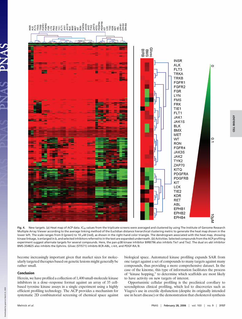
Previously Uncharacterized Activities. The data were next scanned toidentify whether any known drugs and well characterized com-pounds from the chemical collection displayed any previouslyunknown activities. The data were configured into a heat map byhierarchical clustering for ease of analysis (Fig. 4a). The data wereexamined to find targets for existing molecules and to understandthe activity of molecules on related targets. For Glivec, activityagainst Bcr-Abl, platelet-derived growth factor, and c-kit were allmanifest (see Fig. 4b). Interesting cross-activities for other less wellcharacterized kinase inhibitors currently under clinical investigationwere also discovered. For example, BIRB796, a kinase inhibitor forall p38 kinase isoforms (24, 25), was also shown to inhibit Tie2 andto a lesser extent Tie1 (Fig. 4b). These activities were confirmed tobe target-related and not dependent on inhibition of p38 byWestern blot of both Tel-Tie1 and Tel-Tie2 immunoprecipitatedfrom Ba�F3 cells and probed with antiphosphotyrosine antibodies(data not shown). Tie2 has recently been implicated in angiogenesisas well as stem cell quiescence and mobilization during chemother-apy (24–27). Therefore, expansion to Tie2 as a target couldpotentially increase the utility of this compound.
Although BIRB796 displayed high selectivity versus other ty-rosine kinases in our experiment, BMS-354825, a dual inhibitor ofSrc and Abl (28), was shown to inhibit multiple other kinases,including several Ephrin receptors (B1, B2, and B4). Ephrinreceptors have been implicated in both tumor angiogenesis andgrowth and survival of tumor cells (29–31). BMS-354825 is cur-rently being tested in humans for its potential to overcome Glivec-resistant Bcr-Abl-positive chronic mylogenous and acute lympho-blastic leukemia (32). Our results indicate the potential forexpansion into other tumor types. In summary, these data demon-strate the power of the aforementioned ACP in identifying novelindications for known drugs and drug candidates, a fact that will
Fig. 3. Global data analysis. (a) Selectivity�potency correlation. The com-pounds were clustered based on chemical structure (Pipeline Pilot, Scitegic,San Diego) to yield 94 clusters. The test points for 14 clusters (those with 20 ormore compounds) were plotted broken out by cluster to assess the specificity–potency relationships for focused structural classes. The clusters are numberedfrom 1 to 14, where blue dots represent compounds, and are distributed byspecificity (abscissa) and potency (GI50; ordinate). (b) Relationship of struc-tural similarity and biological similarity. Each marker represents a pair ofcompounds, with the pairwise chemical distance (Tanimoto coefficient) on theabscissa and the pairwise biological profile distance (Pearson correlationcoefficient) on the ordinate. The line shown is the ordinary least-squares fit.The triangle encloses the outliers, which are discussed in the text. Markers arecolored according to an alternative measure of biological profile similarity,the cosine (or ‘‘uncentered’’) correlation coefficient.
3156 � www.pnas.org�cgi�doi�10.1073�pnas.0511292103 Melnick et al.
Dow
nloa
ded
by g
uest
on
Mar
ch 2
1, 2
020
become increasingly important given that market sizes for molec-ularly targeted therapies based on genetic lesions might generally berather small.
ConclusionHerein, we have profiled a collection of 1,400 small-molecule kinaseinhibitors in a dose–response format against an array of 35 cell-based tyrosine kinase assays in a single experiment using a highlyefficient profiling technology. The ACP provides a mechanism forsystematic 2D combinatorial screening of chemical space against
biological space. Automated kinase profiling expands SAR fromone target against a set of compounds to many targets against manycompounds, thus providing a more comprehensive dataset. In thecase of the kinome, this type of information facilitates the processof ‘‘kinase hopping,’’ to determine which scaffolds are most likelyto have activity on new targets of interest.
Opportunistic cellular profiling is the preclinical corollary toserendipitous clinical profiling, which led to discoveries such asViagra’s use in erectile dysfunction (despite its originally intendeduse in heart disease) or the demonstration that cholesterol synthesis
Fig. 4. New targets. (a) Heat map of ACP data. IC50 values from the triplicate screens were averaged and clustered by using The Institute of Genome ResearchMultiple Array Viewer according to the average linking method of the Euclidian distance hierarchical clustering metric to generate the heat map shown in thelower left. The scale ranges from 0 (green) to 10 �M (red), as shown in the right-hand color triangle. The dendrogram associated with the heat map, showingkinase linkage, is enlarged in b, and selected inhibitors referred to in the text are expanded underneath. (b) Activities. Selected compounds from the ACP profilingexperiment suggest alternate targets for several compounds. Here, the pan-p38 kinase inhibitor BIRB796 also inhibits Tie1 and Tie2. The dual src-abl inhibitorBMS-354825 also inhibits the Ephrins. Glivec (STI571) inhibits BCR-ABL, c-kit, and PDGF-RA�B.
Melnick et al. PNAS � February 28, 2006 � vol. 103 � no. 9 � 3157
CELL
BIO
LOG
Y
Dow
nloa
ded
by g
uest
on
Mar
ch 2
1, 2
020

inhibitors, statins, reduce CD69 T cell antigen levels in T cells, whichmay extend the statin’s benefits to immune regulation (33–35). Asdemonstrated here, the ACP experiment identified PDGFR andc-kit as side activities for Glivec, which are under investigation foralternative treatments of asthma and gastrointestinal stromal dis-order (GIST). Similarly, the activities identified for the p38 kinaseinhibitor BIRB796 and dual src�abl inhibitor BMS-354825 mayprove useful as tools to validate Tie2 and the Ephrins as drug targetsin angiogenesis.
ACP profiling of molecular libraries against diverse cellularassays can be applied to many other problems as well. For example,it may be possible to identify novel ligands for whole panels oforphan G-protein-coupled receptors by profiling collections ofdiverse lipid, metabolite, and neuropeptide hormone libraries. Itmay also be possible to identify combinations of drugs that actsynergistically against panels of patient-derived tumor cell lines. Forpharmacogenomics, disease-associated SNPs identified by haplo-type mapping can be engineered into SNP-dependent cellularassays and profiled against panels of preclinical drug candidates toprospectively match patient variants with treatment. The configu-ration of the ACP also allows screens for ligands with enhancedpotency, selectivity, stability, or expression levels from evolvedprotein libraries.
Materials and MethodsAutomated Cellular Assays. The assay portion of the ACP consists ofthe same control software as the automated tissue culture station,a noncontact 1,536-well reagent dispenser (dispense range of 250 nlto 5 �l at 5% coefficient of variation (CV), dispense time of �1min�plate at 5 �l), a 1,536-pin transfer device (validated at 9 nl and�10% CVs), room-temperature hotels, the same incubators asdescribed above, and a Perkin–Elmer ViewLux plate reader. Torequest cells for an assay, the operator inputs the desired finalnumber of assay plates, cell density, and well volume. The auto-mated tissue culture station calculates the correct number of
daughter flasks for pooling (if the number of cells required isunavailable, the cell propagation protocol is enacted). When daugh-ter flasks have incubated for a defined amount of time, they arepooled in a matrix fashion into several empty recipient flasks. Anequal volume from each daughter cell flask will be deposited intoeach recipient flask. A sample from one pooled flask will becounted (only one sample is necessary, because all of the flasks havebeen adjusted to the same cell density by the pooling process). Allof the flasks will have their volume adjusted to the desired celldensity for plating. This pooling function, although complex, obvi-ates the requirement for a common pooling container, thus de-creasing the possibility of contamination. The flasks are then movedto the cell dispenser, where cells are pulled from flasks anddispensed into 1,536-well assay plates, typically 5 �l�well. The platesof cells are placed into an incubator and the empty flasks discarded.For the assay described in this paper, compounds are introduced tothe system in 1,536-well plates through the room-temperaturehotels. Cell plates are requested at 105 cells�ml, 5 �l�well, in1,536-well plates, for each of the 35 Ba�F3 cell lines. The cellrequests are processed as described above. After cell plating,preplated compounds in single-dose or dose–response format aretransferred from the compound plates to the assay plates by usingthe 1,536 pintool. The assay plates are incubated in the environ-mentally controlled (37°C, 95% humidity, 5% CO2) incubators for48 h (a custom lid design minimizes edge effects from evaporationand enables 5-�l cell-based assays to be incubated for up to 5 days).After incubation, 5 �l�well Cell Titer Glo (Promega) is added withthe reagent dispenser, incubated for 10 min at room temperature,and luminescence quantitated with the Perkin–Elmer ViewLux. Aluminescence value less than the control value indicates a decreasein either the number of cells or cell viability.
Supporting Text. Protocols detailing the automatic propagation ofcell lines using the ACP, as well as the construction of theBa�F3�Tel-kinase library, are presented in Supporting Text, whichis published as supporting information on the PNAS web site.
1. Plavec, I., Sirenko, O., Privat, S., Wang, Y., Dajee, M., Melrose, J., Nakao, B.,Hytopoulos, E., Berg, E. L. & Butcher, E. C. (2004) Proc. Natl. Acad. Sci. USA101, 1223–1228.
2. Rabow, A. A., Shoemaker, R. H., Sausville, E. A. & Covell, D. G. (2002) J. Med.Chem. 45, 818–840.
3. Butcher, E. C., Berg, E. L. & Kunkel, E. J. (2004) Nat. Biotechnol. 22,1253–1259.
4. Blume-Jensen, P. & Hunter, T. (2001) Nature 411, 355–365.5. Hunter, T. (1998) Harvey Lect. 94, 81–119.6. Seidler, J., McGovern, S. L., Doman, T. N. & Shoichet, B. K. (2003) J. Med.
Chem. 46, 4477–4486.7. Knight, Z. A. & Shokat, K. M. (2005) Chem. Biol. 12, 621–637.8. Krause, D. S. & Van Etten, R. A. (2005) N. Engl. J. Med. 353, 172–187.9. Golub, T. R., Barker, G. F., Lovett, M. & Gilliland, D. G. (1994) Cell 77,
307–316.10. Papadopoulos, P., Ridge, S. A., Boucher, C. A., Stocking, C. & Wiedemann,
L. M. (1995) Cancer Res. 55, 34–38.11. Lacronique, V., Boureux, A., Valle, V. D., Poirel, H., Quang, C. T.,
Mauchauffe, M., Berthou, C., Lessard, M., Berger, R., Ghysdael, J., et al. (1997)Science 278, 1309–1312.
12. Eguchi, M., Eguchi-Ishimae, M., Tojo, A., Morishita, K., Suzuki, K., Sato, Y.,Kudoh, S., Tanaka, K., Setoyama, M., Nagamura, F., et al. (1999) Blood 93,1355–1363.
13. Daley, G. Q. & Baltimore, D. (1988) Proc. Natl. Acad. Sci. USA 85, 9312–9316.14. Bai, R. Y., Dieter, P., Peschel, C., Morris, S. W. & Duyster, J. (1998) Mol. Cell.
Biol. 18, 6951–6961.15. Hayakawa, F., Towatari, M., Kiyoi, H., Tanimoto, M., Kitamura, T., Saito, H.
& Naoe, T. (2000) Oncogene 19, 624–631.16. Weisberg, E., Boulton, C., Kelly, L. M., Manley, P., Fabbro, D., Meyer, T.,
Gilliland, D. G. & Griffin, J. D. (2002) Cancer Cell 1, 433–443.17. Shah, N. P., Tran, C., Lee, F. Y., Chen, P., Norris, D. & Sawyers, C. L. (2004)
Science 305, 399–401.18. Weisberg, E., Manley, P. W., Breitenstein, W., Bruggen, J., Cowan-Jacob,
S. W., Ray, A., Huntly, B., Fabbro, D., Fendrich, G., Hall-Meyers, E., et al.(2005) Cancer Cell 7, 129–141.
19. Christensen, J. G., Schreck, R., Burrows, J., Kuruganti, P., Chan, E., Le, P.,Chen, J., Wang, X., Ruslim, L., Blake, R., et al. (2003) Cancer Res. 63,7345–7355.
20. Manley, P. W., Bold, G., Bruggen, J., Fendrich, G., Furet, P., Mestan, J.,Schnell, C., Stolz, B., Meyer, T., Meyhack, B., et al. (2004) Biochim. Biophys.Acta 1697, 17–27.
21. Ding, S., Gray, N. S., Ding, Q. & Schultz, P. G. (2001) J. Org. Chem. 66,8273–8276.
22. Gallion, S. L. & Qian, D. (2005) Curr. Opin. Drug Discov. Dev. 8, 638–645.23. Gill, A. L., Frederickson, M., Cleasby, A., Woodhead, S. J., Carr, M. G.,
Woodhead, A. J., Walker, M. T., Congreve, M. S., Devine, L. A., Tisi, D., etal. (2005) J. Med. Chem. 48, 414–426.
24. Regan, J., Breitfelder, S., Cirillo, P., Gilmore, T., Graham, A. G., Hickey, E.,Klaus, B., Madwed, J., Moriak, M., Moss, N., et al. (2002) J. Med. Chem. 45,2994–3008.
25. Pargellis, C., Tong, L., Churchill, L., Cirillo, P. F., Gilmore, T., Graham, A. G.,Grob, P. M., Hickey, E. R., Moss, N., Pav, S. & Regan, J. (2002) Nat. Struct.Biol. 9, 268–272.
26. Oliner, J., Min, H., Leal, J., Yu, D., Rao, S., You, E., Tang, X., Kim, H., Meyer,S., Han, S. J., et al. (2004) Cancer Cell 6, 507–516.
27. Kobayashi, H. & Lin, P. C. (2005) Front. Biosci. 10, 666–674.28. Lombardo, L. J., Lee, F. Y., Chen, P., Norris, D., Barrish, J. C., Behnia, K.,
Castaneda, S., Cornelius, L. A., Das, J., Doweyko, A. M., et al. (2004) J. Med.Chem. 47, 6658–6661.
29. Kullander, K. & Klein, R. (2002) Nat. Rev. Mol. Cell Biol. 3, 475–486.30. Brantley-Sieders, D. M. & Chen, J. (2004) Angiogenesis 7, 17–28.31. Brantley-Sieders, D., Parker, M. & Chen, J. (2004) Curr. Pharm. Des. 10,
3431–3442.32. Shah, N. P. (2005) Hematology 1, 183–187.33. Kunkel, E. J., Plavec, I., Nguyen, D., Melrose, J., Rosler, E. S., Kao, L. T.,
Wang, Y., Hytopoulos, E., Bishop, A. C., Bateman, R., et al. (2004) Assay DrugDev. Technol. 2, 431–441.
34. Boolell, M., Allen, M. J., Ballard, S. A., Gepi-Attee, S, Muirhead, G. J., Naylor,A. M., Osterloh, I. H. & Gingell, C. (1996) Int. J. Impot. Res. 8, 47–52.
35. Padma-Nathan, H. (1999) Am J. Cardiol. 84, 18N–23N.
3158 � www.pnas.org�cgi�doi�10.1073�pnas.0511292103 Melnick et al.
Dow
nloa
ded
by g
uest
on
Mar
ch 2
1, 2
020