an architecture for encoding sentence meaning in left mid … · an architecture for encoding...
TRANSCRIPT

An architecture for encoding sentence meaning in leftmid-superior temporal cortexSteven M. Franklanda,b,1 and Joshua D. Greenea,b
aDepartment of Psychology, Harvard University, Cambridge, MA 02138; and bCenter for Brain Science, Harvard University, Cambridge, MA 02138
Edited by Stanislas Dehaene, INSERM U992CEA/Saclay, College de France, Gif/Yvette, France, and approved July 24, 2015 (received for review December2, 2014)
Human brains flexibly combine the meanings of words to composestructured thoughts. For example, by combining the meanings of“bite,” “dog,” and “man,” we can think about a dog biting a man,or a man biting a dog. Here, in two functional magnetic resonanceimaging (fMRI) experiments using multivoxel pattern analysis (MVPA),we identify a region of left mid-superior temporal cortex (lmSTC)that flexibly encodes “who did what to whom” in visually presentedsentences. We find that lmSTC represents the current values of ab-stract semantic variables (“Who did it?” and “To whomwas it done?”)in distinct subregions. Experiment 1 first identifies a broad regionof lmSTC whose activity patterns (i) facilitate decoding of structure-dependent sentence meaning (“Who did what to whom?”) and(ii) predict affect-related amygdala responses that depend on thisinformation (e.g., “the baby kicked the grandfather” vs. “the grand-father kicked the baby”). Experiment 2 then identifies distinct, butneighboring, subregions of lmSTC whose activity patterns carry in-formation about the identity of the current “agent” (“Who did it?”)and the current “patient” (“To whom was it done?”). These neigh-boring subregions lie along the upper bank of the superior temporalsulcus and the lateral bank of the superior temporal gyrus, respec-tively. At a high level, these regions may function like topographi-cally defined data registers, encoding the fluctuating values ofabstract semantic variables. This functional architecture, which inkey respects resembles that of a classical computer, may play a crit-ical role in enabling humans to flexibly generate complex thoughts.
fMRI | cognitive architecture | compositionality | comprehension | PBEC
Yesterday, the world’s tallest woman was serenaded by 30pink elephants. The previous sentence is false, but perfectly
comprehensible, despite the improbability of the situation itdescribes. It is comprehensible because the human mind can flex-ibly combine the meanings of individual words (“woman,” “sere-nade,” “elephants,” etc.) to compose structured thoughts, such asthe meaning of the aforementioned sentence (1, 2). How the brainaccomplishes this remarkable feat remains a central, but unan-swered, question in cognitive science.Given the vast number of sentences we can understand and
produce, it would be implausible for the brain to allocate individualneurons to represent each possible sentence meaning. Instead, it islikely that the brain employs a system for flexibly combining rep-resentations of simpler meanings to compose more complexmeanings. By “flexibly,” we mean that the same meanings can becombined in many different ways to produce many distinct complexmeanings. How the brain flexibly composes complex, structuredmeanings out of simpler ones is a matter of long-standing debate(3–10).At the cognitive level, theorists have held that the mind encodes
sentence-level meaning by explicitly representing and updating thevalues of abstract semantic variables (3, 5) in a manner analogousto that of a classical computer. Such semantic variables correspondto basic, recurring questions of meaning such as “Who did it?” and“To whom was it done?” On such a view, the meaning of a simplesentence is partly represented by filling in these variables withrepresentations of the appropriate semantic components. For ex-ample, “the dog bit the man” would be built out of the same
semantic components as “the man bit the dog,” but with a reversalin the values of the “agent” variable (“Who did it?”) and the“patient” variable (“To whom was it done?”). Whether and howthe human brain does this remains unknown.Previous research has implicated a network of cortical regions in
high-level semantic processing. Many of these regions surround theleft sylvian fissure (11–19), including regions of the inferior frontalcortex (13, 14), inferior parietal lobe (12, 20), much of the superiortemporal sulcus and gyrus (12, 15, 21), and the anterior temporallobes (17, 20, 22). Here, we describe two functional magnetic res-onance imaging (fMRI) experiments aimed at understanding howthe brain (in these regions or elsewhere) flexibly encodes themeanings of sentences involving an agent (“Who did it?”), an action(“What was done?”), and a patient (“To whom was it done?”).First, experiment 1 aims to identify regions that encode struc-
ture-dependent meaning. Here, we search for regions that differ-entiate between pairs of visually presented sentences, where thesesentences convey different meanings using the same words (as in“man bites dog” and “dog bites man”). Experiment 1 identifies aregion of left mid-superior temporal cortex (lmSTC) encodingstructure-dependent meaning. Experiment 2 then asks how thelmSTC represents structure-dependent meaning. Specifically, wetest the long-standing hypothesis that the brain represents andupdates the values of abstract semantic variables (3, 5): here, theagent (“Who did it?”) and the patient (“To whom was it done?”).We search for distinct neural populations in lmSTC that encodethese variables, analogous to the data registers of a computer (5).
Experiment 1In experiment 1, subjects undergoing fMRI read sentences de-scribing simple events. Each sentence expressed a meaning, or
Significance
The 18th-century Prussian philosopher Wilhelm von Humboltfamously noted that natural language makes “infinite use of fi-nite means.” By this, he meant that language deploys a finite setof words to express an effectively infinite set of ideas. As theseat of both language and thought, the human brain must becapable of rapidly encoding the multitude of thoughts that asentence could convey. How does this work? Here, we find evi-dence supporting a long-standing conjecture of cognitive science:that the human brain encodes the meanings of simple sentencesmuch like a computer, with distinct neural populations repre-senting answers to basic questions of meaning such as “Who didit?” and “To whom was it done?”
Author contributions: S.M.F. and J.D.G. designed research; S.M.F. performed research;S.M.F. and J.D.G. contributed new reagents/analytic tools; S.M.F. analyzed data; andS.M.F. and J.D.G. wrote the paper.
The authors declare no conflict of interest.
This article is a PNAS Direct Submission.
Freely available online through the PNAS open access option.1To whom correspondence should be addressed. Email: [email protected].
This article contains supporting information online at www.pnas.org/lookup/suppl/doi:10.1073/pnas.1421236112/-/DCSupplemental.
11732–11737 | PNAS | September 15, 2015 | vol. 112 | no. 37 www.pnas.org/cgi/doi/10.1073/pnas.1421236112
Dow
nloa
ded
by g
uest
on
Mar
ch 1
9, 2
020
“proposition,” which could be conveyed in either the active orpassive voice (e.g., “the ball hit the truck”/“the truck was hit bythe ball”). Each such sentence could be reversed to yield a mirrorimage proposition (e.g., “the truck hit the ball”/“the ball was hitby the truck”), which was also included in the stimulus set. Wecall these “mirror image proposition pairs.”Members of these pairscontain the same words and have the same syntactic structure, butthe words are differentially assigned to the agent and patient rolesto form different sentence-level meanings.A region encoding the meanings of these sentences should have
the following two properties. First, patterns of activity in such aregion should differentially encode members of mirror imagepropositions pairs. For example, the propositions conveyed by “thetruck hit the ball” and “the ball hit the truck” should elicit distinctpatterns of activity. Second, the instantiation of such patternsshould predict downstream neural responses that depend on un-derstanding “who did what to whom.” For example, patterns re-lated to sentence-level meaning should predict differential affectiveresponses to “the grandfather kicked the baby” and “the babykicked the grandfather.” Experiment 1 used two key analyses,corresponding to these two functional properties. First, we ap-plied multivoxel pattern analysis (23–25) and a whole-brainsearchlight procedure (26) to identify sets of contiguous voxelsthat distinguish between members of mirror image propositionpairs. Second, we developed a pattern-based effective connec-tivity (PBEC) analysis to determine whether patterns related toaffectively salient sentences (e.g., “the grandfather kicked thebaby”) mediate the relationship between the sentence presentedand affective responses elsewhere in the brain. Jointly, theseanalyses establish candidate regions for encoding structure-dependent meaning that can be further probed in experiment 2.
Whole-Brain Searchlight Analysis. First, using a linear classifier, wesearched for regions whose patterns of activity distinguishedbetween members of mirror image proposition pairs: for exam-ple, between the proposition conveyed by “the truck hit the ball”(as well as “the ball was hit by the truck”) and the propositionconveyed by “the ball hit the truck” (as well as “the truck was hitby the ball”). The use of mirror image propositions ensures thatbasic lexico-semantic content, syntactic structure, and summedword frequency are matched between the propositions to bediscriminated. Active and passive forms of each proposition weretreated as identical in all analyses, allowing us to identify un-derlying semantic representations, controlling for visual featuresof the stimuli and surface syntax. All propositions were pre-sented separately, and multiple times, to better estimate thepattern of activity evoked by each proposition. For experiment 1,classifiers were thus tested on their ability to discriminate be-tween new tokens of the mirror image propositions on whichthey were trained.For this initial searchlight analysis, we used four mirror image
pairs of propositions, two involving animate entities and twoinvolving inanimate entities. For each subject (n = 16), we av-eraged classification accuracies across these four pairwise classifi-cation problems to yield a map of the mean classification accuracyby region. Group-level analysis identified a region of lmSTC (k =123; Talairach center: −59, −25, 6) that reliably distinguished be-tween mirror image propositions (P < 0.0001, corrected; meanaccuracy, 57%) (see left temporal region in Fig. 1). This result wasnot driven by a particular subset of the stimuli (Supporting In-formation). A second significant cluster was discovered along theright posterior insula/extreme capsule region (P < 0.001, corrected;37, −9, 6; mean accuracy, 56.4%). However, this second regionfailed to meet additional, minimal functional criteria for encodingsentence meaning (Supporting Information).
PBEC Analysis. The foregoing searchlight analysis suggests thatlmSTC represents critical aspects of sentence-level meaning. If
this hypothesis is correct, then the particular pattern instantiatedin lmSTC should also predict downstream neural responses whenthose responses depend on an understanding of “who did what towhom.” Our second analysis in experiment 1 attempts to de-termine whether the patterns of activity in lmSTC predict af-fective neural responses elsewhere in the brain.To test this hypothesis, we used, within the same experiment, an
independent set of mirror image proposition pairs in which oneproposition is more affectively salient than its counterpart, as in“the grandfather kicked the baby” and “the baby kicked thegrandfather.” (Differences in affective salience were verified withindependent behavioral testing. See Supporting Information.) Wepredicted that patterns of activity in lmSTC (as delineated by theindependent searchlight analysis) would statistically mediate therelationship between the sentence presented and the affectiveneural response, consistent with a causal relationship (27). ThisPBEC analysis proceeded in three steps.First, we confirmed that patterns of activity in the region of
lmSTC identified by the searchlight analysis can discriminatebetween these new mirror image propositions [t(15) = 3.2; P =0.005; mean accuracy, 58.3%], thus replicating the above findingswith new stimuli. Second, we identified brain regions that re-spond more strongly to affectively salient propositions (e.g., “thegrandfather kicked the baby” > “the baby kicked the grandfa-ther”). This univariate contrast yielded effects in two brain re-gions, the left amygdala (−28, −7, −18) and superior parietallobe (−38, −67, 47), (P < 0.001, corrected). Given its well-knownrole in affective processing (28), we interpreted this amygdalaresponse as an affective signal and focused on this region in oursubsequent mediation analysis. Third, and most critically, weexamined the relationship between patterns of activity in lmSTCand the magnitude of the amygdala’s response. The first of theabove analyses shows that “the grandfather kicked the baby”produces a different pattern in lmSTC than “the baby kicked thegrandfather” (etc.). If these patterns actually reflect structure-dependent meaning, then these patterns should mediate therelationship between the sentence presented and the amygdala’sresponse on a trial-by-trial basis.To quantify the pattern of activity in lmSTC on each trial, we
used the signed distance of each test pattern from the classifier’sdecision boundary (Supporting Information). This signed distancevariable reflects the content of the classifier’s decision regardingthe sentence (the sign), as well as what one may think of as its“confidence” in that decision (the distance). According to ourhypothesis, trials in which the pattern is confidently classified as“the grandfather kicked the baby” (etc.), rather than “the babykicked the grandfather” (etc.), should be trials in which theamygdala’s response is robust. Here, we are supposing that theclassifier’s “confidence” will reflect the robustness of the se-mantic representation, which in turn may influence downstreamaffective responses in the amygdala.As predicted, the pattern of activity instantiated in lmSTC pre-
dicted the amygdala’s response [t(15) = 3.96, P = 0.0013], over andabove both the mean signal in lmSTC and the content of thestimulus. The pattern of activity in the lmSTC explains uniquevariance in the amygdala’s response, consistent with a causal modelwhereby information flows from the sentence on the screen, to apattern of activity in the lmSTC, to the amygdala [P < 0.01, byMonte Carlo simulation (29, 30); Sobel test (27), z = 2.47, P =0.013] (Fig. 1). The alternative model reversing the direction ofcausation between the lmSTC and amygdala was not significant(Monte Carlo, P > 0.10; Sobel, z = 1.43, P = 0.15), further sup-porting the proposed model.There are several possible sources of trial-to-trial variability in
lmSTC’s responses (see Supporting Information for more dis-cussion). For example, a participant’s inattention might disruptthe semantic representation in lmSTC, making the trial moredifficult to classify and, at the same time, making the amygdala
Frankland and Greene PNAS | September 15, 2015 | vol. 112 | no. 37 | 11733
PSYC
HOLO
GICALAND
COGNITIVESC
IENCE
S
Dow
nloa
ded
by g
uest
on
Mar
ch 1
9, 2
020

response weaker than otherwise expected. Regardless of thesource of the variation in these patterns, the present data provideevidence that neural representations of structure-dependent mean-ings in lmSTC predict downstream affective responses, consistentwith our causal model.Thus, experiment 1 shows that a region of lmSTC meets our two
initial functional criteria for a region encoding structure-dependentsentence meaning. First, its patterns of activity differentiate be-tween mirror image propositions containing the same words andsyntactic structure. Second, these patterns statistically mediate therelationship between the sentence presented and affective neuralresponses that depend on understanding “who did what to whom.”Experiment 1 does not, however, explain how this region encodessuch information. Experiment 2 aims to further validate the resultsof experiment 1 and to illuminate the mechanism by which thisregion encodes these structure-dependent meanings.
Experiment 2In experiment 2, we test the hypothesis that lmSTC flexibly en-codes these meanings (at least in part) by explicitly representingthe values of the agent (“Who did it?”) and the patient (“Towhom was it done?”) (5). To evaluate this possibility, wesearched for subregions of lmSTC whose patterns of activityreflect the current value of these variables. We performed sep-arate searches for each variable, searching for subregionsencoding “Who did it?” and “To whom it was done?” across verbcontexts. Thus, we aimed to identify regions that are specializedfor representing the agent and patient variables as such.Experiment 2 (n = 25) used a stimulus set in which four nouns
(“man,” “girl,” “dog,” and “cat”) were assigned to the agent andpatient roles for each of five verbs (“chased,” “scratched,” etc.),in both active and passive forms (Fig. 2A). Thus, subjects un-dergoing fMRI read sentences such as “the dog chased the man”and “the girl was scratched by the cat,” exhausting all meaningfulcombinations, excluding combinations assigning the same nounto both roles (e.g., “the man chased the man”).We acquired partial-volume, high-resolution (1.5-mm3 iso-
tropic voxels) functional images covering the lmSTC. We usedseparate searchlight analyses within each subject to identifysubregions of lmSTC encoding information about the identity of
the agent or patient (Fig. 2 B and C). For our principal search-light analyses, four-way classifiers were trained to identify theagent or patient using data generated by four out of five verbs.The classifiers were then tested on data from sentences con-taining the withheld verb. For example, the classifiers weretested using patterns generated by “the dog chased the man,”having never previously encountered patterns generated bysentences involving “chased,” but having been trained to identify“dog” as the agent and “man” as the patient in other verb con-texts. This procedure was repeated holding each verb’s data outof the training set, and the results were averaged across cross-validation iterations. Thus, this analysis targets regions that in-stantiate consistent patterns of activity for (for example) “dog asagent” across verb contexts, discriminable from “man as agent”(and likewise for other nouns). A region that carries this in-formation therefore encodes “Who did it?” across the nouns andverb contexts tested. This same procedure was repeated to de-code the identity of the patient.These searchlight analyses revealed distinct subregions of lmSTC
that reliably carry information about the identity of the agent andthe patient (Fig. 3A). Within the anterior portion of lmSTC, amedial subregion located on the upper bank of the superior tem-poral sulcus (STS) encoded information about the identity of theagent (P < 0.01, corrected; −46, −18, 1). A spatially distinct lateralsubregion, encompassing part of the upper bank of the STS, as wellas the lateral superior temporal gyrus (STG) carried patient in-formation (P < 0.0001, corrected; −57, −10, 2) across subjects.These anterior agent and patient clusters are adjacent, but non-overlapping in this analysis. A follow-up analysis using independentdata to define each participant’s agent and patient clusters foundthat these subregions are significantly dissociable by their in-formational content [FRegion×Content(1,24) = 12.99, Pperm = 0.001](Fig. 3). This searchlight analysis also revealed a second agentcluster, posterior and superior to the clusters described above, lo-cated primarily within the posterior STS (P < 0.02, corrected; −57,−37, 7). Post hoc analyses found the classification accuracies drivingthese results to be only modestly above chance levels of 25%, butstatistically reliable across our set of 25 subjects (mean accuraciesacross subjects: anterior agent, 27.1%; posterior agent, 28.1%;patient, 26.6%).
A B
C
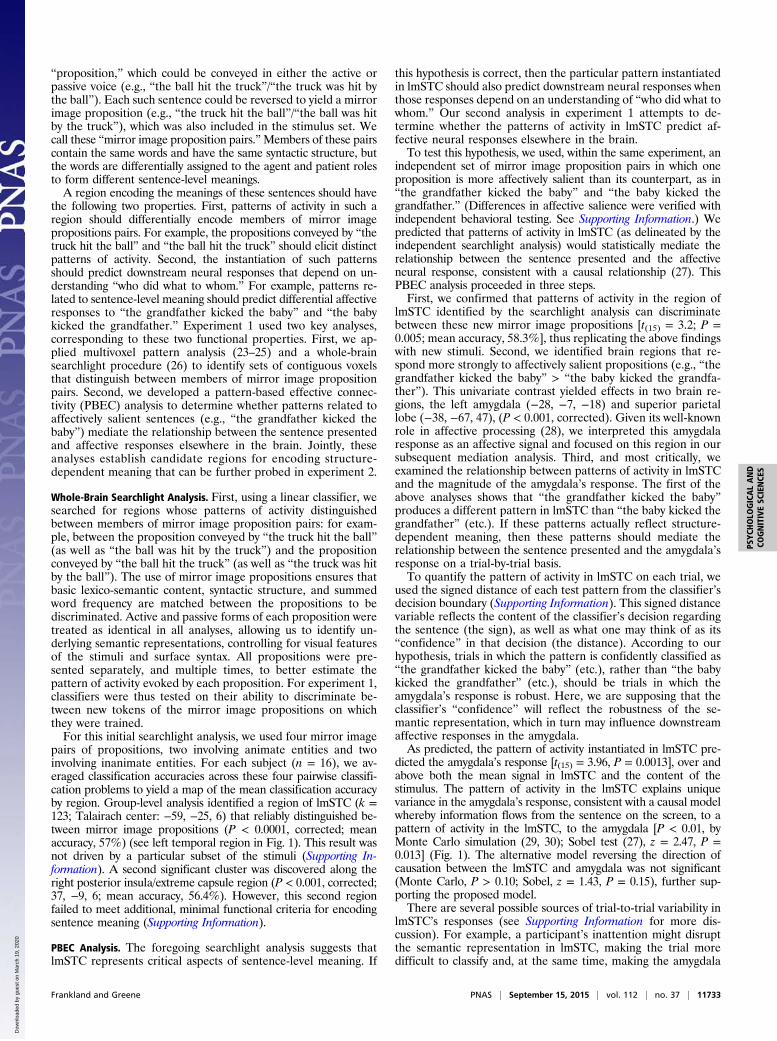
Fig. 1. Model of information flow from stimulus tolmSTC to amygdala in experiment 1. (A) A patternclassifier determines which of two propositions waspresented using activity in lmSTC. Distance from theclassification boundary indicates the extent to whicha learned pattern was instantiated. The red regioncorresponds to the emotionally evocative proposi-tion (e.g., “the grandfather kicked baby”), whereasblue corresponds to the less evocative proposition(“the baby kicked grandfather”). (B) For each trial,the classifier’s signed distance from the classificationboundary was transformed by a sigmoidal functionand used to predict the mean level of activity in theleft amygdala. (C) Patterns in lmSTC mediate therelationship between the proposition on the screenand the amygdala’s response, consistent with amodel according to which the lmSTC encodes thestructured representations necessary to generate anemotional response.
11734 | www.pnas.org/cgi/doi/10.1073/pnas.1421236112 Frankland and Greene
Dow
nloa
ded
by g
uest
on
Mar
ch 1
9, 2
020

As in experiment 1, post hoc analyses ruled out the possibilitythat these results were driven by a subset of items, as these regionswere relatively consistent in their ability to discriminate betweenparticular pairs of nouns and to generalize across the five verbcontexts. (See Supporting Information for detailed procedures andresults for these post hoc analyses.) These results thus suggest thatthe regions identified by the experiment 2 searchlight analyses aregenerally involved in encoding noun–role bindings across the nounsand verbs used. No regions of lmSTC carried information aboutthe surface subject and surface object of the sentence. For example,no lmSTC region encoded “the dog chased the man” and “the dogwas chased by the man” as similar to each other, but different from“the man chased the dog” and “the man was chased by the dog.”Within lmSTC, the encoding appears, instead, to be based ondeeper semantics, encoding the underlying agent and patient of thesentence, independent of which noun serves as the sentence’ssurface subject or object, consistent with experiment 1.These findings provide preliminary evidence that these sub-
regions of lmSTC encode the values of the agent and patientvariables. However, it remains open whether and to what extentthese subregions are specialized for representing agent and pa-tient information—that is, whether they tend to represent onekind of information and not the other. To address this question,we conducted planned post hoc analyses that separately definedagent and patient regions within each subject using data from theremaining subjects. We assessed the significance of these effectsusing both conventional parametric statistics and permutation
tests (Supporting Information). Within subjects’ independentlylocalized patient regions, patient identification accuracy wassignificantly greater than agent identification accuracy acrosssubjects [lateral lmSTC: t(24) = 2.96, P = 0.006; permutation test:0.006]. Within the posterior agent region, agent identificationwas significantly above chance [t(24) = 2.38, P = 0.01; permuta-tion test: P = 0.008]. Within the anterior agent region, theclassification effect was somewhat weaker [t(24) = 2.04, P = 0.02;permutation test: P = 0.055]. As expected, patient identificationwas at chance in both the anterior agent region [t(24) = 0.86, P =0.2; permutation test: P = 0.22] and the posterior agent region[t(24) = −0.29, P = 0.39; permutation test: P = 0.38]. However,the direct comparison of accuracy levels for agent and patientidentification was not statistically significant in the anterior agentregion (P = 0.27; permutation test: P = 0.26) or the posterioragent region (P = 0.15; permutation test: P = 0.15). See Fig. 3B.To further assess the role specificity of these subregions, we
localized a large portion of the anterior lmSTC in a manner thatwas unbiased with respect to its role preference, and thenquantified the average preferences of slices of voxels at each Xcoordinate (Supporting Information). We found a clear trend inrole preference along the medial-lateral axis, with medial por-tions preferentially encoding agent information and lateral por-tions preferentially encoding patient information (Fig. 3C).From the present data, we cannot determine whether the ob-served graded shift in role preference exists within individuals, or
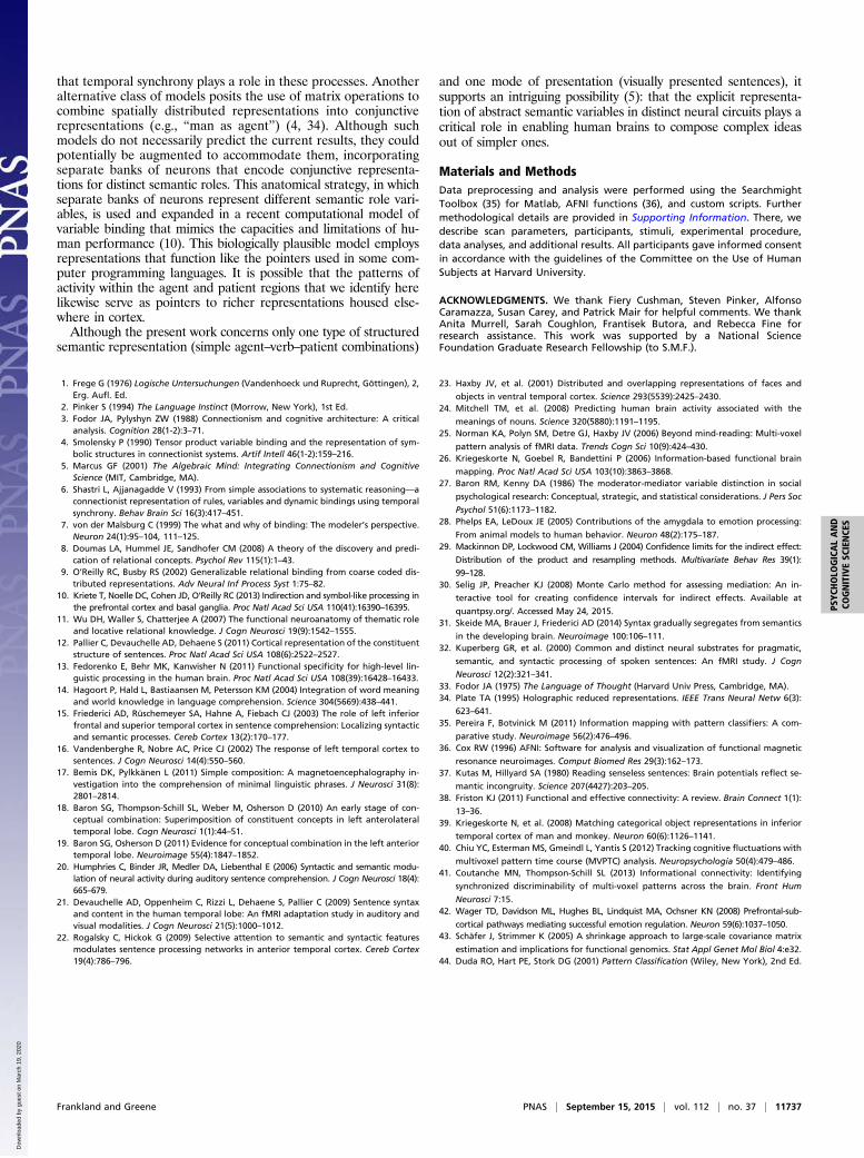
A B CFig. 2. Experiment 2 design. (A) Subjects read senten-ces constructed from a menu of five verbs and fournouns, with one noun in the agent role and another inthe patient role. (B) For each trial, separate patternclassifiers attempted to identify the agent and thepatient based on activity within subregions of lmSTC.(C) Classifiers were trained using data from four of fiveverbs and tested on data from the withheld verb. Thisrequired the classifiers to identify agents and patientsbased on patterns that are reused across contexts.
A C
BFig. 3. (A) Searchlight analyses identified adjacent,but nonoverlapping subregions of anterior lmSTCthat reliably encoded information about agentidentity (medial, blue) and patient identity (lateral,red). (B) Post hoc analyses find that these adjacentregions differ significantly in the information theyencode. These analyses define each subject’s agentand patient subregions using data from othersubjects, and the statistics computed within eachsubject’s agent/patient region reflect the averageaccuracy of all voxel neighborhoods across that re-gion. (C) Across subjects, medial portions of ante-rior lmSTC preferentially encode agent information,whereas lateral portions of anterior lmSTC prefer-entially encode patient information.
Frankland and Greene PNAS | September 15, 2015 | vol. 112 | no. 37 | 11735
PSYC
HOLO
GICALAND
COGNITIVESC
IENCE
S
Dow
nloa
ded
by g
uest
on
Mar
ch 1
9, 2
020

simply results from averaging across individuals exhibiting moreabrupt transitions.A final searchlight analysis within lmSTC identified two addi-
tional subregions supporting identification of the present verb(Supporting Information). The anterior verb subregion (P < 0.025;−61, −15, 2) was adjacent to the patient subregion. The posteriorverb subregion (P < 0.0001; −55, −49, 5) in the posterior STSpartially overlapped with the posterior agent region.The foregoing analyses strongly suggest that a lateral sub-
region of anterior lmSTC selectively encodes information aboutthe identity of the current patient, and somewhat less strongly,that a medial portion of anterior lmSTC selectively encodes in-formation about the identity of the current agent. In addition, weidentified two subregions of lmSTC supporting classification of theverb present on a given trial (Supporting Information). Together,these results indicate that distinct subregions of lmSTC separatelyand dynamically represent the semantic information sufficient tocompose complex representations involving an agent, a patient,and an action.A third experiment replicates the findings of experiment 2.
Once again, we find that a medial region of lmSTC encodesinformation about the agent while a neighboring lateral regionencodes information about the patient (Supporting Information).
DiscussionThe experiments presented here begin to address an importantunanswered question in cognitive neuroscience (2–6): How doesthe brain flexibly compose structured thoughts out of simplerideas? We provide preliminary evidence for a long-standingtheoretical conjecture of cognitive science: that the brain, onsome level, functions like a classical computer, representingstructured semantic combinations by explicitly encoding thevalues of abstract variables (3, 5). Moreover, we find evidencethat the agent and patient variables are topographically repre-sented across the upper bank of the left STS and lateral STG,such that adjacent cortical regions are differentially involved inencoding the identity of the agent and patient. At a high level,these regions may be thought of as functioning like the dataregisters of a computer, in which time-varying activity patternstemporarily represent the current values of these variables (5). Thisfunctional architecture could support the compositional encodingof sentence meaning involving an agent and a patient, as theserepresentations can be simultaneously instantiated in adjacent re-gions to form complex representations with explicit, constituentstructure. These structured representations may in turn be read byother neural systems that enable reasoning, decision making, andother high-level cognitive functions.The present results are broadly consistent with previous re-
search concerning the neural loci of sentence-level semanticprocessing while, at the same time, offering new insight into howsuch semantic information is represented. With respect tofunctional localization, previous research has implicated thelmSTC in phrase and sentence-level semantic processing usingboth functional neuroimaging and lesion data (11–13, 15, 18, 21).However, lmSTC is by no means the only region consistentlyimplicated in higher-order semantic processing, as research hasreliably documented the involvement of the anterior regions of thetemporal lobe (20, 22), left inferior parietal lobe (12, 20), and leftinferior frontal cortices (13, 14). The two studies presented heresuggest that lmSTC may be more narrowly involved in encodingthe values of semantic role variables. This narrower claim is con-sistent with multiple pieces of preexisting experimental evidence.First, fMRI studies (15, 31) have found increased activation in
a similar region of mid-left STG/STS in response to implausiblenoun–verb combinations that violate a verb’s selectional re-strictions (e.g., “the thunderstorm was ironed”) (but see ref. 32for conflicting results). More directly, an fMRI study (21) findsthat the repetition of a sentence’s meaning produces adaptation
effects in the lmSTC, even when that meaning is expressed usingdifferent surface syntactic forms, such as the active and passivevoice. These semantic adaptation effects occur in mid-STG andmiddorsal MTG/ventral STS when sentences are presented au-rally, and in middorsal MTG/midventral STS when presentedvisually. Finally, and perhaps of most direct relevance, patientswith damage to lmSTC have been found to have specific deficitsin determining “who did what to whom” in response to bothsentences and visual scenes representing actions (11). Here, thelocus of damage that most consistently predicts impaired per-formance across tasks appears to correspond to the anteriorsubregion of lmSTC in which we find the agent and patientvariables to be topographically represented.The present results build on this literature and extend our un-
derstanding in several key ways. First, experiment 1 uses multivar-iate methods to demonstrate that lmSTC carries information aboutsentence-level meaning. Second, experiment 1 employs a PBECanalysis to link these patterns of activity to affect-related amygdalaresponses, consistent with a model whereby lmSTC enables thecomprehension necessary to produce an appropriate affective re-sponse to a morally salient sentence. Third, and most critically,experiment 2 provides insight into how the lmSTC encodes sen-tence-level meaning, namely by representing the values of the agentand patient variables in spatially distinct neural populations.Given that the present results were generated using only lin-
guistic stimuli, the current data are silent as to whether theserepresentations are part of a general, amodal “language ofthought” (33), or whether they are specifically linguistic. In par-ticular, it is not known whether results would be similar using al-ternative modes of presentation, such as pictures. We note that theaforementioned lesion study of ref. 11 reports deficits in compre-hension of pictorial stimuli following damage to this region. How-ever, linguistic deficits could disrupt comprehension of pictures ifpictorial information is normally translated into words. Althoughsuch questions remain open, we emphasize that the representationsexamined here are related to the underlying semantic properties ofour stimuli, for reasons explained in detail above. They encodeinformation that would have to be encoded, in some form, by anysemantic system capable of supporting genuine comprehension.In evaluating the significance of the present results, we note
that the classification accuracies observed here are rather mod-est. Thus, we are by no means claiming that it is now possible to“read” people’s thoughts using patterns of activity in lmSTC. Norare we claiming that the lmSTC is the unique locus of complexthought. On the contrary, we suspect that the lmSTC is merelypart of a distributed neural system responsible for accessing andcombining representations housed elsewhere in the cortex (10).We regard the observed effects as significant, not because oftheir size, but because they provide evidence for a distinctivetheory of high-level semantic representation. We find evidencefor a functional segregation, and corresponding spatial segrega-tion, based on semantic role, which may enable the compositionof complex semantic representations. Such functional segrega-tion need not take the form of spatial segregation, but insofar asit does, it becomes possible to provide evidence for functionalsegregation using fMRI, as done here.A prominent alternative model for the encoding of complex
meanings holds that binding is signaled through the synchroni-zation (or desynchronization) of the firing phases of neuronsencoding a complex representation’s constituent semantic ele-ments (6–8). Given the limited temporal resolution of fMRI, thecurrent design cannot provide direct evidence for or againsttemporal synchrony as a binding mechanism. However, thepresent data suggest that such temporal correlations may beunnecessary in this case, because these bindings may instead beencoded through the instantiation of distributed patterns of ac-tivity in spatially dissociable patches of cortex devoted to rep-resenting distinct semantic variables. Nevertheless, it is possible
11736 | www.pnas.org/cgi/doi/10.1073/pnas.1421236112 Frankland and Greene
Dow
nloa
ded
by g
uest
on
Mar
ch 1
9, 2
020
that temporal synchrony plays a role in these processes. Anotheralternative class of models posits the use of matrix operations tocombine spatially distributed representations into conjunctiverepresentations (e.g., “man as agent”) (4, 34). Although suchmodels do not necessarily predict the current results, they couldpotentially be augmented to accommodate them, incorporatingseparate banks of neurons that encode conjunctive representa-tions for distinct semantic roles. This anatomical strategy, in whichseparate banks of neurons represent different semantic role vari-ables, is used and expanded in a recent computational model ofvariable binding that mimics the capacities and limitations of hu-man performance (10). This biologically plausible model employsrepresentations that function like the pointers used in some com-puter programming languages. It is possible that the patterns ofactivity within the agent and patient regions that we identify herelikewise serve as pointers to richer representations housed else-where in cortex.Although the present work concerns only one type of structured
semantic representation (simple agent–verb–patient combinations)
and one mode of presentation (visually presented sentences), itsupports an intriguing possibility (5): that the explicit representa-tion of abstract semantic variables in distinct neural circuits plays acritical role in enabling human brains to compose complex ideasout of simpler ones.
Materials and MethodsData preprocessing and analysis were performed using the SearchmightToolbox (35) for Matlab, AFNI functions (36), and custom scripts. Furthermethodological details are provided in Supporting Information. There, wedescribe scan parameters, participants, stimuli, experimental procedure,data analyses, and additional results. All participants gave informed consentin accordance with the guidelines of the Committee on the Use of HumanSubjects at Harvard University.
ACKNOWLEDGMENTS. We thank Fiery Cushman, Steven Pinker, AlfonsoCaramazza, Susan Carey, and Patrick Mair for helpful comments. We thankAnita Murrell, Sarah Coughlon, Frantisek Butora, and Rebecca Fine forresearch assistance. This work was supported by a National ScienceFoundation Graduate Research Fellowship (to S.M.F.).
1. Frege G (1976) Logische Untersuchungen (Vandenhoeck und Ruprecht, Göttingen), 2,Erg. Aufl. Ed.
2. Pinker S (1994) The Language Instinct (Morrow, New York), 1st Ed.3. Fodor JA, Pylyshyn ZW (1988) Connectionism and cognitive architecture: A critical
analysis. Cognition 28(1-2):3–71.4. Smolensky P (1990) Tensor product variable binding and the representation of sym-
bolic structures in connectionist systems. Artif Intell 46(1-2):159–216.5. Marcus GF (2001) The Algebraic Mind: Integrating Connectionism and Cognitive
Science (MIT, Cambridge, MA).6. Shastri L, Ajjanagadde V (1993) From simple associations to systematic reasoning—a
connectionist representation of rules, variables and dynamic bindings using temporalsynchrony. Behav Brain Sci 16(3):417–451.
7. von der Malsburg C (1999) The what and why of binding: The modeler’s perspective.Neuron 24(1):95–104, 111–125.
8. Doumas LA, Hummel JE, Sandhofer CM (2008) A theory of the discovery and predi-cation of relational concepts. Psychol Rev 115(1):1–43.
9. O’Reilly RC, Busby RS (2002) Generalizable relational binding from coarse coded dis-tributed representations. Adv Neural Inf Process Syst 1:75–82.
10. Kriete T, Noelle DC, Cohen JD, O’Reilly RC (2013) Indirection and symbol-like processing inthe prefrontal cortex and basal ganglia. Proc Natl Acad Sci USA 110(41):16390–16395.
11. Wu DH, Waller S, Chatterjee A (2007) The functional neuroanatomy of thematic roleand locative relational knowledge. J Cogn Neurosci 19(9):1542–1555.
12. Pallier C, Devauchelle AD, Dehaene S (2011) Cortical representation of the constituentstructure of sentences. Proc Natl Acad Sci USA 108(6):2522–2527.
13. Fedorenko E, Behr MK, Kanwisher N (2011) Functional specificity for high-level lin-guistic processing in the human brain. Proc Natl Acad Sci USA 108(39):16428–16433.
14. Hagoort P, Hald L, Bastiaansen M, Petersson KM (2004) Integration of word meaningand world knowledge in language comprehension. Science 304(5669):438–441.
15. Friederici AD, Rüschemeyer SA, Hahne A, Fiebach CJ (2003) The role of left inferiorfrontal and superior temporal cortex in sentence comprehension: Localizing syntacticand semantic processes. Cereb Cortex 13(2):170–177.
16. Vandenberghe R, Nobre AC, Price CJ (2002) The response of left temporal cortex tosentences. J Cogn Neurosci 14(4):550–560.
17. Bemis DK, Pylkkänen L (2011) Simple composition: A magnetoencephalography in-vestigation into the comprehension of minimal linguistic phrases. J Neurosci 31(8):2801–2814.
18. Baron SG, Thompson-Schill SL, Weber M, Osherson D (2010) An early stage of con-ceptual combination: Superimposition of constituent concepts in left anterolateraltemporal lobe. Cogn Neurosci 1(1):44–51.
19. Baron SG, Osherson D (2011) Evidence for conceptual combination in the left anteriortemporal lobe. Neuroimage 55(4):1847–1852.
20. Humphries C, Binder JR, Medler DA, Liebenthal E (2006) Syntactic and semantic modu-lation of neural activity during auditory sentence comprehension. J Cogn Neurosci 18(4):665–679.
21. Devauchelle AD, Oppenheim C, Rizzi L, Dehaene S, Pallier C (2009) Sentence syntaxand content in the human temporal lobe: An fMRI adaptation study in auditory andvisual modalities. J Cogn Neurosci 21(5):1000–1012.
22. Rogalsky C, Hickok G (2009) Selective attention to semantic and syntactic featuresmodulates sentence processing networks in anterior temporal cortex. Cereb Cortex19(4):786–796.
23. Haxby JV, et al. (2001) Distributed and overlapping representations of faces and
objects in ventral temporal cortex. Science 293(5539):2425–2430.24. Mitchell TM, et al. (2008) Predicting human brain activity associated with the
meanings of nouns. Science 320(5880):1191–1195.25. Norman KA, Polyn SM, Detre GJ, Haxby JV (2006) Beyond mind-reading: Multi-voxel
pattern analysis of fMRI data. Trends Cogn Sci 10(9):424–430.26. Kriegeskorte N, Goebel R, Bandettini P (2006) Information-based functional brain
mapping. Proc Natl Acad Sci USA 103(10):3863–3868.27. Baron RM, Kenny DA (1986) The moderator-mediator variable distinction in social
psychological research: Conceptual, strategic, and statistical considerations. J Pers Soc
Psychol 51(6):1173–1182.28. Phelps EA, LeDoux JE (2005) Contributions of the amygdala to emotion processing:
From animal models to human behavior. Neuron 48(2):175–187.29. Mackinnon DP, Lockwood CM, Williams J (2004) Confidence limits for the indirect effect:
Distribution of the product and resampling methods. Multivariate Behav Res 39(1):
99–128.30. Selig JP, Preacher KJ (2008) Monte Carlo method for assessing mediation: An in-
teractive tool for creating confidence intervals for indirect effects. Available at
quantpsy.org/. Accessed May 24, 2015.31. Skeide MA, Brauer J, Friederici AD (2014) Syntax gradually segregates from semantics
in the developing brain. Neuroimage 100:106–111.32. Kuperberg GR, et al. (2000) Common and distinct neural substrates for pragmatic,
semantic, and syntactic processing of spoken sentences: An fMRI study. J Cogn
Neurosci 12(2):321–341.33. Fodor JA (1975) The Language of Thought (Harvard Univ Press, Cambridge, MA).34. Plate TA (1995) Holographic reduced representations. IEEE Trans Neural Netw 6(3):
623–641.35. Pereira F, Botvinick M (2011) Information mapping with pattern classifiers: A com-
parative study. Neuroimage 56(2):476–496.36. Cox RW (1996) AFNI: Software for analysis and visualization of functional magnetic
resonance neuroimages. Comput Biomed Res 29(3):162–173.37. Kutas M, Hillyard SA (1980) Reading senseless sentences: Brain potentials reflect se-
mantic incongruity. Science 207(4427):203–205.38. Friston KJ (2011) Functional and effective connectivity: A review. Brain Connect 1(1):
13–36.39. Kriegeskorte N, et al. (2008) Matching categorical object representations in inferior
temporal cortex of man and monkey. Neuron 60(6):1126–1141.40. Chiu YC, Esterman MS, Gmeindl L, Yantis S (2012) Tracking cognitive fluctuations with
multivoxel pattern time course (MVPTC) analysis. Neuropsychologia 50(4):479–486.41. Coutanche MN, Thompson-Schill SL (2013) Informational connectivity: Identifying
synchronized discriminability of multi-voxel patterns across the brain. Front Hum
Neurosci 7:15.42. Wager TD, Davidson ML, Hughes BL, Lindquist MA, Ochsner KN (2008) Prefrontal-sub-
cortical pathways mediating successful emotion regulation. Neuron 59(6):1037–1050.43. Schäfer J, Strimmer K (2005) A shrinkage approach to large-scale covariance matrix
estimation and implications for functional genomics. Stat Appl Genet Mol Biol 4:e32.44. Duda RO, Hart PE, Stork DG (2001) Pattern Classification (Wiley, New York), 2nd Ed.
Frankland and Greene PNAS | September 15, 2015 | vol. 112 | no. 37 | 11737
PSYC
HOLO
GICALAND
COGNITIVESC
IENCE
S
Dow
nloa
ded
by g
uest
on
Mar
ch 1
9, 2
020