an annotated list of ornamentals naturally found infected ......an annotated list of ornamentals...
TRANSCRIPT

Kitajima et al.348
Sci. Agric. (Piracicaba, Braz.), v.67, n.3, p.348-371, May/June 2010
Rewiew
An annotated list of ornamentals naturally found infected byBrevipalpus mite-transmitted viruses
Elliot Watanabe Kitajima1*; José Carlos Verle Rodrigues2,3; Juliana Freitas-Astua4,5
1USP/ESALQ - Depto. de Fitopatologia e Nematologia, C.P. 9 - 13418-900 – Piracicaba, SP - Brasil.
2University of Puerto Rico, Jardim Botanico Sur, San Juan, PR 00926 – USA.
3Centro de Energia, Meio Ambiente e Biodiversidade, Av. Carvalho Leal, 1777 – 69065 - Manaus, AM – Brasil.
4Embrapa Mandioca e Fruticultura Tropical, C.P. 7 - 44370-000 – Cruz das Almas, BA - Brasil.
5Centro APTA Citros Sylvio Moreira, C.P. 4 – 13490-970 – Cordeirópolis, SP - Brasil.
*Corresponding author <[email protected]>
ABSTRACT: The first cases of ornamental plants found infected by Brevipalpus transmitted viruses (BTV)were described in the 1990’s from the region of Piracicaba, State of São Paulo, Brazil; subsequent cases werefrom other regions in the country and other American countries. Currently, 37 ornamental plant species (forthe sake of simplicity, orchids being considered as a single species), belonging to 18 families of dicotyledons,have been reported hosting BTV. Because of the non systemic type of infection of these viruses, the localizeddiseases they cause are unimportant usually, but they have the potential to cause economic losses if severeoutbreaks of Brevipalpus mite populations occur. Some ornamentals may serve as reservoirs to BTV known tocause serious damage to food crops as Citrus leprosis virus- cytoplasmic type (CiLV-C), passion fruit green spotvirus (PFGSV) and Coffee ringspot virus (CoRSV).Key words: Acari, Tenuipalpidae, ornamental industry
Lista comentada de plantas ornamentais naturalmente infectadas porvírus transmitidos por ácaros Brevipalpus
RESUMO: Os primeiros casos de plantas ornamentais encontradas naturalmente infetadas por vírus transmitidospor Brevipalpus (Acari: Tenuipalpidae) (VTB) foram registrados nos anos 1990 na região de Piracicaba, Estadode São Paulo, e ocorrências subseqüentes foram observadas em várias outras regiões do país e de outros países dasAméricas. Atualmente acham-se relatadas 37 espécies de ornamentais (para efeito de simplificação, orquídeasforam consideradas como única espécie) pertencentes a 18 famílias botânicas. Pelo fato de causarem apenasinfecções localizadas, geralmente nas folhas, VTB em ornamentais não causam preocupações aos produtores,mas potencialmente podem causar perdas econômicas se ocorrerem explosões populacionais do ácaro vetor.Plantas ornamentais podem servir de reservatório de VTB de importância econômica como os vírus da leprosedos citros-tipo citoplasmático (CiLV-C), da mancha verde do maracujá (PFGSV) e da mancha anular docafeeiro (CoRSV).Palavras-chave: Acari, Tenuipalpidae, indústria de ornamentais
BREVIPALPUS MITES
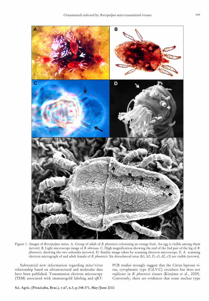
Brevipalpus (Figure 1 A-E) is a genus ofTenuipalpidae (Acari: Prostigmata), family whosemembers are usually referred to as false spider mites.They are ubiquitous in tropical and subtropical regionsaround the world. This genus includes more than 200species (Mesa et al., 2009), but only three are known tobe involved in the transmission of plant viruses, namelyB. californicus Banks, B. obovatus Donnadieu and B.phoenicis (Geijskes) (Childers et al., 2003a), which havebeen found naturally infesting nearly 1000 differentplant species belonging to more than 100 botanicalfamilies throughout the world (Childers et al., 2003b).Brevipalpus mites have a peculiar biology: most of theindividuals in a colony are clones, generated by hap-loid females reproducing by thelytokous parthenogen-
esis (i.e, females begetting females). Only rarely aremales produced (Childers et al., 2003a). It has been re-ported that these haploid mites remain as females be-cause of the infection by bacterial symbionts of the ge-nus Cardinium (Weeks et al., 2001) present in most ofthe organs of the mite (Kitajima et al., 2007). Non-in-fected eggs generate males. The reproductive role ofthe male is unknown. Although it copulates, it appar-ently does not fertilize females (Pijnacker et al., 1981).The three vector Brevipalpus species are distinguishedbased on few external characteristics, as the numberof distal solenidia on tarsus leg II (Figure 1 C, D) andthe number of dorsolateral body setae (Figure 1 E)(Wellbourn et al., 2003). Genetically distinct genotypeswere reported and cryptic species occur among thethree major recognized morphospecies (Rodrigues etal., 2004; Groot, 2006).
Ornamentals infected by Brevipalpus mite-transmitted viruses 349
Sci. Agric. (Piracicaba, Braz.), v.67, n.3, p.348-371, May/June 2010
Substantial new information regarding mite/virusrelationship based on ultrastructural and molecular datahave been published. Transmission electron microscopy(TEM) associated with immunogold labeling and qRT-
PCR studies strongly suggest that the Citrus leprosis vi-rus, cytoplasmic type (CiLV-C) circulates but does notreplicate in B. phoenicis tissues (Kitajima et al., 2009).Conversely, there are evidences that some nuclear type
Figure 1 - Images of Brevipalpus mites. A. Group of adult of B. phoenicis colonizing an orange fruit. An egg is visible among them(arrow). B. Light microscope image of B. obovaus. C. High magnification showing the end of the 2nd pair of the leg of B.phoenicis, showing the two solenidia (arrows). D. Similar image taken by scanning electron microscope. E. A scanningelectron micrograph of and adult female of B. phoenicis. Six dorsolateral setae (h1, h2, f3, e3, d2, c3) are visible (arrows).

Kitajima et al.350
Sci. Agric. (Piracicaba, Braz.), v.67, n.3, p.348-371, May/June 2010
of the viruses transmitted by Brevipalpus (N-BTV) as Or-chid fleck virus (OFV), Coffee ringspot virus (CoRSV),Clerodendrum chlorotic spot virus (ClCSV) and Citrusleprosis virus, nuclear type (CiLV-N) multiply in themite vector’s body (Kitajima et al., 2009).
Plant viruses transmitted by Brevipalpus mitesUntil the beginning of 2000, only a few BTV were
known. The economically most important and the firstto be identified as such was the Citrus leprosis (CL),originally described in Florida at early 20th century(Hume, 1901; Fawcett, 1911). In the 1930’s, it was foundin Argentina, Uruguay, Paraguay and Brazil. Since then,it has been reported from other South American coun-tries (Bolivia, Colombia and Venezuela) as well as fromCentral American countries (Panama, Costa Rica, Hon-duras, Guatemala, Mexico) (Rodrigues et al., 2003;Bastianel et al., 2006). The disease is of concern for cit-rus growers in the Caribbean region, where the virus isabsent, and in the US, where it has not been found sincethe 1960’s (Childers et al., 2003c). The transmission ofCL virus by Brevipalpus mites was initially shown inArgentina by Frezzi (1940) and subsequently confirmedin Brazil and US by Musumeci and Rossetti (1963) andKnorr (1968), respectively. It was found that two distinctviruses, based on cytopathic effects, both transmitted byB. phoenicis, respectively cytoplasmic (CiLV-C) andnuclear (CiLV-N), cause leprosis symptoms, being theCiLV-C the prevalent (Rodrigues et al., 2003). The com-plete sequence of the genome of CiLV-C was obtained(Pascon et al., 2006) being a bipartite positive sensessRNA distinct from known viruses. As a consequence,a new genus, Cilevirus, was proposed for this virus(Locali-Fabris et al., 2006).
Another well-known BTV is the Coffee ringspot vi-rus (CoRSV) first described by Bitancourt (1938) in cof-fee (Coffea arabica L.) plantations of the state of SãoPaulo. This virus causes localized ringspots on leavesand berries. Ultrastructural studies demonstratednuclear type of cytopathology in the tissues of the le-sions (Kitajima and Costa, 1972). Its transmission by B.phoenicis and by mechanical means was demonstratedby Chagas (1973) and Chagas et al. (1961). Part of its ge-nome has been sequenced, which allowed the design ofprimers that specifically amplify CoRSV by RT-PCR(Locali et al., 2005).
Passion fruit green spot virus (PFGSV) is another cy-toplasmic type BTV that devastated passion fruitgrooves at Vera Cruz, state of São Paulo, in the 1990s’.The virus is vectored by B. phoenicis and causes greenspots on yellow fruits and in senescent yellowish leavesand necrotic lesions on stems. When infection is heavy,stem lesions fuse causing annealing and consequent deathof the infected plant (Kitajima et al., 2003b). The genomeof PFGSV has been partially sequenced and specific prim-ers based on these sequences have been designed andused for its detection (Antonioli-Luizon et al., 2008). Be-cause cytophatic effects caused by BTV-cytoplasmic typewere only seen in these green areas of senescent leaves,
it is possible that senescence is retarded in the virus-in-fected leaf tissues. This finding suggested that otherplants could have senescent leaves with green spots,ringspots, brown spots, besides green leaves with chlo-rotic and/or necrotic ringspot, associated withBrevipalpus. Many suspected cases were found, mostlyin ornamentals infested by Brevipalpus. Examination ofthe tissues of the lesion by TEM led to the discovery ofBTV in almost 40 different plant species, mostly orna-mentals (Kitajima et al., 2003a).
Symptoms caused by Brevipalpus-transmitted virusesA common characteristic of the symptoms caused
by BTV is that they are always localized: chlorotic and/or necrotic spots and ringspots on green leaves and greenspots or ringspots on senescent leaves; chlorotic and/ornecrotic spots on the stems, chlorotic or brown spotsusually depressed on the fruits; brown spots on the flow-ers (Kitajima et al., 2003a). The process which keeps theinfection by BTV restricted to the lesion is not under-stood yet. It may result from a tug-of-war between theviral replication and the cell defense mechanism and theinability of the BTV to infect the vascular region(Marques et al., 2007). This, however, does not alwaysapply since systemic infection was observed when Che-nopodium quinoa Wild. and C. amaraticolor Coste &Reyn. were mechanically inoculated with three virusescausing nuclear type of cytopathology (OFV, CoRSVand Clerodendrum chlorotic spot virus [ClCSV]) andkept at high temperatures (28-30oC) for about two weeks(Kondo et al., 1995; Boari et al., 2004; Kitajima et al.,2008). Non-inoculated leaves of these plants exhibitedchlorotic spots and chlorotic veins. In these plants, themechanism which hinders the invasion of phloem ves-sels by the virus is somehow overcome and the patho-gen spreads to the rest of the plant.
Cytopathology of Brevipalpus-transmitted virusesAnother important feature of BTV is the alteration
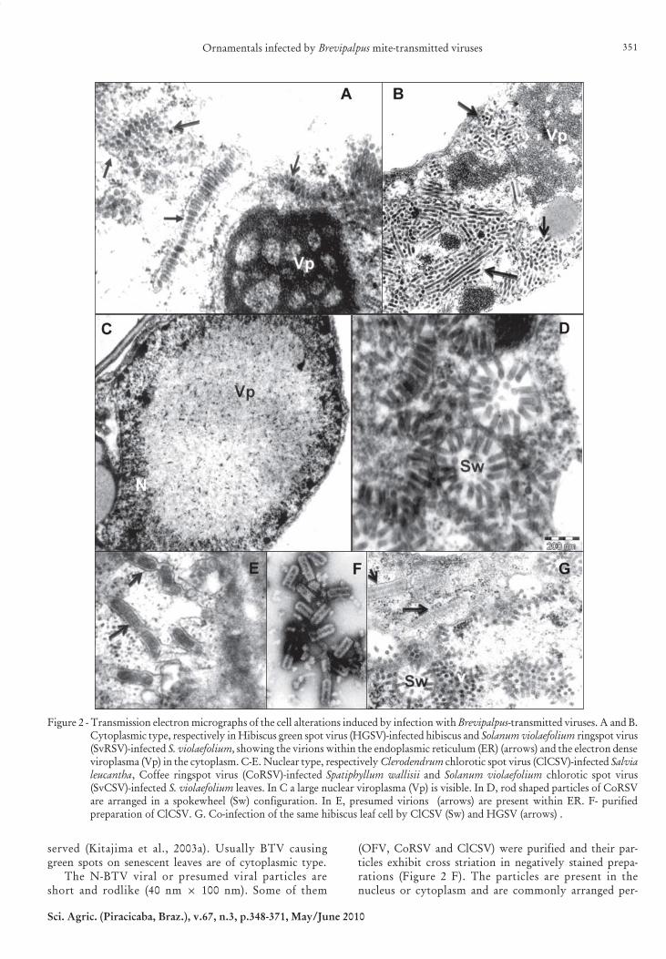
they induce in infected cells. Ultrastructural studies byTEM consistently revealed two types of cytopathology(Figure 2) (Kitajima et al., 2003a): Cytoplasmic type re-ferred to now on as C-BTV and Nuclear type (N-BTV).C-BTV is characterized by the presence of short, bacil-liform, membrane-bounded particle (60-70 nm × 110-120nm) which occurs single or grouped within cisternae ofthe endoplasmic reticulum and by the appearance of anelectron- dense, vacuolated inclusion, referred to asviroplasma, in the cytoplasm (Figure 2 A, B). Usually,there is a single, large viroplasma with elliptical or ir-regular profile. Immunogold labeling experiments withan antibody specific to the CiLV-C p29 (putativecapsidal) protein produces heavy labeling of theviroplasma, in addition to the bacilliform particles, in-dicating that it is a site of accumulation of viral proteins.C-BTV was first described in cells infected by CiLV-C(Colariccio et al., 1995). In some instances, evidence ofviral morphogenesis by budding process of viroplasmamaterial into the endoplasmic reticulum has been ob-
Ornamentals infected by Brevipalpus mite-transmitted viruses 351
Sci. Agric. (Piracicaba, Braz.), v.67, n.3, p.348-371, May/June 2010
served (Kitajima et al., 2003a). Usually BTV causinggreen spots on senescent leaves are of cytoplasmic type.
The N-BTV viral or presumed viral particles areshort and rodlike (40 nm × 100 nm). Some of them
(OFV, CoRSV and ClCSV) were purified and their par-ticles exhibit cross striation in negatively stained prepa-rations (Figure 2 F). The particles are present in thenucleus or cytoplasm and are commonly arranged per-
Figure 2 - Transmission electron micrographs of the cell alterations induced by infection with Brevipalpus-transmitted viruses. A and B.Cytoplasmic type, respectively in Hibiscus green spot virus (HGSV)-infected hibiscus and Solanum violaefolium ringspot virus(SvRSV)-infected S. violaefolium, showing the virions within the endoplasmic reticulum (ER) (arrows) and the electron denseviroplasma (Vp) in the cytoplasm. C-E. Nuclear type, respectively Clerodendrum chlorotic spot virus (ClCSV)-infected Salvialeucantha, Coffee ringspot virus (CoRSV)-infected Spatiphyllum wallisii and Solanum violaefolium chlorotic spot virus(SvCSV)-infected S. violaefolium leaves. In C a large nuclear viroplasma (Vp) is visible. In D, rod shaped particles of CoRSVare arranged in a spokewheel (Sw) configuration. In E, presumed virions (arrows) are present within ER. F- purifiedpreparation of ClCSV. G. Co-infection of the same hibiscus leaf cell by ClCSV (Sw) and HGSV (arrows) .

Kitajima et al.352
Sci. Agric. (Piracicaba, Braz.), v.67, n.3, p.348-371, May/June 2010
pendicularly onto the membranes of the endoplasmicreticulum or nuclear envelope. Sometimes this mem-brane/virion complex produces cylindrical structureswhich in cross section exhibit radial arrangement ofparticles surrounded by a membrane ring, generatinga configuration referred to as “spokewheel” (Figure 2D). Also groups of viral particles may form a large,side-by-side, sheet-like, aggregate. An additional char-acteristic feature of N-BTV infection is the presenceof an electron lucent mass, the viroplasma, in thenucleus (Figure 2 C). In the case of OFV, antibody pro-duced against purified particles heavily impregnates theviroplasma, indicating that it may represent, as withthe cytoplasmic viroplasma, a site of accumulation ofviral materials. A possible variant of N-BTV seems tobe the cases in which short, membrane bounded,rhabdoviruslike particles appear within endoplasmicreticulum with rare nuclear viroplasma as in the caseof CoRSV-SP, Solanum violaefolium chlorotic spot vi-rus (SvCSV) (Kitajima et al., 2004) (Figure 2 E) andCestrum ringspot virus (CeRSV) (Kitajima et al., 2003a).In a few cases, as in Clerodendrum thomsoniae and Hi-biscus rosa-sinensis, co-infection of the same cell by C-BTV and N-BTV has been observed (Figure 2 G).
Brevipalpus-transmitted viruses and ornamentalsAs our knowledge about possible BTV advanced
in the last decade, a large number of cases were de-tected, mostly in ornamentals, causing localized le-sions. One reason why these viruses have been ne-glected is the lack of systemic infection. If the mitepopulation declines, due to seasonal factors, and theinfected organs fall off or die, the sources of inocu-lum are reduced, and the disease literally disappears.But they are potentially important in cases when fa-vorable environmental conditions (climate, host spe-cies, lack of predators, etc.) result in outbreaks of themite vector. In this scenario, a heavy infection mayoccur causing economic damage.
Except for OFV which has a world wide distribu-tion (Kondo et al., 2003) because of the extensive inter-national trade that spreads both the vector and the vi-rus, other known BTV have been registered only in theAmerican continent. However, growing internationaltrade, including living plants, may help the dispersionof BTV and viruliferous mite vectors to other parts ofthe world. The situation becomes more complex becauseBrevipalpus mites have already been found in most ofthe tropical and subtropical regions of the world, infest-ing many plant species. Additionally, economically im-portant BTV, such as CiLV-C and CoRSV are able toinfect other plant species including some ornamentals(Bastianel et al., 2006; Novelli et al., 2008), under eithernatural or experimental conditions. Thus these virusesmay accidentally be introduced into disease-free regionsby infected ornamentals.
An annotated list of ornamental plants that can beinfected by BTV is subsequentely presented, with com-ments on the corresponding diseases, known vectors and
other known information including natural infection byother viruses (Albouy and Devergne, 1999; Alexandre etal., 2005). For this list, we considered BTV to have beenidentified, when at least experimental mite transmissionhas been reported. In other cases, we are referring to thepresumed BTV found by TEM associated with the le-sions as “unidentified BTV”. Table 1 summarizes thisannotated list. The description of the ornamental prop-erties of the cited plant species were mostly based onLorenzi and de Souza (2001), whereas plant names werebased on the International Plant Name Index(www.inpi.org).
I. ACANTHACEAE
1. Thunbergia erecta (Benth.) T. Anderson - King’smantle (manto-do-rei)
King’s mantle is a woody, erect shrub, 2-2.5 m tall,from Tropical Africa, with ovate, thick, brilliant leaves.The blue to purple flowers are tubular with yellowthroat and they bloom nearly the entire year. Althoughgrown in isolation, they are most commonly used ashedge. T. erecta plants with ringspot on leaves (Figure 3A) and infested by B. phoenicis were found in a residen-tial garden at Águas de São Pedro, SP. TEM of the le-sions revealed the presence of cytopathic effect causedby C-BTV (Nogueira et al., 2003). Similar case was foundwith king’s mantle being used as hedge in a hotel atJaguariúna, SP (E.W. Kitajima, unpublished data). Nofurther work to characterize and identify the virus wasconducted.
No other virus has been reported infecting T. erectain Brazil but Broad bean wilt virus (BBWV) and Daturayellow vein virus (DYVV) have been recorded elsewhere(Albouy and Devergne, 1999).
II. AGAVACEAE
2. Cordyline terminalis (L.) Kunth. - Ti-plant (dracenavermelha)
This plant was formerly classified as Liliaceae, butaccording to the International Plant Name Index (INPI)it is now considered a member of the family Agavaceae.Ti-plant is a semi-woody, erect shrub, which may reach2 m in height, with terminal tufts of elongated, smooth,flexible white or colorful (green to red, sometimesstriped) leaves. Ti-plants are planted in vases to be keptindoors or in outdoor rows. It is native to India, Malay-sia and Polynesia. A few plants with pink spots andringspots on red leaves (Figure 3 B) were found at thecampus of Escola Superior de Agricultura “Luiz deQueiroz”, Universidade de São Paulo (ESALQ/USP),Piracicaba, state of São Paulo (SP). Tissues of these le-sions exhibited cytopathic effects typical of C-BTV(Ferreira et al., 2004a). The causal virus may be relatedto the Hibiscus green spot virus (HGSV) because theseTi-plants were growing next to affected hibiscus. Apotyvirus has been reported in this plant associated withleaf discoloration and ringspots in Croacia and Italy(Albouy and Devergne, 1999).
Ornamentals infected by Brevipalpus mite-transmitted viruses 353
Sci. Agric. (Piracicaba, Braz.), v.67, n.3, p.348-371, May/June 2010
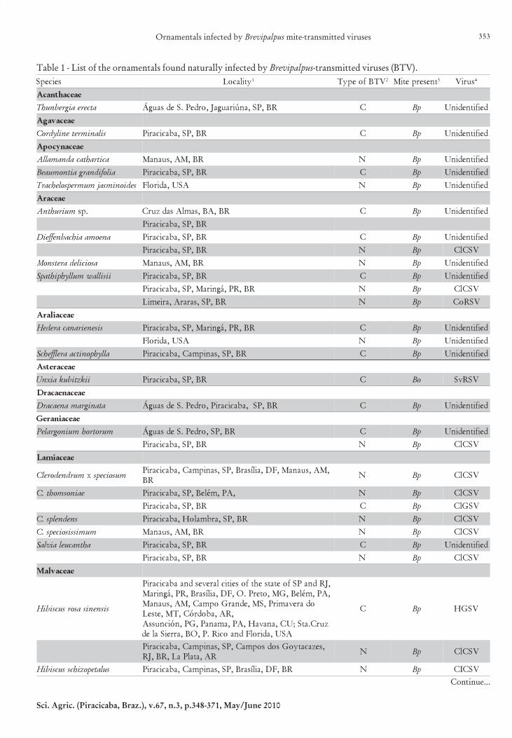
Table 1 - List of the ornamentals found naturally infected by Brevipalpus-transmitted viruses (BTV).
Continue...
seicepS ytilacoL 1 VTBfoepyT 2 tneserpetiM 3 suriV 4
eaecahtnacA
atcereaigrebnuhT RB,PS,anúiraugaJ,ordeP.SedsaugÁ C pB deifitnedinU
eaecavagA
silanimretenilydroC RB,PS,abacicariP C pB deifitnedinU
eaecanycopA
acitrahtacadnamallA RB,MA,suanaM N pB deifitnedinU
ailofidnargaitnomuaeB RB,PS,abacicariP C pB deifitnedinU
sedionimsajmumrepsolehcarT ASU,adirolF N pB deifitnedinU
eaecarA
muiruhtnA .ps RB,AB,samlAsadzurC C pB deifitnedinU
RB,PS,abacicariP
aneomaaihcabneffeiD RB,PS,abacicariP C pB deifitnedinU
RB,PS,abacicariP N pB VSClC
asoiciledaretsnoM RB,MA,suanaM N pB deifitnedinU
iisillawmullyhpihtapS RB,PS,abacicariP C pB deifitnedinU
RB,RP,ágniraM,PS,abacicariP N pB VSClC
RB,PS,sararA,ariemiL N pB VSRoC
eaecailarA
siseneiranacaredeH RB,RP,ágniraM,PS,abacicariP C pB deifitnedinU
ASU,adirolF N pB deifitnedinU
allyhponitcaarelffehcS RB,PS,sanipmaC,abacicariP C pB deifitnedinU
eaecaretsA
iikztibukaixnU RB,PS,abacicariP C oB VSRvS
eaecaneacarD
atanigramaneacarD RB,PS,abacicariP,ordeP.SedsaugÁ C pB deifitnedinU
eaecainareG
murotrohmuinograleP RB,PS,ordeP.SedsaugÁ C pB deifitnedinU
RB,PS,abacicariP N pB VSClC
eaecaimaL
murdnedorelC x musoiceps,MA,suanaM,FD,ailísarB,PS,sanipmaC,abacicariP
RBN pB VSClC
eainosmoht.C ,AP,méleB,PS,abacicariP N pB VSClC
RB,PS,abacicariP C pB VSGlC
snednelps.C RB,PS,arbmaloH,abacicariP N pB VSClC
mumissisoiceps.C RB,MA,suanaM N pB VSClC
ahtnacuelaivlaS RB,PS,abacicariP C pB deifitnedinU
RB,PS,abacicariP N pB VSClC
eaecavlaM
sisnenisasorsucsibiH
,JRdnaPSfoetatsehtfoseiticlarevesdnaabacicariP,AP,méleB,GM,oterP.O,FD,ailísarB,RP,ágniraM
odarevamirP,SM,ednarGopmaC,MA,suanaM,RA,abodróC,TM,etseL
zurC.atS;UC,anavaH,AP,amanaP,GP,nóicnussAASU,adirolFdnaociR.P,OB,arreiSaled
C pB VSGH
,sezacatyoGsodsopmaC,PS,sanipmaC,abacicariPRA,atalPaL,RB,JR
N pB VSClC
sulatepozihcssucsibiH RB,FD,ailísarB,PS,sanipmaC,abacicariP N pB VSCIC

Kitajima et al.354
Sci. Agric. (Piracicaba, Braz.), v.67, n.3, p.348-371, May/June 2010
Table 1 - Continuation.
RB,PS,abacicariP C pB VSGH
sucairyssucsibiH RB,PS,sanipmaC,PS,abacicariP C pB VSGH
sueniccocsucsibiH RB,PS,abacicariP N pB VSClC
sunibannacsusibiH RB,PS,abacicariP N pB VSClC
RB,PS,abacicariP C pB VSGH
suerobrasucsivavlaMPS,sanipmaC,abacicariP
RB,JR,orienaJ.RN pB VSClC
eaecaelO
mudiculmurtsugiL RB,RP,abitiruC,PS,abacicariP C pB VSRgiL
sisnenismurtsugiLetnoM,abacicariP,ordeP.SedsaugÁ,RA,aidrócnoC
RB,FD,ailísarB,PS,ergelAC pB VSRgiL
eaecadihcrO
seicepsdnaareneglareveS dlrowehtnisecalplareveS N cB VFO
,enygoleoC,aissarB,suiahP,ailemmuJ,muidimroH
muibolyXRB,PS,abacicariP C - deifitnedinU
anidnurA ,.ps sisponealahP.ps
RB,MA,suanaM,PS,açnagarB C - deifitnedinU
murdnedipE .ps RB,TM,etseLodarevamirP C - deifitnedinU
RB,PS,abacicariP
muidicnO .ps RB,PS,abacicariP C pB VSRvS
muibordneD .ps RB,PS,abacicariP N pB VSClC
eaecaropsottiP
aribotmuropsottiP ASU,adirolF N pB deifitnedinU
eaecanigabmulP
ataluciruaogabmulP RB,PS,aiabitA C pB deifitnedinU
eaecalumirP
arolfitsegnocaihcamisyL RB,PS,ordeP.SedsaugÁ C pB deifitnedinU
eaecaibuR
sedionimsajainedraG RB,MA,ucurU N pB deifitnedinU
allyhporhtyreadneassuM RB,MA,suanaM N pB deifitnedinU
eaecanaloS
arolficuapaislefnurB RA,atalPaL N pB deifitnedinU
arolfinuaislefnurB RB,PS,abacicariP,ordeP.SedsaugÁ C pB deifitnedinU
munrutconmurtseC RB,PS,aiabitA N oB VSReC
RB,PS,abacicariP N pB VSClC
muilofealoivmunaloS RB,PS,abacicariP C oB VSRvS
RB,PS,sanipmaC N pB VSCvS
eaecaloiV
atarodoaloiV SUA,ruobmaN N - VFO1AR- Argentina; AUS- Australia; BO- Bolivia; BR- Brazil; CO- Colombia; CU- Cuba; PA- Panama; PG- Paraguay. 2C- Brevipalpustransmitted virus cytoplasmic type; N- idem, nuclear type. 3Bc- Brevipalpus californicus; Bo- B. obovatus; Bp- B. phoenicis .4Unidentified:when only symptoms, association with Brevipalpus and cytopathology are described.
III. APOCYNACEAE
3. Allamanda cathartica L. - Allamanda (alamanda)Allamanda is a tropical climbing vine with milky
sap, bright and thick leaves with trumpet shaped yellowflowers, native to the Brazilian coast. It is widely usedas hedge or arbors. Plants with green spots and ringspots
on senescent leaves (Figure 3 C) were found in residen-tial garden in Manaus, AM, associated with infestationby B. phoenicis and cytopathic effects of N-BTV(Rodrigues et al., 2008). No additional work was carriedout to identify the causal virus. Cucumber mosaic virus(CMV) has been found naturally infecting allamanda inBrazil (Alexandre et al., 2005).

Ornamentals infected by Brevipalpus mite-transmitted viruses 355
Sci. Agric. (Piracicaba, Braz.), v.67, n.3, p.348-371, May/June 2010
4. Beaumontia grandiflora Wall. - Herald’s trumpet(trombeta do arauto)
This is an evergreen, branched woody twiningplant with large, thick and glossy leaves and large,white, fragrant, trumpet shaped flowers. It is nativeto tropical Himalaya and it is suitable for arbors oras isolated plants. Some plants growing in the cam-pus of ESALQ, Piracicaba, SP, were found with greenspots and ringspots on senescent leaves (Figure 3 D)in which cytopathology caused by C-BTV was ob-
served (Kitajima et al., 2006). The causal virus remainsunidentified.
5. Trachelospermum jasminoides Lem. - star jasmine(jasmim estrelado)
Star jasmine is an evergreen, branched, lactiferous,climbing vine, with small dark green leaves and abun-dant pure-white, intensely fragrant flowers with tubularcorolla opening out into five petal-like twisted lobes. Itis native to Eastern and Southeastern Asia, and it iswidely used in arbors and as hedge. Star jasmine plants
Figures 3-11 - Symptoms caused by natural infection by Brevipalpus-transmitted viruses (BTV) on ornamentals. Fig. 3. A. Green spotson senescent leaves of Thunbergia erecta caused by a cytoplasmic type of BTV (C-BTV). B. Ringspot on the leavesof Cordyline terminalis caused by a C-BTV. C. Green spots on Allamanda cathartica from Manaus, AM, BR, caused bya nuclear type of BTV (N-BTV). D. Green spots on senescent leaves of Beaumontia grandiflora caused by a C-BTV. E.Chlorotic ringspot on the leaves of Trachelospermum jasminoides collected at Florida, US, caused by a N-BTV. F.Chlorotic spots on the leaves of Anthurium sp. caused by a C-BTV.

Kitajima et al.356
Sci. Agric. (Piracicaba, Braz.), v.67, n.3, p.348-371, May/June 2010
with chlorotic ringspots (Figure 3 E) were found in aresidential garden in North Central Florida, USA, asso-ciated with B. phoenicis infestation and N-BTV cytopa-thology (Rodrigues et al., 2004b). The causal virus is stillunidentified.
IV. ARACEAE
6. Anthurium sp. - Anthurium (antúrio)The genus Anthurium contains 600-800 species with
neotropical distribution mostly from Central and SouthAmerica. They grow as twining epiphytes or terrestrial.Several species are used as ornamentals due to the inflo-rescence, which has a large spathe of varied colors andsmall flowers arranged on a fleshy axis, the spadix.Leaves are usually large, spatulate or round. Sample ofanthurium leaves, possibly A. andraeanum Linden, show-ing chlorotic spots and ringspots (Figure 3 F) from acommercial nursery of Cruz das Almas, BA, associatedwith Brevipalpus mite infestation were examined by TEMand revealed C-BTV cytopathology (Ferreira et al.,2004a). The same finding was made in a plant growingin the campus of ESALQ at Piracicaba, SP (E.W.Kitajima, unpublished data).
7. Dieffenbachia amoena Hort. ex. Gentil - dumbcane(comigo-ninguém-pode)
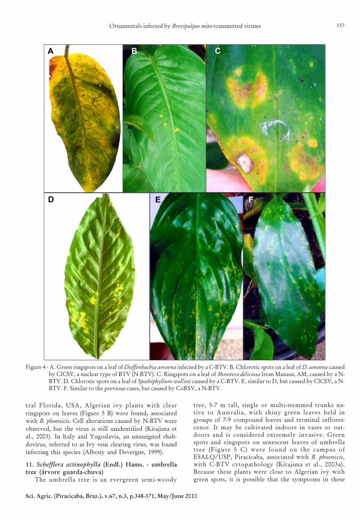
Dumbcane is a perennial, herbaceous, erect plantreaching about 1.5 m, with very ornamental, dark greenleaves with irregular white zones along the veins. It iswidely used in interiorscape at half shade in vases or ingardens. The sap of dumbcane is poisonous. Adumbcane plant showing ringspots on the leaves (Fig-ure 4 A), associated with infestation by B. phoenicis, wasfound in a residential garden in Piracicaba, SP. Cellsfrom the lesions showed cyopathology of C-BTV (E.W.Kitajima, unpublished data). Because this plant was to-gether with Salvia leucantha also infected by an uniden-tified C-BTV, it is possible that the same virus was in-fecting both species. A plant of dumbcane growing nearClCSV-infected bleeding heart, in the campus ofESALQ/USP developed chlorotic spots (Figure 4 B).The plant was infested by B. phoenicis and TEM exami-nation of the lesion tissues revealed cell alterations in-duced by N-BTV. Further analysis revealed that thisplant was infected by ClCSV, the bleeding heart actingas the virus source (Kitajima et al., 2008).
8. Monstera deliciosa Liebm. - widowleaf (costela-de-adão)
The widowleaf is an evergreen, vigorous liana,with large, coriaceous and perforated leaves and growswell in half shade. It usually grows over wall, mosspillar or tree. Plants of M. deliciosa showing ringspoton their leaves (Figure 4 C) were found in a residen-tial garden in Manaus, AM, associated with infesta-tion by B. phoenicis. TEM examination revealed cyto-pathology of N-BTV in the tissues of the lesion(Rodrigues et al., 2008). The virus remains unidenti-fied.
9. Spathiphyllum wallisii Regel - Peace lily (lírio-da-paz)
Vigorous, perennial, erect plant, 30-40 cm tall withbright, coriaceous leaves originated from Colombiaand Venezuela. It produces inflorescence with charac-teristic white spathe that turns green as it ages, withoutfragrance. Peace lily is cultivated in flower beds or vasesat half shade, and also in aquarium. Several plants culti-vated in a vase in a bakery at Piracicaba, SP, exhibitedringspots on their leaves (Figure 4 D), associated withinfestation with B. phoenicis, and the cells of the lesionsshowed cytopathic effects of C-BTV. The causal viruswas not identified yet. As in the case of dumbcane, S.wallisii plants growing next to a ClCSV-infected bleed-ing heart in the campus of ESALQ/USP at Piracicaba,SP, were infected under natural conditions by this vi-rus, developing chlorotic spots (Figure 4 E). These symp-toms were reproduced by experimental transmission ofthe ClCSV by the mite B. phoenicis and the presence ofthe virus was confirmed by TEM, RT-PCR and serol-ogy (Kitajima et al., 2008). Peace lily plants with chlo-rotic spots were also found in a public park at Maringá,PR. Lesions contained unidentified N-BTV (E.W.Kitajima, unpublished data). A peace lily plant pur-chased in a nursery at Limeira, SP, developed chloroticspots (Figure 4 F) initially taken as a result of infectionby ClCSV and indeed showing cytopathology of N-BTV,but RT-PCR assays indicated that this particular plantwas infected by an isolate of CoRSV (Novelli et al.,2008). Thus peace lily seems to be susceptible to severalBTV. Another case of natural infection of peace lily withCoRSV was found in a plant collected in Araras, SP.This plant was co-infected by PFGSV as revealed by RT-PCR and TEM assays (V.M. Novelli, unpublished data).
Some ornamental araceae as Anthurium andDieffenbachia have been found naturally infected byDasheen mosaic virus (DsMV) in several parts of Brazil(Alexandre et al., 2005). Elsewhere, natural infection ofornamental araceae by viruses like Tomato spotted wiltvirus (TSWV), Alfalfa mosaic virus (AMV), CMV, Arabismosaic virus (ArMV) and Potato virus X (PVX), besidesDsMV, are reported (Albouy and Devergne, 1999).
V. ARALIACEAE
10. Hedera canariensis Willd. - Algerian or CanaryIsland ivy (hera)
H. canariensis is a semi-herbaceous, clinging vine withornamental, lobate leaves. Aerial roots permit the plantto cling on walls and trees. It is also used as ground cover.Algerian ivy, clinging to trees in the campus of ESALQ/USP, Piracicaba, was found showing large green patcheson senescent leaves (Figure 5 A) associated with B.phoenicis. TEM examination revealed C-BTV cytopathol-ogy (Kitajima et al., 2003a) in the leaf tissues of the greenareas. A similar case was observed in Algerian ivy plantsgrowing in a residential garden at Maringá, PR (E.W.Kitajima, unpublished data). The causal virus is still uni-dentified. In a survey conducted at the northern and cen-
Ornamentals infected by Brevipalpus mite-transmitted viruses 357
Sci. Agric. (Piracicaba, Braz.), v.67, n.3, p.348-371, May/June 2010
tral Florida, USA, Algerian ivy plants with clear
ringspots on leaves (Figure 5 B) were found, associatedwith B. phoenicis. Cell alterations caused by N-BTV were
observed, but the virus is still unidentified (Kitajima etal., 2003). In Italy and Yugoslavia, an unassigned rhab-
dovirus, referred to as Ivy vein clearing virus, was foundinfecting this species (Albouy and Devergne, 1999).
11. Schefflera actinophylla (Endl.) Hams. - umbrellatree (árvore guarda-chuva)
The umbrella tree is an evergreen semi-woody
tree, 5-7 m tall, single or multi-stemmed trunks na-tive to Australia, with shiny green leaves held ingroups of 7-9 compound leaves and terminal inflores-cence. It may be cultivated indoors in vases or out-doors and is considered extremely invasive. Greenspots and ringspots on senescent leaves of umbrellatree (Figure 5 C) were found on the campus ofESALQ/USP, Piracicaba, associated with B. phoenicis,with C-BTV cytopathology (Kitajima et al., 2003a).Because these plants were close to Algerian ivy withgreen spots, it is possible that the symptoms in these
Figure 4 - A. Green ringspots on a leaf of Dieffenbachia amoena infected by a C-BTV. B. Chlorotic spots on a leaf of D. amoena causedby ClCSV, a nuclear type of BTV (N-BTV). C. Ringspots on a leaf of Monstera deliciosa from Manaus, AM, caused by a N-BTV. D. Chlorotic spots on a leaf of Spathiphyllum wallisii caused by a C-BTV. E. similar to D, but caused by ClCSV, a N-BTV. F. Similar to the previous cases, but caused by CoRSV, a N-BTV.

Kitajima et al.358
Sci. Agric. (Piracicaba, Braz.), v.67, n.3, p.348-371, May/June 2010
two species are caused by the same virus. A similarcase was found at the Universidade Estadual deCampinas (Unicamp), at Campinas, SP (E.W.Kitajima, unpublished data).
Schefflera is also susceptible to Schefflera ringspot vi-rus (SRV) found in Australia and Europe (Albouy andDevergne, 1999) and also in Brazil (Alexandre et al.,2005).
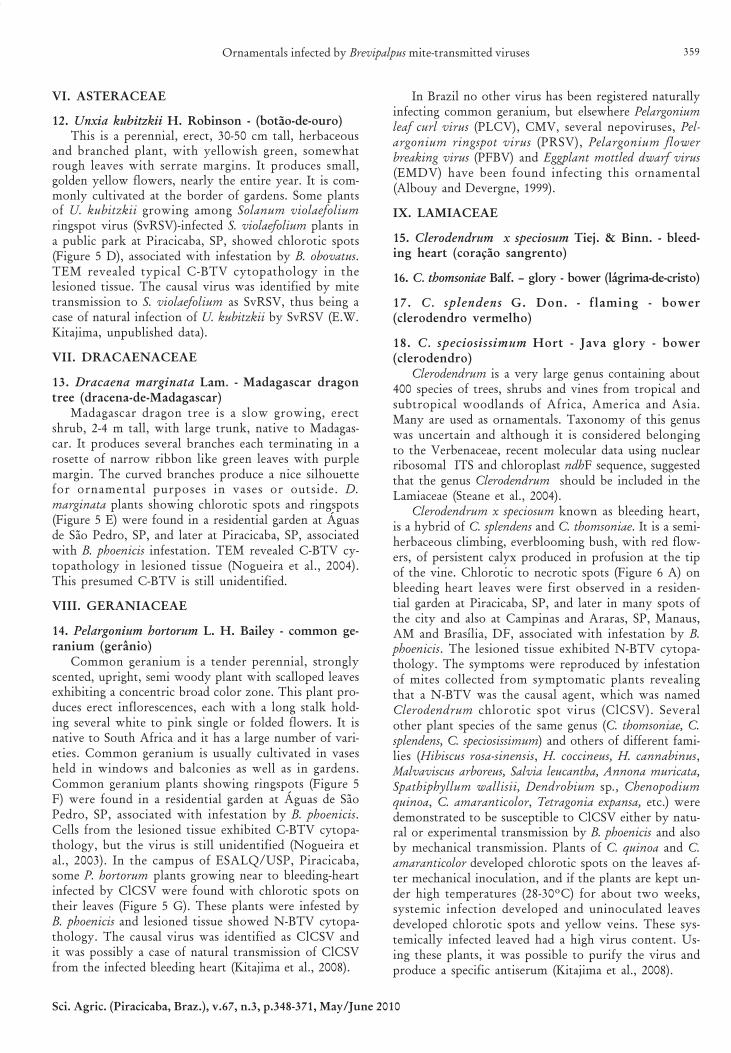
Figure 5 - A. Green spots on a senescent leaf of Hedera canariensis caused by a C-BTV. B. Ringpot on a green leave of H. canariensisfound at Florida, USA, caused by a N-BTV. C. Green spots and ringsppots on a senescent leaf of Schefflera actinophyllaassociated with a C-BTV. D. Chlorotic spots on a leaf of Unxia kubitzkii caused by natural infection by Solanum violaefoliumringspot virus (SvRSV), a C-BTV. E. Chlorotic spots on a leaf of Dracaena marginata associated with a C-BTV. F. Greenspots and ringspots on a senescent leaf of Pelargonium hortorum associated with a C-BTV. G. Chlorotic spots on a leaf ofP. hortorum caused by ClCSV.
Ornamentals infected by Brevipalpus mite-transmitted viruses 359
Sci. Agric. (Piracicaba, Braz.), v.67, n.3, p.348-371, May/June 2010
VI. ASTERACEAE
12. Unxia kubitzkii H. Robinson - (botão-de-ouro)This is a perennial, erect, 30-50 cm tall, herbaceous
and branched plant, with yellowish green, somewhatrough leaves with serrate margins. It produces small,golden yellow flowers, nearly the entire year. It is com-monly cultivated at the border of gardens. Some plantsof U. kubitzkii growing among Solanum violaefoliumringspot virus (SvRSV)-infected S. violaefolium plants ina public park at Piracicaba, SP, showed chlorotic spots(Figure 5 D), associated with infestation by B. obovatus.TEM revealed typical C-BTV cytopathology in thelesioned tissue. The causal virus was identified by mitetransmission to S. violaefolium as SvRSV, thus being acase of natural infection of U. kubitzkii by SvRSV (E.W.Kitajima, unpublished data).
VII. DRACAENACEAE
13. Dracaena marginata Lam. - Madagascar dragontree (dracena-de-Madagascar)
Madagascar dragon tree is a slow growing, erectshrub, 2-4 m tall, with large trunk, native to Madagas-car. It produces several branches each terminating in arosette of narrow ribbon like green leaves with purplemargin. The curved branches produce a nice silhouettefor ornamental purposes in vases or outside. D.marginata plants showing chlorotic spots and ringspots(Figure 5 E) were found in a residential garden at Águasde São Pedro, SP, and later at Piracicaba, SP, associatedwith B. phoenicis infestation. TEM revealed C-BTV cy-topathology in lesioned tissue (Nogueira et al., 2004).This presumed C-BTV is still unidentified.
VIII. GERANIACEAE
14. Pelargonium hortorum L. H. Bailey - common ge-ranium (gerânio)
Common geranium is a tender perennial, stronglyscented, upright, semi woody plant with scalloped leavesexhibiting a concentric broad color zone. This plant pro-duces erect inflorescences, each with a long stalk hold-ing several white to pink single or folded flowers. It isnative to South Africa and it has a large number of vari-eties. Common geranium is usually cultivated in vasesheld in windows and balconies as well as in gardens.Common geranium plants showing ringspots (Figure 5F) were found in a residential garden at Águas de SãoPedro, SP, associated with infestation by B. phoenicis.Cells from the lesioned tissue exhibited C-BTV cytopa-thology, but the virus is still unidentified (Nogueira etal., 2003). In the campus of ESALQ/USP, Piracicaba,some P. hortorum plants growing near to bleeding-heartinfected by ClCSV were found with chlorotic spots ontheir leaves (Figure 5 G). These plants were infested byB. phoenicis and lesioned tissue showed N-BTV cytopa-thology. The causal virus was identified as ClCSV andit was possibly a case of natural transmission of ClCSVfrom the infected bleeding heart (Kitajima et al., 2008).
In Brazil no other virus has been registered naturallyinfecting common geranium, but elsewhere Pelargoniumleaf curl virus (PLCV), CMV, several nepoviruses, Pel-argonium ringspot virus (PRSV), Pelargonium flowerbreaking virus (PFBV) and Eggplant mottled dwarf virus(EMDV) have been found infecting this ornamental(Albouy and Devergne, 1999).
IX. LAMIACEAE
15. Clerodendrum x speciosum Tiej. & Binn. - bleed-ing heart (coração sangrento)
16. C. thomsoniae Balf. – glory - bower (lágrima-de-cristo)
17. C. splendens G. Don. - flaming - bower(clerodendro vermelho)
18. C. speciosissimum Hort - Java glory - bower(clerodendro)
Clerodendrum is a very large genus containing about400 species of trees, shrubs and vines from tropical andsubtropical woodlands of Africa, America and Asia.Many are used as ornamentals. Taxonomy of this genuswas uncertain and although it is considered belongingto the Verbenaceae, recent molecular data using nuclearribosomal ITS and chloroplast ndhF sequence, suggestedthat the genus Clerodendrum should be included in theLamiaceae (Steane et al., 2004).
Clerodendrum x speciosum known as bleeding heart,is a hybrid of C. splendens and C. thomsoniae. It is a semi-herbaceous climbing, everblooming bush, with red flow-ers, of persistent calyx produced in profusion at the tipof the vine. Chlorotic to necrotic spots (Figure 6 A) onbleeding heart leaves were first observed in a residen-tial garden at Piracicaba, SP, and later in many spots ofthe city and also at Campinas and Araras, SP, Manaus,AM and Brasília, DF, associated with infestation by B.phoenicis. The lesioned tissue exhibited N-BTV cytopa-thology. The symptoms were reproduced by infestationof mites collected from symptomatic plants revealingthat a N-BTV was the causal agent, which was namedClerodendrum chlorotic spot virus (ClCSV). Severalother plant species of the same genus (C. thomsoniae, C.splendens, C. speciosissimum) and others of different fami-lies (Hibiscus rosa-sinensis, H. coccineus, H. cannabinus,Malvaviscus arboreus, Salvia leucantha, Annona muricata,Spathiphyllum wallisii, Dendrobium sp., Chenopodiumquinoa, C. amaranticolor, Tetragonia expansa, etc.) weredemonstrated to be susceptible to ClCSV either by natu-ral or experimental transmission by B. phoenicis and alsoby mechanical transmission. Plants of C. quinoa and C.amaranticolor developed chlorotic spots on the leaves af-ter mechanical inoculation, and if the plants are kept un-der high temperatures (28-30ºC) for about two weeks,systemic infection developed and uninoculated leavesdeveloped chlorotic spots and yellow veins. These sys-temically infected leaved had a high virus content. Us-ing these plants, it was possible to purify the virus andproduce a specific antiserum (Kitajima et al., 2008).

Kitajima et al.360
Sci. Agric. (Piracicaba, Braz.), v.67, n.3, p.348-371, May/June 2010
The purified virus permitted sequencing of part ofits genome, and to design pair of primers that can beused for specific molecular detection of ClCSV by RT-PCR (Kubo et al., 2007). ClCSV was also found natu-rally infecting glory - bower (C. thomsoniae ) atPiracicaba, SP and Belém, state of Pará, causing chlo-rotic/necrotic spots on the leaves (Figure 6 B). At leastin one instance, brownish spots were observed in thewhite sepal (Figure 6 C) in which virus was present(Kitajima et al., 2008). This plant, native to West Africa,is similar to bleeding heart but the flowers have a whiteand inflated sepal and an expanded red corolla.
Flaming bower (C. splendens) plants growing in resi-dential gardens at Piracicaba and Holambra, SP, werealso found naturally infected by ClCSV, showing chlo-rotic spots (Figure 6 D) (Kitajima et al., 2008). This spe-cies native to tropical Africa has oval and wrinkledleaves, and scarlet flowers arranged in dense terminalclusters.
During a survey of plant viruses in the Amazon ba-sin, a plant of Java glory - bower (C. speciosissimum) wasfound infected by ClCSV. Affected leaves showed dark
green spots and ringspots (Figure 6 E) on their leaves(Rodrigues et al., 2008). This Clerodendrum species isnative to Sri Lanka and Java and has broad dark greenleaves and a large upright panicle of fiery scarlet flow-ers produced practically all over the year. Glory - bower(C. thomsoniae) plants were found at Piracicaba, SP,showing green spots on senescent leaves (Figure 6 F) inwhich C-BTV cytopathology was observed. These symp-toms were reproduced by infestation of healthy glory -bower with mites collected from affected leaves, and thevirus was named Clerodendrum green spot virus (ClGSV)(Kitajima et al., 2003a). In a few instances, these lesionsalso contained N-BTV and co-infection in the same leafparenchymal cell by both N- and C- BTV were observed(Kitajima et al., 2003a). This is considered strong evi-dence that C- and N-BTV are unrelated because they donot interfere with the replication of each other.
Ringspots on leaves of C. x speciosum were found atCordeirópolis, SP, associated with the infection of anunidentified tospovirus (E.W. Kitajima, unpublisheddata). Elsewhere there is a report of the infection ofClerodendrum by TSWV (Aubouy and Devergne, 1999).
Figure 6 - A-E. Localized symptoms caused by Clerodendrum chlorotic spot virus (ClCSV). A. Chlorotic spots on the leaves ofbleeding heart (Clerodendrum x speciosum). B. Similar symptoms on the leaves of glory - bower (C. thomsoniae) as well asbrown spots on the sepal of the flower (C). D. Chlorotic spots on the leaves of flaming bower (C. splendens). E. A leafof Java glory - bower (C. speciosissimum) from Manaus, AM, showing green spots and ringspots on the leaves of glory -bower associated with an unidentified N-BTV. F. Green spots on a senescent leaf of C. thomsoniae caused by an unidentifiedC-BTV.

Ornamentals infected by Brevipalpus mite-transmitted viruses 361
Sci. Agric. (Piracicaba, Braz.), v.67, n.3, p.348-371, May/June 2010
19. Salvia leucantha Cav. - Mexican bush sage (salviabranca)
S. leucantha is a perennial, erect, bushy evergreenshrub, 50-90 cm tall, with long branches and lanceolateleaves, with white and woolly underneath. Its flowersare white with lavender-blue calyces, blooming nearlythe entire year. Mexican bush sage is native to Mexico.It is planted mostly at the edges of gardens. Mites cantransmit ClCSV to S. leucantha producing chlorotic spots(Figure 7 A) (Kitajima et al., 2008). S. leucantha was alsofound infected by an unidentified C-BTV in a residen-tial garden at Piracicaba, SP, showing green spots on se-nescent leaves (Figure 7 B) (Kitajima et al., 2003a).
X. MALVACEAE
20. Hibiscus rosa sinensis L. - Chinese hibiscus(hibisco, mimo-de-Venus)
21. H. schizopetalus Hook. f. - Japanese lantern(hibisco crespo)
22. H. syriacus L. - rose of Sharom (rosa-de-sarom,hibisco-da-Síria)
23. H. coccineus Walter- scarlet hibiscus
24. H. cannabinus L. - kenaf (kenaf)The genus Hibiscus comprises more than 200 species
native to tropical and subtropical regions, and includesannual and perennial herbaceous plants as well aswoody shrubs and small trees. Flowers in this genus arecharacteristically trumpet shaped with five or more col-orful petals. Many Hibiscus species are ornamental, butthere are fibrous plants like kenaf (H. cannnabinus) andothers are edible (H. sabdariffa).
Chinese hibiscus is probably the most widely culti-vated Hibiscus species in tropical and subtropical re-gions. It is an evergreen shrub native to tropical Asia, 3-5 m tall, and presents a large number of varieties. Iso-lated flowers are large without scent and colors varyfrom red, white, yellow, orange and pink with single ordouble sets of petals, blooming nearly the entire yearusually attracting hummingbirds. Chinese hibiscus maybe cultivated as isolated plants or in rows, forming liv-ing fences. H. schizopetalus is native to eastern Africa, 3-4 m tall with long arching branches and narrow lan-ceolate leaves. Solitary flowers hang at the end of the
Figure 7 - A. Green spots on the leaves of Salvia leucantha caused by an unidentified C-BTV. B. Chlorotic spots on the leaves of thesame species, caused by ClCSV. C. Green and brown spots on the leaves of Hibiscus rosa sinensis caused by HGSV. D.Green ringspots on the leaves of H. schizopetalus caused by HGSV. E. The same, on the leaves of H. syriacus. F. Green spotson a leaf of kenaf (H. cannabinus) caused by HGSV.
Kitajima et al.362
Sci. Agric. (Piracicaba, Braz.), v.67, n.3, p.348-371, May/June 2010
stems with a long pendunculus with fringed and lacy pet-als, usually red or pink. H. syriacus, originated in Asia,is a woody shrub with upright habit, reaching 2-3 m inheight with oval shaped leaves on open and loosebranches. Flowers, smaller than those of Chine hibis-cus, are of varied colors (white, pink, red, lavender) andproduced during the entire year.
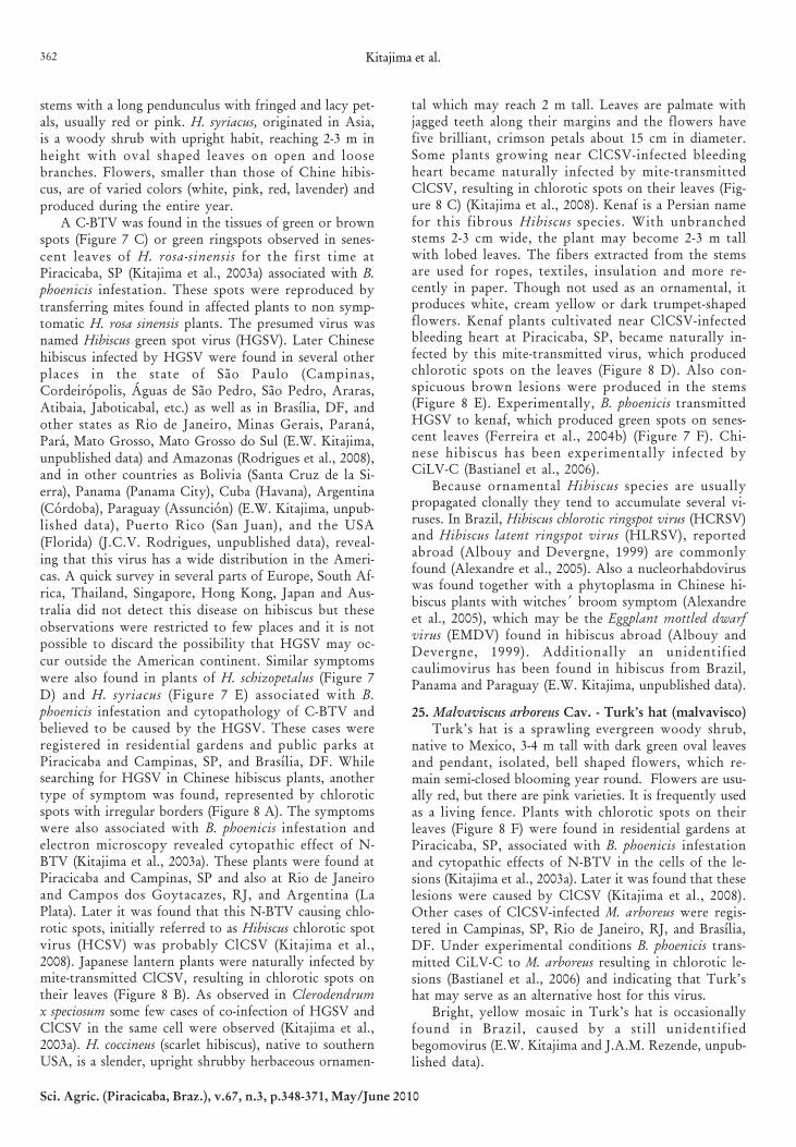
A C-BTV was found in the tissues of green or brownspots (Figure 7 C) or green ringspots observed in senes-cent leaves of H. rosa-sinensis for the first time atPiracicaba, SP (Kitajima et al., 2003a) associated with B.phoenicis infestation. These spots were reproduced bytransferring mites found in affected plants to non symp-tomatic H. rosa sinensis plants. The presumed virus wasnamed Hibiscus green spot virus (HGSV). Later Chinesehibiscus infected by HGSV were found in several otherplaces in the state of São Paulo (Campinas,Cordeirópolis, Águas de São Pedro, São Pedro, Araras,Atibaia, Jaboticabal, etc.) as well as in Brasília, DF, andother states as Rio de Janeiro, Minas Gerais, Paraná,Pará, Mato Grosso, Mato Grosso do Sul (E.W. Kitajima,unpublished data) and Amazonas (Rodrigues et al., 2008),and in other countries as Bolivia (Santa Cruz de la Si-erra), Panama (Panama City), Cuba (Havana), Argentina(Córdoba), Paraguay (Assunción) (E.W. Kitajima, unpub-lished data), Puerto Rico (San Juan), and the USA(Florida) (J.C.V. Rodrigues, unpublished data), reveal-ing that this virus has a wide distribution in the Ameri-cas. A quick survey in several parts of Europe, South Af-rica, Thailand, Singapore, Hong Kong, Japan and Aus-tralia did not detect this disease on hibiscus but theseobservations were restricted to few places and it is notpossible to discard the possibility that HGSV may oc-cur outside the American continent. Similar symptomswere also found in plants of H. schizopetalus (Figure 7D) and H. syriacus (Figure 7 E) associated with B.phoenicis infestation and cytopathology of C-BTV andbelieved to be caused by the HGSV. These cases wereregistered in residential gardens and public parks atPiracicaba and Campinas, SP, and Brasília, DF. Whilesearching for HGSV in Chinese hibiscus plants, anothertype of symptom was found, represented by chloroticspots with irregular borders (Figure 8 A). The symptomswere also associated with B. phoenicis infestation andelectron microscopy revealed cytopathic effect of N-BTV (Kitajima et al., 2003a). These plants were found atPiracicaba and Campinas, SP and also at Rio de Janeiroand Campos dos Goytacazes, RJ, and Argentina (LaPlata). Later it was found that this N-BTV causing chlo-rotic spots, initially referred to as Hibiscus chlorotic spotvirus (HCSV) was probably ClCSV (Kitajima et al.,2008). Japanese lantern plants were naturally infected bymite-transmitted ClCSV, resulting in chlorotic spots ontheir leaves (Figure 8 B). As observed in Clerodendrumx speciosum some few cases of co-infection of HGSV andClCSV in the same cell were observed (Kitajima et al.,2003a). H. coccineus (scarlet hibiscus), native to southernUSA, is a slender, upright shrubby herbaceous ornamen-
tal which may reach 2 m tall. Leaves are palmate withjagged teeth along their margins and the flowers havefive brilliant, crimson petals about 15 cm in diameter.Some plants growing near ClCSV-infected bleedingheart became naturally infected by mite-transmittedClCSV, resulting in chlorotic spots on their leaves (Fig-ure 8 C) (Kitajima et al., 2008). Kenaf is a Persian namefor this fibrous Hibiscus species. With unbranchedstems 2-3 cm wide, the plant may become 2-3 m tallwith lobed leaves. The fibers extracted from the stemsare used for ropes, textiles, insulation and more re-cently in paper. Though not used as an ornamental, itproduces white, cream yellow or dark trumpet-shapedflowers. Kenaf plants cultivated near ClCSV-infectedbleeding heart at Piracicaba, SP, became naturally in-fected by this mite-transmitted virus, which producedchlorotic spots on the leaves (Figure 8 D). Also con-spicuous brown lesions were produced in the stems(Figure 8 E). Experimentally, B. phoenicis transmittedHGSV to kenaf, which produced green spots on senes-cent leaves (Ferreira et al., 2004b) (Figure 7 F). Chi-nese hibiscus has been experimentally infected byCiLV-C (Bastianel et al., 2006).
Because ornamental Hibiscus species are usuallypropagated clonally they tend to accumulate several vi-ruses. In Brazil, Hibiscus chlorotic ringspot virus (HCRSV)and Hibiscus latent ringspot virus (HLRSV), reportedabroad (Albouy and Devergne, 1999) are commonlyfound (Alexandre et al., 2005). Also a nucleorhabdoviruswas found together with a phytoplasma in Chinese hi-biscus plants with witches´ broom symptom (Alexandreet al., 2005), which may be the Eggplant mottled dwarfvirus (EMDV) found in hibiscus abroad (Albouy andDevergne, 1999). Additionally an unidentifiedcaulimovirus has been found in hibiscus from Brazil,Panama and Paraguay (E.W. Kitajima, unpublished data).
25. Malvaviscus arboreus Cav. - Turk’s hat (malvavisco)Turk’s hat is a sprawling evergreen woody shrub,
native to Mexico, 3-4 m tall with dark green oval leavesand pendant, isolated, bell shaped flowers, which re-main semi-closed blooming year round. Flowers are usu-ally red, but there are pink varieties. It is frequently usedas a living fence. Plants with chlorotic spots on theirleaves (Figure 8 F) were found in residential gardens atPiracicaba, SP, associated with B. phoenicis infestationand cytopathic effects of N-BTV in the cells of the le-sions (Kitajima et al., 2003a). Later it was found that theselesions were caused by ClCSV (Kitajima et al., 2008).Other cases of ClCSV-infected M. arboreus were regis-tered in Campinas, SP, Rio de Janeiro, RJ, and Brasília,DF. Under experimental conditions B. phoenicis trans-mitted CiLV-C to M. arboreus resulting in chlorotic le-sions (Bastianel et al., 2006) and indicating that Turk’shat may serve as an alternative host for this virus.
Bright, yellow mosaic in Turk’s hat is occasionallyfound in Brazil, caused by a still unidentifiedbegomovirus (E.W. Kitajima and J.A.M. Rezende, unpub-lished data).
Ornamentals infected by Brevipalpus mite-transmitted viruses 363
Sci. Agric. (Piracicaba, Braz.), v.67, n.3, p.348-371, May/June 2010
XI. OLEACEAE
26. Ligustrum lucidum W.T. Aiton- tree privet(alfeneiro)
27. L. sinensis Lour. - chinese privet (alfeneiro-da-China)
The genus Ligustrum has 40-50 species of evergreen
shrubs and small trees, native to Europe, Asia,Australasia and Africa. They are used for privacy hedg-ing. They have small fragrant flowers, borne in panicles.The tree privet (L. lucidum) is the largest species in thegenus and may reach up to 20 m tall. It is used for shadein the streets and also as living fence. A disease similarto citrus leprosis, referred to as “lepra explosiva” andtransmitted by Brevipalpus mites was first reported in
Figure 8 - A-D. Chlorotic spots on the leaves of Hibiscus rosa sinensis (A), H. schizopetalus (B), H. coccineus (C) and kenaf (D), causedby ClCSV. E. Necrotic lesions on the stems of kenaf caused by ClCSV. F. Chlorotic spots on a leaf of Malvaviscus arboreuscaused by ClCSV. G-H. Chlorotic spots and ringspots on the leaves of respectively Ligustrum lucidum and L. sinensiscaused by Ligustrum ringspot virus (LigRSV), a C-BTV.

Kitajima et al.364
Sci. Agric. (Piracicaba, Braz.), v.67, n.3, p.348-371, May/June 2010
Concórdia, Argentina in L. sinensis (Vergani, 1942). L.lucidum with ringspots (Figure 8 G) was found inCuritiba, PR, and Piracicaba, SP, exhibiting cytopathiceffects of what now we consider C-BTV in the cells ofthe lesion. The disease was transmitted by B. phoenicis(Kitajima et al., 2003a). L. sinensis is smaller, bushy, withsmaller leaves and commonly used as living fences andfor bonsai. There is a variegated form widely used ingardens. Chlorotic spots on the leaves of the Chineseprivet (Figure 8 H) were found associated with cyto-pathic effect of C-BTV and infestation by B. phoenicis atÁguas de São Pedro and Piracicaba, SP (Nogueira et al.,2004). Similar occurrence was observed at Monte Alegre,SP, and Brasília, DF (E.W. Kitajima and J.C.V.Rodrigues, unpublished data).
No other virus was found naturally infectingLigustrum in Brazil, but in Europe several soil-borne vi-ruses as nepoviruses, Tobacco rattle virus (TRV), Tomatobushy stunt virus (TBSV), Tobacco necrosis virus (TNV),Tobacco mosaic virus (TMV) and CMV have been de-scribed infecting plants of the genus Ligustrum (Albouyand Devergne, 1999).
XII. ORCHIDACEAE
28. OrchidsOrchidaceae is the largest plant family comprising
more than 800 genera and 22,000 accepted species. Or-chids are found all around the world, mostly in tropicalareas of Asia, Africa, Central and South America, thoughthey have been found in sub-Antarctic and sub-Arcticregions. Most orchids are epiphytes but there are litho-phytes and terrestrial species. They are perennial herbslacking woody structure with simple parallel innervatedleaves with variable shape and size, and their structuredepend on the plant habitat. Terrestrial orchids are pro-vided with rhizomes or form tubers while the epiphyticones have modified aerial roots. In several epiphyticalspecies the base of the stem is thickened to form a res-ervoir structure referred to as pseudobulb. Orchid flow-ers present many structural variations, some producingsingle flowers while most produce racemose inflores-cence. These flowers usually have three sepals, and threepetals, one of them is modified and enlarged and calledlabellum. Some orchids such as Vanilla are economicallyimportant as foodstuff flavoring while others are impor-tant in perfume industry. But most of the orchids areappreciated for ornamental purpose all around the globeattracting collectors and associations and business result-ing in intense international movement.
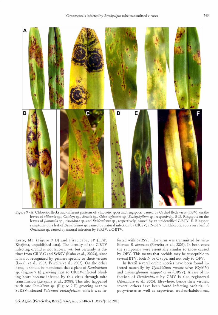
Orchid fleck is a viral disease resulting in chloroticspots and ringspots on the leaves (Figure 9 A). It wasfirst described on Cymbidium sp. in Japan and soon af-ter in several parts of the world in different orchid spe-cies and genera (Doi et al., 1977; Freitas-Astúa et al.,1999; Kitajima et al., 2001; Kondo et al., 2003). The viralnature was suggested by the first reported case of N-BTVcytopathology and later by the mechanical infection ofsome herbaceous species (Doi et al., 1977). The virus was
named Orchid fleck virus (OFV). Kondo et al. (1995) firstpurified the virus and produced a specific antiserum andsubsequently managed to sequence the viral genome re-vealing that it is bipartite (ca. 6 kb each) negative sensessRNA with genomic organization similar to that of rhab-doviruses and they proposed a new genusDichorhabdovirus in this family to accommodate OFV(Kondo et al., 2006). Primers are also available to detectOFV by RT-PCR (Blanchfield et al., 2001; Kubo et al.,2009a). The vector was identified as B. californicus andthe virus/vector relationship is of circulative/propaga-tive type (Kondo et al., 2003). An in situ immunocy-tochemical study demonsrated that the rodlike particlesseen in the tissues represent the OFV, that the nuclearviroplasm contain OFV structural proteins and that theantiserum produced against a Japanese isolate of OFVrecognizes OFV isolates from Brazil and Australia(Kitajima et al., 2001). Some genomic variation amongOFV isolates was detected using single strand confor-mation polymorphism technique (Kubo et al., 2009b)confirming previous works by Blanchfield et al. (2001).OFV has been registered in several orchid generathroughout the world: Angraecum (AUS), Aspasia (BR),Baptistonia (AUS), Bifrenaria (BR), Brassia (BR, USA),Bulbophyllm (AUS), Calanthe (JP), Cattleya (AUS, KOR),Coelogyne (BR), Colmanara (JP), Cymbidium (AUS, JP,KOR, USA), Dendrobium (AUS, BR, DEN, GE, JP,KOR), Diplocaulobium (AUS) , Dockrillia (AUS) ,Encyclia (BR), Flickingeria (AUS), Hormidium (AUS,BR), Liparia (AUS), Masdevallia (AUS), Maxillaria (AUS,BR), Miltonia (AUS, BR, GE), Odontoglossum (AUS, BR,GE, JP), Oncidium (AUS, BR, GE, JP, KOR), Oncidiumx Odontoglossum (BR), Paphiopedilum (BR, GE),Pascatorea (JP), Phaius (BR), Phalaenopsis (DEN) ,Polystachya (AUS), Renanthera (GE), Stanhopea (AUS,GE), Stenia (AUS), Trigonidium (BR), Vanda (GE) ,Zygopetalum (KOR) (compilation made by Kitajima etal., 2001). After this literature survey was made, new ob-servations made at ESALQ, included some new cases:Eria (AUS), Oncidum (CO, CR), Trichopilia (CR),Xylobium (BR) (Freitas-Astúa et al., 2002; E.W. Kitajima,unpublished data) [Key for country abbreviation-AUS-Australia, BR- Brazil, CO- Colombia, CR- Costa Rica,DEN- Denmark, GE- Germany, JP- Japan, KOR- Korea,USA - United States of America].
During a survey made on viruses occurring in orchidsin Brazil, several isolates of OFV were found infectingdifferent orchid genera. However in two of them (Phaius,and Jummelia- Figure 9 B), with symptomsundistinguishable from those of OFV, cytopathology re-vealed the presence of C-BTV (Freitas-Astúa et al., 1999).Later other cases of the presence of C-BTV on orchidswere observed in Brassia, Coelogyne, Hormidium,Xylobium and Arundina, in the state of São Paulo (Kuboet al., 2009a). Another case on Arundina (Figure 9 C) wasfound in the Amazon Basin (Rodrigues et al., 2008) andBragança Paulista (E.W. Kitajima and R. Gioria unpub-lished data), and also in Epidendrum at Primavera do
Ornamentals infected by Brevipalpus mite-transmitted viruses 365
Sci. Agric. (Piracicaba, Braz.), v.67, n.3, p.348-371, May/June 2010
Leste, MT (Figure 9 D) and Piracicaba, SP (E.W.Kitajima, unpublished data). The identity of the C-BTVinfecting orchid is not known yet, but certainly is dis-tinct from CiLV-C and SvRSV (Kubo et al., 2009a), sinceit is not recognized by primers specific to these viruses(Locali et al., 2003; Ferreira et al., 2007). On the otherhand, it should be mentioned that a plant of Dendrobiumsp. (Figure 9 E) growing next to ClCSV-infected bleed-ing heart became infected by this virus through mitetransmission (Kitajima et al., 2008). This also happenedwith one Oncidium sp. (Figure 9 F) growing near toSvRSV-infected Solanum violaefolium which was in-
fected with SvRSV. The virus was transmitted by viru-liferous B. obovatus (Ferreira et al., 2007). In both casesthe symptoms were essentially similar to those causedby OFV. This means that orchids may be susceptible toseveral BTV, both N or C type, and not only to OFV.
In Brazil several orchid species have been found in-fected naturally by Cymbidium mosaic virus (CyMV)and Odontoglossum ringspot virus (ORSV). A case of in-fection of Dendrobium by CMV is also registered(Alexandre et al., 2005). Elsewhere, beside these viruses,several others have been found infecting orchids: 13potyviruses as well as nepovirus, nucleorhabdovirus,
Figure 9 - A. Chlorotic flecks and different patterns of chlorotic spots and ringspots, caused by Orchid fleck virus (OFV) on theleaves of Miltonia sp., Cattleya sp., Brassia sp., Odontoglossum sp., Bulbophyllum sp., respectively. B-D. Ringspots on theleaves of Jummelia sp., Arundina sp. and Epidendrum sp., respectively, caused by an unidentified C-BTV. E. Ringspotsymptoms on a leaf of Dendrobium sp. caused by natural infection by ClCSV, a N-BTV. F. Chlorotic spots on a leaf ofOncidium sp. caused by natural infection by SvRSV, a C-BTV.
Kitajima et al.366
Sci. Agric. (Piracicaba, Braz.), v.67, n.3, p.348-371, May/June 2010
tombusvirus and tospovirus (Albouy and Devergne, 1999;Gibbs et al., 2000).
XIII. PITTOSPORACEAE
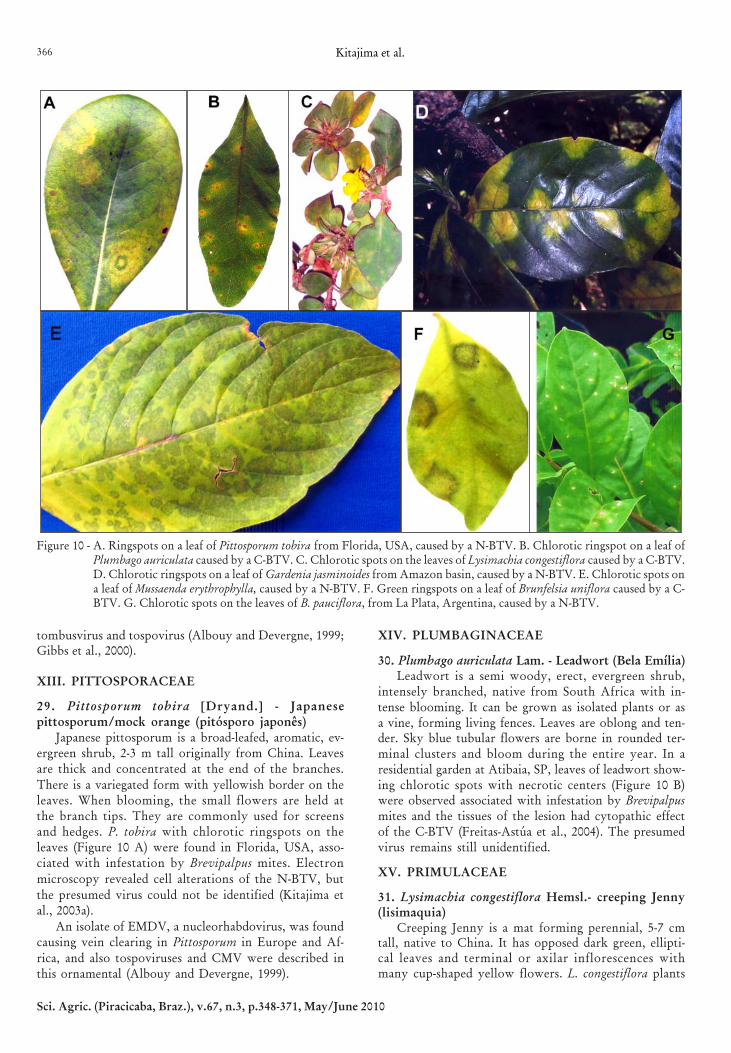
29. Pittosporum tobira [Dryand.] - Japanesepittosporum/mock orange (pitósporo japonês)
Japanese pittosporum is a broad-leafed, aromatic, ev-ergreen shrub, 2-3 m tall originally from China. Leavesare thick and concentrated at the end of the branches.
There is a variegated form with yellowish border on theleaves. When blooming, the small flowers are held atthe branch tips. They are commonly used for screensand hedges. P. tobira with chlorotic ringspots on theleaves (Figure 10 A) were found in Florida, USA, asso-ciated with infestation by Brevipalpus mites. Electron
microscopy revealed cell alterations of the N-BTV, but
the presumed virus could not be identified (Kitajima etal., 2003a).
An isolate of EMDV, a nucleorhabdovirus, was foundcausing vein clearing in Pittosporum in Europe and Af-
rica, and also tospoviruses and CMV were described inthis ornamental (Albouy and Devergne, 1999).
XIV. PLUMBAGINACEAE
30. Plumbago auriculata Lam. - Leadwort (Bela Emília)Leadwort is a semi woody, erect, evergreen shrub,
intensely branched, native from South Africa with in-tense blooming. It can be grown as isolated plants or asa vine, forming living fences. Leaves are oblong and ten-der. Sky blue tubular flowers are borne in rounded ter-minal clusters and bloom during the entire year. In aresidential garden at Atibaia, SP, leaves of leadwort show-ing chlorotic spots with necrotic centers (Figure 10 B)were observed associated with infestation by Brevipalpusmites and the tissues of the lesion had cytopathic effectof the C-BTV (Freitas-Astúa et al., 2004). The presumedvirus remains still unidentified.
XV. PRIMULACEAE
31. Lysimachia congestiflora Hemsl.- creeping Jenny(lisimaquia)
Creeping Jenny is a mat forming perennial, 5-7 cmtall, native to China. It has opposed dark green, ellipti-cal leaves and terminal or axilar inflorescences withmany cup-shaped yellow flowers. L. congestiflora plants
Figure 10 - A. Ringspots on a leaf of Pittosporum tobira from Florida, USA, caused by a N-BTV. B. Chlorotic ringspot on a leaf ofPlumbago auriculata caused by a C-BTV. C. Chlorotic spots on the leaves of Lysimachia congestiflora caused by a C-BTV.D. Chlorotic ringspots on a leaf of Gardenia jasminoides from Amazon basin, caused by a N-BTV. E. Chlorotic spots ona leaf of Mussaenda erythrophylla, caused by a N-BTV. F. Green ringspots on a leaf of Brunfelsia uniflora caused by a C-BTV. G. Chlorotic spots on the leaves of B. pauciflora, from La Plata, Argentina, caused by a N-BTV.
Ornamentals infected by Brevipalpus mite-transmitted viruses 367
Sci. Agric. (Piracicaba, Braz.), v.67, n.3, p.348-371, May/June 2010
with chlorotic spots on the leaves (Figure 10 C) werefound in a residential garden at Águas de São Pedro, SP,associated with infestation of Brevipalpus mites. Cells ofthe lesions showed cytopathic effect of C-BTV(Nogueira and Rossi, 2005). The presumed virus was notidentified.
There is a report of infection of Lysimachia by CMVelsewhere (Albouy and Devergne, 1999).
XVI. RUBIACEAE
32. Gardenia jasminoides Ellis - gardenia (gardenia)Gardenia is a fragrant flowering evergreen tropical,
semi woody bushy plant, native to China and cultivatedworldwide. It may become 1 to 1.5 m tall with dark green,glossy leaves. Flowers are small and white, extremelyfragrant becoming yellowish as they age. It may beplanted isolated or in rows. During a survey for BTV inthe Amazon basin at Urucu, AM, an isolated base foroil prospection, gardenia plants growing in a residen-tial garden were found with chlorotic spots (Figure 10D) associated with Brevipalpus mite infestation. Cellsfrom the lesions exhibited alterations of the N-BTV(Rodrigues et al., 2008). The virus could not be identi-fied. Gardenia is also susceptible to CMV (Albouy andDevergne, 1999).
33. Mussaenda erythrophylla Schumach. & Thonn. -ashanti blood (mussaenda vermelha)
This is a semi-deciduous rambler with multiple stemsthat may grow 3 m or more, native to East and CentralAfrica. Opposite leaves are round to ovate, pubescentat underside and strongly veined. M. erythrophylla pro-duces dense inflorescence with several flowers borne inbranching terminal panicles. Flowers are small withcreamy funnel shaped corolla and a red felt center.Ashanti blood may be cultivated isolated or in rows inparks and gardens. A few plants in a small nursery atManaus, AM, were found with diffuse chlorotic spotson the leaves (Figure 10 E) associated with Brevipalpusmite infestation. Electron microscopy of the tissues fromthe lesions revealed cytopathic effect of the N-BTV(Rodrigues et al., 2008). The presumptive BTV could notbe identified.
XVII. SOLANACEAE
34. Brunfelsia uniflora D. Don. – Yesterday – today -tomorrow (manacá)
35. B. pauciflora Benth -Yesterday – today - tomor-row (jasmim paraguayo)
Brunfelsia is a genus with about 40 species ofneotropical shrubs and small trees. Both B. uniflora andB. pauciflora are very similar, native to Brazil. They arewoody shrubs with several stems which may reach 1-3m tall, with oval, smooth and dark green leaves. WhileB. uniflora produces solitary flowers at the tip of thestems, in B. pauciflora they are produced in clusters. Flow-ers are fragrant and produced in the spring and summer.They open purple, then turn pale lavender and finally
white. They are cultivated isolated or in clusters, form-ing hedges. B. uniflora plants with green spots andringspot on senescent leaves (Figure 10 F) were foundin a residential garden at Águas de São Pedro (Nogueiraet al., 2003) associated with Brevipalpus mite infestationand cytopathic effect of C-BTV in the lesions. Later simi-lar symptoms were found in several plants growing inparks and residential gardens at Piracicaba, Araras andAtibaia, SP. RT-PCR using primers for CiLV-C, SvRSVand PFGSV did not result in amplification of the viralgenome, suggesting that the virus causing the green spotson B. uniflora leaves is different from these viruses. In asurvey carried out in several flower nurseries at LaPlata, Argentina, B. pauciflora plants were found withchlorotic spots (Figure 10 G) associated with Brevipalpusmite infestation. The lesion cells exhibited alterationsof N-BTV (Dal Bo et al., 2007). These viruses are stillunidentified.
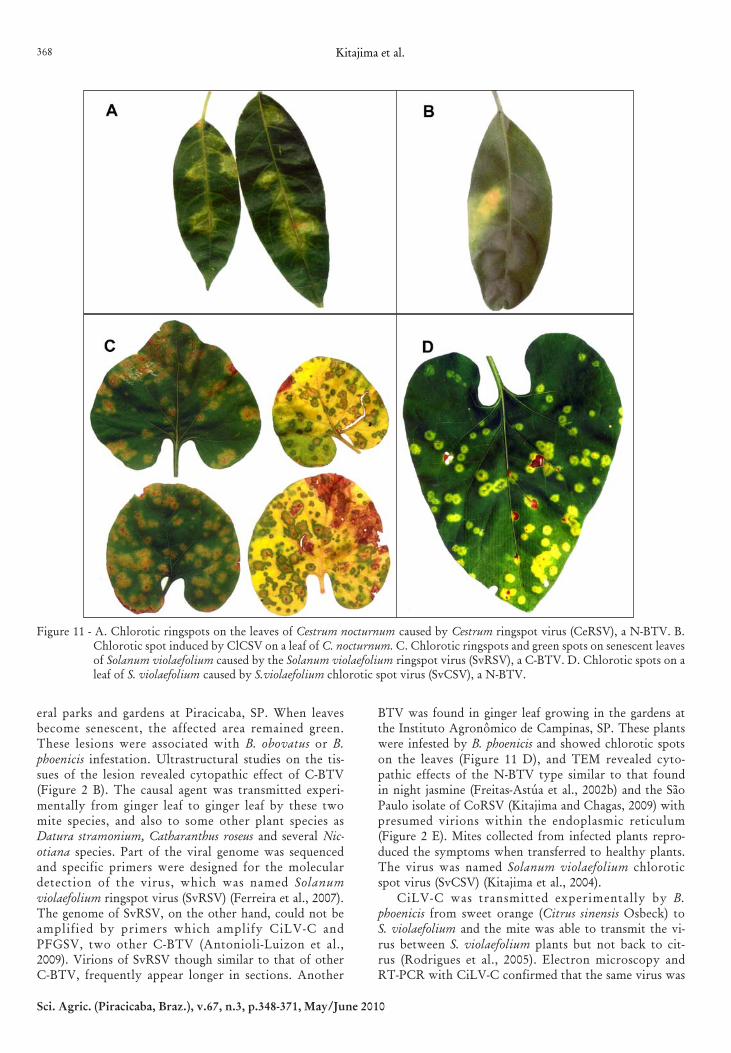
36. Cestrum nocturnum L. - night jasmine (dama-da-noite)
Night jasmine is a semi woody, evergreen shrub na-tive to the Antilles, 1.5-3 m tall with narrow, lanceolate,bright green, glossy, coriaceous leaves. Flowers aresmall, tubular, strongly scented during the night, creamyellowish produced in numerous cymose inflorescences.Plants are usually cultivated isolated. Night jasmineplants showing chlorotic ringspots (Figure 11 A) on theirleaves were found in a residential garden at Atibaia, SP,associated with infestation by B. obovatus (Kitajima etal., 2003a; Guidotti et al., 2006). Mites collected from af-fected plants reproduced the symptoms on healthy oneindicating that the symptoms were due to a BTV, whichwas named Cestrum ringspot virus (CeRSV). This viruswas also experimentally transmitted by B. phoenicis (J.Freitas-Astúa, unpublished data). Cytopathological ob-servations on the tissues of the lesion revealed a patternof N-BTV. However, instead the typical N-BTV, pre-sumed virions were found within endoplasmic reticu-lum and nuclear viroplasma was rarely seen (Kitajimaet al., 2003a). On the other hand, a plant of C. nocturnumgrowing near bleeding heart infected by ClCSV present-ing chlorotic spots on their leaves (Figure 11 B) wasfound at Piracicaba, SP. Further serological and molecu-lar studies indicated that this plant was naturally in-fected by ClCSV (Kitajima et al., 2008).
37. Solanum violaefolium (Schott.) (=Solanumasarifolium Kunth. & Bouch., Lycianthes asarifoliaBitter) - ginger leaf (solano violeta)
Ginger leaf has a prostrate, trailing growth habit withstolons that root and produce dark green, cordiformleaves at every node. Small, white flowers are formedbetween the leaves during the entire year. It may reach10-15 cm tall. Stolons produce a mat near the surface ofthe soil and do not appear to maintain structures thatbecome buried. This plant prefers wet and shady areas.S. violaefolium plants showing conspicuous chloroticringspots on the leaves (Figure 11 C) were found in sev-
Kitajima et al.368
Sci. Agric. (Piracicaba, Braz.), v.67, n.3, p.348-371, May/June 2010
eral parks and gardens at Piracicaba, SP. When leavesbecome senescent, the affected area remained green.These lesions were associated with B. obovatus or B.phoenicis infestation. Ultrastructural studies on the tis-sues of the lesion revealed cytopathic effect of C-BTV(Figure 2 B). The causal agent was transmitted experi-mentally from ginger leaf to ginger leaf by these twomite species, and also to some other plant species asDatura stramonium, Catharanthus roseus and several Nic-otiana species. Part of the viral genome was sequencedand specific primers were designed for the moleculardetection of the virus, which was named Solanumviolaefolium ringspot virus (SvRSV) (Ferreira et al., 2007).The genome of SvRSV, on the other hand, could not beamplified by primers which amplify CiLV-C andPFGSV, two other C-BTV (Antonioli-Luizon et al.,2009). Virions of SvRSV though similar to that of otherC-BTV, frequently appear longer in sections. Another
BTV was found in ginger leaf growing in the gardens atthe Instituto Agronômico de Campinas, SP. These plantswere infested by B. phoenicis and showed chlorotic spotson the leaves (Figure 11 D), and TEM revealed cyto-pathic effects of the N-BTV type similar to that foundin night jasmine (Freitas-Astúa et al., 2002b) and the SãoPaulo isolate of CoRSV (Kitajima and Chagas, 2009) withpresumed virions within the endoplasmic reticulum(Figure 2 E). Mites collected from infected plants repro-duced the symptoms when transferred to healthy plants.The virus was named Solanum violaefolium chloroticspot virus (SvCSV) (Kitajima et al., 2004).
CiLV-C was transmitted experimentally by B.phoenicis from sweet orange (Citrus sinensis Osbeck) toS. violaefolium and the mite was able to transmit the vi-rus between S. violaefolium plants but not back to cit-rus (Rodrigues et al., 2005). Electron microscopy andRT-PCR with CiLV-C confirmed that the same virus was
Figure 11 - A. Chlorotic ringspots on the leaves of Cestrum nocturnum caused by Cestrum ringspot virus (CeRSV), a N-BTV. B.Chlorotic spot induced by ClCSV on a leaf of C. nocturnum. C. Chlorotic ringspots and green spots on senescent leavesof Solanum violaefolium caused by the Solanum violaefolium ringspot virus (SvRSV), a C-BTV. D. Chlorotic spots on aleaf of S. violaefolium caused by S.violaefolium chlorotic spot virus (SvCSV), a N-BTV.
Ornamentals infected by Brevipalpus mite-transmitted viruses 369
Sci. Agric. (Piracicaba, Braz.), v.67, n.3, p.348-371, May/June 2010
responsible for the infections. However, the dsRNA pro-file from infected S. violaefolium and sweet orange weredifferent, indicating possible changes in the viral genome.
An isolate of Eggplant mosaic virus (EMV) was foundat Piracicaba, SP, causing mosaic symptoms on S.violaefolium leaves (E.W. Kitajima, unpublished data).Also, an isolate of TRV was found co-infecting S.violaefolium plants with SvRSV inducing a larger chlo-rotic lesions (Alexandre et al., 2005).
XVIII. VIOLACEAE
38. Viola odorata L. - Sweet violet (violeta)Besides orchids, this is the only case of the occurrence
of BTV outside the American continent. Violet is an ev-ergreen, perennial, herbaceous, woodland plant native toEurope, Africa and Asia. Plants have cordiform leaves andspreads with stolons. Dark blue flowers have long pedun-culus and sweet scent. It makes excellent weed-excludingground cover. In Nambour, Queensland, Australia,Gowanlock and Dietzegen (1995) reported the occurrenceof chlorotic spots on the leaves of violet and TEM revealedcytopathic effects of N-BTV. It is likely that the causalagent may be OFV, since specific primers for this virusresulted in amplification of cDNA by RT-PCR (A. Gibbs,personal communication). There is no information regard-ing infestation by Brevipalpus mites.
Violet is also susceptible to other viruses as Violamottle virus (VMoV) (Albouy and Devergne, 1999).
Acknowledgements
This work received financial support from FAPESP(20007/50809-0 and 2008/52691-9) and CNPq (47.1552/2007-0). The authors thanks to the criticisms and sugges-tions made by the two anonymous referees, whichhelped to improve considerably this text and Dr. Neusade Lima Nogueira, for providing images of localizedsymptoms in some of the described ornamentals.
References
Albouy, J.; Devergne, J.C. 1999. Diseases produced by viruses onornamental plants. Ediciones Mundi-Prensa, Madrid, Spain. 480p. (in Spanish).
Alexandre, M.A.V.; Rivas, E.B.; Tozetto, A.R.P.; Duarte, L.M.L.2005. An annotated list on the natural occurrence of viruses inornamental plants in Brazil. Instituto Biológico, São Paulo, SP,Brazil. 54 p. (in Portuguese).
Antonioli-Luizon, R.; Freitas-Astúa, J.; Locali-Fabris, E.C.;Machado, M.A.; Kitajima, E.W. 2009. Detection of the passionfruit green spot virus (PFGSV) by RT-PCR. Abstracts BrazilianCongress of Genetics. Sociedade Brasileira de Genética,Salvador, BA, Brazil, CDRom (in Portuguese).
Bastianel, M.; Freitas-Astúa, J.; Kitajima, E.W.; Machado, M.A.2006. The citrus leprosis pathosystem. Summa Phytopathologica32: 211-220.
Bitancourt, A.A. 1938. Ringspot, a new disease of the coffee. OBiológico 4: 404-405 (in Portuguese).
Blanchfield, A.L.; Mackenzie, A.M.; Gibbs, A.; Kondo, H.; Tamada,T.; Wilson, C.R. 2001. Identification of orchid fleck virus byreverse transcriptase polymerase chain reaction and analysis ofisolate relationships. Journal of Phytopathology 149: 713-718.
Boari, A.J.; Freitas-Astúa, J.; Ferreira, P.T.O.; Neder, D.G.;Nogueira, N.L.; Rossi, M.L.; Kitajima, E.W. 2004. Purificationand serology of the coffee ringspot virus. SummaPhytopathologica 30: 453-458.
Chagas, C.M. 1973. Association of the mite Brevipalpus phoenicis(Geijskes) with the coffee ringspot. O Biológico 39: 229-232 (inPortuguese).
Chagas, C.M.; July, J.R.; Alba, A.P.C. 1961. Mechanicaltransmission and structural features of coffee ringspot virus.Phytopathologische Zeitschrift 102: 100-106.
Childers, C.C.; French, J.V.; Rodrigues, J.C.V. 2003a. Brevipalpuscalifornicus, B. obovatus, B. phoenicis, and B. lewisi (Acari:Tenuipalpidae): a review of their biology, feeding injury and economicimportance. Experimental and Applied Acarology 30:5-28.
Childers, C.C.; Rodrigues, J.C.V.; Welbourn, W.C. 2003b. Hostplants of Brevipalpus californicus, B. obovatus and B. phoenicis(Acari: Tenuipalpidae) and their potential involvement in thespread of viral diseases vectored by these mites. Experimentaland Applied Acarology 30: 29-105.
Childers, C.C.; Rodrigues, J.C.V.; Derrick, K.S.; Achor. D.S.;French, J.V.; Welbourn, W.C.; Ochoa, R.; Kitajima, E.W.2003c. Citrus leprosis and its status in Florida and Texas: pastand present. Experimental and Applied Acarology 30: 181-202.
Colariccio, A.; Lovisolo, O.; Chagas, C.M.; Galletti, S.R.; Rossetti,V.; Kitajima, E.W. 1995. Mechanical transmission andultrastructural aspects of citrus leprosis disease. FitopatologiaBrasileira 20: 208-213.
Dal Bo, E.; Peña, E.; Fernandez, R.; Kubo, K.; Freitas-Astúa, J.;Bedendo, I.P.; Kitajima, E.W. 2007. Preliminary survey of virusdiseases in ornamentals at La Plata, Argentina. FitopatologiaBrasileira 32 (supplement): S135, 2007 (in Spanish).
Doi, Y.; Chang, M.U.; Yora, K. 1977. Orchid fleck virus.Description of Plant Viruses, No. 183. ComonwealthMycological Institute/Association of Applied Biology,Wellesbourne, UK, 4 p.
Fawcett, H.S. 1911. Scaly bark or nail-head rust of citrus. BulletinNo. 106, Florida Agriculture Experimental Station. Gainesville,FL, US, 41p.
Ferreira, P.T.O.; Kubo, K.S.; Kitajima, E.W. 2004a. Ringspots inAnthurium sp. and Cordyline terminalis associated withcytoplasmic type of Brevipalpus-borne viruses. Virus Reviewand Research 9 (supplement): 249, 2004a.
Ferreira, P.T.O.; Buso Jr.; A.A.; Freitas-Astúa, J.; Kitajima, E.W.2004b. Kenaf (Hibiscus cannabis): experimental host for theHibiscus green spot and Hibiscus chlorotic spot viruses. SummaPhytopathologica 30: 68-69 (In Portuguese).
Ferreira, P.T.O.; Locali-Fabris, E.C.; Freitas-Astúa, J.; Antonioli-Luizon, R.; Gomes, R.T.; Machado, M.A.; Kitajima, E.W. 2007.Characterization of a bacilliform virus isolated from Solanumviolaefolium transmitted by the tenuipalpid mites Brevipalpusphoenicis and B. obovatus. Summa Phytopathologica 33: 264-269(in Portuguese, with abstract in English).
Freitas-Astúa, J.; Rezende, J.A.M.; Kitajima, E.W. 1999. Incidenceof orchid viruses in the state of São Paulo, Brazil. FitopatologiaBrasileira 24: 125-130.
Freitas-Astúa, J.; Moreira, L.; Rivera, C.; Rodriguez C.M.; Kitajima,E.W. 2002. First report of orchid fleck vírus in Costa Rica.Plant Disease 86: 1402.
Freitas-Astúa, J.; Kitajima, E.W.; Astúa-Monge, G.; Locali, E.C.;Machado, M.A. 2004. Leadwort (Plumbago auriculata): a newhost for a bacilliform, rhabdoviruslike virus. SummaPhytopathologica 30: 80 (in Portuguese).
Frezzi, M.S. 1940. The citrus leprosis. Researches conducted at thelaboratory of pathology of Bella Vista (Corrientes). BoletínFrutas y Hortalizas. 5. Ministerio de la Agricultura de la Nación,Buenos Aires, Argentina. 16p (in Spanish).
Gibbs, A.; Mackenzie, A.; Blanchfield, A.; Cross, P.; Wilson, C.;Kitajima, E.W., Nightingale, M.; Clemens, M. 2000. Viruses oforchids in Australia: their identification, biology and control.Orchid Review 65 (3); 10-21.

Kitajima et al.370
Sci. Agric. (Piracicaba, Braz.), v.67, n.3, p.348-371, May/June 2010
Gowanlock, D.H.; Dietzegen, R.G. 1995. Small rhabdovirus-likeparticles in violet (Viola spp.). Australasian Plant Pathology24: 215-216.
Groot, TVM. 2006. The effect of symbiont induced haploidthelytoky on the evolution of Brevipalpus mites. PhDdissertation, Institute of Biodiversity and EcosystemDynamics. University of Amsterdam, Amsterdam, theNetherlands, 154p.
Guidotti, A.R.; Rodrigues, V.; Machado, M.A.; Freitas-Astúa, J.2006. Transmission of the Cestrum ringspot virus by differentactive phases of the mite Brevipalpus obovatus. SummaPhytopathologica 32 (supplement): S 75 (in Portuguese).
Hume, H.H. 1901. Some fungus diseases of citrus and othersfruits. Proceedings Florida State Horticulture Society 14: 64–70.
Kitajima, E.W.; Chagas, C.M. 2009. Viral diseases in coffee. p.550-556. In Wintgens, J.N. ed. Coffee: growing, processing,sustainable production. 2nd ed, Wiley-VCH, Weinheim,Germany. 976p.
Kitajima, E.W.; Costa, A.S. 1972. Bacilliform particles associatedwith the coffee ringspot. Ciência e Cultura 24: 542-545 (inPortuguese, with abstract in English).
Kitajima,E.W.; Kondo, H.; Mackenzie, A.; Rezende, J.A.M.;Gioria, R.; Gibbs, A.; Tamada, T. 2001. Comparativecytopathology and immunocytochemistry of Japanese,Australian and Brazilian isolates of Orchid fleck virus. Journalof General Plant Pathology 67: 231-237.
Kitajima, E.W.; Chagas, C.M.; Rodrigues, J.C.V. 2003a.Brevipalpus-transmitted plant vírus and virus-like diseases:cytopathology and some recent cases. Experimental andApplied Acarology 30: 135-160.
Kitajima, E.W.; Rezende, J.A.M.; Rodrigues, J.C.V. 2003b. Passionfruit green spot virus vectored by Brevipalpus phoenicis (Acari:Tenuipalpidae). Experimental and Applied Acarology 30: 225-231.
Kitajima, E.W.; Kubo, K.S.; Gomes, R.T.; Ferreira, P.T.O. 2004.Chlorotic spots on Solanum violaefolium, transmissible byBrevipalpus mite, associated with a nuclear type of virus.Fitopatologia Brasileira 29 (supplement): S65 (in Portuguese).
Kitajima, E.W.; Boari, A.J.; Salaroli, R.B. 2006. Green spots onBeaumontia leaves associated with a cytoplasmic type ofBrevipalpus transmitted virus. Summa Phytopathologica 32(supplement): S 11 (in Portuguese).
Kitajima, E.W.; Groot, T.V.M.; Freitas-Astúa, J.; Alberti, G.;Morais, G.J. 2007. In situ detecion of the Cardinium symbiontsof Brevipalpus (Acari: Tenuipalpidae) by electron microscopy.Experimental and Applied Acarology 43: 263-271.
Kitajima, E.W.; Kubo, K.S.; Ferreira, P.T.O.; Alcântara, B.K.;Boari, A.J.; Gomes, R.T.; Freitas-Astúa, J.; Rezende, J.A.M.;Morais, G.J.; Salaroli R.B. 2008. Chlorotic spots onClerodendrum, a disease caused by a nuclear type of Brevipalpus(Acari: Tenuipalpidae) transmitted virus. Scientia Agricola65: 36-49.
Kitajima, E.W.; Calegario, R.F.; Freitas-Astúa, J.; Alberti, G.2009. Electron microscopic evidences that Citrus leprosisvirus, cytoplamisc type (CiLV-C) only circulate in the vectorBrevipalpus phoenicis (Acari: Tenuipalpid), while othernuclear type Brevipalpus transmitted viruses do replicate.Abstracts from Milbenkundliches Klloquium in Posen. 7.Uniwersytet im Adama Mickiewicza. Posnan, Polland, p.28-29.
Knorr, L.C. 1968. On the etiology of leprosis in Citrus. Proceedinsfrom Conference International Organization of CitrusVirologists; 4. Rome, Italy, 1966. University of Florida Press,Gainesville, FL, USA, p.332-341.
Kondo, H.; Matsumoto, J.; Maeda, T.; Inouye, N. 1995. Hostrange and some properties of orchid fleck virus isolated fromoriental Cymbidium in Japan. Bulletin Research Institute ofBioresource, Okayama University 2: 151-161 (in Japanese, withabstract in English).
Kondo, H.; Maeda, T.; Tamada, T. 2003. Orchid fleck virus:Brevipalpus californicus mite transmission, biological propertiesand genome structure. Experimental and Applied Acarology30: 215-223.
Kondo, H.; Maeda, T.; Shirako, Y.; Tamada, T. 2006. Orchid fleckvirus is a rhabdovirus with an unusual bipartite genome Journalof General Virology 87: 2413-2421.
Kubo, K.S.; Antonioli-Luizon, R.; Freitas-Astúa, J.; Kitajima, E.W.2007. Molecular diagnosis of the Clerodendron chlorotic spotvirus (ClCSV). Summa Phytopathologica 33 (supplement): S12(in Portuguese).
Kubo, K.S.; Freitas-Astúa, J.; Machado, M.A.; Ktajima, E.W. 2009a.Orchid fleck symptoms may be caused naturally by two differentBrevipalpus transmitted viruses. Journal of General PlantPathology 75: 250-256.
Kubo, K.S.; Stuart, R.M.; Freitas-Astua, J.; Antonioli-Luizon, R.;Locali-Fabris, E.C.; Coletta F.,H.D; Machado, M.A.; Kitajima,E.W. 2009b. Evaluation of the genetic variability of orchid fleckvirus by single-strand conformational polymorphism analysisand nucleotide sequencing of a fragment from the nucleocapsidgene. Archives of Virology 154: 1009-1014.
Locali, E.C.; Freitas-Astúa, J.; Souza, A.A.; Takita, M.A.; Astúa-Monge, G.; Antonioli-Luizon, R.; Kitajima, E.W.; Machado,M.A. 2003. Development of a molecular tool for the diagnosisof leprosis, a major threat to citrus production in the Americas.Plant Disease 87: 1317-1321.
Locali, E.C.; Freits-Astúa, J.; Antonioli-Lauizon, R.; Boari, A.J.;Machado, M.A. 2005. Diagnosis of the coffee ringspot by RT-PCR. Fitopatolologia Brasileira 30 (supplement): S 185 (inPortuguese).
Locali-Fabris, E.C.; Freitas-Astúa, J.; Souza, A.A.; Takita, M.A.;Astúa-Monge, G.; Antonioli-Luizon, A.; Rodrigues, V.; Targon,M.L.P.N.; Machado, M.A. 2006. Complete nucleotidesequence, genomic organization and phylogenetic analysis ofCitrus leprosis virus cytoplasmic type. Journal of GeneralVirology 87: 2721-2729.
Lorenzi, H.; Souza, H.M. 2001. Ornamental plants in Brazil. Woody,herbaceous and climbing. 3 ed. Instituto Plantarum, NovaOdessa, SP, Brazil. 1088 p. (in Portuguese).
Marques, J.P.R.; Freitas-Astúa, J.; Kitajima, E.W.; Appezzato-da-Glória, B. 2007. Leaf and stem lesions of sweet orangeplants caused by the Citrus leprosis vírus. PesquisaAgropecuária Brasileira 42: 1531-1536 (in Portuguese, withEnglish abstract).
Musumeci, M.R.; Rossetti, V.V. 1963. Transmission of citrusleprosis symptoms by the mite Brevipalpus phoenicis. Ciência eCultura 15: 228 (in Portuguese).
Mesa, N.C.; Ochoa, R.; Welbourn, W.C.; Evans, G.A.; Moraes,G.J. 2009. A catalog of the tenuipalpid (Acari) of the world witha key to genera. Zootaxa 2008: 1-185.
Nogueira, N.L.; Rossi, M.L. 2005. Rhabdovirus-like bacilliformparticles in the leaves of creeping Jenny (Lysimaquia congestifoliaHemsi, Primulaceae) associated to typical lesions caused bythe mite Brevipalpus. Fitopatologia Brasileira 30 (supplement):S 189 (in Portuguese).
Nogueira, N.L.; Rodrigues, J.C.V.; Rossi, M.L. 2003. Rhabdovirus-like particles in three ornamental species showing local lesionsand the presence of Brevipalpus mites. Summa Phytopathologica29: 278-282 (in Portuguese, with abstract in English).
Nogueira, N.L.; Rodrigues, J.C.V.; Rossi, M.L. 2004. Dracaenamarginata Lam and Ligustrum sinensis Loureiro, two new hostplants for Brevipalpus borne vírus. Fitopatologia Brasileira 29:S234 (In Portuguese).
Novelli, V.M.; Kubo, K.S.; Freitas-Astúa, J.; Machado, M.A.,Salaroli, R.B.; Kitajima, E.W. 2008. Natural infection of peacelily (Spatiphyllum wallisii) by a nuclear type of Brevipalpus-transmitted virus. Abstracts Simposio Latinamericano y delCaribe “La Biodiversidad acarina: utilización, protección yconservación”, 2, Centro Nacional de Sanidad Agropecuaria,Havana, Cuba, 2008, CDRom, Resumen BA-P14.
Ornamentals infected by Brevipalpus mite-transmitted viruses 371
Sci. Agric. (Piracicaba, Braz.), v.67, n.3, p.348-371, May/June 2010
Pascon, R.C.; Kitajima, J.P.; Breton, M.C.; Assumpção, L.,Greggio, C., Zanca, A.S.; Okura, V.K.; Alegria, M.C.;Camargo, M.E.; Silva, G.G.C.; Cardozo, J.C.; Vallin, M.A.;Franco, S.F.; Silva, V.H., Jordão Jr., H.; Oliveira, F.; Giachetto,P.F.; Ferrri, F.; Aguillar-Vildoso, C.I.; Francischini, F.J.B.;Silva, J.M.F.; Arruda, P.; Ferro, J.A.; Reinach, F.; Silva A.C.R.2006. The complete nucleotide sequence and genomicorganization of Citrus leprosis associated vírus, cytoplasmictype (CiLV-C). Vírus Genes 32: 289-298.
Pijnacker, L.P.; Ferwerda, M.A.; Helle, W. 1981. Cytologicalinvestigations on the female and male reproductive system ofthe parthenogetetic privet mite, Brevipalpus obovatus(Donnadieu) (Phytoptipalpidae, Acari). Acarologia 22: 157-163.
Rodrigues, J.C.V.; Kitajima, E.W.; Childers, C.C.; Chagas, C.M.2003. Citrus leprosies virus vectored by Brevipalpus phoenicis(Acari: Tenuipalpidae) on citrus in Brazil. Experimental andApplied Acarology 30: 161-179.
Rodrigues, J.C.V.; Gallo-Meagher, M.; Ochoa, R.; Childers, C.C.;Adams, B.J. 2004. Mitochondrial DNA and RAPDpolymorphisms in the haploid mite Brevipalpus phoenicis (Acari:Tenuipalpidae). Experimental and Applied Acarology 34: 275–290.
Rodrigues, J.C.V.; Locali, E.C.; Freitas-Astúa, J.; Kitajima, E.W.2005. Transmissibility of citrus leprosis virus by Brevipalpusphoenicis in Solanum violaefolium. Plant Disease 89: 911.
Rodrigues, J.C.V.; Antony, L.M.K.; Salaroli, R.B.; Kitajima, E.W.2008. Brevipalpus-associated viruses in the central Amazon basin.Tropical Plant Pathology 33: 12-19.
Steane, D.A.; De Kok, R.P.J.; Olmstead, R.G. 2004. Phylogeneticrelationships between Clerodendrum (Lamiaceae) and otherAjugoid genera inferred from nuclear and chloroplast DNAsequence data. Molecular Phylogenetics and Evolution 32: 39-45.
Vergani, A.R. 1942. The transmission of the “lepra explosiva” ofthe tree privet by mites. Revista Agricultura y Agronomia 9:292-294 (in Spanish).
Weeks, A.R.; Marec, F.; Breeuwer, J.A.J. 2001. A mite species thatconsists entirely of haploid females. Science 292: 2479-2483.
Welbourn, W.C.; Ochoa, R.; Kane, E.C.; Erbe, E.F. 2003.Morphological observations on Brevipalpus phoenicis (Acari:Tenuipalpidae) including comparisons with B. californicus andB. obovatus. Experimental and Applied Acarology 30: 107-133.
Received May 13, 2009Accepted February 26, 2010