an analysis of anterior mandibular anatomy using cone beam
TRANSCRIPT

University of ConnecticutOpenCommons@UConn
Master's Theses University of Connecticut Graduate School
6-28-2016
An Analysis of Anterior Mandibular AnatomyUsing Cone Beam Computed Tomography: AStudy of Dentate and Edentulous MandiblesRoberta A. Wright D.M.D.University of Connecticut School of Medicine and Dentistry, [email protected]
This work is brought to you for free and open access by the University of Connecticut Graduate School at OpenCommons@UConn. It has beenaccepted for inclusion in Master's Theses by an authorized administrator of OpenCommons@UConn. For more information, please [email protected].
Recommended CitationWright, Roberta A. D.M.D., "An Analysis of Anterior Mandibular Anatomy Using Cone Beam Computed Tomography: A Study ofDentate and Edentulous Mandibles" (2016). Master's Theses. 973.https://opencommons.uconn.edu/gs_theses/973

An Analysis of Anterior Mandibular Anatomy Using Cone Beam Computed
Tomography: A Study of Dentate and Edentulous Mandibles
Roberta Anne Wright, D.M.D.
B.S., University of Illinois at Urbana-Champaign, 2009
D.M.D., University of Connecticut School of Dental Medicine, 2013
A Thesis
Submitted in Partial Fulfillment of the
Requirements for the Degree of
Master of Dental Science
at the
University of Connecticut
2016

ii
APPROVAL PAGE
Master of Dental Science Thesis
An Analysis of Anterior Mandibular Anatomy Using Cone Beam Computed Tomography:
A Study of Dentate and Edentulous Mandibles
Presented by
Roberta Anne Wright, D.M.D.
Major Advisor ________________________________________________
Avinash Bidra, B.D.S., M.S.
Associate Advisor _____________________________________________
John Agar, D.D.S., M.A.
Associate Advisor _____________________________________________
Thomas Taylor, D.D.S., M.S.D.
Associate Advisor _____________________________________________
Alan Lurie, D.D.S., Ph.D.
Associate Advisor _____________________________________________
Aditya Tadinada, B.D.S., M.D.Sc.
University of Connecticut
2016

iii
ABSTRACT
Statement of Problem: The anterior mandible has conventionally been deemed as a relatively
“safe zone” for dental implants due to perceived lack of innervation to the area as well as its
relatively thick cortices and dense bone. However, with the evolution of cone beam computed
tomography (CBCT), a number of anatomic challenges have been identified by clinicians that
can lead to neuropathy and life-threatening hemorrhage if violated. The three critical anatomic
structures in this area that pertain to implant placement are the sublingual artery (SLA),
submental artery (SMA), and the mandibular incisive canal. Currently, there is a lack of
knowledge regarding average measurements of these anatomic structures in relation to a specific
non-variable landmark. Furthermore, it is not known if there are any significant variations of
these anatomic structures in dentate and edentulous patients. While these structures may be
identifiable on a CBCT scan, mandatory CBCTs are not required by practitioners in order to
perform implant surgery in the anterior mandible.
Purpose: To determine if standardized average values can be obtained for the sublingual artery
(SLA), submental artery (SMA), and mandibular incisive canal (MIC), and if differences exist
between dentate and edentulous patients.
Materials and Methods: CBCTs of 125 edentulous and 100 dentate subjects were evaluated at
the anterior mandible for incidence of visualization of the SLA, SMA, and MIC. Measurements
of these three structures were also made from the inferior cortical border of the mandible to the
superior border of each structure in order to gain average anatomical measurements. The cross-
sectional shapes of anterior mandibles were also categorized and prevalence of each shape in this
sample was calculated.

iv
Results: The incidence of visualization of the SLA on CBCT was found to be 100% for
edentulous subjects and 98% for dentate subjects. The SLA was located approximately 15mm
above the inferior border of the mandible. The incidence of visualization of the SMA on CBCT
was 94% for edentulous subjects and 88% for dentate subjects. The SMA was located
approximately 5mm above the inferior border of the mandible. The incidence of visualization of
the MIC on CBCT was 61% for edentulous subjects and 59% for dentate subjects. The MIC was
found to be approximately 1.5mm in diameter at the lateral incisor and canine regions. The MIC
was located approximately 11mm above the inferior border of the mandible in edentulous
patients, and approximately 14mm above the inferior border of the mandible in dentate patients.
The edentulous mandibular ridge attained a buccal-lingual width of 6 mm at a mean distance of 4
mm below the ridge crest in this patient sample. A new classification system for the cross-
sectional morphology of the anterior mandible was characterized and includes the following
shapes: hourglass, ovoid, pear, sickle, and triangular. The pear was the most commonly
visualized cross-sectional morphology among both edentulous and dentate patients.
Conclusions: The sublingual artery and submental artery can be consistently identified in the
anterior mandible using CBCT, both in dentate and edentulous patients. The SLA was located
approximately 15mm above the inferior border of the mandible and the SMA was located
approximately 5mm above the inferior border of the mandible. The mandibular incisive canal
was not consistently visualized. The cross-sectional morphology of the anterior mandible is
diverse in dentate and edentulous mandibles with pear shape being the most common in both
situations. These findings should be taken into consideration when treatment planning for
implants using CBCT or panoramic radiography.

v
TABLE OF CONTENTS
INTRODUCTION ……………………………………………………………………………. 1
Literature Review 1
Rationale for the Study 9
OBJECTIVES AND HYPOTHESIS ………………………………………………………... 11
Research Objectives 11
Hypothesis 12
MATERIALS AND METHODS …………………………………………………………….. 13
CBCT Selection 13
CBCT Analysis 14
Statistical Analysis 16
RESULTS …………………………………………………………………………………....... 19
Intra-Operator Reliability 19
Demographic Data 20
Incidence of Visualization of Key Anatomic Structures 20
Measurements to Anatomic Structures 20
Minimum Mandibular Anterior Width for Implants 22
Mandibular Cross-Sectional Morphology 23
DISCUSSION ………………………………………………………………………………… 25
STUDY LIMITATIONS ……………………………………………………………………... 31
CONCLUSIONS ……………………………………………………………………………... 32
FUTURE RESEARCH ………………………………………………………………………. 34
REFERENCES ……………………………………………………………………………….. 35
FIGURES AND TABLES ……………………………………………………………………. 41

1
INTRODUCTION
Literature Review
Over the past three decades, dental implants have emerged as a mainstay in the esthetic,
functional, and prosthetic rehabilitation of patients with partial and complete edentulism.1 Dental
implants are widely used in a variety of prosthodontic treatments such as single tooth
replacement, multiple teeth replacement, support for complete arch fixed dental prostheses, and
retention for removable complete and partial overdentures. Historically, dental implants were
primarily intended for placement in atrophic edentulous mandibles for metal-resin screw-retained
fixed complete dentures.2 Currently, dental implants in the anterior mandible have become
increasingly common due to popular prosthetic treatments such as two-implant retained
mandibular overdentures, mandibular fixed implant-supported prostheses, and implant-supported
four-to-six-unit anterior fixed dental prostheses on partially edentulous patients. Each of these
treatment modalities includes at least two implants in the anterior mandible, typically at the
positions of the lateral incisors or canines.
The mandibular anterior region has historically been considered a “safe and predictable
zone” for implant surgery.3,4
The predictability was attributed to the relatively thick cortices and
dense bone; however, the “safe zone” concept was a misnomer, primarily due to the lack of
knowledge and appreciation of anatomic structures in this region. According to Per-Ingvar
Branemark’s original protocol, the dental implant placement procedure was to be completed in
the surgical operating room in a hospital setting rather than an out-patient setting, as largely
performed today.5 Performing implant surgery in a hospital operating room allowed clinicians to
better manage any intra-operative complications, such as arterial bleeding, that were
unexpectedly encountered. The same argument holds true for other oral surgical procedures

2
performed in the anterior mandible, such as orthognathic surgeries, and oncologic or trauma
related resections that are all currently performed in a hospital setting. As an increasing number
of clinicians converted from the hospital operating room environment to the outpatient setting in
a dental chair environment, numerous reports have emerged recounting adverse outcomes related
to implant surgery in the anterior mandible.6 Recent literature discusses a number of adverse
events ranging from neurosensory disturbances to life-threatening complications in this region,
including formation of sublingual hematoma, upper airway obstruction, and profuse, pulsatile
bleeding.7 Therefore, thorough knowledge of surgical anatomy of the anterior mandible is
critical for many routine dental procedures including dental implant placement, dental
extractions, donor site bone harvesting of mandibular symphysis, and torus removal.8
There are three significant anatomic structures in the anterior mandible that deem surgery
in this region challenging. These include the sublingual artery, the submental artery, and the
mandibular incisive canal that houses the incisive neurovascular bundle.9 The sublingual artery
is a terminal branch of the lingual artery. The lingual artery arises from the external carotid
artery between the superior thyroid artery inferiorly and the facial artery superiorly. It travels
deep to the hypoglossal nerve, the stylohyoid muscle, and posterior belly of the digastric muscle.
It then dives deep to the hyoglossus muscle.10
The sublingual branch arises at the anterior border
of the hyoglossus muscle, and courses forward in the anterior floor of the mouth above the
mylohyoid muscle to anastomose with the contralateral artery, as well as the submental branch of
the facial artery. This anastomosis may result in small alveolar branches which penetrate the
lingual cortex of the mandible at the lingual foramen.10
The sublingual artery provides the major
blood supply to the floor of the mouth; its branches perfuse the sublingual salivary gland, the

3
mylohyoid and surrounding muscles, and the gingiva and mucosa of the mandibular anterior
teeth.9,11,12
The submental artery is a terminal branch of the facial artery, which originates from the
external carotid artery. The facial artery emerges either superior to the lingual artery or in
common with it. It passes deep to the stylohyoid and digastric muscles, and loops anteriorly on
the inferior border of the mandible to travel a deep groove in the submandibular salivary gland.10
At this point, the submental branch travels anteriorly on the surface of the mylohyoid muscle and
anastomoses with the sublingual branch of the lingual artery and the mylohyoid branch of the
inferior alveolar artery.12
These terminal anastomosing branches may penetrate the lingual
cortex via the lingual foramen or may enter via accessory lingual foramina.13,14
A study by
Loukas et al in 2008 found that 27% of human cadavers studied had the sublingual artery arising
from the submental artery rather than the lingual artery.15
Anatomical aberrations such as this
may make surgical complications in the anterior mandibular region of atrophic mandibles
unpredictable without proper imaging.
A number of authors have described anatomic findings regarding this intricate perfusion
of the anterior mandible in cadaver specimens and radiographic studies; however, all of these
studies examined limited variables of anatomy. Numerous studies have dissected cadaver
specimens to study variable arterial branching patterns, as well as the location of arterial
perforation of the mandibular lingual cortical plate.13,15-17
However, these studies were all
completed on human cadavers or dried mandibles. Radiographic studies of the blood supply to
the anterior mandible have also been conducted to support cadaver evidence. In a study by
Tepper et al in 2001, traditional computed tomography (CT) scans were used to evaluate the
presence of sublingual canals, the distance of these canals to the menton, and the intraosseous

4
paths of these canals.18
This study used 70 total scans, but did not describe the dental status
(dentate or edentulous) of the patients.18
Lustig et al in 2003 also evaluated the sublingual
arterial supply. However, this study employed the use of ultrasound/doppler to evaluate only the
diameter of the sublingual artery and blood flow patterns through this artery.19
No evaluation of
precise location of bony perforation of this artery was attempted. In 2007, Longoni et al
compared 100 dry skull cadaver mandibles and 100 traditional CTs of mandibles of living
patients to examine location and number of lingual vascular canals in the mandible.20
This study
was novel in that it examined CTs of living patients; however, the data collected were limited to
the number and diameter of lingual vascular canals and the location of these canals relative to the
dentition. No measurement of superior-inferior location within the mandible was described, nor
were measurements taken of edentulous mandibles, which is of particular interest to the
clinician.20
Finally, in 2009, Tagaya et al evaluated cadaver sections and 200 traditional CT
scans of living patients.21
This study determined the number and position of lingual foramina of
the mandible relative to the mental spine and midline.21
While this study used CT scans of a
large sample of living patients, no absolute measurements were made that would be of
significant clinical use to the practitioner. Despite the wealth of information gained from these
studies, no examination of differences between dentate and edentulous mandibles was
mentioned, nor was any standardized measurement attempted from a non-variable anatomic
structure (such as the inferior border of the mandible) to derive average measurements.
Furthermore, no studies investigated the presence and location of the submental artery. Further
studies using current 3-dimensional CBCT technology are needed in order to provide clinically
applicable data for practicing dentists.

5
The mandibular incisive canal (MIC) houses the incisive artery, vein, and nerve which
perfuse and innervate the anterior mandibular teeth, including the first premolars, canines, and
incisors.10
The incisive artery is a branch of the inferior alveolar artery. The inferior alveolar
artery arises from the maxillary artery, a terminal branch of the external carotid artery. The
inferior alveolar artery courses inferiorly, giving off the mylohyoid artery before entering the
mandibular foramen and travelling through the mandibular canal. In this canal, it divides into
two branches.10
The mental branch exits out through the mental foramen in the area of the
premolars to perfuse the chin and lower lip. The incisal branch remains in the mandibular canal
anteriorly to the mental foramen and sends perfusing branches to the incisor teeth as well as to
the contralateral artery.12
The mandibular incisive nerve is the final branch of the inferior alveolar nerve, a branch
of the mandibular division of the trigeminal nerve.12
It enters the lingual aspect of the posterior
mandible via the mandibular foramen and travels anteriorly within the mandibular canal. The
inferior alveolar nerve traverses the mandible from lingual to buccal, and splits into the mental
and incisive nerves in the area of the premolars.10
The mental nerve emerges from the mental
foramen and travels with the mental artery to supply innervation to the skin of the lower lip and
chin, and the gingiva and mucosa anterior to the second premolar. The incisive nerve remains in
the canal medial to the mental foramen, termed the mandibular incisive canal. The incisive
nerve provides innervation to the mandibular anterior teeth, including the first premolars,
canines, and incisors.22,23
However, in a number of cases, the MIC may be indistinct, suggesting
that this important neurovascular bundle may simply travel through the medullary spaces of the
mandibular trabecular bone.24

6
Numerous studies have examined anatomy of the mandibular incisive canal in cadaver
specimens, panoramic radiographs, and computed tomography.23,25-30
These studies have mostly
focused on percent occurrence, estimated diameter of the mandibular incisive canal, and distance
from root apices of lateral incisors and canines. Mardinger et al in 2000 and Mraiwa et al in
2003 both examined percent occurrence and diameter of the mandibular incisive canal (MIC)
using cadaver specimens only.25,26
Jacobs et al in 2002 and 2004 also examined occurrence and
diameter of the MIC.27,28
These studies used traditional spiral computed tomography and
panoramic radiography respectively. In these studies, the presence of the MIC via panoramic
radiography was only present in 15% of patients, and had “good visibility” in only 1%, which is
insufficient.27,28
Uchida et al. in 2007, and again in 2009, evaluated MIC anatomy in cadaver
specimens, and cadavers along with CBCT respectively.29,30
This was the first study of this type
to use CBCT, the standard three-dimensional imaging in dental medicine to evaluate anatomy;
however, this study only investigated four CBCT scans which were taken of cadavers, not living
patients. Finally, Apostolakis et al in 2013, used CBCT of living patients to examine dimensions
of the MIC only and determined mean distances of the MIC to apices of adjacent teeth, not an
unchanging structure through time.23
Furthermore, no measurements were noted for edentulous
mandibles. Currently, there is a lack of data comparing the anatomy of the MIC in dentate and
edentulous patients using conventional three-dimensional CBCT analysis in living patients.
While description of these anatomic structures seems straightforward, individual
variation can be vast. This variability may be complicated by the presence or absence of
dentition, as degree of alveolar bone atrophy can have a large influence on anatomic variation.31
In the edentulous mandible, impingement of the MIC during implant surgery is typically not of
significant concern for three reasons: 1) sclerosis of the incisive artery with age, 2) hemorrhage

7
may be controlled by implant placement itself, and 3) absence of mandibular anterior teeth may
render the innervation of the incisive nerve inconsequential.9 However, a number of cases have
reported surgical complications in patients with enlarged MIC, including extreme post-operative
pain from neural injury and severe pulsatile hemorrhage from implant invasion of the mandibular
incisive artery.8,22
Other complications reported for dentate and edentulous patients include life-
threatening vascular injuries. Numerous reports discuss surgical cases involving perforation of
the lingual cortex of the mandible resulting in large sublingual hematoma formation causing
near-fatal airway obstruction. In all reported cases, the sublingual or submental artery had been
lacerated during osteotomy preparation and necessitated emergency hospitalization.3,32-34
The current consensus, or the standard of care, for surgical intervention in the anterior
mandible does not require the use of three-dimensional imaging. However, the ability of
conventional two-dimensional radiographic imaging (panoramic and periapical radiography) to
reveal these important structures are severely limited in appreciation of the MIC and incapable of
visualization of the sublingual and submental arteries.26,28
The use of three-dimensional cross-
sectional imaging using CBCT has resulted in better visualization of alveolar ridge topography
and proximity of vital anatomic structures.35
However, current guidelines continue to only
recommend the use of CBCT on an individual needs basis as an alternative to conventional
imaging.35
Not all cases warrant full CBCT analysis, which potentially increases the cost of
treatment as well as radiation exposure to the patient. Therefore, a need for standardized
measurements and incidence of visualization of important anatomy using the best clinical
visualization practices is needed.
Two additional topics of interest in this study involve the buccal-lingual width of the
alveolar crest in the anterior mandible, and the related cross-sectional shape of the anterior

8
mandibular bone. Rehabilitation of the dentition using dental implants requires a minimum
amount of buccal-lingual width of alveolar bone for implant success. Placement of the minimum
acceptable diameter implant (3.3mm) in the mandibular anterior with adequate remaining
thickness of bone on the buccal and lingual (1mm each) yields a minimum bone thickness of
5.3mm, or more clinically pertinent, 6mm of bone.36
In many patients, the implant bed
preparation requires flattening and reduction of the ridge crest in order to achieve the 6mm
minimum thickness. Necessary reduction of crestal bone to attain the minimum 6mm buccal-
lingual thickness potentially brings the three key anatomic structures studied here closer to the
osteotomy site, increasing the risk of encroaching upon to these structures. Similarly, restoration
of dentition using fixed dental prosthetics also requires a minimum amount of apico-coronal
space for prosthetic parts involved.36
The amount of prosthetic space varies depending on the
restorative material and type of final prosthesis. In many patients, reduction of alveolar bone is
required in order to gain the minimum prosthetic space. Reduction of alveolar bone in the
anterior mandible, again, has the potential to bring the crest closer to the three key anatomic
structures studied here. Therefore, it is pertinent to examine edentulous scans to determine the
distance from the residual ridge crest inferiorly to the region of the mandible where the buccal-
lingual width is at least 6mm.
Finally, the cross-sectional shape of the anterior mandibular bone is of significant
interest. Implant-based rehabilitation in the anterior mandible can be compromised in cases of
severe alveolar constriction, the so-called “hourglass mandible” variant.37
It is defined as an
osseous constriction at the alveolar-basal bone junction. According to Butura et al in 2011, the
approximate incidence of the hourglass variant is about 3.98%.37
This extreme narrowing of
bone makes dental implant placement difficult, and often requires bone grafting procedures.38,39
9
Another treatment approach to dental implant placement in the hourglass mandible is to
complete ostectomy past the bony constriction to an optimal width (6mm as discussed above).
Furthermore, reduction of the alveolar bone potentially brings crestal bone nearer to the three
key structures. Therefore, it is beneficial to further examine cross-sectional patterns of bone to
not only identify the incidence of the hourglass variant, but also other potentially remarkable
bony variations as well.
Rationale for the Study
Dental implants have become an important treatment modality in the esthetic, functional,
and prosthetic rehabilitation of patients with partial and complete edentulism.1 Dental implants
have gained widespread popularity in prosthodontic rehabilitation including single tooth
replacement, multiple teeth replacement, support for complete arch fixed dental prostheses, and
retention for removable complete and partial overdentures. Each of these treatment modalities
includes at least two implants in the anterior mandible, usually at the positions of the lateral
incisors or canines. There are three key anatomic structures in this area that are at significant
risk of injury during surgical implant placement: the sublingual artery, the submental artery, and
the mandibular incisive canal (MIC).
Quantitative data regarding precise location or percentage of variability of the sublingual
artery, submental artery, and the MIC in dentate and edentulous patients is helpful to clinicians
planning surgery in the anterior mandible. Currently, there is a lack of standardized
measurements of these three vital anatomic structures. Furthermore, anatomy can vary in dentate
and edentulous patients. As current clinical consensus or standard of care does not require a
CBCT analysis prior to implant surgery, the risk for vascular and neurologic complications

10
during surgery performed in an outpatient setting is significant for clinical and medico-legal
reasons. Furthermore, potential negative outcomes of these injuries have been documented to
result in substantial paresthesia and pain from neural damage, and life-threatening hemorrhage
leading to airway compromise from vascular injury.
From a clinical standpoint, CBCT analysis of all cases requiring surgery of the anterior
mandible may be prudent, but it is not always possible or realistic. Modern CBCT (or CBVI)
machines provide minimal radiation exposure and have been accepted to be very safe.40
With the
popularity of implant dentistry and advancement of associated technology, there is a need for
scientific standards for clinicians to use when CBCT analysis is not employed. Therefore, this
observational study seeks to determine if any standardized measurements of key anatomic
structures can be determined via CBCT in dentate and edentulous patients in order to accurately
predict mandibular surgical anatomy. Additionally, results from this study can offer “safe zones”
and measurement guidelines to clinicians (not using CBCT) to help improve their surgical
outcomes and minimize risks of morbidity for patients.

11
OBJECTIVES & HYPOTHESIS
Research Objectives
This study used cone beam computed tomography to examine the following characteristics of
125 edentulous and 100 dentate mandibles in order to know:
The incidence of visualization of the perforation of the lingual cortex and silhouette of
the sublingual artery at the midline in dentate and edentulous patients.
The incidence of visualization of the perforation of the lingual cortex and silhouette of
the submental artery at the midline in dentate and edentulous patients.
The average distance from the inferior cortical border of the anterior mandible to the
superior border of the sublingual artery at the midline in dentate and edentulous patients.
The average distance from the inferior cortical border of the anterior mandible to the
superior border of the submental artery at the midline in dentate and edentulous patients.
The incidence of visualization of the silhouette of the mandibular incisive canal in the
anterior mandible bilaterally in dentate and edentulous patients.
The average distance from the inferior cortical border of the anterior mandible to the
superior border of the mandibular incisive canal measured at lateral incisor and canine
regions bilaterally in dentate and edentulous patients.
The average diameter of the mandibular incisive canal in the anterior mandible measured
at lateral incisor and canine regions bilaterally in dentate and edentulous patients.
The average distance from the residual ridge crest inferiorly to the region of the mandible
where the buccal-lingual width is at least 6mm at lateral incisor and canine regions
bilaterally in edentulous patients.
The various cross-sectional shapes of the anterior mandible at the midline.

12
Hypothesis
This study is a cross-sectional, non-experimental, observational study to define baseline
information related to anatomic landmarks in the anterior mandible. As a result, there are no null
hypotheses that were constructed in this study.
13
MATERIALS AND METHODS
CBCT Selection
University of Connecticut Health Center Institutional Review Board approval was
obtained and was granted an exemption, as non-identifiable data was examined and collected in
this study (Project number: UCHC-14-1107). A total of 225 cone beam computed tomography
(CBCT) scans were selected for use from the Oral and Maxillofacial Radiology archives at the
University of Connecticut School of Dental Medicine, Farmington, CT, and Pi Dental Center,
Fort Washington, PA. This amounted to CBCT scans of 100 dentate subjects and 125
edentulous subjects.
The inclusion criteria for CBCT selection was as follows:
Dentate scans must have had at least the mandibular six anterior teeth present (canine to
canine)
Edentulous scans must have been either completely edentulous in the mandible or
partially edentulous with at least the mandibular anterior teeth missing (canine to canine)
The anterior mandible must have included the entire height of the mandible with no field-
of-view cuts
Scans must have been full-volume, or at least have included the maxillary hard palate
Images must have been of adequate resolution/diagnostic quality
The exclusion criteria for eliminating a CBCT scan was as follows:
Any scan that did not satisfy any of the requirements listed in the inclusion criteria
Any scan with “radiographic noise” that did not allow measurements to be recorded in
the planning software

14
Any scan that did not allow adequate manipulation of the image in the planning software
due to technical errors
Any scan that included maxillofacial trauma, orthognathic surgery, congenital anomalies,
pathology, or reconstruction
CBCT Analysis
No personal identifiers were recorded from scans that were identified for use in the study.
Demographic information including age and gender were recorded. The selected CBCT scans
were copied to an encrypted and passcode protected external hard drive for use by the sole
evaluator. Analysis of each CBCT scan was completed as follows:
One investigator underwent calibration training with a board-certified oral and
maxillofacial radiologist prior to commencement of the study.
In order to assess intra-operator reliability, repeated measurements on a random set of 30
samples (15 edentulous subjects, and 15 dentate subjects) was completed.
Digital Imaging and Communications in Medicine (DICOM) files were analyzed on an
implant planning software program (InVivo 5: Anatomage, San Jose, Calif). This
software program was used on a single encrypted and passcode protected computer.
CBCT scans were evaluated in a standardized position with the hard palate oriented
parallel to the horizontal axis
A standardized measurement tool within the software program was used to make
measurements of the structures of interest to the nearest hundredth of a millimeter
Serial sagittal sections were viewed perpendicular to the anterior mandible. Occurrence
of visualization of the cross-sections of the sublingual and submental arteries at the

15
mandibular midline were recorded. Incidence was calculated after all scans were
analyzed.
Upon visualization of the sublingual and submental arteries at the midline in sagittal
sections perpendicular to the anterior mandible, measurements were recorded from the
inferior-most border of the mandible to the superior-most border of the sublingual and
submental arteries as they enter the lingual surface of the mandibular bone.
Serial sagittal sections were viewed perpendicular to the anterior mandible. Occurrence
of visualization of the cross-section of the mandibular incisive canal was recorded. If
visualized, measurements were taken from the inferior-most border of the mandible to the
superior-most border of the mandibular incisive canal at the lateral incisor and canine
regions bilaterally. A standardized mesial-distal distance deviating 5mm from the midline
was used to denote the lateral incisor region and another 5mm from this site was used to
denote the canine region in edentulous scans.
Serial sagittal sections were viewed perpendicular to the anterior mandible. If
visualization of the cross-section of the mandibular incisive canal was observed, the
greatest dimension diameter of the mandibular incisive canal was measured to the nearest
hundredth of a millimeter at the lateral incisor and canine regions bilaterally.
Edentulous scans only: Serial sagittal sections were viewed perpendicular to the anterior
mandible. Using the measurement tool, the distance from the superior-most position on
the crest of the residual ridge to the height at which the facial-lingual thickness of the
mandible is 6mm in the area of the lateral incisors and canines was measured bilaterally.
A cross-section perpendicular to the anterior mandible at the midline was viewed and
mandibular cross-sectional bone patterns were recorded.

16
Statistical Analysis
Data subjected to statistical analysis:
Percent occurrence of visualization of the sublingual artery at the midline
Percent occurrence of visualization of the submental artery at the midline
Mean distance of the sublingual artery to the inferior cortical border of the anterior
mandible at the midline
o Difference in the mean distance of sublingual artery to the inferior cortical border
in dentate and edentulous scans
Mean distance of the submental artery to the inferior cortical border of the anterior
mandible at the midline
o Difference in the mean distance of sublingual artery to the inferior cortical border
in dentate and edentulous scans
Mean distance from the superior border of the incisive canal to the inferior cortical border
of the anterior mandible
o Difference in the mean distance of superior border of the incisive canal to the
inferior cortical border in dentate and edentulous scans
Mean diameter of the incisive canal in the anterior mandible
o Difference in the mean diameter of the incisive canal in dentate and edentulous
scans
Mean distance from the edentulous residual ridge crest inferiorly to the region of the
mandible at lateral incisor and canine region where the buccal-lingual width is at least
6mm

17
Percent occurrence of different cross-sectional shapes of the anterior mandible at the
midline.
Association of age with cross-sectional mandibular shape
All data were recorded in Microsoft Excel data sheets before statistical analysis. All of
the statistical analyses were performed the statistical software R 3.1.2. (R Core Team (2014). R:
A language and environment for statistical computing. R Foundation for Statistical Computing,
Vienna, Austria. URL http://www.R-project.org/)
First, the variables examined in this study were classified into categorical variables and
discrete/continuous variables. In the descriptive data analysis, each categorical variable was
summarized with frequencies and percentages. Each continuous/discrete variable was
summarized with mean and standard deviation. Among the discrete/continuous variables, the left
and right side data for bilateral measurements were averaged. The intra-class correlation co-
efficient with a 95% confidence interval were calculated to evaluate the reliability of each
distance measurement using the twice-evaluated data of 15 edentulous scans and 15 dentate
scans.
Edentulous and dentate scans were compared using population means with respect to
each continuous variable by two-sample t-test or Wilcoxon rank sum test. The Shapiro-Wilk test
was applied to each sample to test for normality of the distribution. If both p-values were greater
than 0.05, a two-sample t-test was performed; otherwise, a Wilcoxon rank sum test was used. For
each categorical variable with two outcomes, the outcome distributions between edentulous and
dentate scans were compared using the Fisher’s exact test. For the only categorical variable with
more than 2 outcomes, cross-sectional shape of the anterior mandible, the distributions of each

18
mandibular shape were compared using a Fisher’s exact test. An overall chi-square test was not
used for mandibular shape. Instead, multiple Fisher’s exact tests were used because of the rarity
of some shapes within the edentulous and dentate groups. An alpha value of 0.05 was chosen to
test for any statistical significance.

19
RESULTS
This study sought to uncover average measurement values of anterior mandibular
anatomic structures and compare similarities or differences in these measures between
edentulous and dentate populations. In all, this study utilized a total of 125 edentulous CBCT
scans and 100 dentate CBCT scans with ages ranging from 18 to 85 years old.
Intra-Operator Reliability
In order to determine the validity of any results that would be obtained in this study, a
pilot evaluation of 15 edentulous and 15 dentate CBCTs in duplicate was first carried out to
establish intra-rater reliability of the sole evaluator. Measures for the first and second replicates
were recorded and intra-class correlation coefficients (ICC) were established for all
measurements (Table 1). Most measures demonstrated a high degree of reliability between the
first and second replicates with ICC values exceeding 0.95, with some notable exceptions. The
distributions of these few exceptions are depicted in Figure 1, and consist of ICC of 0.89 and
0.88 for the diameter of the MIC at the lateral incisor position on the left and right sides
respectively in edentulous scans. ICCs of 0.22 and 0.83 were also noted for the diameter of the
MIC at the canine position on the left and right respectively in dentate scans. On closer
examination, this was most likely due outliers, small N, and a small scale on the order of
hundredths of a millimeter. Structures that were not analyzed, or where no comparison could be
made were recorded as “NA.”

20
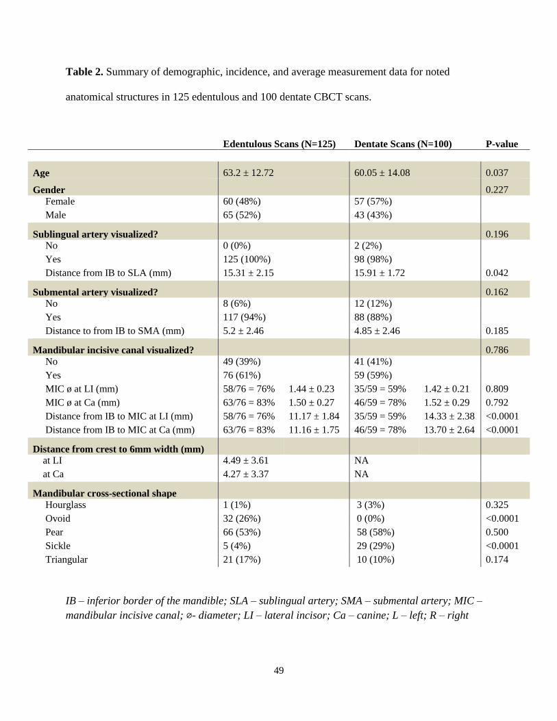
Demographic Data
This study recorded basic demographic data, such as age and gender, for further exploration
of possible anatomical changes related to age or gender. The age range of subjects in this study
spanned from 18 to 85 years old. The gender distribution in edentulous scans was 48% female
and 52% male, while the dentate scans had 57% female and 43% male (Table 2).
Incidence of Visualization of Key anatomic Structures
The overall incidence of visualization of the SLA in edentulous scans was 100%, and in
dentate scans was 98% (Figure 2, Table 2). The incidence of visualization of the SMA in
edentulous scans was found to be 94%, and in dentate scans was 88% (Figure 2, Table 2). The
MIC had the lowest visualization rate, at 61% for edentulous scans, and 59% for dentate scans
(Figure 2, Table 2). Though each group demonstrated slight differences, none of the differences
in visualization noted between dentate and edentulous CBCTs were significant.
Measurements to Anatomic Structures
Sublingual Artery
Measurements from the inferior border of the mandible to the superior-most border where
the sublingual artery (SLA) pierces the mandibular lingual cortex were made in edentulous and
dentate scans. The average distance from the inferior border to the SLA in edentulous scans was
15.31mm ± 2.15 (mean ± SD) (Figure 3A). For dentate scans, the average distance was 15.91mm
± 1.72 (mean ± SD). This difference was statistically significant with a p-value of 0.042 (Table
2).

21
Submental Artery
Measurements from the inferior border of the mandible to the superior-most border where
the submental artery (SMA) pierces the mandibular lingual cortex were made in edentulous and
dentate scans. The average distance from the inferior border to SMA in edentulous scans was
5.2mm ± 2.46, and in dentate scans was measured to be 4.85mm ± 2.46 (Figure 3B). The
difference between edentulous and dentate scans was not significant, with a p-value of 0.185
(Table 2).
Mandibular Incisive Canal
Table 3 depicts the bilateral comparison of subjects for measurements from the inferior
border of the mandible to the MIC, as well as the diameter of the MIC at the locations of the
lateral incisors and canines for edentulous and dentate CBCT scans. The majority of these
measures were not significantly different between the right and left sides, which allowed us to
combine data from the right and left sides with confidence. However, of note, there were two
measures that were determined to be statistically significant between the right and left sides.
These measures were the distance from the inferior border of the mandible to the MIC at the
lateral incisor and canine sites. However, upon further inspection, the mean values for each
comparison were separated by less than 0.9mm, and thus the difference was determined not to be
clinically significant.
In edentulous scans, the MIC at the positions of the lateral incisors and canines was
visualized 76% and 83% of the time, respectively. The mean diameter of the MIC was
determined to be 1.44mm ± 0.23 at the lateral incisor position, and 1.50mm ± 0.27 at the canine
position (Table 2). In dentate scans, the MIC at the positions of the lateral incisors and canines
22
was visualized 59% and 78% of the time, respectively. The mean diameter was noted to be
1.42mm ± 0.21 at the lateral incisor site, and 1.52mm ± 0.29 at the canine site. The difference in
MIC diameter measured between edentulous and dentate scans was determined not to be
statistically significant (Figure 3C-D).
The distance from the inferior border of the mandible to the superior border of the MIC
was also of interest. At the lateral incisor site, this distance was 11.17mm ± 1.85 in edentulous
scans, and 14.33mm ± 2.38 in dentate scans (Table 2). This was difference was determined to be
statistically significant with a p-value <0.0001 (Figure 3E). Likewise, at the canine site, this
distance was measured to be 11.16mm ± 1.75 in edentulous scans, and 13.70mm ± 2.64 in
dentate scans. This difference, too, was considered to be statistically significant with a p-value
<0.0001 (Figure 3F). These differences between dentate and edentulous scans could potentially
be attributed to bony changes during remodeling post-extraction of teeth.
Minimum Anterior Mandibular Width for Implants
Rehabilitation of the dentition using dental implants requires a minimum amount of
buccal-lingual width of alveolar bone. The minimum buccal-lingual thickness of bone required
for a regular diameter implant (4 mm) is approximately 6mm. In edentulous patients, implant
bed preparation requires reduction of the ridge crest in order to achieve this minimum thickness.
Because implants in the anterior mandible of edentulous patients are most commonly placed
bilaterally in the lateral incisor or canine sites, measurements were taken at each of these two
sites bilaterally. It was necessary to evaluate the measurements for the right and left sides of
each CBCT to determine if the values for each site could be combined. Table 3 depicts the
bilateral comparison of measurements from the alveolar crest inferiorly to where the ridge

23
thickness was 6mm at locations of the lateral incisors and canines for edentulous CBCT scans.
The differences between the right and left at the lateral incisor and canine sites were not
statistically significant, and therefore, could be combined confidently into one measure. The
distance from the crest of the residual ridge to a minimum 6mm width at the lateral incisor site
was 4.49mm ± 3.61, and the canine site was 4.27mm ± 3.37. This suggests that on average, in
edentulous ridges, approximately 4-5mm of alveoloplasty may be required to achieve the
minimum buccal-lingual width of bone for implant placement.
Mandibular Cross-Sectional Morphology
An alternate goal of this study was to examine the diversity of cross-sectional shapes of
the anterior mandible at the midline and potentially classify the shapes. Based on commonly
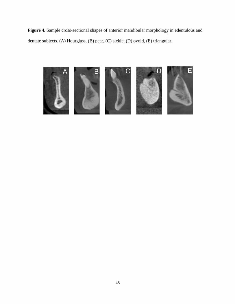
occurring morphological shapes, a classification system was developed. Figure 4 depicts these
morphological shapes, and categorizes mandibular cross-sections into the following groups:
hourglass, pear, sickle, ovoid, and triangular.
The incidence distribution for these shapes varied between edentulous and dentate
mandibles. For edentulous patients, the percentage of occurrence was as follows: hourglass 1%,
ovoid 26%, pear 53%, sickle 4%, and triangular 17% (Figure 5, Table 2). For dentate patients,
the percentage of occurrence was slightly different: hourglass 3%, ovoid 0%, pear 58%, sickle
29%, and triangular 10%. The differences between dentate and edentulous mandibles were not
significant for the hourglass, pear, and triangular shapes. However, the differences between
edentulous and dentate mandibles were significant for ovoid shaped, and sickle shaped
mandibles with p-values <0.0001for both groups. This suggests that perhaps these two

24
morphological variants are highly dependent on the dental status of the patient, whereas the other
three shapes are not.
Finally, the last comparison that was evaluated explored if any association existed
between the age of the subject at the time of CBCT and the mandibular cross-sectional
morphological shape. As demonstrated in figure 6, for both the edentulous and dentate groups,
no statistical significance was found between mandibular shape and age.
25
DISCUSSION
The purpose of this study was to explore surgically pertinent anatomical differences
between edentulous and dentate mandibles, and establish average measurement guidelines for
particular anatomic structures, namely the sublingual artery, submental artery, and the
mandibular incisive canal. Extensive literature has documented the general morphological
changes that take place over time in the edentulous mandible after loss of dentition.42
However,
to date, no known studies document the specific changes that may happen to the surrounding key
anatomical structures. This study sought to explore if any differences in specific anatomy could
be determined in edentulous and dentate mandibles.
First, calibration of the evaluator for this study was of prime importance in order to
establish reliability of the results. Overall, reliability was very high for the majority of categories.
In the four categories where reliability was <0.95, the groups had small sample numbers and
contained one or two severe outliers. This test of intra-rater correlation established the reliability
of all of the results in the study.
The reference point for data measurements that was used in this study was the inferior
border of the mandible. The inferior border of the mandible is considered to be a stable
landmark throughout life, and its position is not affected by the presence or absence of teeth and
subsequent alveolar resorption. The demographic composition of the edentulous and dentate
groups demonstrated a significant difference in age between the edentulous and dentate groups
with the mean ages being 63.2 and 60.05 years old, respectively. Though this difference was
statistically significant, the clinical significance between 63 and 60 years old most likely
irrelevant. The composition of males and females in both the edentulous and dentate groups was
not significantly different.

26
The sublingual artery (SLA) and submental artery (SMA) both had high incidence of
visualization on CBCT in both edentulous and dentate scans. The SLA was visualized 100% of
the time in edentulous scans, and 98% of the time in dentate scans. Furthermore, the SMA was
visualized 94% of the time in edentulous scans, and 88% of the time in dentate scans. As these
two vascular structures are in a potentially hazardous area with serious implications if injured
during implant surgery, providers utilizing CBCT imaging should always attempt to locate these
structures with a do so with high degree of success. Additionally, clinicians not current utilizing
CBCT for anterior mandibular implant placement should reconsider this option. On the other
hand, the mandibular incisive canal (MIC) was only visualized 61% of the time in edentulous
scans, and 59% of the time in dentate scans. This is most likely due to the intraosseous nature of
the canal. As it courses medially from split at the mental foramen, it traverses through trabecular
bone, and thus approximately 40% of the time can be lost in medullary spaces. This finding is of
important value to clinicians as large mandibular incisive canals may be misdiagnosed as
trabecular spaces and larger trabecular spaces may be potentially misdiagnosed as mandibular
incisive canals.
This study sought to establish average measurements that may be useful to clinicians as
guidelines for the location of the SLA, SMA, and MIC. On average, the SLA in edentulous
subjects was 15.31mm from the inferior border of the mandible, and in dentate subjects was
found to be 15.91mm from the inferior border of the mandible. Though this difference was
found to be statistically significant, it is likely not clinically significant. As a result, a clinician
not utilizing CBCT for anterior mandibular implant placement may use a distance approximately
15-16mm superior to the inferior border of the mandible as a general guideline for the location of
the SLA in both edentulous and dentate patients.

27
Additionally, the SMA was found on average to be 5.2mm from the inferior border in
edentulous subjects, and 4.85mm from the inferior border in dentate subjects. This difference
was not found to be statistically significant. Thus, a generalized measurement of approximately
5mm superior to the inferior border of the mandible can serve as a general guideline for the
location of the SMA in both edentulous and dentate patients.
Measurements for the MIC were conducted bilaterally. This was done in order to account
for the bilateral and separate nature of this anatomic structure. The MIC is the medial
continuation of the inferior alveolar canal after the mental foramen. Because of the slight
anatomical variation that might exist bilaterally, both sides were accounted for. The average
diameter of the MIC at the lateral incisor and canine regions for edentulous and dentate subjects
ranged from 1.42-1.52mm. This is consistent with other studies in the literature which cite
average diameters of 1.2mm,43
1.3mm,26
and a range from 0.48mm-2.9mm.25
Furthermore,
average measurements from the inferior border of the mandible to the superior border of the MIC
at the lateral incisor and canine regions yielded averages of 11.17mm and 11.16mm respectively
in edentulous subjects. In dentate subjects, these averages were statistically significantly
different yielding averages of 14.33mm and 13.70mm at the lateral incisor and canine
respectively. Thus, guideline measurements for the MIC in edentulous subjects can be
approximately 11mm from the inferior border of the mandible, and in dentate subjects
approximately 14mm from the inferior border of the mandible. This difference might possibly be
explained by the remodeling of bone post-extraction in edentulous patients as root apices are in
close proximity to this anatomical structure. These measurements are inconsistent with existing
literature by Mraiwa et al which sites this distance to be approximately 7.2mm from the inferior

28
border in cadaver studies.26
This study, however, did not note the dental status of the cadavers
and only included an evaluation of 50 samples.
Rehabilitation of the edentulous anterior mandible using implants requires a minimum
buccal-lingual bone width of 6mm for regular diameter implants for predictable implant
success.36
Another facet of this study sought to investigate the average amount of alveolar bone
reduction needed in order to achieve a minimum buccal-lingual bone width of 6mm. Many
times, the implant bed preparation requires flattening and reduction of the ridge crest in order to
achieve this minimum thickness. On average, the lateral incisor site required 4.49mm of bone
reduction, and the canine site required 4.27mm. However, for both of these sites, the standard
deviations were comparatively large yielding ±3.61mm and ±3.31mm for the lateral incisor and
canine respectively. Ostectomy (formerly called aveolectomy) in this region brings the crestal
bone closer to the three anatomic structures discussed in this project: the SLA, SMA, and MIC.
As a guideline, clinicians may use an approximated 4-5mm rule for bone reduction; however,
good clinical judgement based on specific bone width and prosthetic space should ultimately be
favored.
Finally, the cross-sectional shape of the anterior mandible was of particular interest in
this study. To date, no classification system for cross-sectional shape of the anterior mandible
exists. This is of significant interest as implant-based rehabilitation in the anterior mandible may
be compromised in cases of severe alveolar constriction, better known as the so-called
“hourglass mandible” variant.37
To date, this is the only classified morphological shape, and is
defined as an osseous constriction at the alveolar-basal bone junction.37
As other morphological
variants may have implications on implant placement, a classification system was defined based
on commonly occurring cross-sectional shapes noted during CBCT evaluation. The classified

29
variants are as follows: hourglass, ovoid, pear, sickle, and triangular. The incidence of the
hourglass variant was 1% in edentulous subjects, and 3% in dentate subjects, with no statistical
difference between these two groups. This is within range reported by Butura et al in 2011 in
which the incidence of the hourglass variant was reported to be 3.98%.37
In general, the majority
of edentulous and dentate subjects were noted to have the pear shaped cross-section with 53%
and 58% respectively. This difference was also not significant. The ovoid shape mandibles were
significantly different between edentulous and dentate patients with 26% and 0% respectively.
This is likely due to the absence of teeth causing to the rounded appearance of the alveolar crest.
Furthermore, the difference between edentulous and dentate subjects was significant for the
sickle shaped mandibles, totaling 4% and 29% respectively. The greater proportion of sickle
shaped mandibles in dentate subjects is likely influenced by the presence of teeth in the alveolus.
Finally, the mandibular cross-sectional shapes were compared with age to see if any
trends were present. No trends were noted, and no relationships could be established with the
present data. This could potentially be due to limited variation in age of subjects examined, or
ethnic or skeletal differences, and warrants future exploration.
The average anatomic guidelines presented in this project can be used not only directly in
a surgical setting in order to predict the location of the SLA, SMA, and MIC; but, also may be
helpful in as means of “feasibility analysis” on a 2-dimensional panoramic radiograph. The most
superior to these structures would be the first encountered in a top-down osteotomy preparation
technique. In the majority of cases, the most superior structure is the SLA, which is
approximately 15mm above the inferior border of the mandible. After consideration of
prosthetic space and implant length, if the provider determines that surgical intervention will
likely encroach on this region 15mm above the inferior border, a full CBCT analysis is

30
warranted. If not, then anatomical averages and 2-dimensional radiography may provide the
operator with enough information to confidently proceed with implant rehabilitation.

31
STUDY LIMITATIONS
The nature of this study included the following limitations:
Though best efforts were made to avoid using CBCTs which appeared to have fresh
extraction sites, time post-extraction or length of time edentulous could not be fully
accounted for in the analysis of the edentulous subjects
Though age range of patients was widespread (from 18-85 years old), the age
distribution was generally clustered between the ages of 55-70 years old.
Racial or skeletal associations (eg: class I, II, or III) could not be recorded due to the
purely radiographic nature of this project.
All observations were made by 1 observer, which may introduce bias in data
gathering. However, this may also be viewed as a strength, as it eliminates
heterogeneity; additionally, the intra-operator reliability testing for the pilot study of
30 samples showed high consistency in measurements.

32
CONCLUSIONS
As dental implants have gained widespread popularity in prosthodontic rehabilitation, the
number of reports citing adverse events has increased as well. Quantitative data regarding precise
location or percentage of variability of the sublingual artery, submental artery, and the
mandibular incisive canal in dentate and edentulous patients is helpful to clinicians planning
surgery in the anterior mandible. The current standard of care does not require CBCT analysis
prior to implant surgery. Yet countless negative reports of severe injuries have been documented
and can result in substantial paresthesia and pain from neural damage, or life-threatening
hemorrhage leading to airway compromise from vascular injury.
Though CBCT analysis of all cases requiring surgery of the anterior mandible may be
prudent, it is not always necessary or possible. With the increasing popularity of implant
dentistry and advancement of associated technology, there is a need for scientific standards for
clinicians to use whether CBCT analysis is utilized or not.
Based on the results of this study, the following conclusions were drawn:
The incidence of visualization of the sublingual artery on CBCT was 100% for
edentulous subjects and 98% for dentate subjects
The sublingual artery was located approximately 15mm above the inferior border of the
mandible
The incidence of visualization of the submental artery on CBCT was 94% for edentulous
subjects and 88% for dentate subjects
The submental artery was located approximately 5mm above the inferior border of the
mandible

33
The incidence of visualization of the mandibular incisive canal on CBCT was 61% for
edentulous subjects and 59% for dentate subjects
The mandibular incisive canal was approximately 1.5mm in diameter at the lateral incisor
and canine regions
The mandibular incisive canal was located approximately 11mm above the inferior
border of the mandible in edentulous patients, and approximately 14mm above the
inferior border of the mandible in dentate patients
The average distance from the residual ridge crest inferiorly to the region of the mandible
where the buccal-lingual width is at least 6mm was approximately 4mm at the lateral
incisor and canine regions in edentulous patients
A new classification system for the cross-sectional morphology of the anterior mandible
was characterized, and includes the following shapes: hourglass, ovoid, pear, sickle, and
triangular. The pear shaped was the most common among both edentulous and dentate
patients.
Overall, the results of this study may aid clinicians in achieving better clinical confidence
and improved clinical outcomes for implant placement in the anterior mandible.

34
FUTURE RESEARCH
The use of implants in dentistry grows more widespread daily. The results of the current
study offer some clinical guidelines for practitioners who perform any surgical or prosthetic
intervention in the anterior mandible. Future studies can expand upon the foundation laid by
exploring potential anatomic changes with age or length of time the subjects are edentulous.
Furthermore, future correlations can be made exploring possible relationships between
mandibular cross-sectional morphology and race, skeletal classification, or length of time that the
subject has been edentulous. Finally, future studies could be conducted in a similar manner
concerning the posterior mandible. This area also has a significant number of important, and
challenging anatomic structures providers must be fully aware of prior to any surgical
intervention.

35
REFERENCES
1. Tagliareni GM, Clarkson E. Basic Concepts and Techniques of Dental Implants. Dent Clin
North Am 2015;2:255-264.
2. Adell R, Lekholm U, Rockler B, Branemark PI. A 15-year Study of Osseointegrated
Implants in the Treatment of the Edentulous Jaw. Int J Oral Surg 1981;10:387-416.
3. Dubois L, de Lange J, Baas E, Van Ingen J. Excessive bleeding in the Floor of the Mouth
after Endosseous Implant Placement: a Report of Two Cases. Int J Oral Maxillofac Surg
2010;39:412–415.
4. Lee KA, Kim M, Hong J, Lee J, Choi S, Chai J, Jung U. Anatomical Topography of the
Mandibular Symphysis in the Korean Population: A Computed Tomography Analysis.
Clin Anat 2013; doi: 10.1002/ca.22315.
5. Branemark PI, Zarb GA, Albrektsson T. Tissue Integrated Prostheses: Osseointegration in
Clinical Dentistry. 1st ed. Chicago: Quintessence Publishing Co., Inc; 1985. p. 211-232.
6. Kutuk N, Gonen ZB, Kilic E, Alkan A. Anterior Mandibular Zone Safe for Implants. J
Craniofac Surg 2013;24:e405-e408.
7. Jaju P, Jaju S. Lingual Vascular Canal Assessment by Dental Computed Tomography: a
Retrospective Study. Indian J Dent Res 2011;22:232-236.
8. Lee C, Yanagihara LC, Suzuki JB. Brisk, Pulsatile Bleeding From the Anterior
Mandibular Incisive Canal During Implant Surgery: A Case Report and Use of an Active
Hemostatic Matrix to Terminate Acute Bleeding. Implant Dent 2012;21:368-373.
9. Bidra AS. Flapless Implant Surgery to Overcome Anatomic Challenges in the Anterior
Mandible for Overdenture Therapy: a Clinical Report. J Prosthet Dent 2014;111:175-80.

36
10. Standring S. Gray’s Anatomy: The Anatomical Basis of Clinical Practice. 40th
ed. Spain:
Churchill Livingstone/Elsevier; 2008. p. 499-506, 530-533, 538-546.
11. Romanos GE, Gupta B, Crespi R. Endosseous Arteries in the Anterior Mandible:
Literature Review. Int J Oral Maxillofac Implants 2012;27:90-94.
12. Moore KL, Dalley AF. Clinically Oriented Anatomy. 5th
ed. Baltimore: Lippincott
Williams & Wilkins; 2006. p. 886-1122, 1138-1143.
13. McDonnell D, Nouri MR, Todd ME. The Mandibular Lingual Foramen: a Consistent
Arterial Foramen in the Middle of the Mandible. J Anat 1994;184:363-369.
14. Fujita S, Ide Y, Abe S. Variations of Vascular Distribution in the Mandibular Anterior
Lingual Region: a High Risk of Vascular Injury During Implant Surgery. Implant Dent
2012;21:259-264.
15. Loukas M, Kinsella CR, Kapos T. Tubbs RS, Ramachandra S. Anatomical Variation in
Arterial Supply of the Mandible with Special Regard to Implant Placement. Int J Oral
Maxillofac Surg 2008;37:367-371.
16. Liang X, Jacobs R, Lanbrichts I, Vandewalle G. Lingual Foramina on the Mandibular
Midline Revisited. Clin Anat 2007;20:246-251.
17. Mardinger O, Manor Y, Mijiritsky E, Hirshberg A. Lingual Perimandibular Vessels
Associated with Life-threatening Bleeding: an Anatomic Study. Int J Oral Maxillofac
Implants 2007;22:127-131.
18. Tepper G, Hofschneider UB, Gahleitner A, Ulm C. Computed Tomographic Diagnosis
and Localization of Bone Canals in the Mandibular Interformainal Region for Prevention
of Bleeding Complications During Implant Surgery. Int J Oral Maxillofac Implants
2001;16:68-72.

37
19. Lustig JP, London D, Dor BL, Yanko R. Ultrasound Identification and Quantitative
Measurement of Blood Supply to the Anterior Mandible. Oral Surg Oral Med Oral Pathol
Oral Radiol Endod 2003;96:625-629.
20. Longoni S, Sartori M, Braun M, Bravetti P, Lapi A, Baldoni M, Tredici G. Lingual
Vascular Canals of the Mandible: the Risk of Bleeding Complications During Implant
Procedures. Implant Dent 2007;16:131-138.
21. Tagaya A, Matsuda Y, Nakajima K, Seki K, Okano T. Assessment of the Blood Supply to
the Lingual Surface of the Mandible for Reduction of Bleeding During Implant Surgery.
Clin Oral Implants Res 2009;4:351-355.
22. Romanos GE, Greenstein G. The Incisive Canal. Considerations During Implant
Placement: Case Report and Literature Review. Int J Oral Maxillofac Implants
2009;24:740-745.
23. Apostolakis D, Brown JE. The Dimensions of the Mandibular Incisive Canal and its
Spatial Relationship to Various Anatomical Landmarks of the Mandible: a Study Using
Cone Beam Computed Tomography. Int J Oral Maxillofac Implants 2013;28:117-124.
24. Polland KE, Munro S, Redford G, Lockhart A, Logan G, Brocklebank L, McDonald SW.
The Mandibular Canal of the Edentulous Jaws. Clin Anat 2001;14:445-452.
25. Mardinger O, Chaushu G, Arensburg B, Taicher S, Kaffe I. Anatomic and Radiologic
Course of the Mandibular Incisive Canal. Surg Radiol Anat 2000;22:157-161.
26. Mraiwa N, Jacobs R, Moerman P, Lambrichts I, van Steenberghe D, Quirynen M.
Presence and Course of the Incisive Canal in the Human Mandibular Interforaminal
Region: Two-dimensional Imaging Versus Anatomical Observations. Surg Radiol Anat
2003;25:416-423.
38
27. Jacobs R, Mraiwa N, van Steenberghe D, Gijbels F, Quirynen M. Appearance, Location,
Course, and Morphology of the Mandibular Incisive Canal: an Assessment on Spiral CT
Scan. Dentomaxillofac Rad 2002;31:322-327.
28. Jacobs R, Mraiwa N, van Steenberghe D, Sanderink G, Guirynen M. Appearance of the
Mandibular Incisive Canal on Panoramic Radiographs. Surg Radiol Anat 2004;26:329-
333.
29. Uchida Y, Yamashita Y, Goto M, Hanihara T. Measurement of Anterior Loop Length of
the Mandibular Canal and Diameter of the Mandibular Incisive Canal to Avoid Nerve
Damage when Installing Endosseous Implants in the Interforaminal Region. J Oral
Maxillofac Surg 2007;65:1772-1779.
30. Uchida Y, Noguchi N, Goto M, Yamashita Y, Hanihara T, Takamori H, Sato I, et al.
Measurement of Anterior Loop Length of the Mandibular Canal and Diameter of the
Mandibular Incisive Canal to Avoid Nerve Damage when Installing Endosseous Implants
in the Interforaminal Region: a Second Attempt Introducing Cone Beam Computed
Tomography. J Oral Maxillofac Surg 2009;67:744-750.
31. Juodzbalys G, Wang HL, Sabalys G. Anatomy of the Mandibular Vital Structures. Part II:
Mandibular Incisive Canal, Mental Foramen and Associated Neurovascular Bundles in
Relation to Dental Implantology. J Oral Maxillofacial Res 2010; doi:
10.5037/jomr.2010.1103.
32. Mordenfeld A, Andersson L, Bergstrom B. Hemorrhage in the Floor of the Mouth During
Implant Placement in the Edentulous Mandible: a Case Report. Int J Oral Maxillofac
Implants 1997;12:558-561.

39
33. Niamtu J. Near-fatal Airway Obstruction After Routine Implant Placement. Oral Surg
Oral Med Oral Pathol Oral Radiol Endod 2001;92:597-600.
34. Woo BM, Al-Bustani S, Ueeck BA. Floor of Mouth Hemorrhage and Life-threatening
Airway Obstruction During Immediate Implant Placement in the Anterior Mandible. Int J
Oral Maxillofac Surg 2006;35:961-964.
35. Benavides E, Rios HF, Ganz SD, An CH, Resnik R, Reardon GT, Feldman SJ, et al. Use
of Cone Beam Computed Tomography in Implant Dentistry: the International Cognress of
Oral Implantologists Consensus Report. Implant Dent 2012;21:78-86.
36. Straumann Dental Implant System: Basic Information on Surgical Procedures. Can be
accessed at: www.straumann.com
37. Butura CC, Galindo DF, Cottam J, Adams M, Jensen O. Hourglass Mandibular Anatomic
Variant Incidence and Treatment Considerationsfor All-on-Four Implant Therapy: Report
of 10 Cases. J Oral Maxillofacial Surg 2001;69:2135-2143.
38. Moghadam HG. Vertical and Horizontal Bone Augmentation with the Intraoral
Autogenous J-graft. Implant Dent 2009;18:230-238.
39. Pelo S, Boniello R, Moro A, Gasparini G, Amaroso PF. Augmentation of the Atrophic
Edentulous Mandible by a Bilateral Two-step Osteotomy with Autogenous Bone Graft to
Place Osseointegrated Dental Implants. Int J Oral Maxillofac Surg 2010;39:227-234.
40. Chan HL, Misch K, Wang HL. Dental Imaging in Implant Treatment Planning. Implant
Dent 2010;19:288-298.
41. Douglass CW, Shih A, Ostry L. Will There be a Need for Complete Dentures in the
United States in 2020? J Prosthet Dent 2002;87:5-8.

40
42. Lekholm U, Zarb G. (1985) Patient selection and preparation. Tissue-Integrated
Prosthesis: Osseointegration in Clinical Dentistry, 199 - 209. Chicago: Quintessence.
43. Bavitz JV, Harn SD, Hansen CA, et al. An anatomical study of mental neurovascular
bundle-implant relationships. Int J Oral Maxillofac Implants 1993;8:563-567.

41
Figure 1. (A) Distributions of outlier intra-class correlation coefficients of mandibular incisive
canal diameter measurements at the lateral incisor position bilaterally in edentulous CBCTs. (B)
Distributions of outlier intra-class correlation coefficients of mandibular incisive canal diameter
measurements at the canine position bilaterally in dentate CBCTs. Note the presence of outliers
in each distribution.
Edentulous
Dentate
A
B

42
Figure 2. Incidence of visualization of the sublingual artery (SLA), submental artery (SMA),
and mandibular incisive canal (MIC) in CBCT scans of 125 edentulous and 100 dentate CBCTs.

43
Figure 3. Scatter plots of measurements of noted anatomic structures. This includes the (A)
distance measured from the inferior border of the mandible to the sublingual artery, (B) and the
submental artery in edentulous and dentate CBCTs. (C) Measured diameter of the mandibular
incisive canal at the lateral incisor region, and (D) the canine region in edentulous and dentate
CBCTs. (E) Distance measured from the inferior border of the mandible to the mandibular
incisive canal at the lateral incisor region, and (F) the canine region in edentulous and dentate
CBCTs. All figures are presented as mean ± standard error of the mean (SEM) where error bars
represent SEM. *P<0.05, **P<0.01, ***P<0.001, ****P<0.0001

44
45
Figure 4. Sample cross-sectional shapes of anterior mandibular morphology in edentulous and
dentate subjects. (A) Hourglass, (B) pear, (C) sickle, (D) ovoid, (E) triangular.

46
Figure 5. Comparison of percent incidence of cross-sectional anterior mandibular shapes in 125
edentulous and 100 dentate CBCT scans.

47
Figure 6. Scatter-plot distribution of mandibular cross-sectional shapes based on subject’s age
in edentulous and dentate CBCT scans. All figures are presented as mean ± standard error of the
mean (SEM) where error bars represent SEM.

48
Table 1. Intra-class correlation coefficients for assessing operator reliability between first and
second replicates. Structures that were not analyzed, or where no comparison could be made
were recorded as “NA.”
Edentulous Scans (N=15) Dentate Scans (N=15)
N Intra-class
correlation
N Intra-class correlation
Distance (mm)
IB to SLA 15 1 (0.99, 1) 14 1 (0.99, 1)
IB to SMA 12 1 (0.98, 1) 11 1 (0.99, 1)
IB to MIC at LI - L 8 0.99 (0.97, 1) 1 NA
IB to MIC at LI - R 9 0.99 (0.97, 1) 1 NA
IB to MIC at Ca - L 10 0.99 (0.94, 1) 6 1 (0.98, 1)
IB to MIC at Ca - R 9 0.98 (0.93, 1) 6 1 (0.99, 1)
Crest to 6mm width at LI - L 15 1 (0.99, 1) NA NA
Crest to 6mm width at LI - R 15 0.99 (0.96, 1) NA NA
Crest to 6mm width at Ca - L 15 1 (0.99, 1) NA NA
Crest to 6mm width at Ca - R 15 0.98 (0.93, 0.99) NA NA
Diameter (mm)
MIC ø at LI - L 8 0.89 (0.54, 0.98)* 1 NA
MIC ø at LI - R 9 0.88 (0.57, 0.97)* 1 NA
MIC ø at Ca - L 10 0.97 (0.88, 0.99) 6 0.22 (-0.57, 0.83)*
MIC ø at Ca - R 9 0.95 (0.74, 0.99) 6 0.83 (0.20, 0.97)*
IB – inferior border of the mandible; SLA – sublingual artery; SMA – submental artery; MIC –
mandibular incisive canal; ⌀- diameter; LI – lateral incisor; Ca – canine; L – left; R – right
*Measurements of lower reliability due to exceptional outliers
49
Table 2. Summary of demographic, incidence, and average measurement data for noted
anatomical structures in 125 edentulous and 100 dentate CBCT scans.
IB – inferior border of the mandible; SLA – sublingual artery; SMA – submental artery; MIC –
mandibular incisive canal; ⌀- diameter; LI – lateral incisor; Ca – canine; L – left; R – right
Edentulous Scans (N=125) Dentate Scans (N=100) P-value
Age 63.2 ± 12.72 60.05 ± 14.08 0.037
Gender 0.227
Female 60 (48%) 57 (57%)
Male 65 (52%) 43 (43%)
Sublingual artery visualized? 0.196
No 0 (0%) 2 (2%)
Yes 125 (100%) 98 (98%)
Distance from IB to SLA (mm) 15.31 ± 2.15 15.91 ± 1.72 0.042
Submental artery visualized? 0.162
No 8 (6%) 12 (12%)
Yes 117 (94%) 88 (88%)
Distance to from IB to SMA (mm) 5.2 ± 2.46 4.85 ± 2.46 0.185
Mandibular incisive canal visualized? 0.786
No 49 (39%) 41 (41%)
Yes 76 (61%) 59 (59%)
MIC ø at LI (mm) 58/76 = 76% 1.44 ± 0.23 35/59 = 59% 1.42 ± 0.21 0.809
MIC ø at Ca (mm) 63/76 = 83% 1.50 ± 0.27 46/59 = 78% 1.52 ± 0.29 0.792
Distance from IB to MIC at LI (mm) 58/76 = 76% 11.17 ± 1.84 35/59 = 59% 14.33 ± 2.38 <0.0001
Distance from IB to MIC at Ca (mm) 63/76 = 83% 11.16 ± 1.75 46/59 = 78% 13.70 ± 2.64 <0.0001
Distance from crest to 6mm width (mm)
at LI 4.49 ± 3.61 NA
at Ca 4.27 ± 3.37 NA
Mandibular cross-sectional shape
Hourglass 1 (1%) 3 (3%) 0.325
Ovoid 32 (26%) 0 (0%) <0.0001
Pear 66 (53%) 58 (58%) 0.500
Sickle 5 (4%) 29 (29%) <0.0001
Triangular 21 (17%) 10 (10%) 0.174

50
Table 3. Comparison of bilateral right and left measurements of average distances from the
inferior border of the mandible to the mandibular incisive canal, and average diameter of the
mandibular incisive canal in edentulous and dentate CBCT scans; and, average distance from the
alveolar crest inferiorly to a minimum of 6mm anterior mandibular bone width in edentulous
CBCT scans.
IB – inferior border of the mandible; SLA – sublingual artery; SMA – submental artery; MIC –
mandibular incisive canal; ⌀ - diameter; LI – lateral incisor; Ca – canine; L – left; R – right
Edentulous Scans Dentate Scans
Left Right P-value Left Right P-value
Distance (mm)
IB to MIC at LI 11.12 ±
2.28
11.25 ±
1.88
0.341 13.68 ± 2.56 14.58 ± 2.62 <0.000
1
IB to MIC at Ca 11.11 ±
1.95
11.2 ± 1.77 0.410 13.26 ± 2.72 13.9 ± 2.84 0.025
Crest to 6mm width at LI 4.37 ± 3.68 4.61 ± 3.79 0.517 NA NA NA
Crest to 6mm width at Ca 4.28 ± 3.55 4.27 ± 3.54 0.974 NA NA NA
Diameter (mm)
MIC ø at LI 1.45 ± 0.27 1.42 ± 0.26 0.232 1.45 ± 0.28 1.42 ± 0.2 0.757
MIC ø at Ca 1.5 ± 0.29 1.54 ± 0.36 0.118 1.52 ± 0.31 1.51 ± 0.28 0.888