an advanced solution for sfa disease: vascular mimetic
TRANSCRIPT
38 INSERT TO ENDOVASCULAR TODAY OCTOBER 2014
FEATURED TECHNOLOGY: SUPERA® PERIPHERAL STENT SYSTEM
Sponsored by Abbott Vascular
Perspectives on the use of the Supera® peripheral stent system.
WITH CRAIG WALKER, MD, AND JOSEPH J. RICOTTA II, MD, MS
An Advanced Solution for SFA Disease: Vascular Mimetic Implants
W ith this year’s US Food and Drug Administration approval of the Supera® peripheral stent system (Abbott Vascular), intervention-
ists across the United States have had the chance to use a truly unique tool for femoro popliteal disease. Endovascular Today recently asked two leading endo-vascular specialists, Craig Walker, MD, and Joseph J. Ricotta II, MD, MS, to discuss how and why they use vascular mimetic implants to treat lesions in the superficial femoral (SFA) and proximal popliteal arter-ies. Their perspectives and recommendations of this
unique class of technology are based upon both the relevant clinical evidence and their own experience.
COMBINING THE IDEAL ATTRIBUTES OF AN SFA TREATMENT
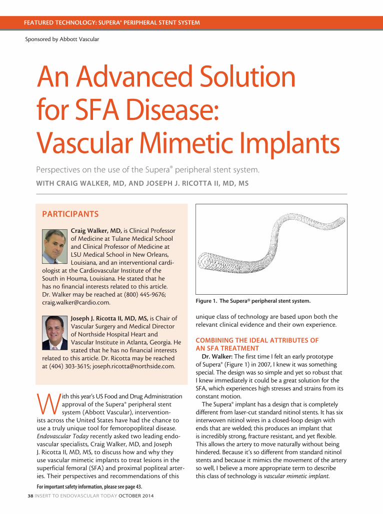
Dr. Walker: The first time I felt an early prototype of Supera® (Figure 1) in 2007, I knew it was something special. The design was so simple and yet so robust that I knew immediately it could be a great solution for the SFA, which experiences high stresses and strains from its constant motion.
The Supera® implant has a design that is completely different from laser-cut standard nitinol stents. It has six interwoven nitinol wires in a closed-loop design with ends that are welded; this produces an implant that is incredibly strong, fracture resistant, and yet flexible. This allows the artery to move naturally without being hindered. Because it’s so different from standard nitinol stents and because it mimics the movement of the artery so well, I believe a more appropriate term to describe this class of technology is vascular mimetic implant.
PARTICIPANTS
Craig Walker, MD, is Clinical Professor of Medicine at Tulane Medical School and Clinical Professor of Medicine at LSU Medical School in New Orleans, Louisiana, and an interventional cardi-
ologist at the Cardiovascular Institute of the South in Houma, Louisiana. He stated that he has no financial interests related to this article. Dr. Walker may be reached at (800) 445-9676; [email protected].
Joseph J. Ricotta II, MD, MS, is Chair of Vascular Surgery and Medical Director of Northside Hospital Heart and Vascular Institute in Atlanta, Georgia. He stated that he has no financial interests
related to this article. Dr. Ricotta may be reached at (404) 303-3615; joseph.ricotta@ northside.com.
For important safety information, please see page 43.
Figure 1. The Supera® peripheral stent system.
OCTOBER 2014 INSERT TO ENDOVASCULAR TODAY 39
FEATURED TECHNOLOGY: SUPERA® PERIPHERAL STENT SYSTEM
Sponsored by Abbott Vascular
Dr. Ricotta: I agree that preserving the natural move-ment of the anatomy is critical to ensuring durable clinical outcomes. A natural-moving mimetic implant such as Supera® can more comprehensively address the common issues that we see with the current modalities of SFA treatment.
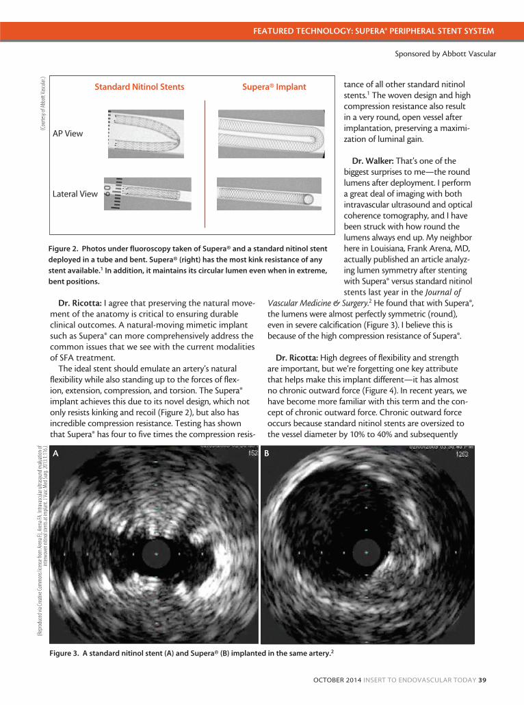
The ideal stent should emulate an artery’s natural flexibility while also standing up to the forces of flex-ion, extension, compression, and torsion. The Supera® implant achieves this due to its novel design, which not only resists kinking and recoil (Figure 2), but also has incredible compression resistance. Testing has shown that Supera® has four to five times the compression resis-
tance of all other standard nitinol stents.1 The woven design and high compression resistance also result in a very round, open vessel after implantation, preserving a maximi-zation of luminal gain.
Dr. Walker: That’s one of the biggest surprises to me—the round lumens after deployment. I perform a great deal of imaging with both intravascular ultrasound and optical coherence tomography, and I have been struck with how round the lumens always end up. My neighbor here in Louisiana, Frank Arena, MD, actually published an article analyz-ing lumen symmetry after stenting with Supera® versus standard nitinol stents last year in the Journal of
Vascular Medicine & Surgery.2 He found that with Supera®, the lumens were almost perfectly symmetric (round), even in severe calcification (Figure 3). I believe this is because of the high compression resistance of Supera®.
Dr. Ricotta: High degrees of flexibility and strength are important, but we’re forgetting one key attribute that helps make this implant different—it has almost no chronic outward force (Figure 4). In recent years, we have become more familiar with this term and the con-cept of chronic outward force. Chronic outward force occurs because standard nitinol stents are oversized to the vessel diameter by 10% to 40% and subsequently
Figure 2. Photos under fluoroscopy taken of Supera® and a standard nitinol stent
deployed in a tube and bent. Supera® (right) has the most kink resistance of any
stent available.1 In addition, it maintains its circular lumen even when in extreme,
bent positions.
Figure 3. A standard nitinol stent (A) and Supera® (B) implanted in the same artery.2
A B
(Cou
rtesy
of A
bbot
t Vas
cular
.)(R
epro
duce
d via
Crea
tive C
omm
ons l
icens
e fro
m A
rena
FJ, A
rena
FA. In
trava
scula
r ultr
asou
nd ev
aluat
ion of
int
erw
oven
nitin
ol ste
nts a
t im
plant
. J V
asc M
ed Su
rg. 2
013:1
:116.)
Standard Nitinol Stents Supera® Implant
AP View
Lateral View
40 INSERT TO ENDOVASCULAR TODAY OCTOBER 2014
FEATURED TECHNOLOGY: SUPERA® PERIPHERAL STENT SYSTEM
Sponsored by Abbott Vascular
push outward on the vessel wall with a constant force to try to reach their equilibrium state. This outward force is believed to irritate the vessel and may be a catalyst for accelerated formation of neointimal hyperplasia. We’ve adjusted down our oversizing with some devices, such as covered self-expanding stents, but Supera® has an innate advantage for two reasons: (1) its woven design has minimal outward force, and (2) it’s also sized 1:1 with the vessel.
Dr. Walker: That’s right, Dr. Ricotta. That’s one rea-son there are limitations with drug-eluting peripheral stents; even if these laser-cut stents were to have a paclitaxel coating, the fundamental mechanical struc-ture is still a standard nitinol stent. Once the drug is gone after 2 or 3 days, a bare-metal standard nitinol
stent remains and imposes a persistent chronic out-ward force on the vessel.
CLINICAL DATA Dr. Walker: I had faith that this design would work well
in the SFA even before the robust clinical evidence came out. Since the SUPERB trial data3 have been released, we’ve learned even more about this device, and they have affirmed our hypotheses. Even before I saw the clinical data, I really believed in the design because I had felt the device and knew it combined the appropriate properties to help revolutionize SFA treatment.
Dr. Ricotta: I agree with Dr. Walker. When initial data were released in late 2012 and early 2013,4 people really took notice of this device. The Leipzig studies5,6 in par-ticular really caught my eye.
The SUPERB pivotal trial achieved an excellent 12-month primary patency rate of 86.3% (Kaplan-Meier [K-M]). Freedom from reintervention (target lesion revascularization [TLR]) was also excellent at 89% (1 year) and 84% (2 years). This was in a relatively tough patient population that consisted of 45% of patients with severe calcium and 25% with total occlusions.3
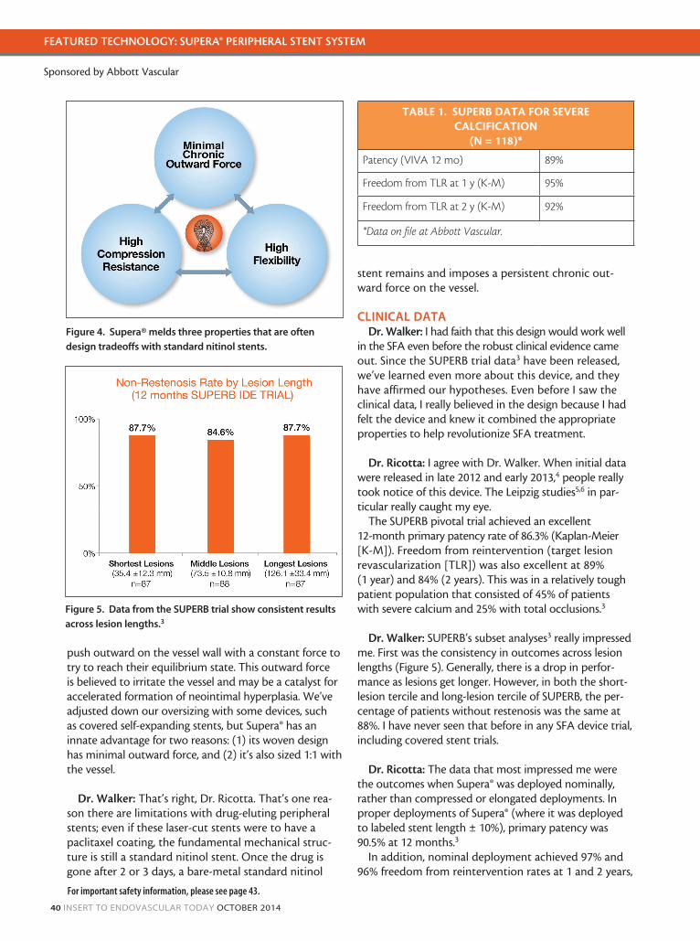
Dr. Walker: SUPERB’s subset analyses3 really impressed me. First was the consistency in outcomes across lesion lengths (Figure 5). Generally, there is a drop in perfor-mance as lesions get longer. However, in both the short-lesion tercile and long-lesion tercile of SUPERB, the per-centage of patients without restenosis was the same at 88%. I have never seen that before in any SFA device trial, including covered stent trials.
Dr. Ricotta: The data that most impressed me were the outcomes when Supera® was deployed nominally, rather than compressed or elongated deployments. In proper deployments of Supera® (where it was deployed to labeled stent length ± 10%), primary patency was 90.5% at 12 months.3
In addition, nominal deployment achieved 97% and 96% freedom from reintervention rates at 1 and 2 years,
TABLE 1. SUPERB DATA FOR SEVERE CALCIFICATION
(N = 118)*
Patency (VIVA 12 mo) 89%
Freedom from TLR at 1 y (K-M) 95%
Freedom from TLR at 2 y (K-M) 92%
*Data on file at Abbott Vascular.
Figure 4. Supera® melds three properties that are often
design tradeoffs with standard nitinol stents.
Figure 5. Data from the SUPERB trial show consistent results
across lesion lengths.3
For important safety information, please see page 43.
OCTOBER 2014 INSERT TO ENDOVASCULAR TODAY 41
FEATURED TECHNOLOGY: SUPERA® PERIPHERAL STENT SYSTEM
Sponsored by Abbott Vascular
respectively. However, we found that lower primary patency and freedom from reintervention rates (TLR) were associated with severely elongated deployments. That’s why it is important to deploy properly and size appropriately to optimize Supera® nominal deploy-ment.3
Dr. Walker: I agree. In deploying Supera® nomi-nally, you can really get outstanding results. I was also recently made aware of the SUPERB data in severe cal-cium (Table 1). Forty-five percent of the patients in the SUPERB trial had severe calcification.3 In those patients, the 12-month patency was 89% (VIVA [binary] meth-od), and the freedom from reintervention rate was 95%. At 24 months, this number decreased only slightly to 92%.7 The similarities between 12- and 24-month data show that this is really a durable solution.
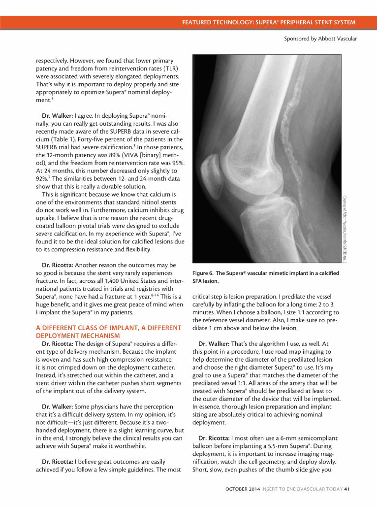
This is significant because we know that calcium is one of the environments that standard nitinol stents do not work well in. Furthermore, calcium inhibits drug uptake. I believe that is one reason the recent drug-coated balloon pivotal trials were designed to exclude severe calcification. In my experience with Supera®, I’ve found it to be the ideal solution for calcified lesions due to its compression resistance and flexibility.
Dr. Ricotta: Another reason the outcomes may be so good is because the stent very rarely experiences fracture. In fact, across all 1,400 United States and inter-national patients treated in trials and registries with Supera®, none have had a fracture at 1 year.8-14 This is a huge benefit, and it gives me great peace of mind when I implant the Supera® in my patients.
A DIFFERENT CLASS OF IMPLANT, A DIFFERENT DEPLOYMENT MECHANISM
Dr. Ricotta: The design of Supera® requires a differ-ent type of delivery mechanism. Because the implant is woven and has such high compression resistance, it is not crimped down on the deployment catheter. Instead, it’s stretched out within the catheter, and a stent driver within the catheter pushes short segments of the implant out of the delivery system.
Dr. Walker: Some physicians have the perception that it’s a difficult delivery system. In my opinion, it’s not difficult—it’s just different. Because it’s a two-handed deployment, there is a slight learning curve, but in the end, I strongly believe the clinical results you can achieve with Supera® make it worthwhile.
Dr. Ricotta: I believe great outcomes are easily achieved if you follow a few simple guidelines. The most
critical step is lesion preparation. I predilate the vessel carefully by inflating the balloon for a long time: 2 to 3 minutes. When I choose a balloon, I size 1:1 according to the reference vessel diameter. Also, I make sure to pre-dilate 1 cm above and below the lesion.
Dr. Walker: That’s the algorithm I use, as well. At this point in a procedure, I use road map imaging to help determine the diameter of the predilated lesion and choose the right diameter Supera® to use. It’s my goal to use a Supera® that matches the diameter of the pre dilated vessel 1:1. All areas of the artery that will be treated with Supera® should be predilated at least to the outer diameter of the device that will be implanted. In essence, thorough lesion preparation and implant sizing are absolutely critical to achieving nominal deployment.
Dr. Ricotta: I most often use a 6-mm semicompliant balloon before implanting a 5.5-mm Supera®. During deployment, it is important to increase imaging mag-nification, watch the cell geometry, and deploy slowly. Short, slow, even pushes of the thumb slide give you
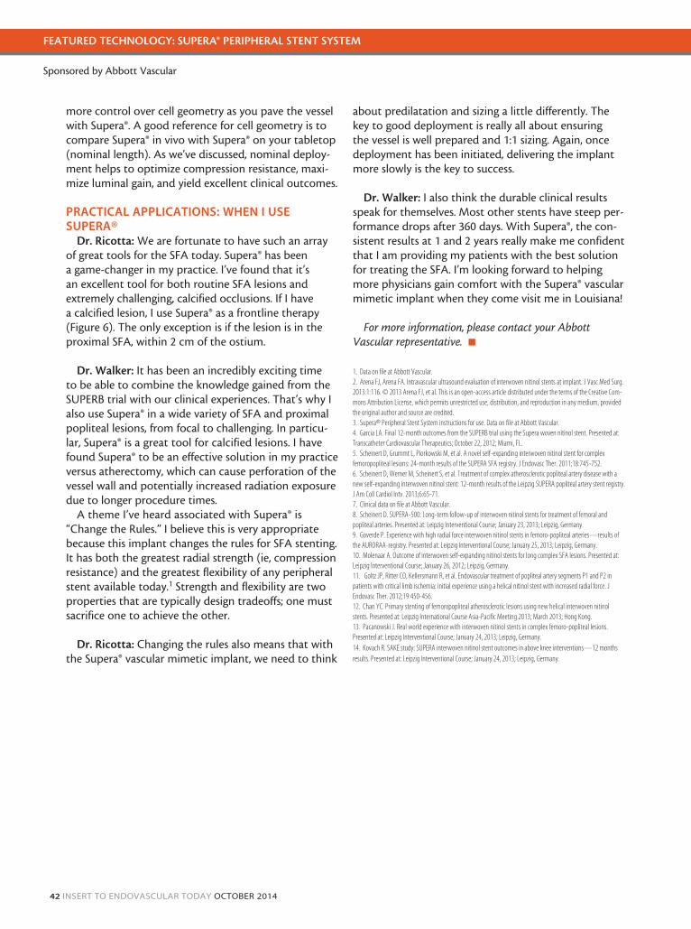
Figure 6. The Supera® vascular mimetic implant in a calcified
SFA lesion.
(Courtesy of Abbott Vascular, from the SUPERB trial.)
42 INSERT TO ENDOVASCULAR TODAY OCTOBER 2014
FEATURED TECHNOLOGY: SUPERA® PERIPHERAL STENT SYSTEM
Sponsored by Abbott Vascular
more control over cell geometry as you pave the vessel with Supera®. A good reference for cell geometry is to compare Supera® in vivo with Supera® on your tabletop (nominal length). As we’ve discussed, nominal deploy-ment helps to optimize compression resistance, maxi-mize luminal gain, and yield excellent clinical outcomes.
PRACTICAL APPLICATIONS: WHEN I USE SUPERA®
Dr. Ricotta: We are fortunate to have such an array of great tools for the SFA today. Supera® has been a game-changer in my practice. I’ve found that it’s an excellent tool for both routine SFA lesions and extremely challenging, calcified occlusions. If I have a calcified lesion, I use Supera® as a frontline therapy (Figure 6). The only exception is if the lesion is in the proximal SFA, within 2 cm of the ostium.
Dr. Walker: It has been an incredibly exciting time to be able to combine the knowledge gained from the SUPERB trial with our clinical experiences. That’s why I also use Supera® in a wide variety of SFA and proximal popliteal lesions, from focal to challenging. In particu-lar, Supera® is a great tool for calcified lesions. I have found Supera® to be an effective solution in my practice versus atherectomy, which can cause perforation of the vessel wall and potentially increased radiation exposure due to longer procedure times.
A theme I’ve heard associated with Supera® is “Change the Rules.” I believe this is very appropriate because this implant changes the rules for SFA stenting. It has both the greatest radial strength (ie, compression resistance) and the greatest flexibility of any peripheral stent available today.1 Strength and flexibility are two properties that are typically design tradeoffs; one must sacrifice one to achieve the other.
Dr. Ricotta: Changing the rules also means that with the Supera® vascular mimetic implant, we need to think
about predilatation and sizing a little differently. The key to good deployment is really all about ensuring the vessel is well prepared and 1:1 sizing. Again, once deployment has been initiated, delivering the implant more slowly is the key to success.
Dr. Walker: I also think the durable clinical results speak for themselves. Most other stents have steep per-formance drops after 360 days. With Supera®, the con-sistent results at 1 and 2 years really make me confident that I am providing my patients with the best solution for treating the SFA. I’m looking forward to helping more physicians gain comfort with the Supera® vascular mimetic implant when they come visit me in Louisiana!
For more information, please contact your Abbott Vascular representative. n
1. Data on file at Abbott Vascular. 2. Arena FJ, Arena FA. Intravascular ultrasound evaluation of interwoven nitinol stents at implant. J Vasc Med Surg. 2013:1:116. © 2013 Arena FJ, et al. This is an open-access article distributed under the terms of the Creative Com-mons Attribution License, which permits unrestricted use, distribution, and reproduction in any medium, provided the original author and source are credited.3. Supera® Peripheral Stent System instructions for use. Data on file at Abbott Vascular.4. Garcia LA. Final 12-month outcomes from the SUPERB trial using the Supera woven nitinol stent. Presented at: Transcatheter Cardiovascular Therapeutics; October 22, 2012; Miami, FL.5. Scheinert D, Grummt L, Piorkowski M, et al. A novel self-expanding interwoven nitinol stent for complex femoropopliteal lesions: 24-month results of the SUPERA SFA registry. J Endovasc Ther. 2011;18:745-752. 6. Scheinert D, Werner M, Scheinert S, et al. Treatment of complex atherosclerotic popliteal artery disease with a new self-expanding interwoven nitinol stent: 12-month results of the Leipzig SUPERA popliteal artery stent registry. J Am Coll Cardiol Intv. 2013;6:65-71.7. Clinical data on file at Abbott Vascular.8. Scheinert D. SUPERA-500: Long-term follow-up of interwoven nitinol stents for treatment of femoral and popliteal arteries. Presented at: Leipzig Interventional Course; January 23, 2013; Leipzig, Germany. 9. Goverde P. Experience with high radial force interwoven nitinol stents in femoro-popliteal arteries—results of the AURORAA-registry. Presented at: Leipzig Interventional Course; January 25, 2013; Leipzig, Germany. 10. Molenaar A. Outcome of interwoven self-expanding nitinol stents for long complex SFA lesions. Presented at: Leipzig Interventional Course; January 26, 2012; Leipzig, Germany.11. Goltz JP, Ritter CO, Kellersmann R, et al. Endovascular treatment of popliteal artery segments P1 and P2 in patients with critical limb ischemia: initial experience using a helical nitinol stent with increased radial force. J Endovasc Ther. 2012;19:450-456.12. Chan YC. Primary stenting of femoropopliteal atherosclerotic lesions using new helical interwoven nitinol stents. Presented at: Leipzig International Course Asia-Pacific Meeting 2013; March 2013; Hong Kong. 13. Pacanowski J. Real world experience with interwoven nitinol stents in complex femoro-popliteal lesions. Presented at: Leipzig Interventional Course; January 24, 2013; Leipzig, Germany. 14. Kovach R. SAKE study: SUPERA interwoven nitinol stent outcomes in above knee interventions—12 months results. Presented at: Leipzig Interventional Course; January 24, 2013; Leipzig, Germany.
INDICATIONSThe Supera Peripheral Stent System is indicated to improve luminal diam-eter in the treatment of patients with symptomatic de novo or restenotic native lesions or occlusions of the superficial femoral artery (SFA) and/or proximal popliteal artery with reference vessel diameters of 4.0 to 6.5 mm, and lesion lengths up to 140 mm.
CONTRAINDICATIONSThe Supera Peripheral Stent System is contraindicated in:• patients who are judged to have a lesion that prevents complete infla-tion of an angioplasty balloon or proper placement of the stent or stent delivery system • patients who cannot receive antiplatelet or antico-agulation therapy. Based on in vivo thrombogenicity testing, the device should not be used in patients who cannot be anticoagulated as there may be some thrombus formation in the absence of anticoagulation.
WARNINGS• This device is intended for single-use only. Do not reuse. Do not resterilize. Do not use if the package is opened or damaged. • Use this device prior to the “Use By” date as specified on the device package label. Store in a dry, dark, cool place. • DO NOT use if it is suspected that the sterility of the device has been compromised. • Persons with known hypersensitivi-ties to Nitinol and/or its components (e.g. nickel titanium) may suffer an allergic reaction to this implant. • Administer appropriate antiplatelet therapy pre- and post-procedure. • Careful attention should be paid when sizing and deploying the stent to prevent stent elongation. In the SUPERB clinical study, stent elonga-tion was associated with a decrease in patency at 12 months.
PRECAUTIONSThe Supera Peripheral Stent System should only be used by physicians and medical personnel trained in vascular interventional techniques and trained
on the use of this device. • The long-term safety and effective-ness of the Supera Peripheral Stent System has not been established beyond two years. • The safety and effectiveness of the Supera Peripheral Stent System has not been established in patients who:• are less than 18 years old • are preg-nant or lactating • have in-stent reste-nosis of the target lesion • have known hypersensitivity to any component of the stent system (e.g., nickel) • cannot tolerate contrast media and cannot be pre-treated • have uncontrolled hypercoaguability and/or other coag-ulopathy• This device is not designed for use with contrast media injection systems or power injection systems. • The flex-ible design of the Supera stent may result in variation in the deployed stent length.
Magnetic Resonance Imaging (MRI)Non-clinical testing has demonstrated the Supera Stents are MR Conditional for lengths up to 250 mm. A patient with this stent can be scanned safely, immediately after placement, under the following conditions:• Static magnetic field of 1.5 or 3.0 Tesla • Highest spatial gradient magnetic field of 2,500 Gauss/cm or less • Maximum MR system reported whole body averaged specific absorp-tion rate (SAR) of m 2 W/kg for landmarks (i.e. cen-ter of RF coil) above the umbilicus m 1 W/kg for landmarks below the umbilicus and above the mid-thigh m 0.5 W/kg for landmarks below the mid-thigh for 15 minutes of scan-ning (per pulse sequence), operat-ing in the Normal Operating Mode (i.e., MR system mode of operation where there is no physiological stress to the patient).
POTENTIAL ADVERSE EVENTSPotential adverse events include, but are not limited to: • Abrupt stent closure • Allergic reac-tion (contrast medium; drug; stent material) • Amputation or limb loss • Aneurysm or pseudoaneurysm
in vessel or at vascular access site • Angina or coronary ischemia • Arrhythmia (including premature beats, bradycardia, atrial or ventricu-lar tachycardia, atrial or ventricular fibrillation) • Arteriovenous fistula • Bleeding complications from anti-coagulant or antiplatelet medication requiring transfusion or surgical inter-vention • Death • Detachment of a system component or implantation in an unintended site • Embolization, arterial or other (e.g. air, tissue, plaque, thrombotic material, or stent) • Fever • Hematoma or hemor-rhagic event, with or without surgical repair • Hypertension/Hypotension • Infection, local or systemic, including bacteremia or septicemia • Ischemia requiring intervention (bypass or amputation of toe, foot, or leg) • Ischemia or infarction of tissue or organ (e.g., occlusion of SFA/PPA or distal vasculature) • Myocardial Infarction • Pain (leg, foot, and/or insertion site) • Partial stent deploy-ment • Pulmonary embolism • Renal failure insufficiency secondary to con-trast medium (with or without treat-ment including dialysis) • Restenosis of vessel in stented segment • Shock • Stent malapposition or migration, which may require emergency sur-gery to remove stent • Stent strut fracture • Stent thrombosis or occlu-sion • Stroke • Thrombosis/occlusion at the puncture site, treatment site or remote site • Transient ischemic attack • Venous Thromboembolism • Vessel dissection, perforation or rupture • Vessel spasm or recoil • Worsening claudication or rest pain
Prior to use, please reference the Instructions for Use at www.abbottvascular.com/ifu for more information on indications, contrain-dications, warnings, precautions, and adverse events.
Supera is a trademark of the Abbott Group of Companies.
©2014 Abbott. All rights reserved. AP2940369-US Rev. A 09/14
Supera® Peripheral Stent System