amplification of cytokine-specific elisas increases the sensitivity of detection to 5–20 picograms...
TRANSCRIPT

Ž .Journal of Immunological Methods 229 1999 155–160www.elsevier.nlrlocaterjim
Protocol
Amplification of cytokine-specific ELISAs increases thesensitivity of detection to 5–20 picograms per milliliter
Eric O’Connor, Edda M. Roberts, Joanna D. Davies )
Department of Immunology, The Scripps Research Institute, 10550 N. Torrey Pines Road, IMM-23, La Jolla, San Diego, CA 92037, USA
Received 7 December 1998; received in revised form 28 June 1999; accepted 26 July 1999
Abstract
The ability to detect a protein is always limited to the sensitivity of the assays available. Progress in improving thesensitivity of protein detection will allow a more complete understanding of biological systems. Of particular interest to thefield of immunology is the ability to characterize an immune response based upon the pattern of cytokines that are releasedin response to antigen. A Th1 response is characterized by the presence of IL-2, IL-12, TNF and IFN-g, whereas a Th2response is characterized by IL-4, IL-5, IL-6 and IL-10. Often, these cytokines are present in in vitro-derived culturesupernatants at extremely low concentrations and are therefore very difficult to detect. Although a number of improvementshave been made to the sensitivity of the relevant detection assays, the most successful assays involve the presence of thecells being cultured thereby limiting the number of tests per culture to one. Here we describe an enhanced ELISA protocolwhere the sensitivity is equivalent or better than corresponding cell-based assays. This protocol will permit the sensitivemeasurement of multiple cytokines per single culture supernatant. q 1999 Elsevier Science B.V. All rights reserved.
Keywords: ELISA; Cytokine; Supernatant; Biotin; Avidin
1. Type of research
Ž .The enzyme-linked immunosorbent assay ELISAhas been used extensively in laboratories to quanti-tate cytokines and other proteins in solution and isdescribed in detail in many laboratory technique text
Ž .books Hudson and Hay, 1991; Wadhwa et al., 1995 .The sensitivity and specificity of the assay are of
critical importance. The specificity is controlled byŽ .the antigen protein -specific antibodies used in the
assay. The sensitivity of the assay is a limitationwhich controls the level of our understanding of the
) Corresponding author. Tel.: q1-619-784-9022; fax: q1-619-784-9096; e-mail: [email protected]
biological system being investigated. Consequently,many laboratories have sought to improve the sensi-tivity of the ELISA as well as other methods usedfor protein detection. For the detection of cytokinessecreted from cultured cells, the sensitivity of theassay has been increased by including the cultured
Žcells in the assay Hutchings et al., 1989; Beech et.al., 1997 .
A growing number of cytokines can now beŽmeasured using cytokine-dependent cell lines Swain
et al., 1981; Wadhwa et al., 1995; Mire-Sluis and.Thorpe, 1998 . However, such bioassays are not
available for all cytokines. In contrast, the ELISA islimited only by the availability of the relevant cy-tokine-specific antibodies.
0022-1759r99r$ - see front matter q 1999 Elsevier Science B.V. All rights reserved.Ž .PII: S0022-1759 99 00117-9

( )E. O’Connor et al.rJournal of Immunological Methods 229 1999 155–160156
Despite the increase in sensitivity of the assay byŽthe introduction of cells Hutchings et al., 1989;
.Beech et al., 1997 , the ease and flexibility of theELISA makes it an attractive assay for many of thelaboratories which depend on the accurate measure-
Žment of cytokines Rogers et al., 1998; Shibuya et.al., 1998 .
Here we take advantage of the strength of theŽbiotin–avidin interaction Bayer et al., 1979; Wilchek
.and Bayer, 1984 and the availability of a biotin-con-Žjugated avidin-specific monoclonal antibody Cas-
.sano, 1989 to enhance the cytokine-specific ELISAby 10–20 fold resulting in a sensitivity of detectionof all cytokines so far tested to 5–20 pgrml.
2. Time required — 48 h
Please note that steps 9–12 are only requiredwhen performing a single amplification of the ELISA.Steps 9–16 are required when performing a doubleamplification. For no amplification of the ELISA, godirectly to step 17 from step 8.1. Preparation of plates coated with cytokine-
specific monoclonal antibody: 3 min per plate.2. Coating plates: 16 h.3. Washing plates and putting on blocking buffer: 5
min per plate.4. Blocking plates: 4 h.5. Washing plates and preparation of serial dilu-
tions of cytokine: 10 min per plate.6. Duration of standards on plates: 16 h.7. Washing plates and putting on biotinylated cy-
tokine-specific antibody: 5 min per plate.8. Duration of biotinylated antibody on plates: 2 h.9. Washing plates and putting on Extravidin: 5 min
per plate.10. Duration of Extravidin on plates: 30 min.11. Washing plates and putting on biotinylated
avidin-specific monoclonal antibody: 5 min perplate.
12. Duration of avidin-specific antibody on plates: 1h.
13. Washing plates and putting on Extravidin: 5 minper plate.
14. Duration of Extravidin on plates: 30 min.15. Washing plates and putting on biotinylated
avidin-specific monoclonal antibody: 5 min perplate.
16. Duration of avidin-specific antibody on plates: 1h.
17. Washing plates and putting on Extravidin perox-idase: 5 min per plate.
18. Duration of Extravidin peroxidase on plates: 1 h.19. Washing plates and putting on substrate: 5 min
per plate.20. Duration of substrate on the plate: 30 min.21. Putting stop solution on the plates: 1 min per
plate.22. Reading plates on ELISA plate reader: 1 min per
plate.
3. Materials
3.1. Special equipment
ŽELISA plates Nunc ELISA MaxiSorp microwell.plates, Fisher Scientific, Santa Clara, CA, USA
were read on a Vmax, Molecular Devices ELISAŽ .plate reader Sunnyvale, CA, USA using SOFTmax-
PRO version 1.2.0 software.
3.2. Chemicals and reagents
3.2.1. Monoclonal antibodiesThe following monoclonal antibodies were pur-
Ž .chased from PharMingen La Jolla, CA, USA : anti-Ž .IL-2 Catalog number 18161D , biotinylated anti-IL-
Ž . Ž2 catalog number 18172D , anti-IL-6 catalog num-. Žber 18071D , biotinylated anti-IL-6 catalog number
. Ž .18082D , anti-IL-12 catalog number 18491D , bio-Ž .tinylated anti-IL-12 catalog number 18482D , bio-Ž .tinylated anti-IL-4 catalog number 18042D , anti-
Ž .IL-10 catalog number 18141D , biotinylated anti-Ž .IL-10 catalog number 18152D and biotinylated
Ž .anti-IFN-g catalog number 18112D . Anti-IL-4Ž . Ž .11B11 and anti-IFN-g R46A.2 were purified inthe laboratory from culture supernatants using stan-dard methods. The 11B11 and R4-6A2 cell lines areavailable from ATCC.
3.2.2. Recombinant cytokinesAll cytokines were purchased from PharMingen:Ž . ŽIL-2 catalog number 19211T , IL-6 catalog number
. Ž .19251V , IL-12 catalog number 19401W , IL-4Ž . Žcatalog number 19231V , IL-10 catalog number
. Ž .19281V and IFN-g catalog number 19301T .
( )E. O’Connor et al.rJournal of Immunological Methods 229 1999 155–160 157
3.2.3. Carbonate bufferŽ15 mM Na CO catalog number S-2127, Sigma,2 3. ŽSt. Louis, MO, USA and 35 mM NaHCO catalog3.number S-8875, Sigma , pH 9.6.
3.2.4. Citrate phosphate bufferŽ26 mM citric acid catalog number C-0759,
. ŽSigma and 120 mM Na HPO catalog number2 4.S-0876, Sigma , pH 5.0.
3.2.5. Enzyme and substrateŽExtravidin peroxidase catalog number E 2886,
.Sigma . Prepare stock at 2 mgrml o-Phenylen-Žediamine dihydrochloride catalog number P-1526,
.Sigma and 40 mgrml Urea hydrogen peroxideŽ .catalog number U-1753, Sigma in distilled H O.2
Dilute OPD stock 10 fold and UPO stock 200 fold incitrate phosphate buffer immediately before use.
3.2.6. Amplification reagentsŽ .ExtrAvidin catalog number E 2511, Sigma and
biotinylated avidin-specific monoclonal antibodyŽclone WC19.10 Cassano, catalog number B 9655,
.Sigma .
3.2.7. Blocking solutionŽ1% Bovine Serum Albumin Grade V, catalog
.number A-9647, Sigma , 0.05% polyoxyethylene–Žsorbitan monolaurate Tween 20, catalog number P
. Ž1379, Sigma and 10% Fetal Bovine Serum catalognumber 16000-044, Gibco BRL, Grand Island, NY,
.USA in PBS.
3.2.8. Wash solution0.05% polyoxyethylene–sorbitan monolaurate
Ž .Tween 20, Sigma in PBS.
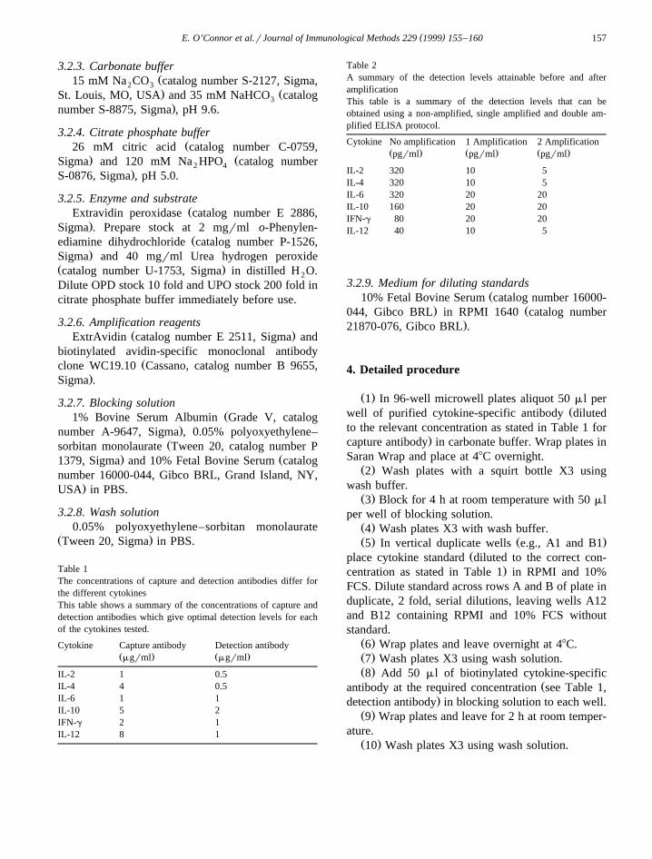
Table 1The concentrations of capture and detection antibodies differ forthe different cytokinesThis table shows a summary of the concentrations of capture anddetection antibodies which give optimal detection levels for eachof the cytokines tested.
Cytokine Capture antibody Detection antibodyŽ . Ž .mgrml mgrml
IL-2 1 0.5IL-4 4 0.5IL-6 1 1IL-10 5 2IFN-g 2 1IL-12 8 1
Table 2A summary of the detection levels attainable before and afteramplificationThis table is a summary of the detection levels that can beobtained using a non-amplified, single amplified and double am-plified ELISA protocol.
Cytokine No amplification 1 Amplification 2 AmplificationŽ . Ž . Ž .pgrml pgrml pgrml
IL-2 320 10 5IL-4 320 10 5IL-6 320 20 20IL-10 160 20 20IFN-g 80 20 20IL-12 40 10 5
3.2.9. Medium for diluting standardsŽ10% Fetal Bovine Serum catalog number 16000-
. Ž044, Gibco BRL in RPMI 1640 catalog number.21870-076, Gibco BRL .
4. Detailed procedure
Ž .1 In 96-well microwell plates aliquot 50 ml perŽwell of purified cytokine-specific antibody diluted
to the relevant concentration as stated in Table 1 for.capture antibody in carbonate buffer. Wrap plates in
Saran Wrap and place at 48C overnight.Ž .2 Wash plates with a squirt bottle X3 using
wash buffer.Ž .3 Block for 4 h at room temperature with 50 ml
per well of blocking solution.Ž .4 Wash plates X3 with wash buffer.Ž . Ž .5 In vertical duplicate wells e.g., A1 and B1
Žplace cytokine standard diluted to the correct con-.centration as stated in Table 1 in RPMI and 10%
FCS. Dilute standard across rows A and B of plate induplicate, 2 fold, serial dilutions, leaving wells A12and B12 containing RPMI and 10% FCS withoutstandard.
Ž .6 Wrap plates and leave overnight at 48C.Ž .7 Wash plates X3 using wash solution.Ž .8 Add 50 ml of biotinylated cytokine-specific
Žantibody at the required concentration see Table 1,.detection antibody in blocking solution to each well.
Ž .9 Wrap plates and leave for 2 h at room temper-ature.
Ž .10 Wash plates X3 using wash solution.

( )E. O’Connor et al.rJournal of Immunological Methods 229 1999 155–160158
Ž .11 Add 50 ml ExtrAvidin diluted to 1 mgrml inblocking solution.
Ž .12 Wrap and leave at room temperature for 30min.
Fig. 1. A single but not a double amplification step improves the level of detection of all cytokines tested. The ELISAs for each cytokineŽ . ŽAs IL-2, Bs IL-4, Cs IL-6, Ds IL-10, Es IFN-g, Fs IL-12 were set up separately. The three levels of amplification Bsno
.amplification, Issingle amplification, v sdouble amplification were set up on three separate plates. Points shown are means ofduplicate values. The data shown are representative of more than five experiments for each cytokine.

( )E. O’Connor et al.rJournal of Immunological Methods 229 1999 155–160 159
Ž .13 Wash X3 in wash solution.Ž .14 Add 50 ml biotinylated avidin-specific anti-
body diluted to 0.5 mgrml in blocking solution.Ž .15 Wrap and leave at room temperature for 1 h.Ž .16 Repeat steps 10–15 for a double amplifica-
tion step.Ž .17 Wash plates X3 using wash solution.Ž .18 Add 75 ml of Extravidin peroxidase diluted
to 1.1 mgrml in blocking solution to each well.Ž .19 Wrap plates and leave for 1 h at room
temperature.Ž .20 Wash plates X3 using wash solution.Ž . Ž .21 Add 100 ml OPD 0.2 mgrml and UPO
Ž .0.2 mgrml diluted in citrate phosphate buffer toeach well.
Ž .22 Wrap plates in aluminum foil and leave for30 min at room temperature.
Fig. 2. The specificity of the protocol is shown by testing for thepresence of IL-2, IL-12, IL-4 and IFN-g in Th1- and Th2-typecultures. C57BLr6 splenocytes were isolated. After red blood celllysis, the cells were resuspended to a concentration of 2=106
cellsrml and added to a culture flask previously coated with 10Žmgrml of CD3-specific mAb 10 mgrml 145 2C11 in PBS for 2
.h at 378C . The cells were stimulated under two conditions. Th1cells were generated by the addition of recombinant IL-2 andIL-12 and the IL-4-specific mAb, 11B11, and Th2 cells weregenerated by the addition of recombinant IL-2 and IL-4 and the
Ž .IFN-g-specific mAb, XMG1.2 using published protocols 12 . Thecells were re-stimulated 5 days later at a concentration of 1=106
cellsrml in tissue culture flasks coated with 10 mgrml CD3-specific mAb. Culture supernatants were collected 24 h later. TheTh1 and Th2 culture supernatants were tested for the presence ofIL-2, IL-4, IL-12 and IFN-g. In each case, the culture supernatantswere tested as concentrated supernatants and in 2-fold serialdilutions to a total of 10 dilutions. The data shown are concentra-tions of each of the cytokines at a single dilution as determined
Ž . Ž .using the single IFN-g or the double IL-4, IL-12 amplificationprotocol.
Ž .23 After 30 min, add 50 ml of 25% H SO to2 4
each well to stop the reaction.Ž .24 Read plates on the ELISA plate reader at 490
nm.
5. Results
ELISA procedures were set up where the effect ofa single and double amplification step on the detec-tion level of various cytokines was compared to thedetection level using the standard non-amplifiedELISA protocol. The detection level without amplifi-cation varied enormously from one cytokine to an-
Ž .other Table 2 and Fig. 1 . Interestingly, after asingle amplification step, the detection of all cy-
Žtokines tested was within a small range 20–10.pgrml . A further amplification step reproducibly
improved the detection level of IL-2, IL-4 and IL-12but not of IL-6, IL-10 and IFN-g. The specificity ofthe protocol was shown using supernatants from
Ž .Th1- and Th2-type cultures Fig. 2 . As expected,the Th1-type culture contained IL-2, IL-12 and IFN-gbut not IL-4 while the Th2-type culture containedIL-4 but not IL-2, IL-12 and IFN-g.
6. Discussion
By exploiting the strength of the biotin–avidinŽinteraction Bayer et al., 1979; Wilchek and Bayer,
.1984 we have successfully enhanced the sensitivityof cytokine detection by 4–64 fold. The data arehighly reproducible and the amplification step issimple and inexpensive.
6.1. Trouble-shooting
The substrate constituents UPO and OPD are verysensitive to temperature. OPD stock should be storedat y208C and thawed directly before use. UPO stockshould be stored at 48C. The OPD and UPO shouldbe mixed immediately before adding to the assay.Recombinant cytokines are very sensitive to freeze–thaw conditions and should be stored in one-time-usealiquots at y708C.

( )E. O’Connor et al.rJournal of Immunological Methods 229 1999 155–160160
6.2. AlternatiÕe and support protocols
The sensitivity of detection of IL-2, IL-4 andIL-12 was improved when the assay was amplifiedtwice vs. once. It is possible therefore that the sensi-tivity could be further increased by repeated amplifi-cation steps.
7. Essential literature cited
Standard protocols for the detection and quantita-tion of cytokines can be found in Wadhwa et al.Ž .1995 .
8. Quick procedure
1. Coat plates with capture antibody2. Wash plates3. Block plates4. Wash plates5. Dilute cytokine standard on plates6. Leave plates at 48C7. Wash plates8. Put biotinylated antibody on plates9. Leave plates at room temperature
10. Wash plates11. Add extravidin to plates12. Leave plates at room temperature13. Wash plates14. Add biotinylated avidin-specific antibody to
plates15. Leave plates at room temperature16. Repeat steps 10–15 one time for a single
amplification step and twice for a doubleamplification step
17. Wash plates18. Add Extravidin peroxidase to plates19. Leave plates at room temperature20. Wash plates21. Add substrate to plates22. Leave plates at room temperature
23. Stop reaction with H SO2 4
24. Read plates
Acknowledgements
This work was supported by grants from NovartisPharma and from the National Institutes of HealthŽ .DK55099 . This is manuscript number 12113-IMMfrom The Scripps Research Institute.
References
Bayer, E.A., Skutelsky, E., Wilchek, M., 1979. The avidin–biotincomplex in affinity cytochemistry. Methods Enzymol. 62, 308.
Beech, J.T., Bainbridge, T., Thompson, S.J., 1997. Incorporationof cells into an ELISA system enhances antigen-driven lym-phokine detection. J. Immunol. Methods 205, 163.
Cassano, W.F., 1989. Murine monoclonal anti-avidin antibodiesenhance the sensitivity of avidin–biotin immunoassays andimmunohistologic staining. J. Immunol. Methods 117, 169.
Hudson, L., Hay, F.C., 1991. Practical Immunology. Blackwell,Oxford, UK, pp. 348–350.
Hutchings, P.R., Cambridge, G., Tite, J.P., Meager, T., Cooke, A.,1989. The detection and enumeration of cytokine-secretingcells in mice and man and the clinical application of theseassays. J. Immunol. Methods 120, 1.
Mire-Sluis, A.R., Thorpe, R., 1998. Laboratory protocols for thequantitation of cytokines by bioassay using cytokine respon-sive cell lines. J. Immunol. Methods 211, 199.
Rogers, P.R., Huston, G., Swain, S.L., 1998. High antigen densityand IL-2 are required for generation of CD4 effectors secret-ing Th1 rather than Th0 cytokine. J. Immunol. 161, 3844.
Shibuya, K., Robinson, D., Zonin, F., Hartley, S.B., Macatonia,S.E., Somoza, C., Hunter, C.A., Murphy, K.M., O’Garra, A.,1998. IL-1a and TNF-a are required for IL-12-induced devel-opment of Th1 cells producing high levels of IFN-g inBALBrc but not C57BLr6 mice. J. Immunol. 160, 1708.
Swain, S.L., Dennert, G., Warner, J., Dutton, R.W., 1981. Culturesupernatants of a stimulated T cell line have helper activitythat acts synergistically with interleukin-2 in the response of Bcells to antigen. Proc. Natl. Acad. Sci. U.S.A. 78, 2517.
Wadhwa, M., Bird, C., Page, L., Mire-Sluis, A., Thorpe, R., 1995.Quantitative biological assays for individual cytokines. In:
Ž .Rickwood, D., Hames, B.D. Eds. , Cytokines, A PracticalApproach. IRL Press at Oxford University Press, Oxford, UK,p. 357.
Wilchek, M., Bayer, E.A., 1984. The avidin–biotin complex inimmunology. Immunol. Today 5, 39.