amniotic membrane transplantation in bacterial and...
TRANSCRIPT
457
http://journals.tubitak.gov.tr/medical/
Turkish Journal of Medical Sciences Turk J Med Sci(2016) 46: 457-462© TÜBİTAKdoi:10.3906/sag-1501-6
Amniotic membrane transplantation in bacterial and herpetic stromal keratitis
Yeşim ALTAY*, Sema TAMER, Ayşe BURCU, Özgür BALTADepartment of Ophthalmology, Ankara Training and Research Hospital, Ankara, Turkey
* Correspondence: [email protected]
1. IntroductionMicrobial keratitis or infectious corneal ulcer is due to the proliferation of microorganisms and associated inflammation, and tissue destruction within the corneal tissue. It is a potentially sight-threatening condition (1). Various mechanisms are involved in the pathogenesis of this corneal destruction; they include production of bacterial enzymes and toxins that injure cellular components and the extracellular matrix of the cornea, and activation of cornea-degrading enzymes from inflammatory cells (2).
Herpes simplex keratitis is a common cause of corneal ulceration and blindness worldwide. When antiviral agents become ineffective, corneal perforation frequently results in destructive complications. Additionally, in these cases, if the patients undergo corneal transplantation, there is an increasing incidence of immune rejection due to the presence of ongoing active inflammation (3,4).
Bacterial keratitis is also a serious ocular condition and requires treatment with intensive topical broad-spectrum antibiotics that are potentially toxic to the corneal epithelium (5).
The main therapeutic goals for infectious keratitis are to eliminate the pathogens and to prevent irreversible corneal structural damage (6). Various adjunctive modalities have been used in addition to antibiotic/antiviral therapy, which include steroids, nonsteroidal antiinflammatory drugs, ascorbic acid, doxycycline, hyperbaric oxygen, and amniotic membrane transplantation (AMT) (6–9).
In 1997, Lee and Tseng first used human amniotic membrane (AM) for epithelial defects (10). The AM does not express the antigens HLA-A, -B, or -DR and therefore poses no problem of immunological rejection (11). AM has been used in vivo as a substrate for epithelial growth, in the management of persistent epithelial defects following infection, in neurotrophic corneas, in chemical injuries, and for recurrent erosion syndrome and persistent epithelial defects associated with cicatricial conditions (9,12). Other indications include pterygium surgery and pain relief in bullous keratopathy (13). The AM, consisting of a thick basement membrane and avascular stromal matrix, is able to express multiple antiinflammatory, antimicrobial, and antiangiogenic factors and protease inhibitors (13,14). AMT is effective in promoting epithelial healing and in
Background/aim: The aim of this study was to describe the results of amniotic membrane transplantation (AMT) in patients with bacterial and herpetic stromal keratitis.
Materials and methods: This was a retrospective chart review study including 42 patients with herpetic keratitis (group 1) and 42 patients with bacterial keratitis (group 2). AMT was performed in addition to antimicrobial therapy. Topical steroids were administered after surgery. The outcome parameters evaluated were epithelialization time, decrease of stromal inflammation, and uncorrected visual acuity (UCVA).
Results: The average age of our patients was 55.85 ± 19.07 years, and average follow-up was 14.70 ± 11.75 months. The period of epithelialization was 19.23 ± 7.32 days in the herpetic group and 19.31 ± 6.30 days in the bacterial group. Descemetocele developed in 2 patients of the herpetic group. Other patients in both groups completed epithelialization after AMT procedures with varying amounts of corneal scarring. The bacterial group showed an improvement in UCVA, but the herpetic group showed no improvement in UCVA.
Conclusion: AMT is a convenient approach for the treatment of corneal keratitis resistant to conventional treatment and allows the use of early topical steroid application. It provides patients with corneal scarring an opportunity for subsequent keratoplasty by arresting the inflammatory response.
Key words: Amniotic membrane transplantation, bacterial keratitis, herpetic keratitis, topical steroids
Received: 04.01.2015 Accepted/Published Online: 08.05.2015 Final Version: 17.02.2016
Research Article
458
ALTAY et al. / Turk J Med Sci
reducing inflammation, scarring, and angiogenesis (15).We report a series of patients with herpetic and
bacterial ulcers who were treated with AMT combined with antimicrobial and corticosteroid therapy.
2. Materials and methodsChart review of 84 patients with bacterial and herpetic stromal keratitis who underwent AMT between 2010 and 2013 was performed retrospectively. The study was approved by the Institutional Review Board and was in accordance with the principles of the Declaration of Helsinki. All patients gave informed consent before treatment.
Patients were divided into 2 groups according to etiology of ulcerative corneal keratitis: group 1 due to herpetic stromal keratitis (42 patients), and group 2 due to bacterial keratitis (42 patients). Clinical data including patients’ demographic features, etiology, ulcer localization, dimension and depth, surgical procedure, epithelialization time after AMT, uncorrected visual acuity (UCVA), and follow-up period were retrieved retrospectively. Surgical success was defined as complete epithelialization of ocular surface, cessation of stromal inflammation, and formation of a visible stromal thickness.
Briefly, patients with ulcerative keratitis demonstrate an ulcer with a dense, whitish inflammatory infiltration with stromal edema. Additionally, in herpetic keratitis, keratic endothelial precipitates confined to the area of corneal involvement are commonly present. Diagnostic criteria were based on ocular examination, previous reports, and smears and cultures of corneal tissue. The majority of patients with herpetic keratitis had a history of recurrent episodes of corneal disease.
Eyes with perforated corneal ulcers, children under 12 years old, cases of concomitant stem-cell deficiency, and patients undergoing immunosuppressive treatment were excluded from the study.
Photographs of the eyes were taken at the initial presentation and follow-up visits. The size of the corneal ulcer was measured at its greatest diameter. Depth of the ulcer was also noted (as deep as or less than one-half of corneal thickness). Smears and cultures were obtained for microbiological studies at the first visit. In the herpetic keratitis group, systemic acyclovir at 5 × 400 mg daily was administered for 1 month, and then 2 × 400 mg daily for 11 months. In the bacterial keratitis group, frequent topical antibiotic treatment was continued according to clinical response. In culture-positive patients, antibiotic treatment was modified according to culture results. After 2 to 5 days of initial treatment, AMT was performed in all eyes. Antiviral and antibiotic medications were continued; topical 0.1% prednisolone acetate 3 × 1 and intensive
lubrication with artificial tear drops were administered after surgery.
Amniotic membrane transplantations were performed using subtenon or general anesthesia. The entire cornea and limbus were covered by a single or double layer of cryopreserved AM, which was sutured epithelial side up at 2 mm posterior to the limbus on the episclera by a continuous 10–0 nylon suture (overlay technique). The amniotic basement membrane affords a more suitable substratum for corneal or conjunctival epithelial cells to grow on compared to amniotic stroma. The wider spaced collagen of the stroma retards epithelial spread. Because of this, AM is placed onto the ocular surface with the basement membrane (epithelial) side up (13). If the ulcer depth was more than half the corneal thickness, double-layer AMT was performed, and each layer of AM was sutured to the episclera separately.
Human AM was obtained under sterile conditions from planned cesarean sections after screening the donor for HIV, hepatitis B and C, and syphilis. The placenta was cleaned of blood clots with a sterile phosphate-buffered saline solution containing penicillin, 50 µg/mL; streptomycin, 50 µg/mL; tobramycin, 100 µg/mL; and amphotericin B, 2.5 µg/mL. The amnion was separated from the chorion by blunt dissection and flattened onto nitrocellulose paper with the epithelium side up. The paper with adherent AM was cut into sheets of 3 × 3 cm and stored before transplantation at –80 °C in a sterile vial containing Dulbecco’s modified Eagle medium and glycerol at a ratio of 1/1. The membrane was thawed for 15 min before use.
All patients were examined on postoperative day 1, then weekly until complete epithelialization occurred, then monthly for 3 months, and every 3 months thereafter.
While the AM was in place, epithelialization could not be easily assessed. After the membrane was largely dissolved (mean: 10 days), the nylon suture was removed and the corneal surface was assessed for fluorescein staining. If epithelialization was not complete and unsatisfactory, a second AMT procedure was performed.
Statistical analyses were performed by using SPSS 15. The variables were investigated using visual (histograms) and analytical (Kolmogorov–Smirnov test) methods to determine whether or not they were normally distributed. Descriptive analyses were presented using means and standard deviations for normally distributed variables. Comparison of normally distributed variables according to the type of keratitis were made using Student’s t-test. Abnormally distributed or ordinal variables were compared by using a chi-square test. A 5% type-I error level was used to infer statistical significance.
459
ALTAY et al. / Turk J Med Sci
3. ResultsEighty-four patients (51 male, 33 female) were enrolled in this retrospective study. The average age of our patients was 55.85 ± 19.07 years (range: 17–93), and the average duration of follow-up was 14.70 ± 11.75 months (range: 3–52). Patients were sorted into 2 groups according to ulcer etiology. Group 1 (42 eyes) included herpetic ulcers and group 2 (42 eyes) included bacterial ulcers.
There were no statistically significant differences between the 2 groups in age, initial UCVA, or size and depth of corneal ulcer at initial presentation (P = 0.33, P = 0.23, P = 0.22, P = 0.49, respectively). Although herpetic ulcers were localized more centrally, there was no significant difference between groups (P = 0.19). In the herpetic group, 24 patients had corneal scars from previous herpetic attacks at initial presentation. In the bacterial group, all patients were primary cases. In this group, 15 patients had a history of foreign bodies, and 9 patients were chronic contact lens users.
There were only 24 cases with positive culture results, which might be due to antibiotic treatment that was started previously. Culture results were as follows: methicillin-resistant Staphylococcus epidermidis (8 cases), Streptococcus pneumoniae (6 cases), Staphylococcus aureus (6 cases), and Pseudomonas aeruginosa (4 cases).
The time of epithelialization was 19.23 ± 7.32 days (range: 10–36) in the herpetic group compared with 19.31 ± 6.30 days (range: 10–35) in the bacterial group. The difference between groups was not significant (P = 0.95).
Table 1 shows distribution of age, lesion diameter, lesion depth, and localization between groups with herpetic ulcers and bacterial ulcers before AMT.
Because of incomplete epithelialization or relapsing ulcers, 13 eyes (31%) in the herpetic group and 10 eyes
(23.8%) in the bacterial group required a repeat AMT procedure (Table 2). After mean follow-up of 17.17 ± 13.84 months (range: 5–52), we observed 6 (14.28%) recurrences in the herpetic ulcer group. No recurrences were observed in the bacterial ulcer group.
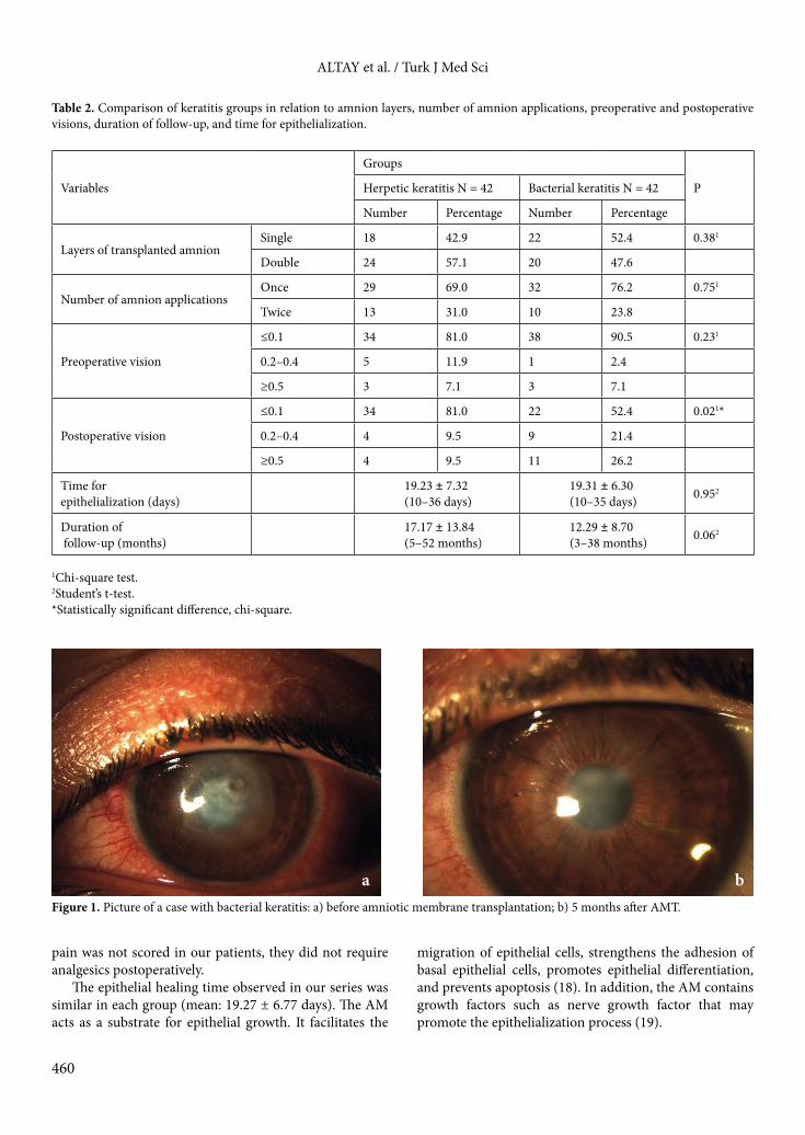
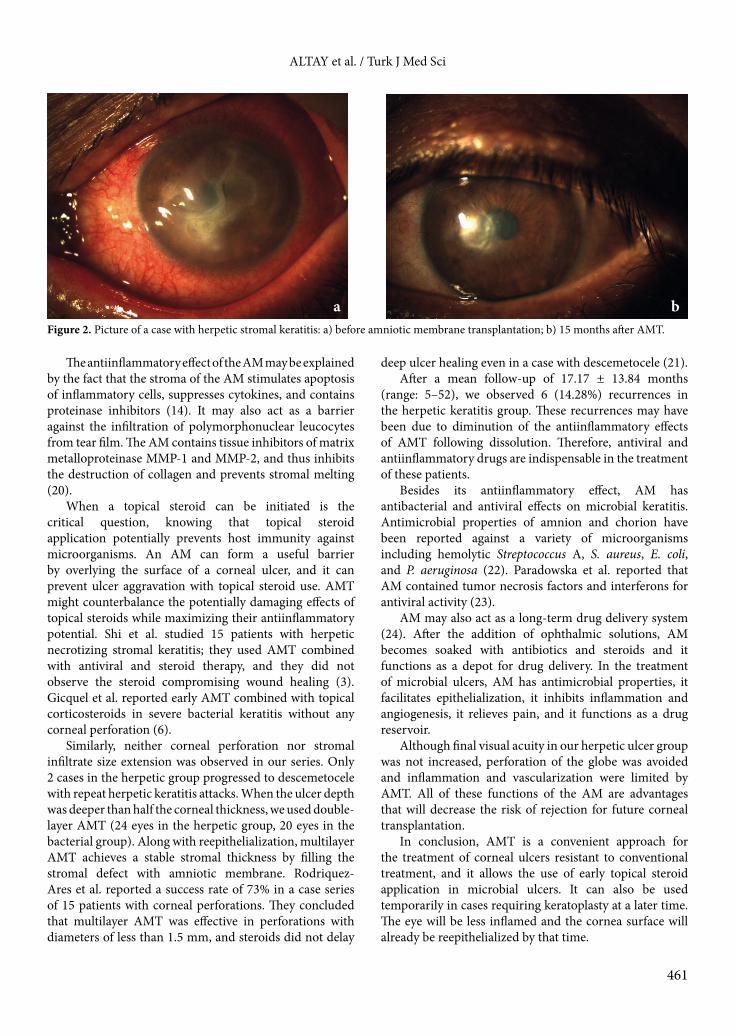
No perforations occurred in eyes of patients in either group. Only 2 patients in the herpetic group developed significant corneal thinning and descemetocele; penetrating keratoplasty was performed for these eyes. Other patients in both groups completed epithelialization after 1 or 2 AMT procedures with varying amounts of corneal scarring (Figures 1 and 2).
Of the herpetic ulcer group, mean initial UCVA less than 0.1 was 81%, between 0.2 and 0.4 was 11.9%, and more than 0.5 was 7.1%. Final visual acuity in this group was 81%, 9.5%, and 9.5%, respectively. There was no difference between initial and final visual acuity for the herpetic ulcer group.
In the bacterial ulcer group, initial UCVA less than 0.1 was 90.5%, between 0.2 and 0.4 was 2.4%, and more than 0.5 was 7.1%. Final UCVA in this group was 52.4%, 21.4%, and 26.2%, respectively. There was a significant improvement in UCVA for the bacterial ulcer group (P = 0.02).
4. DiscussionWe have presented the results of AMT in the treatment of herpetic and bacterial corneal ulcers with a combination of antimicrobial and topical steroid therapy.
The presence of antiangiogenic and antiinflammatory factors in the AM helps decrease inflammation and neovascularization, and it functions as a biological barrier (14,16). The AM serves as a bandage contact lens before epithelial healing. This mechanical or physical bandage effect may decrease pain after AMT surgery (17). Although
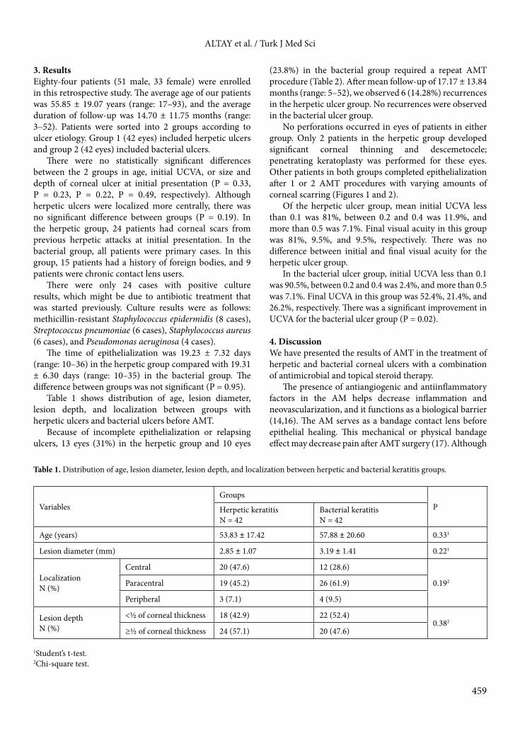
Table 1. Distribution of age, lesion diameter, lesion depth, and localization between herpetic and bacterial keratitis groups.
VariablesGroups
PHerpetic keratitisN = 42
Bacterial keratitisN = 42
Age (years) 53.83 ± 17.42 57.88 ± 20.60 0.331
Lesion diameter (mm) 2.85 ± 1.07 3.19 ± 1.41 0.221
LocalizationN (%)
Central 20 (47.6) 12 (28.6)
0.192Paracentral 19 (45.2) 26 (61.9)
Peripheral 3 (7.1) 4 (9.5)
Lesion depthN (%)
<½ of corneal thickness 18 (42.9) 22 (52.4)0.382
≥½ of corneal thickness 24 (57.1) 20 (47.6)
1Student’s t-test.2Chi-square test.
460
ALTAY et al. / Turk J Med Sci
pain was not scored in our patients, they did not require analgesics postoperatively.
The epithelial healing time observed in our series was similar in each group (mean: 19.27 ± 6.77 days). The AM acts as a substrate for epithelial growth. It facilitates the
migration of epithelial cells, strengthens the adhesion of basal epithelial cells, promotes epithelial differentiation, and prevents apoptosis (18). In addition, the AM contains growth factors such as nerve growth factor that may promote the epithelialization process (19).
Table 2. Comparison of keratitis groups in relation to amnion layers, number of amnion applications, preoperative and postoperative visions, duration of follow-up, and time for epithelialization.
Variables
Groups
PHerpetic keratitis N = 42 Bacterial keratitis N = 42
Number Percentage Number Percentage
Layers of transplanted amnionSingle 18 42.9 22 52.4 0.381
Double 24 57.1 20 47.6
Number of amnion applicationsOnce 29 69.0 32 76.2 0.751
Twice 13 31.0 10 23.8
Preoperative vision
≤0.1 34 81.0 38 90.5 0.231
0.2–0.4 5 11.9 1 2.4
≥0.5 3 7.1 3 7.1
Postoperative vision
≤0.1 34 81.0 22 52.4 0.021*
0.2–0.4 4 9.5 9 21.4
≥0.5 4 9.5 11 26.2
Time for epithelialization (days)
19.23 ± 7.32(10–36 days)
19.31 ± 6.30(10–35 days) 0.952
Duration of follow-up (months)
17.17 ± 13.84(5–52 months)
12.29 ± 8.70(3–38 months) 0.062
1Chi-square test.2Student’s t-test.*Statistically significant difference, chi-square.
Figure 1. Picture of a case with bacterial keratitis: a) before amniotic membrane transplantation; b) 5 months after AMT.
a b
461
ALTAY et al. / Turk J Med Sci
The antiinflammatory effect of the AM may be explained by the fact that the stroma of the AM stimulates apoptosis of inflammatory cells, suppresses cytokines, and contains proteinase inhibitors (14). It may also act as a barrier against the infiltration of polymorphonuclear leucocytes from tear film. The AM contains tissue inhibitors of matrix metalloproteinase MMP-1 and MMP-2, and thus inhibits the destruction of collagen and prevents stromal melting (20).
When a topical steroid can be initiated is the critical question, knowing that topical steroid application potentially prevents host immunity against microorganisms. An AM can form a useful barrier by overlying the surface of a corneal ulcer, and it can prevent ulcer aggravation with topical steroid use. AMT might counterbalance the potentially damaging effects of topical steroids while maximizing their antiinflammatory potential. Shi et al. studied 15 patients with herpetic necrotizing stromal keratitis; they used AMT combined with antiviral and steroid therapy, and they did not observe the steroid compromising wound healing (3). Gicquel et al. reported early AMT combined with topical corticosteroids in severe bacterial keratitis without any corneal perforation (6).
Similarly, neither corneal perforation nor stromal infiltrate size extension was observed in our series. Only 2 cases in the herpetic group progressed to descemetocele with repeat herpetic keratitis attacks. When the ulcer depth was deeper than half the corneal thickness, we used double-layer AMT (24 eyes in the herpetic group, 20 eyes in the bacterial group). Along with reepithelialization, multilayer AMT achieves a stable stromal thickness by filling the stromal defect with amniotic membrane. Rodriquez-Ares et al. reported a success rate of 73% in a case series of 15 patients with corneal perforations. They concluded that multilayer AMT was effective in perforations with diameters of less than 1.5 mm, and steroids did not delay
deep ulcer healing even in a case with descemetocele (21).After a mean follow-up of 17.17 ± 13.84 months
(range: 5–52), we observed 6 (14.28%) recurrences in the herpetic keratitis group. These recurrences may have been due to diminution of the antiinflammatory effects of AMT following dissolution. Therefore, antiviral and antiinflammatory drugs are indispensable in the treatment of these patients.
Besides its antiinflammatory effect, AM has antibacterial and antiviral effects on microbial keratitis. Antimicrobial properties of amnion and chorion have been reported against a variety of microorganisms including hemolytic Streptococcus A, S. aureus, E. coli, and P. aeruginosa (22). Paradowska et al. reported that AM contained tumor necrosis factors and interferons for antiviral activity (23).
AM may also act as a long-term drug delivery system (24). After the addition of ophthalmic solutions, AM becomes soaked with antibiotics and steroids and it functions as a depot for drug delivery. In the treatment of microbial ulcers, AM has antimicrobial properties, it facilitates epithelialization, it inhibits inflammation and angiogenesis, it relieves pain, and it functions as a drug reservoir.
Although final visual acuity in our herpetic ulcer group was not increased, perforation of the globe was avoided and inflammation and vascularization were limited by AMT. All of these functions of the AM are advantages that will decrease the risk of rejection for future corneal transplantation.
In conclusion, AMT is a convenient approach for the treatment of corneal ulcers resistant to conventional treatment, and it allows the use of early topical steroid application in microbial ulcers. It can also be used temporarily in cases requiring keratoplasty at a later time. The eye will be less inflamed and the cornea surface will already be reepithelialized by that time.
Figure 2. Picture of a case with herpetic stromal keratitis: a) before amniotic membrane transplantation; b) 15 months after AMT.
a b
462
ALTAY et al. / Turk J Med Sci
References
1. Edelstein SL, Wichiensin P, Huang AJW. Bacterial keratitis. In: Krachmer JH, Mannis M, Holland EJ, editors. Cornea Fundamentals, Diagnosis and Management. Volume 1-A. 3rd ed. Beijing, China: Mosby Elsevier; 2010. pp. 919–944.
2. Kreger AS. Pathogenesis of Pseudomonas aeruginosa ocular diseases. Rev Infect Dis 1983; 5: S931–S935.
3. Shi W, Chen M, Xie L. Amniotic membrane transplantation combined with antiviral and steroid therapy for herpes necrotizing stromal keratitis. Ophthalmol 2007; 114: 1476–1481.
4. Knickelbein JE, Hendricks RL, Charukamnoetkanok P. Management of herpes simplex virus stromal keratitis: an evidence based review. Surv Ophthalmol 2009; 54: 226–234.
5. Gangopadhyay N, Daniell M, Weih L, Taylor HR. Fluoroquinolone and fortified antibiotics for treating bacterial and corneal ulcers. Br J Ophthalmol 2000; 84: 378–384.
6. Gicquel JJ, Bejjani RA, Ellies P, Mercie M, Dighiero P. Amniotic membrane transplantation in severe bacterial keratitis. Cornea 2007; 26: 27–33.
7. Kheirkhah A, Tabatabaei A, Zavareh MK, Khodabandeh A. A controlled study of amniotic membrane transplantation for acute Pseudomonas keratitis. Can J Ophthalmol 2012; 47: 305–311.
8. Heiligenhaus A, Li H, Hernandez Galindo EE, Koch JM, Steuhl KP, Meller D. Management of acute ulcerative and necrotising herpes simplex and zoster keratitis with amniotic membrane transplantation. Br J Ophthalmol 2003; 87: 1215–1219.
9. Brijacak N, Dekaris I, Gagro A, Gabric N. Therapeutic effect of amniotic membrane in persistent epithelial defects and corneal ulcers in herpetic keratitis. Coll Antropol 2008; 32: 21–25.
10. Lee S, Tseng SCG. Amniotic membrane transplantation for persistent epithelial defects with ulceration. Am J Ophthalmol 1997; 123: 303–312.
11. Dua HS, Azuara-Blanco A. Amniotic membrane transplantation. Br J Ophthalmol 1999; 83: 748–752.
12. Khokhar S, Natung T, Sony P, Sharma N, Agarwal N, Vajpayee R. Amniotic membrane transplantation in refractory neurotrophic corneal ulcers: a randomized controlled trial. Cornea 2005; 24: 654–660.
13. Dua HS, Gomes JAP, King AJ, Maharajan SV. The amniotic membrane in ophthalmology. Surv Ophthalmol 2004; 49: 51–77.
14. Hao Y, Ma DH, Hwang DG, Kim WS, Zhang F. Identification of antiangiogenic and antiinflammatory proteins in human amniotic membrane. Cornea 2000; 19: 348–352.
15. Bouchard CS, John T. Amniotic membrane transplantation in the management of severe ocular surface disease: indications and outcomes. Ocul Surf 2004; 2: 201–211.
16. Yildiz EH, Nurozler AB, Aksoy NO, Altiparmak UE, Onat M, Karaguzel H, Duman S. Amniotic membrane transplantation: Indications and results. Eur J Ophthalmol 2008; 18: 685–690.
17. Espana EM, Grueterich M, Sandoval H, Solomon A, Alfonso E, Carp CL, Fantes F, Tseng SC. Amniotic membrane transplantation for bullous keratopathy in eyes with poor visual potential. J Cataract Refract Surg 2003; 29: 279–284.
18. Wang MX, Gray TB, Park WC, Prabhasawat P, Culbertson W, Forster R, Hanna K, Tseng SCG. Reduction in corneal haze and apoptosis by amniotic membrane matrix in excimer laser photoablation in rabbits. J Cataract Refract Surg 2001; 27: 310–319.
19. Touhami A, Grueterich M, Tseng SC. The role of NGF signaling in human limbal epithelium expanded by amniotic membrane culture. Invest Ophthalmol Vis Sci 2002; 43: 987–994.
20. Kim JS, Kim JC, Na BK, Jeong JM, Song CY. Amniotic membrane patching promotes healing and inhibits protease activity on wound healing following acute corneal alkali burn. Exp Eye Res 2000; 70: 329–337.
21. Rodriquez-Ares MT, Tourino R, Lopez-Valladares MJ, Gude F. Multilayer amniotic membrane transplantation in the treatment of corneal perforations. Cornea 2004; 23: 577–583.
22. Kjaaergaard N, Hein M, Hyttel L, Helmig RB, Schonheyder HC, Uldbjerg N, Madsen H. Antibacterial properties of human amnion and chorion in vitro. Eur J Obstet Gynecol Reprod Biol 2001; 94: 224–229.
23. Paradowska E, Blach-Olszewska Z, Sender J, Jaroz W. Antiviral nonspecific immunity of human placenta at term: possible role of endogenous tumor necrosis factors and interferons. J Interferon Cytokine Res 1996; 16: 941–948.
24. Resch MD, Resch BE, Csizmazia E, Imre L, Nemeth J, Szabo-Revesz P, Csanyi E. Drug reservoir function of human amniotic membrane. J Ocular Pharma Therapeutics 2011; 27: 323–326.