amelioration of cognitive deficit by embelin in a
TRANSCRIPT

fphar-09-00665 June 21, 2018 Time: 15:56 # 1
ORIGINAL RESEARCHpublished: 25 June 2018
doi: 10.3389/fphar.2018.00665
Edited by:Muhammad Ayaz,
University of Malakand, Pakistan
Reviewed by:Sagheer Ahmed,
Shifa Tameer-e-Millat University,Pakistan
Alla B. Salmina,Krasnoyarsk State Medical University
named afterProf. V.F.Voino-Yasenetski, Russia
*Correspondence:Yatinesh Kumari
[email protected] Farooq Shaikh
[email protected];[email protected]
Specialty section:This article was submitted to
Ethnopharmacology,a section of the journal
Frontiers in Pharmacology
Received: 16 March 2018Accepted: 04 June 2018Published: 25 June 2018
Citation:Bhuvanendran S, Kumari Y, Othman I
and Shaikh MF (2018) Ameliorationof Cognitive Deficit by Embelin in aScopolamine-Induced Alzheimer’s
Disease-Like Condition in a RatModel. Front. Pharmacol. 9:665.doi: 10.3389/fphar.2018.00665
Amelioration of Cognitive Deficit byEmbelin in a Scopolamine-InducedAlzheimer’s Disease-Like Conditionin a Rat ModelSaatheeyavaane Bhuvanendran, Yatinesh Kumari* , Iekhsan Othman andMohd Farooq Shaikh*
Neuropharmacology Research Laboratory, Jeffrey Cheah School of Medicine and Health Sciences, Monash UniversityMalaysia, Bandar Sunway, Malaysia
Embelin (2,5-dihydroxy-3-undecyl-1,4-benzoquinone) is one of the active components(2.3%) found in Embelia ribes Burm fruits. As determined via in vitro AChE inhibitionassay, embelin can inhibit the acetylcholinesterase enzyme. Therefore, embelin canbe utilized as a therapeutic compound, after further screening has been conductedfor its use in the treatment of Alzheimer’s disease (AD). In this study, the nootropicand anti-amnesic effects of embelin on scopolamine-induced amnesia in rats wereevaluated. Rats were treated once daily with embelin (0.3 mg/kg, 0.6 mg/kg, 1.2 mg/kg)and donepezil (1 mg/kg) intraperitoneally (i.p.) for 17 days. During the final 9 daysof treatment, a daily injection of scopolamine (1 mg/kg) was administered to inducecognitive deficits. Besides that, behavioral analysis was carried out to assess the rats’learning and memory functions. Meanwhile, hippocampal tissues were extracted forgene expression, neurotransmitter, and immunocytochemistry studies. Embelin wasfound to significantly improve the recognition index and memory retention in the novelobject recognition (NOR) and elevated plus maze (EPM) tests, respectively. Furthermore,embelin at certain doses (0.3 mg/kg, 0.6 mg/kg, and 1.2 mg/kg) significantly exhibiteda memory-enhancing effect in the absence of scopolamine, besides improving therecognition index when challenged with chronic scopolamine treatment. Moreover, inthe EPM test, embelin treated rats (0.6 mg/kg) showed an increase in inflection ratioin nootropic activity. However, the increase was not significant in chronic scopolaminemodel. In addition, embelin contributed toward the elevated expression of BDNF,CREB1, and scavengers enzymes (SOD1 and CAT) mRNA levels. Next, pretreatmentof rats with embelin mitigated scopolamine-induced neurochemical and histologicalchanges in a manner comparable to donepezil. These research findings suggest thatembelin is a nootropic compound, which also possesses an anti-amnesic ability thatis displayed against scopolamine-induced memory impairment in rats. Hence, embelincould be a promising compound to treat AD.
Keywords: embelin, Alzheimer’s disease, cognition, neuroprotective, anti-amnesic effect
Frontiers in Pharmacology | www.frontiersin.org 1 June 2018 | Volume 9 | Article 665

fphar-09-00665 June 21, 2018 Time: 15:56 # 2
Bhuvanendran et al. Embelin as Anti-amnesic in AD Like Condition
INTRODUCTION
Alzheimer’s disease (AD) is known as the leading cause ofdementia amongst people aged 65 and older (Ghumatkar et al.,2015). This age-related disease affects millions of individuals,and it is estimated that by 2050, 1 in 85 people worldwide willbe suffering from AD (Brookmeyer et al., 2007). According toTanzi and Bertram (2005), AD is a progressive and chronicneurodegenerative disorder which displays global cognitivedecline involving memory, orientation, judgment, and reasoning.The key features of AD’s pathogenesis are the gradual amassingof the protein fragment beta-amyloid (plaques) and twistedfibers of the protein tau (tangles), outside and inside neuronsin the brain, respectively (Alzheimer’s Association, 2017). Beta-amyloid plaques function as a neurotoxin by intervening inneuron-to-neuron communication at synapses. On the otherhand, tau tangles prevent the passage of essential moleculesand nutrients inside neurons, which causes axonal transportdysfunction and neuronal loss (Ali et al., 2015; Alzheimer’sAssociation, 2017).
Apart from that, memory impairment is associated withcholinergic system dysfunction, which involves cholinergicneurons, neurotransmitters, and their receptors (Bartus et al.,1982; Lee et al., 2015). Cholinergic system dysfunction resultsfrom a loss of cholinergic neurons in the basal forebrain andhippocampus, which diminishes cognitive capability (Bartuset al., 1982; Lee et al., 2015). In healthy individuals, activationof the central cholinergic system enhances hippocampalneurogenesis through the cAMP response element-bindingprotein/brain-derived neurotrophic factor (CREB/BDNF)pathway (Lee et al., 2015). At present, one of the treatmentsfor AD is a dispensation of acetylcholinesterase (AChE)inhibitors like tacrine or donepezil that increase the availabilityof acetylcholine at cholinergic synapses (Pandareesh et al.,2016). Moreover, oxidative stress plays an important role inAD, with some studies suggesting that beta-amyloid toxicityis linked to an increment in reactive oxygen species (ROS),including H2O2 (Butterfield and Lauderback, 2002), and lipidperoxidation in neuronal cultures (Yatin et al., 1999). Highoxidative stress can cause memory deficits via impairmentof hippocampal synaptic plasticity (Serrano and Klann,2004) and oxidative damage in neurodegenerative diseases(Ding et al., 2007).
Current pharmacological options for AD, only have a partialeffect and poor control over the disease-causing neuronslinked with Alzheimer’s symptoms and lethal complications(Alzheimer’s Association, 2017). As such, the available drugsin the market mainly focus on the improving memory byinhibiting the AChE enzyme (Ghumatkar et al., 2015). However,AD is not a result of a single factor like AChE, but ratheris a multifactorial condition and this needs to be consideredwhen designing a drug. Other factors such as oxidativestress and synaptic dysfunction play a significant role in thecognitive deficits in AD. Natural products could be a sourceof neuroprotective drugs as they can maintain normal cellularinteraction in the brain and reduce the loss of neuronalfunctions in pathological circumstances (Hritcu et al., 2014).
Presently, many AD research groups have already exploredthe potential of using natural products as neuroprotectiveagents.
One such potential natural product is embelin (2,5-dihydroxy-3-undecyl-1,4-benzoquinone), which is the mainactive constituent in the fruits of Embelia ribes Burm (Family:Myrsinaceae), commonly known as “False Black Pepper”(Kundap et al., 2017a). The bright orange fruits of E. ribeshave been utilized in traditional medicinal practice for treatingcentral nervous system (CNS) disorders such as mentaldisorders and as a brain tonic (Poojari, 2014). Moreover,embelin has displayed anti-inflammatory, antioxidant, analgesic,antifertility, antitumor, wound healing, hepatoprotective, andantibacterial activities (Mahendran et al., 2011). Additionally,it has been reported that embelin is neuroprotective andpossesses anticonvulsant ability when tested using animal models(Mahendran et al., 2011).
Embelin possesses all the features of a compound that cantraverse the blood-brain barrier (BBB) and prompt a reactionin the CNS (Pathan et al., 2009; Kundap et al., 2017a). Eventhough embelin has various uses, there have been no studies ofits neuropharmacological activities against AD-like conditions.Thus, in the present study, the anti-amnesic potential of embelinon memory deficits in a rat model of cognitive impairment causedby scopolamine was examined.
MATERIALS AND METHODS
Animal CareIn-house bred Sprague Dawley rats weighing between 180–200 gand between 6–8 weeks old were housed in the animal facilityof the Jeffrey Cheah School of Medicine and Health Sciences,Monash University Malaysia. The rats were kept in cagesand maintained under standard husbandry conditions (12:12 hlight/dark cycle, controlled room temperature (23± 2◦C), stress-free, ad libitum water, standard diets, and sanitary conditions).Before commencing the experiment, the rats were allowed toacclimatize for a period of 1 week to reduce stress. The MonashAnimal Research Platform (MARP) Animal Ethics Committee inAustralia approved all the animal experimentations conducted inthis study.
Experimental DesignDrug TreatmentEmbelin (98%) batch number (Yucca/EM/2015/01/01) waspurchased from Yucca Enterprises, Mumbai, India. The rangeof doses for embelin was determined based on pre-screeningresults. Embelin was solubilized in DMSO and then dissolvedin saline. Donepezil and scopolamine were prepared in saline.Normal control rats were administered saline throughout theexperiment. The treatments were given intraperitoneally (i.p) ata volume corresponding to 0.1 ml/100 g of body weight.
All experiments were performed in a balanced design (9animals/group) to avoid being influenced by order and time. Thebehavioral studies were divided into two categories namely thenootropic and scopolamine models.
Frontiers in Pharmacology | www.frontiersin.org 2 June 2018 | Volume 9 | Article 665

fphar-09-00665 June 21, 2018 Time: 15:56 # 3
Bhuvanendran et al. Embelin as Anti-amnesic in AD Like Condition
Nootropic Model(i) Group 1: Control (Saline) (n = 9);
(ii) Group 2: Positive control (donepezil (DPZ) 1 mg/kg);(iii) Group 3: Low dose of embelin (EMB) 0.3 mg/kg;(iv) Group 4: Medium dose of EMB 0.6 mg/kg;(v) Group 5: High dose of EMB 1.2 mg/kg
For nootropic activity, all the groups received pretreatment viathe intraperitoneal route, for 8 days. All these rats were subjectedto a battery of behavioral tests from day six onward until day eightfor NOR and EPM (Figure 1).
Scopolamine Model(i) Group 1: Control (Saline) (n = 9);
(ii) Group 2: Negative control [scopolamine (SCP) 1 mg/kg](n = 9);
(iii) Group 3: Positive control [donepezil (DPZ) 1 mg/kg] +(SCP 1 mg/kg) (n = 9);
(iv) Group 4: Low dose of embelin (EMB) 0.3 mg/kg + (SCP1 mg/kg) (n = 9);
(v) Group 5: Medium dose of EMB 0.6 mg/kg+ (SCP 1 mg/kg)(n = 9);
(vi) Group 6: High dose of EMB 1.2 mg/kg + (SCP 1 mg/kg)(n = 9)
For scopolamine, amnesia was induced in all the groupsexcept the control group by daily intraperitoneal injectionsof scopolamine (1 mg/kg) for 9 days after embelin pre-treatment (day nine to day 17). Half an hour after scopolamineadministration, NOR was conducted on day 15, and EPM wascarried out on day 16 and 17 of the study. At the end of theexperiment, the rats were sacrificed, and their brains were isolatedfor further biochemical and immunohistochemistry analysis.
Novel Object Recognition (NOR)For the object recognition task, an open field box(40 × 40 × 20 cm) composed of black acrylic material wasutilized as the experimental apparatus. This method is similarto that used by Ennaceur and Delacour (1988), with minormodifications. Besides that, behavioral testing was carried outbetween 9:00 am and 6:00 pm under red light illumination. Thescrutinized objects were two similar transparent culture flaskscontaining water and a Lego toy of similar height as that ofthe flask (new object). Both objects types presented during thetest session varied in texture, color, and size. This assessment
FIGURE 1 | Schematic representation of the experimental procedure.
has three phases: (i) habituation; (ii) training, and (iii) test. Onthe first day, each rat was allowed to become familiarized withthe open field box without the presence of an object for about10 min. On the second day, each rat was placed in the openfield for 5 min and allowed to freely explore the two identicalobjects (transparent cultured flask with water). After an intervalof 90 min post-training session, one of the old objects used wassubstituted with a new object and the rats were subjected to a2 min test run. The time spent with each object was recorded andevaluated using SMART software version 3.0. The open field boxwas cleaned with 70% ethanol between runs to minimize scenttrails. The recognition index was calculated using the formula[TB/(TA + TB)∗100] where TA and TB are time spent exploringfamiliar object A and novel object B respectively (Batool et al.,2016). Exploration of an object was noted when a rat sniffed ortouched the object with its nose and/or forepaws.
Elevated Plus Maze (EPM)The EPM device was comprised of four arms sharing the samedimensions, i.e., two open arms (50 × 10 cm) that crossedover two closed arms with 40 cm high walls. These arms wereconnected using a central square (10 × 10 cm), thus giving theapparatus plus sign look. Furthermore, the EPM was elevated50 cm above floor level. This technique is almost similar toone reported by Halder et al. (2011). The behavioral testing wasconducted between 9:00 am and 6:00 pm under dim red lightillumination. Assessment of memory via EPM was done in twosessions. During the training phase, each rat was placed at the endof an open arm and by using a stopwatch, transfer latency time(s), which is the time each rat took to enter (with all four paws)into either closed arm, was noted. The maze was cleaned with70% ethanol between runs to minimize scent trails. To evaluatememory retention, a test phase was conducted 24 h (retention)after a training session. The cut-off time for each rat to explorethe maze in both the phases (training and test) was 90 s. A dropin transfer latency time during test sessions was taken as an indexof memory improvement.
Tissue ProcessingAll the rats were sacrificed under ketamine and xylazineanesthesia 1 h after completing the behavioral test. In eachgroup, five rat brains were fixed in 4% paraformaldehyde, andhippocampi of remaining four rats were used for real-time PCRand neurotransmitter analysis. One part of the hippocampus wasused for isolation of RNA and another part of the hippocampuswas homogenized on ice using methanol containing formicacid.
Total RNA Extraction and Real-Time PCRTotal RNA was extracted from the rat brain’s hippocampal regionand was similar to the method used by Kundap et al. (2017b),with some minor modifications. One part of the hippocampustissue was momentarily homogenized in Trizole solution. Themixture was extracted using chloroform and centrifuged at13,500 rpm at 4◦C. Then, the aqueous phase was precipitatedwith isopropanol and followed by centrifugation at 13,500 rpm at4◦C. The volume of isopropanol added was same as the volume
Frontiers in Pharmacology | www.frontiersin.org 3 June 2018 | Volume 9 | Article 665

fphar-09-00665 June 21, 2018 Time: 15:56 # 4
Bhuvanendran et al. Embelin as Anti-amnesic in AD Like Condition
of the supernatant from the aqueous phase. After that, the alcoholwas removed. The pellet on the other hand, was rinsed twice with70% ethanol and resuspended in 20 µL of RNase free water. RNAconcentration was ascertained via absorbance at 260 nm usinga Nanodrop machine. The total RNA (500 ng) was then reversetranscribed to synthesize cDNA using a QuantiTect R© ReverseTranscription Kit, according to the manufacturer’s protocol.Next, the mRNA expression of genes encoding cAMP responseelement-binding protein (CREB1), brain-derived neurotrophicfactor (BDNF), superoxide dismutase 1 (SOD1), catalase (CAT),and IMPDH2 in the hippocampus, was measured by real-timePCR using the StepOne Real-Time PCR system. Subsequently,cDNA from the reverse transcription reaction was subjected toreal-time PCR using a QuantiNovaTM SYBR R© Green PCR kitaccording to manufacturer’s protocol. A comparative threshold(CT) cycle method was applied to normalize cDNA contentof samples, which involves of normalization of a numberof target gene copies against the endogenous reference gene,IMPDH2.
Neurotransmitter Analysis Using LiquidChromatography-Tandem MassSpectrometry (LC-MS/MS)The brain levels of neurotransmitters like dopamine (DA),glutamate (Glu), norepinephrine (NE), and acetylcholine (ACh)were estimated using LC-MS/MS in a similar manner to thatused by Kundap et al. (2017b), with some modifications.For all these standard neurotransmitters, stock solutions of1 mg/ml were prepared in methanol (0.1% formic acid) andthen stored at 4◦C until use. Four calibration standards withthe concentration ranges of 0.25–200.00, 250.00–20,000.00, 0.50–200.00, and 0.25–200.00 ng/mL were used for validation ofDA, Glu, NE, and ACh respectively. In brief, hippocampaltissue was homogenized in ice-cold methanol containing formicacid. Then, the homogenate was vortex-mixed followed bycentrifugation at 14,000 rpm for 10 min at 4◦C. Finally, thesupernatant was subjected to LC-MS/MS analysis, which wasrun on an Agilent 1290 Infinity UHPLC, coupled with an auto-sampler system comprising of Agilent 6410 Triple Quad LC/MS,ZORBAXEclipse plus C18 RRHD 2.1 × 150.0 mm and 1.8-micron (P/N959759-902) column (Agilent Technologies, SantaClara, CA, United States). The mobile phase consisted of 0.1%formic acid in (i) water (Solvent A) and (ii) acetonitrile (SolventB). It was used with a gradient elution: (i) 0–3 min, 50% B;(ii) 3–6 min, 95% B; (iii) 6–7 min, 95% B at a flow rate of0.1 mL/min.
Immunohistochemical Stain AnalysisImmunohistochemical stain analysis was conducted viaassessment of neurogenesis using Doublecortin (DCX) andlipid peroxidation with 4-hydroxy-2-nonenal (4HNE) stainingin the hippocampus. Five brain samples from each groupwere immersed in 4% paraformaldehyde overnight. Thesamples were methodically cryoprotected in 10, 20, and 30%sucrose for 24 h. Next, the brains were embedded in 15%polyvinylpyrrolidone (PVP), frozen using dry ice, and cut
into 40 µm frozen coronal sections using a Leica CM3050cryostat. All sections were then stored in an anti-freeze buffer.Endogenous quenching using 1% H2O2 in methanol for 30 minwas performed on the free-floating sections. After washing withphosphate buffered saline (PBS), the tissues were treated withblocking buffer (1.0% bovine serum albumin in PBS and 0.3%Triton X-100) for 1 h, followed by incubation with primaryDCX (1:500, Abcam) and 4HNE (1:250, Abcam) antibodiesovernight at 4◦C. The tissues were then incubated with abiotinylated goat anti-rabbit secondary antibody (Abcam) for2 h after being washed with PBS. Subsequently, the tissueswere exposed for 2 h to an avidin-biotin-peroxidase complex(Vectastain ABC kit, Vector). Peroxidase activity was visualizedusing a stable diaminobenzidine solution (DAB, Sigma). Allimmunoreactions were monitored via a microscope (BX41,Olympus) and using the DigiAcquis 2.0 software, results werecalculated.
Statistical AnalysisAll findings were expressed as mean ± standard error of themean (SEM). These data were analyzed using one-way analysisof variance (ANOVA) followed by Dunnett’s tests. The P-valuesof ∗P < 0.05, ∗∗P < 0.01, and ∗∗∗P < 0.001 were consideredas statistically significant. All the experimental groups werecompared with the SCP 1 mg/kg group.
RESULTS
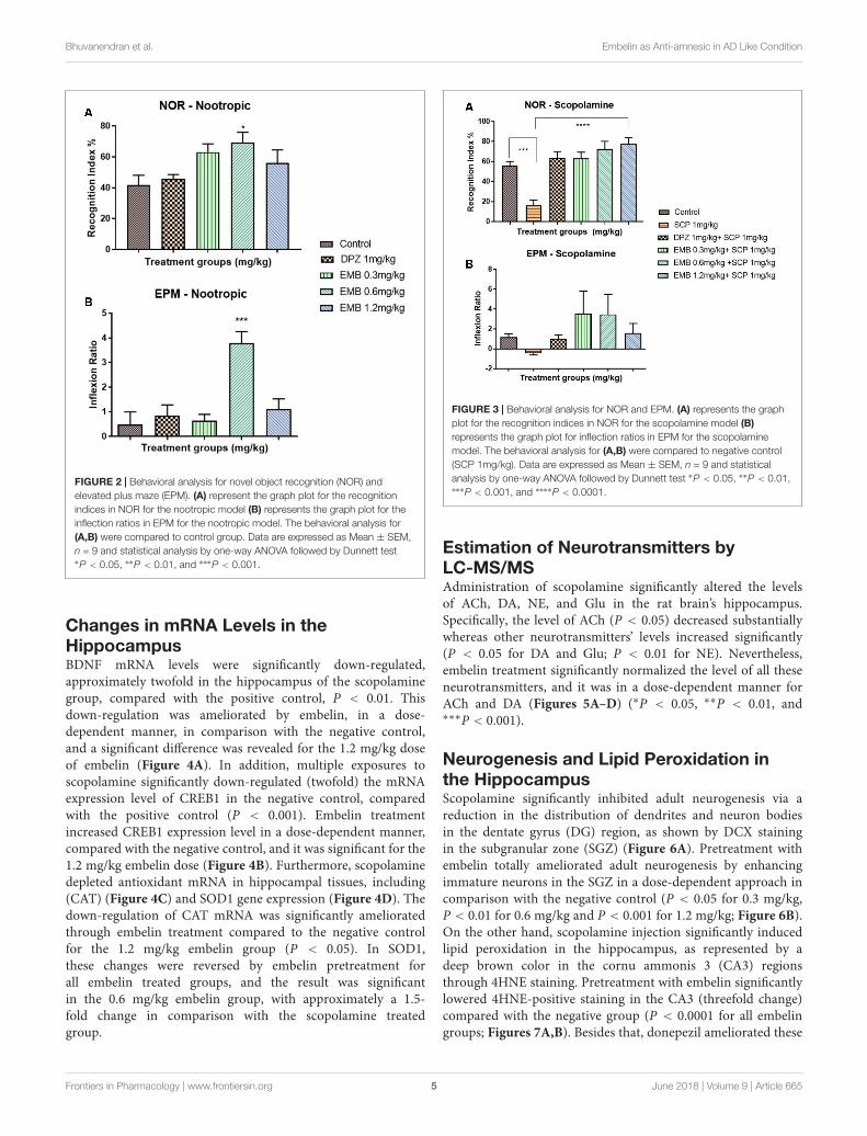
Nootropic effect of EmbelinFindings obtained from the NOR test for embelin nootropicactivity are illustrated in Figure 2A. The effect of differentembelin doses on memory function were assessed following7 days of pretreatment. The results were expressed as recognitionindex (%) for the novel object. Based on the outcomes, thepretreated groups of embelin showed an increase in recognitionindex for novel object compared with the control group anddonepezil groups. Only 0.6 mg/kg of embelin showed statisticallysignificant results with p value of <0.05. In EPM, the inflectionratio was significantly increased in 0.6 mg/kg embelin treatedgroups when compared with the control (Figure 2B). There wasno significant difference in other treated groups.
Anti-amnesic Effect of Embelin in RatsWith Scopolamine-Induced AmnesiaThe NOR test showed a reduction in recognition indexpercentage for the negative group (SCP 1 mg/kg) in the chronicscopolamine model (Figure 3A). Moreover, the recognitionindex percentage for all the embelin treated groups were highand comparable with donepezil (1 mg/kg) group. A significantdifference in the recognition index percentage was observedbetween all embelin treated and the negative group (P < 0.05). InEPM, inflection ratio analysis showed that there was an increasein retention memory in all embelin treated groups comparedwith the negative control group; however, it was statistically notsignificant (Figure 3B).
Frontiers in Pharmacology | www.frontiersin.org 4 June 2018 | Volume 9 | Article 665
fphar-09-00665 June 21, 2018 Time: 15:56 # 5
Bhuvanendran et al. Embelin as Anti-amnesic in AD Like Condition
FIGURE 2 | Behavioral analysis for novel object recognition (NOR) andelevated plus maze (EPM). (A) represent the graph plot for the recognitionindices in NOR for the nootropic model (B) represents the graph plot for theinflection ratios in EPM for the nootropic model. The behavioral analysis for(A,B) were compared to control group. Data are expressed as Mean ± SEM,n = 9 and statistical analysis by one-way ANOVA followed by Dunnett test∗P < 0.05, ∗∗P < 0.01, and ∗∗∗P < 0.001.
Changes in mRNA Levels in theHippocampusBDNF mRNA levels were significantly down-regulated,approximately twofold in the hippocampus of the scopolaminegroup, compared with the positive control, P < 0.01. Thisdown-regulation was ameliorated by embelin, in a dose-dependent manner, in comparison with the negative control,and a significant difference was revealed for the 1.2 mg/kg doseof embelin (Figure 4A). In addition, multiple exposures toscopolamine significantly down-regulated (twofold) the mRNAexpression level of CREB1 in the negative control, comparedwith the positive control (P < 0.001). Embelin treatmentincreased CREB1 expression level in a dose-dependent manner,compared with the negative control, and it was significant for the1.2 mg/kg embelin dose (Figure 4B). Furthermore, scopolaminedepleted antioxidant mRNA in hippocampal tissues, including(CAT) (Figure 4C) and SOD1 gene expression (Figure 4D). Thedown-regulation of CAT mRNA was significantly amelioratedthrough embelin treatment compared to the negative controlfor the 1.2 mg/kg embelin group (P < 0.05). In SOD1,these changes were reversed by embelin pretreatment forall embelin treated groups, and the result was significantin the 0.6 mg/kg embelin group, with approximately a 1.5-fold change in comparison with the scopolamine treatedgroup.
FIGURE 3 | Behavioral analysis for NOR and EPM. (A) represents the graphplot for the recognition indices in NOR for the scopolamine model (B)represents the graph plot for inflection ratios in EPM for the scopolaminemodel. The behavioral analysis for (A,B) were compared to negative control(SCP 1mg/kg). Data are expressed as Mean ± SEM, n = 9 and statisticalanalysis by one-way ANOVA followed by Dunnett test ∗P < 0.05, ∗∗P < 0.01,∗∗∗P < 0.001, and ∗∗∗∗P < 0.0001.
Estimation of Neurotransmitters byLC-MS/MSAdministration of scopolamine significantly altered the levelsof ACh, DA, NE, and Glu in the rat brain’s hippocampus.Specifically, the level of ACh (P < 0.05) decreased substantiallywhereas other neurotransmitters’ levels increased significantly(P < 0.05 for DA and Glu; P < 0.01 for NE). Nevertheless,embelin treatment significantly normalized the level of all theseneurotransmitters, and it was in a dose-dependent manner forACh and DA (Figures 5A–D) (∗P < 0.05, ∗∗P < 0.01, and∗∗∗P < 0.001).
Neurogenesis and Lipid Peroxidation inthe HippocampusScopolamine significantly inhibited adult neurogenesis via areduction in the distribution of dendrites and neuron bodiesin the dentate gyrus (DG) region, as shown by DCX stainingin the subgranular zone (SGZ) (Figure 6A). Pretreatment withembelin totally ameliorated adult neurogenesis by enhancingimmature neurons in the SGZ in a dose-dependent approach incomparison with the negative control (P < 0.05 for 0.3 mg/kg,P < 0.01 for 0.6 mg/kg and P < 0.001 for 1.2 mg/kg; Figure 6B).On the other hand, scopolamine injection significantly inducedlipid peroxidation in the hippocampus, as represented by adeep brown color in the cornu ammonis 3 (CA3) regionsthrough 4HNE staining. Pretreatment with embelin significantlylowered 4HNE-positive staining in the CA3 (threefold change)compared with the negative group (P < 0.0001 for all embelingroups; Figures 7A,B). Besides that, donepezil ameliorated these
Frontiers in Pharmacology | www.frontiersin.org 5 June 2018 | Volume 9 | Article 665

fphar-09-00665 June 21, 2018 Time: 15:56 # 6
Bhuvanendran et al. Embelin as Anti-amnesic in AD Like Condition
FIGURE 4 | Gene expression in the rat hippocampi determined by real time-PCR. The genes included are (A) BDNF, (B) CREB1, (C) Catalase, and (D) SuperoxideDismutase. All changes in the expressions levels were compared to the negative control group (SCP 1 mg/kg). Data are expressed as Mean ± SEM, n = 4 andstatistical analysis by one-way ANOVA followed by Dunnett test ∗P < 0.05, ∗∗P < 0.01, ∗∗∗P < 0.001, and ∗∗∗∗P < 0.0001.
alterations triggered by scopolamine, as displayed through bothDCX and 4-HNE staining.
DISCUSSION
This work aims to determine whether embelin has an anti-amnesic effect by modulating the cholinergic pathway. An animalmodel of hippocampal memory damage due to intraperitonealinjection of scopolamine was adopted to verify this hypothesis.The experiments comprised of two parts: Experiment 1(pretreatment with embelin without scopolamine injectionduring training) to test embelin’s nootropic effects on learningand memory process, and Experiment 2 (multiple exposures ofscopolamine injection) to assess the effect of embelin on anti-amnesic activities and biochemical aspects during learning andmemory process.
At the beginning of this experiment, we conducted a dosedeciding study to find the therapeutic dose of embelin. A priorliterature search determined that the range of embelin dosewas between 2.5 mg/kg to 10 mg/kg for the intraperitonealroute in CNS related animal models (Mahendran et al., 2011;Afzal et al., 2012). However, our preliminary study using these
range of embelin doses resulted in a neurobehavioral effect oncoordination and motor activity whereby the treated rats wereimmobile and kept falling from the behavioral apparatus. Thus,we decided 1.2 mg/kg as the highest dose as the LD50 valuefor embelin was 44 mg/kg for intraperitoneal administrationreported by Poojari (2014). Furthermore, we decided 0.3 mg/kgand 0.6 mg/kg would be the low dose and medium doserespectively, and all these 3 doses were effective therapeutic dosesfor our study as we noticed no side effects.
In this experiment, NOR and EPM were applied as behavioralmodels to evaluate learning and memory. The NOR test isparticularly relevant in AD research as it allows the assessmentof visual recognition memory, which is affected early in ADprogression, involving brain regions similar to those affectedby this devastating and debilitating neurodegenerative disease(Grayson et al., 2015). On the other hand, EPM is a behavioraltest employed to study long-term spatial memory (Uddin et al.,2016b). Certain EPM parameters like retention transfer latencyare utilized for the evaluation of memory. A decrease in transferlatency on the second day, which is after 24 h, indicates animprovement of memory and vice-versa (Dhingra and Kumar,2012). The findings of this study showed that embelin at0.6 mg/kg displayed nootropic activity in both the recognition
Frontiers in Pharmacology | www.frontiersin.org 6 June 2018 | Volume 9 | Article 665

fphar-09-00665 June 21, 2018 Time: 15:56 # 7
Bhuvanendran et al. Embelin as Anti-amnesic in AD Like Condition
FIGURE 5 | The concentration of neurotransmitters in the rat hippocampi after chronic scopolamine. The figure represents the rat hippocampal neurotransmitterlevels of (A) Acetylcholine, (B) Dopamine, (C) Norepinephrine, and (D) Glutamate. All changes in the neurotransmitter levels were compared to the negative controlgroup (SCP 1 mg/kg). Data are expressed as Mean ± SEM, n = 4 and statistical analysis by one-way ANOVA followed by Dunnett test ∗P < 0.05, ∗∗P < 0.01, and∗∗∗P < 0.001.
index and inflection ratio in the NOR and EPM tests, respectively(Figures 2A,B). However, the nootropic activity of embelin inboth behavioral paradigms was found to be dose independent.This could be explained that at a higher dose, the drug reaches itsmaximum effect so increasing the drug dosage does not increaseits effectiveness, but on the contrary, effectiveness decreases.This theory is supported by the fact that CNS drugs such asantipsychotic drugs produce maximum dopaminergic blockageat high doses. However, further dose increments will not produceany dopamine blockage but eventually lead to other side effectssuch as anticholinergic activity (Bridges, 1981). It is possiblethat in this experiment, the 1.2 mg/kg embelin group hasreached its maximum effect and therefore cognitive ability hasdeclined. Based on the behavioral results obtained, it can besuggested that embelin is a nootropic drug that acts as a naturalcognitive enhancer. These findings show that supplementationof embelin significantly amplified the rats’ memory function and0.6 mg/kg of embelin demonstrated significant nootropics effects.Nootropic drugs are used to treat cognition deficits in patientswith AD, schizophrenia, stroke, attention deficit hyperactivitydisorder (ADHD), and vascular dementia (VaD) (Birks andGrimley Evans, 2009; Froestl et al., 2012).
Scopolamine-induced dementia has been used extensivelyto assess potential therapeutic agents for treating AD
(Kwon et al., 2009). Scopolamine is a nonselective muscariniccholinergic receptor antagonist associated with cholinergicdysfunction, which causes performance deficits in learning andmemory (Heo et al., 2014). Therefore, in this study, scopolaminewas administered to rodents for 1 week to induce cholinergicneurodegeneration along with cognitive deficits. Following6 days of scopolamine administration, the scopolamine treatedgroup had less than 20% of the recognition index of other groups.Pretreatment with embelin ameliorated memory impairmentcaused by scopolamine (Figure 3A), with the recognitionindex being twofold more, in comparison with scopolaminetreated group in a dose-dependent manner. These resultsexposed that embelin was as effective as the donepezil-treatedgroup. Moreover, the findings showed that embelin treatmentattenuated amnesic behavior in EPM, but it was insignificant(Figure 3B). Hence, these outcomes suggest that embelin had ananti-amnesic effect in the scopolamine model.
The brain is susceptible to oxidative stress because it consumeshuge amounts of oxygen, has an abundant lipid content, anda low antioxidant level compared than other organs (Serranoand Klann, 2004). Furthermore, it is well known that thehippocampus region in the brain is crucial for learning andmemory, and the formation of spatial memory (Huang et al.,2015; Lee et al., 2016). The scopolamine-induced memory
Frontiers in Pharmacology | www.frontiersin.org 7 June 2018 | Volume 9 | Article 665

fphar-09-00665 June 21, 2018 Time: 15:56 # 8
Bhuvanendran et al. Embelin as Anti-amnesic in AD Like Condition
FIGURE 6 | DCX immunohistochemical analysis of the effects of embelin in improving scopolamine-induced suppression of neurogenesis in the dentate gyrus.(A) DCX-positive staining in immature neurons is shown in the subgranular zone of the dentate gyrus. Photomicrographs of the hippocampal section of treatmentgroups was (i) Control (ii) SCP 1 mg/kg alone (iii) DPZ 1 mg/kg + SCP 1 mg/kg (iv) EMB 0.3 mg/kg + SCP 1 mg/kg (v) EMB 0.6 mg/kg + SCP 1 mg/kg (vi) EMB1.2 mg/kg + SCP 1 mg/kg. Representative photomicrographs were taken at magnifications of 40X and 200X. (B) Quantification of DCX population. Data areexpressed as means Mean ± SEM, n = 5 and statistical analysis by one-way ANOVA followed by Dunnett test ∗P < 0.05, ∗∗P < 0.01, and ∗∗∗P < 0.001.
deficit model demonstrated that prominent oxidative stress andmemory deficits in a rodent model is similar to that in ADpatients, even though the mechanism of action remains unclear(Lee et al., 2015). The change in the mRNA levels of antioxidantsin the hippocampus after embelin pretreatment was examinedusing the scopolamine model in this present study. Scopolamineinjection induced oxidative stress in the hippocampus, as evidentby the decreased levels of CAT and SOD1 mRNA levels inthe scopolamine alone treated negative group. To prevent orslow down the progression of free radical-mediated oxidativestress, brain antioxidant defense enzymes such as CAT andSOD play a vital role in protecting tissues against oxidativedamage (Uddin et al., 2016a). Antioxidant mRNA alterationcaused by scopolamine injection was significantly amelioratedfor SOD1 via pretreatment with embelin. However, CAT mRNAlevel was decreased by scopolamine induction, but it was notsignificant (Figures 4C,D). Additionally, scopolamine-inducedlipid peroxidation in the hippocampus’s CA3 was shown aspositively stained 4HNE cells. Nonetheless, pretreatment withembelin completely attenuated the over-production of 4HNEcells (Figures 7A,B). These results propose that the protective
antioxidant gene response by embelin pretreatment reduced lipidperoxidation induced by scopolamine. An increase in 4HNE cellsis a key histopathological feature of neurodegenerative diseaseslike AD (Serrano and Klann, 2004).
Expression of BDNF and CREB1 mRNA levels inscopolamine-induced hippocampal tissue were examined toinvestigate the role of embelin in neurogenesis and synapticplasticity. In this study, hippocampal BDNF and CREB1were markedly reduced due to scopolamine injection, andpretreatment with embelin increased the mRNA expressionlevel of both BDNF and CREB1. A high dose of embelin at1.2 mg/kg exhibited maximum protection by increasing thelevels of BDNF and CREB1. Other than that, cAMP responseelement binding protein (CREB) plays a crucial role in neuronalgrowth, proliferation, differentiation, and survival (Lee et al.,2016). In our results, the explanation for the increased in dosedependency for both BDNF and CREB 1 could possibly be thatembelin may be responsible for visual recognition memoryin NOR through this BDNF/CREB pathway. We noticed thatat the 1.2 mg/kg dose, embelin expressed high mRNA levelsof BDNF and CREB1 and this could be the reason for a 60%
Frontiers in Pharmacology | www.frontiersin.org 8 June 2018 | Volume 9 | Article 665

fphar-09-00665 June 21, 2018 Time: 15:56 # 9
Bhuvanendran et al. Embelin as Anti-amnesic in AD Like Condition
FIGURE 7 | 4HNE immunohistochemical analysis illustrating the inhibitory effects of embelin against scopolamine-induced lipid peroxidation in the hippocampus.(A) The 4HNE-positive stained cells in the CA3 region of the hippocampus are indicative of lipid peroxidation. Photomicrographs hippocampal section of treatmentgroups was (i) Control (ii) SCP 1 mg/kg alone (iii) DPZ 1 mg/kg + SCP 1 mg/kg (iv) EMB 0.3 mg/kg + SCP 1 mg/kg (v) EMB 0.6 mg/kg + SCP 1 mg/kg (vi) EMB1.2 mg/kg + SCP 1 mg/kg. Representative photomicrographs were taken at magnifications of 40X, and 200X. (B) Quantification of 4HNE protein in CA3. Data areexpressed as means Mean ± SEM, n = 5 and statistical analysis by one-way ANOVA followed by Dunnett test ∗P < 0.05, ∗∗P < 0.01, ∗∗∗P < 0.001, and∗∗∗∗P < 0.0001.
increase in visual recognition index in NOR when comparedwith the scopolamine treated group. Thus, this validates therole of BDNF-CREB signaling in visual recognition memory,particularly for hippocampus-dependent learning.
Likewise, adult hippocampal neurogenesis plays a key role inhippocampal memory function (Mu and Gage, 2011). Altmanand Das (1965) first reported on the continual production ofnew neurons in the adult hippocampus. These new neuronsoriginated from adult neural stem cells (NSCs) residing in theSGZ of DG (Bonaguidi et al., 2011). In this present research,a significantly reduced level of immature neurons, revealedthrough DCX staining of scopolamine-induced rat hippocampuswas determined, while pretreatment with embelin distinctlyameliorated repression of the SGZ region’s neuronal precursorcells in a dose-dependent manner (Figures 6A,B).
Numerous studies have reported that most classicalneurotransmitter systems such as ACh, NE, Glu, and DA,influence learning and memory (Myhrer, 2003). We adoptedLC-MS/MS method as it is a simple, sensitive and simultaneouslyable to quantify the four major neurotransmitters from rathippocampal tissue in a single run (Zheng et al., 2012). The
extraction of the neurotransmitters from rat hippocampus wasdone with utmost care and prior to LC-MS/MS analysis toavoid any possibilities of sample degradation and oxidationas described by He et al. (2013). The neurotransmitters’concentrations were expressed as a ratio of total proteinconcentration in order to get correct value and to avoidpossible variation in sample when subjected to LC-MS/MS.In AD patients, pathological changes affecting glutamatergic,cholinergic, noradrenergic, and serotonergic systems havebeen revealed (Francis et al., 1999). In this study, the effect ofembelin on brain neurotransmitter levels in rats administeredscopolamine was investigated. ACh plays an essential rolein learning process and memory as a key transmitter in thecholinergic system (Chen et al., 2016). A decrease in AChlevels is reported in this study as a biomarker of scopolamine-induced cognitive impairment in the rat hippocampus. Embelinadministered at a dose of 0.3 mg/kg significantly increasedACh levels, subsequently improving cholinergic function.Interestingly, Arora and Deshmukh (2017) reported that embelintreatment in a streptozotocin-induced rat model decreasedAChE activity, which is the enzyme that metabolizes ACh
Frontiers in Pharmacology | www.frontiersin.org 9 June 2018 | Volume 9 | Article 665

fphar-09-00665 June 21, 2018 Time: 15:56 # 10
Bhuvanendran et al. Embelin as Anti-amnesic in AD Like Condition
FIGURE 8 | Schematic diagram showing the effect of embelin in a diseased condition. The figure depicts cholinergic dysfunction, oxidative stress, andneurodegeneration which contributes to neuronal loss, synaptic dysfunction, and an AD-like condition. Embelin could act as memory enhancer by stimulating thecholinergic systems via acetylcholine release and inhibition of AChE. In the oxidative defense pathway, embelin reduces oxidative stress by increasing SOD and CATmRNA levels and eventually reducing lipid peroxidation. In addition to these effects, a rise in DCX expression by embelin treatment contributes to neurogenesis andan increase in BDNF-CREB levels may contribute to synaptic plasticity.
into choline and acetate. Therefore, a reduction in AChE levelindicates a high level of ACh as a result of embelin treatment,which is similar to our results. In the current research, Glulevels were raised after being treated with scopolamine. Similaroutcomes were reported by Pandareesh et al. (2016) and Aroraand Deshmukh (2017). Administration of 0.6 mg/kg embelinsignificantly lowered the level of Glu. A rise in Glu level hasbeen reported to cause excitotoxic neuronal damage and lossof cognitive function (Arora and Deshmukh, 2017) and alsoassociated with excitotoxicity in AD brains (Jackson, 2014).Scopolamine treatment also caused an increment in the levelsof DA and NE in the hippocampus. Earlier reports suggests thatan increase in DA and NE levels leads to amnesia and memorydeficits. Wu et al.’s (2014) study, demonstrated that donepeziltreatment can modulate the increase levels of DA and NE indisease control group. Interestingly, a similar protective effectwas observed with embelin pre-treatment in amnesia condition.
In this scopolamine model, our results are unusual, withembelin causing different dose dependency in the behavioralmodel and neurotransmitters, particularly ACh when comparedto other reported studies that utilized embelin. This could beexplained by embelin being neuroprotective in a scopolamine-induced amnesia model via visual recognition memory but not inlong-term spatial memory. This theory is supported by our resultsas there was a dose dependency in embelin treatment in NORand the result of embelin is comparable with the donepezil group.
However, we could not see this pattern in EPM. Whilst embelinimproved visual recognition in dose dependency manner, it alsoreduced the level of ACh in a dose-dependent manner as well.This discrepancy could be because at a dose of 0.3 mg/kg,embelin might be effectively increasing the level of ACh but stopsfurther production of ACh at 1.2 mg/kg. At this particular dose,embelin probably plays a different role in inhibiting the enzymeAChE. This could be the reason that at 1.2 mg/kg of embelin,we observed a high recognition index in NOR of scopolamine-induced amnesia rats.
CONCLUSION
In conclusion, the results from this study have demonstratedthat embelin displays nootropic and neuroprotective abilitiesin scopolamine-induced amnesia in rats. Nootropic effects maybe attributed to an increase in visual recognition and spatialmemory in both NOR and EPM. Embelin possesses anti-amnesic effects, which could be mediated by an antioxidantgene response particularly though SOD1, the CREB-BDNFpathway, hippocampal neurogenesis, and cholinergic activity.The anti-amnesic effect of embelin is also comparable to thatof donepezil at a specific concentration even though it isnot in a dose-dependent manner in certain cases. Therefore,embelin could be a promising treatment for patients sufferingfrom neurodegenerative diseases. Figure 8 shows the potential
Frontiers in Pharmacology | www.frontiersin.org 10 June 2018 | Volume 9 | Article 665

fphar-09-00665 June 21, 2018 Time: 15:56 # 11
Bhuvanendran et al. Embelin as Anti-amnesic in AD Like Condition
mechanism of action of embelin in scopolamine-inducedmemory impairment in rodents.
ETHICS STATEMENT
The experimental protocol was approved by the Monash AnimalResearch Platform (MARP) Animal Ethics Committee, MonashUniversity, Australia (MARP/2016/054).
AUTHOR CONTRIBUTIONS
SB performed all the experiments and was responsible forthe writing of the manuscript in its entirety. YK helped indesigning gene expression study, result analysis and figures inthe manuscript. IO helped in LC-MS/MS method. MS helpedin conceptualizing, designing the study, result analysis, andmanuscript writing. All authors gave their final approval for thesubmission of the manuscript.
REFERENCESAfzal, M., Gupta, G., Kazmi, I., Rahman, M., Upadhyay, G., Ahmad, K., et al.
(2012). Evaluation of anxiolytic activity of embelin isolated from Embelia ribes.Biomed. Aging Pathol. 2, 45–47. doi: 10.1016/j.biomag.2012.03.003
Ali, T., Yoon, G. H., Shah, S. A., Lee, H. Y., and Kim, M. O. (2015). Osmotinattenuates amyloid beta-induced memory impairment, tau phosphorylationand neurodegeneration in the mouse hippocampus. Sci. Rep. 5:11708.doi: 10.1038/srep11708
Altman, J., and Das, G. D. (1965). Autoradiographic and histological evidence ofpostnatal hippocampal neurogenesis in rats. J. Comp. Neurol. 124, 319–335.doi: 10.1002/cne.901240303
Alzheimer’s Association (2017). 2017 Alzheimer’s disease facts andfigures. Alzheimers Dement. 13, 325–373. doi: 10.1016/j.jalz.2017.02.001
Arora, R., and Deshmukh, R. (2017). Embelin attenuates intracerebroventricularstreptozotocin-induced behavioral, biochemical, and neurochemicalabnormalities in rats. Mol. Neurobiol. 54, 6670–6680. doi: 10.1007/s12035-016-0182-y
Bartus, R. T., Dean, R. L., Beer, B., and Lippa, A. S. (1982). The cholinergichypothesis of geriatric memory dysfunction. Science 217, 408–414. doi: 10.1126/science.7046051
Batool, Z., Sadir, S., Liaquat, L., Tabassum, S., Madiha, S., Rafiq, S., et al. (2016).Repeated administration of almonds increases brain acetylcholine levels andenhances memory function in healthy rats while attenuates memory deficitsin animal model of amnesia. Brain Res. Bull. 120, 63–74. doi: 10.1016/j.brainresbull.2015.11.001
Birks, J., and Grimley Evans, J. (2009). Ginkgo biloba for cognitive impairmentand dementia. Cochrane Database Syst. Rev. CD003120. doi: 10.1002/14651858.CD003120.pub3
Bonaguidi, M. A., Wheeler, M. A., Shapiro, J. S., Stadel, R. P., Sun, G. J., Ming, G.-L.,et al. (2011). In vivo clonal analysis reveals self-renewing and multipotent adultneural stem cell characteristics. Cell 145, 1142–1155. doi: 10.1016/j.cell.2011.05.024
Bridges, P. K. (1981). Disturbed behavior induced by high-dose antipsychoticdrugs. Br. Med. J. (Clin. Res. Ed.) 282:313.
Brookmeyer, R., Johnson, E., Ziegler-Graham, K., and Arrighi, H. M. (2007).Forecasting the global burden of Alzheimer’s disease. Alzheimer’s Dement. 3,186–191. doi: 10.1016/j.jalz.2007.04.381
Butterfield, D. A., and Lauderback, C. M. (2002). Lipid peroxidation andprotein oxidation in Alzheimer’s disease brain: potential causes andconsequences involving amyloid β-peptide-associated free radical oxidativestress1, 2. Free Radic. Biol. Med. 32, 1050–1060. doi: 10.1016/S0891-5849(02)00794-3
Chen, L.-E., Wu, F., Zhao, A., Ge, H., and Zhan, H. (2016). Protection efficacy of theextract of Ginkgo biloba against the learning and memory damage of rats underrepeated high sustained. Evid. Based Complement. Alternat. Med. 2016:6320586.doi: 10.1155/2016/6320586
Dhingra, D., and Kumar, V. (2012). Memory-enhancing activity of palmatine inmice using elevated plus maze and morris water maze. Adv. Pharmacol. Sci.2012:357368. doi: 10.1155/2012/357368
Ding, Q., Dimayuga, E., and Keller, J. N. (2007). Oxidative damage, proteinsynthesis, and protein degradation in Alzheimer’s disease. Curr. Alzheimer Res.4, 73–79. doi: 10.2174/156720507779939788
Ennaceur, A., and Delacour, J. (1988). A new one-trial test for neurobiologicalstudies of memory in rats. 1: behavioral data. Behav. Brain Res. 31, 47–59.doi: 10.1016/0166-4328(88)90157-X
Francis, P. T., Palmer, A. M., Snape, M., and Wilcock, G. K. (1999). The cholinergichypothesis of Alzheimer’s disease: a review of progress. J. Neurol. Neurosurg.Psychiatry 66, 137–147. doi: 10.1136/jnnp.66.2.137
Froestl, W., Muhs, A., and Pfeifer, A. (2012). Cognitive enhancers (nootropics).Part 1: drugs interacting with receptors. J. Alzheimers Dis. 32, 793–887.doi: 10.3233/JAD-2012-121186
Ghumatkar, P. J., Patil, S. P., Jain, P. D., Tambe, R. M., and Sathaye, S.(2015). Nootropic, neuroprotective and neurotrophic effects of phloretin inscopolamine induced amnesia in mice. Pharmacol. Biochem. Behav. 135, 182–191. doi: 10.1016/j.pbb.2015.06.005
Grayson, B., Leger, M., Piercy, C., Adamson, L., Harte, M., and Neill, J. C. (2015).Assessment of disease-related cognitive impairments using the novel objectrecognition (NOR) task in rodents. Behav. Brain Res. 285, 176–193. doi: 10.1016/j.bbr.2014.10.025
Halder, S., Mehta, A. K., Kar, R., Mustafa, M., Mediratta, P. K., and Sharma, K. K.(2011). Clove oil reverses learning and memory deficits in scopolamine-treatedmice. Planta Med. 77, 830–834. doi: 10.1055/s-0030-1250605
He, B., Bi, K., Jia, Y., Wang, J., Lv, C., Liu, R., et al. (2013). Rapid analysisof neurotransmitters in rat brain using ultra-fast liquid chromatographyand tandem mass spectrometry: application to a comparative study innormal and insomnic rats. J. Mass Spectrom. 48, 969–978. doi: 10.1002/jms.3243
Heo, Y.-M., Shin, M.-S., Lee, J.-M., Kim, C.-J., Baek, S.-B., Kim, K.-H., et al.(2014). Treadmill exercise ameliorates short-term memory disturbance inscopolamine-induced amnesia rats. Int. Neurourol. J. 18, 16–22. doi: 10.5213/inj.2014.18.1.16
Hritcu, L., Noumedem, J. A., Cioanca, O., Hancianu, M., Kuete, V., andMihasan, M. (2014). Methanolic extract of Piper nigrum fruits improvesmemory impairment by decreasing brain oxidative stress in amyloid beta(1–42) rat model of Alzheimer’s disease. Cell Mol. Neurobiol. 34, 437–449.doi: 10.1007/s10571-014-0028-y
Huang, T.-T., Leu, D., and Zou, Y. (2015). Oxidative stress and redox regulation onhippocampal-dependent cognitive functions. Arch. Biochem. Biophys. 576, 2–7.doi: 10.1016/j.abb.2015.03.014
Jackson, W. S. (2014). Selective vulnerability to neurodegenerative disease: thecurious case of Prion protein. Dis. Models Mech. 7, 21–29. doi: 10.1242/dmm.012146
Kundap, U. P., Bhuvanendran, S., Kumari, Y., Othman, I., and Shaikh, M. F.(2017a). Plant derived phytocompound, embelin in CNS disorders: a systematicreview. Front. Pharmacol. 8:76. doi: 10.3389/fphar.2017.00076
Kundap, U. P., Kumari, Y., Othman, I., and Shaikh, M. F. (2017b). Zebrafishas a model for epilepsy-induced cognitive dysfunction: a pharmacological,biochemical and behavioral approach. Front. Pharmacol. 8:515. doi: 10.3389/fphar.2017.00515
Kwon, S.-H., Kim, H.-C., Lee, S.-Y., and Jang, C.-G. (2009). Loganin improveslearning and memory impairments induced by scopolamine in mice. Eur. J.Pharmacol. 619, 44–49. doi: 10.1016/j.ejphar.2009.06.062
Lee, J.-S., Hong, S.-S., Kim, H.-G., Lee, H.-W., Kim, W.-Y., Lee, S.-K., et al.(2016). Gongjin-Dan enhances hippocampal memory in a mouse model ofscopolamine-induced amnesia. PLoS One 11:e0159823. doi: 10.1371/journal.pone.0159823
Frontiers in Pharmacology | www.frontiersin.org 11 June 2018 | Volume 9 | Article 665

fphar-09-00665 June 21, 2018 Time: 15:56 # 12
Bhuvanendran et al. Embelin as Anti-amnesic in AD Like Condition
Lee, J.-S., Kim, H.-G., Lee, H.-W., Han, J.-M., Lee, S.-K., Kim, D.-W., et al.(2015). Hippocampal memory enhancing activity of pine needle extract againstscopolamine-induced amnesia in a mouse model. Sci. Rep. 5:9651. doi: 10.1038/srep09651
Mahendran, S., Thippeswamy, B., Veerapur, V., and Badami, S. (2011).Anticonvulsant activity of embelin isolated from Embelia ribes. Phytomedicine18, 186–188. doi: 10.1016/j.phymed.2010.04.002
Mu, Y., and Gage, F. H. (2011). Adult hippocampal neurogenesis and its role inAlzheimer’s disease. Mol. Neurodegener. 6:85. doi: 10.1186/1750-1326-6-85
Myhrer, T. (2003). Neurotransmitter systems involved in learning and memory inthe rat: a meta-analysis based on studies of four behavioral tasks. Brain Res. Rev.41, 268–287. doi: 10.1016/S0165-0173(02)00268-0
Pandareesh, M., Anand, T., and Khanum, F. (2016). Cognition enhancing andneuromodulatory propensity of Bacopa monniera extract against scopolamineinduced cognitive impairments in rat hippocampus. Neurochem. Res. 41, 985–999. doi: 10.1007/s11064-015-1780-1
Pathan, S. A., Iqbal, Z., Zaidi, S., Talegaonkar, S., Vohra, D., Jain, G. K., et al. (2009).CNS drug delivery systems: novel approaches. Recent Pat. Drug Deliv. Formul.3, 71–89. doi: 10.2174/187221109787158355
Poojari, R. (2014). Embelin–a drug of antiquity: shifting the paradigm towardsmodern medicine. Expert Opin. Investig. Drugs 23, 427–444. doi: 10.1517/13543784.2014.867016
Serrano, F., and Klann, E. (2004). Reactive oxygen species and synaptic plasticityin the aging hippocampus. Ageing Res. Rev. 3, 431–443. doi: 10.1016/j.arr.2004.05.002
Tanzi, R. E., and Bertram, L. (2005). Twenty years of the Alzheimer’s diseaseamyloid hypothesis: a genetic perspective. Cell 120, 545–555. doi: 10.1016/j.cell.2005.02.008
Uddin, M. S., Al Mamun, A., Hossain, M. S., Ashaduzzaman, M., Noor, M. A. A.,Hossain, M. S., et al. (2016a). Neuroprotective effect of Phyllanthus acidus L.on learning and memory impairment in scopolamine-induced animal model ofdementia and oxidative stress: natural wonder for regulating the development
and progression of Alzheimer’s disease. Adv. Alzheimers Dis. 5, 53–72.doi: 10.4236/aad.2016.52005
Uddin, M. S., Nasrullah, M., Hossain, M. S., Rahman, M. M., Sarwar, M. S.,Amran, M. S., et al. (2016b). Evaluation of nootropic activity ofPersicaria flaccida on cognitive performance, brain antioxidantmarkers and acetylcholinesterase activity in rats: implication for themanagement of Alzheimer’s disease. Am. J. Psychiatry Neurosci. 4, 26–37.doi: 10.11648/j.ajpn.20160402.12
Wu, C.-R., Lin, H.-C., and Su, M.-H. (2014). Reversal by aqueous extractsof Cistanche tubulosa from behavioral deficits in Alzheimer’s disease-likerat model: relevance for amyloid deposition and central neurotransmitterfunction. BMC Complement. Altern. Med. 14:202. doi: 10.1186/1472-6882-14-202
Yatin, S., Varadarajan, S., Link, C., and Butterfield, D. (1999). In vitro andin vivo oxidative stress associated with Alzheimer’s amyloid ß-peptide (1–42).Neurobiol. Aging 20, 325–330.
Zheng, X., Kang, A., Dai, C., Liang, Y., Xie, T., Xie, L., et al. (2012).Quantitative analysis of neurochemical panel in rat brain and plasma by liquidchromatography-tandem mass spectrometry. Anal. Chem. 84, 10044–10051.doi: 10.1021/ac3025202
Conflict of Interest Statement: The authors declare that the research wasconducted in the absence of any commercial or financial relationships that couldbe construed as a potential conflict of interest.
Copyright © 2018 Bhuvanendran, Kumari, Othman and Shaikh. This is an open-access article distributed under the terms of the Creative Commons AttributionLicense (CC BY). The use, distribution or reproduction in other forums is permitted,provided the original author(s) and the copyright owner are credited and that theoriginal publication in this journal is cited, in accordance with accepted academicpractice. No use, distribution or reproduction is permitted which does not complywith these terms.
Frontiers in Pharmacology | www.frontiersin.org 12 June 2018 | Volume 9 | Article 665