ambiguity in detection of necrosis in ivus plaque characterization
TRANSCRIPT
Ambiguity in Detection of Necrosis in IVUS
Plaque Characterization Algorithms
and SDH as Alternative Solution
Amin Katouzian, Ph.D., Debdoot Sheet, M.S., Abouzar Eslami, Ph.D.,
Athanasios Karamalis, M.Sc., Andreas König, MD,
Stephane G. Carlier, MD, Nassir Navab, Ph.D.
Conflict of interest
None

Background
• Intravascular ultrasound (IVUS)
– Provides information about arterial wall and extend of atherosclerosis.
– Grayscale and radiofrequency (RF) signals are used for
atherosclerotic tissue characterization (TC).
– Has potential to identify factors associated with vulnerable plaques.
• Lipid pool.
• Necrotic core.
– Existing TC techniques fail to detect it reliably.
• Calcification patterns.
• Thin cap fibroatheroma.
– Hard to detect due to resolution limitation.
• Shear stress.
3
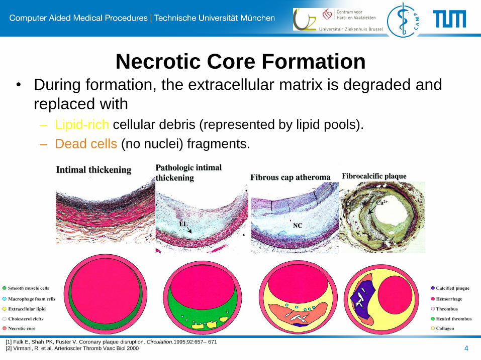
Necrotic Core Formation • During formation, the extracellular matrix is degraded and
replaced with
– Lipid-rich cellular debris (represented by lipid pools).
– Dead cells (no nuclei) fragments.
4 [1] Falk E, Shah PK, Fuster V. Coronary plaque disruption. Circulation.1995;92:657– 671
[2] Virmani, R. et al. Arterioscler Thromb Vasc Biol 2000
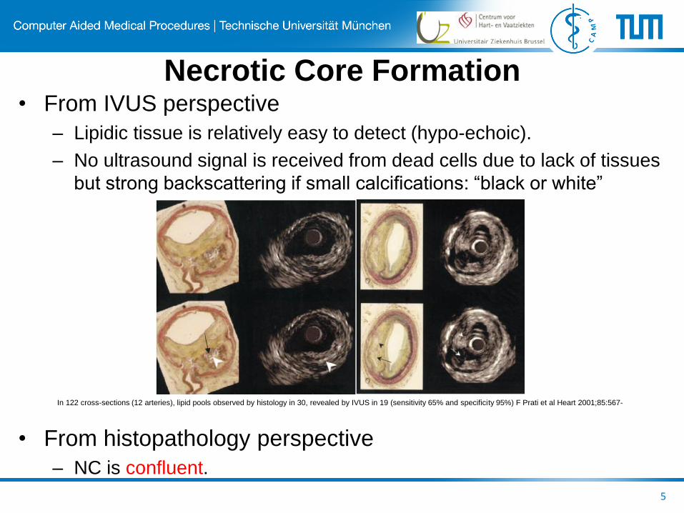
Necrotic Core Formation • From IVUS perspective
– Lipidic tissue is relatively easy to detect (hypo-echoic).
– No ultrasound signal is received from dead cells due to lack of tissues
but strong backscattering if small calcifications: “black or white”
• From histopathology perspective
– NC is confluent.
5
In 122 cross-sections (12 arteries), lipid pools observed by histology in 30, revealed by IVUS in 19 (sensitivity 65% and specificity 95%) F Prati et al Heart 2001;85:567-
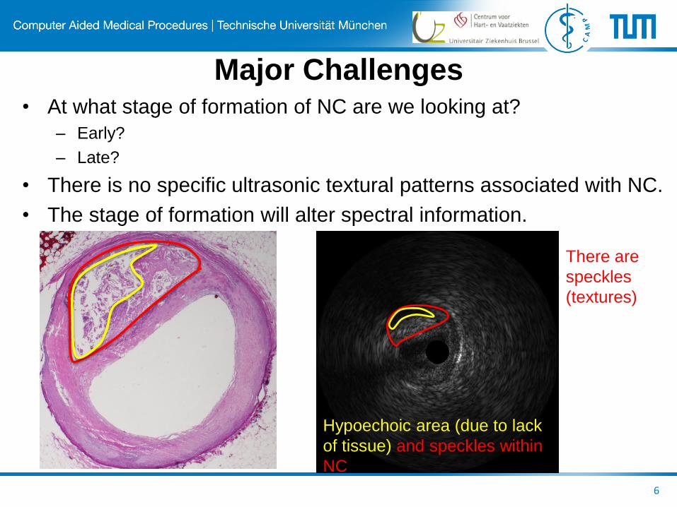
Major Challenges • At what stage of formation of NC are we looking at?
– Early?
– Late?
• There is no specific ultrasonic textural patterns associated with NC.
• The stage of formation will alter spectral information.
6
There are
speckles
(textures)
Hypoechoic area (due to lack
of tissue) and speckles within
NC

• We are able to generate VH-like images, coined Prognosis
Histology (PH)[1], based only on textural information.
• Correlation between detected tissues in PH and VH images in
155 cross sections collected in 4 patients.
– Calcified: 93.1±6.1%, Necrosis: 87.5±9.5%, Fibrotic: 78.4±17.6%, and
Fibrofatty: 61.3±21.3%.
• Observation:
– VH detects necrosis around calcified tissues.
– NC appears sparse and mainly superficially in VH images.
– VH systematically detects fibrotic followed by fibrofatty tissues in
shadowed regions behind arc of calcified plaques.
7
Iterative self-organizing atherosclerotic
tissue labeling: Prognosis Histology
[1] Amin Katouzian, Athanasios Karamalis, Debdoot Sheet, Elisa E. Konofagou, Babak Baseri, Stephane G. Carlier, Abouzar Eslami, Andreas König, Nassir Navab, Andrew F. Laine,
“Iterative self-organizing atherosclerotic tissue labeling in intravascular ultrasound images and comparison with virtual histology,” Accepted in IEEE TBME, 2012.

8
VH vs PH for the detection of NC
[1] Amin Katouzian, Athanasios Karamalis, Debdoot Sheet, Elisa E. Konofagou, Babak Baseri, Stephane G. Carlier, Abouzar Eslami, Andreas König, Nassir Navab, Andrew F. Laine,
“Iterative self-organizing atherosclerotic tissue labeling in intravascular ultrasound images and comparison with virtual histology,” Accepted in IEEE TBME, 2012.
VH PH
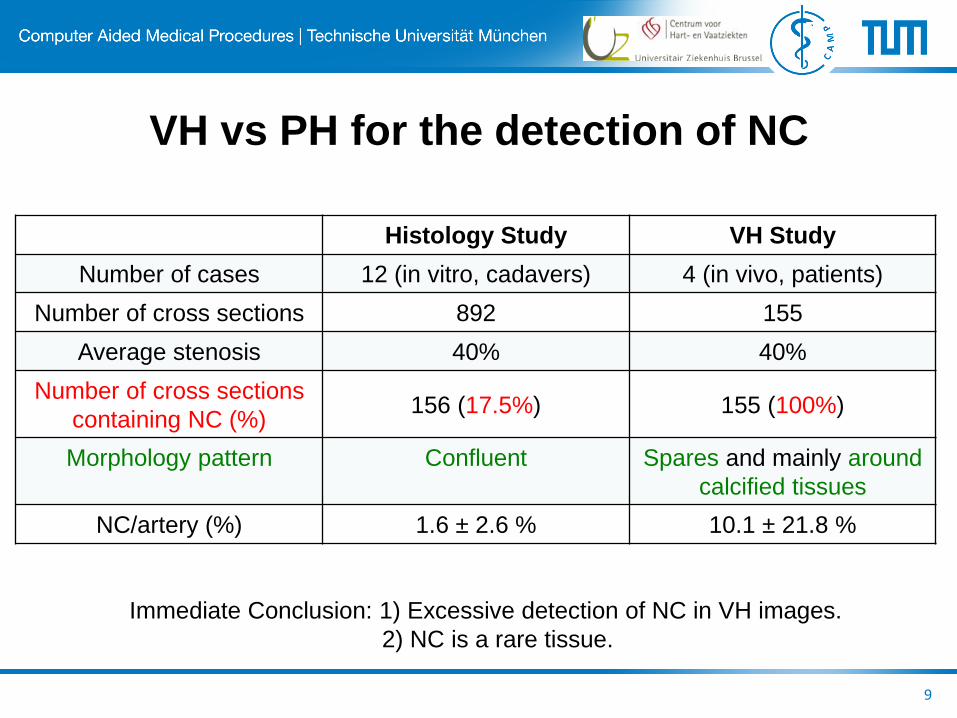
Histology Study VH Study
Number of cases 12 (in vitro, cadavers) 4 (in vivo, patients)
Number of cross sections 892 155
Average stenosis 40% 40%
Number of cross sections
containing NC (%) 156 (17.5%) 155 (100%)
Morphology pattern Confluent Spares and mainly around
calcified tissues
NC/artery (%) 1.6 ± 2.6 % 10.1 ± 21.8 %
9
Immediate Conclusion: 1) Excessive detection of NC in VH images.
2) NC is a rare tissue.
VH vs PH for the detection of NC

Histopathology Perspective
10 Thim T et al. Circ Cardiovasc Imaging
2010;3:384-391
Fibrous lesion with dense collagen
displayed as necrotic core by VH
IVUS. A, Gray-scale IVUS with lumen
and external elastic lamina borders.
Fibroatheroma with large necrotic core
missed by VH IVUS. A, Gray-scale
IVUS with lumen and external elastic
lamina border contours.
Granada J F et al. Arterioscler Thromb Vasc Biol
2007;27:387-393
Movat pentachrome section showing a fibrolipidic
plaque with no evidence of calcification.

Alternative Solution for Tissue
Classification and NC Identification
• Combination of both textural and RF information.
• Deployment of features with ultrasonic physics background.
• Superior machine learning algorithm.
• Reliability measure for estimation of tissues.
• Extensive and proper in vitro and in vivo validation.
11
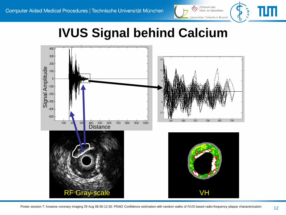
IVUS Signal behind Calcium
12 Poster session 7: Invasive coronary imaging 29 Aug 08:30-12:30: P5462 Confidence estimation with random walks of IVUS based radio-frequency plaque characterization
RF Gray-scale VH
Distance
Sig
na
l Am
plit
ud
e
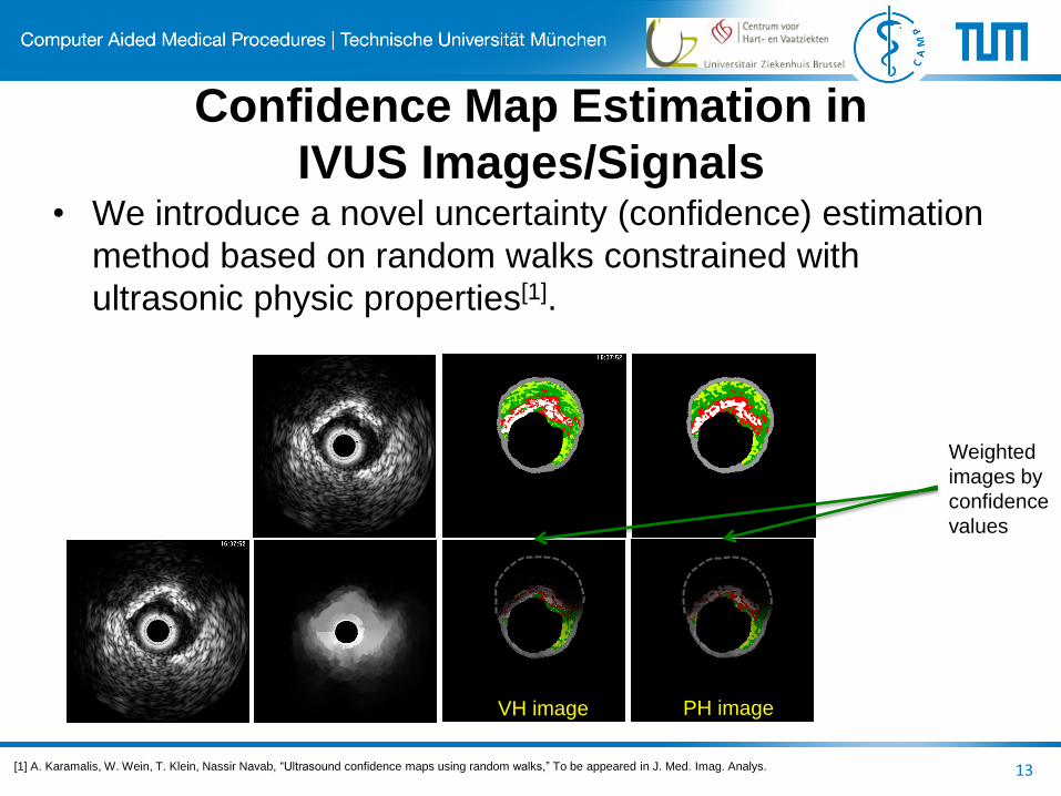
Confidence Map Estimation in
IVUS Images/Signals
13
• We introduce a novel uncertainty (confidence) estimation
method based on random walks constrained with
ultrasonic physic properties[1].
VH image PH image
Weighted
images by
confidence
values
[1] A. Karamalis, W. Wein, T. Klein, Nassir Navab, “Ultrasound confidence maps using random walks,” To be appeared in J. Med. Imag. Analys.
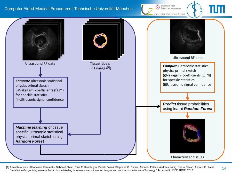
Ultrasound RF data Tissue labels (PH images[1])
Compute ultrasonic statistical physics primal sketch (i)Nakagami coefficients (Ω,m) for speckle statistics (ii)Ultrasonic signal confidence
Machine learning of tissue
specific ultrasonic statistical
physics primal sketch using
Random Forest
Compute ultrasonic statistical physics primal sketch (i)Nakagami coefficients (Ω,m) for speckle statistics (ii)Ultrasonic signal confidence
Ultrasound RF data
Predict tissue probabilities
using learnt Random Forest
Characterized tissues
[1] Amin Katouzian, Athanasios Karamalis, Debdoot Sheet, Elisa E. Konofagou, Babak Baseri, Stephane G. Carlier, Abouzar Eslami, Andreas König, Nassir Navab, Andrew F. Laine,
“Iterative self-organizing atherosclerotic tissue labeling in intravascular ultrasound images and comparison with virtual histology,” Accepted in IEEE TBME, 2012. 14

Ultrasonic Stochastic Driven Histology
(SDH)[1]
• Extraction of features with ultrasonic physics background.
• Superior machine learning algorithm.
• Reliability measure for estimation of tissues.
15
Probability of Calcified
tissues
Probability of Fibrotic
tissues
Probability of Lipidic
tissues Probability of Necrotic
tissues
[1] D. Sheet, Katouzian, A. Karamalis, A. Eslami, P. Noel, A. Koening, N. Navab, J. Chatterjee, A. Ray, A. Laine, S. G. Carlier, A. Katouzian,
“Joint Learning of Ultrasonic Statistical Physics and Signal Confidence Primal using Random Forests for Plaque Characterization in Intravascular Ultrasound,” under review.
Calcified
Lipidic
Fibrotic
Necrotic

Qualitative In Vitro Results
16
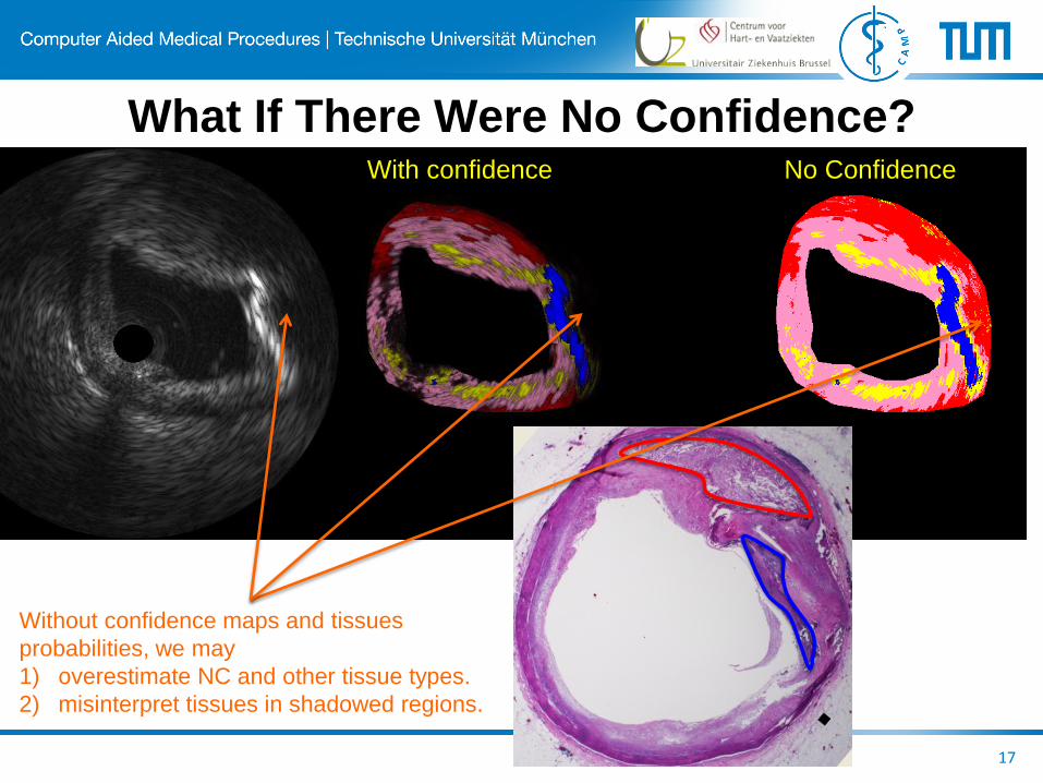
What If There Were No Confidence?
17
With confidence No Confidence
Without confidence maps and tissues
probabilities, we may
1) overestimate NC and other tissue types.
2) misinterpret tissues in shadowed regions.
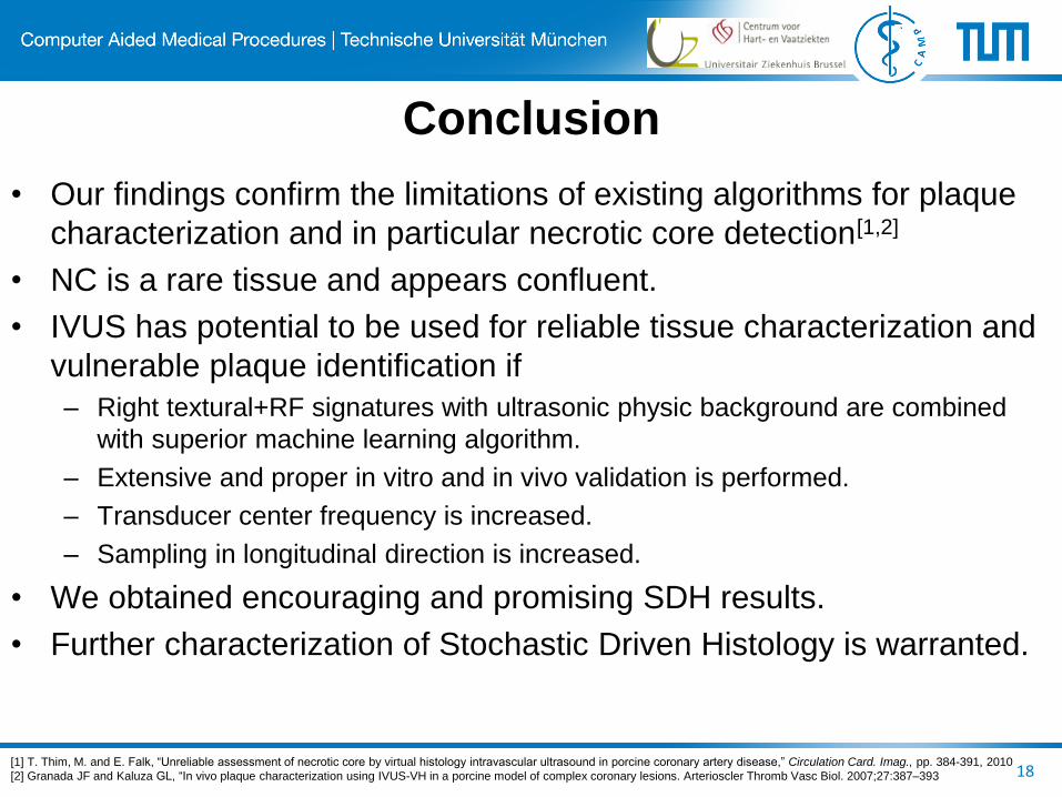
Conclusion
• Our findings confirm the limitations of existing algorithms for plaque
characterization and in particular necrotic core detection[1,2]
• NC is a rare tissue and appears confluent.
• IVUS has potential to be used for reliable tissue characterization and
vulnerable plaque identification if
– Right textural+RF signatures with ultrasonic physic background are combined
with superior machine learning algorithm.
– Extensive and proper in vitro and in vivo validation is performed.
– Transducer center frequency is increased.
– Sampling in longitudinal direction is increased.
• We obtained encouraging and promising SDH results.
• Further characterization of Stochastic Driven Histology is warranted.
18 [1] T. Thim, M. and E. Falk, “Unreliable assessment of necrotic core by virtual histology intravascular ultrasound in porcine coronary artery disease,” Circulation Card. Imag., pp. 384-391, 2010
[2] Granada JF and Kaluza GL, “In vivo plaque characterization using IVUS-VH in a porcine model of complex coronary lesions. Arterioscler Thromb Vasc Biol. 2007;27:387–393
19
Amin Katouzian Debdoot Sheet Abouzar Eslami
Ph.D. MS Ph.D.
Athanasios Karamalis Nassir Navab
M.Sc. Ph.D.
THANK YOU!