alveolar bone
DESCRIPTION
the basic anatomy of the alveolar bone has been described. will be most useful for 1st year dental students.TRANSCRIPT

ALVEOLAR BONE

BONE COMPOSITION OF BONE CLASSIFICATION OF BONE ALVEOLAR PROCESS DEVELOPMENT STRUCTURE INTERNAL RECONSTRUCTION FUNCTIONS AGE CHANGES
CONTENTS

Bone is a living tissue, which makes up the body skeleton and is one of the hardest structures of the animal body.
It provides shape and support for the body. Bone serves as a storage site for minerals and
provides the medium, the marrow for the development and storage of blood cells
Bone

Composition of bone
Inorganic- hydrox-yapatite crystals
67%
Collagen fibers28%
5%Oteonectin, osteo-calcin, phosphopro-tien, sialoprotien,
BSP
BONE

The bones can be classified on many basis, but their histological classification is of more relevance to us
Classification of bone
bone
mature
Compact or cortical
Cancellous or spongy
immature
Woven bone

Compact bone (cortical bone) consists of tightly packed osteons or
haversian systems forming a solid mass. since the bone mass is arranged in layers, it
is called lamellar bone Cancellous bone (spongy bone) it has a honeycomb appearance, with large
marrow cavities and sheets of trabeculae of bone in the form of bars and plates
Mature bone

Compact bone
1. Haversian
system
(osteon)
2. Haversian
canal
3.Interstitial
lamella
4. osteocytes
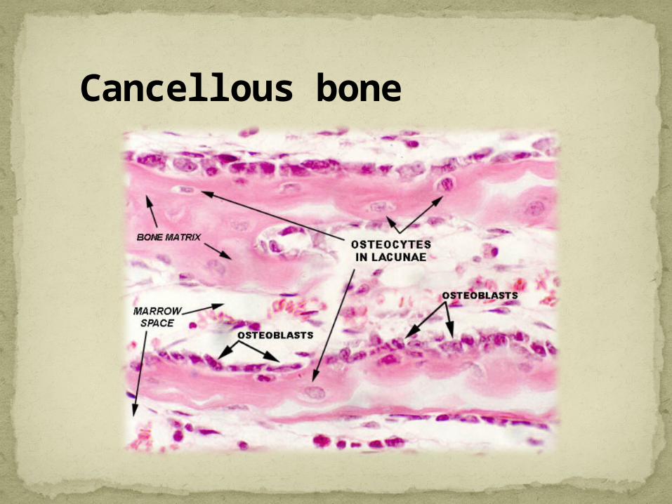
Cancellous bone

Woven or immature bone is the first formed bone with irregularly oriented collagen fibers of varying diameters.
usually not seen after birth However, it is seen in alveolar bone and
during healing of fractures.
Immature bone

Woven bone
1.Intercellular
bone matrix
2.Osteocytes
3.Periosteum
4.Osteoclast

mandible
maxilla
Alveolar process

alveolar process is defined as that part of mandible and maxilla that forms and supports the sockets of the teeth
They develop during the eruption of tooth and disappear after the tooth is extracted or lost
Introduction

Development The alveolar bone develops from the dental
follicle. From the 2nd month of fetal life, the maxilla and
mandible form a groove that open towards the surface of oral cavity.
The ectomesenchymal cells of dental follicle differentiate into osteoblasts lay down a matrix called osteoid.
As tooth germs start to develop, bony septa form gradually.
The alveolar process start deveoping strictly during tooth eruption

Bud Stage
dental papilla enamel organ
dental follicle
Developing alveolar process

Developing alveolar process
developing tooth
Meckel’s cartilage
bone of mandible

Structure
Alveolar
bone proper
•Bundle •Lamellar boneSuppor
ting alveolar bone
•Cortical plates•Spongy bone

The alveolar bone consists partly of lamellar bone and partly bundle bone and is about 0.1-0.4 mm thick
bundle bone
cementum
lamellated bone
dentin
haversian system
Bundles of PDL
Alveolar bone proper

The alveolar bone proper which forms the inner wall of socket is perforated and is known as cribriform plate.

interdental septum
Bone between the teeth is called interdental septum and interradicular septum.
interradicular
septum

interdental
septum
The interdental and interradicular canal contain the perforating canals of zuckerkandl and hirschfeld
Interradicular septum

The alveolar bone proper surrounds the root of tooth and gives attachment to principle fibers of PDL
The lamellar bone has many osteons each of which has a blood vessel in a haversian canal.
Bundle bone Bundle bone is that bone in which the principle
fibers of periodontal ligament are anchored. Radiographically it is referred to as lamina dura.
Lamellar bone

lamellated bone
dentin sharpey’s fibers
cementum bundle bone
PDL lamina dura
reversal line
bundle bone

Supporting alveolar bone The supporting alveolar bone consists of two
parts :- cortical plates spongy bone
cortical plates
spongy bone
Alveolar bone proper
PDL
tooth

Cortical plates consist of compact bone and form the outer and inner plates of the alveolar processes.
The cortical plates are thin in maxilla than in mandible.
They are thickest in premolar and molar region of lower jaw
cortical plates
Cortical plates

Spongy bone fills the area between the cortical plates and alveolar bone proper.
spongy bone
It is of 2 main types type 1:- the interdental and inter radicular
trabeculae are regular and horizontal type 2:- shows irregularly arranged,
numerous, delicate interdental and interradicular trabeculae.
Spongy bone

Bone re-modeling is an unique and important process that takes place in the bones.
It involves the bone resorption by the osteoclasts on one hand and the matrix deposition by osteoblasts on the other hand.
Periods of resorption alternate with periods of rest and repair.
Islands of bundle bone are separated from the lamellated bone by reversal lines
Internal reconstruction

Bone remodeling

Houses the roots of teeth
Anchors the roots of teeth to the alveoli
Helps to move the teeth for better occlusion
Helps to absorb and distribute occlusal forces generated during tooth contact
Supplies vessels to PDL
Houses and protects developing permanent teeth while supporting primary teeth
Organizes eruption of primary and permanent teeth
functions

Alveolar bones appear jagged and uneven
The marrow spaces have fatty infiltration
The alveolar process in edentulous jaws decreases in size
Loss of maxillary bone is accompanied by increase in size of maxillary sinus
Internal trabecular arrangement is more open, which indicates bone loss.
The distance between the crest of alveolar bone and CEJ increases with age (2.81 mm approx)
Age changes

THANK YOU