alsharqia echo club al qusaibi hotel al khobar may 23 rd 2013 eric mcwilliams mb, frcpi, frcp, facc...
TRANSCRIPT
ALSHARQIA ECHO CLUB Al Qusaibi HotelAl KhobarMay 23rd 2013
Eric McWilliams MB, FRCPI, FRCP, FACCConsultant Cardiologist Dhahran Health CenterDiplomate of the Certification Board of Cardiovascular Computed Tomography 2008 – 2018Honorary Clinical Senior Lecturer, Brighton and Sussex Medical School August 2009 – August 2014

History
75 yr lady dyspnoea on exertion
Type 2 Diabetes Hypertension Hypothyroidism Hyperlipidemia Glaucoma
No murmurs Chest clear

Echo
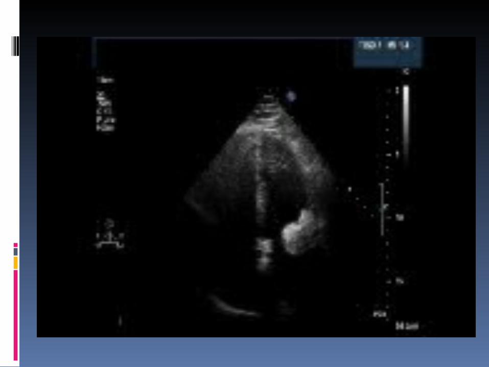

Echo
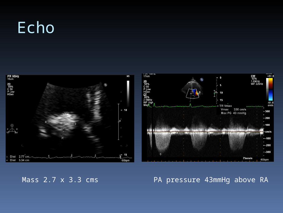
Mass 2.7 x 3.3 cms PA pressure 43mmHg above RA
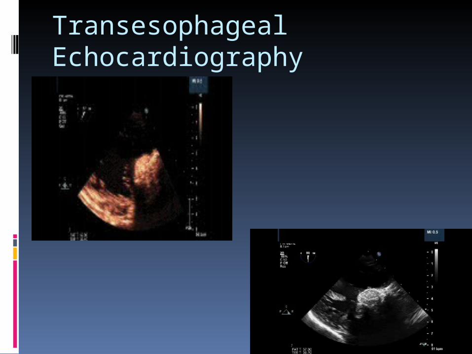
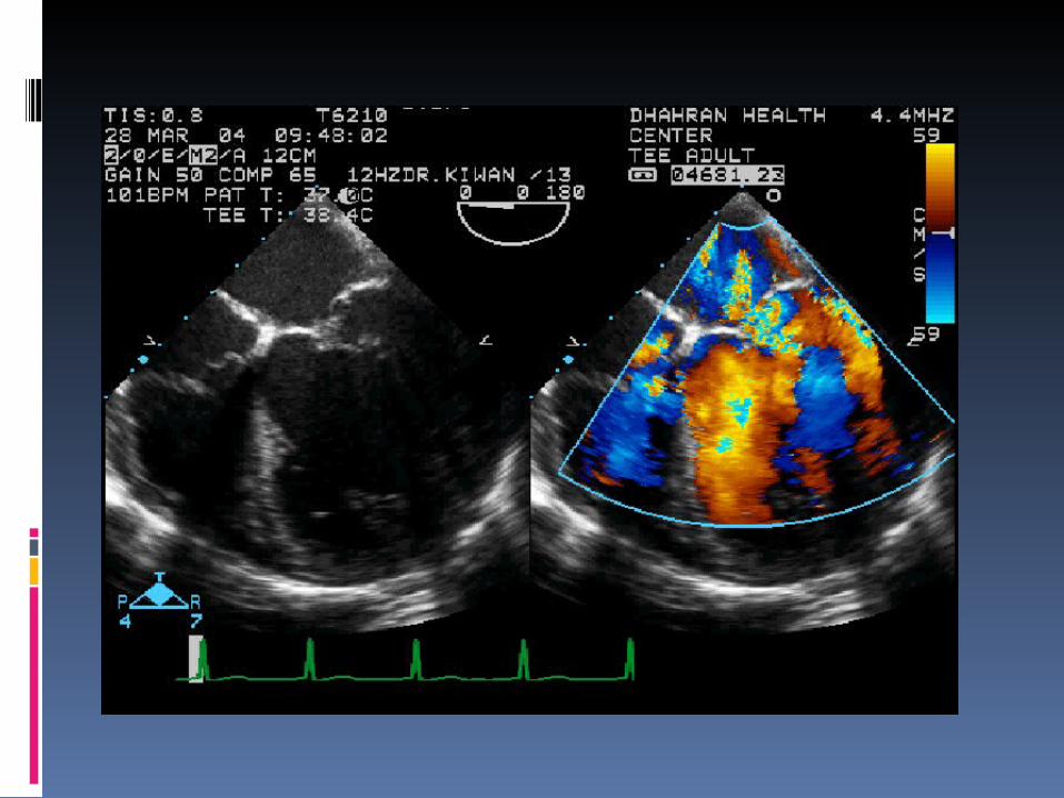
Transesophageal Echocardiography

3D RTE Left Atrial Surgeons View
Phillips IE 33
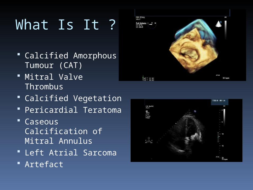
What Is It ?
Calcified Amorphous Tumour (CAT)
Mitral Valve Thrombus
Calcified Vegetation Pericardial Teratoma Caseous Calcification
of Mitral Annulus Left Atrial Sarcoma Artefact

CXR

Cardiac CT

CTA

CMR
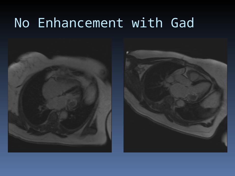
No Enhancement with Gad

CT CMR
What is it ?

What Is It ?
Calcified Amorphous Tumour (CAT)
Mitral Valve Thrombus
Calcified Vegetation Pericardial Teratoma Caseous Calcification
of Mitral Annulus Left Atrial Sarcoma Artefact
Mitral Annular Calcification Caseous Calcification
Degenerative abnormality 10% of >50yr olds commoner in women
Deposition of calcium between the basal infero-lateral ventricular wall and posterior mitral leaflet
Typically posterior mitral annulus
Soft periannular calcification : calcium, fatty acids and cholesterol
Toothpaste like material
Caseous calcification of the mitral annulus

Caseous Calcificationof Mitral Annulus Rare 0.6% of MAC patients
in echo series 2.7% necropsy series Usually incidental
finding Mitral stenosis can
occur Misdiagnosed as
abscess, cardiac tumour etc
MAC and Caseous calcification similar baseline characteristics:
Elderly, hypertension,coronary artery disease, aortic disease etc
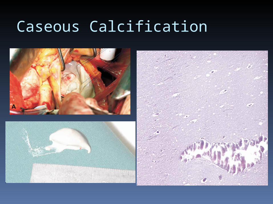
Caseous Calcification

CAT
Calcified amorphous tumor (CAT) was coined in 1997 by Reynolds et al,1 who described 11 cases with non neoplastic cardiac masses characterized by a pedicle and diffuse calcification

Evaluation of Intra- and Extra-Cardiac
Structures Cardiac CT CMR
Evaluation of cardiac mass (suspected tumor or thrombus) A (8)
Patients with technically limited images from echocardiogram, MRI, or TEE
Evaluation of pericardial conditions
Evaluation of cardiac mass (suspected tumor or thrombus) A (9)
Use of contrast for perfusion and enhancement
Evaluation of pericardial conditions (pericardial mass, constrictive pericarditis) A (8)
Appropriateness Criteria ACCF/ACR/SCCT/SCMR/ASNC/NASCI/SCAI/SIR

Ghada Al Dossary, BS, RDCS Senior Echo TechnologistEric McWilliams MB, FRCPI,FRCP,FACCDhahran Health Center
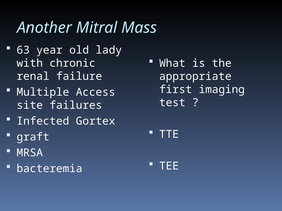
Another Mitral Mass 63 year old lady
with chronic renal failure
Multiple Access site failures
Infected Gortex graft MRSA bacteremia
What is the appropriate first imaging test ?
TTE
TEE
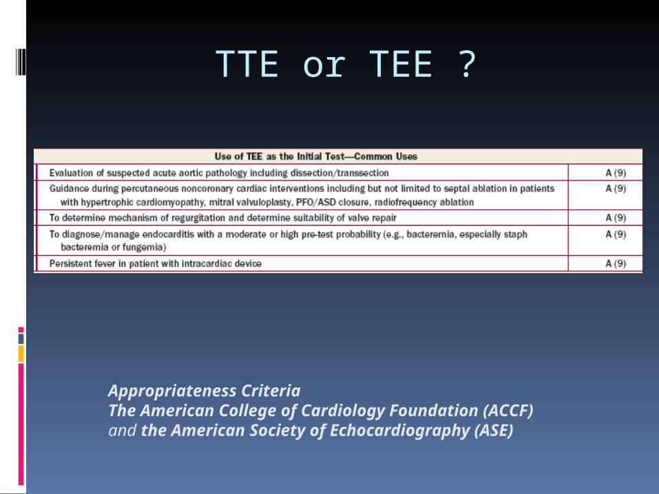
TTE or TEE ?
Appropriateness CriteriaThe American College of Cardiology Foundation (ACCF)and the American Society of Echocardiography (ASE)
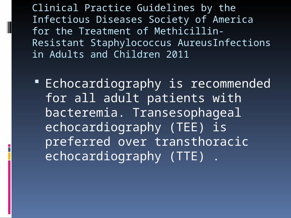
Clinical Practice Guidelines by the Infectious Diseases Society of America for the Treatment of Methicillin-Resistant Staphylococcus AureusInfections in Adults and Children 2011
Echocardiography is recommended for all adult patients with bacteremia. Transesophageal echocardiography (TEE) is preferred over transthoracic echocardiography (TTE) .

Another Mitral Mass 63 year old lady
with chronic renal failure
Multiple Access site failures
Infected Gortex graft MRSA bacteremia

TEE May 21st,2013

TEE May 21st,2013
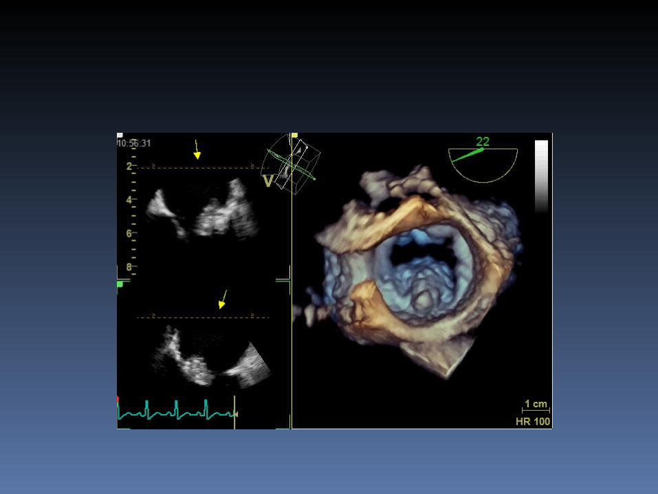

What is It ?
Caseous Calcification of Mitral Annulus
Mitral Cusp Prolapse Vegetation Papillary Fibroelastoma of Mitral
Valve Mitral Thrombus
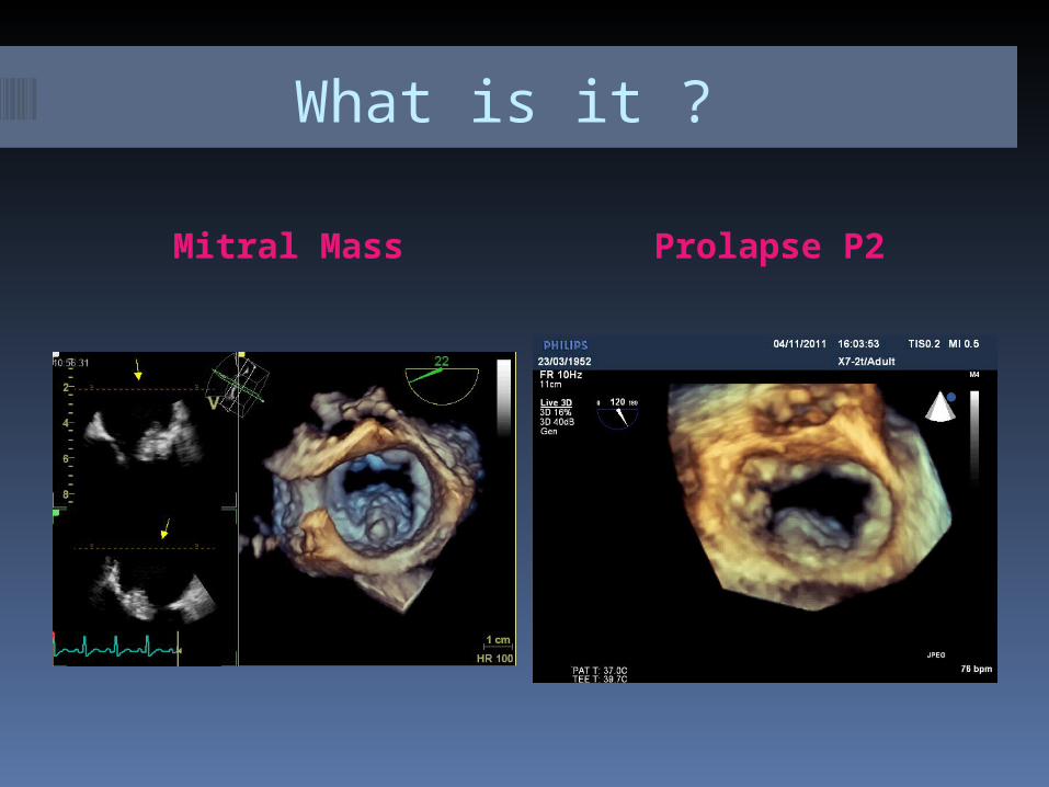
What is it ?
Mitral Mass Prolapse P2
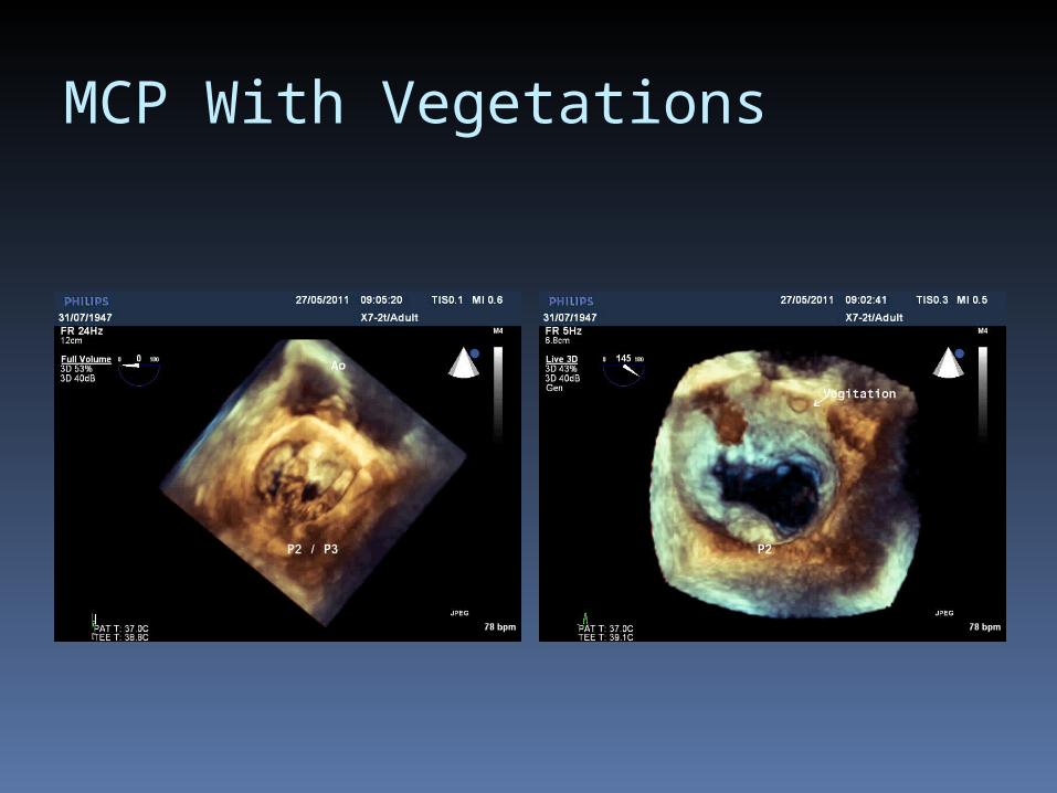
MCP With Vegetations

Mitral Papillary Fibroelastoma

Staph Aureus Bacteremia
13% of hospital acquired Staphylococcus aureus infection develop endocarditis
31% Staph aureus bacteremia : endocarditis
Echocardiography TEE

MRSA Endocarditis
The S. aureus organism carries particular adhesin molecules : can attack structurally Normal valves
The bacteria can either internalise and persist locally, protected from antibiotic therapy and host defences, or lyse the endothelial cells, causing local tissue destruction and distant emboli
S. aureus endocarditis can present either acutely or with a more indolent presentation
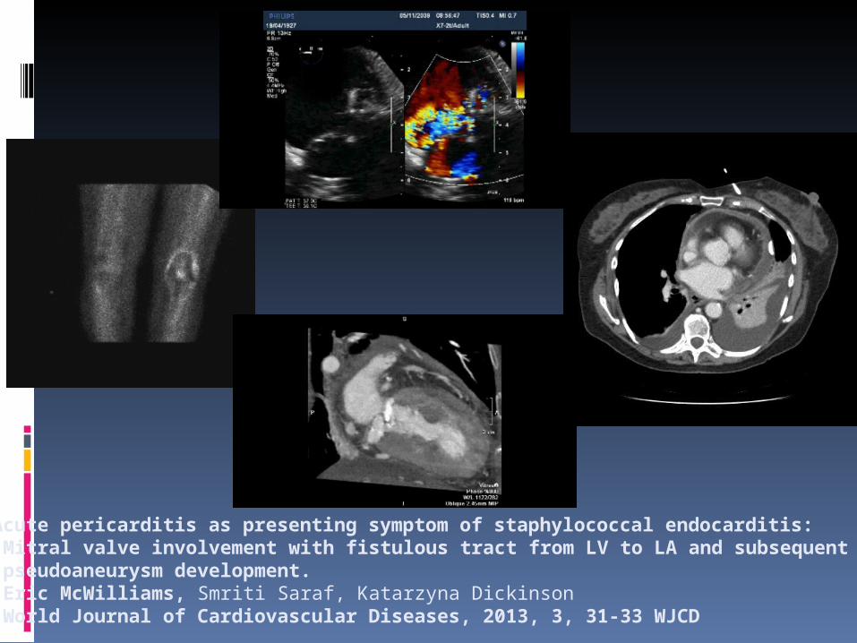
Acute pericarditis as presenting symptom of staphylococcal endocarditis: Mitral valve involvement with fistulous tract from LV to LA and subsequent pseudoaneurysm development. Eric McWilliams, Smriti Saraf, Katarzyna Dickinson World Journal of Cardiovascular Diseases, 2013, 3, 31-33 WJCD
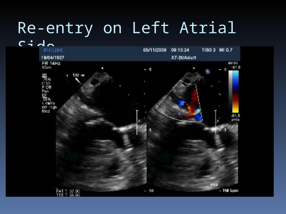
Re-entry on Left Atrial Side

MRSA Endocarditis


TTE versus TEE
The sensitivity for detecting vegetation with 2D TTE is 65 to 80 percent and 95 percent with TEE but depends on vegetation size, location, and the echocardiographic window
The yield of TTE is poor in patients with prosthetic valve endocarditis, but the sensitivity for TEE is 90 percent
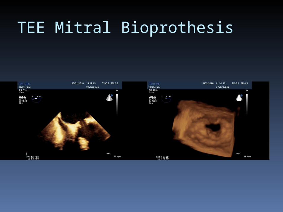
TEE Mitral Bioprothesis
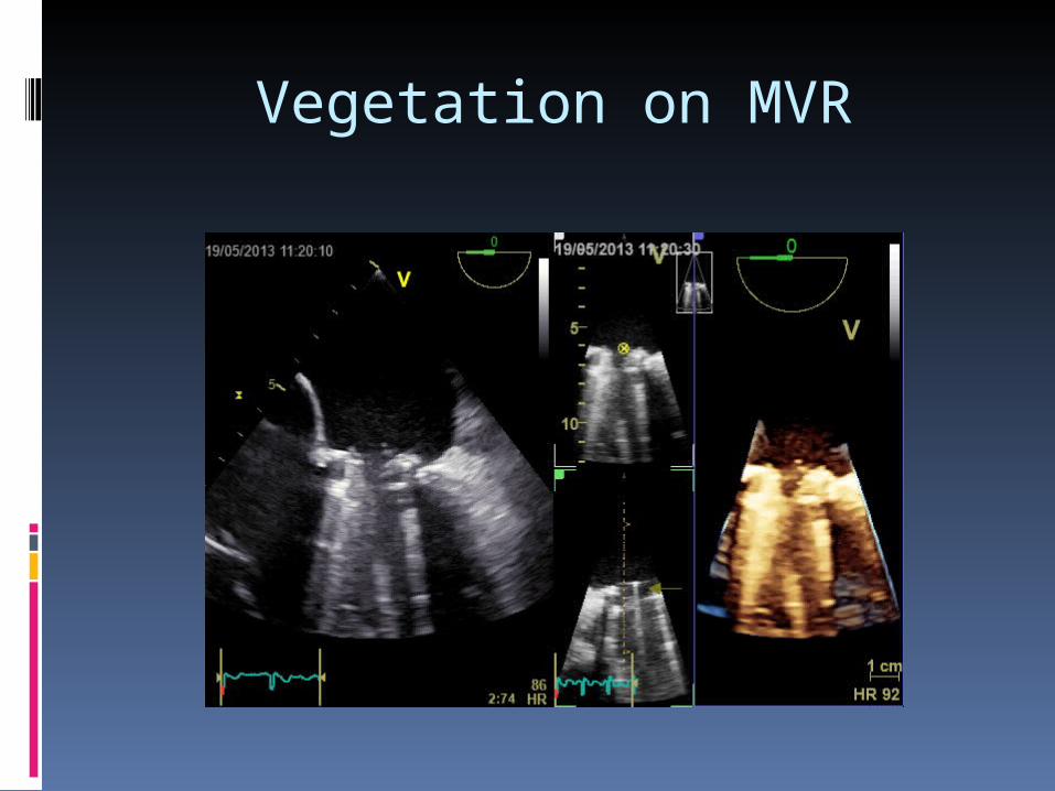
Vegetation on MVR