alport syndrome 4.14 - nyu langone health · 2016-07-12 · alport syndrome 4.14.15 sushma bhusal...
TRANSCRIPT
Alport Syndrome 4.14.15
Sushma Bhusal Nephrology Fellow
Case presentation
26 yo white Ukrainian male with no known PMH, sent to ED by PCP for – Cr 12 with severe electrolytes abnormalities – Pancytopenia – HTN
HPI
c/o fatigue , malaise and leg/hand cramping , post-prandial vomiting and pallor for months
SOB with exertion over the past weeks, associated with LE swelling
Easy bruising , several episodes of epistaxis
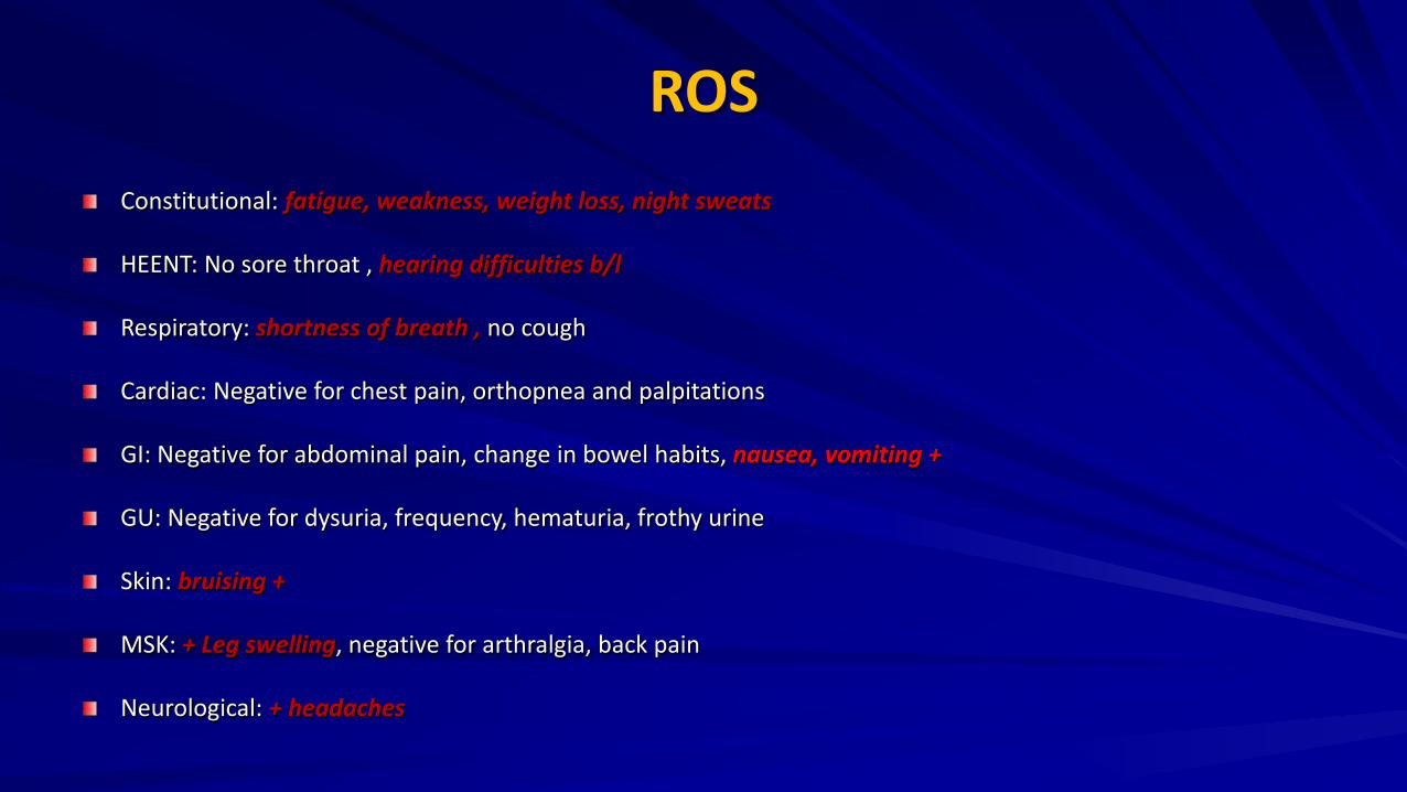
ROS Constitutional: fatigue, weakness, weight loss, night sweats
HEENT: No sore throat , hearing difficulties b/l Respiratory: shortness of breath , no cough Cardiac: Negative for chest pain, orthopnea and palpitations GI: Negative for abdominal pain, change in bowel habits, nausea, vomiting + GU: Negative for dysuria, frequency, hematuria, frothy urine Skin: bruising + MSK: + Leg swelling, negative for arthralgia, back pain Neurological: + headaches
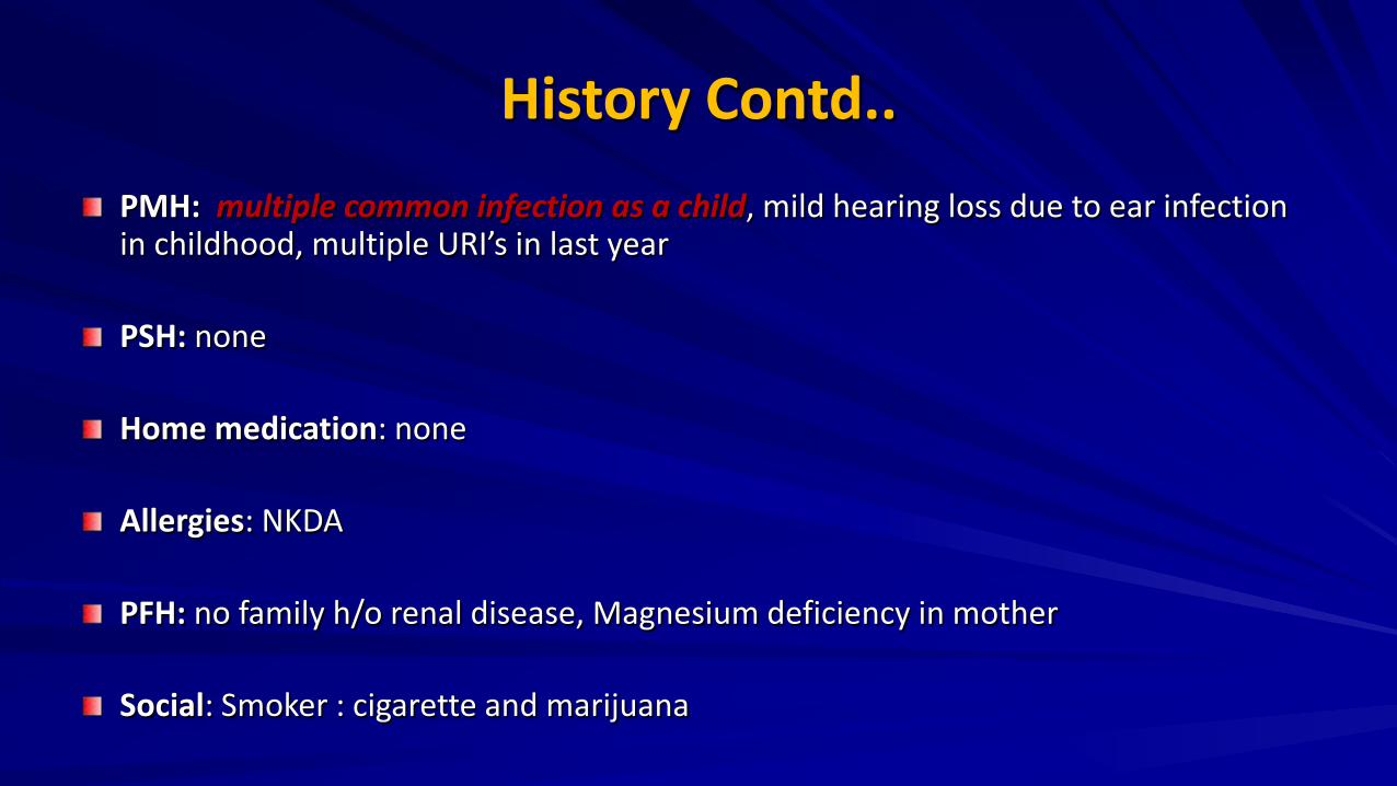
History Contd.. PMH: multiple common infection as a child, mild hearing loss due to ear infection in childhood, multiple URI’s in last year
PSH: none
Home medication: none
Allergies: NKDA PFH: no family h/o renal disease, Magnesium deficiency in mother Social: Smoker : cigarette and marijuana
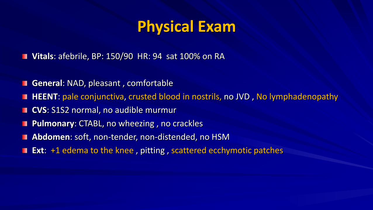
Physical Exam
Vitals: afebrile, BP: 150/90 HR: 94 sat 100% on RA General: NAD, pleasant , comfortable HEENT: pale conjunctiva, crusted blood in nostrils, no JVD , No lymphadenopathy CVS: S1S2 normal, no audible murmur Pulmonary: CTABL, no wheezing , no crackles Abdomen: soft, non-tender, non-distended, no HSM Ext: +1 edema to the knee , pitting , scattered ecchymotic patches
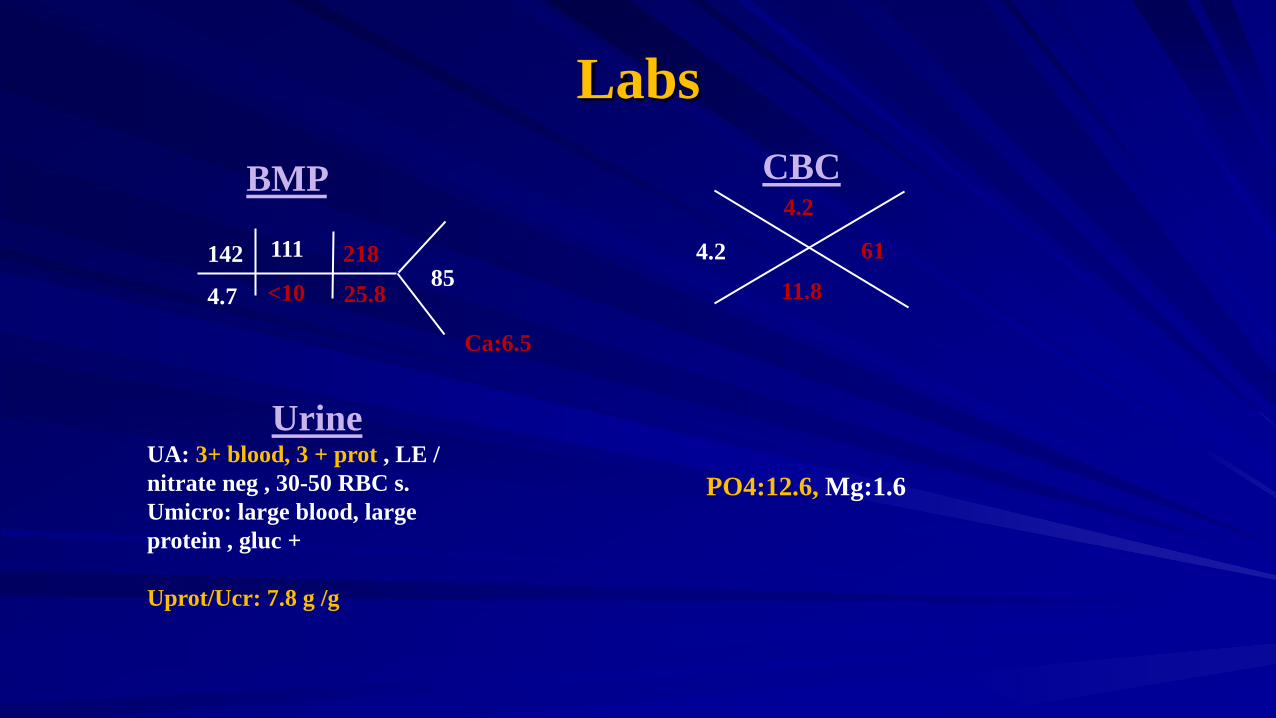
Labs CBC
4.2 11.8
4.2
61
BMP
UA: 3+ blood, 3 + prot , LE / nitrate neg , 30-50 RBC s. Umicro: large blood, large protein , gluc + Uprot/Ucr: 7.8 g /g
Urine
<10 4.7
142
25.8
218 85
111
PO4:12.6, Mg:1.6
Ca:6.5
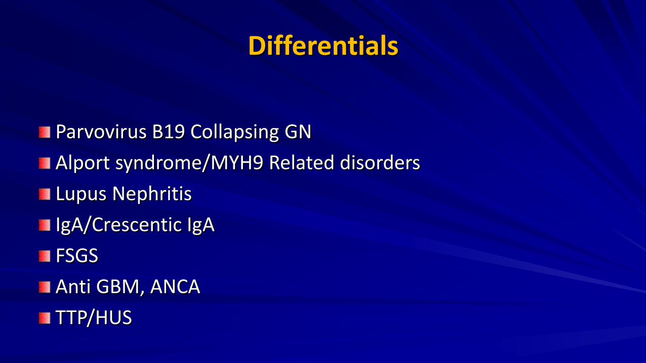
Differentials
Parvovirus B19 Collapsing GN Alport syndrome/MYH9 Related disorders Lupus Nephritis IgA/Crescentic IgA FSGS Anti GBM, ANCA TTP/HUS

Additional Labs PTH: 1500 LFTs; WNL Alb: 3.4 Chol : 173 Uric acid: 9.4 Lactic acid: 0.6 Retic: 2.88 ESR: 80 CRP: 3.57 Hep B, Hep c serolgoy neg C3 C4 WNL, ASO neg , P –ANCA, C-ANCA neg UPEP/SPEP neg, Parvovirus: + Ig G
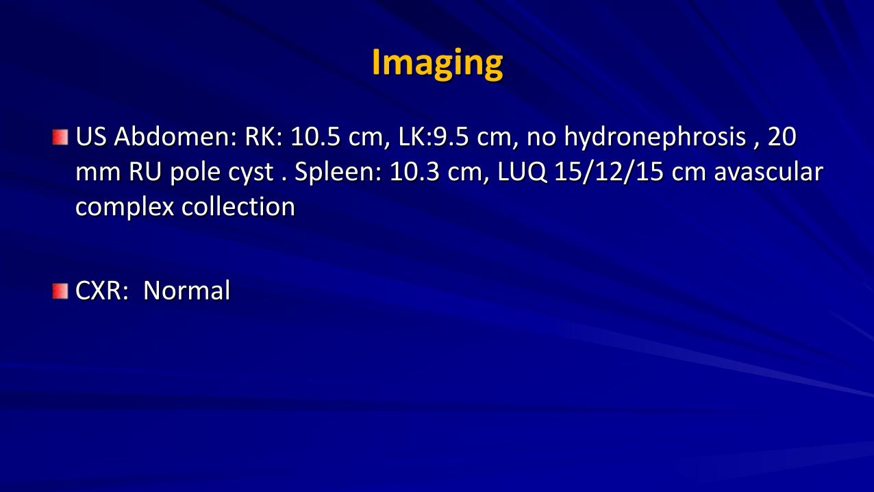
Imaging
US Abdomen: RK: 10.5 cm, LK:9.5 cm, no hydronephrosis , 20 mm RU pole cyst . Spleen: 10.3 cm, LUQ 15/12/15 cm avascular complex collection CXR: Normal

Renal Biopsy
MAB 3
POSITIVE CONTROL PATIENT
MAB 5
POSITIVE CONTROL PATIENT
EM
Hospital Course
Dialyzed
Received blood and platelet transfusions Hematology involved for pancytopenia
ALPORT SYNDROME

Outline
History Epidemiology Pathophysiology Inheritance Pattern Clinical Features Diagnosis Treatment Prognosis
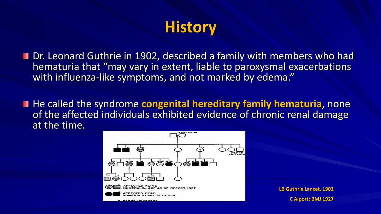
History Dr. Leonard Guthrie in 1902, described a family with members who had hematuria that “may vary in extent, liable to paroxysmal exacerbations with influenza-like symptoms, and not marked by edema.”
He called the syndrome congenital hereditary family hematuria, none of the affected individuals exhibited evidence of chronic renal damage at the time.
LB Guthrie Lancet, 1902
C Alport: BMJ 1927
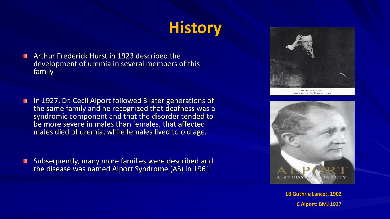
History Arthur Frederick Hurst in 1923 described the development of uremia in several members of this family
In 1927, Dr. Cecil Alport followed 3 later generations of the same family and he recognized that deafness was a syndromic component and that the disorder tended to be more severe in males than females, that affected males died of uremia, while females lived to old age.
Subsequently, many more families were described and the disease was named Alport Syndrome (AS) in 1961.
LB Guthrie Lancet, 1902
C Alport: BMJ 1927
Epidemiology Overall incidence in the general population is unknown Accounts for 3% of children and 0.2 % of adults with ESRD, 0.6 % in Europe Incidence varies from 1 to 11% depending upon the indications for biopsy The gene frequency of AS in the US estimated at 1:5000 to 10,000, approx. 30,000 to 60,000 affected individuals in the US
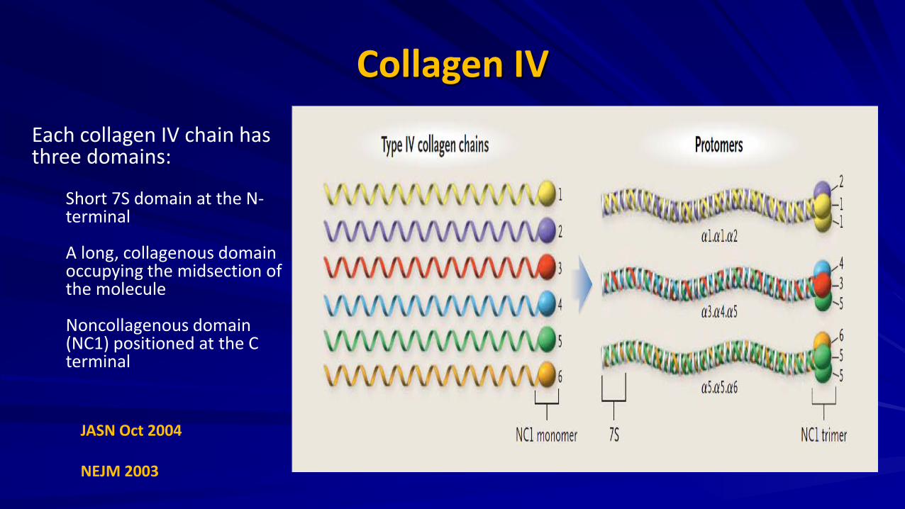
Collagen IV
JASN Oct 2004 NEJM 2003
Each collagen IV chain has three domains:
Short 7S domain at the N-terminal A long, collagenous domain occupying the midsection of the molecule Noncollagenous domain (NC1) positioned at the C terminal
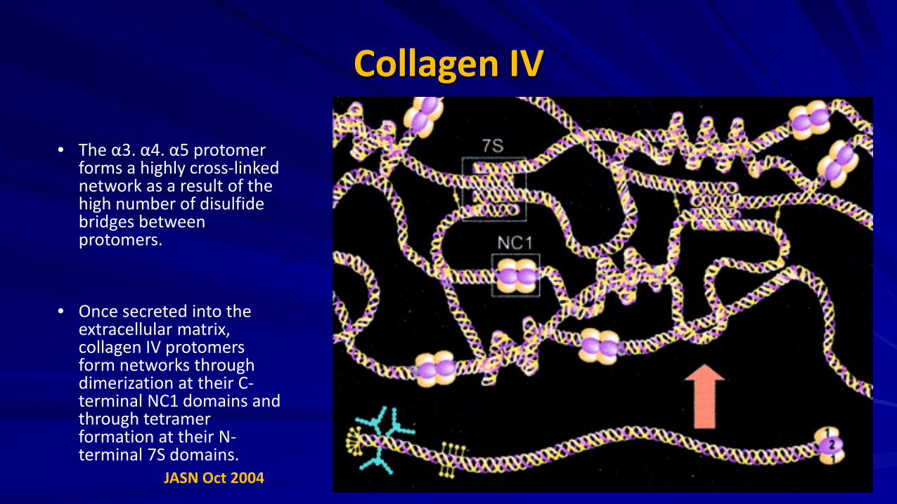
Collagen IV • The α3. α4. α5 protomer
forms a highly cross-linked network as a result of the high number of disulfide bridges between protomers.
• Once secreted into the extracellular matrix, collagen IV protomers form networks through dimerization at their C-terminal NC1 domains and through tetramer formation at their N-terminal 7S domains.
JASN Oct 2004
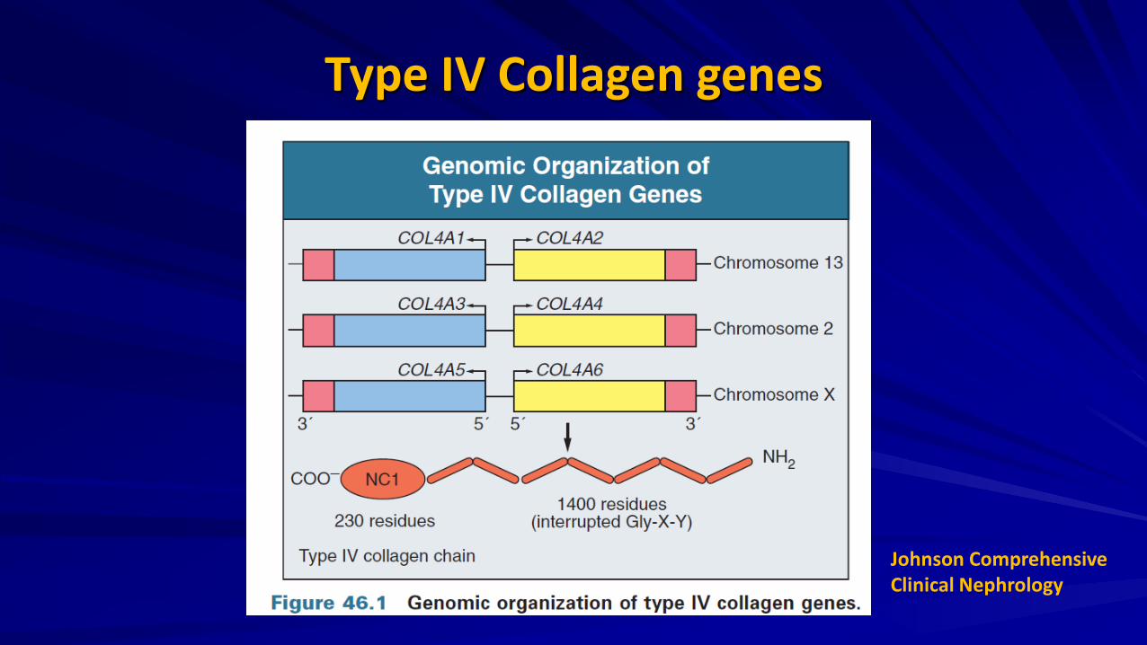
Type IV Collagen genes
Johnson Comprehensive Clinical Nephrology

Pathogenesis
JASN Oct 2004
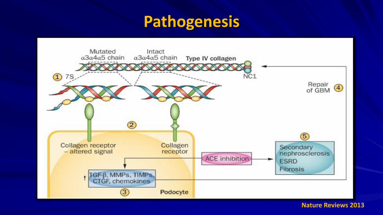
Pathogenesis
Nature Reviews 2013
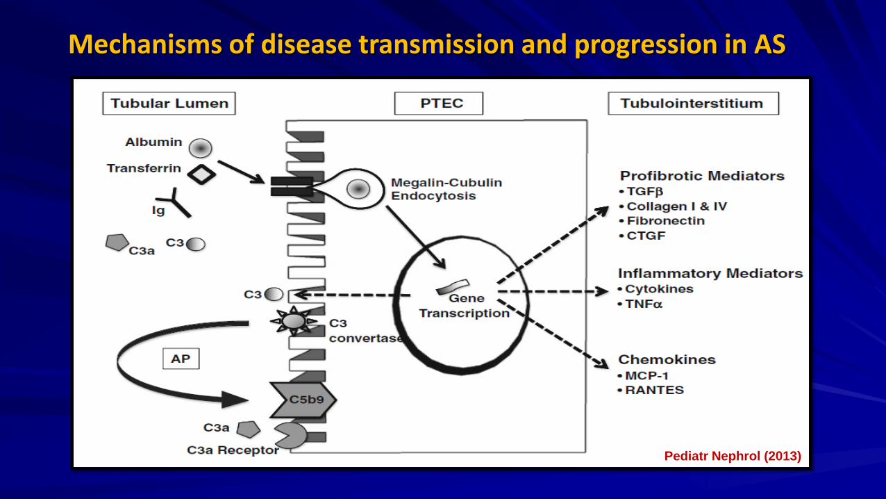
Mechanisms of disease transmission and progression in AS
Pediatr Nephrol (2013)
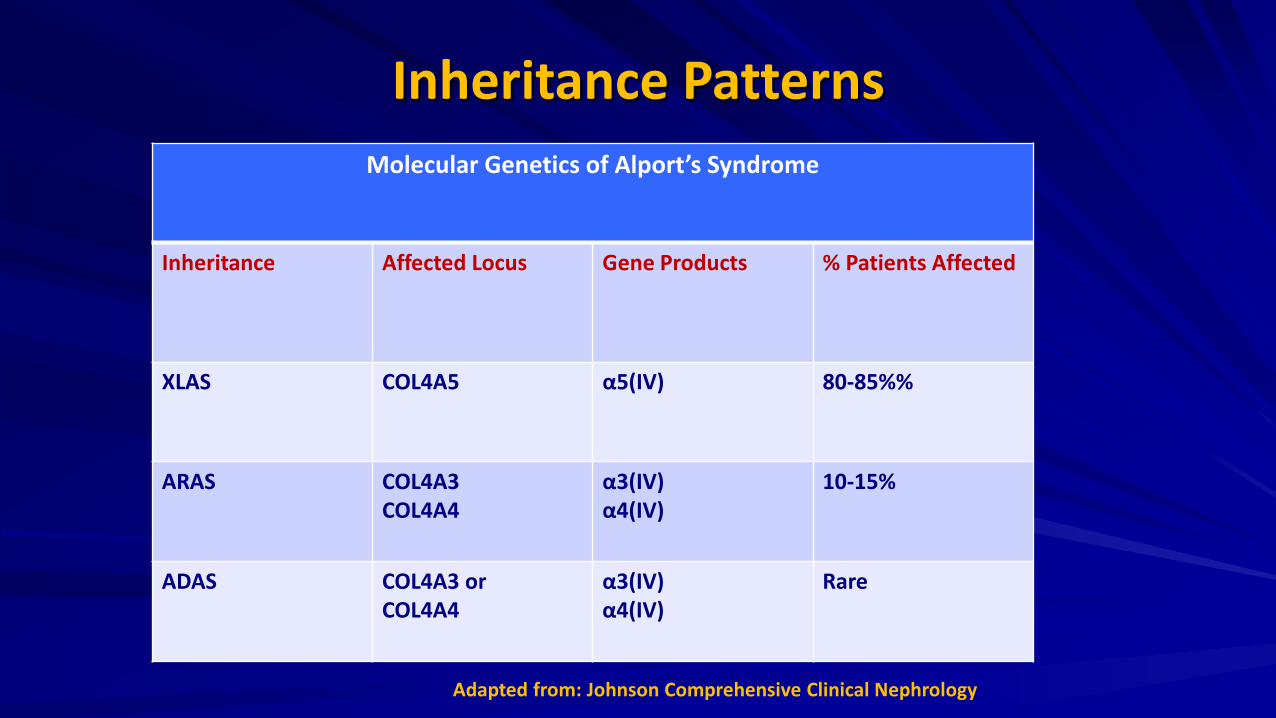
Inheritance Patterns Molecular Genetics of Alport’s Syndrome
Inheritance Affected Locus Gene Products % Patients Affected
XLAS COL4A5 α5(IV) 80-85%%
ARAS COL4A3 COL4A4
α3(IV) α4(IV)
10-15%
ADAS COL4A3 or COL4A4
α3(IV) α4(IV)
Rare
Adapted from: Johnson Comprehensive Clinical Nephrology
X Linked Mutations In the COL4A5 genes from the families with XLAS, more than 300 gene mutations have been reported.
Most COL4A5 mutations are small and include missense mutations, splice-site mutations, and small deletions where renal failure and deafness occur after 30 years of age (adult form).
Approximately 20% of the mutations are major rearrangements at the COL4A5 locus (i.e., large deletions, reading frame shifts, etc) in which patients are symptomatic before the age of 30 (juvenile form).
A rare large deletion spanning COL4A5 and COL4A6 genes is associated with a combination of XLAS and diffuse leiomyomatosis. Johnson Comp Clinical Nephrology
Nature Reviews 2013
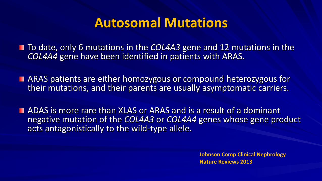
Autosomal Mutations To date, only 6 mutations in the COL4A3 gene and 12 mutations in the COL4A4 gene have been identified in patients with ARAS.
ARAS patients are either homozygous or compound heterozygous for their mutations, and their parents are usually asymptomatic carriers.
ADAS is more rare than XLAS or ARAS and is a result of a dominant negative mutation of the COL4A3 or COL4A4 genes whose gene product acts antagonistically to the wild-type allele.
Johnson Comp Clinical Nephrology Nature Reviews 2013
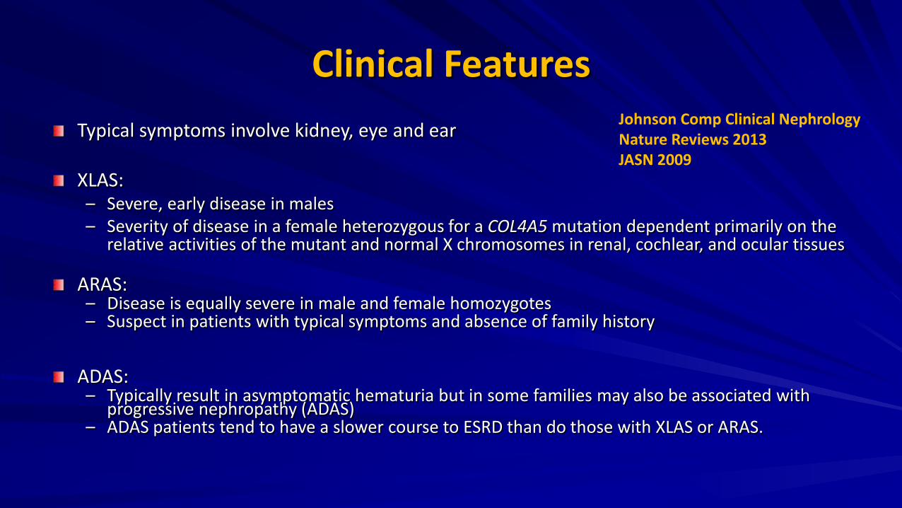
Clinical Features Typical symptoms involve kidney, eye and ear
XLAS: – Severe, early disease in males – Severity of disease in a female heterozygous for a COL4A5 mutation dependent primarily on the
relative activities of the mutant and normal X chromosomes in renal, cochlear, and ocular tissues
ARAS: – Disease is equally severe in male and female homozygotes – Suspect in patients with typical symptoms and absence of family history
ADAS: – Typically result in asymptomatic hematuria but in some families may also be associated with
progressive nephropathy (ADAS) – ADAS patients tend to have a slower course to ESRD than do those with XLAS or ARAS.
Johnson Comp Clinical Nephrology Nature Reviews 2013 JASN 2009
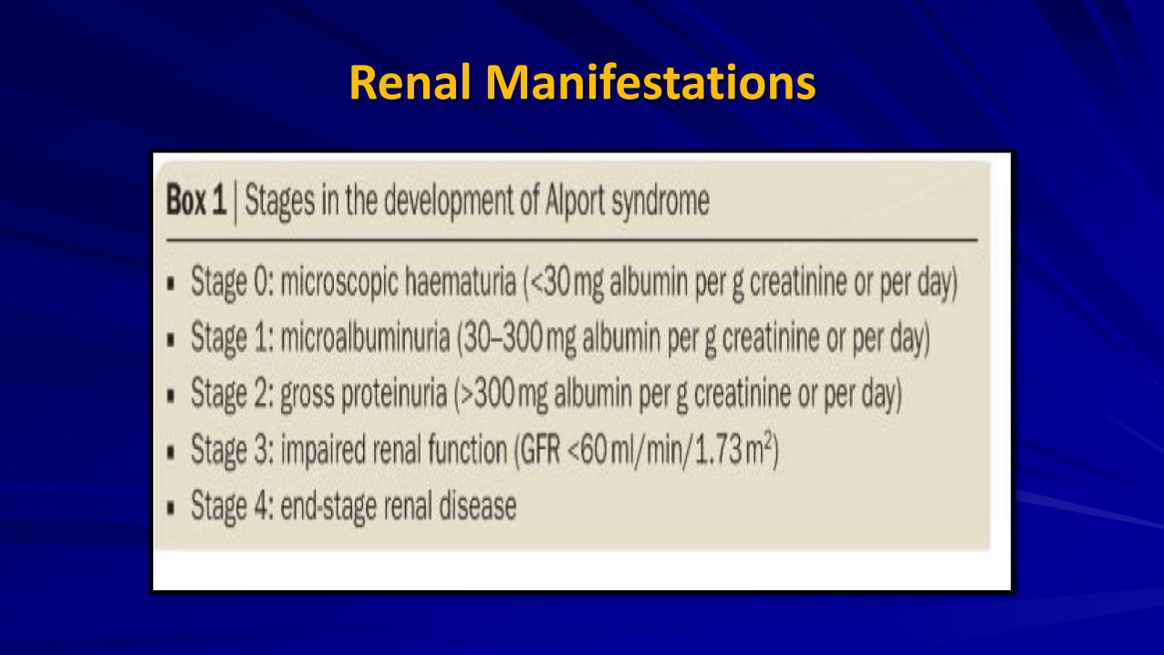
Renal Manifestations
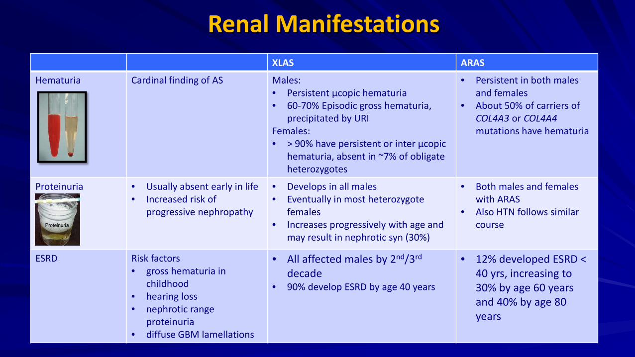
Renal Manifestations XLAS ARAS
Hematuria
Cardinal finding of AS
Males: • Persistent μcopic hematuria • 60-70% Episodic gross hematuria,
precipitated by URI Females: • > 90% have persistent or inter μcopic
hematuria, absent in ~7% of obligate heterozygotes
• Persistent in both males and females
• About 50% of carriers of COL4A3 or COL4A4 mutations have hematuria
Proteinuria • Usually absent early in life • Increased risk of
progressive nephropathy
• Develops in all males • Eventually in most heterozygote
females • Increases progressively with age and
may result in nephrotic syn (30%)
• Both males and females with ARAS
• Also HTN follows similar course
ESRD Risk factors • gross hematuria in
childhood • hearing loss • nephrotic range
proteinuria • diffuse GBM lamellations
• All affected males by 2nd/3rd decade
• 90% develop ESRD by age 40 years
• 12% developed ESRD < 40 yrs, increasing to 30% by age 60 years and 40% by age 80 years
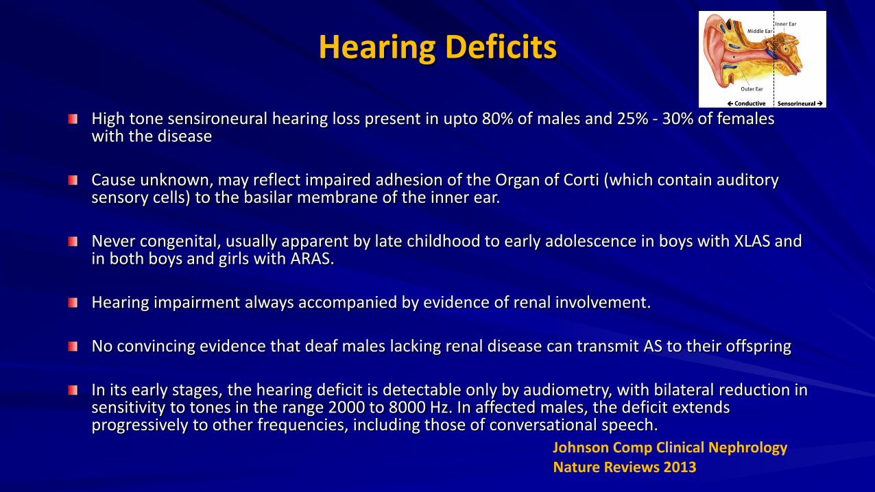
Hearing Deficits
High tone sensironeural hearing loss present in upto 80% of males and 25% - 30% of females with the disease Cause unknown, may reflect impaired adhesion of the Organ of Corti (which contain auditory sensory cells) to the basilar membrane of the inner ear. Never congenital, usually apparent by late childhood to early adolescence in boys with XLAS and in both boys and girls with ARAS. Hearing impairment always accompanied by evidence of renal involvement. No convincing evidence that deaf males lacking renal disease can transmit AS to their offspring In its early stages, the hearing deficit is detectable only by audiometry, with bilateral reduction in sensitivity to tones in the range 2000 to 8000 Hz. In affected males, the deficit extends progressively to other frequencies, including those of conversational speech.
Johnson Comp Clinical Nephrology Nature Reviews 2013
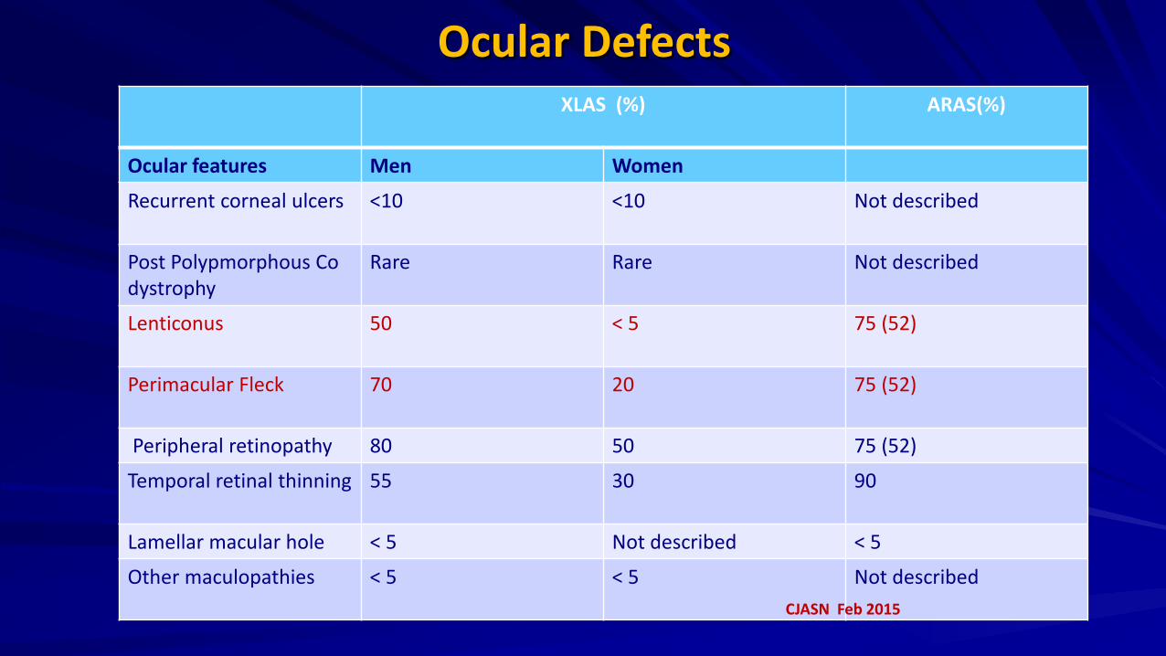
Ocular Defects XLAS (%)
ARAS(%)
Ocular features Men Women
Recurrent corneal ulcers <10 <10 Not described
Post Polypmorphous Co dystrophy
Rare Rare Not described
Lenticonus 50 < 5 75 (52)
Perimacular Fleck 70 20 75 (52)
Peripheral retinopathy 80 50 75 (52)
Temporal retinal thinning
55 30 90
Lamellar macular hole < 5 Not described < 5
Other maculopathies < 5 < 5 Not described CJASN Feb 2015
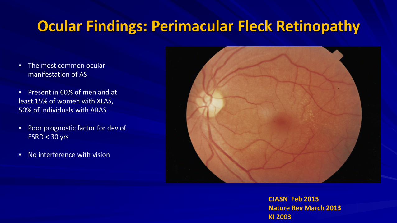
Ocular Findings: Perimacular Fleck Retinopathy
• The most common ocular manifestation of AS
• Present in 60% of men and at least 15% of women with XLAS, 50% of individuals with ARAS • Poor prognostic factor for dev of
ESRD < 30 yrs
• No interference with vision
CJASN Feb 2015 Nature Rev March 2013 KI 2003
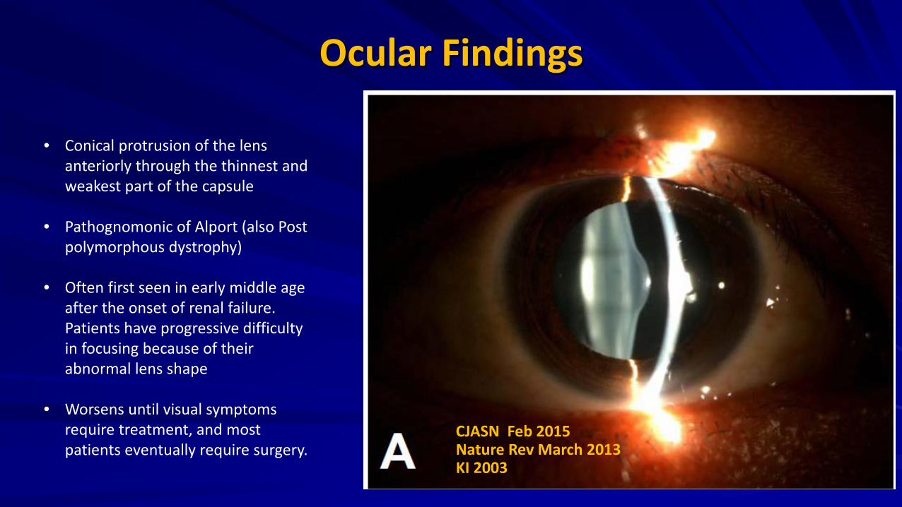
Ocular Findings
• Conical protrusion of the lens anteriorly through the thinnest and weakest part of the capsule
• Pathognomonic of Alport (also Post polymorphous dystrophy)
• Often first seen in early middle age after the onset of renal failure. Patients have progressive difficulty in focusing because of their abnormal lens shape
• Worsens until visual symptoms require treatment, and most patients eventually require surgery.
CJASN Feb 2015 Nature Rev March 2013 KI 2003
Leiomyomatosis Characterized by benign nodular tumors consisting of smooth muscle cells present in some families with Alport Can affect GI, trachea, bronchial tre and genital tract Genetic defect usually a large deletion in the COL4A5 gene, including COL4A6 gene, normally expressed in sm muscle cells of GE tract and encodes collagen α5 or α5α6 chains Symptoms usually appear in late childhood and include dysphagia, postprandial vomiting, substernal or epigastric pain, recurrent bronchitis, dyspnea, cough, and stridor Nature Rev March 2013
Diagnosis Family history and typical symptoms UA, Slit lamp exam (presence of anterior lenticonus and posterior polymorphous corneal dystrophy pathognomonic) and audiometry for hearing loss Molecular genetic testing (gold std) in presymptomatic children Tissue biopsy: skin/kidney biopsy
Nature Rev March 2013
Skin Biopsy
Absence of α5(IV) chains in the epidermal basement membrane on skin biopsy is diagnostic of XLAS (observed in only 80% of males with XLAS)
α3(IV) and α4(IV) chains are normally absent in the epidermal basement membrane, cannot diagnose ARAS and ADAS.
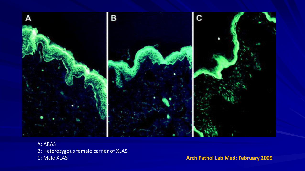
A: ARAS B: Heterozygous female carrier of XLAS C: Male XLAS Arch Pathol Lab Med: February 2009

Renal Biopsy : LM
A: Podocyte hypertrophy on an early biopsy specimen B: Rigidity and moderate thickening of GBM and segmental glomerular sclerosis C: Irregular thickening of the GBM
JASN 2009
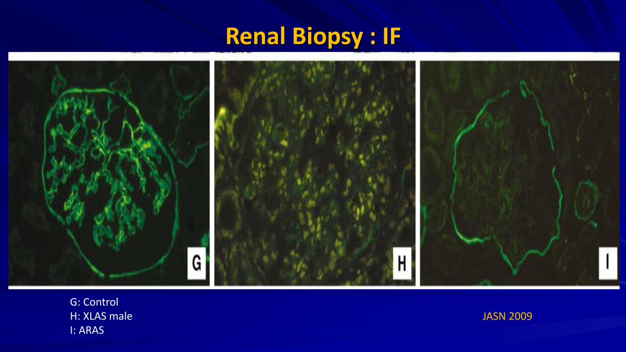
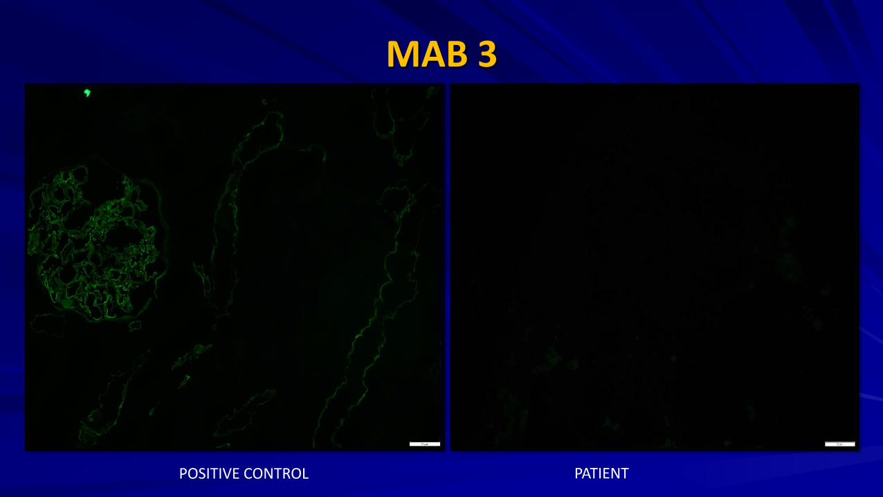
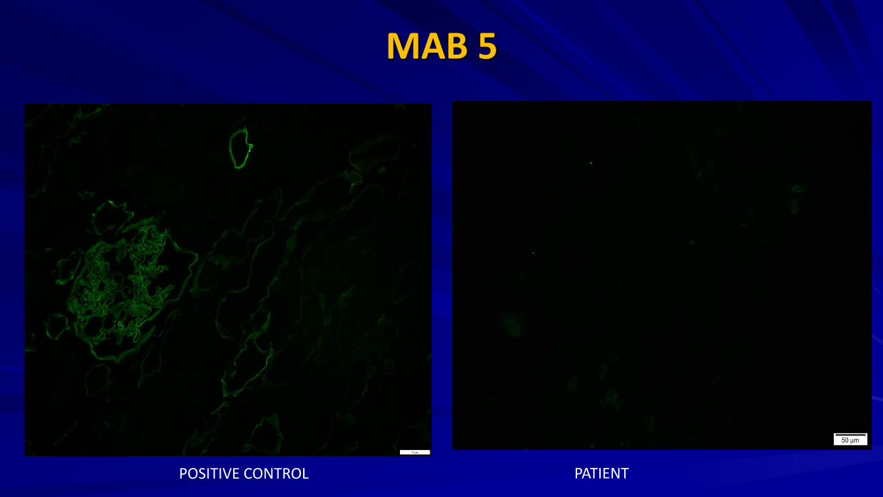
Renal Biopsy : IF
JASN 2009 G: Control H: XLAS male I: ARAS
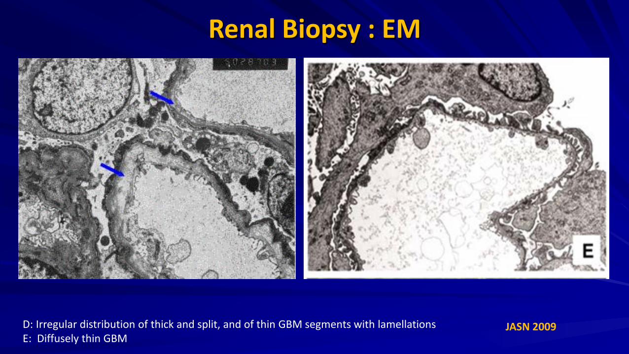
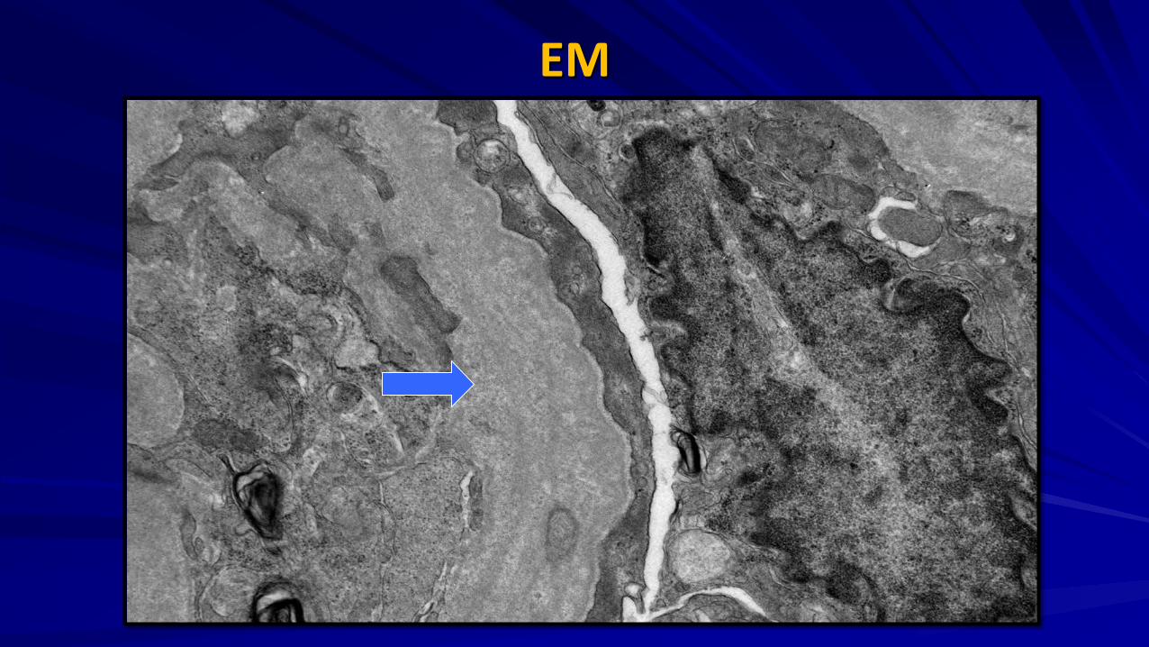
Renal Biopsy : EM
JASN 2009 D: Irregular distribution of thick and split, and of thin GBM segments with lamellations E: Diffusely thin GBM
Treatment
Treatments targeting proteinuria – RAAS Blockade – Calcineurin Inhibitors
Treatments targeting inflammation and fibrosis – Complement inhibition – Chemokine receptor antagonists – Bone morphogenetic protein-7 – Matrix metalloproteinase inhibitors
Treatments targeting the GBM pathology of Alport syndrome – Discoidin domain receptor 1 and integrin α2β1 antagonism
Bone marrow transplant/stem cell therapy Renal transplant Pediatr Nephrol (2013)
NDT 2014
NDT 2014
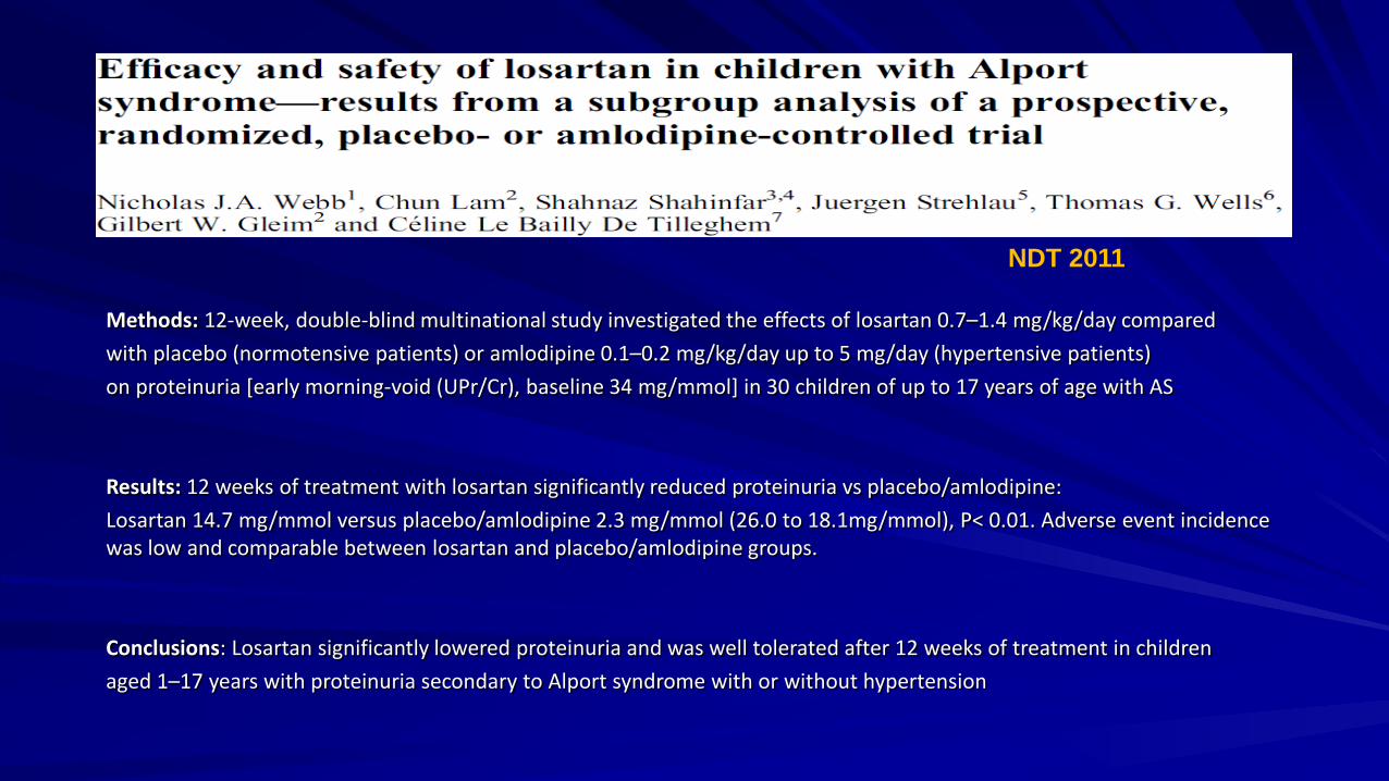
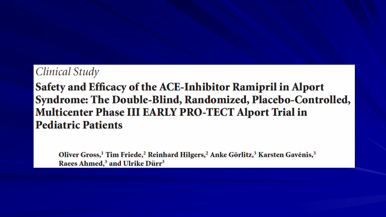
Methods: 12-week, double-blind multinational study investigated the effects of losartan 0.7–1.4 mg/kg/day compared with placebo (normotensive patients) or amlodipine 0.1–0.2 mg/kg/day up to 5 mg/day (hypertensive patients) on proteinuria [early morning-void (UPr/Cr), baseline 34 mg/mmol] in 30 children of up to 17 years of age with AS Results: 12 weeks of treatment with losartan significantly reduced proteinuria vs placebo/amlodipine: Losartan 14.7 mg/mmol versus placebo/amlodipine 2.3 mg/mmol (26.0 to 18.1mg/mmol), P< 0.01. Adverse event incidence was low and comparable between losartan and placebo/amlodipine groups. Conclusions: Losartan significantly lowered proteinuria and was well tolerated after 12 weeks of treatment in children aged 1–17 years with proteinuria secondary to Alport syndrome with or without hypertension
NDT 2011
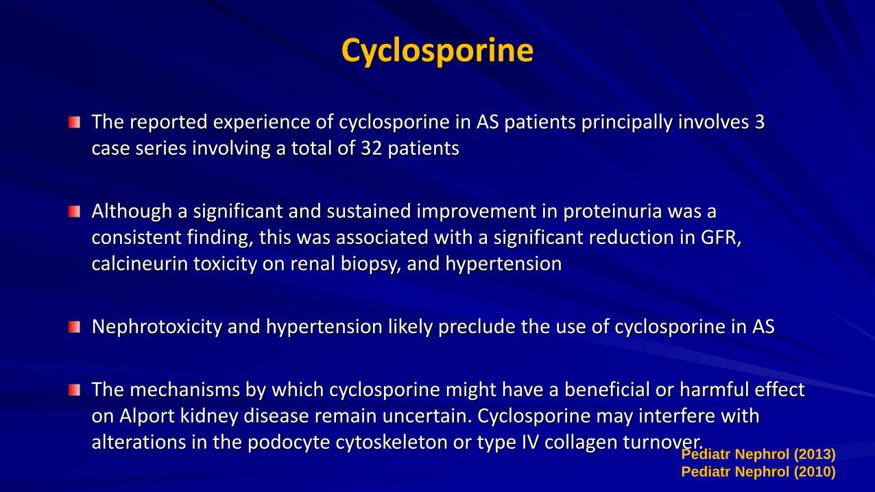
Cyclosporine
The reported experience of cyclosporine in AS patients principally involves 3 case series involving a total of 32 patients Although a significant and sustained improvement in proteinuria was a consistent finding, this was associated with a significant reduction in GFR, calcineurin toxicity on renal biopsy, and hypertension Nephrotoxicity and hypertension likely preclude the use of cyclosporine in AS The mechanisms by which cyclosporine might have a beneficial or harmful effect on Alport kidney disease remain uncertain. Cyclosporine may interfere with alterations in the podocyte cytoskeleton or type IV collagen turnover.
Pediatr Nephrol (2013) Pediatr Nephrol (2010)
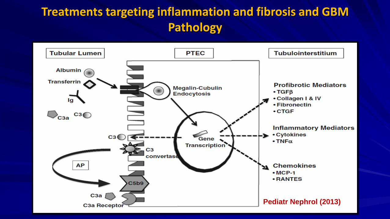
Treatments targeting inflammation and fibrosis and GBM Pathology
Pediatr Nephrol (2013)

Stem Cell Based Therapies
Could potentially be “Holy Grail” in the treatment of AS Several groups have claimed podocyte differentiation from stem cells in Alport mice, these publications still need validation and do not convincingly demonstrate that podocyte differentiation and consequent restoration of the GBM really occurs Differentiation of injected stem cells (amniotic fluid stem cells) into podocytes does not occur in vivo in an animal model of AS, despite significant protection of glomerular structure and function
Gross et al. NDT 2014
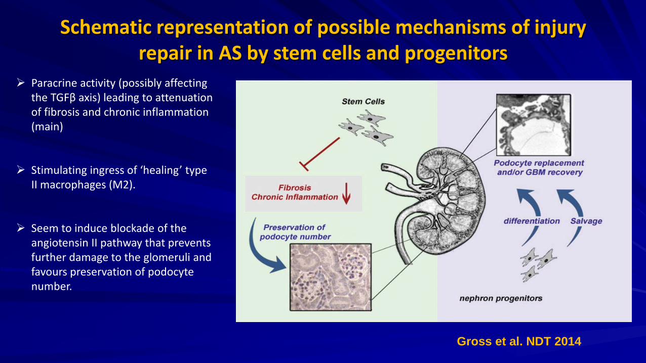
Schematic representation of possible mechanisms of injury repair in AS by stem cells and progenitors
Paracrine activity (possibly affecting the TGFβ axis) leading to attenuation of fibrosis and chronic inflammation (main)
Stimulating ingress of ‘healing’ type II macrophages (M2).
Seem to induce blockade of the angiotensin II pathway that prevents further damage to the glomeruli and favours preservation of podocyte number.
Gross et al. NDT 2014
Renal Transplant AS is essentially cured with renal transplantation The most significant and devastating, albeit rare, complication of transplantation is antiglomerular basement membrane nephritis.
Approximately 3-5% of patients with Alport syndrome who receive a transplant develop anti-GBM antibody to the NC1 component of the α3(IV) chain.
Post-transplant anti-GBM nephritis usually develops within the first year of the transplant.
Kashtan CE. J Am Soc Nephrol. 1998;9:1736.
Renal Transplant Low risk: normal hearing, late progression to ESRD, or females with XLAS.
Unlike de novo anti-GBM nephritis, pulmonary hemorrhage is never observed because the patient's lung tissue does not contain the antigen.
Treatment with plasmapheresis and cyclophosphamide is usually unsuccessful, and most patients lose the allograft.
Re-transplantation in most patients results in recurrence of anti-GBM nephritis despite the absence of detectable circulating anti-GBM antibodies before transplantation.
Kashtan CE. J Am Soc Nephrol. 1998;9:1736.

Goodpasture Autoantibodies and Alport Alloantibodies
NEJM 2003

Thank You