aligned electrospun polymer fibres for...
TRANSCRIPT

193 www.ecmjournal.org
KJ Aviss et al. Polymer fibres for skeletal muscle regeneration.European Cells and Materials Vol. 19 2010 (pages 193-204) DOI: 10.22203/eCM.v019a19 ISSN 1473-2262
Abstract
Skeletal muscle repair is often overlooked in surgicalprocedures and in serious burn victims. Creating a tissue-engineered skeletal muscle would not only provide agrafting material for these clinical situations, but could alsobe used as a valuable true-to-life research tool into diseasesaffecting muscle tissue. Electrospinning of the elastomerPLGA produced aligned fibres that had the correct topologyto provide contact guidance for myoblast elongation andalignment. In addition, the electrospun scaffold requiredno surface modifications or incorporation of biologicmaterial for adhesion, elongation, and differentiation ofC2C12 murine myoblasts.
Keywords: Electrospinning, myoblasts, alignment, PLGA,surface topography.
*Address for correspondence:Sandra DownesSchool of Materials, Biomaterials GroupThe University of ManchesterManchester, M1 7HS, UK
E-mail: [email protected]
Introduction
Skeletal muscle is a highly organised and hierarchicaltissue. Creating and maintaining myoblast to myotubemorphology has been a challenge in tissue engineeringof skeletal muscle (Huang et al., 2006; Riboldi et al.,2005). Grown on surfaces with no contact guidance,myoblasts grow in random swirling patterns; which is notconducive to the formation of efficient contraction. Inorder to contract as a syncytium, muscle fibres must growparallel to one another with identical anisotropy. This canbe done by using a scaffold with the relevant topographyto induce this behaviour via contact guidance (Bashur etal., 2006; Dalby et al., 2003). It is known that the structuralcomponents of the extracellular matrix (ECM) and theirinteraction with transmembrane proteins, such as integrinsplay a role in the organisation of tissues, including theorganisation of myoblasts (Bray et al., 2008; Schiaffinoand Partridge, 2008). These ECM cues provide topologyand mechanical properties relevant to the tissue andchanges in ECM structure can affect cellular behaviour.To aid the regeneration of skeletal muscle, an orientedscaffold that provides a template for alignment could beused to encourage this organisation in myoblasts as theyfuse and differentiate to form multinucleated myofibres.By electrospinning an elastomeric polymer, poly(lactide-co-glycolide), it is possible to create such a scaffold thatmay provide the topographical cues and contact guidancefor alignment of myoblasts (Choi et al., 2008), whileproviding an elastic substrate for myotube differentiation.The technique of electrospinning is attractive to use inthis application because of the amount of control availableby altering various parameters. For example, the size ofthe fibres, from the micrometer to the nanometre range,can be controlled by changing the concentration ofpolymer, flow rate and the distance from needle tocollector plate. A comprehensive review of theelectrospinning process has been carried out by Subbiahet al. (2004).
Alignment of polymer nanofibres can be controlledby selecting the relevant collector plate: stationary or veryslow rotation of the collector plate can be used tomanufacture randomly oriented fibres, whereas a high-speed rotating mandrel can be used to create aligned fibres(Bian and Bursac, 2009; Blackwood et al., 2008; Courtneyet al., 2006; Wang et al., 2009).
To date polyesters such as PCL (polycaprolactone),PLA (poly (lactic acid)), PGA (poly (glycolic acid)), andtheir co-polymer PLGA (poly (lactide-co-glycolide)) havebeen used in the form of foams, films, and electrospunfibres for tissue engineering applications (Huang et al.,2006; Huang et al., 2005; Singh et al., 2004). Levenberget al. (2005) used a sponge of PLLA (poly-L-lactic acid)and PLGA to form a highly porous three-dimensional (3D)
ALIGNED ELECTROSPUN POLYMER FIBRES FOR SKELETAL MUSCLEREGENERATION
K.J. Aviss, J.E. Gough, and S. Downes*
School of Materials, Biomaterials Group, The University of Manchester, Manchester, M1 7HS, UK

194 www.ecmjournal.org
KJ Aviss et al. Polymer fibres for skeletal muscle regeneration.
scaffold for vascularised skeletal muscle development,with the addition of Matrigel for increased cell infiltration.Choi et al. (2008) electrospun PCL with collagen to forma biodegradable, oriented, fibrous scaffold to inducemyotube alignment. Both these studies used polyesters thatincorporated biologic materials to enhance cell attachment;these biologic components decrease relevancy for clinicalapplications. Inclusion of animal products, e.g., collagenand Matrigel could provoke an immune response in a hostif used as an implantable construct. Eliminating thesebiologic materials by using a totally synthetic andcontrollable polymer decreases the risk of an immuneresponse to the construct. A totally synthetic biocompatiblescaffold for skeletal muscle engineering, which includestopographical cues for myoblast alignment, wouldtherefore be advantageous.
Previous work in skeletal muscle regeneration hasshown that using flat, inflexible scaffolds for myoblastdifferentiation is ineffective as the myoblastsspontaneously contract as they near the end of thedifferentiation process and often detach away from stiffscaffolds (Cooper et al., 2004; Engler et al., 2004; Ren etal., 2008). Using the elastomeric polymer PLGA mayovercome this problem thus creating a scaffold which maybe compatible for long term culture of skeletal muscle cells,with potential for creation of a 3D tissue construct.
PLGA undergoes elastic deformation in response tocyclic strain (Webb et al., 2004), as is expected from anelastomeric polymer. For this study the scaffold needs towithstand the stresses and strains of a monolayer ofdifferentiating myoblasts into myotubes. An elastic, yetcomparatively stiff scaffold would allow myoblasts todifferentiate into myotubes without them detaching fromthe surface (Engler et al., 2004). Levy-Mishali et al. (2009)investigated a range of PLGA scaffolds with differingstiffnesses and found that those with a Young’s modulusof greater than 200 kPa, such as their PL75GA25 enabledmyotube formation, and that those with a lower Young’smodulus value collapsed under the forces of the myotubes(Levi-Mishali et al., 2009).
Degradation products can be an issue when syntheticpolymers are used as biomaterials. PLGA degrades intolactic acid and glycolic acid, both of which occur naturally,and should be easily excreted from the highly metabolicskeletal muscle tissue. It is possible to control thedegradation time of PLGA by altering the ratio oflactide:glycolide (Kim et al., 2003; Zong et al., 2003).Groups have researched PLGA degradation and found nochange in pH over 22 days (Zong et al., 2005; Zong et al.,2003). This would indicate PLGA is compatible for long-term tissue culture as a change in pH could affect cellulargrowth and survival. The degradation of electrospunPL85GA15 has not been investigated, however, Kaetsu etal. (1987) found solid PL85GA15 degraded in 6 weeks.This may not be the same as for electrospun fibres as bulkphase degradation may not occur due to the small size ofthe fibres and the ease of water infiltration washing anydegradation products away halting autocatalysis.
The objective of this study is to determine whethermurine myoblasts (C2C12) adhere and proliferate uponan electrospun synthetic polymer (PLGA) scaffold, with
no surface modifications to the polymer or the use of afeeder layer; and whether the oriented fibres within thescaffold can provide adequate topographical cues andcontact guidance for myoblast elongation and alignment.
Materials and Methods
MaterialsPDL85GA15 (Average molecular weight (Mw) 50,000 –70,000), hexafluoroisopropanol (HFIP), and FITC-conjugated phalloidin were purchased from Sigma Aldrich(St. Louis, MO, USA). C2C12 murine myoblasts werepurchased from the European Collection of Cell Cultures(ECACC) (Sigma Aldrich) (91031101). 4',6-diamidino-2-phenylindole (DAPI) Prolong Gold Antifade Reagentwas purchased from Invitrogen (Carlsbad, CA, USA).Mouse monoclonal IgG [MY-32] to fast skeletal myosinwas purchased from Abcam (Cambridge, MA, USA)(ab7784-250). Sarcomeric myosin was detected using theprimary antibody A1025, which was a very generous giftfrom Dr. Michelle Peckham, University of Leeds.Secondary antibodies were purchased from Invitrogen’sMolecular Probes (Alexa Fluor 488 and 546).
ElectrospinningA 20% w/v solution of PLGA in HFIP was loaded into a10 ml syringe with a blunted BD (Franklin Lakes, NJ,USA) Microlance 3 21G needle. A 1 ml/hour flow ratewas applied with a voltage of 25 kV, and a needle tip tocollector plate distance of 15 cm. A rotating mandrel withdimensions of 11 cm in length, and 3.5 cm in diameter,was used as the earthed collector plate. To create randomlyoriented fibres the rotation speed was 300 rpm, for alignedfibres this was increased to 1500 rpm. Annotations to eachfibre type will be according to mandrel rotation speed:RPM 300 (random), and RPM 1500 (aligned).Electrospinning produced thin sheets of fibrous meshes,approximately 0.1-0.3 mm thick. Prior to cell seeding theelectrospun sheets were cut to fit into a sterile 12 wellplate Scaffdex well insert (C00002S, Scaffdex, Tampere,Finland) to hold them at the bottom of the well plate, andsterilised under UV light in a laminar flow hood for 10minutes on either side.
Fibre characterisationA Topcon (Tokyo, Japan) scanning electron microscope(SEM) was used to visualise the fibrous sheets with andwithout cells. Cell seeded samples were fixed with 3%glutaraldehyde for 30 minutes at 4ºC and rinsed in 0.1Mphosphate buffer. Cellular samples were dehydrated usinga series of increasing ethanol concentration incubationsfollowed by an overnight incubation in hexa-methyldisilasane (HMDS). Samples were mounted oncarbon tabs and gold sputter coated. Fibre diameter andangles for alignment were determined manually from theSEM images using the software ImageJ, and the Students’t-test was used to analyse any statistical difference betweenRPM 300 and RPM 1500 fibres (n=50, 10 fibremeasurements from 5 different SEM images). Mechanicaltesting was undertaken using an Instron (High Wycombe,

195 www.ecmjournal.org
KJ Aviss et al. Polymer fibres for skeletal muscle regeneration.
Figure 1: Graph showing Young’s modulusof electrospun scaffolds RPM 1500 and RPM300. Scaffolds were tested along the paralleldirectional axis of the fibres andperpendicular to the directional axis of thefibres. Bars indicate standard deviation; NSover brackets indicates no significantdifference.
U.K.) 1122 tensile tester applying 200 g/cm2 load at 5 mm/min until breakage was detected. Breakage was detectedwhen no resistance to load was recorded. Scaffolds weretested along the directional axis of the fibres, i.e., alongthe length of the fibres as spun (n=8), and perpendicularto the directional axis of the fibres as spun (n=4). Young’smodulus was calculated by dividing maximum stress bymaximum strain.
Cell culture and seedingC2C12 murine myoblasts were cultured in DMEM(Dulbecco’s modified Eagle’s medium) supplemented with10% FBS (foetal bovine serum) and 1% penicillin/streptomycin, grown in a humidified incubator at 37oC,with 5% CO2. For differentiation induction, the serumcontent in the growth media was decreased to 5% after 24hours. For all studies cells were seeded at 25,000 cells/ml.Electrospun scaffolds were held at the bottom of a 12 wellplate with Scaffdex 12 well plate inserts.
Fluorescence stainingCellular morphology and alignment was investigated byfluorescently staining the f-actin cytoskeleton usingfluorescein isothiocyanate (FITC)-conjugated phalloidin,and nuclear staining with DAPI-Prolong. Cells were fixedwith 3% paraformaldehyde (PFA) for 15 minutes at roomtemperature before being permeabilised and blocked usingICC block buffer (1% goat serum, 1 mg/ml bovine serumalbumin (BSA), and 0.1% Triton X-100) at roomtemperature for 30 minutes. For each sample 5 μl of a 6μM stock solution of FITC-phalloidin was added to 200μl ICC buffer and incubated at room temperature for 30minutes. Samples were analysed using a fluorescencemicroscope (Nikon Eclipse 50i) (Nikon, Tokyo, Japan) at10 x and 20 x magnification, and a Leica (Wetzlar,Germany) TCS SP5 confocal microscope for highermagnifications for more detailed inspection of thecytoskeleton. Cell elongation was analyzed by measuringthe aspect ratio (length/width) of each cell (n = 20 perscaffold type) via ImageJ. A Student’s t-test was used todetermine statistical differences between the scaffold types.
Immunostaining was performed to detect adult fastmyosin heavy chain differentiation marker. Cells were
fixed with 3% PFA and treated with ICC buffer as above.The primary antibody (mouse monoclonal IgG [MY-32]to fast skeletal myosin - ab7784-250) was added at a 1:1000dilution in ICC block buffer and incubated at roomtemperature for 1 hour. Samples were washed twice withPBS and the secondary antibody (goat anti-mouse AlexaFluor 488) was added at a 1:1000 dilution in ICC blockbuffer and incubated at room temperature for 1 hour beforesamples were washed with PBS and mounted with DAPI-Prolong and visualized under the fluorescence microscopeas above. Fusion index was calculated by dividing thenumber of green positive multi-nucleated myotubes by thetotal number of nuclei present in the image thenmultiplying by 100.
Sarcomeric myosin was detected using the A1025primary antibody from Dr. Michelle Peckham at aconcentration of 1:100 in ICC block buffer and incubatedat room temperature for 1 hour. Secondary antibody wasgoat anti-mouse antibody (Alexa Fluor 546), which wasadded at 1:1000 in ICC block buffer for 1 hour at roomtemperature. Sarcomeric banding was visualized using aLeica TCS SP5 confocal microscope.
Results
Fibre characterisationResults in Fig. 1 illustrate the Young’s modulus of theelectrospun scaffolds. These results show that the RPM1500 fibres tested along the length of the directional axisas spun are much stiffer, with a higher modulus value thanRPM 300 fibres tested in the same way and also whentested with load applied perpendicular to the directionalaxis.
The differences between the two fibre types testedperpendicular are not significant from each other; there isalso no significant difference between RPM 300 fibrestested parallel to the directional axis as spun and the RPM1500 fibres tested perpendicular. RPM 1500 fibres testedparallel to the directional axis as spun are significantlydifferent to all other fibre types tested either way.Elongation at break was also calculated with RPM 1500fibres tested parallel to the directional axis as spun, with a

196 www.ecmjournal.org
KJ Aviss et al. Polymer fibres for skeletal muscle regeneration.
Figure 2: Scanning electron micrographs of RPM 300 (A and B) and RPM 1500 (C and D) electrospun PLGAfibres, (A) and (C) 3000 x magnification, (B) and (D) 700 x magnification; histogram (E) shows fibre diameterdistribution in both fibre types. No significant difference was found between the fibre diameter of the different fibretypes: p >0.05 as found by the students t-test (n = 50 per fibre type).
Fibre diameter (μμμμμm)
Freq
uenc
y

197 www.ecmjournal.org
KJ Aviss et al. Polymer fibres for skeletal muscle regeneration.
percentage elongation of 45%, RPM 300 fibres tested inthe same way had a percentage elongation of 50%.Elongation of the fibres tested perpendicular to thedirectional axis as spun was much higher; RPM 1500 fibreselongated over 250%, and RPM 300 fibres 175%.
Micrographs A-D in Fig. 2 illustrate the observationaldifferences between RPM 300 and RPM 1500; RPM 1500have a more organized, aligned arrangement compared toRPM 300. These images suggest that RPM 1500 representsaligned electrospun fibres, where RPM 300 representsrandomly oriented fibres. The histogram in Fig. 2 (E)illustrates the fibre diameter distribution as a function offrequency (number of fibres that lie in the fibre diametergroup). There is a greater variety in fibre diameter in theRPM 300 fibres with the majority of fibre diameters withinthe 0.4-0.8 μm range. In RPM 1500 fibres the diameterappears more controlled, with the majority of the fibrediameters within the 0.6 μm-0.9 μm range. RPM 1500fibres appear, on average, 80 nm wider than RPM 300fibres, but this is not significantly different (p > 0.05 (0.52),n = 50 per fibre type).
Fibre alignment was quantified by measuring the meanfibre angle from 5 SEM images (n = 50 per fibre type);these values were then normalized to 90 degrees and plottedin a histogram (Fig. 3) with the moving average line ofbest fit added to illustrate the distribution clearer. Thishistogram shows that RPM 300 have a broad distributionindicating lack of directional order, compared to RPM 1500where the distribution is mainly between 80.5 to 110degrees normalised to 90 degrees. Closer to 90 degreesindicates more alignment. Measuring the standarddeviation of mean angles measured in the two fibre typesalso shows the variation in mean angle: a larger standarddeviation would indicate more variation in anglemeasurements and less alignment, and vice versa. Standarddeviation of each cohort of angle measurements was alsocalculated to show the amount of variation in each fibretype cohort (Table 1).
Results presented in Table 1 indicate that the RPM 1500fibres show significantly more alignment (p <0.05, n =
108 angles per fibre type) than RPM 300 fibres due to asignificantly lower deviation value in RPM 1500 comparedto RPM 300.
Cellular alignmentSEM analysis, shown in figure 4, allows visualization ofboth the cellular morphology and the orientation of thefibrous scaffolds. The cells appear to align along thedirectionality of the RPM 1500 fibres with an elongatedmorphology, and appear more polygonal, and lessorganised on the RPM 300 fibres. After 7 days culture, thecells on the RPM 300 fibres appear similar in morphologyto those at the same time on RPM 1500 fibres, but lookingat the overall directionality of the cells, it can be notedthat on the RPM 300 there is a curvature in the directionof cell growth compared to the parallel growth of cells onRPM 1500.
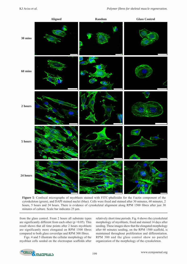
Fluorescence staining for the f-actin component of thecytoskeleton shown in Fig. 5 illustrates how, after 30minutes C2C12 myoblast cells have adhered to all thesurfaces. At this early time point the myoblasts on theRPM 1500 fibres have begun to organize their f-actin intoparallel arrays of stress fibres, whereas on the RPM 300fibres the f-actin is much less organised. On the coverslips,the actin is concentrated around the cell peripheryindicating the active adhesion and spreading process. After1 hour, the cells have formed a more defined cytoskeletalarrangement with stress fibres on the glass coverslips, andhave more defined peripheral actin staining indicating the
Figure 3: Histogram showingnormalized (to 90 degrees) fibreangles in electrospun RPM1500 andRPM300 fibres. Black (RPM 1500)and grey (RPM 300) lines indicate theaverage for each cohort. The majorityof fibres spun at RPM 1500 showalignment to 90-110 degrees.
RPM 300 RPM 1500
Standard Deviation 74.7 19.5
Table 1: Standard deviation from mean of alignmentin electrospun PLGA fibres (n = 50). RPM1500 fibresshowed significantly less deviation (p <0.05) than RPM300 fibres (n = 108 angles per fibre type).

198 www.ecmjournal.org
KJ Aviss et al. Polymer fibres for skeletal muscle regeneration.
cells are still not fully spread on the glass surface. Cellsupon the RPM 1500 show a degree of elongation withactin stress fibres becoming parallel compared to the RPM300 fibres and the glass coverslips, where actin stress fibrescreate a polygonal cell morphology. By 24 hours the cellsseeded on the glass coverslip control surface have a morepolygonal morphology, compared to those seeded on theRPM 1500 electrospun fibres which have elongated in abipolar manner. On the RPM 300 electrospun fibres, by24 hours the cells are less polygonal than those on glass
coverslips, but elongated to the same degree as those onRPM 1500 fibres after 5 hours.
Fig. 6 shows the quantification of elongation from thecells in figure 4. To quantify the degree of elongation, theaspect ratio of the cells was measured (n = 20 per timepoint and sample type). The student’s t-test was employedto determine any significant difference between the aspectratios in each time point and sample type. After 30 minutesand 1 hour, elongation of cells on electrospun fibres issimilar (p >0.05), but is significantly different (p <0.05)
24 hours
3 days
7 days
RPM 1500 RPM 300
Figure 4: Scanning electron micrographs of myoblasts seeded on electrospun RPM 1500 and RPM 300 PLGAscaffolds. Myoblasts were fixed and viewed after 24 hours, 3 days, and 7 days in culture. Images show alignment ofcells along the RPM 1500 fibres.
199 www.ecmjournal.org
KJ Aviss et al. Polymer fibres for skeletal muscle regeneration.
from the glass control. From 2 hours all substrate typesare significantly different from each other (p <0.05). Thisresult shows that all time points after 2 hours myoblastsare significantly more elongated on RPM 1500 fibrescompared to both glass coverslips and RPM 300 fibres.
Figs. 4 and 5 illustrate the cellular morphology of themyoblast cells seeded on the electrospun scaffolds after
relatively short time periods. Fig. 6 shows the cytoskeletalmorphology of myoblasts, fixed and stained 14 days afterseeding. These images show that the elongated morphologyafter 60 minutes seeding, on the RPM 1500 scaffold, ismaintained throughout proliferation and differentiation.RPM 300 and the glass control show no parallelorganization of the morphology of the cytoskeleton.
30 mins
60 mins
2 hours
5 hours
24 hours
Aligned Random Glass Control
Figure 5: Confocal micrographs of myoblasts stained with FITC-phalloidin for the f-actin component of thecytoskeleton (green), and DAPI stained nuclei (blue). Cells were fixed and stained after 30 minutes, 60 minutes, 2hours, 5 hours and 24 hours. There is evidence of cytoskeletal alignment along RPM 1500 fibres after just 30minutes of culture. Scale bar indicates 25 μm.

200 www.ecmjournal.org
KJ Aviss et al. Polymer fibres for skeletal muscle regeneration.
DifferentiationDifferentiation of myoblasts into myofibres can bemonitored by immunostaining of differentiation markers,proteins that are only expressed as the cell differentiates.Fast myosin heavy chain is a sarcomeric protein that isclassically expressed after 7 days in cells in differentiationmedia.
Immunostaining of the differentiation marker fastmyosin heavy chain (fast MyHC) is illustrated in Fig. 8.This result concurs with previous work (Engler et al., 2004)which suggests that myoblasts will differentiate morereadily upon elastic or softer surfaces. This result showsthat C2C12 myoblasts will express normal differentiationproteins and begin to fuse into multinucleated myotubes,especially upon the electrospun PLGA fibres. Themorphology of the cells shown in Fig. 8 show that themultinucleated myotubes are aligned in the same directionand are fused together to form long myofibre-like cells,compared to those on the glass where disordered fusionhas taken place to create myotubes with a less fibre-likemorphology.
Quantification of differentiation using lowmagnification (10 x) ICC images of fast myosin heavychain (fast MyHC) staining. To quantify the differentiation,the fusion index was calculated: number of fast MyHCpositive multinucleated myotubes was divided by the totalnumber of nuclei and converted into a percentage value,these values were then plotted as a bar chart as shown inFig. 9. The graph in Fig.9 shows that after 14 days inculture, there is significantly more (p <0.05) differentiationon RPM 1500 fibres compared to both glass and RPM300 fibres.
ContractilityAs discussed, myoblast and consequently myofibreelongation is thought to be essential for efficientcontraction to take place. Immunostaining of a proteinpresent in the sarcomere allows visualization of thecapability to contract.
Low magnification images of myoblast cells stainedfor sarcomeric myosin (red) shown in Fig. 10 not onlyillustrate the distinct, and expected cell elongation along
Figure 6: Quantification of elongation inmyoblasts seeded on glass control andelectrospun PLGA fibres. Values representmeans ± standard deviation, where n = 20.Brackets indicate not significantly different:p >0.05 as found by the student’s t-test, allother data sets are significantly different fromeach other (p <0.05). Cells are significantlymore elongated on RPM 1500 fibres.
RPM 1500 RPM 300 Glass Control
Figure 7: F-actin and nuclear staining, with FITC-phalloidin and DAPI respectively, of myoblast cells seeded on theelectrospun scaffolds, fixed and stained after 14 days. Scale bar indicates 50 μm; arrow implies direction of cellalignment.

201 www.ecmjournal.org
KJ Aviss et al. Polymer fibres for skeletal muscle regeneration.
one axis as shown in figures 4, 5, and 6, but also showsthe expression of sarcomeric myosin. This expression, asshown by red staining in Fig. 10 is more abundant on RPM1500 fibres compared to RPM 300 and the glass control.
Confocal images shown in Fig. 11 illustrate theindication of correct sarcomeric protein organization.
These images show sarcomeric myosin in red, it is thesered striations that indicate correct organization, as theyrepresent the myosin in the A band of the sarcomere. Fromthese images it can be concluded that the myotubes formedon the electrospun fibres are able to organize the correctsarcomeric protein array.
Day 3
Day 7
Day 14
RPM 1500 RPM 300 Glass Control
Figure 8: Fluorescence micrographs of myoblast cells stained for fast myosin heavy chain (green) and nuclear staining(blue). Cells were fixed and stained after 3 days, 7 days, and 14 days in differentiation media. Scale bar indicates 50μm (magnification 20 x).
Figure 9: Graph presenting percentage fastmyosin heavy chain positive multinucleatedmyotubes, counted using ImageJ software.Bars indicate standard deviation, star indicatessignificant difference (p < 0.05, n = 5 persubstrate type and time point) as shown bystudents’ t-test. There is a significantly highernumber of differentiated myotubes on the RPM1500 fibres.

202 www.ecmjournal.org
KJ Aviss et al. Polymer fibres for skeletal muscle regeneration.
Figure 10: Myoblast cells fixed and stained for sarcomeric myosin (red), f-actin (green), and nuclei (blue) after 14days culture in differentiation media. Original magnification is 10 x; scale bar indicates 200 μm.
RPM 1500 RPM 300 Glass Control
Figure 11: Confocal micrographs of myoblasts seeded on electrospun scaffolds and glass control stained for sarcomericmyosin (red), f-actin (green), and nuclei (blue) after 14 days in differentiation media. Arrows point to individualbands of sarcomeric myosin; scale bar shows 25 μm.

203 www.ecmjournal.org
KJ Aviss et al. Polymer fibres for skeletal muscle regeneration.
Discussion
Regeneration of a fully functioning skeletal muscle patchfor grafting or as a true-to-life research tool for diseasesaffecting the tissue is a challenge for bioengineers. Creatingand maintaining cellular organization and architecturebeing the primary difficulty. Synthetic materials, such asPLGA, may create a biodegradable scaffold with thetopography to introduce contact guidance to the cells inorder for them to grow parallel to each other in onedirection (Bian and Bursac, 2009; Choi et al., 2008; Lamet al., 2009). Contact guidance is the reaction of cells totopographical features (Dalby et al., 2003). Berendse etal. (2003) and Peckham (2008) report that parallel f-actincytoskeletal arrangement is important for successfulsarcomeric formation (Berendse et al., 2003; Peckham,2008). Due to the inherent alignment within the electrospunPLGA scaffold described in this paper and the profoundeffect on cellular alignment and cytoskeletal arrangementit could become the basis for eventual 3D tissue cultureincorporating different cell types for vascularisation. Thistype of electrospun scaffold does not allow for cellularinfiltration thus would be used as a template to create andmaintain the alignment of the myoblast cells. Used inconjunction with other materials e.g. a gel it could be usedto create a 3D tissue construct. Levenberg et al. (2005)created a PLLA and PLGA porous foam and seeded it witha tri-culture of cells to encourage vascularisation to occursimultaneously with myoblast differentiation (Levenberget al., 2005). Lam et al. (2009) used a PDMS scaffold as atemplate for aligned myotube growth then transferred thecells onto a fibrin gel for 3D cell infiltration into the gel,maintaining the alignment (Lam et al., 2009).
In the current study it is shown that highly alignedelectrospun PLGA fibres (RPM 1500) can be reproduciblymade with diameters ranging from 100 nm to 1.4 μm. Thisalignment can be quantified by measuring the standarddeviation from the mean; randomly oriented fibres (RPM300 in this study) would be expected to have a largestandard deviation, compared to aligned fibres (RPM 1500)that would yield a smaller standard deviation. Our resultsconcur with this theory; RPM 300 fibres have a largerstandard deviation than RPM 1500 fibres. It is also shownthat this highly aligned scaffold can provide thetopographical cues for the alignment of C2C12 murinemyoblasts. Using both SEM and fluorescence imaging itis possible to illustrate that myoblasts elongate and alignalong the parallel axis of the electrospun PLGA fibres.Quantification of elongation from the fluorescence imagesstatistically shows that the cellular morphology on alignedfibres (RPM 1500) is more elongated; looking at this inconjunction with the SEM images the cellular morphologyresponds to the alignment in the scaffold, and is maintainedthrough proliferation over 14 days. Along with controllingcellular organisation with aligned sub-micron fibres, thePLGA scaffolds also encourage myoblast differentiation.Quantification of differentiation, measured by number ofmultinucleated (>2 nuclei) myotubes expressing thedifferentiation marker fast myosin heavy chain as shownin Fig. 9 indicates around 13% multinucleated myotubespresent on the RPM 1500 fibres which is significantly
higher (p <0.05) than those on RPM 300 fibres or the glasscontrol. This figure is relative to other studies where nobiologic components were added to the cellular substrate(Stern et al., 2009). This differentiation, along with theimmunostaining of sarcomeric myosin (see Figs. 10 and11), show that myotube alignment introduced by theelectrospun fibres leads to preferential expression ofdifferentiation markers.
This could be due to the myoblasts being organised toencourage fusion and consequent differentiation. Resultsshown in Figs. 10 and 11 illustrate that the sarcomericcontractile machinery is arranged correctly forming thetypical banding pattern characteristic of skeletal musclefibres. This banding is clearer and more defined inmyotubes seeded on the electrospun scaffolds. Due to thisstereotypical highly organized arrangement of thecontractile proteins the cells have the ability to contractwhen given the appropriate stimulus. Levi-Mishali et al.(2009) showed that PLGA scaffolds with at least 50:50lactide:glycolide ratio are elastic enough to allow myotubeformation without detatchment, this study uses PL85GA15which falls into the criteria stated by Levi-Mishali et al.stating what elasticity is optimum for myoblastdifferentiation (Levi-Mishali et al., 2009).
Previous studies have shown that aligned fibres caninfluence the orientation of the cell type seeded upon them(Bashur et al., 2006; Bian and Bursac, 2009; Choi et al.,2008; Courtney et al., 2006; Huang et al., 2006; Lehnertet al., 2004; Riboldi et al., 2005; Wang et al., 2009). Otherstudies have incorporated natural biologic materials e.g.collagen, to improve biocompatibility (Choi et al., 2008;Lam et al., 2009). This study illustrates a totally syntheticscaffold with no surface modifications or feeder layer/co-culture can support the attachment, proliferation anddifferentiation of C2C12 myoblasts. Future work includesan in vitro degradation study comparing random andaligned electrospun fibres with spin coated PLGA, lookingat mass loss, surface morphology changes, pH changes,and molecular weight modulations.
Acknowledgements
The BBSRC is gratefully acknowledged for funding theresearch studentship (KJA) and Confocal microscope (BB/FO11547/1). Thanks to Dr. Steve Eichhorn and The RoyalSociety for use and funding of the electrospinningequipment.
References
Bashur CA, Dahlgren LA, Goldstein AS (2006) Effectof fiber diameter and orientation on fibroblast morphologyand proliferation on electrospun poly(D,L-lactic-co-glycolic acid) meshes. Biomaterials 27: 5681-5688.
Berendse M, Grounds MD, Lloyd CM (2003) Myoblaststructure affects subsequent skeletal myotube morphologyand sarcomere assembly. Exp Cell Res 291: 435-450.
Bian W, Bursac N (2009) Engineered skeletal muscletissue networks with controllable architecture. Biomaterials30: 1401-1412.

204 www.ecmjournal.org
KJ Aviss et al. Polymer fibres for skeletal muscle regeneration.
Blackwood KA, McKean R, Canton I, Freeman CO,Franklin KL, Cole D, Brook I, Farthing P, Rimmer S,Haycock JW (2008) Development of biodegradableelectrospun scaffolds for dermal replacement. Biomaterials29: 3091-3104.
Bray M-A, Sheehy SP, Parker KK (2008) Sarcomerealignment is regulated by myocyte shape. Cell MotilCytoskel 65: 641-651.
Choi J S, Lee S J, Christ G J, Atala A, Yoo J J (2008)The influence of electrospun aligned poly(caprolactone)/collagen nanofiber meshes on the formation of self-alignedskeletal muscle myotubes. Biomaterials 29: 2899-2906.
Cooper S T, Maxwell A L, Kizana E, Ghoddusi M,Hardeman E C, Alexander I E, Allen D G, North K N (2004)C2C12 co-culture on a fibroblast substratum enablessustained survival of contractile, highly differentiatedmyotubes with peripheral nuclei and adult fast myosinexpression. Cell Motil Cytoskel 58: 200-211.
Courtney T, Sacks M S, Stankus J, Guan J, Wagner WR (2006) Design and analysis of tissue engineeringscaffolds that mimic soft tissue mechanical anisotropy.Biomaterials 27: 3631-3638.
Dalby M J, Childs S, Riechle MO, Johnstone HJH,Affrossman S, Curtis ASG (2003) Fibroblast reaction toisland topography: changes in cytoskeleton andmorphology with time. Biomaterials 24: 927-935.
Engler J, Griffin MA, Sen S, Bonnemann CG, SweenyHL, Discher DE (2004) Myotubes differentiate optimallyon substrates with tissue-like stiffness: pathologicalimplications for soft of stiff microenvironments. J Cell Biol166: 877-887.
Huang NF, Patel S, Thakar RG, Wu J, Hsiao BS, ChuB, Lee RJ, Lee S (2006) Myotube assembly on nanofibrousand micropatterned polymers. Nano Letters 6: 537-542.
Huang Y-C, Dennis RG, Larkin L, Baar K (2005) Rapidformation of functional muscle in vitro using fibrin gels. JApp Phys 98: 706-713.
Kaetsu I, Yoshida M, Asano M, Yamanaka H, Imai K,Yuasa H, Mashimo T, Suzuki K, Katakai R, Oya M (1987)Biodegradable implant composites for local therapy. JControl Release 6: 249-263.
Kim K, Yu M, Zong X, Chiu J, Fang D, Seo Y-S, HsiaoBS, Chu B, Hadjiargyrou M (2003) Control of degradationrate and hydrophillicity in electrospun non-wovenpoly(D,L-lactide) nanofiber scaffolds for biomedicalapplications. Biomaterials 24: 4977-4985.
Lam MT, Huang Y-C, Birla RK, Takayama S (2009)Microfeature guided skeletal muscle tissue engineering forhighly organised 3-dimensional free-standing constructs.Biomaterials 30: 1150-1155.
Lehnert D, Wehrle-Haller B, David C, Weiland U,Ballestrem C, Imhof BA, Bastmeyer M (2004) Cellbehaviour on micropatterned substrata: limits of
extracellular matrix geometry for spreading and adhesion.J Cell Sci 117: 41-52.
Levenberg S, Rouwkema J, Macdonald M, Garfein ES,Kohane DS, Darland DC, Marini R, Van Blitterswijk CA,Mulligan RC, D’Amore PA (2005) Engineeringvascularised skeletal muscle tissue. Nat Biotechnol 23:879-884.
Levi-Mishali M, Zoldan J, Levenberg S (2009) Effectof scaffold stiffness on myoblast differentiation. TissueEng 15: 935-944.
Peckham M (2008) Engineering a multinucleatedmyotube, the role of the actin cytoskeleton. J Microsc 231:486-493.
Ren K, Crouzier T, Roy C, Picart C (2008)Polyelectrolyte multilayer films of controlled stiffnessmodulate myoblast differentiation. Adv Funct Mater18:1378-1389.
Riboldi S A, Sampaolesi M, Neuenschwander P, CossuG, Mantero S (2005) Electrospun degradablepolyesterurethane membranes: potential scaffolds forskeletal muscle tissue engineering. Biomaterials 26: 4606-4615.
Schiaffino S, Partridge T (eds) (2008) Skeletal MuscleRepair and Regeneration, Advances in Muscle Research,Vol 3. Springer Netherlands, Dordrecht.
Singh L, Kumar V, Ratner B D (2004) Generation ofporous microcellular 85/15 poly(DL-lactide-co-glycolide)foams for biomedical applications. Biomaterials 25: 2611-2617.
Stern M, Myers R L, Hammam N, Stern KA, Eberli D,Kritchevsky SB, Soker S, Dyke M V (2009) The influenceof extracellular matrix derived from skeletal muscle tissueon the proliferation and differentiation of myogenicprogenitor cells ex vivo. Biomaterials 30: 2393-2399.
Subbiah T, Bhat G S, Tock R W, Parameswaran S,Ramkumar S S (2004) Electrospinning of nanofibres. JAppl Polym Sci 96: 557-569.
Wang HB, Mullins ME, Cregg JM, Hurtado A, OudegaM, Trombley MT, Gilbert RJ (2009) Creation of highlyaligned electrospun poly-L-lactide acid fibres for nerveregeneration applications. J Nerual Eng 6: 016001 (15pp).
Webb AR, Yang J, Ameer GA (2004) Biodegradablepolyester elastomers in tissue engineering. Exp Opin BiolTher 4: 801-812.
Zong X, Bien H, Chung C-Y, Yin L, Fang D, HsiaoBS, Chu B, Entcheva E (2005) Electrospun fine-texturedscaffolds for heart tissue constructs. Biomaterials 26: 5330-5338.
Zong X, Ran S, Kim K-S, Fang D, Hsiao BS, Chu B(2003) Structure and morphology changes during in vitrodegradation of electrospun poly(glycolide-co-lactide)nanofibre membrane. Biomacromolecules 4: 416-423.