alginate encapsulation parameters influence the differentiation of microencapsulated embryonic stem...
TRANSCRIPT
Ac
ce
pte
dP
re
pr
i nt
Tissue Engineering and Delivery Systems Biotechnology and Bioengineering DOI 10.1002/bit.25121
Alginate Encapsulation Parameters Influence the Differentiation of Microencapsulated Embryonic Stem Cell Aggregates†
Jenna L. Wilson1, Mohamad Ali Najia1, Rabbia Saeed1, and Todd C. McDevitt1,2
* 1 The Wallace H. Coulter Department of Biomedical Engineering, Georgia Institute of Technology and Emory University, Atlanta, GA, USA 2 The Parker H. Petit Institute for Bioengineering and Bioscience, Georgia Institute of Technology, Atlanta, GA, USA * Address for Correspondence: Todd C. McDevitt, Ph.D. 313 Ferst Drive, Suite 2102, Atlanta GA, 30332-0532 Phone: 404-385-6647 Fax: 404-894-4243 Email: [email protected] †This article has been accepted for publication and undergone full peer review but has not been through the copyediting, typesetting, pagination and proofreading process, which may lead to differences between this version and the Version of Record. Please cite this article as doi: [10.1002/bit.25121]
© 2013 Wiley Periodicals, Inc.
Received June 6, 2013; Revision Received August 26, 2013; Accepted September 17, 2013
Ac
ce
pte
dP
re
pr
i nt
Abstract
Pluripotent embryonic stem cells (ESCs) have tremendous potential as tools for regenerative medicine
and drug discovery, yet the lack of processes to manufacture viable and homogenous cell populations of
sufficient numbers limits the clinical translation of current and future cell therapies. Microencapsulation of
ESCs within microbeads can shield cells from hydrodynamic shear forces found in bioreactor environments
while allowing for sufficient diffusion of nutrients and oxygen through the encapsulation material. Despite
initial studies examining alginate microbeads as a platform for stem cell expansion and directed differentiation,
the impact of alginate encapsulation parameters on stem cell phenotype has not been thoroughly investigated.
Therefore, the objective of this study was to systematically examine the effects of varying alginate compositions
on microencapsulated ESC expansion and phenotype. Pre-formed aggregates of murine ESCs were encapsulated
in alginate microbeads composed of a high or low ratio of guluronic to mannuronic acid residues (High G and
High M, respectively), with and without a poly-L-lysine coating, thereby providing four distinct alginate bead
compositions for analysis. Encapsulation in all alginate compositions was found to delay differentiation, with
encapsulation within High G alginate yielding the least differentiated cell population. The addition of a PLL
coating to the High G alginate prevented cell escape from beads for up to 14 days. Furthermore, encapsulation
within High M alginate promoted differentiation toward a primitive endoderm phenotype. Taken together, the
findings of this study suggest that distinct ESC expansion capacities and differentiation trajectories emerge
depending on the alginate composition employed, indicating that encapsulation material physical properties can
be used to control stem cell fate.
Key words: microencapsulation, alginate, stem cells, bioprocessing

Ac
ce
pte
dP
re
pr
i nt
Introduction
Stem cells, including pluripotent embryonic stem cells (ESCs), have tremendous potential as tools for
regenerative medicine and drug discovery. However, there are many challenges to overcome before stem cell-
derived therapies can be made broadly available, including the lack of processes to manufacture viable and
homogenous cell populations of sufficient numbers (Kirouac and Zandstra, 2008). Currently, the large scale
production of mammalian cells typically occurs in suspension bioreactors, which impart hydrodynamic forces
that can adversely affect stem cell viability and influence their phenotype (Gareau et al., 2012; Leung et al.,
2011; Sargent et al., 2010; Wolfe and Ahsan, 2013). Stem cells are very sensitive to environmental stimuli, as
external signals in the culture environment provide cues that determine whether stem cells continue to self-
renew or differentiate into specific cell types. Because most current bioprocesses are designed around
suspension culture systems, investigation into systems to culture anchorage-dependent stem cells in suspension
is critical for translating process technology and for achieving the high cell densities required for therapeutic
doses (Kehoe et al., 2010; Serra et al., 2012).
One approach to the challenge of scalable bioprocessing is to encapsulate stem cells in hydrogels, such
as alginate, to better control microenvironmental cues and enable suspension culture. Microencapsulation has
been used for decades to protect enclosed cells from the host immune system upon transplantation, but it can
also shield cells from hydrodynamic shear forces found in bioreactor environments and prevent agglomeration
of stem cell aggregates while permitting diffusion of nutrients and oxygen through the encapsulation material
(Wilson and McDevitt, 2013). Several investigations of ESC microencapsulation, mostly with single cells, have
been previously performed with alginate capsule formulations (Serra et al., 2011; Siti-Ismail et al., 2008; Wang
et al., 2006), and directed differentiation has been achieved, in most cases through soluble factor addition,
toward osteogenic (Hwang et al., 2009a; Tang et al., 2012), cardiac (Bauwens et al., 2005; Jing et al., 2010),
hematopoietic (Dang et al., 2004; Rahman et al., 2010), neural (Li et al., 2011b), pancreatic (Chayosumrit et al.,
2010; Wang et al., 2009), and hepatocytic lineages (Fang et al., 2007; Maguire et al., 2005). Although most
previous studies have examined the encapsulation of single ESCs, aggregates of ESCs are also important to

Ac
ce
pte
dP
re
pr
i nt
investigate for a number of reasons. First, using pre-formed aggregates provides a consistent initial size that can
be compared directly to unencapsulated controls, which can also be cultured in suspension. Though
microencapsulation-based protocols have been developed to form multicellular aggregates (Magyar et al., 2001;
Sakai et al., 2008; Wang et al., 2006), using microwell formation (Ungrin et al., 2008) may provide more
consistency so that the impact of encapsulation parameters on cell phenotype is not confounded by the
additional parameters of aggregate size or the aggregation kinetics (Kinney et al., 2012). Furthermore,
microencapsulation of aggregates has been previously determined to improve cell viability (Serra et al., 2011),
as ESCs generally require maintenance of intercellular adhesions for optimal survival (Watanabe et al., 2007).
Finally, culture as 3D aggregates can initiate differentiation of ESCs as embryoid bodies (EBs) (Bratt-Leal et al.,
2009; Doetschman et al., 1985), providing a platform to study the impact of microencapsulation parameters on
differentiation trajectory.
While encapsulation has been explored as a tool for stem cell expansion and directed differentiation, the
impact of alginate encapsulation parameters on stem cell phenotype has not been systematically examined.
Alginate is a biocompatible polymer purified from brown seaweed that is commonly used to encapsulate
mammalian cells due to its relative abundance and mild cross-linking requirements. Alginate is composed of
two anionic monomers, α-L-guluronic acid (G) and β-D-mannuronic acid (M), which are arranged in both
homopolymeric regions (GG blocks and MM blocks) and mixed monomeric regions (MG blocks) (Haug et al.,
1966). Cross-linking occurs at G residues through binding of divalent cations such as calcium, therefore
alginates with higher G content and longer GG blocks produce stiffer beads due to greater cross-linking than
alginates with higher M content. Because native alginate is non-adhesive and therefore cannot interact directly
with cells, the physical and chemical properties of the polymer dictate the relationship with the enclosed cells. A
handful of studies have examined the impact of G and M content on cell proliferation, metabolism, and
secretion. Experiments with murine insulinoma βTC3 cells found that alginates with high G content inhibited
cell growth, leading to decreased metabolic and secretory activity (Stabler et al., 2001). Alternatively, studies of
encapsulated neural stem cells observed improved secretion of neurotrophic factors when the cells were cultured
in alginate with high G content, citing poor capsule stability in alginate with high M content (Purcell et al.,

Ac
ce
pte
dP
re
pr
i nt
2009). Despite such examples of reported differences based on G and M content, most studies do not specify the
composition of alginate species used, thus provoking questions about the potential influence of the
encapsulation material utilized on the phenotype of the enclosed cells.
An additional microencapsulation property that can be varied is the presence of a coating surrounding
the capsule, typically with a polycation like poly-L-lysine (PLL), which has historically served as an added
barrier to the host immune system upon transplantation by decreasing the permeability of larger molecules, such
as antibodies, into and out of the bead (Lim and Sun, 1980). However, coating with PLL also confers additional
mechanical stability (De Castro et al., 2005; Strand et al., 2003; Thu et al., 1996a; Thu et al., 1996b) which can
be beneficial for maintaining capsule integrity over time in culture. Many studies have described issues with
single cells escaping or leaking out of the capsules (Chang et al., 1994; Cheng et al., 2006; Jing et al., 2010;
Pajić-Lijaković et al., 2007), an issue which may be ameliorated through PLL coating. However, the issue of
cell escape may be increased when encapsulating pre-formed aggregates due to their large size and potential
proximity to the bead edge, and it has yet to be determined whether addition of PLL is sufficient to prevent
escape in this context, a question also relevant for the encapsulation of other aggregated cells.
Overall, the inconsistency in the composition of alginate and use of a coating layer in previous studies
with microencapsulated cells raises questions about whether disparate results result from differing material
compositions. The potential impacts of microencapsulation material on cell phenotype are particularly relevant
for the culture of ESCs due to their particular sensitivities to the surrounding physical microenvironment (Li et
al., 2011a). Thus, the objective of this study was to systematically examine the impact of varying alginate
compositions on microencapsulated ESC expansion and phenotype. Aggregates of murine ESCs were pre-
formed and encapsulated in alginate microbeads composed of a high or low ratio of G to M residues, with and
without a PLL coating, providing four distinct alginate bead compositions for analysis. Mechanical
characterization of each bead composition was performed, as well as analysis of encapsulated cell aggregate
viability, morphology, and phenotype via gene and protein expression. The results of this study revealed
distinctions in the growth rate, morphology, and differentiation trajectories of ESCs in each of the encapsulated

Ac
ce
pte
dP
re
pr
i nt
formats over 14 days of culture, suggesting that alginate bead composition is an important parameter to consider
when designing microencapsulation-based expansion and directed differentiation protocols.
Methods
Embryonic stem cell culture
Murine ESCs (D3 cell line; chosen due to its widespread use) were cultured on tissue culture treated polystyrene
dishes (Corning Inc., Corning, NY) adsorbed with gelatin (0.1% solution in diH2O). Undifferentiated ESC
culture media consisted of Dulbecco’s modified Eagle’s medium (DMEM) (Mediatech) supplemented with 15%
fetal bovine serum (Hyclone, Logan, UT), 100 U/mL penicillin, 100 μg/mL streptomycin, and 0.25 μg/mL
amphotericin (Mediatech, Herndon, VA), 2 mM L-glutamine (Mediatech), 1x MEM non-essential amino acid
solution (Mediatech), 0.1 mM 2- mercaptoethanol (Fisher Scientific, Fairlawn, NJ), and 103 U/mL of leukemia
inhibitory factor (LIF) (ESGRO, Chemicon, Temecula, CA). Cultures were replenished with fresh media every
other day and passaged prior to reaching 70% confluence (percent of plate covered with cells).
ESC aggregate formation and culture
A single cell suspension of undifferentiated ESCs was obtained through dissociation of monolayer cultures with
0.05% trypsin-EDTA (Mediatech). Defined, serum-free N2B27 media (Ying and Smith, 2003) was used for all
aggregate cultures and consisted of DMEM/F12 (50/50) medium (Gibco) supplemented with N2 (Gibco), 25
μg/L bovine serum albumin (BSA), 100 U/mL penicillin, 100 μg/mL streptomycin, and 0.25 μg/mL
amphotericin (Mediatech, Herndon, VA), 2 mM L-glutamine (Mediatech), all combined 1:1 with Neurobasal
medium (Gibco) supplemented with B27 (Gibco). N2B27 basal media has been widely used by several groups
for a number of different applications, including undifferentiated ESC culture (Ying et al., 2008), differentiation
of ESCs to insulin-producing cells (Wang et al., 2009), and hematopoietic differentiation of ESCs (Rahman et
al., 2010). Additionally, neural ectoderm differentiation is considered the “default” pathway for ESC

Ac
ce
pte
dP
re
pr
i nt
differentiation (Tropepe et al, Neuron, 2001) in the absence of any exogenous factors, thus N2B27 media is an
appropriate choice for basal media to examine ESC differentiation. Aggregation of ESCs was achieved by
centrifugation (200 rcf) of ESCs into 400 μm square polydimethylsiloxane (PDMS) micro-wells (AggrewellTM,
Stem Cell Technologies, Vancouver, Canada) as previously reported (Kinney et al., 2012; Ungrin et al., 2008).
The cell seeding density yielded approximately 500 cells per individual well. The ESCs were incubated in the
wells for approximately 20 hours in serum-free N2B27 culture media to allow for aggregate formation. The
resulting aggregate population was either immediately transferred to suspension culture (approximately 1500
aggregates in 10 mL of serum-free culture media) for the unencapsulated condition or transferred after
subsequent encapsulation. Aggregates were cultured in sterile 100 x 15 mm bacteriological grade polystyrene
Petri dishes (BD, Franklin Lakes, NJ) and maintained on rotary orbital shakers (Carpenedo et al., 2007) at ~45
rpm. A 90% media exchange was performed every three days following gravity-induced sedimentation of the
aggregates in 15 mL conical tubes. Suspension cultures were maintained for up to 14 days of differentiation.
Aggregate microencapsulation
Two different medium viscosity alginates were used in this study, ultrapure MVG (Pronova) which contains
greater than 60% G residues (High G) and ultrapure MVM (Pronova) which contains greater than 50% M
residues (High M). Alginate solutions were prepared at 1.5 wt% in calcium-free DMEM (Gibco) and autoclaved
for sterilization. Once the solutions were cooled to 37°C, aggregates were re-suspended in the sterile alginate at
a density of 12,000 aggregates per mL of alginate (approximately 6 x 10⁶ cells/mL). An electrostatic bead
generator (Nisco) was utilized for encapsulation. The alginate solution was extruded through a 400 μm nozzle
using a syringe pump at a flow rate of 6 mL/hour and dropped into a 100 mM calcium chloride (EMD)
hardening bath. After encapsulation, the beads were rinsed with serum-free media 3x, and half of the beads from
each alginate type were coated with 0.05% poly-L-lysine (MW 15,000-30,000; Sigma) for 2 minutes prior to
three additional media rinses. All encapsulated aggregates were cultured in serum-free N2B27 media on a rotary
orbital shaker as previously described.
Ac
ce
pte
dP
re
pr
i nt
Mechanical characterization of microbeads
Alginate beads of both High G and High M were formed (without cells) and some coated with PLL as described.
The beads were maintained in serum-free (N2B27) media on a rotary orbital shaker to simulate conditions
experienced during culture. At days 1, 7, and 14 following formation, the mechanical properties of six alginate
beads were assessed through micron-scale parallel-plate compression using a CellScale MicroSquisher and the
associated SquisherJoy software (Baraniak et al., 2012). The upper compression plate of a 302.8 μm cantilever
compressed the samples at 40% strain over a period of 40 seconds, held at constant deformation for 10 seconds,
and released over a period of 40 seconds to document the magnitude of hysteresis. To determine the Young’s
modulus, Hertzian theory for non-adhesive elastic contact was used to fit a linear regression line to 30 data
points above and below the 20% strain data point on the plot of force (F) vs. deformation2/3 (d). The resultant
slope in addition to the initial radius (R) of the bead was used to calculate the Young’s modulus of the sample
(E), as described by the following standard expression:
3/22/1
34 dREF ××=
During testing, all samples were held in a PBS fluid bath (pH 7.4, 0.1 g/L Ca2+ and 0.1 g/L Mg2+).
Cell number quantification
Samples were collected from each group at days 4, 7, 10, and 14 of differentiation. Encapsulated aggregates
were released from beads through 10 minute incubation with TrypLETM (Invitrogen) for PLL-coated beads only,
trituration, and 5 minute incubation with 55 mM sodium citrate (Sigma) for all beads, as previously described
(Jing et al., 2010). The cells were centrifuged at 200 rcf for 5 minutes and rinsed 3x with PBS. Cells from all
conditions were pelleted at 375 rcf for 4 minutes followed by supernatant removal and storage at -20°C. A

Ac
ce
pte
dP
re
pr
i nt
CyQUANT Cell Proliferation Assay Kit (Molecular Probes Inc., Eugene, OR) was used to determine cell
number, with a standard curve created using undifferentiated ESCs that were counted using a hemocytometer.
The fluorescence was read using a Synergy H4 plate reader (Biotek). The net growth rate (μnet) was determined
by examining change in cell density (X) over given periods of time (t) using the following standard expression:
XdtdX
netμ=
Viability staining and imaging
Cell viability was assessed at day 14 of differentiation using a LIVE/DEAD kit (Molecular Probes Inc., Eugene,
OR). Samples were incubated in PBS containing 0.1 μM calcein AM and 8 μM ethidium homodimer-1 at 4°C
for 1 hour. The samples were washed with PBS, transferred to a glass-bottomed 24-well plate, and immediately
imaged using a Zeiss LSM 700-405 confocal microscope (Carl Zeiss Inc.).
Histological analysis
Encapsulated and unencapsulated aggregates were sampled at days 4, 7, and 14 of differentiation, rinsed with
PBS, fixed in 4% paraformaldehyde for 30 minutes with rotation at room temperature, rinsed with PBS, and
stored at 4°C. Fixed aggregates were embedded in Histogel (Richard-Allen Scientific, Kalamazoo, MI),
processed via a series of graded ethanol and xylene rinses, and embedded in paraffin. Paraffin-embedded
samples were sectioned at a thickness of 5 μm using a rotary microtome (Microtom HM310). For histological
analysis, sections were de-paraffinized through a series of xylene and graded ethanol concentrations, followed
by staining with hematoxylin and eosin (H&E). Stained sections were imaged using a Nikon Eclipse 80i
equipped with a SpotFlex digital camera (Diagnostic Instruments).
Ac
ce
pte
dP
re
pr
i nt
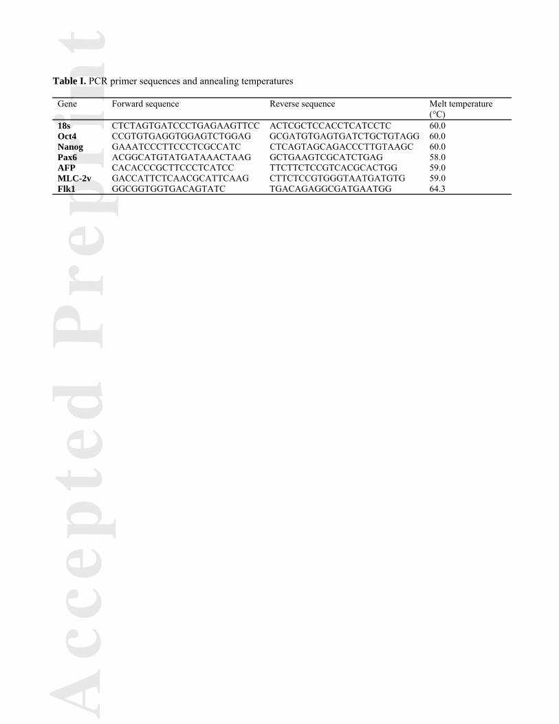
Quantitative reverse-transcription polymerase chain reaction (qRT-PCR)
Encapsulated aggregates were released from beads through 10 minute incubation with TrypLETM (Invitrogen)
for PLL-coated capsules only, trituration, and 5 minute incubation with 55 mM sodium citrate (Sigma) for all
beads. The cells were centrifuged at 200 rcf and rinsed 3x with PBS to remove residual PLL. RNA was
extracted from the aggregates with the RNeasy Mini kit (Qiagen Inc, Valencia, CA). The RNA (300 ng/sample)
was converted to complimentary DNA using the iScript cDNA synthesis kit (Bio-Rad, Hercules, CA) and
analyzed using real time PCR (MyIQ cycler, Bio-Rad). Forward and reverse primers for 18s, Oct4, Nanog,
Pax6, AFP, MLC-2v, and Flk1 were designed with Beacon Designer software (sequences and conditions are
given in Table I) and purchased from Invitrogen. Oct4 and Nanog gene expression were calculated with respect
to undifferentiated ESC expression levels as previously described (Pfaffl, 2001). Pax6, AFP, MLC-2v, and Flk1
concentrations were determined using a standard curve and normalized to 18s expression levels.
Immunofluorescent staining
Encapsulated and unencapsulated aggregates were sampled at days 4, 7, and 14 of differentiation, rinsed with
PBS, fixed in 4% paraformaldehyde for 30 minutes with rotation at room temperature, rinsed with PBS, and
stored at 4°C. Fixed aggregates were embedded in Histogel (Richard-Allen Scientific, Kalamazoo, MI) and
subject to graded sucrose and OCT infiltration prior to rapid freezing in a dry ice-ethanol bath and storage at -
80°C. OCT-embedded samples were sectioned at a thickness of 10 μm using a CryoStar NX70 cryostat and
allowed to dry at room temperature. Each section was surrounded using a PAP hydrophobic barrier pen and
rinsed with PBS 3x for 5 minutes. The slides were blocked and permeabilized with 3% donkey serum and 0.05%
Triton X-100 for 45 minutes at room temperature. After rinsing with PBS 2x for 5 minutes, primary antibody
solution diluted in blocking buffer (3% donkey serum in PBS) was added and incubated overnight at 4°C.
Primary antibodies against OCT-4 (Santa Cruz Biotechnology sc-8628; goat polyclonal; 1:100), AFP (Dako
A000829-2; rabbit polyclonal; 1:100), and α-SMA (Dako M0851; mouse monoclonal; 1:100) were used.
Following overnight incubation, slides were rinsed with PBS 3x for 5 minutes and incubated with secondary
Ac
ce
pte
dP
re
pr
i nt
solutions diluted in blocking buffer (1:1000 AlexaFluor®488 donkey anti-goat, 1:1000 Alexa Fluor 488 donkey
anti-rabbit, and 1:1000 Alexa Fluor 488 donkey anti-mouse) for 1 hour at room temperature. Slides were rinsed
with PBS 3x for 5 minutes and incubated with Hoechst dye (1:100) for 10 minutes at room temperature.
Following a final PBS rinse, coverslips were mounted with Fluoromount-G (SouthernBiotch) and sealed with
clear nail polish. The slides were imaged using a Zeiss LSM 700-405 confocal microscope (Carl Zeiss Inc.).
Statistics
All experiments were performed with replicate samples from independent conditions (n=6 for mechanical
testing, n=3 for cell counts, n=5 for PCR). The data is represented as the mean of the independent replicates, and
the error bars represent the standard error of the mean. Before performing statistical analysis, data were
normalized using a Box–Cox power transformation to normalize data variance. Two-way ANOVA was
calculated between different conditions (unencapsulated, High G, High G + PLL, High M, and High M + PLL)
and time points, followed by post hoc Tukey analysis to determine significant differences (p < 0.05). All
statistical analysis was performed using SYSTAT software.
Results
Characterization of alginate beads and aggregate encapsulation
Embryonic stem cell (ESC) spheroids were encapsulated in alginate with a high (High G) or low (High
M) ratio of guluronic acid to mannuronic acid. Additionally, a portion of the beads were coated with poly-L-
lysine (PLL), creating High G + PLL and High M + PLL beads in addition to uncoated beads. The seeding
density of spheroids in alginate gave rise to a small number (<15%) of empty beads, with the majority of beads
(>71%) containing one to three spheroids, consistent with the expected Poisson distribution assuming an
average rate of two aggregates per capsule (Fig. 1A). The average bead diameter (~734 μm) was relatively
uniform for all of the bead compositions examined over the 14 day culture period (Fig. 1B).

Ac
ce
pte
dP
re
pr
i nt
The bulk mechanical properties of each bead type were determined by parallel plate compression
testing. The Young’s modulus ranged from 5-18 kPa (Fig. 1C) and was consistently greater in the PLL-coated
beads, likely due to the increased mechanical stability provided by the external polyelectrolyte layer. The
uncoated High G alginate was significantly stiffer than the uncoated High M alginate at days 1 (p = 0.011) and 7
(p < 0.001) following formation, consistent with previous literature reports that alginate containing a higher
proportion of guluronic acid residues enhances the mechanical strength of the hydrogels (Uludag et al., 2000).
The modulus remained constant over the course of the culture period for each composition tested, indicating that
the beads were physically stable for at least two weeks when cultured dynamically without enclosed cells in
standard culture media.
Although the beads appeared similar after one week, unencapsulated spheroids (Fig. 1D) and aggregates
within each bead type (Fig. 1E-H) exhibited distinct morphologies depending on the type of alginate used. All of
the encapsulated spheroids appeared smaller than unencapsulated spheroids, with those in High G and High G +
PLL remaining spherical (Fig.1E, 1F) and those in High M and High M + PLL appearing more ovoid (Fig. 1G,
1H).
ESC escape from and proliferation within alginate beads
Aggregate escape from the alginate beads was observed as early as four days post-encapsulation for
aggregates within the High M alginate (Fig. 2A-E). After 14 days in culture, aggregate escape was more
frequent, with many of the initially encapsulated aggregates no longer residing within the alginate beads (Fig.
2F-J). In general, the High G alginate contained the aggregates more effectively than the High M alginate, with
the addition of the PLL coating improving retention. The High G + PLL formulation maintained all spheroids
within beads (Fig. 2K), while the uncoated High M only retained ~4% of the aggregates within beads after 14
days.
The proliferation rates of the ESCs cultured in different configurations (Fig. 2L) diverged, with the
unencapsulated spheroids having the highest initial net growth rate (0.38 day-1 versus 0.12 day-1 for High G,

Ac
ce
pte
dP
re
pr
i nt
0.32 day-1 for High G + PLL, 0.24 day-1 for High M, and 0.22 day-1 for High M + PLL). After 14 days of
culture, the number of cells in the unencapsulated aggregates was significantly greater than in the High G (p =
0.016), High G + PLL (p < 0.001), and High M + PLL (p = 0.035) conditions. Cell growth appeared most
stunted in the cells within the stiffer High G and High G + PLL beads, though the beads coated with PLL
exhibited overall similar growth trends to each other after day 4. A marked increase in cell number was
observed after the majority of encapsulated aggregates escaped from the beads (day 7 for High M and day 10 for
High G), indicating that the cells proliferated more robustly when not encapsulated. Cell growth plateaued at a
cell density of ~1.8 x 10⁶ cells/mL, likely due to the volumetric capacity of this particular culture system and
exhaustion of nutrients from the culture media.
Encapsulated ESC viability and aggregate morphology
Cell viability was assessed following 14 days in culture for each of the encapsulation conditions for both
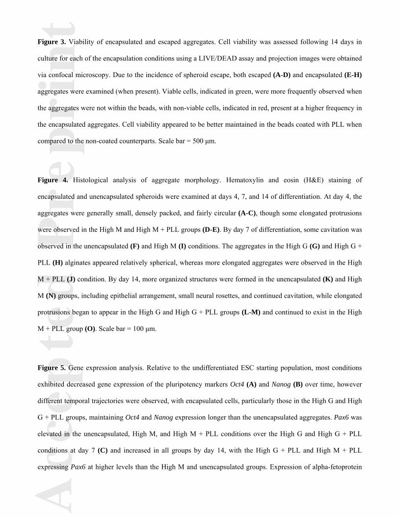
encapsulated and escaped aggregates due to the incidence of spheroid escape. Viable cells were more prevalent
when the aggregates were not within the beads (Fig. 3A-D), with non-viable cells present more frequently in the
encapsulated aggregates. The non-viable cells tended to be localized to the more rounded region of aggregates,
with more viable cells present in the elongated region of aggregates when present. In addition, more viable cells
were observed in the beads coated with PLL.
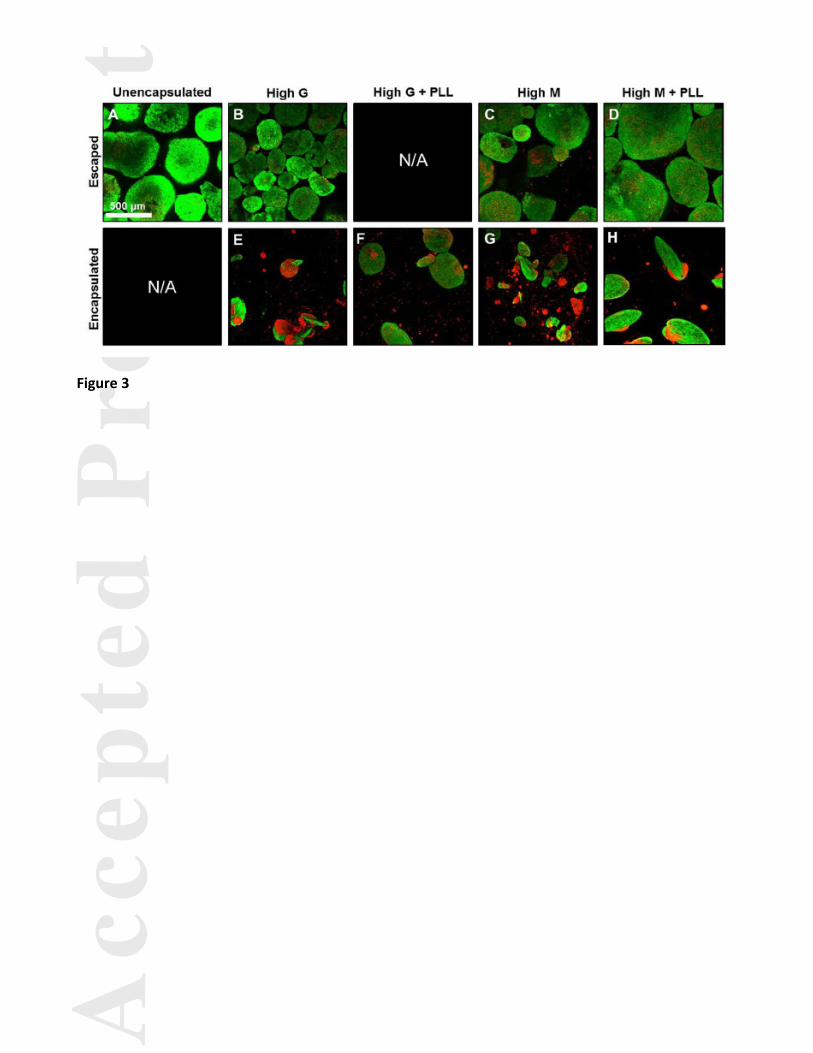
The elongated morphology that was observed in some groups (Fig. 1D-E, Fig. 2E, Fig. 3E-H) prompted
further examination into the morphological differences observed based on encapsulation conditions. At day 4,
the ESC aggregates were generally small and fairly round (Fig. 4A-C), although some elongated protrusions
were observed in the High M and High M + PLL groups (Fig. 4D-E). The aggregates were densely packed
without any morphogenic structures, suggesting that a primarily undifferentiated phenotype persisted. By day 7
of differentiation, some cavitation was observed in the unencapsulated (Fig. 4F) and High M (Fig. 4I) conditions
(which by day 7 had only half of the spheroids within beads), suggesting that differentiation was progressing
more rapidly in the unencapsulated and High M conditions. While the aggregates in the High G (Fig. 4G) and

Ac
ce
pte
dP
re
pr
i nt
High G + PLL (Fig. 4H) alginate beads still appeared relatively round, more distinguished extension of the
ovoid geometry occurred in the High M + PLL (Fig. 4J) condition. After two weeks in culture, more
differentiated structures were formed in the unencapsulated (Fig. 4K) and High M (Fig. 4N) groups, including
epithelial layer arrangement, small neural rosettes, and cavitation. The unencapsulated and High M conditioned
aggregates appeared larger, consistent with the cell proliferation data (Fig. 2G) previously described. In contrast,
aggregates encapsulated in the High G (Fig. 4L) and High G + PLL (Fig. 4M) beads continued to exhibit a
smaller cross-sectional area, though more multicellular protrusions were observed than at earlier time points.
The significantly elongated morphology in the High M + PLL condition at day 14 (Fig. 4O) was present at
similar levels to the High M + PLL at day 7 and at greater frequency than any of the other experimental groups.
Alginate compositional influences on stem cell phenotype
To determine what, if any, impact encapsulation in alginate with varying compositions had on stem cell
differentiation, gene and protein expression were examined over the course of two weeks. Relative to the
undifferentiated ESC starting population, most conditions exhibited decreased gene expression of the
pluripotency markers Oct4 (Fig. 5A) and Nanog (Fig. 5B) over time. However, different temporal trajectories
were observed, with the cells encapsulated in the High G and High G + PLL beads exhibiting significantly
higher Oct4 levels at day 7 compared to cells in unencapsulated (p < 0.001 and p < 0.001, respectively), High M
(p < 0.001 and p < 0.001), and High M + PLL (p < 0.001 and p = 0.006,) conditions. Furthermore, the cells
within the High M and High M + PLL had significantly higher levels of Oct4 expression when compared to the
unencapsulated spheroids (p < 0.001 and p < 0.001, respectively), signifying that residence within the alginate
beads, even temporarily, may delay differentiation. By day 14, cells in the High G and High G + PLL beads
continued to express higher levels of Oct4 compared to the unencapsulated, High M, and High M + PLL
conditions. A similar trend was observed in the expression of Nanog, with cells in the High G alginate
exhibiting higher levels compared to unencapsulated cells at day 4 and aggregates in the High G and High G +
PLL conditions having higher expression than the unencapsulated aggregates at day 7. After 14 days, the cells in
the High G + PLL beads maintained significantly increased expression over the unencapsulated and High M
groups. The steady Nanog levels observed in the High G + PLL group, as opposed to the decrease observed in
Ac
ce
pte
dP
re
pr
i nt
all other groups, was noteworthy since High G + PLL was the only condition to retain 100% of the spheroids
over the entire two week period of culture.
Genes representative of the three germ lineages were also examined to determine if encapsulation
conditions could impact differentiated phenotype(s), since culture as aggregates can lead to spontaneous
differentiation as embryoid bodies (EBs). Pax6, an early ectodermal marker, was elevated in the unencapsulated,
High M, and High M + PLL conditions compared to the High G and High G + PLL conditions at day 7 (Fig. 5C)
and increased in all groups by day 14, with the High G + PLL and High M + PLL groups having significantly
higher levels than the High M and unencapsulated groups. Investigation of alpha-fetoprotein (AFP) expression,
an endodermal marker, revealed dramatic increases in the High M and High M + PLL groups (Fig. 5D), with
significantly higher expression compared to all other groups observed both at day 7 and 14 of differentiation.
Spontaneous contractile beating was observed in approximately half of the unencapsulated aggregates
and approximately one-third of the High M aggregates at days 10 and 14, motivating examination into
mesodermal cardiac differentiation. Expression of MLC-2v, an isoform of myosin light chain-2, was elevated in
High M and High M + PLL conditions at day 7 (Fig. 5E). Furthermore, increased MLC-2v expression was
observed in the unencapsulated and High M groups at day 14, consistent with the visually observed beating
activity. In addition, expression of Flk1, which encodes for VEGF receptor-2 and is a common marker of
primitive mesoderm differentiation, was increased in the unencapsulated, High M, and High M + PLL
conditions at day 7 (Fig. 5F), though only transient expression was observed, as levels were decreased in all
groups by day 14 compared to day 7.
To determine spatial patterns of phenotype expression within encapsulated aggregates, samples from
each group were immunostained for OCT-4, AFP, and alpha smooth muscle actin (α-SMA). Similar to what was
observed in the gene expression data, the relative intensity of OCT-4 expression in the encapsulated groups (Fig.
6B-E) was elevated compared to the unencapsulated group (Fig. 6A) at day 7. AFP was expressed most
frequently in cells from the High M condition (Fig. 6I) at day 14, with little to no expression detected in the
other groups (Fig. 6G-H, 6J-K). In the High M conditions, AFP was commonly observed throughout the
aggregates or in central localized regions surrounded by an epithelial layer, as displayed in Fig. 6I. Expression
Ac
ce
pte
dP
re
pr
i nt
of α-SMA was observed frequently at localized regions near the aggregate exterior in the unencapsulated (Fig.
6K) and High M (Fig. 6N) groups at day 14, the same conditions in which spontaneous beating was observed.
More diffuse expression of α-SMA was observed in the High G (Fig. 6L), High G + PLL (Fig. 6M), and High M
+ PLL groups (Fig. 6O), none of which exhibited spontaneous beating in culture.
Discussion
The findings of this study indicate that the composition of alginate beads can have a dramatic impact on
the expansion and phenotype of microencapsulated ESCs. Examination of four different bead compositions
(High G, High G + PLL, High M, and High M + PLL) yielded divergent phenotypes of the enclosed ESC
aggregates. The addition of a PLL coating to alginate beads dramatically reduced the rate of cell escape from the
microbeads, completely preventing escape in the case of the High G + PLL condition. Encapsulation generally
retarded cell growth while also inhibiting the loss of pluripotency, particularly moreso for cells cultured in the
stiffer High G alginate than cells in the more flexible High M alginate. These results demonstrate that
encapsulation material properties and format impact pluripotent stem cell expansion and differentiation.
To define the physical microenvironment experienced by the encapsulated ESCs, the elastic modulus
was determined, and the values of the different alginate beads (5-18 kPa) were similar to previously reported
literature values (Banerjee et al., 2009; Capone et al., 2013; Chan et al., 2011; Hwang et al., 2009a; Yeatts et al.,
2011). PLL-coated alginate beads were stiffer than the non-coated beads (Fig. 1C), consistent with the increased
stability exhibited following polycation addition. However, the calculated bulk modulus values may result from
the increased exterior strength and not accurately reflect the mechanical forces encapsulated cells experience
within beads. Uncoated High G alginate was significantly stiffer (~1.3-fold) compared to the High M alginate,
indicating that the cells within the High G beads may be experiencing a more constrained microenvironment
than those in the High M beads. No significant decrease in modulus within the same group was observed over
two weeks of culture, which was unexpected based on previous reports that bead stability decreases with time

Ac
ce
pte
dP
re
pr
i nt
(Purcell et al., 2009; Shoichet et al., 1996; Thu et al., 1996b) and the data presented here regarding aggregate
escape from the beads (Fig. 2F).
The difference between the constant elastic modulus over time and the increasing frequency of
aggregate escape may be due to the discrepancy between a bulk mechanical measure like the Young’s modulus
and the incidence of local fissures in the material that can create pathways for cell escape. The formation and
propagation of local cracks in the beads is further supported by the correlation between the extended, ovoid
morphology and the frequency of escape (Fig. 2), indicating that the aggregates appear to grow and expand
along the axis of weakness prior to escape. Although some previous reports of elongated, “lens-like”
morphology of microencapsulated cells have been described (Capone et al., 2013; Magyar et al., 2001), no clear
explanation for this phenomenon has been offered. The correlation between cell growth and cell escape is likely
related to differences in gel mechanics, however it is unclear whether stiffer alginate materials impede cell
growth (and therefore the expansion of aggregates out of the bead) directly, or whether less stress is exerted on
the stiffer alginate due to less net cell growth, leading to prolonged bead stability. It is also possible that local
gradients in calcium due to cellular calcium use may contribute to the generation of local weak points in the
alginate. In general, greater cell numbers and higher cell viability were observed in the conditions with more
elongated aggregates, indicating that in cases where elongation was not observed, cell growth may have been
physically impeded. Increased initial cell growth rates (Fig. 2L) and higher cell viability (Fig. 3) were observed
in the PLL-coated capsules, which is consistent with previous studies that directly compared PLL-coated and
uncoated alginate capsules (Capone et al., 2013; Purcell et al., 2009). While the direct mechanism behind the
differences observed in PLL-coated and non-coated beads is unknown, it is possible that the increased barrier to
diffusion in the PLL-coated beads (Capone et al., 2013) could result in “trapping” of autocrine growth factors
within the bead, leading to increased cell proliferation and viability. While others have found that encapsulation
within alginate can slow cell proliferation (Banerjee et al., 2009; Markusen et al., 2006; Stabler et al., 2001), it is
not universally observed, indicating that the diverse alginate compositions reported in the literature have yielded
varying conclusions regarding the impact of microencapsulation on cell growth.
Ac
ce
pte
dP
re
pr
i nt
In addition to proliferative and morphologic changes, differences in phenotype were observed for cells
from the different encapsulation conditions, implying that the bead configuration can impact the differentiation
trajectory of the entrapped cells. Generally, the longer that cells were entrapped within the beads without escape,
the more prolonged the expression of pluripotency markers was observed. The High M group, which had the
highest incidence of aggregate escape, exhibited pluripotency marker expression similar to the unencapsulated
aggregates, whereas the cells in the High G + PLL group, which contained the aggregates for the entire 14 days
of culture, had the highest expression of both Oct4 and Nanog at day 14. In the High G + PLL group, Nanog
expression remained relatively unchanged from undifferentiated ESC levels even as Oct4 expression decreased,
indicating that regulation of Nanog may be occurring independently from the other pluripotent transcription
factors (Faunes et al., 2013; Muñoz Descalzo et al., 2012; Navarro et al., 2012). Persistent OCT-4 signal in the
encapsulated conditions was also observed via immunostaining (Fig. 6A-E). The correlation between lower cell
growth and high expression of the pluripotency markers appears somewhat counterintuitive given that
undifferentiated cells are commonly believed to proliferate at a faster rate. However, recent work has
demonstrated that inhibiting cell division of hESCs led to increased NANOG levels (Wu and Tzanakakis, 2012),
implying that a similar mechanism of action may be responsible for the high Oct4 and Nanog levels observed in
the groups which grew more slowly. Previous reports have indicated that microencapsulation can be used to
prevent or delay differentiation of both single ESCs (Siti-Ismail et al., 2008; Wang et al., 2006) and ESC
aggregates (Serra et al., 2011), which is interesting given that aggregation of ESCs is often deemed to be an
important step in many differentiation protocols and indicates that differentiation within alginate hydrogels
alters the kinetics and trajectory of differentiation. Therefore, when directed differentiation within alginate is
desired, the presence of additional cues, either soluble or co-entrapped within the material matrix, may be
critical for efficient ESC differentiation.
Along with changes in the loss of pluripotency, modulation of differentiation lineage commitment was
also observed depending on the bead configuration, which has been the focus of few investigations thus far. The
most striking difference was the increased propensity of cells within High M beads to differentiate toward an
endodermal lineage, evidenced by alpha-fetoprotein (AFP) gene (Fig. 5D) and protein (Fig. 6I) expression. In

Ac
ce
pte
dP
re
pr
i nt
some cases, the AFP+ cells were localized to regions surrounded by an epithelial-looking layer, similar to
structures observed via histology (Fig. 4N). Interestingly, even though 96% of the aggregates in the High M
condition escaped the bead prematurely and therefore were actually unencapsulated at day 14 of differentiation,
dramatic differences in AFP were observed between the High M group and the unencapsulated group, indicating
that the earlier encapsulation may have primed the differentiation of the entrapped cells toward an endodermal
fate. The other major phenotypic distinction observed was the high frequency of spontaneous contractile beating
in the unencapsulated and High M cultures, and the complete lack of beating in the other conditions. MLC-2v
levels were elevated in the unencapsulated, High M, and High M + PLL conditions (Fig. 5E), and staining for
alpha smooth muscle actin (α-SMA) exhibited strong localized expression in the unencapsulated and High M
groups. There are several hypotheses for the increased incidence of cardiac differentiation which may not be
directly related to encapsulation parameters. First, culture of EBs under rotary orbital suspension has been
previously implicated in inducing cardiomyogenic differentiation (Sargent et al., 2009), and the unencapsulated
condition was directly exposed to the hydrodynamic fluid shear throughout the culture period. Due to the early
escape of cells from the beads (Fig. 2F), the High M group was also exposed to hydrodynamic forces for a more
prolonged duration than the other encapsulated conditions that were shielded within the beads for a longer time
period. A second hypothesis is based on the recent finding that high lactate culture environments lead to
preferential survival of cardiomyocytes (Tohyama et al., 2012). Since the unencapsulated and High M
conditions consistently had higher cell densities than the other conditions (Fig. 2G), there were possibly higher
levels of lactate present in the culture media. Finally, while effort was put forth to control for initial aggregate
size, the aggregates in the unencapsulated and High M groups resulted in larger diameter EBs. Previous work
investigating the impact of aggregate size on differentiation has observed that larger aggregates have a greater
tendency to undergo cardiac differentiation (Bauwens et al., 2008; Hwang et al., 2009b; Mohr et al., 2010).
Overall, these results suggest that sustained encapsulation impedes cardiomyogenic differentiation, which along
with sustained expression of pluripotency markers, signifies that encapsulation is a capable platform for
maintenance and/or expansion of ESCs in a less differentiated state.
Ac
ce
pte
dP
re
pr
i nt
Overall, this study establishes the significant impacts of alginate bead composition on the expansion and
phenotype of encapsulated pluripotent stem cells (PSCs). Alginate beads with a high ratio of guluronic acid
residues maintain a less differentiated phenotype and addition of a PLL coating to the High G alginate can
prevent cell escape from the bead while maintaining cell viability. Additionally, employing alginate with a high
ratio of mannuronic to guluronic acid residues induced differentiation toward an endodermal lineage. Future
work with human pluripotent stem cells (hPSCs) may provide further insight into the impact of encapsulation
parameters on hPSC behavior and determine whether alginate microencapsulation is a feasible expansion and/or
differentiation platform for hPSC bioprocessing. Overall, the findings of this study suggest that selecting
different alginate compositions for PSC expansion or differentiation provides a facile approach to control stem
cell fate, as distinct phenotypes may be yielded by different material compositions and hydrogel properties.

Ac
ce
pte
dP
re
pr
i nt
Acknowledgements
This work was supported by funding from the NIH (R01 EB010061) and the Regenerative Engineering and
Medicine (REM) center at Georgia Tech and Emory University. J.L.W. has previously been supported by a
GAANN Fellowship (Department of Education P200A090099) and an NSF IGERT on Stem Cell
Biomanufacturing (DGE 0965945).
Ac
ce
pte
dP
re
pr
i nt
References
Banerjee A, Arha M, Choudhary S, Ashton RS, Bhatia SR, Schaffer DV, Kane RS. 2009. The influence of
hydrogel modulus on the proliferation and differentiation of encapsulated neural stem cells.
Biomaterials 30:4695-9.
Baraniak PR, Cooke MT, Saeed R, Kinney MA., Fridley KM, McDevitt TC. 2012. Stiffening of human
mesenchymal stem cell spheroid microenvironments induced by incorporation of gelatin microparticles.
J. Mech. Behav. Biomed. Mater. 11:1-9.
Bauwens C, Yin T, Dang S, Peerani R, Zandstra PW. 2005. Development of a perfusion fed bioreactor for
embryonic stem cell-derived cardiomyocyte generation: oxygen-mediated enhancement of
cardiomyocyte output. Biotechnol. Bioeng. 90:452-61.
Capone SH, Dufresne M, Rechel M, Fleury M-J, Salsac A-V, Paullier P, Daujat-Chavanieu M, Legallais C.
2013. Impact of Alginate Composition: From Bead Mechanical Properties to Encapsulated HepG2/C3A
Cell Activities for In Vivo Implantation. PloS One 8:e62032.
Carpenedo RL, Sargent CY, McDevitt TC. 2007. Rotary suspension culture enhances the efficiency, yield, and
homogeneity of embryoid body differentiation. Stem Cells 25:2224-34.
De Castro M, Orive G, Hernández RM, Gascón AR, Pedraz JL. 2005. Comparative study of microcapsules
elaborated with three polycations (PLL, PDL, PLO) for cell immobilization. J. Microencapsulation
22:303-15.
Chan E-S, Lim T-K, Voo W-P, Pogaku R, Tey BT, Zhang Z. 2011. Effect of formulation of alginate beads on
their mechanical behavior and stiffness. Particuology 9:228-234.
Chang PL, Hortelano G, Awrey DE, Tse M. 1994. Growth of recombinant fibroblasts in alginate microcapsules.
Biotechnol. Bioeng. 43:925-33.

Ac
ce
pte
dP
re
pr
i nt
Chayosumrit M, Tuch B, Sidhu K. 2010. Alginate microcapsule for propagation and directed differentiation of
hESCs to definitive endoderm. Biomaterials 31:505-14.
Cheng S, Constantinidis I, Sambanis A. 2006. Use of Glucose-Responsive Material to Regulate Insulin Release
From Constitutively Secreting Cells. Biotechnol. Bioeng. 93:1-10.
Dang SM, Gerecht-Nir S, Chen J, Itskovitz-Eldor J, Zandstra PW. 2004. Controlled, Scalable Embryonic Stem
Cell Differentiation Culture. Stem Cells 22:275-282.
Fang S, Qiu Y-D, Mao L, Shi X-L, Yu D-C, Ding Y-T. 2007. Differentiation of embryoid-body cells derived
from embryonic stem cells into hepatocytes in alginate microbeads in vitro. Acta Pharmacol. Sin.
28:1924-30.
Gareau T, Lara GG, Shepherd RD, Krawetz R, Rancourt DE, Rinker KD, Kallos MS. 2012. Shear stress
influences the pluripotency of murine embryonic stem cells in stirred suspension bioreactors. J. Tissue
Eng. Regener. Med.
Groot MD, Leuvenink HGD, Keizer PPM, Fekken S, Schuurs TA, Schilfgaarde RV. 2002. Technical Note
Effective removal of alginate-poly- L -lysine microcapsules from pancreatic islets by use of trypsin –
EDTA. J. Biomed. Mater. Res., Part A 67A:679-683.
Haug A, Larsen B, Smidsrod O. 1966. A Study of the Constitution of Alginic Acid by Partial Acid Hydrolysis.
Acta Chem. Scand. 20:183-190.
Hwang Y-S, Cho J, Tay F, Heng JYY, Ho R, Kazarian SG, Williams DR, Boccaccini AR, Polak JM, Mantalaris
A. 2009. The use of murine embryonic stem cells, alginate encapsulation, and rotary microgravity
bioreactor in bone tissue engineering. Biomaterials 30:499-507.
Jing D, Parikh A, Tzanakakis ES. 2010. Cardiac cell generation from encapsulated embryonic stem cells in
static and scalable culture systems. Cell Transplant. 19:1397-1412.
Ac
ce
pte
dP
re
pr
i nt
Kehoe DE, Jing D, Lock LT, Tzanakakis ES. 2010. Scalable Stirred-Suspension Bioreactor Culture of Human
Pluripotent Stem Cells. Tissue Eng., Part A 16:405-421.
Kinney MA, Saeed R, McDevitt TC. 2012. Systematic analysis of embryonic stem cell differentiation in
hydrodynamic environments with controlled embryoid body size. Integr. Biol. 4:641-50.
Kirouac DC, Zandstra PW. 2008. The systematic production of cells for cell therapies. Cell Stem Cell 3:369-81.
Leung HW, Cand PD, Chen A, D P, Choo ABH, Reuveny S, Oh SKW. 2011. Agitation can Induce
Differentiation of Human Pluripotent Stem Cells in Microcarrier Cultures. Pharm. Biotechnol. 17:1-8.
Li D, Zhou J, Chowdhury F, Cheng J, Wang N, Wang F. 2011a. Role of mechanical factors in fate decisions of
stem cells. Regener. Med. 6:229-240.
Li L, Davidovich AE, Schloss JM, Chippada U, Schloss RR, Langrana NA, Yarmush ML. 2011b. Neural
lineage differentiation of embryonic stem cells within alginate microbeads. Biomaterials 32:4489-97.
Lim F, Sun AM. 1980. Microencapsulated Islets as Bioartificial Endocrine Pancreas. Science 210:908-910.
Maguire T, Novik E, Schloss R, Yarmush M. 2005. Alginate-PLL Microencapsulation: Effect on the
Differentiation of Embryonic Stem Cells Into Hepatocytes. Biotechnol. Bioeng. 93:581-591.
Magyar JP, Nemir M, Ehler E, Suter N, Perriard JC, Eppenberger HM. 2001. Mass production of embryoid
bodies in microbeads. Ann. N. Y. Acad. Sci. 944:135-43.
Markusen JF, Mason C, Hull DA, Town MA, Tabor AB, Clements M, Boshoff CH, Dunnill P. 2006. Behavior
of adult human mesenchymal stem cells entrapped in alginate-GRGDY beads. Tissue Eng. 12:821-30.
Pajić-Lijaković I, Bugarski D, Plavšić M, Bugarski B. 2007. Influence of microenvironmental conditions on
hybridoma cell growth inside the alginate-poly-l-lysine microcapsule. Process Biochem. 42:167-174.
Pfaffl MW. 2001. A new mathematical model for relative quantification in real-time RT-PCR. Nucleic Acids
Res. 29:e45.

Ac
ce
pte
dP
re
pr
i nt
Purcell EK, Singh A, Kipke DR. 2009. Alginate composition effects on a neural stem cell-seeded scaffold.
Tissue Eng., Part C 15:541-50.
Rahman N, Purpura KA, Wylie RG, Zandstra PW, Shoichet MS. 2010. The use of vascular endothelial growth
factor functionalized agarose to guide pluripotent stem cell aggregates toward blood progenitor cells.
Biomaterials 31:8262-70.
Robitaille R, Leblond A, Henley N, Prud J, Drobetsky E. 1999. Alginate-poly- L-lysine microcapsule
biocompatibility: A novel RT-PCR method for cytokine gene expression analysis in pericapsular
infiltrates. J. Biomed. Mater. Res., Part A 45:223-230.
Sakai S, Hashimoto I, Kawakami K. 2008. Production of Cell-Enclosing Hollow-Core Agarose Microcapsules
Via Jetting in Water-Immiscible Liquid Paraffin and Formation of Embryoid Body-Like Spherical
Tissues From Mouse ES Cells Enclosed Within These Microcapsules. Biotechnol. Bioeng. 99:235-243.
Sargent CY, Berguig GY, Kinney MA, Hiatt LA, Carpenedo RL, Berson RE, McDevitt TC. 2010.
Hydrodynamic modulation of embryonic stem cell differentiation by rotary orbital suspension culture.
Biotechnol. Bioeng. 105:611-26.
Sargent CY, Berguig GY, McDevitt TC. 2009. Cardiomyogenic differentiation of embryoid bodies is promoted
by rotary orbital suspension culture. Tissue Eng., Part A 15:331-42.
Serra M, Brito C, Correia C, Alves PM. 2012. Process engineering of human pluripotent stem cells for clinical
application. Trends Biotechnol. 30:350–359.
Serra M, Correia C, Malpique R, Brito C, Jensen J, Bjorquist P, Carrondo MJT, Alves PM. 2011.
Microencapsulation Technology: A Powerful Tool for Integrating Expansion and Cryopreservation of
Human Embryonic Stem Cells. PLoS One 6:e23212.
Shoichet MS, Li RH, White ML, Winn SR. 1996. Stability of hydrogels used in cell encapsulation: An in vitro
comparison of alginate and agarose. Biotechnol. Bioeng. 50:374-81.

Ac
ce
pte
dP
re
pr
i nt
Siti-Ismail N, Bishop AE, Polak JM, Mantalaris A. 2008. The benefit of human embryonic stem cell
encapsulation for prolonged feeder-free maintenance. Biomaterials 29:3946-52.
Stabler C, Wilks K, Sambanis A, Constantinidis I. 2001. The effects of alginate composition on encapsulated
TC3 cells. Biomaterials 22:1301-1310.
Tang M, Chen W, Weir MD, Thein-Han W, Xu HHK. 2012. Human embryonic stem cell encapsulation in
alginate microbeads in macroporous calcium phosphate cement for bone tissue engineering. Acta
Biomater. 8:3436-45.
Thu B, Bruheim P, Espevik T, Smidsrød O, Soon-Shiong P, Skjåk-Braek G. 1996. Alginate polycation
microcapsules II. Some functional properties. Biomaterials 17:1069-79.
Tohyama S, Hattori F, Sano M, Hishiki T, Nagahata Y, Matsuura T, Hashimoto H, Suzuki T, Yamashita H,
Satoh Y, Egashira T, Seki T, Muraoka N, Yamakawa H, Ohgino Y, Tanaka T, Yoichi M, Yuasa S,
Murata M, Suematsu M, Fukuda K. 2012. Distinct Metabolic Flow Enables Large-Scale Purification of
Mouse and Human Pluripotent Stem Cell-Derived Cardiomyocytes. Cell Stem Cell 12:1-11.
Uludag H, De Vos P, Tresco PA. 2000. Technology of mammalian cell encapsulation. Adv. Drug Delivery Rev.
42:29-64.
Ungrin MD, Joshi C, Nica A, Bauwens C, Zandstra PW. 2008. Reproducible, ultra high-throughput formation of
multicellular organization from single cell suspension-derived human embryonic stem cell aggregates.
PloS One 3:e1565.
Wang N, Adams G, Buttery L, Falcone FH, Stolnik S. 2009. Alginate encapsulation technology supports
embryonic stem cells differentiation into insulin-producing cells. J. Biotechnol. 144:304-12.
Wang X, Wang W, Ma J, Guo X, Yu X, Ma X. 2006. Proliferation and differentiation of mouse embryonic stem
cells in APA microcapsule: A model for studying the interaction between stem cells and their niche.
Biotechnol. Prog. 22:791-800.

Ac
ce
pte
dP
re
pr
i nt
Wilson JL, McDevitt TC. 2013. Stem cell microencapsulation for phenotypic control, bioprocessing, and
transplantation. Biotechnol. Bioeng. 110:667-682.
Wolfe RP, Ahsan T. 2013. Shear stress during early embryonic stem cell differentiation promotes hematopoietic
and endothelial phenotypes. Biotechnol. Bioeng. 110:1231-42.
Wu J, Tzanakakis ES. 2012. Contribution of stochastic partitioning at human embryonic stem cell division to
NANOG heterogeneity. PloS One 7:e50715.
Yeatts AB, Gordon CN, Fisher JP. 2011. Formation of an Aggregated Alginate Construct in a Tubular Perfusion
System. Tissue Eng., Part C 17:1171-1178.
Ying Q-L, Smith A. 2003. Defined Conditions for Neural Commitment and Differentiation. Methods Enzymol.
365:327–341.
Ac
ce
pte
dP
re
pr
i nt
Table I. PCR primer sequences and annealing temperatures
Gene Forward sequence Reverse sequence Melt temperature (°C)
18s CTCTAGTGATCCCTGAGAAGTTCC ACTCGCTCCACCTCATCCTC 60.0 Oct4 CCGTGTGAGGTGGAGTCTGGAG GCGATGTGAGTGATCTGCTGTAGG 60.0 Nanog GAAATCCCTTCCCTCGCCATC CTCAGTAGCAGACCCTTGTAAGC 60.0 Pax6 ACGGCATGTATGATAAACTAAG GCTGAAGTCGCATCTGAG 58.0 AFP CACACCCGCTTCCCTCATCC TTCTTCTCCGTCACGCACTGG 59.0 MLC-2v GACCATTCTCAACGCATTCAAG CTTCTCCGTGGGTAATGATGTG 59.0 Flk1 GGCGGTGGTGACAGTATC TGACAGAGGCGATGAATGG 64.3

Ac
ce
pte
dP
re
pr
i nt
Figure legends
Figure 1. Characterization of microbead configurations. ESC aggregates (500 cells/aggregate) were
encapsulated in alginate with a high (High G) or low (High M) ratio of guluronic acid to mannuronic acid with
or without a poly-L-lysine (PLL) coating. The seeding density of spheroids in alginate generally followed the
expected Poisson distribution and gave rise to a small number (<15%) of empty beads, with the majority of
beads containing one or two spheroids (A). The average bead diameter (734 ± 41 μm) remained unchanged for
the different types of beads and time in culture (B). The Young’s modulus of each bead type (C) was determined
through parallel plate compression testing and was consistently greater in the PLL-coated beads. Representative
phase images of unencapsulated control spheroids (D) and aggregates within each bead type (E-H) one week
following formation reveal beads of similar appearance, but indicate morphologically distinct encapsulated
aggregates. Scale bar = 100 μm for (D-H). Significant (p < 0.05) increases over unencapsulated (*), High G (#),
High G + PLL ($), High M (+), and High M + PLL (‡) are denoted.
Figure 2. Aggregate growth and escape from microbeads. After 4 days in culture, most aggregates remained
encapsulated (A-E), with the exception of the High M group (D). At day 14 of culture, many of the initially
encapsulated aggregates had escaped from the alginate (F-J), with only the High G + PLL formulation
maintaining all spheroids within beads (K). The proliferation rates for the ESCs cultured in the different
configurations (L) diverged, with the cell density in the unencapsulated condition significantly higher than the
cell density in the High G, High G + PLL, and High M + PLL conditions. Significant (p < 0.05) increases over
unencapsulated (*), High G (#), High G + PLL ($), High M (+), and High M + PLL (‡) are denoted. Scale bar =
500 μm.
Ac
ce
pte
dP
re
pr
i nt
Figure 3. Viability of encapsulated and escaped aggregates. Cell viability was assessed following 14 days in
culture for each of the encapsulation conditions using a LIVE/DEAD assay and projection images were obtained
via confocal microscopy. Due to the incidence of spheroid escape, both escaped (A-D) and encapsulated (E-H)
aggregates were examined (when present). Viable cells, indicated in green, were more frequently observed when
the aggregates were not within the beads, with non-viable cells, indicated in red, present at a higher frequency in
the encapsulated aggregates. Cell viability appeared to be better maintained in the beads coated with PLL when
compared to the non-coated counterparts. Scale bar = 500 μm.
Figure 4. Histological analysis of aggregate morphology. Hematoxylin and eosin (H&E) staining of
encapsulated and unencapsulated spheroids were examined at days 4, 7, and 14 of differentiation. At day 4, the
aggregates were generally small, densely packed, and fairly circular (A-C), though some elongated protrusions
were observed in the High M and High M + PLL groups (D-E). By day 7 of differentiation, some cavitation was
observed in the unencapsulated (F) and High M (I) conditions. The aggregates in the High G (G) and High G +
PLL (H) alginates appeared relatively spherical, whereas more elongated aggregates were observed in the High
M + PLL (J) condition. By day 14, more organized structures were formed in the unencapsulated (K) and High
M (N) groups, including epithelial arrangement, small neural rosettes, and continued cavitation, while elongated
protrusions began to appear in the High G and High G + PLL groups (L-M) and continued to exist in the High
M + PLL group (O). Scale bar = 100 μm.
Figure 5. Gene expression analysis. Relative to the undifferentiated ESC starting population, most conditions
exhibited decreased gene expression of the pluripotency markers Oct4 (A) and Nanog (B) over time, however
different temporal trajectories were observed, with encapsulated cells, particularly those in the High G and High
G + PLL groups, maintaining Oct4 and Nanog expression longer than the unencapsulated aggregates. Pax6 was
elevated in the unencapsulated, High M, and High M + PLL conditions over the High G and High G + PLL
conditions at day 7 (C) and increased in all groups by day 14, with the High G + PLL and High M + PLL
expressing Pax6 at higher levels than the High M and unencapsulated groups. Expression of alpha-fetoprotein

Ac
ce
pte
dP
re
pr
i nt
(AFP) was increased in the High M and High M + PLL groups (D), with significantly higher expression
compared to other groups observed both at day 7 and 14 of differentiation. Expression of MLC-2v was elevated
in High M and High M + PLL conditions at day 7 (E). Flk1 was more highly expressed in the unencapsulated,
High M, and High M + PLL conditions at day 7 (F). Significant (p < 0.05) increases over unencapsulated (*),
High G (#), High G + PLL ($), High M (+), and High M + PLL (‡) are denoted.
Figure 6. Immunostaining analysis. Frozen sections from each group were stained for OCT-4, AFP, and alpha
smooth muscle actin (α-SMA) and counterstained for cell nuclei. The extent of OCT-4 expression was higher in
the encapsulated groups (B-E) compared to the unencapsulated group (A) at day 7. AFP was observed
frequently in the High M condition (I) at day 14, with little to no expression detected in the other groups (G-H,
J-K). Expression of α-SMAwas observed at localized regions in the unencapsulated (K) and High M (N) groups
at day 14. More diffuse expression of α-SMA of a lower relative intensity was observed in the High G (L), High
G + PLL (M), and High M + PLL groups (O). Scale bar = 100 μm.

Ac
ce
pte
dP
re
pr
i nt
Figure 1

Ac
ce
pte
dP
re
pr
i nt
Figure 2
Ac
ce
pte
dP
re
pr
i nt
Figure 3
Ac
ce
pte
dP
re
pr
i nt
Figure 4

Ac
ce
pte
dP
re
pr
i nt
Figure 5

Ac
ce
pte
dP
re
pr
i nt
Figure 6