akhtar et al. stem cell reports (2015)
TRANSCRIPT

Stem Cell Reports
ReportATransposon-Mediated System for Flexible Control of Transgene Expressionin Stem and Progenitor-Derived Lineages
Aslam Abbasi Akhtar,1,3 Jessica Molina,1 Marina Dutra-Clarke,1 Gi Bum Kim,1 Rachelle Levy,1
William Schreiber-Stainthorp,1 Moise Danielpour,1,2 and Joshua J. Breunig1,3,*1Board of Governors Regenerative Medicine Institute2Department of Neurosurgery3Department of Biomedical Sciences
Cedars-Sinai Medical Center, Los Angeles, CA 90048, USA
*Correspondence: [email protected]
http://dx.doi.org/10.1016/j.stemcr.2015.01.013
This is an open access article under the CC BY-NC-ND license (http://creativecommons.org/licenses/by-nc-nd/4.0/).
SUMMARY
Precise methods for transgene regulation are important to study signaling pathways and cell lineages in biological systems where gene
function is often recycled within and across lineages. We engineered a genetic toolset for flexible transgene regulation in these diverse
cellular contexts. Specifically, we created an optimized piggyBac transposon-based system, allowing for the facile generation of stably
transduced cell lineages in vivo and in vitro. The system, termed pB-Tet-GOI (piggyBac-transposable tetracycline transactivator-mediated
flexible expression of a genetic element of interest), incorporates the latest generation of tetracycline (Tet) transactivator and reverse Tet
transactivator variants—along with engineered mutants—in order to provide regulated transgene expression upon addition or removal
of doxycycline (dox). Altogether, the flexibility of the system allows for dox-induced, dox-suppressed, dox-resistant (i.e., constitutive),
and dox-induced/constitutive regulation of transgenes. This versatile strategy provides reversible temporal regulation of transgenes
with robust inducibility and minimal leakiness.
INTRODUCTION
Complex multicellular organisms contain a wide spatial
and temporal diversity of cell types. Gene expression and
signaling mechanisms are tightly coordinated processes
that are recycled throughout development, maturation,
and aging, thusmaintaining a smaller overall gene number.
This makes the investigation of gene function dependent
on the ability to precisely manipulate gene expression in
each spatial and temporal context. In the CNS, one such
example is the Notch signaling pathway, which is involved
in a multitude of cell types from development to late neu-
rodegeneration (Ables et al., 2011). The dynamic cellular
contexts and diverse signaling interactions create the
need to temporally regulate gene activity in order to pre-
cisely study gene function. Moreover, transgene manipula-
tion has emerged as an important technique to study and
treat disease. Specifically, directing neuronal subtype differ-
entiation is important for disease modeling, and regulated
growth factor secretion is a promising therapy for several
neurodegenerative diseases (Behrstock et al., 2006;Marche-
tto et al., 2011).
Tetracycline (Tet)-regulated systems have been used to
temporally and spatially regulate gene expression in
various methodologies (Furth et al., 1994; Gossen and Bu-
jard, 1992). Specifically, the bacterial Tet transactivator
(tTA) has been optimized to silence gene expression down-
stream of a Tet-regulated promoter in the presence of doxy-
cycline (dox), a Tet analog. In addition to this ‘‘Tet-Off’’ sys-
Stem C
tem, a ‘‘Tet-On’’ system uses a reverse tTA (rtTA) in order to
activate transgene expression in the presence of dox (Man-
suy and Bujard, 2000). Tet-inducible systems have suffered
from ‘‘leaky’’ gene expression and low inducibility of trans-
gene expression over baseline (Mansuy and Bujard, 2000).
These drawbacks, along with epigenetic silencing of these
elements through endogenous methylation, contribute to
the limitations related to CNS transgene regulation (Zhu
et al., 2007). While new tTA and rtTA variants help over-
come some of these limitations (Zhou et al., 2006), their
use in neural stem cell populations is unexplored.
Plasmid electroporation (EP) to the brain has become a
commonly usedmethod of in vivo transgenesis in develop-
ment (Breunig et al., 2007; Saito and Nakatsuji, 2001).
However, like most episomal methods of transgenesis, EP
suffers from plasmid dilution (Chen and LoTurco, 2012).
Thus, studying complex lineage trees or postnatal neuro-
and gliogenesis has been limited (Chen and LoTurco,
2012). Tomitigate this, the recently characterized piggyBac
transposon system permits stable integration of transgenes
and can be used with various standard in vitro transfection
methods to generate stable cell lines (Chen and LoTurco,
2012; Garcıa-Marques and Lopez-Mascaraque, 2013; Kah-
lig et al., 2010).
However, the robustness of the new tTA and rtTAvariants
and the ability of pBASE to promote stable genetic integra-
tion of such complex inducible/reversible genetic systems
in stem and progenitor-derived cells have not yet been
described. Our results indicate that this genetic system
ell Reports j Vol. 4 j 323–331 j March 10, 2015 j ª2015 The Authors 323

can be an efficient and highly flexible mediator of trans-
gene expression in diverse biological contexts.
RESULTS
We constructed a custom genetic system for stable Tet-
regulated flexible transgene expression using piggyBac
transposition, termed pB-Tet-GOI (piggyBac-transposable
Tet-mediated flexible expression of a genetic element of
interest). pB-Tet-GOI consists of three plasmids: (1) a
pBase-expressing plasmid for transposon integration, (2) a
reverse Tet transactivator (rtTA) plasmid (or transactivator
variants [tTA2 and tTA2-CA] discussed below) to allow for
dox-regulation of the GOI, and (3) a Tet response GOI
plasmid (Figure 1A). This tripartite systemprovides flexible,
‘‘mix and match’’ use of the different tTA2/rtTA variants
with different GOI response plasmids. pBASE itself recog-
nizes the piggyBac terminal repeats (TRs) that flank the
rtTA and response plasmids and thereby catalyzes stable
genomic insertion of these plasmids to avoid plasmid dilu-
tion with cell division (Chen and LoTurco, 2012). Impor-
tantly, the pBase plasmid is episomal and dilutes with cell
division, preventing the stably inserted rtTA and response
plasmids from continuously ‘‘hopping’’ in and out of the
genome. The rtTA plasmid yields constitutive expression
of rtTA-V10 protein. With dox, the rtTA-V10 binds to the
Tet-Bi promoter of the response plasmid and induces bi-
directional transcription of two genes of interest (i.e.,
‘‘Tet-On’’). Finally, the Tet response plasmid features a
constitutively expressed TagBFP2 reporter with a nuclear
localization signal (NLS) and a V5 epitope tag (Figure 1A)
for fluorescent and immunocytochemical (ICC) identifica-
tion of transgenic cells. In addition, this design was
employed to minimize spurious Tet-responsive gene
expression by using the TagBFP2 cistron as a genetic insu-
lator, minimizing possible upstream promoter/enhancer
activity due to insertion in active chromatin and pre-
venting transcriptional interference—including enhancer
activity attributed to the upstream piggyBac TR (Shi et al.,
2007).
We first generated a Tet response plasmid termed pB-TRE-
Bi-EGFP (Figure 1B) expressing enhanced green fluorescent
protein (EGFP). Next, we incorporated the transcription
factor NEUROGENIN 2 (NEUROG2) that promotes gluta-
matergic-like neuron generation during cortical develop-
ment (Zhang et al., 2013) and the DLX2 homeobox protein
that promotes interneuron generation (Petryniak et al.,
2007) into separate response plasmids to functionally test
the ability of our system to inducibly direct differentiation.
These two plasmids were defined as pB-TRE-Bi-EGFP/Ngn2
(i.e., Neurog2) and pB-TRE-Bi-EGFP/HA-Dlx2 (Dlx2 with a
hemagglutinin [HA] epitope tag) (Figures 1C and 1D). For
324 Stem Cell Reports j Vol. 4 j 323–331 j March 10, 2015 j ª2015 The Auth
initial tests of the system, we transfected mouse N2a cells
and a human cortical progenitor cell line (HuNPC) (Svend-
sen et al., 1998) with the three different response plasmids,
along with rtTA-V10 and pBase. After culture for 4 days
with or without dox, western blot analysis revealed robust
GFP protein levels only in N2a cells grownwith dox and no
substantive GFP production without dox (Figure 1E). These
collective results highlight both the inducibility and non-
leakiness of the system. V5 epitope levels (from the
TagBFP2-NLS in the response plasmid) demonstrated
equivalent transfection and protein loading (Figure 1E).
Moreover, NGN2 and HA (from the Dlx2 construct) were
detected only in their respective groups when ICC analysis
showed that HuNPCs nucleofected with pB-TRE-Bi-EGFP/
HA-Dlx2 and grown with dox produced TagBFP2, GFP,
and HA. In contrast, cells grown without dox produced
only TagBFP2 (Figures 1F–1G1). Similarly, HuNPCs nucleo-
fected with pB-TRE-Bi-EGFP constitutively expressed nu-
clear TagBFP2 but expressed GFP only with dox (Figures
1H and 1H1). Finally, NGN2 and GFP were induced only
with dox in the pB-TRE-Bi-EGFP/Ngn2 group (Figures 1I–
1J1). Interestingly, even though HuNPCs nucleofected
with pB-TRE-Bi-EGFP are cortically derived, they did not
express detectable NGN2 (Figures 1I1 and 1K1). Taken
together, pB-Tet-GOI was readily inducible, non-leaky,
and could be used to bidirectionally express a reporter
and GOI simultaneously in vitro.
To analyzewhether the pB-Tet-GOI system expresses pro-
teins that are biologically active and at sufficient levels to
direct specific neuronal differentiation, HuNPCs were nu-
cleofected with each response plasmid and differentiated
for up to 6 weeks. Live cells were imaged daily for GFP
and TagBFP2 autofluorescence. Notably, each group dis-
played divergent morphological changes and growth pat-
terns over time (Figures S1A–S1C). Sibling cells from these
three groups were fixed at 4 and 14 days for ICC examina-
tion of differentiation into TUJ1+ neurons and glial
fibrillary acidic protein (GFAP)+ astroglia using confocal
microscopy (Figures S1D–S1F2). Induced expression mark-
edly increased neurogenesis at the expense of astrogliogen-
esis as early as 4 days and to a lesser extent at 14 days
in both the pB-TRE-Bi-EGFP/HA-Dlx2 and pB-TRE-Bi-
EGFP/Ngn2 groups (Figures 1L and 1M). The divergent
morphologies were most clearly seen by computer-assisted
cell tracing of the GFP+ cells at this time point (Figures
S1G–S1I).
To assess the ability of our system to generate pure popu-
lations of stable undifferentiated human cell lines, we nu-
cleofected three groups of HuNPCs as mentioned above,
expanded them in culture without dox, and then sorted
cells to isolate BFP+ cells (Figures S1J–S1L). All groups,
expanded as undifferentiated neurospheres without dox,
did not express GFP but did express the neural progenitor
ors

Figure 1. Validating the pB-Tet-GOI Sys-tem for Transgene Manipulation(A) Schematic of pB-Tet-GOI system fortransposon-mediated integration alongwith inducible and reversible transgeneexpression.(B–D) Response plasmids utilized fordirected differentiation experiments.(E) Western blot analysis of response groupsgrown with and without dox.(F–K) Immunocytochemical staining for HA(DLX2 epitope tag), GFP (dox reporter),TagBFP2 (constitutive reporter), and NGN2in HuNPCs nucleofected with indicatedresponse plasmids.Scale bar, 100 mm.(L and M) Quantification of HuNPCsharboring indicated response plasmids anddifferentiated for 4 days (I) or 2 weeks (J)after dox.Error bars represent mean ± SEM. *p < 0.05,**p < 0.01; n = 3 biological replicates percondition per time point.See also Figure S1.
marker, SOX2 (Figures S1M–S1M2 and data not shown).
Following dox addition, pB-TRE-Bi-EGFP/Ngn2 gained
GFP expressionwhile SOX2 expressionwas lost, suggesting
that the immature cells can differentiate on demand by dox
addition (Figures S1N–S1N2 and S1O–S1O2).
In addition to rtTA-V10 (Tet-On), we engineered tTA2
(‘‘Tet-Off’’) and tTA2-CA (‘‘Tet-insensitive’’) plasmids (Fig-
Stem C
ure 2A). tTA2-CA has a H100Y mutation, making it dox
insensitive (Hecht et al., 1993) to provide ubiquitous GOI
expression without dox. Finally, we incorporated tTA2-
CA into the response plasmid (instead of the transactivator
plasmid) to permit temporally inducible but subsequently
permanent expression of genes under the Tet-Bi promoter,
yielding pB-TRE-Bi-EGFP/i-tTA2-CA). More precisely, dox
ell Reports j Vol. 4 j 323–331 j March 10, 2015 j ª2015 The Authors 325
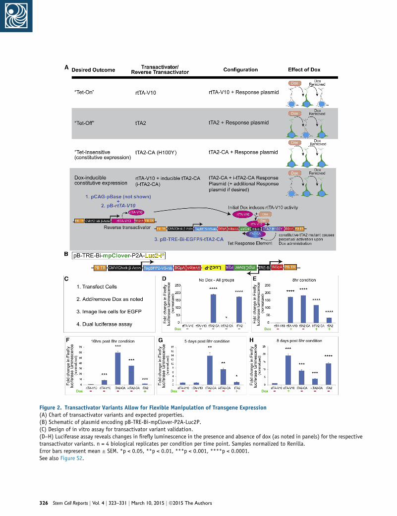
Figure 2. Transactivator Variants Allow for Flexible Manipulation of Transgene Expression(A) Chart of transactivator variants and expected properties.(B) Schematic of plasmid encoding pB-TRE-Bi-mpClover-P2A-Luc2P.(C) Design of in vitro assay for transactivator variant validation.(D–H) Luciferase assay reveals changes in firefly luminescence in the presence and absence of dox (as noted in panels) for the respectivetransactivator variants. n = 4 biological replicates per condition per time point. Samples normalized to Renilla.Error bars represent mean ± SEM. *p < 0.05, **p < 0.01, ***p < 0.001, ****p < 0.0001.See also Figure S2.
326 Stem Cell Reports j Vol. 4 j 323–331 j March 10, 2015 j ª2015 The Authors

induces rtTA-V10 activation, leading to i-tTA2-CA expres-
sion and continual TRE-Bi self-activation (Figure 2A).
In order to rapidly and quantitatively assay these transac-
tivator variants, we incorporated a destabilized firefly lucif-
erase (Luc2P) gene (Figures 2B and 2C). Further, Luc2P was
co-expressed using a P2A element with myristolated/
palmitolated-Clover (mpClover; Figure 2B), which is an
EGFP variant (Lam et al., 2012) tagged to membranes. pB-
TRE-Bi-mpClover-P2A-Luc2P-transduced cells did not
exhibit mpCLOVER expression or LUC2P activity without
dox (Figures 2D–2H; Figures S2A–S2E and SA1). With dox,
rtTA-V10 induced mpCLOVER at 8 hr, which was detect-
able up to 16 hr after dox removal (Figures S2B1–S2C1).
However, LUC2P displayed more rapid kinetics in that
luminescence was noticeably diminished at 16 hr (Fig-
ure 2F). This may reflect the longer half-life of mpCLOVER
when compared with destabilized LUC2P. Five days after
dox removal, neither mpCLOVER expression nor LUC2P
activity was detectable (Figure 2G; Figure S2D1). Dox was
subsequently re-added to the culture, and mpCLOVER
expression and LUC2P activity was detected (Figure 2H;
Figure S2E1). This demonstrates that the system can be
induced, reversed, and subsequently induced (i.e., On/
Off/On).
As expected, cells expressing the pB-TRE-Bi-EGFP/
tTA2-CA (Tet-insensitive) plasmid displayed constitutive
mpCLOVER expression and luminescence without dox
(Figures 2D–2H; Figures S2A2–S2E2). In contrast, cells ex-
pressing the pB-TRE-Bi-EGFP/i-tTA2-CA combined plasmid
did not exhibit basal mpCLOVER expression or lumines-
cence without dox (Figure 2D; Figure S2A3). However, after
a single dox administration, both were constitutively ex-
pressed—even after dox removal (Figures 2E–2H; Figures
S2B3–S2E3).
tTA2 (Tet-Off) exhibited high basal luciferase activity and
mpCLOVER expression, similar to that of dox-induced
rtTA-V10 (Figure 2D; Figure S2A4). mpCLOVER expression
decreased with dox addition until it was undetectable at
5 days, and it rebounded 3 days after dox removal (Figures
S2B4–S2D4). Luminescence decreased more rapidly (again,
presumably due to the attached PEST destabilization
domain), approaching baseline at 16 hr after dox addition,
though rebounding (along with mpCLOVER fluorescence)
after dox removal (Figures 2E–2H; Figure S2E4).
To confirm transposition, we nucleofected neural stem
cells with our pB-Tet-GOI system, alternate piggyBac
expression vectors encoding fluorescent proteins and
episomal expression vectors expressing analogous fluores-
cent proteins in various combinations—with and without
pBase. In the presence of pBase (and dox), pB-Tet-Bi-EGFP
and pB-mCherry demonstrated negligible loss of co-expres-
sion and retained high levels of fluorescence when assayed
by vital imaging at 3 and 6 days post-transduction (group 1;
Stem C
Figures S2F and S2G). Without pBase, the identical combi-
nation of plasmids (with dox) displayed relatively similar
co-expression at 3 days, but both GFP and mCHERRY
expression were almost undetectable by 6 days (group 2;
Figures S2F and S2G). Mixing different piggyBac or
episomal vectors in the presence of pBase demonstrated
that the episomal plasmid expression was lost at 3–6 days
(groups 3 and 4; Figures S2F and S2G). Conversely, the pig-
gyBac transposed fluorescent proteins were perdurant,
arguing against the possibility that non-specific toxicity
caused loss of fluorescent protein expression rather than
plasmid dilution (groups 3 and 4; Figures S2F and S2G).
tTA systems have shown promise in cell culture, but effi-
cacy in the CNS has been inconsistent. To test the robust,
non-leaky, inducible, and reversible characteristics of our
pB-Tet-GOI system in vivo, pB-TRE-Bi-EGFP was intro-
duced into ventricular zone (VZ) stem and progenitor cells
using postnatal EP, a technique that allows for rapid expres-
sion of plasmid-based transgenes in these cells (Boutin
et al., 2008; Breunig et al., 2012; Chesler et al., 2008). In
the postnatal VZ, radial glia stem and progenitor cells
differentiate into the mature cells of the striatum and pro-
vide neurons to the olfactory bulb (OB) (Alvarez-Buylla and
Garcia-Verdugo, 2002; Carleton et al., 2003; Fernandez
et al., 2011). Five days after EP of pB-TRE-Bi-EGFP, half of
the mice received a single dose of dox followed by brain
removal at 1 hr, 1 day, 4 days, 6 days, 10 days, 2 weeks,
3 weeks, and 4 weeks (Figure 3A; Figure S3). The naive
(un-stained) fluorescence of TagBFP2 and GFP was imaged
to avoid artifactual amplification of these signals (Fig-
ure 3B). Coronal brain sections showed nuclear TagBFP2
expression in the subventricular zone, striatum, and OB
of all animals (Figures 3C–3L). Six days after treatment,
robust GFP expressionwas observed in groups that received
a single dox dose, but not in groups without dox (Figures
3C–3F). This robust expression persisted up to 14 days,
decreasing at 3 weeks (Figures 3J and 3J1; Figure S3). Cells
at 4 weeks after doxwere indistinguishable from cells in an-
imals without dox (Figures 3C–3E, 3K, and 3L), demon-
strating the reversibility of transgene induction. Cell
counts at the OB core of dox-treated animals revealed a
decrease in the GFP+ cell number commencing after
14 days, while TagBFP2+ cell counts remained the same,
indicating that the cells were still present but that their
GFP expression turned off (Figure 3M). Analysis of the
mean fluorescence intensity after the single dox dose re-
vealed the most robust GFP expression at around 6 days,
before declining to baseline at approximately 4 weeks (Fig-
ure 3N). In total, these data indicate that the pB-Tet-GOI
system is a robust, inducible, and reversible method for
transgene manipulation in vivo.
Non-invasively validating transgene expression could
confirmappropriatedelivery/expressionofgenetic elements
ell Reports j Vol. 4 j 323–331 j March 10, 2015 j ª2015 The Authors 327
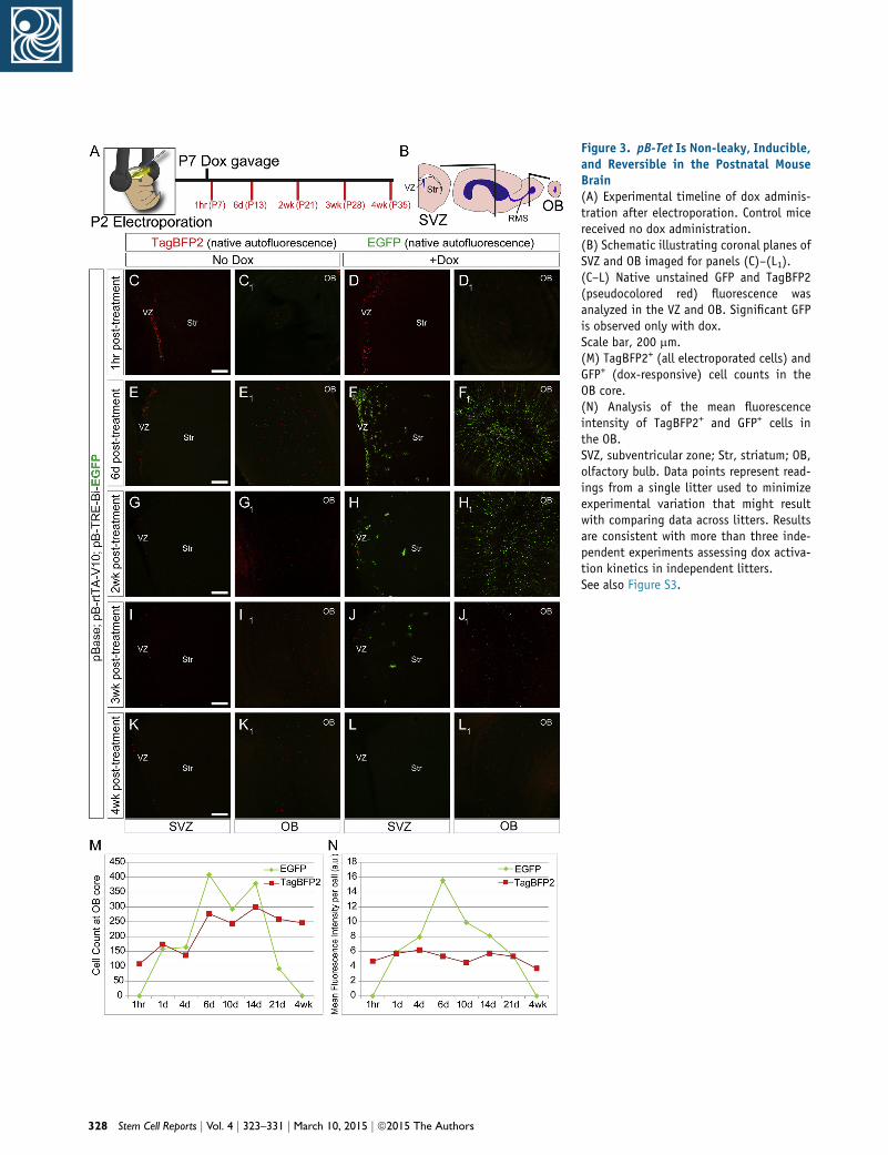
Figure 3. pB-Tet Is Non-leaky, Inducible,and Reversible in the Postnatal MouseBrain(A) Experimental timeline of dox adminis-tration after electroporation. Control micereceived no dox administration.(B) Schematic illustrating coronal planes ofSVZ and OB imaged for panels (C)–(L1).(C–L) Native unstained GFP and TagBFP2(pseudocolored red) fluorescence wasanalyzed in the VZ and OB. Significant GFPis observed only with dox.Scale bar, 200 mm.(M) TagBFP2+ (all electroporated cells) andGFP+ (dox-responsive) cell counts in theOB core.(N) Analysis of the mean fluorescenceintensity of TagBFP2+ and GFP+ cells inthe OB.SVZ, subventricular zone; Str, striatum; OB,olfactory bulb. Data points represent read-ings from a single litter used to minimizeexperimental variation that might resultwith comparing data across litters. Resultsare consistent with more than three inde-pendent experiments assessing dox activa-tion kinetics in independent litters.See also Figure S3.
328 Stem Cell Reports j Vol. 4 j 323–331 j March 10, 2015 j ª2015 The Authors

Figure 4. Addition of Luciferase into pB-Tet-GOI Allows Non-invasive Bioluminescence Imaging of Transgene Expression(A) Bioluminescence analysis revealed firefly activity in mice that received dox (n = 5 biological replicates per condition).(B) Quantification of total flux (p/s) in mice from (A).(C) Bioluminescence analysis of tTA2-CA alongside rtTA-V10 (n = 3, all littermates).(D) Quantification of total flux (p/s) in mice from (C).Error bars represent mean ± SEM. *p < 0.05. See also Figure S4.
and dox administration. We used a non-invasive imaging
method with our well-characterized inducible Luc2P
element (Figure 2B) in order to monitor dox induction of
the pB-Tet-GOI system. Mice electroporated with a pB-
TRE-Bi-mpClover-P2A-Luc2P-containing plasmid, along
with pBase and rtTA-V10, were analyzed using in vivo lumi-
nescence imaging, which showed a significant induction of
luminescence 1 week after dox administration (Figures 4A
and4B; see Figure S4A for timeline).After the initial imaging,
several animals were used for histological examination,
which demonstrated GFP+ cells in the dox-treated animals
and no GFP+ expression in untreated animals (data not
shown). One dox-treated animal not receiving a further
dose subsequentlydisplayed thebasal luminescence reading
of untreated littermates, while another littermate receiving
another dose exhibited continued luminescence (Fig-
ure S4B). Finally, we electroporated littermates with a pB-
TRE-Bi-mpClover-P2A-Luc2-P-containing plasmid along
with rtTA-V10 (two mice) or tTA2-CA (three mice). As ex-
pected, luminescence was observed at similar levels in all
Stem C
animals, with the exception of the littermate that did not
receive dox (Figures 4C and 4D). In summary, these data
demonstrate the ability to monitor expression of the ele-
ments of our genetic system in a non-invasive manner.
DISCUSSION
Here, we characterize pB-Tet-GOI, a customizable, flexible
genetic system that achieves stable transgene integration,
along with inducible and reversible dox-mediated trans-
gene expression in transduced cells and their resulting lin-
eages without the need for viral packaging and generation
of high viral titers. Specifically, we demonstrate the robust
dox-mediated regulation of the latest-generation rtTA and
tTA2 variants and promoter elements, allowing for trans-
gene expression in vitro and in vivo. Additionally, we pro-
vide tTA2-CA and i-tTA2-CA as a constitutive and inducible
constitutive activator, respectively, of TRE-Bi for dox-insen-
sitive expression of transgenes.
ell Reports j Vol. 4 j 323–331 j March 10, 2015 j ª2015 The Authors 329

Through pBase-mediated transposition, stable cell lines
can be rapidly generated. This technology not only
facilitates the investigation of cell biology in vitro but
also permits the in vivo induction of requisite transgenes
from transplanted cell populations. Therapeutic applica-
tions include induced growth factor delivery from trans-
planted cells (Behrstock et al., 2006). Also, induced, direct
differentiation in vivo permits the transplantation of
precursor cells, which expand easier in culture and survive
better post-transplantation, followed by dox-mediated
maturation.
Our pB-Tet-GOI system allows for more precise investi-
gations of gene function and cell lineages using in vivo
EP with the ability to stably genetically trace and revers-
ibly manipulate transgene expression. Notably, the
combination of rtTA-V10 and i-tTA2 permits permanent
cell lineage tracing after a single pulse. Thus, dox can be
used as an orthogonal ligand for gene manipulation
with other chemically induced genetic systems such as
tamoxifen and RU486-induced transgenes. Finally, there
is great potential to combine this system with INTRSECT
technology, including alternate recombinases to achieve
greater control over transgene manipulation (Fenno
et al., 2014).
This genetic system is not limited to use in the CNS and
is broadly applicable to many fields of biology. Any
context requiring temporal control of transgenes may
benefit from the use of the pB-Tet-GOI system. Further,
we have recently confirmed that mir30 and miR-E-based
small hairpin RNAs (Fellmann et al., 2013) can be similarly
induced by our system, providing temporally controlled
knockdown experiments (data not shown). Taken
together, the pB-Tet-GOI tool kit will be useful for the
investigation and elucidation of molecular mechanisms
of biological systems in a wide variety of contexts. Further,
it holds great promise for the fields of reprogramming and
gene therapy.
EXPERIMENTAL PROCEDURES
Mice and ElectroporationCD1 mice were used according to the Cedars-Sinai Medical Center
institutional animal care and use committee. EPs were performed
as described previously (Breunig et al., 2012). Briefly, postnatal
day 2 pups were placed on ice for �8 min until unresponsive to
tail pressure. A total of 1.2 ml of a plasmid cocktail of pBase
(0.5 mg/ml [final]), transactivator (1 mg/ml [final]), and response
plasmid (1 mg/ml [final]) diluted in Tris-EDTA buffer buffer was in-
jected into the left lateral ventricle. Fast green dye was added
(10%v/v) to visualize injections. PlatinumTweezertrodes delivered
five pulses of 120V (50ms; separated by 950ms) from the ECM830
System (Harvard Apparatus). SignaGel was applied to increase
conductance. Mice were then warmed with a heat lamp for
recovery.
330 Stem Cell Reports j Vol. 4 j 323–331 j March 10, 2015 j ª2015 The Auth
Cell Culture and NucleofectionHuNPCs (CNS010 cell line) were expanded as described previously
(Svendsen et al., 1997, 1998). Briefly, HuNPCs were expanded as
neurospheres in Stemline Media (Sigma S3194) with epidermal
growth factor (Sigma E9644), leukemia inhibitory factor (Millipore
LIF1010), and antibiotic-antimycotic (Life Technologies 15240-
062). Spheres were dissociated for nucleofection with pB-Tet
plasmids using the Lonza Nucleofector 2b device per themanufac-
turer’s recommendations. After nucleofection, media contained
B-27 (Life Technologies 12587-010) without growth factors.
DoxycyclineCulture media contained dox (final concentration 100 ng/ml,
Clontech 631311). Mice were gavaged with dox at 33 mg/g weight,
using a 5-mg/ml stock solution.
SUPPLEMENTAL INFORMATION
Supplemental Information includes Supplemental Experimental
Procedures, four figures, and one table and can be found
with this article online at http://dx.doi.org/10.1016/j.stemcr.
2015.01.013.
ACKNOWLEDGMENTS
We thank C. Svendsen for providing the HuNPCs and D. Eisenstat,
M. Lin, and J. Loturco for providing plasmids. We thank G. Gow-
ing, B. Shelley, V. Mattis, D. Sareen, and L. Garcia for experimental
assistance. We thank S. Svendsen for critical review. We acknowl-
edge support from the Samuel Oschin Comprehensive Cancer
Institute Cancer Research Forum Award (M.D., J.J.B.), the Board
ofGovernors RMI of Cedars-Sinai (M.D., J.J.B), the Thrasher-Broidy
Trinity College Research Fellowship (W.S.S), the Smidt Family
Foundation, and the Paul and Vera Guerin Family Foundation.
Received: November 17, 2014
Revised: January 14, 2015
Accepted: January 14, 2015
Published: February 19, 2015
REFERENCES
Ables, J.L., Breunig, J.J., Eisch, A.J., and Rakic, P. (2011). Not(ch)
just development: Notch signalling in the adult brain. Nat. Rev.
Neurosci. 12, 269–283.
Alvarez-Buylla,A., andGarcia-Verdugo, J.M. (2002).Neurogenesis in
adult subventricular zone. The Journal of neuroscience22, 629–634.
Behrstock, S., Ebert, A., McHugh, J., Vosberg, S., Moore, J.,
Schneider, B., Capowski, E., Hei, D., Kordower, J., Aebischer, P.,
and Svendsen, C.N. (2006). Human neural progenitors deliver glial
cell line-derived neurotrophic factor to parkinsonian rodents and
aged primates. Gene Ther. 13, 379–388.
Boutin, C., Diestel, S., Desoeuvre, A., Tiveron, M.C., and Cremer,
H. (2008). Efficient in vivo electroporation of the postnatal rodent
forebrain. PloS one 3, e1883.
Breunig, J.J., Arellano, J.I.,Macklis, J.D., and Rakic, P. (2007). Every-
thing that glitters isn’t gold: a critical review of postnatal neural
precursor analyses. Cell Stem Cell 1, 612–627.
ors
Breunig, J.J., Gate, D., Levy, R., Rodriguez, J., Jr., Kim, G.B., Daniel-
pour, M., Svendsen, C.N., and Town, T. (2012). Rapid genetic tar-
geting of pial surface neural progenitors and immature neurons
by neonatal electroporation. Neural Dev. 7, 26.
Carleton, A., Petreanu, L.T., Lansford, R., Alvarez-Buylla, A., and
Lledo, P.M. (2003). Becoming a new neuron in the adult olfactory
bulb. Nat. Neurosci. 6, 507–518.
Chen, F., and LoTurco, J. (2012). A method for stable transgenesis
of radial glia lineage in rat neocortex by piggyBac mediated trans-
position. J. Neurosci. Methods 207, 172–180.
Chesler, A.T., Le Pichon, C.E., Brann, J.H., Araneda, R.C., Zou, D.J.,
and Firestein, S. (2008). Selective gene expression by postnatal
electroporation during olfactory interneuron nurogenesis. PloS
one 3, e1517.
Fellmann, C., Hoffmann, T., Sridhar, V., Hopfgartner, B., Muhar,
M., Roth, M., Lai, D.Y., Barbosa, I.A., Kwon, J.S., Guan, Y., et al.
(2013). An optimized microRNA backbone for effective single-
copy RNAi. Cell Rep. 5, 1704–1713.
Fenno, L.E., Mattis, J., Ramakrishnan, C., Hyun, M., Lee, S.Y., He,
M., Tucciarone, J., Selimbeyoglu, A., Berndt, A., Grosenick, L.,
et al. (2014). Targeting cells with single vectors using multiple-
feature Boolean logic. Nat. Methods 11, 763–772.
Fernandez, M.E., Croce, S., Boutin, C., Cremer, H., and Raineteau,
O. (2011). Targeted electroporation of defined lateral ventricular
walls: a novel and rapid method to study fate specification during
postnatal forebrain neurogenesis. Neural Dev. 6, 13.
Furth, P.A., St Onge, L., Boger, H., Gruss, P., Gossen, M., Kistner, A.,
Bujard, H., andHennighausen, L. (1994). Temporal control of gene
expression in transgenic mice by a tetracycline-responsive pro-
moter. Proc. Natl. Acad. Sci. USA 91, 9302–9306.
Garcıa-Marques, J., and Lopez-Mascaraque, L. (2013). Clonal iden-
tity determines astrocyte cortical heterogeneity. Cereb. Cortex 23,
1463–1472.
Gossen,M., and Bujard,H. (1992). Tight control of gene expression
in mammalian cells by tetracycline-responsive promoters. Proc.
Natl. Acad. Sci. USA 89, 5547–5551.
Hecht, B., Muller, G., and Hillen, W. (1993). Noninducible Tet
repressor mutations map from the operator binding motif to the
C terminus. J. Bacteriol. 175, 1206–1210.
Kahlig, K.M., Saridey, S.K., Kaja, A., Daniels, M.A., George, A.L., Jr.,
andWilson,M.H. (2010). Multiplexed transposon-mediated stable
gene transfer in human cells. Proc. Natl. Acad. Sci. USA 107, 1343–
1348.
Stem C
Lam, A.J., St-Pierre, F., Gong, Y., Marshall, J.D., Cranfill, P.J., Baird,
M.A., McKeown, M.R., Wiedenmann, J., Davidson, M.W., Schnit-
zer, M.J., et al. (2012). Improving FRET dynamic range with bright
green and red fluorescent proteins. Nat. Methods 9, 1005–1012.
Mansuy, I.M., and Bujard, H. (2000). Tetracycline-regulated gene
expression in the brain. Curr. Opin. Neurobiol. 10, 593–596.
Marchetto,M.C., Brennand, K.J., Boyer, L.F., andGage, F.H. (2011).
Induced pluripotent stem cells (iPSCs) and neurological disease
modeling: progress and promises. Hum. Mol. Genet. 20 (R2),
R109–R115.
Petryniak, M.A., Potter, G.B., Rowitch, D.H., and Rubenstein, J.L.
(2007). Dlx1 and Dlx2 control neuronal versus oligodendroglial
cell fate acquisition in the developing forebrain. Neuron 55,
417–433.
Saito, T., and Nakatsuji, N. (2001). Efficient gene transfer into the
embryonic mouse brain using in vivo electroporation. Dev. Biol.
240, 237–246.
Shi, X., Harrison, R.L., Hollister, J.R., Mohammed, A., Fraser, M.J.,
Jr., and Jarvis, D.L. (2007). Construction and characterization of
new piggyBac vectors for constitutive or inducible expression of
heterologous gene pairs and the identification of a previously un-
recognized activator sequence in piggyBac. BMC Biotechnol. 7, 5.
Svendsen, C.N., Caldwell, M.A., Shen, J., ter Borg, M.G., Rosser,
A.E., Tyers, P., Karmiol, S., andDunnett, S.B. (1997). Long-term sur-
vival of human central nervous system progenitor cells trans-
planted into a rat model of Parkinson’s disease. Exp. Neurol. 148,
135–146.
Svendsen, C.N., ter Borg,M.G., Armstrong, R.J., Rosser, A.E., Chan-
dran, S., Ostenfeld, T., and Caldwell, M.A. (1998). A new method
for the rapid and long term growth of human neural precursor
cells. J. Neurosci. Methods 85, 141–152.
Zhang, Y., Pak, C., Han, Y., Ahlenius, H., Zhang, Z., Chanda, S.,
Marro, S., Patzke, C., Acuna, C., Covy, J., et al. (2013). Rapid sin-
gle-step induction of functional neurons from human pluripotent
stem cells. Neuron 78, 785–798.
Zhou, X., Vink, M., Klaver, B., Berkhout, B., and Das, A.T. (2006).
Optimization of the Tet-On system for regulated gene expression
through viral evolution. Gene Ther. 13, 1382–1390.
Zhu, P., Aller, M.I., Baron, U., Cambridge, S., Bausen, M., Herb, J.,
Sawinski, J., Cetin, A., Osten, P., Nelson, M.L., et al. (2007).
Silencing and un-silencing of tetracycline-controlled genes in neu-
rons. PLoS ONE 2, e533.
ell Reports j Vol. 4 j 323–331 j March 10, 2015 j ª2015 The Authors 331
Stem Cell Reports, Volume 4
Supplemental Information
A Transposon-Mediated System
for Flexible Control of Transgene Expression
in Stem and Progenitor-Derived Lineages
Aslam Abbasi Akhtar, Jessica Molina, Marina Dutra-Clarke, Gi Bum Kim, Rachelle Levy,
William Schreiber-Steinthorp, Moise Danielpour, and Joshua J. Breunig

Figure S1. pB-Tet-mediated directed differentiation and utility for sorting of undifferentiated
populations.
(A-C7) Vital imaging of HuNPCs reveals morphological changes in pB-Tet differentiating cells. HuNPCs were
nucleofected with pB-TRE-Bi-EGFP (ctrl), pB-TRE-Bi-EGFP/HA-Dlx2, or pB-TRE-Bi-EGFP/Ngn2 and
allowed to differentiate without growth factors for 6 weeks. EGFP autofluorescence was captured at denoted
time points in live cells.
Scale Bar = 50μm
(D-F2) Immunocytochemical staining reveals altered differentiation patterns across pB-TRE-Bi-EGFP (ctrl),
pB-TRE-Bi-EGFP/HA-Dlx2, or pB-TRE-Bi-EGFP/Ngn2. Yellow arrow indicates GFAP+ cell with polygonal
astrocyte morphology (D-D2). Red arrows (E-E2) indicate TUJ1+ cells with small, interneuron-like or migratory
neuron morphology. White arrows indicate TUJ1+ cells with long processes containing numerous varicosities
(white arrowheads) (F-F2).
Scale Bar (D, E, F) = 50μm
Scale Bar (D1,2, E1, 2, F1, 2) = 25μm
(G, H, I) Camera-lucida tracing of GFP+ cells at two weeks indicating the presence of mainly morphological
astrocyte-like cells in the pB-TRE-Bi-EGFP (ctrl) group with few morphological neuronal-like cells. Traced
GFP+ cells revealed morphologically small, potentially migratory interneuron-like cells in the pB-TRE-Bi-
EGFP/HA-Dlx2 group and large projection neuron-like cells in the pB-TRE-Bi-EGFP/Ngn2 group.
Scale Bar = 100μm
(J, K, L) FACS graphs indicating BFP+ populations in cells nucleofected with pB-TRE-Bi-EGFP (ctrl), pB-
TRE-Bi-EGFP/HA-Dlx2, or pB-TRE-Bi-EGFP/Ngn2
(M-M2) Sorted cells expanding as relatively pure populations of nucleospheres that are GFP-, BFP+. (pB-TRE-
Bi-EGFP/Ngn2 shown)
Scale Bar = 500μm
(N-O2) Nucleofected cells remain SOX2+ in the absence of dox, and lose SOX2 expression after dox-induced
differentiation. (pB-TRE-Bi-EGFP/Ngn2 shown)
Scale Bar = 50μm
Figure S1 is linked to main Figure 1
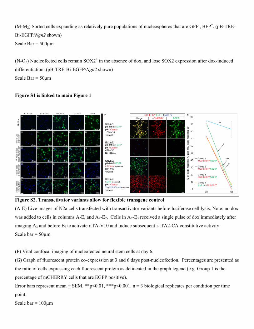
Figure S2. Transactivator variants allow for flexible transgene control
(A-E) Live images of N2a cells transfected with transactivator variants before luciferase cell lysis. Note: no dox
was added to cells in columns A-E, and A2-E2. Cells in A3-E3 received a single pulse of dox immediately after
imaging A3 and before B3 to activate rtTA-V10 and induce subsequent i-tTA2-CA constitutive activity.
Scale bar = 50µm
(F) Vital confocal imaging of nucleofected neural stem cells at day 6.
(G) Graph of fluorescent protein co-expression at 3 and 6 days post-nucleofection. Percentages are presented as
the ratio of cells expressing each fluorescent protein as delineated in the graph legend (e.g. Group 1 is the
percentage of mCHERRY cells that are EGFP positive).
Error bars represent mean + SEM. **p<0.01, ***p<0.001. n = 3 biological replicates per condition per time
point.
Scale bar = 100µm

Figure S2 is linked to main Figure 2
Figure S3. pB-Tet-GOI is non-leaky, inducible and reversible in the mouse brain (full panel set related to
Figure 3)
(A) Experimental timeline for postnatal electroporation and dox activation. (B) Brain regions analyzed in C-
H1. (C-H1) Full set of fluorescence images for main Figure 3. Minimal EGFP expression is observed in
littermates without dox. Examination of the brains of mice electroporated with pBase, pB-rtTA-V10, and pB-
TRE-EGFP reveals detectable EGFP expression at 1 day. Littermates used to decrease experimental variation
across litters. Results are consistent with >3 independent experiments investigating dox activation kinetics in
separate litters.

Scale bar = 200µm
Figure S3 is linked to main Figure 3
Figure S4. Reversibility of non-invasive bioluminescence imaging of pB-Tet-GOI variants
(A) Timeline of experiments for littermates depicted in main Figure 4A and Supp. Figure 4C.
(B) Dox administration was continued (far right) or discontinued (far left) in littermates from Figure 4a.
Figure S4 is linked to main Figure 4
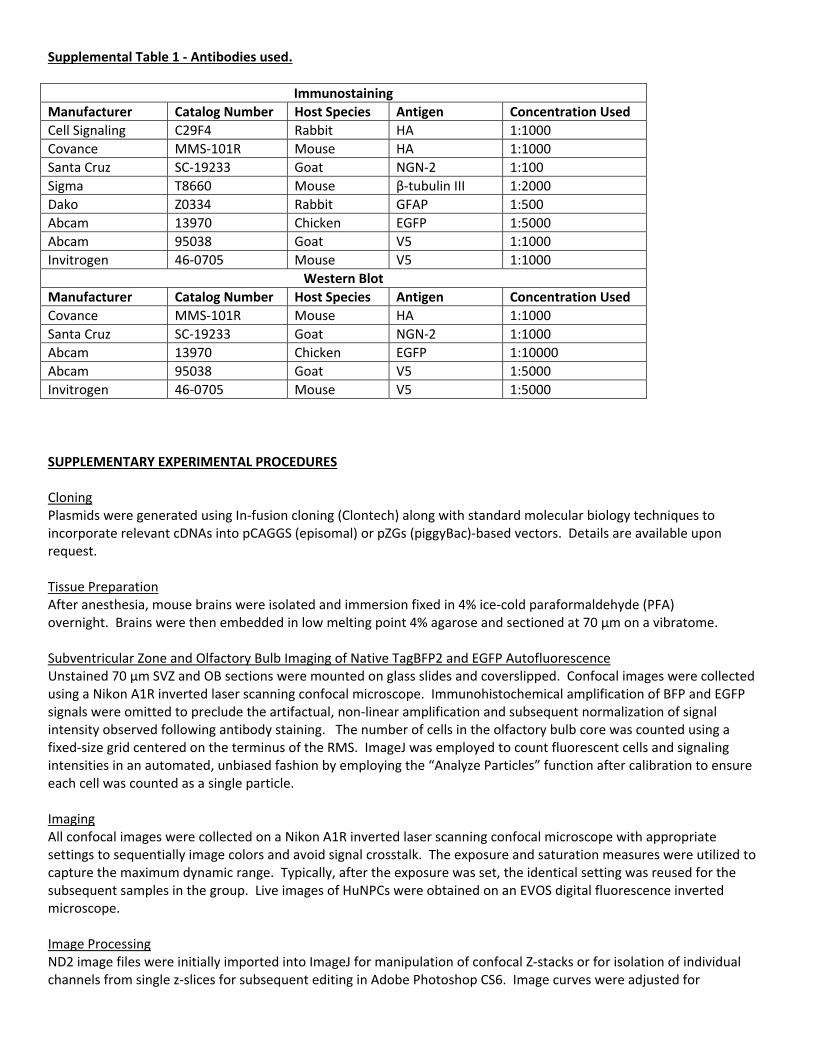
Supplemental Table 1 ‐ Antibodies used.
Immunostaining
Manufacturer Catalog Number Host Species Antigen Concentration Used
Cell Signaling C29F4 Rabbit HA 1:1000
Covance MMS‐101R Mouse HA 1:1000
Santa Cruz SC‐19233 Goat NGN‐2 1:100
Sigma T8660 Mouse β‐tubulin III 1:2000
Dako Z0334 Rabbit GFAP 1:500
Abcam 13970 Chicken EGFP 1:5000
Abcam 95038 Goat V5 1:1000
Invitrogen 46‐0705 Mouse V5 1:1000
Western Blot
Manufacturer Catalog Number Host Species Antigen Concentration Used
Covance MMS‐101R Mouse HA 1:1000
Santa Cruz SC‐19233 Goat NGN‐2 1:1000
Abcam 13970 Chicken EGFP 1:10000
Abcam 95038 Goat V5 1:5000
Invitrogen 46‐0705 Mouse V5 1:5000
SUPPLEMENTARY EXPERIMENTAL PROCEDURES Cloning Plasmids were generated using In‐fusion cloning (Clontech) along with standard molecular biology techniques to incorporate relevant cDNAs into pCAGGS (episomal) or pZGs (piggyBac)‐based vectors. Details are available upon request. Tissue Preparation After anesthesia, mouse brains were isolated and immersion fixed in 4% ice‐cold paraformaldehyde (PFA) overnight. Brains were then embedded in low melting point 4% agarose and sectioned at 70 µm on a vibratome. Subventricular Zone and Olfactory Bulb Imaging of Native TagBFP2 and EGFP Autofluorescence Unstained 70 µm SVZ and OB sections were mounted on glass slides and coverslipped. Confocal images were collected using a Nikon A1R inverted laser scanning confocal microscope. Immunohistochemical amplification of BFP and EGFP signals were omitted to preclude the artifactual, non‐linear amplification and subsequent normalization of signal intensity observed following antibody staining. The number of cells in the olfactory bulb core was counted using a fixed‐size grid centered on the terminus of the RMS. ImageJ was employed to count fluorescent cells and signaling intensities in an automated, unbiased fashion by employing the “Analyze Particles” function after calibration to ensure each cell was counted as a single particle. Imaging All confocal images were collected on a Nikon A1R inverted laser scanning confocal microscope with appropriate settings to sequentially image colors and avoid signal crosstalk. The exposure and saturation measures were utilized to capture the maximum dynamic range. Typically, after the exposure was set, the identical setting was reused for the subsequent samples in the group. Live images of HuNPCs were obtained on an EVOS digital fluorescence inverted microscope. Image Processing ND2 image files were initially imported into ImageJ for manipulation of confocal Z‐stacks or for isolation of individual channels from single z‐slices for subsequent editing in Adobe Photoshop CS6. Image curves were adjusted for

consistency of dynamic range and exposure in Photoshop CS6, cropped, and then imported into Adobe Illustrator CS6 for the preparation of final images. In vitro Phenotypic Quantification Cell phenotype percentages (TUJ1+ and GFAP+ of all GFP+ and BFP+ cells) were quantified in an unbiased manner by an observer blind to the treatment groups. TUJ1+ and GFAP+ quantification was done in triplicates (n= 3 coverslips per condition per timepoint); 100 GFP+ cells per coverslip, performed in triplicate. Nucleofection Human Neural progenitor cell nucleofection was performed using the Amaxa Nucleofector 2b device according to manufacturer’s recommendations (Lonza AG). Unless otherwise noted, plasmids were added to the Lonza Nucleofection Solution in the following amounts (per reaction): Response plasmid(s): 7µg, pBase: 1µg, rtTA‐V10: 3µg. For transposition experiment (Figure S2, F‐G), plasmids were added to their respective groups in the following amounts (per reaction): pBase: 1µg, rtTA‐V10: 3µg, all other plasmids: 7µg. Immunostaining of HuNPCs Immunocytochemistry was performed as previously described (Breunig et al., 2007b). Briefly, a primary antibody mixture was made in PBS‐Triton (PBS, 0.3 % triton) with 3% normal donkey serum (NDS) and the desired primary antibodies at the ratios indicated in Supp. Table 1. Coverslips were incubated with the primary antibody mixture for at least 12 hours at 4°C, followed by three 5 min washes with PBS at room temperature. Secondary antibody mixtures were made with PBS‐T and the appropriate secondary antibodies at a 1:1000 dilution (Jackson Immunoresearch; conjugated with Alexa 405, Fitc, Alexa488, Dylight488, Alexa555, Dylight549, Alexa647, or Dylight649). ). The secondary antibody mixture was added to the coverslips and incubated at room temperature for 1 hour on a shaker. Coverslips were then washed in PBS and mounted on slides with anti‐fade mounting gel medium (Invitrogen ProLong). Western Blot Mouse N2a cells were transfected with Lipofectamine 2000 (Invitrogen 11668019), following the manufacturer’s protocol. After transfection, the cells were grown for three days at 37°C. Cells were harvested by accutase incubation for 3 min at 37°C, followed by re‐suspension in equal amount media, and centrifugation for 3 min at 3000 rpm. The resulting pellet was re‐suspended in laemmli buffer and boiled for 15 min at 95°C. Protein concentrations were measured using a ThermoScientific Nano Drop. Protein separation was performed using SDS‐PAGE separation and transfered onto nitrocellulose membranes, which were incubated overnight at 4°C using primary antibodies listed in Supp. Table 1 diluted in 5% milk in 0.1% PBS‐Tween. All secondary antibodies (Li‐cor IRDye®) were used at a 1:15000 dilution. Infrared detection was accomplished by the Li‐Cor Odyssey® CLX Imaging System. FACS Once harvested cells reached approximately 80% confluency, media was removed and T75 cm2 flasks were passaged with 2 mL of accutase at 37°C in 5% CO2 for 3 minutes. Accutase was neutralized with 2mL of media and cells were centrifuged at 1350 rpm for 3 minutes. Supernatant was removed and cells were resuspended in 2 mL of fresh media, using a P1000 to disrupt the cells in a careful yet vigorous manner. In increments of approximately 200 μL, cells were filtered through a 70 μm filter (BD Falcon). This filtrate was then placed on ice. Cells were then sorted at the Cedars‐Sinai Flow Cytometry Core using a Beckman Coulter MoFlo sorter for blue fluorescence with tight gates making sure only the cells highly expressing BFP were collected, indicative of TagBFP‐V5 expression from the response plasmid. Cells were collected into fresh media and kept cold on ice. Once all cells were sorted, the collected cells were pelleted, washed and placed into new T25 cm2 flasks. IVIS Live Imaging Bioluminescence imaging of animals was performed at the Cedars‐Sinai Medical Center Imaging Core Facility using the Xenogen Spectrum In Vivo Imaging System (IVIS). Animals were subcutaneously injected with 150mg/kg luciferin (VivoGloTM, Promega 1043). Following a 10 minute waiting period to allow for circulation of substrate, animals were imaged in the Xenogen IVIS while under isoflurane anesthesia. Luciferase expression was analyzed and quantified by substracting the background noise (e.g. signal hindlimb regions distal from CNS) from each respective animal.

Dual‐Luciferase Reporter Assay Mouse N2a cells were transfected with Lipofectamine 2000 (Invitrogen 11668019), following the manufacturers protocol. Cells were grown in a 24‐well plate as a monolayer in quadruplets (n=4 per condition) and imaged at respective time points followed by lysis and dual‐luciferase activity assessment using the Promega Dual‐Luciferase® Assay E910 according to the manufacturer’s protocol. Briefly, cells were passively lysed by adding 100ul of Promega® PLB buffer and shaking at room temperature for 15 minutes. 20ul of lysate and 50ul of Promega® LARII substrate was added to a 96‐well assay plate. Firefly luciferase activity was measured using the Wallac Envision Manager Software with a 10 second delay per well. After firefly luciferase activity was measured, 50ul of Promega Stop & Glo® was added and Renilla activity was measured. Statistical Analysis Statistical analyses were carried out using Excel (Microsoft). We compared groups using two sample t tests. For cell counts, at least 100 cells per biological replicate were counted in all cases with the exception of Group 2 in Supplemental Figure S2, where 99 cells were counted.