a.i. procedure manual - the california department of public health
TRANSCRIPT

A.I. Procedure M
anual July 2006
Wildlife Services & State/Tribal Cooperator
Avian Influenza Surveillance
Procedure Manual
July 2006

Procedure Manual for Avian Influenza (AI) Surveillance
Table of Contents I. Introduction ..................................................................................................................................1
A. Purpose...................................................................................................................................1 B. Highly Pathogenic H5N1 Description ...................................................................................1 C. Surveillance Plan Overview...................................................................................................1
II. Detailed Sampling Procedures .....................................................................................................1 A. Sampling Strategies...............................................................................................................1
Investigation of Morbidity/Mortality Events .......................................................................1 Surveillance in Live Wild Birds ..........................................................................................2
Surveillance in Hunter-killed Birds .....................................................................................2 Dead Wild Bird ....................................................................................................................2
Sentinel Species ...................................................................................................................2 Environmental Sampling .....................................................................................................2 B. Personal Safety Guidelines and Equipment ..........................................................................3
Guidelines for Wildlife Biologists Handling Healthy Wild Birds......................................3 Guidelines for Wildlife Biologists Handling Sick or Dead Birds.......................................3
Guidelines for Highly Pathogenic Avian Influenza Response ...........................................3 Safety and Personal Protective Equipment (PPE) ..............................................................4
Web Sites ............................................................................................................................4 C. Procedures for Collecting and Shipping Samples from Wild Birds .....................................4
Cloacal Swabs.....................................................................................................................4 Tracheal Swabs (Morbidity/Mortality events only)............................................................5
Data sheets ..........................................................................................................................6 Instructions for AI Surveillance Field Data Sheets .......................................................6 Instructions for NAHLN Lab Submission Form for AI Samples ..................................7
Proper Labeling of Samples................................................................................................9 Storage of Transport Media ................................................................................................9
Shipping Samples................................................................................................................9 Hand Delivery of Samples to Lab.....................................................................................10 Identification of a NAHLN Laboratory ............................................................................11 Proper Communication of Submitting Specimens............................................................11 Reporting Results..............................................................................................................11 D. Procedures for Collecting and Shipping Environmental Samples ......................................12 Fecal Sample Collection ...................................................................................................12 Water Collection ...............................................................................................................14
III. Data Management Using the HPAI Early Detection System (HEDDS) ..................................14 A. Overview..............................................................................................................................14
Background.......................................................................................................................14 General Information..........................................................................................................14
B. Data Entry ............................................................................................................................15 Web Site Access ...............................................................................................................15 Expected Transitional Phases ...........................................................................................15
C. Obtaining a User Name and Password.................................................................................16 Contact the Wildlife Services Data Administrators..........................................................16
Version 1.0– July 17, 2006 Table of Contents
Procedure Manual for Avian Influenza (AI) Surveillance
D. Data Mapping and Reporting...............................................................................................16
Data Availability for Contributors ....................................................................................16 IV. Appendices ...................................................................................................................................
Appendix A: U.S. Interagency Strategic Plan ........................................................................A1 Appendix B: Bird Capture Equipment.................................................................................... B1
Appendix C: National Wildlife Disease Program Contact Information ................................. C1 Appendix D: USDA APHIS Directive 6800.1 .......................................................................D1 Appendix E: Sample AI Surveillance Field Data Sheet ......................................................... E1
Appendix F: Species Codes .....................................................................................................F1 Appendix G: Sample NAHLN Lab Submission Form for AI Samples..................................G1
Appendix H: Laboratories Approved to Participate in AI Sample Testing ............................H1 Appendix I: Sample Environmental Submission Form ......................................................... I1 Appendix J: Tools and Supplies for Sampling ...................................................................... J1 Appendix K: Communication Protocol in the Event of an HPAI Finding ............................K1
Version 1.0– July 17, 2006 Table of Contents

Procedure Manual for Avian Influenza (AI) Surveillance
I. Introduction
A. Purpose This document describes the guidelines and procedures for Wildlife Services employees and their State/Tribal Cooperators within the framework of the National Early Detection System for Highly Pathogenic H5N1 Avian Influenza in Wild Migratory Birds. The goal of the system is to provide early warning for potentially catastrophic mortality events due to highly pathogenic H5N1 avian influenza (AI) in North American wild birds and to minimize the potential for human and poultry exposures. Surveillance samples will be collected in all 50 U.S. states and select U.S. territories. The purpose of this document is to clarify:
• Capture equipment and sampling kits • Sampling strategies • Protocols used for each sampling technique • Safety and personal protective equipment (PPE) • How to ship AI surveillance samples to the National Animal Health Laboratory
Network (NAHLN) • How to manage, store, and retrieve surveillance data on the USGS HEDDS
B. Highly Pathogenic H5N1 Description Avian influenza (AI) is a type A influenza virus that is naturally found in certain species of waterfowl and shorebirds. However, the recent occurrence of a highly pathogenic avian influenza (HPAI) H5N1 has raised concern about the potential impact on wild birds, domestic poultry, and human health. The virus could enter the U.S. via several routes, including illegal movement of domestic or wild birds, contaminated products, infected travelers, bioterrorism, and migrations of infected wild birds. This plan focuses primarily on the detection of a potential introduction of highly pathogenic avian influenza by migratory birds.
C. Surveillance Plan Overview Please refer to Appendix A entitled: “An Early Detection System for Highly Pathogenic H5N1 Avian Influenza in Wild Migratory Birds U.S. Interagency Strategic Plan.”
• Questions regarding the U.S. Interagency Strategic Plan should be directed to Tom DeLiberto ([email protected] or (970) 266-6088) or Seth Swafford ([email protected] or (301) 734-3570).
II. Detailed Sampling Procedures
A. Sampling Strategies • Investigation of Morbidity/Mortality Events:
Any bird that is found dead and sampled should be classified as a morbidity/mortality event. Paired cloacal and tracheal swabs should be submitted for each dead bird that is sampled (see section II. C.). While agencies or
Version 1.0 – July 17, 2006 Page 1

Procedure Manual for Avian Influenza (AI) Surveillance organizations are encouraged to submit carcasses for a complete necropsy and diagnostic testing, funds received from Wildlife Services for AI surveillance can only be used to diagnose AI from cloacal and tracheal swabs. However, in the case of significant morbidity/mortality events, please contact the Wildlife Disease Coordinator’s office for consultation to ensure the appropriate testing is performed.
• Surveillance in Live Wild Birds: This strategy incorporates sampling of live-captured, apparently healthy wild birds to detect the presence of HPAI virus. Birds are captured using a variety of methods, sampled, and released on site (Appendix B). Cloacal swabs are the only type of sample collected.
• Surveillance in Hunter-killed Birds: Hunter check stations provide an opportunity to conduct surveillance for HPAI. Collection of samples from these species will occur at hunter check stations during hunting seasons in areas where these birds stage during migration. It is unnecessary to pinpoint exact GPS coordinates for each bird that is sampled; the check station may be recorded as the location. Cloacal swabs are the only sample that should be collected (see section II. C.).
• Dead Wild Birds This surveillance category is used to refer to opportunities that Wildlife Services personnel, in particular, have to sample birds that are being removed as part of an operational assignment (goose round-up, etc). Birds taken under this strategy fall under federal and state permits but they are not taken for sport or recreational purposes. Cloacal swabs should be collected from these birds if they are on the state species list for AI.
• Sentinel Species:
The strategic plan discusses implementation of sentinel flocks. Wild flocks also may serve as sentinels. For example, resident ducks and geese at urban parks may serve as sentinels if they can be repeatedly sampled. Cloacal swabs are collected on a regular basis from the sentinel flocks and are the only type of sample that should be collected as long as the flock remains healthy. If the flock becomes sick or a mortality event occurs, then paired samples (cloacal and tracheal) should be collected (see section II. C.). If sentinel flocks will be sampled, please contact the Wildlife Disease Coordinator’s office for assistance (Appendix C).
• Environmental Sampling: Avian influenza viruses are generally released by waterfowl through the intestinal tract and viable virus can be detected in both feces and the water in which the birds swim, defecate, and feed. Analysis of both water and fecal material from waterfowl habitat can provide evidence of HPAI virus circulating in wild bird populations, the specific HPAI subtypes, levels of pathogenicity, and possible risks to poultry and susceptible livestock. Monitoring of water and/or fecal samples gathered from waterfowl habitat is a reasonably cost effective,
Version 1.0 – July 17, 2006 Page 2

Procedure Manual for Avian Influenza (AI) Surveillance technologically achievable method of surveillance. Fecal samples will be collected from the ground primarily by Wildlife Services personnel (except state agencies in Alaska, California, Oregon, and Washington) (see section II. D.). Sampling water for AI is currently considered a research method under development by the National Wildlife Research Center. Once a protocol has been developed, some offices will be requested to collect water samples.
B. Personal Safety Guidelines and Equipment (Appendix D)
• Guidelines for Wildlife Biologists Handling Healthy Wild Birds: 1. Work in a well-ventilated area if working indoors. 2. Work upwind of animals, to the extent possible, to decrease the risk of
inhaling aerosols such as dust, feathers, or dander when working outdoors. 3. Wear rubber or latex gloves that can be disinfected or disposed of. 4. Wash hands with soap and water often and disinfect work surfaces and
equipment between sites. If soap and water are not available, alcohol-based hand cleaner or 10% bleach/water solution should be used.
5. Do not eat, drink, or smoke while handling birds. 6. PPE should include boots, coveralls, and gloves and eye protection.
• Guidelines for Wildlife Biologists Handling Sick or Dead Birds:
1. Work in a well-ventilated area if working indoors. 2. Work upwind of animals, to the extent possible, to decrease the risk of
inhaling aerosols such as dust, feathers, or dander when working outdoors. 3. Wear rubber or latex gloves that can be disinfected or disposed of. 4. Wash hands with soap and water often and disinfect work surfaces and
equipment between sites. If soap and water are not available, alcohol-based hand cleaner or 10% bleach/water solution should be used.
5. Wear protective clothing, including coveralls, rubber boots, and latex or rubber gloves that can be disinfected or disposed of.
6. Minimize exposure to mucosal membranes by wearing protective eyewear (goggles) and a particulate respirator (NIOSH N95 respirator at a minimum).
7. Decontaminate and properly dispose of potentially infectious material including carcasses.
8. Do not eat, drink, or smoke while handling birds.
• Guidelines for Highly Pathogenic Avian Influenza Response: 1. Work in a well-ventilated area if working indoors. 2. Work upwind of animals, to the extent possible, to decrease the risk of
inhaling aerosols such as dust, feathers, or dander when working outdoors. 3. Wear protective clothing, including coveralls, rubber boots, and latex or
rubber gloves that can be disinfected or disposed of. 4. Decontaminate and properly dispose of infectious material including
carcasses. 5. Wash hands with soap and water frequently and disinfect exposed surfaces
and field equipment between work sites. Alcohol-based hand cleaner or 10% bleach/water solution may be used if soap and water are not available.
6. Do not eat, drink, or smoke while handling birds.
Version 1.0 – July 17, 2006 Page 3

Procedure Manual for Avian Influenza (AI) Surveillance 7. Health should be monitored for clinical signs of influenza infection, such as
fever, cough or sore throat, trouble breathing, or eye inflammation, during and for one week after, last exposure to potentially HPAI virus-infected or exposed birds.
8. Contact health care provider if fever, flu-like symptoms, or conjunctivitis (eye inflammation) develops. Inform the provider prior to arrival of potential exposure to HPAI.
9. PPE should include complete coveralls, gloves, and boot covers that are either disposable or that can be disinfected. Goggles, N95 masks (NIOSH respirator preferred) as well as a health monitoring plan are required.
• Safety and Personal Protective Equipment (PPE)
1. N95 masks 2. Gloves – latex or nitrile 3. Protective clothing including:
• Tyvek (or equivalent) apron or body suit • Disposable booties • Goggles or protective face shield
• Web Sites: Please refer to the following web sites for additional guidelines for proper personal protection techniques: 1. http://www.nwhc.usgs.gov/publications/wildlife_health_bulletins/WHB_05_03.jsp 2. http://www.nwhc.usgs.gov/research/WHB/WHB_05_03.html 3. http://www.cdc.gov/flu/avian/professional/protect-guid.htm 4. http://www.cdc.gov/flu/avian/professional/protect-guid.htm
C. Procedures for Collecting and Shipping Samples from Wild Birds (see Appendix J for a list of supplies for sampling)
• Cloacal Swabs 1. Unwrap a Dacron swab from the stem-end of the packaging. (Use a small or
large swab depending on size of the bird) (Small swabs for shorebirds are provided upon request; contact Kerri Pedersen ([email protected] or (970) 266-6272) or Brandon Schmit ([email protected] or (970) 266-6079).
2. Remove swab and insert the tip of the swab into the cloaca of the bird. 3. Gently twirl the swab inside the cloaca taking care to insert the swab just far
enough to completely cover the tip of the swab. 4. Open a vial containing prepared BHI media. 5. Insert the swab into the media. 6. Raise the swab about ¼ inch from the bottom of the vial. While holding the
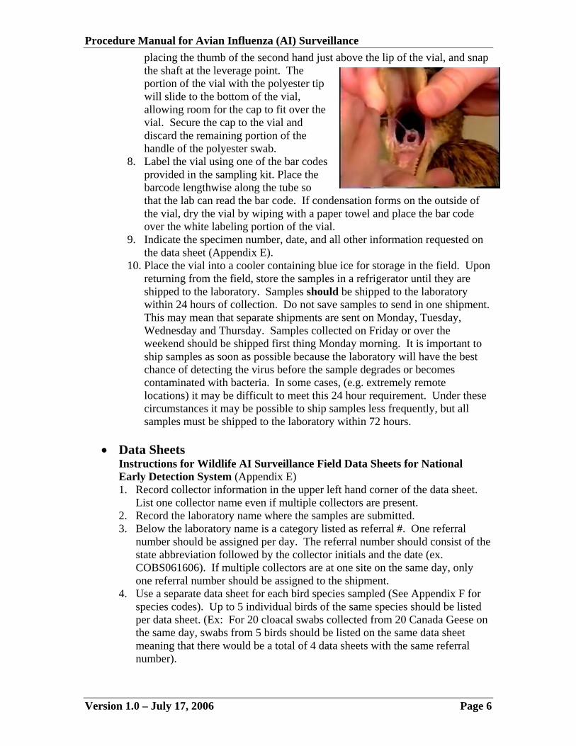
vial in one hand, leverage the shaft of the swab against the lip of the vial, placing the thumb of the second hand just above the lip of the vial, and snap the shaft at the leverage point. The portion of the vial with the polyester tip will slide to the bottom of the vial, allowing room for the cap to fit over the vial. Secure the cap to the vial and discard the remaining portion of the handle of the polyester swab.
Version 1.0 – July 17, 2006 Page 4

Procedure Manual for Avian Influenza (AI) Surveillance 7. Label the vial using one of the bar codes provided in the sampling kit. Place
the barcode lengthwise along the tube so that the lab can read the bar code. If condensation forms on the outside of the vial, dry the vial by wiping with a paper towel and place the bar code over the white labeling portion of the vial.
8. Indicate the sample number, date, and all other information requested on the data sheet (Appendix E).
9. Place the vial into a cooler containing blue ice for storage in the field. Upon returning from the field, store the samples in a refrigerator until they are shipped to the laboratory. Samples should be shipped to the laboratory within 24 hours of collection. Do not save samples to send in one shipment. This may mean that separate shipments are sent on Monday, Tuesday, Wednesday and Thursday. Samples collected on Friday or over the weekend should be shipped first thing Monday morning. It is important to ship samples as soon as possible because the laboratory will have the best chance of detecting the virus before the sample degrades or becomes contaminated with bacteria. In some cases, (e.g. extremely remote locations) it may be difficult to meet this 24 hour requirement. Under these circumstances it may be possible to ship samples less frequently, but all samples must be shipped to the laboratory within 72 hours.
10. Only ONE swab should be taken per bird.
• Tracheal Swabs (morbidity/mortality events only)
1. Be sure that you understand or have been shown the difference between the tracheal and the oral-pharyngeal opening.
2. Gently pinch both sides of the head of the bird near the base of its beak. This will force the bird to open its mouth and expose its oral cavity.
Photo courtesy of USGS.
3. In most waterfowl, you can open the trachea by gently pushing upwards on the neck just below the lower bill.
4. Insert the swab into the trachea while gently swirling the swab in an up and down motion.
5. Open a vial containing prepared BHI media. 6. Insert the swab into the media. 7. Raise the swab about ¼ inch from the bottom of the vial. While holding the
vial in one hand, leverage the shaft of the swab against the lip of the vial,
Version 1.0 – July 17, 2006 Page 5
Procedure Manual for Avian Influenza (AI) Surveillance placing the thumb of the second hand just above the lip of the vial, and snap the shaft at the leverage point. The portion of the vial with the polyester tip will slide to the bottom of the vial, allowing room for the cap to fit over the vial. Secure the cap to the vial and discard the remaining portion of the handle of the polyester swab.
8. Label the vial using one of the bar codes provided in the sampling kit. Place the barcode lengthwise along the tube so that the lab can read the bar code. If condensation forms on the outside of the vial, dry the vial by wiping with a paper towel and place the bar code over the white labeling portion of the vial.
9. Indicate the specimen number, date, and all other information requested on the data sheet (Appendix E).
10. Place the vial into a cooler containing blue ice for storage in the field. Upon returning from the field, store the samples in a refrigerator until they are shipped to the laboratory. Samples should be shipped to the laboratory within 24 hours of collection. Do not save samples to send in one shipment. This may mean that separate shipments are sent on Monday, Tuesday, Wednesday and Thursday. Samples collected on Friday or over the weekend should be shipped first thing Monday morning. It is important to ship samples as soon as possible because the laboratory will have the best chance of detecting the virus before the sample degrades or becomes contaminated with bacteria. In some cases, (e.g. extremely remote locations) it may be difficult to meet this 24 hour requirement. Under these circumstances it may be possible to ship samples less frequently, but all samples must be shipped to the laboratory within 72 hours.
• Data Sheets
Instructions for Wildlife AI Surveillance Field Data Sheets for National Early Detection System (Appendix E) 1. Record collector information in the upper left hand corner of the data sheet.
List one collector name even if multiple collectors are present. 2. Record the laboratory name where the samples are submitted. 3. Below the laboratory name is a category listed as referral #. One referral
number should be assigned per day. The referral number should consist of the state abbreviation followed by the collector initials and the date (ex. COBS061606). If multiple collectors are at one site on the same day, only one referral number should be assigned to the shipment.
4. Use a separate data sheet for each bird species sampled (See Appendix F for species codes). Up to 5 individual birds of the same species should be listed per data sheet. (Ex: For 20 cloacal swabs collected from 20 Canada Geese on the same day, swabs from 5 birds should be listed on the same data sheet meaning that there would be a total of 4 data sheets with the same referral number).
Version 1.0 – July 17, 2006 Page 6
Procedure Manual for Avian Influenza (AI) Surveillance 5. Circle the type of sample that is collected from the birds. If a cloacal and
tracheal sample are collected from the same bird (such as in the case of a morbidity/mortality event), a separate data sheet must be used for each type of sample collected even though the sample is from the same species of bird.
6. Collection site is defined as the refuge, lake, or name used to refer to the area where the samples are collected.
7. The 3 most abundant bird species on the site should be recorded. Use species codes listed on the table. These may differ from the birds captured or sampled at the site. One or more of these fields may be left blank if < 3 bird species are observed at the site.
8. Record the date and GPS location. The GPS unit must be set in the WGS 84 datum and in decimal degrees before recording the location (ddd.ddddd). For hunter check stations, coordinates may be taken at the check station where the birds are sampled.
9. Record the subject ID. The sample bar code # may be used for this if the sample is from any collection strategy other than a morbidity/mortality event. In the case of a morbidity/mortality event (the only time TWO swabs will be collected per bird – tracheal and cloacal), you must assign a unique subject ID so that the tracheal and cloacal swabs can be identified as originating from the same individual. The subject ID can be any identifier you choose as long as it is unique between subjects.
10. Condition need only be filled out for morbidity/mortality events or sentinel animals except in unusual circumstances (e.g., live capture of an obviously sick bird). All birds listed as “sick” will be assumed to have been euthanized and removed from the wild unless otherwise indicated in the comments. For other collection strategies it will be understood that the condition or fate of the bird will be dead for hunter-killed birds and dead wild birds and released and healthy for birds marked as live wild birds unless otherwise indicated in the comments section.
11. When collecting the samples, place one bar code on the sample and one on the field data sheet. (Keep the extra bar codes because they will be used on the laboratory submission form). Circle one each in the categories of collection strategy, sex, condition, and age class. Record any additional information such as band number in the comments section.
12. At the bottom of the data sheet record the date the samples are shipped to the lab and the total number of samples that are included in the shipment. Record the name of the person who actually sends the samples to the lab (this may be different from the person collecting the samples).
13. Make a copy of the field data sheet and send it to Brandon Schmit or Kerri Pedersen at the National Wildlife Research Center 4101 LaPorte Avenue, Fort Collins, CO 80521 or fax to (970) 266-6089 or (970) 266-6203.
Instructions for NAHLN Laboratory Submission Form for AI Samples (Appendix G)
1. Record the collector information in the upper left hand corner of the data sheet. List one collector name even if multiple collectors are present.
2. Record the name and address of the laboratory where the samples are submitted (See Appendix H for a list of NAHLN laboratories).
Version 1.0 – July 17, 2006 Page 7
Procedure Manual for Avian Influenza (AI) Surveillance 3. Use a separate lab submission form for each bird species sampled (See
Appendix F for species codes). Up to 5 individual birds of the same species may be listed per submission form. If birds of the same species were sampled in different locations on the same day they may be combined on the lab submission form.
4. Circle the type of sample that is collected from the birds. If a cloacal and tracheal sample are collected from the same bird (such as in the case of a morbidity/mortality event), a separate lab submission form must be used for each type of sample collected even though the sample is from the same species of bird or even the same individual bird.
5. Record the date, county and state where the samples were collected. 6. List the purchase order number to which the samples should be charged. 7. Below the purchase order number is a category listed as referral #. Use the
referral number from the corresponding field data sheet. 8. Place the bar code that corresponds with the sample collected in the field on
the submission form. 9. Record the subject ID. The sample bar code number may be used for this if
the sample is from any collection strategy other than a morbidity/mortality event. In the case of a morbidity/mortality event (the only time TWO swabs will be collected per bird – tracheal and cloacal), a unique subject ID must be assigned so that the swabs can be traced back to the same individual. The subject ID can be any identifier you choose as long as it is unique between subjects.
10. At the bottom of the data sheet record the date the samples are shipped to the lab and the total number of samples that are included in the shipment. Record the name of the person who actually sends the samples to the lab (this may be different from the person collecting the samples).
11. Include a copy of the laboratory submission form with the samples when submitting them to the laboratory. This form also serves as your itemized contents list that is required for shipping diagnostic specimens. It is not necessary to send a copy of the laboratory submission form to the National Wildlife Research Center.
12. Notify the laboratory of the number of samples to be shipped and confirm prior to sending the samples that the lab can complete the testing within 48 hours. If not, call another NAHLN lab and confirm that they will be able to process the samples in 48 hours. After identifying the new NAHLN lab, Wildlife Services employees should notify the Wildlife Services State Director in the state where the samples are being shipped so that the samples can be credited to the appropriate account. Wildlife Services employees will need to communicate across state lines on these issues to ensure proper accounting and billing. State wildlife agencies should contact their local Wildlife Services State Director to notify them that samples have been sent to a different lab. The Wildlife Services State Director will be responsible for verifying the accuracy of receipts generated by State Wildlife Agency samples.
Version 1.0 – July 17, 2006 Page 8
Procedure Manual for Avian Influenza (AI) Surveillance
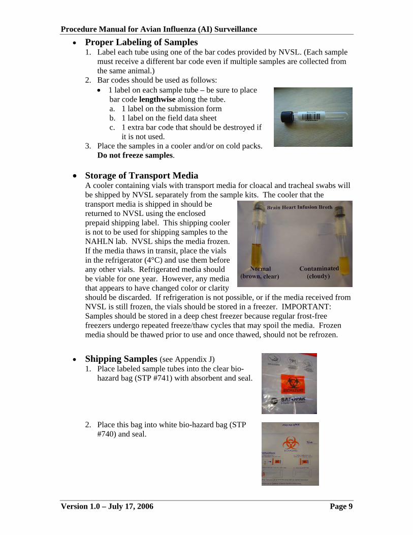
• Proper Labeling of Samples 1. Label each tube using one of the bar codes provided by NVSL. (Each sample
must receive a different bar code even if multiple samples are collected from the same animal.)
2. Bar codes should be used as follows: • 1 label on each sample tube – be sure to place
bar code lengthwise along the tube. a. 1 label on the submission form b. 1 label on the field data sheet c. 1 extra bar code that should be destroyed if
it is not used. 3. Place the samples in a cooler and/or on cold packs.
Do not freeze samples.
• Storage of Transport Media A cooler containing vials with transport media for cloacal and tracheal swabs will be shipped by NVSL separately from the sample kits. The cooler that the transport media is shipped in should be returned to NVSL using the enclosed prepaid shipping label. This shipping cooler is not to be used for shipping samples to the NAHLN lab. NVSL ships the media frozen. If the media thaws in transit, place the vials in the refrigerator (4°C) and use them before any other vials. Refrigerated media should be viable for one year. However, any media that appears to have changed color or clarity should be discarded. If refrigeration is not possible, or if the media received from NVSL is still frozen, the vials should be stored in a freezer. IMPORTANT: Samples should be stored in a deep chest freezer because regular frost-free freezers undergo repeated freeze/thaw cycles that may spoil the media. Frozen media should be thawed prior to use and once thawed, should not be refrozen.
• Shipping Samples (see Appendix J) 1. Place labeled sample tubes into the clear bio-
hazard bag (STP #741) with absorbent and seal.
2. Place this bag into white bio-hazard bag (STP #740) and seal.
Version 1.0 – July 17, 2006 Page 9

Procedure Manual for Avian Influenza (AI) Surveillance 3. Place the white bag into the shipping box.
4. Place frozen ice packs on top of the bag.
5. Place completed NAHLN Laboratory Submission Form for AI Samples on
top of inner styrofoam lid.
• Seal box with packing tape. • Address the box to the NAHLN laboratory where the samples will be
sent. (See Appendix H for a list of NAHLN laboratories). • Place the other required shipping labels on the box.
• Ship by overnight delivery with a carrier that provides overnight
delivery service. • Samples must be shipped Monday, Tuesday, or Wednesday or
Thursday of each week. • Hand Delivery of Samples to Lab
Agencies with the opportunity to hand deliver samples to the lab should: 1. Submit samples with ice packs. 2. Include NAHLN Laboratory Submission Form. 3. Confirm that lab will process the samples in 48 hours prior to submission.
To request additional AI kits or supplies contact Seth Swafford ((301) 734-3570
or [email protected]) or Brandon Schmit (970-266-6079 or [email protected]) or Kerri Pedersen (970-266-6272 or [email protected]).
Version 1.0 – July 17, 2006 Page 10

Procedure Manual for Avian Influenza (AI) Surveillance
• Identification of a NAHLN Laboratory Ship specimens via the overnight contract delivery service to a NAHLN laboratory (See Appendix H for a list of NAHLN laboratories). Call the laboratory before sending samples so that they know a shipment is coming and to make sure that they can test the samples within 48 hours. If not, call another NAHLN lab and confirm that they will be able to process the samples in 48 hours. After identifying the new NAHLN lab, Wildlife Services employees should notify the Wildlife Services State Director in the state where the samples are being shipped so that the samples can be credited to the appropriate account. Wildlife Services employees will need to communicate across state lines on these issues to ensure proper accounting and billing. State wildlife agencies should contact their local Wildlife Services State Director to notify them that samples have been sent to a different lab. The Wildlife Services State Director will be responsible for verifying the accuracy of receipts generated by State Wildlife Agency samples.
• Proper Communication of Submitting Samples It is essential to have secure and reliable communication among the individuals responsible for sample collection and designated NAHLN laboratories. The submitter must: 1. Accurately record all relevant information on the NAHLN Laboratory
Submission Form for AI Samples (Appendix G).
2. Prepare 2 copies of the completed NAHLN Laboratory Submission Form for AI Samples: • One original to accompany the samples shipped to the designated
laboratory. • One photocopy be kept on-file by the submitter
3. Notify the NAHLN laboratory (Appendix H) of incoming samples via fax,
telephone, and/or e-mail. The information to be communicated includes: • The overnight contract delivery service tracking number • The collection site name and address • The unique referral number of the submission • The number of samples
4. Verify, via the overnight contract delivery service tracking system that the
submission has been delivered to the designated laboratory. If the sample does not arrive as expected, the sample submitter should work with the delivery service to determine the location and delivery status of the sample.
• Reporting Results
All results from the laboratory should be sent to Dr. Thomas DeLiberto by fax to (970) 266-6089 or by email to [email protected]. Submitters may view results by logging onto the USGS HEDDS system (see section III).
Version 1.0 – July 17, 2006 Page 11
Procedure Manual for Avian Influenza (AI) Surveillance
D. Procedures for Collecting and Shipping Environmental Samples
Fecal Sample Collection 1. Wear latex or nitrile gloves while handling feces to minimize contamination
of the sample as well as for personal protective purposes. Change gloves if they become soiled or contaminated. When finished collecting, wash your hands with antibacterial soap or antibacterial waterless hand sanitizer.
2. Keep at least 8 ice packs in the freezer for shipping of samples and take enough frozen ice packs (this will vary depending on the size of your field cooler) into the field to keep samples cool after collection.
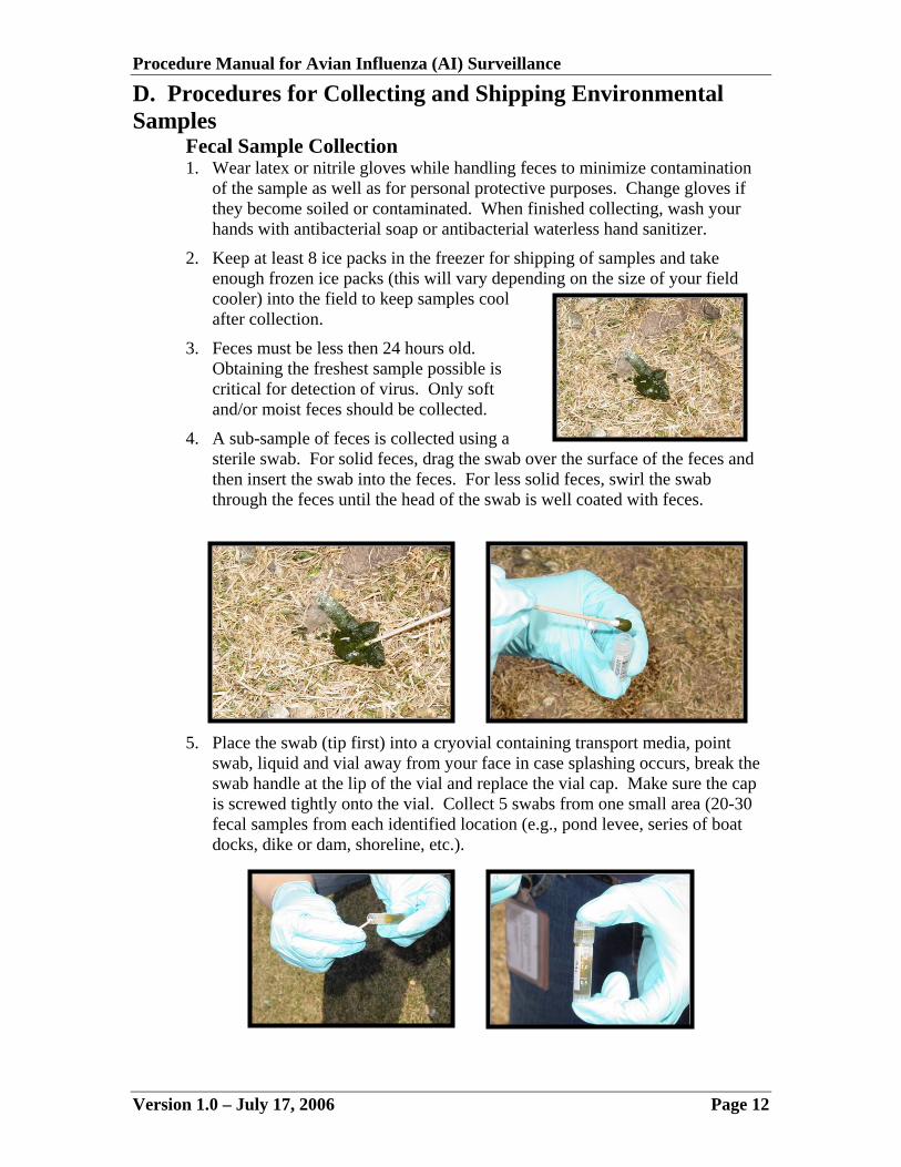
3. Feces must be less then 24 hours old. Obtaining the freshest sample possible is critical for detection of virus. Only soft and/or moist feces should be collected.
4. A sub-sample of feces is collected using a sterile swab. For solid feces, drag the swab over the surface of the feces and then insert the swab into the feces. For less solid feces, swirl the swab through the feces until the head of the swab is well coated with feces.
5. Place the swab (tip first) into a cryovial containing transport media, point swab, liquid and vial away from your face in case splashing occurs, break the swab handle at the lip of the vial and replace the vial cap. Make sure the cap is screwed tightly onto the vial. Collect 5 swabs from one small area (20-30 fecal samples from each identified location (e.g., pond levee, series of boat docks, dike or dam, shoreline, etc.).
Version 1.0 – July 17, 2006 Page 12

Procedure Manual for Avian Influenza (AI) Surveillance 6. Label the cryovial and data sheet with the corresponding bar code label and
return vial to appropriate Whirl-Pak (up to five vials per Whirl-Pak (DO NOT PLACE VIALS FROM MULTIPLE SITES IN THE SAME WHIRL-PAK). Ensure barcode is placed on vial lengthwise, otherwise the scanner will be unable to read it. The end of the bar code will extend beyond the bottom of the tube. Do not affix the bar code to the cap of the tube, or fold the end of the barcode over the cap—it could interfere with barcode reading equipment. DO NOT WRAP THE BAR CODE LABEL AROUND THE CIRCUMFERENCE OF THE VIAL. Ensure that the bar code label on the vial is the same as the bar code label next to the corresponding information on the data sheet (the bar code is used to link the sample in the vial with the corresponding information on the data sheet).
7. Place Whirl-Paks filled with vials containing the fecal samples in a sealed ziploc® bag, seal the ziploc® with duct tape, label the ziploc® bag with name, date, and location with the Sharpie pen and return to chilled field cooler.
8. Fill out data sheet with the necessary information for each swab sample collected including affixing the corresponding bar code label (Appendix I).
9. Dispose of used gloves and used swab handles into a ziploc® bag, seal and send back with the samples. The National Wildlife Research Center (NWRC) will properly dispose of the waste.
10. The samples must be shipped “Priority Overnight” via Federal Express. • Swab samples should be placed in Whirl Paks (up to 5 per Whirl Pak) • Whirl Paks with swab samples inside should be placed in one gallon
ziploc® bags (several per ziploc®). • Ziploc® bags should have Duct tape along the seal. • Place 2 absorbent pads along inside of Styrofoam shipping cooler.
Version 1.0 – July 17, 2006 Page 13

Procedure Manual for Avian Influenza (AI) Surveillance • Ziploc® bags should be placed in the Styrofoam shipping cooler with 4
frozen ice packs. • Place data sheets in a one-gallon ziploc® bag and place in Styrofoam
cooler. • Place sealed ziploc® bag with used gloves and waste in Styrofoam
container. • Place Styrofoam cooler inside of cardboard shipping container. • Affix sticker with UN3373 in diamond to outer shipping container (this
label notifies FedEx that the shipping container includes diagnostic specimens).
• Affix FedEx label provided to the outer shipping container • Make sure the Styrofoam cooler and outer cardboard shipping container
is taped up tightly (No Holes or Leaks), otherwise samples may not be delivered!
11. Notify Ginger Young at 970-266-6078 or [email protected], 24 hours prior to shipping samples. Samples must be shipped to the NWRC within 24 hours of collection. Do not ship any samples Thursday, Friday or Saturday.
Water collection This protocol is still in development. Water samples should not be collected at this time. Once the protocol has been developed it will be provided.
III. Data Management Using the HPAI Early Detection Data System (HEDDS) A. Overview
• Background The U.S. Interagency Strategic Plan stipulates that all surveillance data collected by the cooperating agencies should be stored in a national database. This database is housed at the National Biological Information Infrastructure’s Wildlife Disease Information Node in what is called the HPAI Early Detection Data System (HEDDS). Wildlife Services and its cooperators will enter their surveillance data into the HEDDS. The HEDDS database “provides a secure, accessible platform for the generation of reports, graphs, and maps and can be used for spatial modeling.” It also serves as a resource to keep the public and policy makers informed about Wildlife Services’ and its cooperator’s AI surveillance efforts within the United States.
• General Information
1. The HEDDS Fact Sheet provides a general overview of the system: http://www.nbii.gov/about/pubs/factsheet/pdf/WDIN-HEDDS.pdf
2. Explore the system’s functionality by self-registering with the HEDDS demonstration found at: http://wildlifedisease.nbii.gov/aidemo.
Version 1.0 – July 17, 2006 Page 14

Procedure Manual for Avian Influenza (AI) Surveillance
B. Data Entry • Web site access
http://wildlifedisease.nbii.gov/ai
• Expected transitional phases Many challenges have been associated with developing the required database security features and overall infrastructure required for such a large surveillance effort. Given the complexity of developing as such a comprehensive database, the data entry and management process will be implemented in a series of phases. These phases are as follows: 1. PHASE 1
A copy of all field data sheets from Wildlife Services offices and State/Tribal Cooperators will be sent or faxed to Brandon Schmit or Kerri Pedersen (See Appendix F for contact information). All data will be entered into the HEDDS system by employees of the National Wildlife Disease Program in Fort Collins, Colorado. All contributing Wildlife Services state offices and cooperating state/tribal agencies will have usernames and passwords during phases 1 and 2 and will be able to view all data they have submitted as well as produce reports and maps using HEDDS.
2. PHASE 2 Once phase 2 begins, each collecting agency will be responsible for entering field collection data into a standardized Excel spreadsheet that will be provided to all Wildlife Services employees and state/tribal cooperators. Detailed instructions on filling out the Excel file will also be provided. Each agency will be responsible for forwarding these files electronically to Brandon Schmit ([email protected]) or Kerri Pedersen ([email protected]). Once the Excel files have been submitted they will be uploaded into the HEDDS system by employees of the National Wildlife Disease Program in Fort Collins, Colorado. In addition to the Excel files, copies of all field data sheets should be sent to the attention of Brandon Schmit (Western Regional States) or Kerri Pedersen (Eastern Regional States) (see Appendix F for more additional information). Original field data sheets should be archived by the collecting agency. Note: Phase 2 will begin once a set of instructions has been developed and the standardized Excel spreadsheet has been user tested. Direct entry of AI data using the web-based forms on HEDDS will not occur during phases 1 and 2. After data is entered into HEDDS, it must be verified to affirm that the data are accurate before they appear in maps, reports, or summaries available to the public or other contributing agencies. All GPS locations will be cross checked with the recorded county and state information to ensure accuracy. All data will be verified centrally in Fort Collins during phases 1 and 2.
Version 1.0 – July 17, 2006 Page 15

Procedure Manual for Avian Influenza (AI) Surveillance 3. PHASE 3
When phase 3 begins, each Wildlife Services office and cooperating state agency will be asked to enter data using a web-based data entry system. The web-based data entry system will be available as a link directly on HEDDS. All data storage, reporting, and mapping functions will remain on the HEDDS. Expanded capabilities of the web-based system will include:
• The laboratory submission form will be replaced by a packing slip that
will be automatically generated by the new system for inclusion in each shipment being sent to the lab.
• The NAHLN laboratory will automatically be notified that a shipment is being sent once the data is entered into the system. (The laboratory should still be contacted to confirm that they will be able to process the samples in 48 hours).
• A detailed module with instructions and training for phase 3 is under development and will be available as a new chapter to insert within this manual once completed.
PDA and Tablet PC applications are under development that may eliminate the
need for carrying and filling out data sheets in the field altogether. All data may be recorded directly on the forms within the PDA or Tablet PC and may be uploaded directly to the system from these units once back at the office or when in range of a WiFi hotspot. This may be included after phase 3 has begun.
C. Obtaining a Username and Password
• Contact the Wildlife Services Data Administrators 1. All Western Region Wildlife Services State Offices and their State/Tribal
Cooperating Agencies should contact Brandon Schmit ([email protected]) to request an account on the HEDDS. All requests should include a preferred username as well as all pertinent contact information including agency affiliation.
2. All Eastern Region Wildlife Services State Offices and their State/Tribal Cooperating Agencies should contact Kerri Pedersen ([email protected]) to request an account on the HEDDS. All requests should include a preferred username as well as all pertinent contact information including agency affiliation.
D. Data Mapping and Reporting
• Data Availability for Contributors All contributors must log into the HEDDS system in order to utilize the reporting and mapping features (http://wildlifedisease.nbii.gov/ai). After logging into the HEDDS system, simply click on the reports and maps button within the main page. Screen shots from the reporting and mapping pages are displayed below to provide an idea of what is currently available. It is likely that more options will be added to these reports and maps in the future.
Version 1.0 – July 17, 2006 Page 16

Appendix A: U.S. Interagency Strategic Plan 2006
Version 1.0 – July 17, 2006 U.S. Interagency Strategic Plan: Page A1
An Early Detection System for Highly Pathogenic H5N1
Avian Influenza in Wild Migratory Birds
U.S. Interagency Strategic Plan
Introduction Avian influenza (AI) is a type A influenza virus that is naturally found in certain species of waterfowl and shorebirds. However, the occurrence of highly pathogenic avian influenza (HPAI) subtype highly pathogenic H5N1 avian influenza has raised concern regarding the potential impact on wild birds, domestic poultry, and human health should it be introduced into the United States (U.S.). Numerous potential routes for introduction of the virus into the U.S. exist including illegal movement of domestic or wild birds, contaminated products, via an infected traveler, as a bioterrorism event, and the migration of infected wild birds. This plan focuses primarily on the detection of a potential introduction of highly pathogenic H5N1 avian influenza virus by migratory birds. Avian influenza viruses are classified on the basis of two proteins, hemagglutinin (H) and neuraminidase (N), found on the surface of the virus. Specific viral subtypes have one of 16 different H proteins and one of 9 different N proteins, resulting in 144 possible combinations or subtypes based on this classification scheme. Within each subtype, there are numerous combinations of genetic sequences that determine the pathogenicity of the subtype to an infected host. Wild birds, in particular certain species of waterfowl and shorebirds, are considered to be the natural reservoirs for all 144 subtypes. These subtypes are adapted to survive in these wild species and usually cause little or no disease. However, gradual genetic drift (i.e., mutation) can occur and a particular subtype can become adapted to infect other species of wild birds and domestic birds. Although this slight genetic change in the virus allows it to infect new species, it usually does not cause disease in the new host. The virus can also change if a host is simultaneously infected with another type A influenza virus. In such situations, mixing of the genetic material from the two virus strains (genetic shift) can occur, resulting in the formation of a new strain. The combination of gradual drifts and rapid shifts results in the production of a strain that now causes morbidity and mortality in susceptible hosts. If the morbidity and mortality is significant, the virus is classified as a highly pathogenic avian influenza (HPAI) virus. During 1995-96, it is thought that antigenic drift occurred in an AI virus of wild birds, allowing the virus to infect chickens in China. This was followed by reassortment into the HPAI virus subtype highly pathogenic H5N1 avian influenza. Since that time, this highly pathogenic H5N1 has been circulating in Asian poultry and domestic fowl resulting in significant mortality to these species. Highly pathogenic H5N1 avian influenza likely underwent further antigenic drift and
Appendix A: U.S. Interagency Strategic Plan 2006
Version 1.0 – July 17, 2006 U.S. Interagency Strategic Plan: Page A2
shift allowing infection in additional species of birds, mammals, and humans. More recently, this virus moved back into wild birds resulting in significant mortality of species such as bar-headed geese, brown-headed gulls, black-headed gulls, ruddy shelducks, and great cormorants in China during April 2005. Although the spread of H5N1 in Asia has been primarily due to movement of domestic birds, the movement of this virus into wild birds raised the possibility that these species may also spread the virus. This was thought to be the case in August 2005, when bar-headed geese and whooper swans died on Erkhel Lake, Mongolia, in an area not known to have domestic poultry or fowl nearby. Given the adaptation of highly pathogenic H5N1 avian influenza to wild birds, increasing concern has developed over the potential for migrating species to introduce the virus into new regions of the world such as North America. Therefore, at the request of the Homeland Security Council’s Policy Coordinating Committee for Pandemic Influenza Preparedness, the U.S. Departments of Agriculture (USDA) and Interior (DOI) were asked to develop a coordinated National Strategic Plan for early detection of HPAI introduction into North America by wild birds. Dr. Tom DeLiberto (USDA-APHIS Wildlife Services) and Rick Kearney (USGS Biological Resources Division) convened an interagency Working Group, which consists of representatives from USDA, DOI, U.S. Department of Health and Human Services (HHS), the International Association of Fish and Wildlife Agencies IAFWA), and the state of Alaska (Attachment 1). On 10 August 2005, the Working Group met by teleconference to initiate development of a “Plan For the Detection of HPAI Virus in Migratory Birds in the United States”. After some discussion among the participants it was decided that while the immediate concern was the introduction of highly pathogenic H5N1 avian influenza virus via migratory birds into Alaska and the Pacific Flyway (including Hawaii and other Pacific Islands), the group would also begin to address detection of the virus in all the North American flyways.
Goal of the Strategic Plan The goal of this plan is to describe the essential components of a unified national system for the early detection of HPAI, specifically highly pathogenic H5N1 avian influenza, in migratory birds. While the immediate concern is a potential introduction of highly pathogenic H5N1 avian influenza into the U.S., the development of a system that is capable of detecting the introduction of all HPAI viruses through migratory birds would significantly improve the biosecurity of the Nation. This document provides guidance to Federal, State, university, and non-governmental organizations for conducting HPAI monitoring and surveillance of migratory birds in the U.S. It is expected that this document will be used by agencies and organizations to develop regional and/or state-specific implementation plans for HPAI surveillance. Data collected in accordance with the guidelines presented in this document will be assimilated into a National database for use by all agencies, organizations, and policy makers. Furthermore, although the original charge of the Working Group was to monitor migratory birds as a potential route of entry into the U.S., the standardized methodologies and procedures identified in this

Appendix A: U.S. Interagency Strategic Plan 2006
Version 1.0 – July 17, 2006 U.S. Interagency Strategic Plan: Page A3
document are applicable to other wild birds as well. Agencies and organizations conducting monitoring and surveillance in non-migratory birds are encouraged to follow these guidelines so that their data can be incorporated into and tracked via the National Early Detection System. This system for highly pathogenic H5N1 avian influenza detection will provide early warning for potentially catastrophic mortality events in North American wild birds and poultry, and minimize the potential for human exposures. Agencies and organizations are encouraged to participate in this system by following the guidelines presented in this document when conducting AI sampling in wild birds. While this plan focuses on detection of highly pathogenic H5N1 avian influenza virus, the Working Group fully supports efforts to characterize all AI viruses in wild birds. Such information is critical to our understanding of the ecology of AI viruses and their transmission among wildlife, livestock, and humans. Birds will be sampled in conjunction with existing studies when possible, and additional bird captures will be initiated as necessary to provide a broad species and geographic surveillance effort.
A National Early Detection System for Highly pathogenic H5N1 avian influenza in Migratory Birds The ability to efficiently control the spread of a highly infectious, exotic disease such as highly pathogenic H5N1 avian influenza, is dependent upon the capacity to rapidly detect the pathogen if introduced. For this reason, a National Early Detection System for Highly pathogenic H5N1 avian influenza in Wild Migratory Birds is not only prudent, it is necessary. Effective implementation of this National Detection System will require decentralized planning and execution at regional and state levels, combined with centralized coordination to ensure national level analysis of surveillance data for risk assessment. It also must involve a partnership between public and private interests and include efforts by Federal, State, and local governments as well as nongovernmental organizations, universities, and other interest groups. Lastly, it requires flexibility and commitment by all groups for successful implementation.
Decentralized Planning and Execution Wild migratory birds, by their very nature, are not subject to disease containment controls as are domestic birds and people. While their movements are generally uncontrollable, these movements are largely predictable on both a daily and seasonal basis. Local movements within or between breeding, feeding, and roosting areas are frequently well known by State and local wildlife management authorities and others familiar with local bird populations. Long range movements associated with seasonal migration are also well known for many species, especially those waterfowl and shorebird species of particular interest in highly pathogenic H5N1 avian influenza detection and surveillance. Coordinating groups such as the four Flyway Councils already exist to deal with issues related to migratory bird management on a broad geographic scale. These Councils include representation from each of the States in their respective bird flyways as well as the U.S. Fish and Wildlife Service. Therefore, the planning and execution of local and regional highly pathogenic H5N1

Appendix A: U.S. Interagency Strategic Plan 2006
Version 1.0 – July 17, 2006 U.S. Interagency Strategic Plan: Page A4
avian influenza early detection efforts will best be accomplished by the States in collaboration with Federal agencies.
Centralized Coordination States and flyways are exposed to varying degrees of threat from highly pathogenic H5N1 avian influenza. Each has unique circumstances that will shape the direction and intensity of its early detection efforts. Consequently, gaps among regional programs may emerge over time. Centralized coordination will evaluate the effectiveness of state and regional efforts, allowing for prioritization of available federal resources. Integration of this National Early Detection System with similar influenza surveillance systems in other species (e.g., domestic, feral, zoo) as well as humans will also require centralized coordination. Surveillance data from all of these systems will be incorporated into national risk assessments, and preparedness and response planning efforts.
Geographic Prioritization of Sampling Efforts This Strategic Plan targets bird species in North America that have the highest risk of being exposed to or infected with the highly pathogenic H5N1 avian influenza subtype because of their migratory movement patterns. Currently, these include birds that migrate directly between Asia and North America, birds that may be in contact with species from areas in Asia with reported outbreaks, or birds that are known to be reservoirs of AI. However, should highly pathogenic H5N1 avian influenza virus be detected in domestic birds in the U.S., sampling of wild birds within the affected flyway may become a high priority as well. In general, bird flyways represent migration corridors within continental landmasses. However, Alaska and areas in Eastern Siberia represent a unique situation where major flyway systems cross continental boundaries (Attachment 2, fig. 2-1). Two major Asian flyways (the East Asian-Australasian and East Asian) include both Southeast Asia and the Arctic regions of Siberia, the Russian Far East, and Alaska. The East Asian-Australasian Flyway, defined primarily in the context of shorebird use, extends across 20 countries from the Siberian and Alaskan Arctic through North and Southeast Asia including U.S. trust territories in the Pacific to Australia and New Zealand. Similarly, in North America, the Pacific Flyway extends from Arctic Canada, Alaska, and Eastern Siberia through coastal and western regions of Canada, the United States and Mexico, and on to Central and South America (Attachment 2, Fig. 2-2). Many migratory species that nest in Arctic Siberia, Alaska, and Canada follow the Pacific Flyway to wintering areas. Although not considered a major pathway, birds from both Eastern Siberia and Alaska intermingle in both the Pacific and Central Flyways. The overlap at the northern ends of these flyways and in Hawaii and Oceania establishes a path for potential disease transmission across continents and for mixing, re-assortment, and exchange of genetic material among strains from Eurasia and North America.
Appendix A: U.S. Interagency Strategic Plan 2006
Version 1.0 – July 17, 2006 U.S. Interagency Strategic Plan: Page A5
If highly pathogenic H5N1 avian influenza virus spreads to North America via migratory birds, the above analysis of the major flyways suggests that the virus would most likely arrive first in Alaska. Such a scenario is reasonable, as the contribution of Eurasian AI viruses to the genetic composition of viruses in North American migratory birds has already been demonstrated. Given the current knowledge on highly pathogenic H5N1 avian influenza distribution, the Working Group developed a prioritized sampling approach based on Alaska and the major North American flyways. This approach prioritized the following regions in decreasing order of importance:
1. Alaska, the Pacific Flyway, and Oceania 2. Central Flyway 3 Mississippi Flyway 4. Atlantic Flyway
Agencies participating in the development of this plan are committed to efforts that ensure adequate sampling based on the above prioritization. However experiences with previous introductions of exotic diseases into North America (e.g., West Nile Virus) have demonstrated that detection and surveillance systems must be adaptable to changes in pathogens and risk factors associated with their potential introduction. If changes in the relative risks of highly pathogenic H5N1 avian influenza introduction into the US result in regional reprioritization, agencies must be prepared to redistribute resources accordingly.
Sampling Strategies This strategic plan recommends decentralized planning and execution of highly pathogenic H5N1 avian influenza early detection efforts. To provide a uniform structure for the development of local plans, it recommends the consideration of five strategies for collecting monitoring and surveillance data on highly pathogenic H5N1 avian influenza virus in wild birds. Agencies and organizations are encouraged to use one or more of these strategies when designing AI surveys in wild birds. These strategies are:
Investigation of Morbidity/Mortality Events (Attachment 3): Over 40 species of wild birds have been shown to be susceptible to infection with highly pathogenic H5N1 avian influenza virus. While not all species infected necessarily exhibit disease, the current strain(s) of H5N1 circulating in Asia have been shown to cause morbidity and mortality in a wide variety of these species. The systematic investigation of morbidity and mortality events in wild birds to determine if highly pathogenic H5N1 avian influenza is playing a role in causing illness and death offers the highest and earliest probability of detecting the virus if it is introduced by migratory birds into the United States. State natural resource agencies and Federal refuges and parks, primarily within the DOI’s U.S. Fish and Wildlife Service National Wildlife Refuge System and the National Park Service, are the principal authorities in a position to detect and respond to mortality events involving wild birds. Morbidity and mortality events

Appendix A: U.S. Interagency Strategic Plan 2006
Version 1.0 – July 17, 2006 U.S. Interagency Strategic Plan: Page A6
involving wildlife are often detected by, or reported to, these agencies and entities. This strategy capitalizes on an existing morbidity/mortality program being conducted by DOI and its partners.
Surveillance in Live Wild Birds (Attachment 4): This strategy incorporates sampling of live-captured, apparently healthy wild birds to detect the presence of highly pathogenic H5N1 avian influenza virus. This effort will select bird species in North America that represent the highest risk of being exposed to, or infected with, Highly pathogenic H5N1 avian influenza virus because of their migratory movement patterns, which include birds that migrate directly between Asia and North America, or birds that may be in contact with species from areas in Asia with reported outbreaks. Should highly pathogenic H5N1 avian influenza virus be detected in domestic birds in the U.S., sampling of wild birds in the flyway in the affected area may become a high priority as well. Data collected by organizations currently conducting research and monitoring for avian influenza in Alaska will be incorporated with additional bird captures as necessary to provide a broad species and geographic surveillance effort. This strategy capitalizes on research activities currently being conducted by DOI, USDA and their partners.
Surveillance in Hunter-killed Birds (Attachment 5) Check stations for waterfowl hunting are operated by the US Fish and Wildlife Service and state natural resource agencies. Hunter check stations provide an opportunity to collect additional samples to determine the presence of HPAI and other subtypes of avian influenza viruses and supplement data collected during surveillance of live wild birds. As with surveillance of live wild birds, sampling of hunter-killed birds will focus on hunted species that are most likely to be exposed to HPAI in Asia; have relatively direct migratory pathways from those areas to the U.S. via Alaska or directly to the Pacific Coast; mix in Alaska staging areas with species that could bring the virus from Asia; or should HPAI be detected in domestic birds in the U.S., may mix with wild birds in the flyway of the affected area. Collection of samples from these species will occur at hunter check stations in the lower 48 states during hunting seasons in areas where these birds stage during migration or over-wintering.
Sentinel Species (Attachment 6): Waterfowl, exhibition gamefowl, and poultry flocks reared on backyard premises have been used as sentinels for active surveillance for avian diseases of interest to the commercial poultry industry and regulatory agencies. Currently in Alaska, the State veterinarian uses targeted surveillance of domestic flocks at concentration points due to remote location of villages and lack of resources; enthusiasts travel to poultry exhibitions with birds from distant locations; and. surveillance effectively covers a large geographic area. Enhancement of this approach would be valuable. However, placement of sentinel ducks in strategic locations may also prove useful. Placement of sentinel ducks has been used successfully for surveillance of diseases of importance to the poultry industry, including influenza A. Also, sentinel ducks in wild pelagic bird colonies improved virus detection rates fivefold, suggesting that this approach is advantageous in ecological studies.

Appendix A: U.S. Interagency Strategic Plan 2006
Version 1.0 – July 17, 2006 U.S. Interagency Strategic Plan: Page A7
Environmental Sampling (Attachment 7): Avian influenza viruses are generally released by waterfowl through the intestinal tract and viable virus can be detected in both feces and the water in which the birds swim, defecate and feed. This is the principal means of virus spread to new avian hosts and potentially to poultry, and other susceptible livestock. Analysis of both water and fecal material from waterfowl habitat can provide evidence of AI virus circulating in wild bird populations, the specific AI subtypes, levels of pathogenicity, and possible risks to poultry and susceptible livestock. Monitoring of water and/or fecal samples gathered from waterfowl habitat is a reasonably cost effective, technologically achievable means to assess risks to poultry.
Sample Collection Samples collected for AI surveillance may include carcasses, tracheal and cloacal swabs, feces, and environmental samples (e.g., water). Prior to initiating a surveillance activity, it is important to identify the laboratory in which the samples will be submitted. Sample handling and transportation procedures may differ among laboratories. It is recommended that samples collected for inclusion into the National Early Detection System be submitted to a laboratory that uses standardized procedures identified in the Laboratory Diagnosis section of this document or by using the attached detailed descriptions of sampling methodologies. If birds are found morbid or dead, it is important to use proper personal protection techniques (http://www.nwhc.usgs.gov/research/WHB/WHB_05_03.html, http://www.cdc.gov/flu/avian/professional/protect-guid.htm) and to submit the entire carcass to a veterinary diagnostic laboratory for necropsy (Attachment 8). Field biologists should contact the specific laboratory that they will be working with well in advance of any specimen collection and shipping to receive specific instructions for specimen submissions to that laboratory. Laboratories should always be notified ahead of time when a shipment is being made to their facility. When collecting samples from live or hunter-killed birds, tracheal and cloacal swabs are preferred. Most AI strains tend to replicate more efficiently in the intestinal tract than in the respiratory tract of natural host species (i.e., waterfowl and shorebirds). Consequently, cloacal swabs are generally preferred. However, recent isolations of highly pathogenic H5N1 avian influenza virus in wild birds have documented higher levels of virus in tracheal samples. Therefore, it is recommended that both samples be collected from birds when possible. While the collection of cloacal swabs is a relatively easy procedure, obtaining proper tracheal swabs can be problematic and requires personnel trained in the sampling technique. Examples of tracheal/cloacal swab collection protocols can be found in Attachment 9. Tracheal and cloacal swabs should be placed in separate tubes, and swabs should not be pooled across individuals. Monitoring of water and/or fecal samples gathered from waterfowl habitat is a reasonably cost effective, technologically achievable means to detect the presence of HPAI and alert decision makers to the risks to poultry in the Western Hemisphere from new, potentially highly pathogenic subtypes of AI (Attachment 7). A surveillance system based on water sampling is not ready to implement at the present. However, the validation of this method could come on-

Appendix A: U.S. Interagency Strategic Plan 2006
Version 1.0 – July 17, 2006 U.S. Interagency Strategic Plan: Page A8
line in a short period of time and would represent considerable cost savings without loss of sensitivity. Fecal sampling is an established technique and is ready for use in surveillance with the establishment of sampling guidelines. Both approaches yield advantages where individual bird sampling is too costly or logistically impractical. Either approach could yield a spatial and habitat risk assessment for site contamination with highly pathogenic H5N1 avian influenza virus. The main considerations are where and when to get the samples, ensuring proper storage and transport, and the capacities and capabilities of the laboratories doing the analyses. Real-time reporting and the infrastructure to support such reporting is a serious constraint on any surveillance system. The ability to integrate, analyze, and responsibly disseminate these data is critical and needs to be addressed.
Sample Size Determination Prior to initiating a surveillance program, it is important to determine the sample size necessary to make statistically valid inferences concerning the presence of highly pathogenic H5N1 avian influenza virus in a sample population. In the context of this plan, the population of interest is not defined because this definition will vary by geographic location, time of year, species of interest, and sampling method employed. For example, sampling a breeding population versus a wintering population, for a single species, may result in very different interpretations of the geographic distribution of the population of interest. If water samples are being collected, then the population may consist of several water bodies. Therefore, it is crucial that prior to collections beginning, statistically valid sample size estimations be incorporated into regional and state implementation plans.
Laboratory Diagnostics All samples collected for inclusion in the National Early Detection System should be analyzed in accordance with the standard procedures included in this document. A list of laboratories certified to conduct testing for highly pathogenic H5N1 avian influenza virus is included in Attachment 11. Samples will be analyzed as soon as possible after collection. Tracheal/cloacal swabs and fecal samples will be analyzed by real-time reverse transcriptase-polymerase chain reaction (RT-PCR) using the matrix gene RT-PCR assay (Attachment 12). The matrix gene RT-PCR assay is capable of detecting all 16 hemagglutinin and nine neuraminidase subtypes. Matrix gene RT-PCR-positive samples would indicate the presence of avian influenza and they should be further characterized by the H5- and H7-specific RT-PCR assays of Spackman et al. (2002) as modified in Attachment 11. The H5 RT-PCR test is known to detect the current Highly pathogenic H5N1 avian influenza viruses. Positive H5 and H7 RT-PCR tests would indicate the presence of AI viruses with the potential of causing pathology in domestic poultry. Therefore, all samples positive for H5 and H7 by RT-PCR will be submitted for virus isolation for verification. Samples positive for live virus in virus isolation and positive for H5 or H7 by RT-PCR will be submitted to the USDA APHIS National Veterinary Services Laboratory (NVSL) for confirmation. The NVSL is capable of performing the intracranial chicken pathogenicity index (ICPI) test on the resultant virus to determine directly the pathogenicity of the virus in chickens. Identification of a highly pathogenic H5 or H7 virus is a reportable disease and immediate notification to the agency submitting the sample,

Appendix A: U.S. Interagency Strategic Plan 2006
Version 1.0 – July 17, 2006 U.S. Interagency Strategic Plan: Page A9
the state veterinarian, the area veterinarian in charge (AVIC), the state public health official and the CDC/USDA Select Agent program. Samples will be immediately secured as required by the Select Agent Programs. All positive H5 and H7 samples will also be sent to the USDA Agriculture Research Service Southeastern Poultry Research Laboratory in Athens, GA, for complete molecular sequencing. This will provide for complete typing of the virus and allow for phylogenetic analysis.
Data Management Real-time reporting and the infrastructure to support such reporting is a serious constraint on any surveillance system. The ability to integrate, analyze, and responsibly disseminate these data is critical. In addition, the data collected for this National Surveillance System will consist of samples submitted by many agencies and organizations. This will require a system to manage the input of animal and sample collection data through multiple routes, the ability to easily match, compare, and transfer laboratory data about these samples, and provide a platform in which all data is secure, accessible, and able to be mapped and used for spatial modeling. The National Biological Information Infrastructure Wildlife Disease Information Node (WDIN) managed by the U.S. Geological Survey’s National Wildlife Health Center has created a prototype web-enabled HPAI data management system, which will serve as a template for data collected from live and hunter-killed wild birds. (See http://wildlifedisease.nbii.gov/ai). The WDIN has developed comparable systems for the management of data from multiagency wildlife disease surveillance efforts such as Chronic Wasting Disease, and for USDA APHIS Wildlife Services Plague and Tularemia. General aspects of the proposed WDIN Interagency HPAI Data Management System are described in Appendix 13. Sentinel bird data will be incorporated into a web-enabled, national data management system for backyard and small-flock poultry developed by the USDA APHIS Veterinary Services’ Application Information Management Team at the Centers for Epidemiology and Animal Health. This database system was developed to reduce the number of data-collection problems experienced by field personnel and to generally improve the quality and efficiency of data collection. The initial testing of the design occurred in October 2005, with actual deployment scheduled for December 2005. If the project is successful in the pilot state of California, the system will likely be expanded for national implementation in 2006. This system will allow all necessary data collected in the field to be shared among all approved organizations without the need for manual data entry, and will provide greater chain-of-custody assurance from a legal and diagnostic perspective. Field personnel will be equipped with computer hardware and software which will facilitate the rapid and accurate collection of samples and data. These devices will share the collected information as needed with the diagnostic lab (National Veterinary Services Laboratories), and will send the data to primary information systems within USDA. To assist in data entry and to further improve data quality, bar-coding will be implemented as key identifiers for samples collected and for cases submitted.
Recommendations
Appendix A: U.S. Interagency Strategic Plan 2006
Version 1.0 – July 17, 2006 U.S. Interagency Strategic Plan: Page A10
Given the current state of knowledge of the epidemiology of highly pathogenic H5N1 avian influenza virus it is recommended that a coordinated interagency/organization early detection system be implemented in the U.S. An analysis of risk factors, including current worldwide distribution of the virus and the migratory patterns of wild birds, indicated that this system should primarily focus sampling efforts in Alaska, Oceania, and the Pacific and Central flyways. However, if adequate resources become available, the system should be expanded to include surveillance of migratory birds in the Mississippi and Atlantic Flyways as well. State and Federal agencies should immediately begin developing implementation plans based on the guidance provided in this Strategic Plan. Development of these plans should be conducted with the participation of all relevant management agencies and organizations such that sampling designs are produced that allow for statistically sound inference of the presence or absence of highly pathogenic H5N1 avian influenza virus in wild birds. Furthermore, it is recommended that such coordination be conducted through the Flyway Councils, so that regionally based sampling designs can be implemented. Each Flyway (Atlantic, Mississippi, Central, and Pacific) has a Flyway Council, a formal organization generally composed of one member from each State and Province in that Flyway. The Flyway Councils, established through the International Association of Fish and Wildlife Agencies (IAFWA), also assist in researching and providing management techniques for Federal, State, and Provincial Governments, as well as private groups and the public. State and Federal agencies also should develop communication plans in the event that a HPAI is detected in wild birds. For example, highly pathogenic H5N1 avian influenza virus is a reportable disease that requires notification of the State Veterinarian, and the Area Veterinarian in Charge (AVIC). Highly pathogenic H5N1 avian influenza is also a CDC/USDA Select Agent, requiring notification of the CDC/USDA Select Agent Programs and adherence to Select Agent guidelines is required. Finally, it is recommended that a Steering Committee, consisting of one representative each from USGS, FWS, USDA APHIS, IAFWA, HHS, the National Flyway Council, and the State of Alaska be formed to coordinate wild bird AI surveillance in the United States. Specific roles of this Committee should include:
• Facilitate communication between state and federal agencies, and organizations involved in AI surveillance for wild birds.
• Coordinate implementation and data analysis of AI surveillance programs nationally. • Provide periodic summaries of AI surveillance for wild birds in the United States. • Provide periodic recommendations for AI surveillance in wild birds based on previous
sampling efforts and changes in virus epidemiology. • Facilitate communication and coordination among state and federal agencies for
contingency planning and other preparations for the appearance of highly pathogenic H5N1 avian influenza virus in wild birds in North America.
Sampling strategies to detect highly pathogenic H5N1 avian influenza virus in wild bird populations will change depending upon the risk assessment and management goals and prevailing status of the pathogen in North America. For early detection of highly pathogenic

Appendix A: U.S. Interagency Strategic Plan 2006
Version 1.0 – July 17, 2006 U.S. Interagency Strategic Plan: Page A11
H5N1 avian influenza virus, efforts should focus on likely cross-over routes of birds from Asia to North America (e.g., Alaska). Efforts should focus on areas of high aggregations of waterfowl intersecting with logistical sampling support such as the National Wildlife Refuge System and state waterfowl management areas. If highly pathogenic H5N1 avian influenza virus gains a foothold in North America, the surveillance network should be placed along known waterfowl movement paths from the point of origin (i.e., point of detection). These paths can be inferred from known migration routes of specific species. However, practically, and given the patterns emerging in Eurasia, if highly pathogenic H5N1 avian influenza virus becomes established in North America the likelihood of rapid and diffusive spread across the continent is high. At this point local waterfowl and environmental sampling should target areas of strategic value, e.g., human population centers and areas of high density of poultry production. In the former case, such areas would be represented by urban zoo-parks and lakes. These areas would represent the highest level of risk of human contact with contaminated water and/or waterfowl. In the latter case, ponds, lakes and waterfowl management areas around high density poultry production areas would provide the best ability to assess risk of transmission to humans and poultry. Surveillance efforts patterned on these areas are most amenable to local and state efforts for first detection and subsequent risk assessment if H5N1 subtype(s) achieve enzootic status in North America.

3/14/06
12
ATTACHMENT 1
Interagency highly pathogenic H5N1 avian influenza Early Detection Working Group
Working Group Members
Chairpersons Tom DeLiberto USDA APHIS WS (970) 266-6088 [email protected]
Rick Kearney USGS-BRD (703) 648-4019 [email protected]
Members
US Department of Interior Leslie Dierauf USGS BRD NWHC (608) 270-2401, [email protected] Dirk Derksen USGS BRD ASC (907) 786-3531 [email protected] Chris Brand USGS BRD NWHC (608) 270-2440 [email protected] Mike Higgins USFWS (410) 573-4520 [email protected]
Hon S. Ip USGS BRD NWHC (608) 270-2464 [email protected] Gary Frazer USFWS (703) 648-4059 [email protected] Paul Padding USFWS (301) 497-5982 [email protected] Bob Leedy USFWS (907) 786-3446 [email protected] Margaret Wild NPS (970) 225-3593 [email protected]

3/14/06
13
US Department of Agriculture J. Todd Weaver USDA APHIS VS CEAH (970) 494-7213 [email protected] Larry Clark USDA APHIS WS NWRC (970) 266-6036 [email protected] Tracy DuVernoy USDA APHIS VS (301) 734-7781 [email protected]
Thomas Gomez USDA APHIS VS @ CDC (404) 639-3233 [email protected] Seth Swafford USDA APHIS WS (301) 734-3570 [email protected]
International Association of Fish and Wildlife Agencies Gary Taylor IAFWA (202) 624-7890 [email protected] Russ Mason Nevada Dept. of Wildlife (775) 688-1520 [email protected] Bruce Morrison Nebraska Game and Parks Commission (402) 471-5430 [email protected]
Dan Yparraguirre CA Dept. of Fish and Game (916) 445-3685 [email protected] John Fischer SCWDS (706) 542-1741 [email protected]
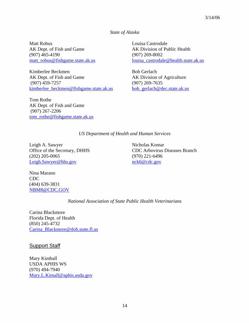
3/14/06
14
State of Alaska Matt Robus AK Dept. of Fish and Game (907) 465-4190 [email protected] Kimberlee Beckmen AK Dept. of Fish and Game (907) 459-7257 [email protected] Tom Rothe AK Dept. of Fish and Game (907) 267-2206 [email protected]
Louisa Castrodale AK Division of Public Health (907) 269-8002 [email protected] Bob Gerlach AK Division of Agriculture (907) 269-7635 [email protected]
US Department of Health and Human Services Leigh A. Sawyer Office of the Secretary, DHHS (202) 205-0065 [email protected] Nina Marano CDC (404) 639-3831 [email protected]
Nicholas Komar CDC Arbovirus Diseases Branch (970) 221-6496 [email protected]
National Association of State Public Health Veterinarians Carina Blackmore Florida Dept. of Health (850) 245-4732 [email protected]
Support Staff Mary Kimball USDA APHIS WS (970) 494-7940 [email protected]

3/14/06
15
ATTACHMENT 2
Migratory Bird Flyways in Asia and North America Figure 2-1: Asian Migratory Bird Flyways
Figure 2-2. North American Migratory Bird Flyways
Pacific Flyway
Atlantic Flyway
Central Flyway
Mississippi Flyway
3/14/06
16
ATTACHMENT 3
Investigation of Morbidity and Mortality Events in Wild Birds
Overview The systematic investigation of morbidity and mortality events in wild birds to determine if the highly pathogenic H5N1 avian influenza subtype of avian influenza (AI) is playing a role in causing illness and death offers the highest and earliest probability of detecting the virus if it is introduced by migratory birds into the United States. There is increasing evidence that highly pathogenic H5N1 avian influenza virus is capable of killing wild birds which is not the usual characteristic of AI viruses. As such, the documentation of the movement of the virus through Asia and into Europe has been discovered in part, through the investigation of mortality events of wild migratory birds. Benefits gained from conducting disease investigations of wildlife mortality events are not unique to AI. Many other important diseases have been discovered and described after initial detection through the wildlife disease investigation process (e.g. West Nile Virus). The investigation of wildlife diseases operates with consistent procedures while maintaining enough flexibility to accommodate the unique characteristics of specific disease agents involved. The initial detection of a mortality event is strongly dependent upon well-trained and observant field personnel. These people in turn communicate with an experienced staff of disease investigation specialists that obtain the maximum amount of information surrounding the event. Depending upon the significance and intensity of the mortality event, these highly trained investigators may visit the site of the mortality event to conduct field investigations so as to obtain further information first hand. In addition to determining a cause of death, disease investigation specialists provide useful management recommendations to potentially reduce further morbidity and mortality. In the event that highly pathogenic H5N1 avian influenza is detected in wild birds, it will be important to investigate the proximity of domestic poultry and swine operations in order to initiate activities to minimize contact between the wild birds and these other animals. Morbidity and mortality of wild birds is most likely to occur in areas where migratory birds infected with highly pathogenic H5N1 avian influenza mingle with other wild bird species, particularly in wetland habitats. Likewise, early outbreaks of highly pathogenic H5N1 avian influenza would most likely occur in Alaska and along the Pacific Flyway of the United States and Canada, where migratory birds from Asia stage in the summer and early fall and subsequently migrate within North America. However, given that migrants also move from Alaska to other parts of North America, surveillance strategies should include other flyways as well. In this surveillance plan, participating state, Federal and tribal agencies, and cooperators will conduct targeted surveillance for highly pathogenic avian influenza (HPAI) both in response to disease outbreaks in wild

3/14/06
17
birds focusing intensively in Alaska, the Pacific Flyway, and Oceania, and in response to mortality events in high-priority (i.e., most likely) species throughout the United States.
Methodology The key to success of this surveillance strategy involves: 1) early detection of morbidity and mortality, 2) rapid reporting and submission of appropriate biological specimens to qualified diagnostic facilities, 3) immediate assessment of the field event (descriptive epidemiology), 4) rapid, accurate, and consistent diagnosis and confirmation, 5) immediate reporting of diagnostic results once confirmed, and 6) pre-planned contingency and response training for the occurrence of HPAI. Specific steps necessary to orchestrate the early detection of highly pathogenic H5N1 avian influenza include:
1) State, Federal, and tribal resource personnel will be instructed to increase
vigilance and to establish routine and systematic monitoring of wild bird populations for morbidity and mortality. Standard guidelines will be prepared with the assistance of the U.S. Geological Survey (USGS) - National Wildlife Health Center (NWHC) and the U.S. Department of Agriculture (USDA) - Wildlife Services (WS) to increase uniformity of effort. The most intensive monitoring will occur in Alaska and selected areas of high risk in the Pacific Flyway and Oceania where migratory birds from affected countries are likely to interact with North American bird species.
2) A uniform protocol for reporting mortality events and instructions for the safe handling and shipment of specimens to identified diagnostic facilities will be developed. Field and response personnel will be trained. Reporting of mortality events will be through appropriate channels within each state, Federal, or tribal entity to the NWHC, where a centralized database (WDIN) will be maintained, made available to contributors, and summarized in modified form for public dissemination.
3) Field personnel or teams designated by respective land management agencies will respond to mortality events by conducting field investigations to determine onset, course, duration, distribution, species, and other environmental conditions associated with mortality events. The NWHC and USDA-WS will assist in developing guidelines and training. In certain circumstances, NWHC and USDA-WS personnel will conduct field investigations or assist other agencies.
4) Representative and suitable carcasses and other biological samples and specimens will be submitted to one or more identified diagnostic facilities capable of conducting immediate necropsy and laboratory analyses. Guidelines will be developed to assure that the appropriate number and types of samples are collected to ensure that there is a statistically-based confidence

3/14/06
18
in the sample size analyzed in response to a mortality event. Necropsies, histology, and laboratory investigations will be utilized to substantiate a diagnosis of highly pathogenic H5N1 avian influenza virus. Virus isolation, hemagglutination inhibition tests, and molecular testing specifically for H5N1 will be performed to detect the presence of the virus in specimens.
5) Reporting of results to submitters will be done as early as possible, including preliminary results that may refute or support the presence of HPAI. Highly pathogenic H5N1 avian influenza virus is a CDC/USDA Select Agent, thus the CDC/USDA Select Agent Programs will be notified immediately upon identification of the virus and all Select Agent guidelines will be followed as required. Final results of HPAI tests will be reported immediately to the submitter. As highly pathogenic H5N1 avian influenza is a reportable disease, the State Veterinarian, and the Area Veterinarian in Charge (AVIC) will be informed simultaneously of the discovery. A final report will also be provided to the WDIN. Public release of information will occur only after these final results are thus reported.
6) Wildlife disease contingency plans will be established at an appropriate landscape scale to enable rapid deployment of personnel and resources to take action. Disease contingency plans can be developed for general response to a mortality event, with special reference and consideration for highly pathogenic H5N1 avian influenza virus. The NWHC and USDA-WS will assist in providing guidelines and training in the establishment of contingency plans.
To increase early detection and response capabilities to the extent needed to protect the United States from highly pathogenic H5N1 avian influenza virus, enhancements to current activities will need to include:
- Personnel and resources in the field to intensively monitor for mortality events, - Systematic methods to detect mortality early in the field, - Resources to fully investigate all such events, and - Surge capacity at wildlife disease diagnostic facilities
Wildlife professionals employed by state natural resource agencies and by the U.S. Department of Interior (DOI) Fish and Wildlife Service and National Park Service are the principal authorities positioned to detect and respond to morbidity and mortality events involving wild birds. The DOI Bureau of Land Management, Tribal Nations, and several other state, Federal, and local agencies (including the U.S. Department of Defense) also have authority over lands that they administer and manage. Morbidity and mortality events involving wildlife are often detected by, or reported to these agencies and entities. Investigations into the causes of wildlife mortality events are dependent on the perceived significance of the event and on the knowledge or availability of disease diagnostic facilities capable of providing assistance. The USGS - NWHC, located in Madison,
3/14/06
19
Wisconsin, is a full-service wildlife diagnostic and research laboratory that assists Federal, state, and tribal agencies in responding to wildlife disease outbreaks. Together with its Honolulu Field Station, which serves Hawaii and Pacific Trust Territories, the NWHC is the principal facility relied upon by the DOI, as well as by most states, to investigate and diagnose wildlife diseases, including those of migratory birds. Numerous state natural resource agencies in the Pacific Flyway also have established wildlife disease laboratories and programs with staff that respond to wildlife disease outbreaks in their respective states. USDA, state and university diagnostic laboratories, and regional entities such as the Southeastern Cooperative Wildlife Disease Study are also involved in wildlife disease investigations. The NWHC maintains an extensive database on wildlife mortality events across the United States and Canada to which Federal, state, provincial, and tribal agencies contribute. The NWHC also supports and houses the Wildlife Disease Information Node (WDIN), a part of the USGS - National Biological Information Infrastructure (NBII).
Discussion The primary strength of the strategy of targeted investigations of avian mortality events is based upon the observation that highly pathogenic H5N1 avian influenza kills some species of wild birds. As such, a wild bird die-off serves as a “trigger event” that immediately focuses the investigation to a given area and species. Further, because the current form of the virus circulating in Asia will be new to North America, the hypothesis is that highly pathogenic H5N1 avian influenza will be detected if it is the cause of an observed mortality event. Therefore concentrating on recovering carcasses and samples from wild bird die-offs affords a timely opportunity to detect HPAI. Conversely, live bird surveillance provides the opportunity to detect birds that may shed the virus without ill-effects and offers the possibility of early detection of the arrival and especially, the spread of highly pathogenic H5N1 avian influenza. Due to the size of the country and the number of species of wild birds involved, careful consideration will be needed to identify relevant species and sampling locations for live bird surveillance. Hunter-harvested birds will provide an opportunity to augment live bird surveillance by providing large numbers of birds using a reduced level of field resources. However, as a limited number of species are targeted for hunting, sound scientific judgment should be exercised in choosing species and locations for analysis. All of the strategies described above will require considerably more resources in personnel to be effective, and the greater number of submissions (surge capacity) will require laboratories to be prepared in advance.
Recommendations Because the primary goal of the process outlined in this plan is the earliest possible detection of highly pathogenic H5N1 avian influenza in wild birds, all of the strategies described are important, but not all strategies are practical to conduct in all areas of the country. The live wild bird surveillance strategy would be a most effective tool to determine the pattern of virus spread subsequent to a die-off attributed to highly pathogenic H5N1 avian influenza. Wild animal die-offs are important to investigate for multiple reasons, however it should be noted that highly pathogenic H5N1 avian

3/14/06
20
influenza will not be the cause of most of the mortality events investigated through a targeted surveillance strategy. Rather, other bacterial and viral diseases that are either zoonotic or important to agriculture may be detected through these surveillance programs. Mortality event investigation provides the opportunity to obtain the greatest amount of information about health and disease in wild birds without an a priori bias. Supplemental wildlife disease information will be prioritized and gathered as funding and personnel allow. Surveillance for Highly pathogenic H5N1 avian influenza will remain the top priority.
Appendix Field Personal Safety: In an area where highly pathogenic H5N1 avian influenza has not been detected, field personnel should follow the recommendations provided in the NWHC Guidelines for Handling Birds (http://www.nwhc.usgs.gov/research/WHB/WHB_05_03.html). Personal protective equipment (PPE) should include boots, coveralls, and gloves. In addition, the use of goggles and N95 masks are recommended. In areas where highly pathogenic H5N1 avian influenza has been detected, especially during a mass mortality event, field personnel should follow the latest guidelines of the CDC (http://www.cdc.gov/flu/avian/professional/protect-guid.htm). PPE should include complete coveralls, gloves, and boot covers that are either disposable or that can be disinfected. Goggles, N95 masks (NIOSH respirator preferred) as well as a health monitoring plan are required.

3/14/06
21
Attachment 4
Surveillance for Highly pathogenic H5N1 avian influenza virus in Live Wild Birds
Overview This surveillance strategy incorporates sampling of live-captured, apparently healthy migratory birds to detect the presence of highly pathogenic avian influenza (HPAI) H5N1 virus. Virus isolation from tracheal and cloacal samples is a common method for detecting avian influenza (AI) viruses and has been used before in various geographic regions, including Alaska (Ito et al., 1995; Hanson et al., 2003; Slemons et al., 2003; Krauss et al., 2004). This effort focuses on bird species in North America that represent the highest risk of being exposed to or infected with highly pathogenic H5N1 avian influenza virus because of their migratory movement patterns, which include birds that migrate directly between Asia and North America, or birds that may be in contact with species from areas in Asia with reported outbreaks. In general, bird flyways represent migration corridors within continental land masses. However, Alaska and corresponding areas in the Russian Far East represent a unique case where major flyway systems cross continental boundaries. Two major Asian flyways (the East Asian-Australasian and East Asian) include Southeast Asia, Oceania, and the arctic regions of Siberia, the Russian Far East, and Alaska. The East Asian-Australasian Flyway, defined primarily in the context of shorebird use, extends from the Siberian and Alaskan arctic through North and Southeast Asia including U.S. trust territories in the Pacific to Australia and New Zealand, covering 20 countries. Similarly, in North America, the Pacific Flyway extends from Arctic Canada, Alaska, and Eastern Siberia through coastal and western regions of Canada, the United States and Mexico, and on to Central and South America. Many migratory species that nest in Arctic Siberia, Alaska, and Canada follow the Pacific Flyway to wintering areas. Although not considered a major pathway, birds from both Eastern Siberia and Alaska intermingle in both the Pacific and Central Flyways. The overlap at the northern ends of these flyways and in Hawaii and Oceania establishes a path for potential disease transmission across continents and for mixing, re-assortment, and exchange of genetic material among strains from Eurasia and North America. There is concern about the spread of HPAI westward from Asia to Europe. However, there is comparatively little movement of wild birds between Europe and North America. Consequently, if highly pathogenic H5N1 avian influenza virus arrives in the U.S. or a U.S. territory in migratory birds, it would most likely arrive first in Alaska or one of the Pacific Islands. Such an event is not unreasonable, as the contribution of Eurasian AI viruses to the genetic composition of viruses in North American wild birds has already been shown.

3/14/06
22
Methodology
Identification of Priority Species Birds should be sampled in conjunction with existing studies when possible, and additional bird captures should be initiated as necessary to provide a broad species and geographic surveillance effort. Initial efforts should focus on one or more species in each of the following three groups that could potentially bring highly pathogenic H5N1 avian influenza virus to the US Pacific Islands and trust territories and/or Alaska and, subsequently, southward through the Pacific and potentially the other North American flyways:
1. Species that travel directly to Alaska or Oceania from Southeast Asia or Australasia. Some of these birds winter in Southeast Asia while others migrate along coastal Southeast Asia to and from wintering areas in Australasia. Based on what is known about the geographic distribution of the current highly pathogenic H5N1 avian influenza virus outbreaks, this is the group most likely to bring the virus to Alaska or the U.S. Islands and trust territories in Oceania. Before any species in this group can be a source of infection for birds in other areas of North America, inter-specific transmission of the virus to temperate migrants must occur in Alaska. Examples include the bar-tailed godwit (Fig. 4-1), dunlin, and red knot. 2. Species that breed in Alaska, with some fraction of the population known to winter in Asia. Although the portion of the population that winters in Asia may be small, some of these species are highly gregarious at other times of the year, particularly during molting, staging, and on their primary wintering grounds. Because the primary wintering grounds of several of these species are in the North American Pacific Flyway, carriers arriving in Alaska from Asia could potentially transmit the virus to a large portion of the North American population. This scenario for highly gregarious species requires only intraspecific transmission in Alaska. The course of events for less gregarious species and those that tend to winter in more northerly latitudes is more likely to require interspecies transmission. Examples include the black brant, northern pintail (Fig. 4-1), long-tailed duck (Fig. 4-2), yellow-billed loon, and red-breasted merganser. 3. Species that intermingle across Siberia, the Russian Far East, and Alaska. This group has become more important with the confirmation of HPAI in poultry near Novosibirsk in Siberia. However, unless highly pathogenic H5N1 avian influenza virus spreads further north and east in this region, the most likely way for this group to become infected would be contact with species that winter in southern Asia and breed in northern Asia. Under such circumstances, inter-specific transmission would be required on both sides of the Bering Strait before the virus could be carried from Alaska to temperate regions of North America. Examples include the Steller’s eider, spectacled eider, emperor goose (Fig. 4-3), sharp-tailed sandpiper, sandhill crane.

3/14/06
23
Figure 4-1. Migratory routes of two species that illustrate movements of birds between Asia and North America
Figure 4-2. Migratory routes of long-tailed ducks between Alaska and Asia

3/14/06
24
Figure 4-3. Migratory pathways of emperor geese between Alaska and Russia.
The strategy for selection of species to be sampled should initially focus on migrants that have the greatest likelihood of making contact with wild migratory birds, domestic flocks, and geographic areas in Asia where highly pathogenic H5N1 avian influenza virus has been documented. Members of three taxonomic groups of waterbirds—loons, waterfowl, and shorebirds—offer the most immediate potential for meeting the selection criteria and being carriers of the virus (see Tables 4-1to 4-3). Table 4-1. Loon species as potential carriers of the highly pathogenic H5N1 avian influenza subtype to North America.
Asian Contact Species Direct Indirect Timing Specifics1 Point of Contact
Yellow-billed Loon Yes Yes Oct-Apr Birds breeding on Alaskan north slope winter off of Japan, Korea, China.
Coastal Japan, Korea, China
Red-throated Loon Yes Yes Oct-Apr Birds breeding on Alaskan north slope winter off of Chukotka and Kamchatka.
Coastal Chukotka and Kamchatka
Pacific/Arctic Loon Little data is available but probably follow similar patterns to yellow-billed and red-throated loons.
1 For the most part, loon species mix with other waterbirds along migration routes and at staging and wintering areas.

3/14/06
25
Table 4-2. Waterfowl species as potential carriers of the Highly pathogenic H5N1 avian influenza subtype to North America. Asian Contact
Species Direct Indirect Timing Specifics1 Point of Contact Tundra Swan Yes Yes Jun-
Sep Portion of breeding range in Chukotka. From there birds migrate to staging and wintering areas throughout Pacific Flyway.
Chukotka
Whooper Swan
Yes Yes Nov - Apr
Eastern Asian population breeds in E. Russia and NW China. Small numbers wintering in Aleutian and Pribilof islands.
E. Russia and NW China; Aleutian and Pribilof islands
Emperor Geese
Yes Yes Year round
Portion of breeding range in Chukotka and some AK breeders molt in Chukotka. From there birds migrate to staging and wintering areas in the W. AK and Aleutian Islands. Small segment of population stage and winter in Commander Islands and Kamchatka.
Chukotka and W. Alaska
Black Brant Yes Yes Jun-Sep
Portion of breeding range in Chukotka, some AK breeders molt on Wrangel Island (Russia), and Siberian breeders molt on Alaskan North Slope (Teshekpuk). Most of population migrates to staging areas in W.AK and winters along Pacific coast to Mexico. Birds breeding west of Kolyma (Russia) winter in Korea, China, and Japan (mixing in fall and to some degree in spring).
Chukotka, and N. and W. Alaska, W. coast of N.A.
Aleutian Canada Geese
Yes Yes Jun-Sep
Portion of population breeds on Commander Island (Russia), then migrate through Alaska into Oregon and California.
Commander Island
Snow Geese Yes Yes Jun-Sep
Portion of population breeds on Wrangel Island (Russia), then migrates through Alaska into Pacific Flyway states (mixing with other waterbird species).
Wrangel Island
Eurasian Wigeon
Yes No Regular vagrant along west coast of North America, especially Aleutian Islands.
W. coast of North America, especially Aleutian Is.
Northern Pintail
Yes Yes May-Sep
Some Siberian breeders winter in W. US (California). Also birds banded in North America have been recovered over large areas of E. Siberia and Kamchatka.
Siberia
Baikal Teal Yes No Occasional vagrant to North America chiefly in Aleutians & extreme Western Alaska.
Aleutians & extreme Western Alaska
Common Pochard
Yes No Vagrant in W. Alaska (Aleutian and Pribilof Islands). W. Alaska (Aleutian and Pribilof Islands)
Steller’s Eiders
Yes Yes Nov-Sep
Most of the population breeds E. Siberian arctic, these birds molt on Alaska Peninsula (Izembek and Nelson Lagoons). Birds winter in largest numbers in Commander and Kuril Islands (Russia) and in smaller numbers in Northern Japan, along Alaska Peninsula, and Aleutian Islands.
Alaska Peninsula, and Aleutian Islands; E. Siberian arctic, Commander and Kuril Islands (Russia), Northern Japan

3/14/06
26
Asian Contact Species Direct Indirect Timing Species Point of Contact
Common Eiders
Yes Yes Year round
Portion of AK and Canada breeders molting and wintering in Chukotka. Portion of E. Siberian breeding population winters in Bering Sea (Aleutian Islands) mixing with AK and Canada breeders.
Chukotka and Aleutian Is.
King Eiders Yes Yes Year round
Portion of AK and Canada breeding population molt and winter in Kamchatka and Kuril islands (Russia). Portion of E. Siberian breeding population winters in Bering Sea (Aleutian Islands) mixing with AK and Canada breeders.
Kamchatka and Kuril islands (Russia) and E. Siberian arctic
Spectacled Eiders
No Yes Nov-Apr
E. Siberian arctic and Alaska breeders mix during winter in Bering Sea. Bering Sea
Long-tailed Ducks
Yes Yes Oct-Apr
Yukon Kuskokwim Delta breeders and North Slope molters molt and winter along Chukotka, Kamchatka, south along the Russian coast, and Kuril and Sakhalin islands.
Chukotka and Kamchatka
Tufted Duck Yes No Regular vagrant along west coast of North America. W. coast of North America
Other Possibilities Species Specifics Greater White-fronted Geese Circumpolar distribution.
Green-winged Teal Breed throughout middle latitude Northern Hemisphere.
Mallard Holarctic distribution.
Northern Shoveler Holarctic distribution.
Gadwall Breed on Alaskan peninsula, Kamchatka, China, Russia.
Greater Scaup Holarctic distribution. AK breeders winter on Atlantic coast
Harlequin Ducks Pacific population breeds from E. Siberia through Alaska to W. Canada.
Black Scoters Pacific population breeds in Siberia and Kamchatka, into western Alaska and sparsely across Canada.
Common Goldeneye Circumpolar distribution.
Red-breasted Merganser Holarctic distribution. 1 For the most part, all the waterfowl species mix with other waterbirds along migration routes and at staging and wintering areas.

3/14/06
27
Table 4-3. Shorebird species as potential carriers of the Highly pathogenic H5N1 avian influenza subtype to North America. Asian Contact
Species Direct Indirect Timing Specifics Point of Contact Pacific Golden-Plover
Yes Yes Jun-Sep
Pop. nesting in Siberia/Chukotka returns to North America; during passage through AK mixes with local nesting Pacific Golden-Plovers that winter in Hawaii and central Oceania. Breeding birds from Siberia thought to also migrate overland to Southeast Asia and Oceania. Birds wintering in the Marshall and Mariana Islands migrate through Southeast Asia whereas birds wintering in the Hawaiian Islands thought to migrate through Alaska.
Russian Far East and w. Alaska; Oceania, main Hawaiian Islands, Northwestern Hawaiian Islands, Marshall Islands, Guam and the Northern Marianas
Black- bellied Plover
Yes Yes Jun-Sep
Pop. nesting in Siberia/Chukotka returns to North America, mixing with birds in w. Alaska before both migrate to nonbreeding areas in North and Central America.
Russian Far East and w. Alaska
Semipalmated Plover
Yes Yes Jun-Sep
North American breeding and North and South American nonbreeding species with small breeding pop. recently established in Chukotka. Chukotka pop. mixes with other species from the East Asian flyway (EAF) before returning to the Americas.
Chukotka, w. Alaska
Bar-tailed Godwit
Yes Yes Apr-Nov
About 90,000 birds migrate along the coast of E. Asia en-route to breeding grounds in n. and w. Alaska. Coastal w. Alaska principal autumn staging area where birds mix with 15-20 spp. of shorebirds and equal number of waterfowl spp. that migrate to the Americas.
Birds spending nonbreeding season in Australia with potential to also mix with other pop. of godwits (L. l. menzberi) that are restricted to coastal E. Asia
Marbled Godwit No Yes Aug-Oct
Mixes with Bar-tailed Godwits at staging sites on AK Pen. Migrates to Pacific NW.
AK Peninsula
Whimbrel No Yes Jul-Aug Contact occurs with Bar-tailed Godwits and plovers (Black-bellied and Pacific Golden) in w. AK & on AK Pen. Estuaries.
AK Peninsula
Bristle-thighed Curlew
Yes Yes May-Sep
Possible direct contact on nonbreeding grounds in Oceania; indirect through contact with Bar-tailed Godwits on w. AK breeding and YKD staging grounds
Seward Pen., Andreafsky Wilderness, Yukon Delta NW Hawaiian Is. and the Marshall Is.
Greater Yellowlegs
No Yes Aug-Sep
Mixes with Bar-tailed Godwits at staging sites on YKD & AK Pen. W. Alaska
Wandering Tattler
Yes Likely May-Sep
Birds breeding in AK migrate to Hawaii and likely elsewhere in Oceania. Bird in Australia during nonbreeding season may be from Alaska and/or part of breeding range in Chukotka.
Chukotka, W. Alaska
Ruddy Turnstone
Possibly Likely May-Sep
Not known if birds nesting on Chukotka come to AK postbreeding. However, birds nesting in AK known to migrate to sites in EAF & Oceania. Birds from Eastern Siberia and Western Alaska also migrate along the East Asian coast, and a portion winter in the Mariana and Marshall Islands.
W. Alaska; Oceania, NW Hawaiian Islands, Marshall Island, Guam and Mariana Islands

3/14/06
28
Asian Contact Species Direct Indirect Timing Specifics Point of Contact Black Turnstone
No Yes Jul-Sep On YKD mingles with species (Bar-tailed Godwit, Sharp-tailed Sandpiper, and C. a. arcticola Dunlin) that pass along EAF during migration.
W. Alaska
Long-billed Dowitcher
Yes Yes Jun-Oct
Birds nesting in Chukotka/Siberia return to AK and then migrate to nonbreeding areas in temp. NA. When in Russia there exists potential to have contact with numerous species that migrate along the EAF.
Siberia, W. Alaska
Surfbird No Yes Jul-Sep At AK Peninsula estuaries mixes with flocks of Bar-tailed Godwits that migrate along the EAF.
SW Alaska
Red Knot Yes Yes May-Oct
Subspecies C. c. roselaari breeds in w. and n. Alaska and on Wrangel l. and spends nonbreeding season along Pacific coast of N. & C. America. Birds staging on YKD in spring possibly mix with C. c. rogersi pop. that reaches Alaska via Australia & EAF. If no mixing with C. c. rogersi, then birds on Wrangel I. are in direct contact with other spp. of waders and waterfowl from EAF.
Wrangel Is., W. Alaska
Sanderling ? Likely Sep-Oct
Birds in autumn in W. Alaska likely from Asian nesting areas where direct contact likely. Nonbreeding areas of birds in w. AK in autumn unknown.
W. Alaska
Semipalmated Sandpiper
No Yes Jun-Aug
Widespread nesting species in n. Alaska where in direct contact with Dunlin (C. a. arcticola) that migrate through and winter in EAF.
N. and W. Alaska
Western Sandpiper
Possibly Yes Jun-Sep
Breeds in Chukotka with birds returning to nonbreeding areas in NA; also has contact with C. a. arcticola Dunlin in N. AK.
N. and W. Alaska
Red-necked Stint
Yes Yes Jun-Sep
Old World species that occasionally nest in w. Alaska with Western Sandpipers.
N. and W. Alaska
Pectoral Sandpiper
Yes Yes Jun-Aug
Birds nesting in Siberia/Russian Far East return to nonbreeding areas in SA via passage through N. America. In Siberia the species is in direct contact with numerous spp. of waders and waterfowl from the EAF.
W. & N Alaska
Sharp-tailed Sandpiper
Yes Yes Aug-Oct
Possibly the entire annual cohort of juveniles comes to w. AK from Siberian nesting grounds where they have had contact with several pop. of birds that have migrated along the EAF.
Mostly W. Alaska
Buff-breasted Sandpiper
Yes Yes Jun-Aug
Breeds in Canada, AK but pop. also nests on Wrangel Is. and migrates through AK & N.A. to reach nonbreeding areas in S.A.
Wrangel Is., W. Alaska

3/14/06
29
Asian contact Species Direct Indirect Timing Specifics Point of Contact Rock Sandpiper
Yes Yes Jun-Oct
C. p. tschuktschorum subspecies has portion of breeding range in Chukotka. Birds from there migrate to nonbreeding areas in the Pacific NW via w. AK staging sites where they mix with Dunlin (C. a. pacifica) & Rock Sandpipers (C. p. ptilocnemis).
W. & SW Alaska
Dunlin Yes Yes May-Oct
C. a. arcticola nests in n. AK and migrates to nonbreeding areas in central EAF (Japan, Korea, Taiwan). While in AK it has contact with numerous spp. of waders and waterfowl that migrate to N, C. & S. America.
N. & W. Alaska
Buff-breasted Sandpiper
Yes Yes Jun-Aug
Breeds in Canada, AK but pop. also nests on Wrangel Is. and migrates through AK & N.A. to reach nonbreeding areas in S.A.
Wrangel Is., W. Alaska
To further focus sampling, five criteria were employed to rank these migratory waterbirds and other migrants that are potential carriers of highly pathogenic H5N1 avian influenza virus (see Tables 4-4 to 4-6 below). These ranking criteria include 1) proportion of the population occurring in Asia, 2) contact with a known area of highly pathogenic H5N1 avian influenza virus, 3) habitats used in Asia, 4) population size in Alaska, and 5) likelihood of obtaining a representative sample of sufficient size. Table 4-7 is a summary of primary and secondary species that should be considered as sampling targets for highly pathogenic H5N1 avian influenza virus in the four major flyways (see also Attachment 5). Table 4-4. Ranking matrix for populations of waterfowl and cranes to be sampled for HPAI during the 2006 field season in Alaska.
Taxon Total or partial contact with Asia
Contact with known "hot
spot"
Habitat used in Asia Pop. in Alaska Can samples be obtained?
Score
Steller's Eider
4 1 4 3 3 15
Most (>90%) of the Pacific-wintering
population (250,000) breeds in northeastern
Asia
No known use of AI-infected areas
Uses estuarine and freshwater habitats
Winter pop approx 80,000 Breeding population <1,000
Relatively easy to trap during fall molting period
Northern Pintail
2 2 4 4 3 15
Unknown number of Siberian-breeding birds migrate through Alaska
to winter in North America
Asian summer range overlaps with known AI-infected areas
Freshwater marshes, ephemeral wetlands
Summer population approximately 1 million
Easy to capture in Alaska in autumn

3/14/06
30
Taxon Total or partial contact with Asia
Contact with known "hot
spot"
Habitat used in Asia Pop. in Alaska Can samples be obtained?
Score
Lesser Snow Goose
5 1 4 3 2 14
The Wrangel Island colony of 110,000 breeding birds is
managed as a discreet population
No known use of AI-infected areas
Freshwater marshes, ephemeral wetlands
Entire breeding population of 110,000 breeding birds plus young of the year migrate through Alaska en-route to the west coast states
Could be difficult to obtain target number
depending on timing and route of migration.
Emperor Goose
2 1 4 3 3 13
Approximately 20,000 birds molt in Chukotka, several thousand breed in the Anadyr lowlands
No known use of AI-infected areas
Breeds moist tundra meadows and near
wetlands
approximately 90% of the population winters in Alaska
and approximately 60% summers in Alaska
Relatively easy to trap during summer and fall
molting period
Black Brant
1 1 4 3 3 12
Several thousand birds nest in the Anadyr lowlands and on Wrangel Island
No known use of AI-infected areas
Breeds in moist sedge coastal tundra areas
Near entire Pacific population of 130,000 birds stage at Izembek Lag prior to fall
migration to winter from B.C. to Mexico
Samples could be obtained easily from fall
birds
Spectacled Eider
4 1 4 2 1 12
Over 90% of the world population (approx
300,000) nests in Arctic Russia
No known use of AI-infected areas
Breeds moist tundra meadows and near
wetlands
Approximately 9,000 birds breed on the Arctic Slope, and 8,000 on the Yukon-
Kuskokwim Delta
Could be difficult to obtain target number
Aleutian Cackling Goose
1 1 4 3 2 11
Small numbers breed on Commander Islands and winter in Asia
No known use of AI-infected areas
Breeds on Aleutian Islands in wet, grassy freshwater
meadows
Approximately 70,000 birds in fall population
Could be difficult to obtain target number
Long-tailed Duck
2 1 2 3 3 11
Approx 250,000 breed in northeastern Russia,
unknown numbers cross to North America
No known use of AI-infected areas
Nests coastal tundra; postbreeding use estuarine
areas
Approx 80,000 summer in western Alaska, 600,000 in
northern Alaska and western Canada
Samples could be obtained easily from fall
birds

3/14/06
31
Taxon Total or partial contact with Asia
Contact with known "hot
spot"
Habitat used in Asia Pop. in Alaska Can samples be obtained?
Score
Tundra Swan
1 1 4 3 2 11
Unknown numbers breed in eastern
Chukotka; may be associated with Pacific
Flyway
No known use of AI-infected areas
Nests coastal tundra; migration and non-breeding
in coastal habitats
Approximately 150,000 summer in Alaska
Could be difficult to obtain target number
Common Eider
2 1 2 3 2 10
Approx 30,000 breed in northeastern Russia
No known use of AI-infected areas
Breeds in wet or moist tundra meadows near wetlands or on barrier
islands
Alaska population believed to be 25,000 western Alaska plus 120,000 in northern
Alaska plus western Canada
Could be difficult to obtain target number in
most locations
King Eider
2 1 2 3 2 10
Approx 150,000 breed in northeastern Russia
No known use of AI-infected areas
Breeds in moist and upland tundra
Approx 360,000 breed in northern Alaska and western
Canada
Could be difficult to obtain target number in
most locations
Lesser Sandhill Crane
2 1 3.5 3 2 11.5
unknown numbers of mid-continent population
breed in Siberia
No known use of AI-infected areas
Breeds in wet or moist tundra meadows near wetlands or on barrier islands, often feeds in
agricultural areas where available
Alaska population believed to be in the low tens of
thousands
Could be difficult to obtain target number
Ranking criteria:
1. Proportion of the population occurring in Asia. Score as 1-5 where 5=100%
2. Contact with a known 'hotspot' or source. Score as 1=no contact, 2=contact
3. Habitats used in context of likelihood of exposure 1=Offshore marine, 2=Estuary, 3=Terrestrial, 4=Freshwater.
4. Population size in Alaska during 2006. Score to the closest number 1=1,000, 2=10,000, 3=100,000, 4=1,000,000
5. Can we obtain a representative sample of sufficient size (n=200)? Score 1=no, 2=maybe, 3=yes.
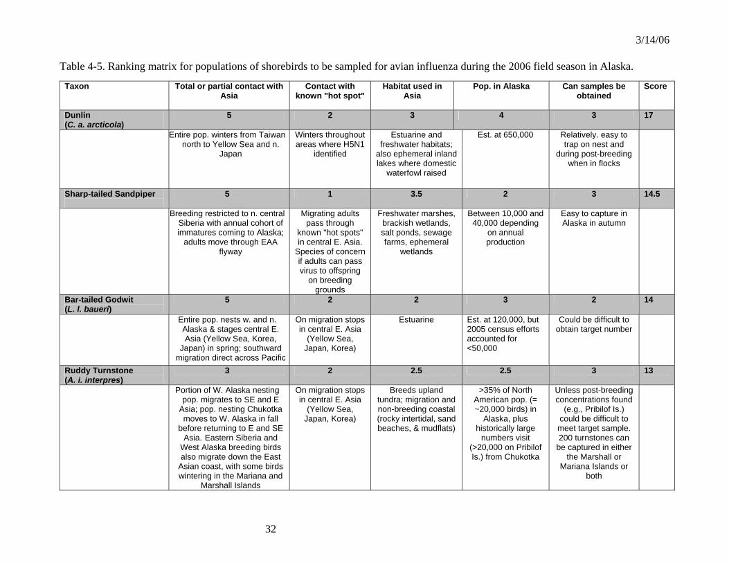
3/14/06
32
Table 4-5. Ranking matrix for populations of shorebirds to be sampled for avian influenza during the 2006 field season in Alaska.
Taxon Total or partial contact with Asia
Contact with known "hot spot"
Habitat used in Asia
Pop. in Alaska Can samples be obtained
Score
Dunlin (C. a. arcticola)
5 2 3 4 3 17
Entire pop. winters from Taiwan north to Yellow Sea and n.
Japan
Winters throughout areas where H5N1
identified
Estuarine and freshwater habitats;
also ephemeral inland lakes where domestic
waterfowl raised
Est. at 650,000 Relatively. easy to trap on nest and
during post-breeding when in flocks
Sharp-tailed Sandpiper
5 1 3.5 2 3 14.5
Breeding restricted to n. central Siberia with annual cohort of immatures coming to Alaska;
adults move through EAA flyway
Migrating adults pass through
known "hot spots" in central E. Asia.
Species of concern if adults can pass virus to offspring
on breeding grounds
Freshwater marshes, brackish wetlands, salt ponds, sewage farms, ephemeral
wetlands
Between 10,000 and 40,000 depending
on annual production
Easy to capture in Alaska in autumn
Bar-tailed Godwit (L. l. baueri)
5 2 2 3 2 14
Entire pop. nests w. and n. Alaska & stages central E. Asia (Yellow Sea, Korea,
Japan) in spring; southward migration direct across Pacific
On migration stops in central E. Asia
(Yellow Sea, Japan, Korea)
Estuarine Est. at 120,000, but 2005 census efforts accounted for <50,000
Could be difficult to obtain target number
Ruddy Turnstone (A. i. interpres)
3 2 2.5 2.5 3 13
Portion of W. Alaska nesting pop. migrates to SE and E
Asia; pop. nesting Chukotka moves to W. Alaska in fall
before returning to E and SE Asia. Eastern Siberia and
West Alaska breeding birds also migrate down the East
Asian coast, with some birds wintering in the Mariana and
Marshall Islands
On migration stops in central E. Asia
(Yellow Sea, Japan, Korea)
Breeds upland tundra; migration and non-breeding coastal (rocky intertidal, sand beaches, & mudflats)
>35% of North American pop. (= ~20,000 birds) in
Alaska, plus historically large
numbers visit (>20,000 on Pribilof Is.) from Chukotka
Unless post-breeding concentrations found
(e.g., Pribilof Is.) could be difficult to meet target sample. 200 turnstones can
be captured in either the Marshall or
Mariana Islands or both

3/14/06
33
Taxon Total or partial contact with Asia
Contact with known "hot
spot"
Habitat used in Asia
Pop. in Alaska Can samples be obtained?
Score
Pectoral Sandpiper
3 1 4 3 2 13
Greater than 50% of pop. nests in Russia west to
Eastern Taimyr Peninsular.
To date no known use of "hot spots"
Breeds marshy/grassy tundra; post-
breeding uses brackish ponds
freshwater marshes
200,000-300,000 Could be difficult to obtain target number
Red Knot (C. c. rogersi & roselaari)
4 2 2 2.5 2 12.5
C.c. roselaari pop. nests Wrangel I. and w. Alaska and
winters Pacific coast of the Americas. C. c. rogersi nests Chukotka/New Siberian Isl. & winters Aust./New Zealand, passing through c. E. Asia
On migration C. c. rogersi passes through areas where H5N1
identified
Estuarine C. c. roselaari <50,000; C. c. rogersi 220,000. C. c. rogersi
thought to stop in Alaska in spring but numbers unknown (possibly several
10,000s)
Could be difficult to obtain target number
Long-billed Dowitcher
3 1 3 3 2 12
>30% of pop. breeds in Russia where range expanding w. to Taimyr Pen.; >95% of entire pop. winters in North and Central America. Unknown numbers winter in Asia (Japan)
To date no known use of "hot spots"
Breeds coastal lowlands in wet,
grassy freshwater meadows; uses estuarine and
managed wetlands during migration &
winter
North American pop. = 450,000 (>90% of this
in Alaska during migration)
Could be difficult to obtain target number
Rock Sandpiper (C. p. tschuktschorum)
3 1 2.5 2 3 11.5
~ 20-30% of pop. nests in Chukotka
To date no known use of "hot spots"
Nests upland tundra; post-breeding use
estuarine areas
Total pop. 50,000. ~20K nest Chukotka
but all return to AK en route to non-breeding areas in Pacific NW
Easy to trap on nest and during post-breeding flocking
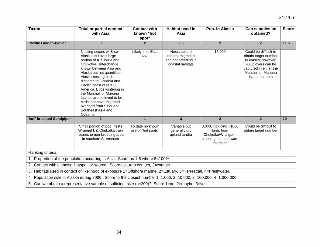
3/14/06
34
Taxon Total or partial contact with Asia
Contact with known "hot
spot"
Habitat used in Asia
Pop. in Alaska Can samples be obtained?
Score
Pacific Golden-Plover
3 2 2.5 2 2 11.5
Nesting occurs w. & sw Alaska and over large portion of n. Siberia and Chukotka. Interchange known between Asia and Alaska but not quantified. Alaska-nesting birds disperse to Oceania and Pacific coast of N & C America. Birds wintering in the Marshall or Mariana Islands are believed to be birds that have migrated overland from Siberia to Southeast Asia and Oceania.
Likely in c. East Asia
Nests upland tundra; migration
and nonbreeding in coastal habitats
16,000 Could be difficult to obtain target number in Alaska; however, 200 plovers can be
captured in either the Marshall or Mariana
Islands or both
Buff-breasted Sandpiper 2 1 3 2 2 10
Small portion of pop. nests Wrangel I. & Chukotka then returns to non-breeding area
in southern S. America
To date no known use of "hot spots"
Variable but generally dry upland tundra
3,000, including ~1000 birds from
Chukotka/Wrangel I. stopping on southward
migration
Could be difficult to obtain target number
Ranking criteria: 1. Proportion of the population occurring in Asia. Score as 1-5 where 5=100% 2. Contact with a known 'hotspot' or source. Score as 1=no contact, 2=contact 3. Habitats used in context of likelihood of exposure 1=Offshore marine, 2=Estuary, 3=Terrestrial, 4=Freshwater. 4. Population size in Alaska during 2006. Score to the closest number 1=1,000, 2=10,000, 3=100,000, 4=1,000,000 5. Can we obtain a representative sample of sufficient size (n=200)? Score 1=no, 2=maybe, 3=yes.

3/14/06
35
Table 4-6. Ranking matrix for populations of passerines and a larid to be sampled for HPAI during the 2006 field season in Alaska. Taxon Proportion of
population in Alaska
Contact with known "hot spot"
Habitat used in Asia Population. in Alaska
Can samples be obtained
Score
Arctic Warbler (Phylloscopus borealis kennicotti)
5 2 3 4 3 17
Endemic subspecies to
Alaska
Winters in Myanmar, Thailand, se. China, Taiwan, Philippines
south to Andaman Is., Malay Peninsula, and
Indonesia east to Moluccas
Terrestrial. Wooded habitats, cultivated areas, grasslands,
gardens, and mangroves
Est. at 2,700,000 Many locations where the most abundant
breeding bird. Easy to capture during breed
and migration
Eastern Yellow Wagtail (Motacilla tschutschensis)
5 2 3.5 4 3 17.5
Endemic species to
Alaska
Taiwan, Indonesia, Sunda Isles, and
Moluccas
Terrestrial. Open areas with water, sugarcane fields, rice
fields, sparse grasslands, cassava plots; usually in association with
wild and domestic grazing mammals
Est. 1,400,000 Easy to capture and areas with known concentration of
breeding birds already identified
Gray-cheeked Thrush (Catharus minimus)
3 2 3 4 3 15
48% of global population in
Alaska
Breeds in E. Siberia Terrestrial. Shrubs often in riparian habitats
Est. 5,000,000 Most abundant bird in many locations.
Already captured at many banding sites.
Glaucous Gull (Larus hyperboreus)
2 2 3 2 3 12
40,000 (100%) of US breeding
population in AK
Contact with humans and garbage dumps
Terrestrial/coastal Approx 40,000 Samples easily obtainable

3/14/06
36
Table 4-7. Suggested migratory bird species for highly pathogenic H5N1 avian influenza surveillance in the four North American flyways. Pacific Flyway Taxon Ranking Tundra Swan (Western Population) Primary Lesser Snow Goose (Wrangel Island Population) Primary Northern Pintail Primary Long-billed Dowitcher Primary Red Knot (small numbers) Primary Pacific Golden Plover (small numbers) Primary Ruddy Turnstone (very small numbers) Primary Black Brant (Pacific Population) Secondary Cackling Goose Secondary Pacific Greater White-fronted Goose Secondary Mallard Secondary American Wigeon Secondary American Green-winged Teal Secondary Northern Shoveler Secondary Central Flyway Taxon Ranking Lesser Sandhill Crane (Mid-continent) Primary Tundra Swan (Eastern Population) Primary Northern Pintail (low percentage from Alaska) Primary Pectoral Sandpiper Primary Buff-breasted Sandpiper Primary Long-billed Dowitcher Primary Greater White-fronted Goose (Mid-continent) Secondary Lesser Snow Goose (Western Central Flyway) Secondary Mallard Secondary American Wigeon Secondary American Green-winged Teal Secondary Northern Shoveler Secondary
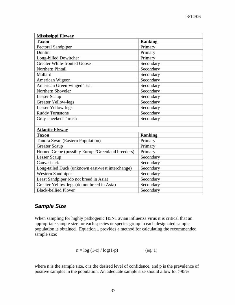
3/14/06
37
Mississippi Flyway Taxon Ranking Pectoral Sandpiper Primary Dunlin Primary Long-billed Dowitcher Primary Greater White-fronted Goose Secondary Northern Pintail Secondary Mallard Secondary American Wigeon Secondary American Green-winged Teal Secondary Northern Shoveler Secondary Lesser Scaup Secondary Greater Yellow-legs Secondary Lesser Yellow-legs Secondary Ruddy Turnstone Secondary Gray-cheeked Thrush Secondary Atlantic Flyway Taxon Ranking Tundra Swan (Eastern Population) Primary Greater Scaup Primary Horned Grebe (possibly Europe/Greenland breeders) Primary Lesser Scaup Secondary Canvasback Secondary Long-tailed Duck (unknown east-west interchange) Secondary Western Sandpiper Secondary Least Sandpiper (do not breed in Asia) Secondary Greater Yellow-legs (do not breed in Asia) Secondary Black-bellied Plover Secondary
Sample Size When sampling for highly pathogenic H5N1 avian influenza virus it is critical that an appropriate sample size for each species or species group in each designated sample population is obtained. Equation 1 provides a method for calculating the recommended sample size: n = log (1-c) / log(1-p) (eq. 1) where n is the sample size, c is the desired level of confidence, and p is the prevalence of positive samples in the population. An adequate sample size should allow for >95%

3/14/06
38
confidence that AI is detected at < 1.5% prevalence. These criteria result in an estimated sample size of 200: n = log (1-.95) / log (1-0.015) = 200 Thus, a minimum of 200 samples should be collected from the population of interest based on an assumed prevalence of 1.5% of highly pathogenic H5N1 avian influenza. We caution that this calculation is very sensitive to the assumed prevalence, which we can not know a priori. For example, if prevalence of the disease at the time of sampling is 0.1% (i.e., 1 in 1000 birds is infected) the necessary sample size is 3000. As prevalence decreases the likelihood of detecting the disease in an individual bird also decreases due to the low probability of detection and practical limitations on laboratory processing capability. We also caution that this formula is weakened here because it is based on assumptions that may not apply to H5N1 virus in wild birds, namely that the agent is homogeneously distributed within a host population that also is homogeneously distributed.
Sample Collection Tracheal and cloacal swabs should be collected from individuals of each species at each location using the procedures identified in Attachment 9.
Discussion Wild birds, particularly waterfowl and other waterbirds, are natural hosts of avian influenza viruses and are believed to play an important role in the epizootiology of these viruses. All hemagglutinin and neuraminidase subtypes have been found in waterfowl and shorebirds (Webster et al., 1992; Krauss et al., 2004; Widjaja et al., 2004). This proposed sampling effort provides the best opportunity for detection of highly pathogenic H5N1 avian influenza virus in live migratory birds that may bring the disease from Asia to Alaska, the Pacific Islands and the west coast of the U.S. The primary advantage to this approach is that species will be sampled that travel directly to Alaska or the US Pacific Islands and trust territories from Southeast Asia or Australasia, have some fraction of the population known to winter in Asia or Pacific trust territories, or intermingle with other species across Siberia, the Russian Far East, and Alaska. The primary disadvantage is the logistical considerations in live capture of the birds in remote areas.
Recommendations Sampling live birds will allow us to determine if they are currently infected with highly pathogenic H5N1 avian influenza virus or other AI viruses. When collecting samples from live birds, tracheal and cloacal swabs are preferred. Most AI strains tend to replicate more efficiently in the intestinal tract than in the respiratory tract of natural host species (i.e., waterfowl and shorebirds). Consequently, cloacal swabs are generally

3/14/06
39
preferred. However, recent isolations of highly pathogenic H5N1 avian influenza virus in wild birds have documented higher levels of virus in tracheal samples. Therefore, it is recommended that both samples be collected from birds when possible. While, the collection of cloacal swabs is a relatively easy procedure, obtaining proper tracheal swabs can be problematic and requires personnel trained in the sampling technique. Examples of tracheal/cloacal swab collection protocols can be found in Attachment 9. Tracheal and cloacal swabs should be placed in separate tubes, and swabs should not be pooled across individuals. Specific implementation plans should be developed for each state/flyway. It is strongly advised that agencies and organizations coordinate their sampling efforts to assure that adequate sample sizes are obtained from each species within each state/flyway. Coordination can be achieved through the existing migratory bird flyway councils.
Literature Cited Hanson B.A., D.E. Stallknecht, D.E. Swayne, L.A. Lewis, and D.A. Senne. 2003. Avian
influenza viruses in Minnesota ducks during 1998-2000. Avian Diseases 47:867-871. Krauss, S., D. Walker, S.P., Pryor, L. Niles, L. Chenghong, V.S. Hinshaw, and R.G.
Webster. 2004. Influenza A viruses of migrating wild aquatic birds in North America. Vector-Bourne and Zoonotic Diseases 4:177-189.
Ito, T., K. Okazaki, Y. Kawaoka, A. Takada, R.G. Webster, and H. Kida. 1995.
Perpetuation of influenza A viruses in Alaskan Waterfowl reservoirs. Archives of Virology 140:1163-1172.
Slemons R.D. et al. 2003. Type A influenza virus surveillance in free-flying,
nonmigratory ducks residing on the eastern shore of Maryland. Avian Diseases 47:1107-1110.
Swayne, D.E. and D.A. Halvorson. Influenza. 2003. Pp 135-160 in Diseases of Poultry,
11th ed. Saif, Y.M., H.J. Barnes, J.R. Glisson, A.M. Fady, L.R. McDougald, and D.E. Swayne (eds.). Iowa State University Press, Ames, IA.
Widjaja, L., S.L. Krauss, R.J. webby, T. Xie, and R.G. Webster. 2004. Matrix gene of
influenza A viruses isolated from wild aquatic birds: ecology and emergence of influenza A viruses. Journal of Virology 78:8771-8779.
Webster, R.G., W.J. Bean, O.T. Gorman, T.M. Chambers, and Y. Kawaoka. 1992.
Evolution and ecology of influenza A viruses. Microbiological Reviews 56:152-179.

3/14/06
40
ATTACHMENT 5
Surveillance for Highly pathogenic H5N1 avian influenza in Hunter-killed Birds
Overview Check stations for waterfowl hunting are operated by some state natural resource agencies and National Wildlife Refuges to collect information on local waterfowl harvest. Hunter check stations provide an opportunity to collect additional samples to monitor for the presence of Highly pathogenic H5N1 avian influenza virus and other avian influenza (AI) virus subtypes. This sampling would supplement live bird surveillance (Attachment 4) by increasing the number of selected species, geographic locations, and time periods represented in surveillance efforts. Previous and current studies that have sampled hunter-killed waterfowl to detect AI include an ongoing 20-year study in Ohio by Dr. Richard Slemons of Ohio State University, current work in North Carolina by Dr. David Stallknecht of the Southeastern Cooperative Wildlife Disease Study (the North Carolina Wildlife Resources Commission has been collecting samples from hunter-killed ducks and tundra swans for Dr. Stallknecht), and sampling in New Mexico, Texas, and Maryland by state wildlife agencies and/or university researchers. A number of the state natural resource agencies and other researchers are developing plans to sample hunter-killed birds for AI in 2005 and/or 2006. This conceptual surveillance strategy includes (1) sampling birds killed in fall by sport hunters and in summer by subsistence hunters in Alaska: (2) sampling birds killed in fall in the lower 48 states. As with surveillance of live-captured birds, sampling hunter-killed birds in Alaska will focus on hunted species that are most likely to be exposed to HPAI in Asia, and that have relatively direct migratory pathways from those areas to Alaska (primary species). Additional samples collected on the wintering grounds in the lower 48 states will include both primary species and species that mix with the primary species in Alaska staging areas (secondary species). Currently, the probability of highly pathogenic H5N1 avian influenza virus transmission from primary species to secondary species is poorly understood, but AI viruses are known to remain viable for months in cold freshwater. From a surveillance standpoint, if secondary transmission proves to be potent and extensive, a very large number of species could be involved. However, this conceptual program is focused on early detection, with adaptation to more intensive efforts as needed. Thus, it is recommended that sampling efforts involving hunter-killed birds in the lower 48 states should concentrate on the species/populations and wintering areas in which the presence of highly pathogenic H5N1 avian influenza virus is most likely to be detected. Some research indicates that susceptibility may vary among game birds. The complete design, implementation and development of an operational plan and the funding necessary for this strategy requires closer coordination with states through the Flyway Council system.

3/14/06
41
A similar approach to investigate the possible movement of Highly pathogenic H5N1 avian influenza virus from Europe could be developed. There are few data on migratory bird movement rates between North America and Europe, but the band recovery data that are available suggest very low exchange rates. Presently we only have data on movement from North America to Europe, but if we make the tenuous assumption that movement rates are similar in both directions, then only 3 species emerge as likely primary species: Eurasian wigeon, northern pintail, and green-winged teal. Of American wigeon banded in the Atlantic Flyway, only 6 of 2,211 recoveries (0.27%) were from Europe. The rates for northern pintail and green-winged teal are 0.06% (3 of 5,341 recoveries) and 0.04% (5 of 12,274 recoveries), respectively. If immigration rates are similar to emigration rates, and if those rates are indicative of the proportion of eastern North American birds that are immigrants from Europe, the chance of sampling even one hunter-killed immigrant is very low. Some common eiders (Keith McAloney, Canadian Wildlife Service, personal communication) and North Atlantic Population Canada geese (Fox et al. 1996) move back and forth between northeastern North America and Greenland, where they could interact with birds from mainland Europe. Likewise, the high-arctic Atlantic brant that breed in North America and winter in Ireland also come into contact with European birds. Thus, these are additional species that should be considered when implementing surveillance for highly pathogenic H5N1 avian influenza virus in the Atlantic flyway.
Methodology
Alaska The surveillance of live-captured birds strategy (Attachment 4) has provided the biological basis for identifying the primary species recommended for sampling in Alaska. Below is a list of these target species and sampling locations recommended for surveillance of hunter-killed birds.
Samples from fall hunters: • Northern pintail: Mendenhall Refuge, Minto Flats, and Cook Inlet • Lesser sandhill crane: Delta Junction • Black brant: Cold Bay • Common eider, king eider, long-tailed duck: Kodiak National Wildlife Refuge,
and perhaps cooperating hunting guides along the coast of Alaska.
Samples from summer hunters: • Seward Peninsula: lesser sandhill crane, bar-tailed godwit, long-billed dowitcher • Yukon-Kuskokwim Delta: black brant, emperor goose, common eider, king
eider, northern pintail, long-tailed duck • Barrow: common eider, king eider, black brant, long-tailed duck, glaucous gull • St. Lawrence Island: emperor goose, black brant, common eider, king eider

3/14/06
42
North American Flyways There are at least 25 states in which either the state wildlife agencies or USFWS routinely check hunter harvested birds: 9 in the Atlantic Flyway, 7 in the Mississippi Flyway, 4 in the Central Flyway, and 5 in the Pacific Flyway. Those 25 states and most other states have already expressed some willingness to collect samples from hunter-killed birds. States are encouraged to develop specific implementation plans in consultation with their respective flyway council using the guidance provided in this strategic plan. Four primary target species/populations have been identified for highly pathogenic H5N1 avian influenza virus sampling in the lower 48 states: northern pintail, Pacific black brant, Wrangel Island snow geese, and lesser sandhill crane (mid-continent population). Also, several secondary species that mix with the primary species in Alaska (and thus have an increased risk of exposure to highly pathogenic H5N1 avian influenza virus) and later winter in the 4 flyways have been identified (see below). Given the ephemeral nature of birds at specific sites along their migration routes, sampling efforts for both primary and secondary species should be concentrated on the wintering grounds. The target species/populations and general sampling locations presented in this document are based on band recovery and in some cases radio telemetry data for birds banded in Alaska. Specific sampling sites (i.e., check stations or other areas where hunter-harvested birds could be sampled) will be determined by the individual states and National Wildlife Refuges that elect to participate in the sampling.
Pacific Flyway: • Northern pintail (primary species): Central Valley of California • Wrangel Island snow geese (primary): Skagit-Fraser Rivers Delta, Washington
and British Columbia • Black brant (primary): Humboldt Bay, California and San Quintin Bay, Mexico if
permits to collect samples and ship them to the U.S. can be obtained • American wigeon combined (secondary): Central Valley of California • American green-winged teal (secondary): Central Valley of California • Northern shoveler (secondary): Central Valley of California • Cackling goose (secondary): Northwest Oregon/Southwest Washington permit
goose areas • Pacific greater white-fronted geese (secondary): Central Valley of California • Tundra Swan (secondary): Montana and Utah • Mallard (secondary): western Washington
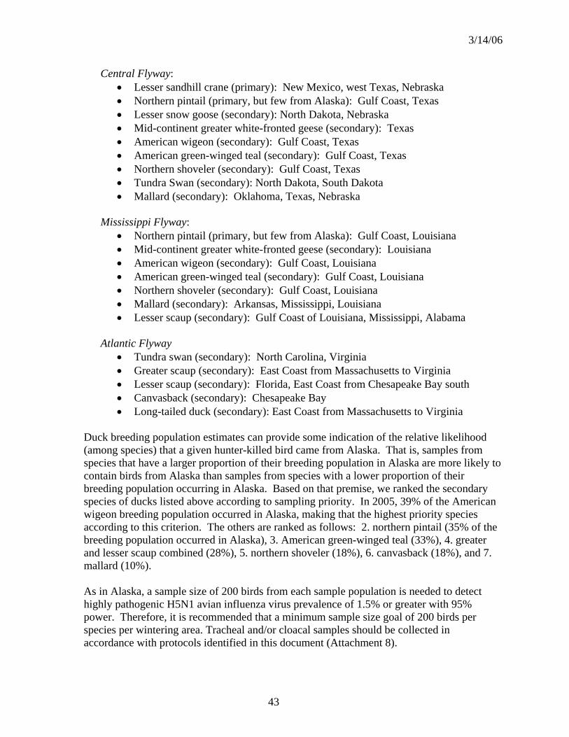
3/14/06
43
Central Flyway: • Lesser sandhill crane (primary): New Mexico, west Texas, Nebraska • Northern pintail (primary, but few from Alaska): Gulf Coast, Texas • Lesser snow goose (secondary): North Dakota, Nebraska • Mid-continent greater white-fronted geese (secondary): Texas • American wigeon (secondary): Gulf Coast, Texas • American green-winged teal (secondary): Gulf Coast, Texas • Northern shoveler (secondary): Gulf Coast, Texas • Tundra Swan (secondary): North Dakota, South Dakota • Mallard (secondary): Oklahoma, Texas, Nebraska
Mississippi Flyway:
• Northern pintail (primary, but few from Alaska): Gulf Coast, Louisiana • Mid-continent greater white-fronted geese (secondary): Louisiana • American wigeon (secondary): Gulf Coast, Louisiana • American green-winged teal (secondary): Gulf Coast, Louisiana • Northern shoveler (secondary): Gulf Coast, Louisiana • Mallard (secondary): Arkansas, Mississippi, Louisiana • Lesser scaup (secondary): Gulf Coast of Louisiana, Mississippi, Alabama
Atlantic Flyway
• Tundra swan (secondary): North Carolina, Virginia • Greater scaup (secondary): East Coast from Massachusetts to Virginia • Lesser scaup (secondary): Florida, East Coast from Chesapeake Bay south • Canvasback (secondary): Chesapeake Bay • Long-tailed duck (secondary): East Coast from Massachusetts to Virginia
Duck breeding population estimates can provide some indication of the relative likelihood (among species) that a given hunter-killed bird came from Alaska. That is, samples from species that have a larger proportion of their breeding population in Alaska are more likely to contain birds from Alaska than samples from species with a lower proportion of their breeding population occurring in Alaska. Based on that premise, we ranked the secondary species of ducks listed above according to sampling priority. In 2005, 39% of the American wigeon breeding population occurred in Alaska, making that the highest priority species according to this criterion. The others are ranked as follows: 2. northern pintail (35% of the breeding population occurred in Alaska), 3. American green-winged teal (33%), 4. greater and lesser scaup combined (28%), 5. northern shoveler (18%), 6. canvasback (18%), and 7. mallard (10%). As in Alaska, a sample size of 200 birds from each sample population is needed to detect highly pathogenic H5N1 avian influenza virus prevalence of 1.5% or greater with 95% power. Therefore, it is recommended that a minimum sample size goal of 200 birds per species per wintering area. Tracheal and/or cloacal samples should be collected in accordance with protocols identified in this document (Attachment 8).

3/14/06
44
Discussion Sampling of hunter-killed birds would supplement targeted surveillance in live wild birds (Attachment 4) and other strategies identified in this strategic plan. The advantage to this approach is that it is cost-effective because for most of the species that are classified as game birds, existing infrastructure (e.g., check stations) is in place in most wintering areas and sufficient numbers of birds are expected to be encountered. The disadvantages to this approach are: 1) most of the sampling in the lower 48 states will be of secondary species, thus the likelihood of sampling birds that have come into contact with infected primary species birds is small, especially in the Atlantic, Mississippi, and Central Flyways; and 2) numerous sampling sites throughout the U.S. will require sufficient training of sampling personnel to ensure samples are properly acquired, preserved, and shipped. There are advantages and disadvantages in terms of public perceptions of sampling hunter-harvested birds. Public perceptions could be positive if user-groups will appreciate that samples are being taken. Conversely, if hunters do not have accurate information about highly pathogenic H5N1 avian influenza virus, they could become unnecessarily alarmed about exposure (especially if agency samplers are wearing protective gear). For spring and summer subsistence users, providing access to birds that were historically taken illegally may make sampling difficult and basic information on highly pathogenic H5N1 avian influenza virus and the sampling program may be harder to deliver.
Recommendation Sampling hunter-killed birds would supplement other approaches in a cost-effective manner and may allow us to determine if certain species of birds (e.g., migratory game birds) are currently infected with highly pathogenic H5N1 avian influenza subtype or other AI viruses. This expanded effort to identify highly pathogenic H5N1 avian influenza virus should be considered a supplemental part of any surveillance system. Specific implementation plans and budgets should be developed in concert with affected state agencies and the flyway councils. Unlike other surveillance approaches, the use of hunter-killed birds has high public profile. Therefore, the implementation of this action should be discussed with agencies and organizations that have experience regarding the public relations aspects of researching and assessing zoonoses (e.g., chronic wasting disease and West Nile virus) to ensure that we develop an appropriate and consistent message to hunters.
References Fox, A.D., Christian Glahder, Carl R. Mitchell, David A. Stroud, Hugh Boyd, and John
Frikke. 1996. North American Canada geese (Branta canadensis) in west Greenland. Auk 113(1): 231-233.

3/14/06
45
ATTACHMENT 6
Sentinel Animal Methods
Overview This methods section reviews two sentinel animal methods that have been used in avian disease surveillance programs and that may be used for the early detection of avian influenza (AI) virus infection along migratory flyways in the U.S.
Non-commercial Backyard Poultry Flocks Backyard poultry are defined as domesticated fowl, including chickens, turkeys, waterfowl, and game birds (except doves and pigeons) maintained for hobby or noncommercial egg and meat production (NAHRS FAQ 2005). Backyard poultry are typically allowed to forage freely or may be confined in partially enclosed fenced areas. The evaluation of poultry flocks reared on backyard premises for diseases of interest to the commercial poultry industry has been used as a surveillance method to estimate seroprevalence of selected disease agents as part of health surveys in backyard flocks adjacent to commercial operations. (McBride; Hird; Carpenter; Snipes; Danaye-Elmi, and Utterback 1991;Johnson; Colby; Tablante; Hegngi; Salem; Gedamu, and Pope C. 2004) In 2005, State animal health officials in Alaska sampled birds at fairs and exhibitions (concentration points). Most exhibitors were 4H or hobby farmers. Fairs and exhibitions are social events and are attended by large numbers of game bird fanciers from remote regions of Alaska. State animal health officials offered testing to exhibitors at three agricultural fairs with the goal of testing every entry to the fair (600 samples representing 100 flocks). This was a voluntary sampling program, but nearly 100% of owners of exhibition game agreed to test in 2005.
• 150 birds were sampled at the Fairbanks fair. Tanana Fair entries represent flocks from a 40 mile radius around the city of Fairbanks (Healy, Tanana, and the North Pole).
• 100 birds were sampled at the Kenai fair. Kenai Fair entries include flocks from Homer to Anchorage
• 300 samples were collected at the Alaskan State Fair in Palmer. Palmer Fair entries include Anchorage, Matanuska Valley (includes flocks as far north as Fairbanks)
In Alaska, poultry chicks are either purchased through mail order or from a few local breeders and may be reared in suburban areas or in remote villages throughout the State. Most backyard birds are reared for egg production and slaughtered for meat prior to the winter season, although there are a growing number of hobbyists that raise show birds. Birds

3/14/06
46
are often reared on open range or in outside enclosures and sometimes have an opportunity to intermingle with wild waterfowl. Limited resources prohibited widespread backyard bird surveillance testing over the large expanse of the state. Backyard flock surveillance is presently passive and owners request testing after morbidity or mortality events occur in their flock or after noting dead waterfowl or sick waterfowl on their premises. There is currently no census available to estimate the number of backyard flocks in Alaska. Cloacal swab samples are placed in ethanol and evaluated at the University of Alaska, at Fairbanks by RT-PCR. If surveillance screening samples are positive by PCR, the premise is placed under quarantine and additional cloacal samples taken during the epidemiological investigation are placed in viral transport media and submitted for virus isolation to NVSL in Ames, Iowa.
In 2006, the Alaska Department of Environmental Conservation, Office of the State Veterinarian will sample backyard flocks, near summer water systems where wild and domestic waterfowl congregate and collect environmental samples (bird droppings, water samples) in six general areas:
• Southeast- 2 cities (Juneau, Ketchikan) • Southcentral- 4 cities (Homer, Soldotna, Anchorage, Matanuska Valley • Interior- 3 cities (Fairbanks, Healy, Talkeetna) • Southwest- 2 cities (Bethel, King Salmon) • Northwest- 2 cities (Nome, Kotezebue) • Aleutians/Bering Sea- 4 cities (Kodiak, Dutch Harbor, Cold Bay, Pribilof Islands)
The areas listed in bold have the highest priority and cover a majority of the population where domestic poultry is kept. The other 3 areas have substantial populations of wild birds but few domestic backyard flocks. A sample size of 11 is needed to detect avian influenza at a prevalence rate of 25% at a 95% confidence interval in flocks ranging from 10 to 10,000 or more birds. The Office of the State Veterinarian will sample poultry exhibited at the six agricultural fairs (concentration points):
• Deltana Fair Date to be announced • Haines Fair 7/26-30/2006 • Tanana Valley Fair 8/4-12/2006 • Kenney Lake Fair Date to be announced
(2nd Week August 2006) • Kenai Peninsula Fair (Ninilchik) 8/18-20/2006 • Alaska State Fair (Palmer) 8/24 - 9/4/2006
The fair boards have expressed interest in promoting this sampling effort and it is expected that all entries will be tested. The state has just finished construction of a new diagnostic laboratory, Alaska Environmental Health Laboratory in Anchorage, and will develop the capacity to run the diagnostic tests using RT-PCR. If surveillance screening samples are positive by PCR, the premise will be placed under quarantine and additional cloacal samples

3/14/06
47
taken during the epidemiological investigation are placed in viral transport media and submitted for virus isolation to NVSL in Ames, Iowa. This approach to sampling non-commercial poultry flocks may be adapted in other areas of the U.S. where there is widespread non-commercial poultry production.
Sentinel Duck Flocks The second method described is the placement of sentinel duck flocks in wetland environments where they are potentially exposed to and infected with disease agents as they commingle with wild birds. The placement of sentinel flocks of domestic ducks has been used to recover AI and detect influenza epizootics in pelagic bird colonies, and yielded much higher isolation rates compared to isolations from wild birds (Turek; Gresikova, and Tumova 1984;Sinnecker; Sinnecker; Zilske, and Koehler 1982;Sinnecker; Sinnecker, and Zilske 1982). Sentinel ducks have been used to determine the presence of AI and timing of infection associated with the arrival of wild migratory waterfowl in wetland habitats adjacent to market turkey production flocks (Halvorson; Karunakaran; Senne; Kelleher; Bailey; Abraham; Hinshaw, and Newman 1983;Halvorson; Kelleher, and Senne 1985;Kelleher; Halvorson; Newman, and Senne 1985). In North America, AI isolations from waterfowl have been reported from approximately 30 locations over the past 35 years (Hanson; Stallknecht; Swayne; Lewis, and Senne 2003). Many of these sites are located along each of the four migratory waterfowl flyways (Pacific, Central, Mississippi, and Atlantic) in the continental U.S. Bodies of water with large concentrations of migratory waterfowl and shorebirds might also serve as sentinel sites.
Ideally, surveillance activities should occur at sites at a time when migratory birds are actively nesting and at locations where they marshal and intermingle with other migratory birds transiting the area prior to winter migration. The onset of avian influenza infection in sentinel ducks has been shown to occur in late July and early August in summer breeding areas (infection of range reared turkey flocks was shown to occur about 6 to 8 weeks later) (Halvorson et al. 1985). Avian influenza virus prevalence estimates from published waterfowl surveys indicate that virus can first be detected in naïve juvenile birds in summer breeding areas in July or August (prevalence ranged from 11% to 61% in published surveys) as juveniles emerge from hiding and intermingle with other broods and a subsequent high rate of re-infection as birds marshal for winter migration in October (Hanson et al. 2003; Hinshaw et al. 1985). Avian influenza virus prevalence generally decreases during late fall and winter and may reach a level of 1% or less in over-wintering areas.(Stallknecht ;Webster; Bean; Gorman; Chambers, and Kawaoka 1992) However, virus was isolated from 11% of teals and from 15% of northern pintails in one recent survey of wintering ducks in Texas, suggesting that the avian influenza season may not be a fall season event (Hanson 2003). As a result of early migration, blue winged teal are thought to serve as an immunologically naïve host in wintering areas. Most virus isolations have occurred in mallards and other species of dabbling ducks, but less commonly in wood ducks and similar species (Stallknecht ). Mallards are commonly
3/14/06
48
associated with habitats located near man, livestock, and poultry and would be more likely to interact with backyard poultry flocks compared with other waterfowl species (Stallknecht and Shane 1988). Although H5, H7, and H9 subtypes have been poorly represented in most waterfowl surveys (H3, H4, or H6 subtypes have been isolated most frequently), pintails and mallards have been shown to be significant reservoirs in one recent survey where H5, H7, and H9 virus subtypes were isolated 21.5% of the time in Minnesota (Hanson and others 2003). The prevalence of AI isolated from blue winged teal on wintering grounds in February in Texas was found to be 22% in 2001 and 15% in 2002 (Hanson 2003). Migration of blue winged teal occurs in late summer and early fall (typically September), prior to the highest period of AI prevalence. Early migration of this species is thought to play a role in maintenance of AI infection on wintering grounds by providing a susceptible population with little or no prior exposure or immunity. However, blue winged teal are less likely to interact with man or livestock, so sites where blue winged teal congregate may not serve as the best sites for surveillance using backyard flocks of domestic waterfowl. The role of shorebirds in avian influenza ecology should be considered separately from that of migratory waterfowl. The highest prevalence of avian influenza virus in shorebirds has been shown to occur in May and in September, which coincides with the times of peak shorebird migration in the northeastern U.S. (Kawaoka; Chambers; Sladen, and Webster 1988). Shorebirds migrating through the Delaware Bay have been shown to have the highest prevalence of AI virus compared with other shorebird populations surveyed at four other locations along the Atlantic flyway (Hanson 2003). Although most isolates reported from shorebirds in this survey were H10 and H12 (H9 and H13 in previous studies), H5 and H7 subtypes were isolated from a small percentage of shorebirds. During May, virus was isolated mostly from ruddy turnstones (9.1%). The approach to the design of a targeted surveillance method for the detection of avian influenza using either of these two sentinel animal methods should incorporate what is presently known about the ecology and natural history of avian influenza infection in wild waterfowl reservoir species. Sentinel animals are most likely to become infected with AI if exposed to reservoirs in nature during periods of highest viral shedding. As described above, prevalence of infection as measured by virus isolations in published waterfowl surveys has been shown to vary temporally by location, age, season, and species. A targeted approach to sentinel animal surveillance should be designed to: • Target specific locations where AI has been isolated from wild waterfowl historically; • Target locations where known primary reservoir species (mallards, blue winged teal,
ruddy turnstones) congregate for breeding (resulting in higher concentrations of juveniles susceptible to infection) or wintering (higher concentrations of species with little or no previous exposure) resulting in a higher prevalence of infection;
• Be timed to coincide with periods (seasons) of highest prevalence in the reservoir species, in particular migratory species that originate from an area having high incidence of AI (Southeast Asia).
3/14/06
49
Methods
Backyard Poultry Surveillance Method
Flock Selection
• Targeted flocks should consist of free range domestic waterfowl or poultry flocks located near marshlands or wetlands.
• Marshlands should contain high density populations of waterfowl or shorebirds. • Flocks should have an opportunity to directly intermingle with waterfowl
(especially mallards) at or near the common watershed via open range or open enclosure or by sharing a common source of water.
• Chose sites adjacent to wetlands where AI virus has been isolated historically.
Timing of Surveillance
• Surveillance should begin in late July and continue through October at sites near northern breeding areas.
• Although, prevalence rates in wild waterfowl were shown to decrease significantly in wintering areas in Louisiana (1%), prevalence in blue winged teal in wintering areas in Texas during February of >10% indicates that some wintering sites may be useful for sentinel surveillance.
• The seasonal peak of AI prevalence in shorebirds occurs in May rather than late summer, so surveillance of backyard flocks in the Delmarva (Delaware Bay) area should be planned for May to coincide with the time of highest prevalence.
Sample Size Estimates
• The average size of backyard poultry flocks in the U.S. is 35 birds (varies from 28
to 49 birds per flock by region). A prevalence estimate for avian influenza of 25% is assumed (NAHMS Poultry '04 Part I 2004).
• A sample size of 11 is needed to detect avian influenza at a prevalence rate of 25% at a 95% confidence interval in flocks ranging from 10 to 10,000 or more birds.(Cannon and Roe 1982)
• Cloacal and tracheal swab samples would be submitted to the appropriate diagnostic labs for RT-PCR testing and to a reference lab for virus isolation.

3/14/06
50
Sentinel Duck Method
Flock Preparation and Placement
• Construct pens or plan for open fenced enclosures that will hold 10 to 20 ducks and allow contact with released “messenger” ducks and wild ducks. Pens should allow exposure to water contaminated with wild duck feces.
• Deploy pens to selected wetlands (or construct fenced enclosures). • Arrange to provide basic husbandry. • Rear one day old ducks in isolation facilities for 6 to 7 weeks. • Establish AI free status by cloacal swabbing and serologic testing. • Release 10 to 20 isolation reared “messenger” pinioned mallard or white Peking
ducks on selected body of water. • Place 10 to 20 ducks in pens on selected body of water to intermingle with
“messenger” ducks and wild ducks. • Periodically bleed ducks to determine serologic status and replace H5 seropositive
ducks with immunologically naïve ducks.
Timing of Surveillance
• Placement of sentinel duck flocks should coincide with backyard flock surveillance seasonally.
Sampling
• Retrieve fecal samples via cloacal swabs from 10 to 20 penned ducks to detect virus weekly and periodically trap messenger ducks for cloacal sampling.
Data Collection
For backyard flocks, a database similar to the one used for Exotic Newcastle Disease (END) surveillance would provide the ability to trace positive samples back to their flock of origin (Accession number; sending facility premises ID; submitter name, address, and contact information; location of animals including premises ID, latitude, and longitude; owner name; flock information including size, number affected, number dead; purpose of submission and relevant clinical information). Data needed to create predictive geospatial models to evaluate spatial and temporal risk for sentinel duck flocks include: (1) lat/long (in unprojected decimal degrees with a WGS-84 or NAD-83 datum) of the sentinel cage's location; (2) front gate coordinates for the premises; (3) name, address, county, zip code, contact information for the land owner/manager, and occupations of all residents; (4) age, sex, and breed of birds; (5) number of sentinel birds and each bird must have a unique ID (e.g., numbered aluminum leg or wing bands work well); (6) environmental description of area where cage containing sentinel birds is placed; (7) AI

3/14/06
51
virus test status (birds are bled periodically to evaluate immune status and need for replacement); (8) presence and approximate distance to other birds and mammals; (9) exposure to wild birds and free ranging domestic birds; (10) estimated density of birds and mammals on premises and in the vicinity of the sentinels; (11) exposure of sentinels to human contact other than the avian phlebotomist; and (12) an environmental assessment of the vicinity (e.g., within 100 meters, within 500 meters, and within 1000 meters). These data should be captured on a site survey form. However, a separate form should be used to record: date and time blood samples were collected, the birds' ID number, and the vial number for the blood specimen. With this basic information, other data sources can be used to evaluate proximity to wetlands, bird roosts, position with in normal flyways, terrain features, and more. Access to extensive datasets (e.g. the National Wetlands Inventory and the National Landcover Dataset) and hydrologic models could be used to identify wetlands.
Discussion
Major advantages of the use of sentinel animals to detect AI:
• Backyard bird surveillance programs are already in existence in most states. • State animal health officials are familiar with a targeted surveillance approach
(i.e. surveillance of backyard flocks within a designated radius adjacent to commercial poultry operations).
• The placement of sentinel ducks has been used successfully to isolate AI from wild waterfowl in previous published surveys.
• Mortality in backyard poultry from H5N1 has occurred in other countries. • Could be done in conjunction with other surveillance methods at the same
location for comparison.
Major disadvantages of the use of sentinel animals:
• Locating suitable surveillance sites will require field surveillance or input from wildlife biologists.
• Expense of rearing AI free birds. • Pen construction and husbandry costs. • Sentinel flocks are subject to predation.
Recommendations In order to implement an efficient active sentinel animal surveillance system, sentinel flock locations should be purposefully chosen. Appropriately allocating limited resources to achieve targeted sampling and reduce costs is an important objective of animal disease surveillance programs (McCluskey 2003). Knowledge of disease distribution allows us to focus surveillance activities. In this case, we can use our knowledge of the most likely entry points for H5N1 through migratory waterfowl to locate sentinel animal flocks. In order to target areas for sentinel surveillance with a higher probability of disease, flyway information
3/14/06
52
should be plotted over waterfowl management areas in order to select sites most likely to have migratory birds from areas where commingling with Eurasian species is most likely to occur. Specific locations in areas where migratory birds from possible northern exposure sites are most likely to be in highest concentration have been identified in other methods sections of this plan. National information on the health and management practices of backyard and small production flocks adjacent to commercial poultry operations in 18 states is available. All of this information should be combined with information on the geographic distribution of poultry producers including sizes and densities of operations in order to produce a risk map. Local animal health officials could then locate sentinel backyard flocks adjacent to waterfowl management areas in poultry dense regions where there is the highest probability of disease transmission. The health status of sentinel backyard flocks could be evaluated on a recurring basis (quarterly, or more often during seasons of the year that pose the highest probability of disease transmission due to higher prevalences) for an active disease surveillance program.
References
NAHMS Poultry '04 Part I; 2004.
NAHRS FAQ [Web Page]. Accessed 2005 Nov 14. Available at: http://www.aphis.usda.gov/vs/ceah/ncahs/nahrs/NAHRS_faq.htm. Notes: The National Animal Health Reporting System (NAHRS) is a cooperative effort between the American Association of Veterinary Laboratory Diagnosticians (AAVLD), the U.S. Animal Health Association (USAHA) and USDA's Animal and Plant Health Inspection Service (APHIS).
Alfonso, C. P.; Cowen, B. S., and van Campen, H. Influenza A viruses isolated from waterfowl in two wildlife management areas of Pennsylvania. J Wildl Dis. 1995 Apr; 31(2):179-85.
Cannon, R. M. and Roe, R. T. Livestock Disease Surveys A Field Manual for Veterinarians Department of Primary Industry Bureau of Rural Science. Canberra: Australian Government Publishing Service; 1982.
Halvorson, D.; Karunakaran, D.; Senne, D.; Kelleher, C.; Bailey, C.; Abraham, A.; Hinshaw, V., and Newman, J. Epizootiology of avian influenza--simultaneous monitoring of sentinel ducks and turkeys in Minnesota. Avian Dis. 1983 Jan-1983 Mar 31; 27(1):77-85.
Halvorson, D. A.; Kelleher, C. J., and Senne, D. A. Epizootiology of avian influenza: effect of season on incidence in sentinel ducks and domestic turkeys in Minnesota. Appl Environ Microbiol. 1985 Apr; 49(4):914-9.
Hanson, B. A. Temporal, spatial and species patterns of avian influenza viruses among wild birds. Athens, Georgia: University of Georgia; 2003.
3/14/06
53
Hanson, B. A.; Stallknecht, D. E.; Swayne, D. E.; Lewis, L. A., and Senne, D. A. Avian influenza viruses in Minnesota ducks during 1998-2000. Avian Dis. 2003; 47(3 Suppl):867-71.
Hinshaw, V. S.; Nettles, V. F.; Schorr, L. F.; Wood, J. M., and Webster, R. G. Influenza virus surveillance in waterfowl in Pennsylvania after the H5N2 avian outbreak. Avian Dis. 1986 Jan-1986 Mar 31; 30(1):207-12.
Hinshaw, V. S.; Wood, J. M.; Webster, R. G.; Deibel, R., and Turner, B. Circulation of influenza viruses and paramyxoviruses in waterfowl originating from two different areas of North America. Bull World Health Organ. 1985; 63(4):711-9.
Johnson, Y. J.; Colby, M. M.; Tablante, N. L.; Hegngi, F. N.; Salem, M.; Gedamu, N., and Pope C. Application of commercial and backyard poultry geographic information system databases for the identification of risk factors for clinical infectious laryngotracheitis in a cluster of cases on the delmarva peninsula. International Journal of Poultry Science. 2004; 3(3):201-205.
Kawaoka, Y.; Chambers, T. M.; Sladen, W. L., and Webster, R. G. Is the gene pool of influenza viruses in shorebirds and gulls different from that in wild ducks? Virology. 1988 Mar; 163(1):247-50.
Kelleher, C. J.; Halvorson, D. A.; Newman, J. A., and Senne, D. A. Isolation of avian paramyxoviruses from sentinel ducks and turkeys in Minnesota. Avian Dis. 1985 Apr-1985 Jun 30; 29(2):400-7.
McBride, M. D.; Hird, D. W.; Carpenter, T. E.; Snipes, K. P.; Danaye-Elmi, C., and Utterback, W. W. Health survey of backyard poultry and other avian species located within one mile of commercial California meat-turkey flocks. Avian Dis. 1991 Apr-1991 Jun 30; 35(2):403-7.
McCluskey, Brian J. Epidemiology of Vesicular Stomatitis Viruses in the Southwestern United States: Colorado State University; c2003.
Sinnecker, H.; Sinnecker, R., and Zilske, E. Detection of influenza A viruses by sentinel domestic ducks in an ecological survey. Acta Virol. 1982 Jan; 26(1-2):102-4.
Sinnecker, H.; Sinnecker, R.; Zilske, E., and Koehler, D. Detection of influenza A viruses and influenza epidemics in wild pelagic birds by sentinels and population studies. Zentralbl Bakteriol Mikrobiol Hyg [A]. 1982 Dec; 253(3):297-304.
Slemons, R. D.; Hansen, W. R.; Converse, K. A., and Senne, D. A. Type A influenza virus surveillance in free-flying, nonmigratory ducks residing on the eastern shore of Maryland. Avian Dis. 2003; 47(3 Suppl):1107-10.
Slemons, R. D.; Johnson, D. C.; Osborn, J. S., and Hayes, F. Type-A influenza viruses isolated from wild free-flying ducks in California. Avian Dis. 1974 Jan-1974 Mar 31; 18(1):119-24.

3/14/06
54
Slemons, R. D.; Shieldcastle, M. C.; Heyman, L. D.; Bednarik, K. E., and Senne, D. A. Type A influenza viruses in waterfowl in Ohio and implications for domestic turkeys. Avian Dis. 1991 Jan-1991 Mar 31; 35(1):165-73.
Stallknecht, D. E. Ecology and epidemiology of avian influenza viruses in wild bird populations: waterfowl, shorebirds, pelicans, cormorants, etc. Proceedings of the Fourth International Symposium on Avian Influenza; Athens, GA. U.S. Animal Health Association.
Stallknecht, D. E. and Shane, S. M. Host range of avian influenza virus in free-living birds. Vet Res Commun. 1988; 12(2-3):125-41.
Stallknecht, D. E.; Shane, S. M.; Zwank, P. J.; Senne, D. A., and Kearney, M. T. Avian influenza viruses from migratory and resident ducks of coastal Louisiana. Avian Dis. 1990 Apr-1990 Jun 30; 34(2):398-405.
Turek, R.; Gresikova, M., and Tumova, B. Isolation of influenza A virus and paramyxoviruses from sentinel domestic ducks. Acta Virol. 1984 Mar; 28(2):156-8.
Webster, R. G.; Bean, W. J.; Gorman, O. T.; Chambers, T. M., and Kawaoka, Y. Evolution and ecology of influenza A viruses. Microbiol Rev. 1992 Mar; 56(1):152-79.
Webster, R. G.; Morita, M.; Pridgen, C., and Tumova, B. Ortho- and paramyxoviruses from migrating feral ducks: characterization of a new group of influenza A viruses. J Gen Virol. 1976 Aug; 32(2):217-25.

3/14/06
55
ATTACHMENT 7
Environmental Sampling (Feces/Water)
Introduction Avian Influenza viruses (AI) are released by waterfowl through the intestinal tract and viable virus can be detected in both feces and the water in which the birds swim, defecate and feed. This is the principle means of virus spread to new avian hosts and potentially to poultry and other susceptible livestock. Analysis of both water and fecal material from waterfowl habitat can provide evidence of AI virus circulating in wild bird populations, the specific AI subtypes, levels of pathogenicity, and possible risks to livestock.
Technical Aspects of Sampling Water AI is relatively stable in water, especially at colder temperatures. The longevity of viable virus (weeks to months) allows for an integration of activity on a site basis. However, in the absence of established serial sampling, pinpointing the time at which a site becomes contaminated would be difficult. The advantages of including waterfowl sites as a point of sampling lie in the ease of collecting samples and the potential to sample the potential contaminating influence many birds at once. This method would provide a cost-effective, geographically explicit methodology. Moreover, given the ease of sampling, more sites could be sampled, providing for a higher resolution surveillance network. Technical aspects to monitor water samples for AI involve collecting specified volumes of water (usually 50-500ml), transporting the samples on ice or frozen, concentrating the virus present either by filtration or precipitation/centrifugation, and inoculating the virus onto chicken eggs or cell culture for virus growth. The virus replicates in the cultures and is characterized by serological or molecular methods to determine specific subtypes. Alternative methods of analysis involve extracting viral genetic material from the sample with detection using molecular techniques such as reverse transcription-polymerase chain reaction (RT-PCR) and subsequent sequencing to determine subtype. Refinement of these methods still needs to be done. However, these techniques have the advantage of rapid results. All of the procedures for monitoring AI in water samples are generally established, and with proper expertise and equipment can be easily adapted to most laboratory settings.
Technical Aspects of Sampling Feces Fecal sampling is used extensively in monitoring studies for AI in wild bird populations. The principal advantages of this method are that the costs and effort of capturing birds are avoided and large sample numbers can be quickly and easily obtained. It also is a

3/14/06
56
good method to determine the presence or absence of virus in bird populations present at a specific location. The disadvantage is that species identification is not always possible, determination of prevalence is complicated by the possibility of repeated sampling of individuals, and the sensitivity of the method is lower than for oral swabs. However, the increased sampling effort occurring because of reduced sampling costs are anticipated to offset any short comings owing to decreased sensitivity. Information from the field could be used to generate an environmental risk map related to specific areas (habitats) associated with potential AIV transmission. Infectivity of the virus is maintained up to 4 days in wet feces at 25oC. Best analytical results come from fresh fecal samples that are either processed quickly or frozen until processing. Thus this method of sampling, while providing good information, is best applied while birds are present at a location such that the samples are as fresh as possible. By restricting fecal collection to fresh samples, it allows for population census data to be collected, and by inference estimates of the species sources of the contamination. Species and individual identification through genetic typing of feces would allow estimates of prevalence.
Accredited laboratories have the capacity and infrastructure to analyze a limited number of samples for AI. The anticipated sampling effort for this surveillance study will require an investment in equipment and staff to provide results in a timely fashion. Equipment needs include real-time PCR thermalcyclers, RNA extraction capabilities, DNA sequencing capabilities, tissue culture and egg culture facilities, ultracold freezers, centrifuges and vacuum pumps.
Methodology Sampling for highly pathogenic avian influenza (HPAI), such as highly pathogenic H5N1 avian influenza, from environmental deposition of virus by waterfowl should be accomplished by collecting and analyzing feces (Attachment 10) and water from areas of known use by high risk species (e.g., transcontinental migrants). The general challenges faced include; 1) Determining locations used by high risk species, 2) Refinement of existing methods for detecting the virus in water and fecal samples and developing the analytical infrastructure and capacity, 3) Design of a sampling system using composite samples for analysis.

3/14/06
57
Table 7-1. Qualitative comparison of environmental sampling methods.
Fecal Sampling
Pros
Cons
Technically easy sample acquisition. Sampling represents non technical approach and would not require extensive training or experience by field personnel.
Viable virus restricted to fresh samples (1-4 days)
Generate large sample numbers quickly. Large sample numbers can swamp lab systems (applies to all methods)
Does not require handling or capturing animals Low cost, well established technique amenable for high through-put screening (modified APHIS RT-PCR method). Sample analysis is transferable across labs.
Capable of identifying HPAI contaminated sites/locations/regions. Prevalence would be estimated on a site basis. Information from the field could be used to generate an environmental risk map related to specific areas (habitats) associated with potential AIV transmission.
Identity of species and individuals unlikely, estimates of prevalence not possible. Species identification possible through molecular fingerprinting, but at additional cost.
BSL-2 laboratory conditions sufficient for initial diagnostic screening.
Requires Biosafety level 3 capabilities for virus isolation
Summary: An approach based on fecal sampling could be immediately implemented and may represent the only reasonable approach in areas where bird capture is not practical.
Water Sampling
Pros
Cons
Low cost Biosafety level 3 capabilities for virus isolation Effectively sample all or most birds present on the body of water
Analyses potentially complicated if multiple strains of AI present in water samples.
Samples easily, quickly obtained Large volumes of water needed to concentrate virus for analysis, transportation and logistical issue
Virus stable, especially at moderate pH and low temperature
Longevity complicates interpretation on initial timing of contamination.
Does not require handling or capturing animals Identity of species and individuals not possible/difficult. Prevalence calculation restricted to a site basis system.
Generate large sample numbers Large sample numbers can swamp lab systems- need analysis infrastructure
Can provide large scale spatial risk assessment of HPAI contamination.
May need to validate technique
Sampling strategies to detect highly pathogenic H5N1 avian influenza virus in waterfowl populations will change depending upon the risk assessment and management goals and prevailing status of the pathogen in North America. For first detection of highly pathogenic H5N1 avian influenza virus in migratory birds efforts should focus on likely cross-over routes of birds from Asia to North America (e.g., Alaska and North Slope). Efforts should focus on areas of high aggregations of waterfowl intersecting with logistical sampling support, e.g., National Wildlife Refuge (NWR) system and state waterfowl management areas. While highly pathogenic H5N1 avian influenza virus may cross from Asia to North America at any point the surveillance network needs to be

3/14/06
58
tactically practical without compromising its ability for detection. Once highly pathogenic H5N1 avian influenza virus gains a foothold in North America the surveillance network should be placed along known waterfowl movement paths from the point of origin (i.e., point of detection). These paths can be inferred from known migration routes based on waterfowl telemetry data. However, practically, and given the patterns emerging in Eurasia, once highly pathogenic H5N1 avian influenza virus gains a foothold in North America the likelihood of rapid and diffusive spread across the continent is high. At this point local waterfowl and environmental sampling should target areas of strategic value, e.g., human population centers and areas of high density of poultry production. In the former case, such areas would be represented by urban zoo-parks and lakes. These areas would represent the highest level of risk of human contact with contaminated water and/or waterfowl. In the latter case, ponds, lakes and waterfowl management areas around high density poultry production areas would provide the best ability to assess risk of transmission to humans and poultry. Surveillance efforts patterned on these areas are best amenable to local and state efforts for first detection and subsequent risk assessment once the highly pathogenic H5N1 avian influenza virus achieves enzootic status in North America. There is an inherent conflict between the need for high resolution surveillance, the number of samples generated, the time to analyze those samples, and the cost of analysis. If the goal is first detection, methods that integrate across many individuals and species at a particular site without loss of sensitivity should be preferred. Currently analysis of fresh fecal samples is the best method to achieve these goals. For logistically practical and economic reasons sample analysis should focus on composite samples on a per site basis; this bulk sample minimizes effort in both data collection and analysis, while greatly increasing the probability of detection. Given the expected rarity of highly pathogenic H5N1 avian influenza virus in current migratory bird populations, this approach will allow for a substantially reduced number of samples to be analyzed. Table 7-2 provides a hypothetical, but plausible, example of the expected number of tests per composite fecal sample necessary to detect Highly pathogenic H5N1 avian influenza virus. When prevalence is very low (e.g., 10-7) almost all composites will test negative and on average only a single test will be needed to determine the absence of highly pathogenic H5N1 avian influenza virus in that composite sample. The approximate sample sizes necessary for assuring a high probability of detecting highly pathogenic H5N1 avian influenza virus depends on its prevalence in the population, which is currently unknown. However, a preliminary estimator is:
p* = 1- (1-r/m)1/n (eq. 1)
where p* is the proportion of infected individual samples across all composite samples, r is the number of composite samples that test positive for the presence of highly pathogenic H5N1 avian influenza virus, m is the total number of composite samples tested, and n is the number of individual samples in each composite sample (e.g., fecal count or volume). Rearranging eq. 1 provides an estimate of the number of individual

3/14/06
59
fecal samples needed to detect highly pathogenic H5N1 avian influenza virus, for a given population level prevalence;
n = ln(1-r/m) / ln(1-p*) (eq. 2) Table 7-2. Expected number of tests needed for a single positive reaction for each composite sample containing 100 individual fecal samples, n, as a function of expected prevalence of HPAI, p. Calculation is based on the binomial probability model describing the average number of tests needed as (n+1) – n(1-p)n
The results for various hypothetical values of r, m, n, and p* are shown in Table 7-2. Thus, if highly pathogenic H5N1 avian influenza virus prevalence is 10-6 and 10,000 independent fecal samples are collected, analysis of 100 composite samples would result in detecting the presence of highly pathogenic H5N1 avian influenza virus in one composite. These two equations allow us to initially estimate the number of fecal samples to be collected and to estimate prevalence of highly pathogenic H5N1 avian influenza virus in the population. Table 7-3. Number of individual fecal samples n, for a fixed prevalence p*, needed to detect the presence of HPAI in 1 out of 100 composite samples. Calculation is based on the probability model given by eq. 2.
Prevalence in
Waterfowl (p)
Individual fecal samples/composite (n)
Mean # composite samples to test
10-3 100 10.5
10-4 100 2.0
10-5 100 1.1
10-6 100 1.0
10-7 100 1.0

3/14/06
60
Safety Given the concern of introduction of highly pathogenic H5N1 avian influenza virus into North America, and the potential for human infection, significant precautions should be taken by workers conducting the environmental sampling and those handling the samples in the laboratory. In the lab, standard BSL-3 precautions are required for virus isolation, and BSL-2 precautions for molecular diagnostics. In the field, workers should wear disposable gloves and garments. Gloves should be decontaminated with 70% ethanol frequently, or changed often as necessary. Mucous membranes (eyes, nose, throat) should be protected from splashes and aerosols. This may require covering with protective equipment such as goggles and hepafiltered masks in some cases. Field workers should avoid direct contact with animals after handling environmental samples until decontamination procedures are completed (e.g. changing garments and gloves). Untrained workers (such as the general public) should be discouraged from collecting and submitting environmental samples for testing.
Summary Monitoring of water and/or fecal samples gathered from waterfowl habitat is a reasonably cost effective, technologically achievable means to assess risks to poultry in the western hemisphere to new, potentially highly pathogenic subtypes of AI. A surveillance system based on water sampling is not ready to implement. However, the validation of this method could come on-line in a short period of time and would represent considerable cost savings without loss of sensitivity. Fecal sampling is an established technique and is ready for use in surveillance with the establishment of sampling guidelines. Both approaches yield advantages where individual bird sampling is too costly or logistically impractical. Either approach could yield a spatial and habitat risk assessment for site contamination with highly pathogenic H5N1 avian influenza virus. The main considerations are where and when to get the samples, ensuring proper storage and
Prevalence in
Waterfowl (p*)
Number of positive composites (r)
Number of composites (m)
Number of individual samples
(n)
10-3 1 100 10
10-4 1 100 100
10-5 1 100 1005
10-6 1 100 10050
10-7 1 100 10050

3/14/06
61
transport, and the capacities and capabilities of the laboratories doing the analyses. Real-time reporting and the infrastructure to support such reporting is a serious constraint on any surveillance system. The ability to integrate, analyze, and responsibly disseminate these data is critical and needs to be addressed.
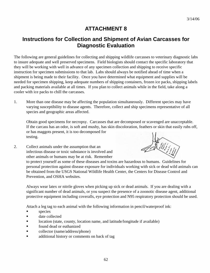
3/14/06
62
ATTACHMENT 8
Instructions for Collection and Shipment of Avian Carcasses for Diagnostic Evaluation
The following are general guidelines for collecting and shipping wildlife carcasses to veterinary diagnostic labs to insure adequate and well preserved specimens. Field biologists should contact the specific laboratory that they will be working with well in advance of any specimen collection and shipping to receive specific instruction for specimen submissions to that lab. Labs should always be notified ahead of time when a shipment is being made to their facility. Once you have determined what equipment and supplies will be needed for specimen shipping, keep adequate numbers of shipping containers, frozen ice packs, shipping labels and packing materials available at all times. If you plan to collect animals while in the field, take along a cooler with ice packs to chill the carcasses. 1. More than one disease may be affecting the population simultaneously. Different species may have
varying susceptibility to disease agents. Therefore, collect and ship specimens representative of all species and geographic areas affected.
Obtain good specimens for necropsy. Carcasses that are decomposed or scavenged are unacceptable. If the carcass has an odor, is soft and mushy, has skin discoloration, feathers or skin that easily rubs off, or has maggots present, it is too decomposed for testing.
2. Collect animals under the assumption that an
infectious disease or toxic substance is involved and other animals or humans may be at risk. Remember to protect yourself as some of these diseases and toxins are hazardous to humans. Guidelines for personal protection against disease exposure for individuals working with sick or dead wild animals can be obtained from the USGS National Wildlife Health Center, the Centers for Disease Control and Prevention, and OSHA websites.
Always wear latex or nitrile gloves when picking up sick or dead animals. If you are dealing with a significant number of dead animals, or you suspect the presence of a zoonotic disease agent, additional protective equipment including coveralls, eye protection and N95 respiratory protection should be used. Attach a leg tag to each animal with the following information in pencil/waterproof ink: species date collected location (state, county, location name, and latitude/longitude if available) found dead or euthanized collector (name/address/phone) additional history or comments on back of tag

3/14/06
63
Place each animal in a plastic bag, tie shut, then place inside a second bag and tie shut. This system of double bagging prevents cross-contamination of individual specimens and leaking shipping containers that can contaminate vehicle surfaces and handlers during transportation. Contact the diagnostic lab for guidance in assistance with collecting samples from animals that are too large to ship.
3. Ship animals in a sturdy hard sided plastic cooler. These
coolers can be disinfected and returned to you if a pre-paid shipping label or commercial shipping company account number is provided to the diagnostic lab. Be sure to provide a street address for return of the cooler.
Line the shipping cooler with a large plastic bag and pack the individually bagged animal(s) in the cooler with enough blue ice to keep carcasses cold. Disperse blue ice packs among the carcasses so that all carcasses are kept chilled. If you are shipping blood tubes, culture tubes, or other specimen
containers along with the carcasses, these specimens should be placed within a sturdy cardboard or plastic box or screw cap container with padding material to prevent breakage. That container should be place next to blue ice packs within the large cooler. Do not use bagged wet ice for shipments in order to avoid fluid leakage during shipment. Do not use dry ice unless instructed to do so by the diagnostic lab. Place crumpled newspaper or similar absorbent material in the cooler with the bagged carcasses to fill unused space, hold the ice in contact with carcasses, provide insulation, and absorb any liquids. Tape the cooler shut with sturdy strapping tape.
Place a detailed history of the animal and circumstances associated with the mortality event in a paper envelope or a plastic sleeve and tape it to the outside of the cooler. A copy of this history should be faxed or e-mailed to the diagnostic lab at the time of shipment. A standard wildlife specimen history form can be found on the last page of these instructions.
4. Prior to shipping contact the diagnostic lab to inform them of
the type and number of specimens being shipped. Ship specimens for next day delivery (overnight service) from Monday through Wednesday to guarantee arrival at the diagnostic lab before the weekend. If specimens are fresh and need to be shipped on Thursday or Friday contact the diagnostic lab to make special arrangements for receipt of specimens.
Freezing and thawing can make isolation of some pathogens difficult and damage tissues needed for microscopic examination. Diagnostic labs prefer unfrozen specimens if

3/14/06
64
they can be sent within 24 – 48 hours of collection or death. The diagnostic lab can provide guidance on when or if to freeze samples on a case-by-case basis. If you are in the field and cannot call or ship within 24-48 hours, freeze the animal(s).
5. Prior to shipping contact the commercial shipping company to obtain guidelines for shipping diagnostic
or biological specimens. Label coolers with clear, legible labels including the diagnostic lab name, street address, and telephone number. In addition to the mailing address, attach a label reading “DIAGNOSTIC SPECIMENS –WILDLIFE” to the side of the cooler. If dry ice was used in the shipment a standard dry ice warning label will be required. These can be obtained from the shipping company. Please make note of the tracking number in case packages are delayed.

3/14/06
65
Wildlife Specimen History Form Always contact the diagnostic lab before shipping specimens!
Submitter’s name: Affiliation: Address: Telephone: E-mail: Date collected: Collector's Name: Method of collection: [found dead, euthanized (describe method) etc.] Method of storage: [chilled, frozen, fixed, etc.] Species Submitted: Specific die-off location: State: County: Latitude/longitude: Environmental factors: (Record conditions such as storms, precipitation, temperature changes, or other changes that may contribute to stress.) Disease onset: (The best estimate of when the outbreak started.) Species affected: (The diversity of species affected may provide clues to the disease involved.) Age/sex: (Any selective mortality related to age and sex.) Morbidity/mortality: (Ratio of sick animals to dead animals.) Known dead: (Actual carcass count) Estimated dead: (Consider removal by scavengers or other means.) Clinical signs: (Any unusual behavior and physical appearance.) Population at risk: (Number of animals in the area that could be exposed to the disease.) Population movement: (Recent changes in the number of animals on the area and their source or destination, if known.) Problem area description: (Land use, habitat types, and other distinctive features.) Comments: (Additional information/observations that may be of value such as past occurrences of disease in area.) PLEASE USE ADDITIONAL SHEETS IF NECESSARY.

3/14/06
66
ATTACHMENT 9
Protocol for the Collection of Tracheal/Cloacal Swab Samples
1. Contact Laboratory to determine specific protocol to use. Laboratories may request samples be
placed in tubes containing Viral Transport Medium (VTM) or brain-heart infusion broth (BHI). 2. Thaw appropriate number of pre-labeled tubes of Viral Transport Medium (VTM) or brain-heart
infusion broth (BHI) at refrigerator temperature (4 °C) overnight and keep chilled with wet/blue ice packs in a cooler during the day of collection.
3. Unwrap a Dacron swab from the stem-end of the packaging. 4. Remove swab and insert the entire head of the swab into the trachea or cloaca. Use gentle
pressure and in a circular motion, swab the inside circumference of the trachea/cloaca two or three times.
5. For Cloacal swabs, shake off large pieces of feces.
6. Inserting the swab into the tube containing VTM or BHI broth. With the swab in the media, swirl the stem end of the swab between fingers vigorously. Lift the swab approximately ¼” from the bottom of the vial and bend the stem over the edge of the vial to break off the stem so that the swab remains in the vial and the cap can be screwed tight. The entire swab end and a portion of the stem will be left in the tube. If the stems are unable to be broken (some small swabs will have metal stems) then they can be cut with scissors. Scissors should be wiped with 70% alcohol each time they are used to cut a stem.
7. Record sample tube number on banding sheet or the Sample History Sheet along with date, species, age, sex, and location data (GIS coordinates if possible)..
8. Replace tube into cooler for transport back to the base camp. Samples should be kept cold (<4 °C, frozen if possible) and out of direct sunlight.
9. At camp, transfer tubes into liquid nitrogen shippers or into a freezer as soon as possible. Note any exceptions to the collection or storage conditions in field sheets and note such information on the "Sample History and Packing List Form".
10. Place tubes into a hard plastic shipping container with enough frozen gel packs to keep samples cold for at least two days.
11. Notify laboratory that samples are being shipped, the method of shipment (FEDEX is preferred), and the expect date of arrival. Packages should only be shipped on Monday, Tuesday, or Wednesday.

3/14/06
67
Sample History and Packing List H5N1 Avian Influenza Project
Submitter ___________________________________________ Address ___________________________________________ ___________________________________________ Phone ___________________________________________ E mail ___________________________________________
Com
men
ts o
r Sam
plin
g pr
oble
ms
Loca
tion
Ban
d N
umbe
r
Sex
Age
B
ird
Spec
ies
Sam
ple
ID
3/14/06
68
ATTACHMENT 10
Fecal Sampling and Shipping Protocol
Fecal Sampling
Purpose The purpose of this standard operating procedure is to describe the essential elements of proper handling and collection of field fecal samples for surveillance of avian influenza.
Procedures
• Before collecting samples, personnel should don the appropriate personal protective equipment (PPE). These include latex or nitrile gloves and face shields, and if it is determined to be necessary, PAPRs.
• Label plastic whirl pack bags with necessary information including date, location
(GPS coordinate if possible), species (if possible), investigator and sample identification. Record all required information on data sheets.
• Feces must be less than 24 hours old. Feces should appear moist.
• For collection, turn a sterile Whirl-Pak inside out and pick up feces using the
Whirl-Pak as a glove, then turn the bag right side in with the feces inside the closed bag. Release as much air from the inside of the bag.
• Label the Whirl-Pak using an indelible ink marker. The sample should be labeled
with the sample number, date, time, collector's name, location, and Quality Assurance number (Protocol Number). This latter information describes in detail the context, purpose, and other procedural and review information of the samples collected.
• Place the Whirl-Paks with fecal samples into a large zip-lock bag, tape (e.g., duct
tape or packaging tape) the opening and label the outer bulk bag with name, date, location, and Protocol number.
• Place the bulk collection bag in a cooler with wet or blue ice to keep the specimen
cool. This precaution is for maximizing the chances for subsequent viral isolation. Pack samples with enough ice or frozen gel packs to keep samples cold for at least two days.
• Maintain the temperature of samples as constant as possible.
3/14/06
69
• Change gloves if soiled or contaminated. When finished collecting, wash hands with suitable antibacterial agent.
• Notify laboratory that samples are being shipped, the method of shipment
(FEDEX is preferred), and the expect date of arrival. Packages should only be shipped on Monday, Tuesday, or Wednesday; this allows the laboratories time to process samples during a normal work-week, or allows for tracking if the shipment is delayed.
Shipping
Purpose
The purpose of this standard operating procedure is to ensure diagnostic specimens are shipped safely and in compliance with governing regulations and requirements. Shippers of diagnostic specimens where a relatively low probability exists that infectious substances are present (diagnostic specimens being transported to undergo routine screening tests or for the purpose of initial diagnosis may be considered to fall under this category) must comply with the International Air Transportation Association (IATA) Dangerous Goods Regulations. The shipper must also ensure that shipments are prepared in such a manner that they arrive at their destination in good condition and that they present no hazard to persons or animals during shipment.
Procedures:
• Federal Express is the preferred carrier for the USDA/NWRC. Use next day service.
• The inner packaging (appropriately labeled) must be comprised of a watertight
primary receptacle, and must no exceed 500 ml total volume. Primary receptacles include those of glass, metal, or plastic (i.e., test tube, plastic jar, or taped zip-loc bag). Positive means of ensuring a leak-proof seal must be provided. Screw caps on primary receptacles must be reinforced with adhesive tape.
• A secondary packaging (also water tight) must be used, but must not exceed 4 L
total volume.
• An absorbent material sufficient to absorb the entire contents of all primary receptacles must be placed between the primary receptacle and the secondary packaging.
• The outer packaging (i.e., cardboard box or cooler) must be of adequate strength
for its capacity, weight, and intended use (capable to withstand being dropped at least 1.2 meters, without leakage of the primary receptacle or significant damage to the outer packaging).
3/14/06
70
• An itemized list of contents must be enclosed between the secondary packaging and the outer packaging. To protect against possible leakage, the list should be enclosed in a sealed plastic bag.
• A Shippers Declaration for Dangerous Goods is not required. However, both the
air waybill and the outer box must show the text "DIAGNOSTIC SPECIMEN PACKED IN COMPLIANCE WITH IATA PACKING INSTRUCTION 650".
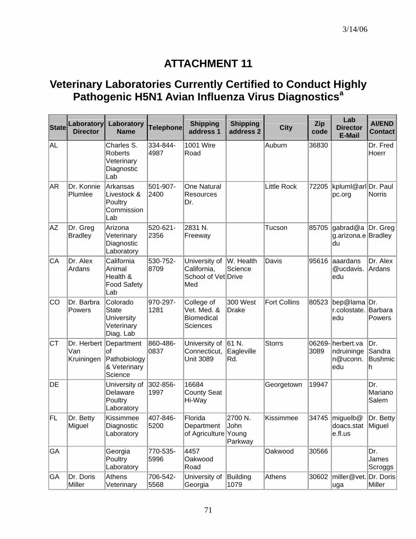
3/14/06
71
ATTACHMENT 11
Veterinary Laboratories Currently Certified to Conduct Highly Pathogenic H5N1 Avian Influenza Virus Diagnosticsa
State Laboratory Director
Laboratory Name Telephone Shipping
address 1 Shipping address 2 City Zip
code Lab
Director E-Mail
AI/END Contact
AL Charles S. Roberts Veterinary Diagnostic Lab
334-844-4987
1001 Wire Road
Auburn 36830 Dr. Fred Hoerr
AR Dr. Konnie Plumlee
Arkansas Livestock & Poultry Commission Lab
501-907-2400
One Natural Resources Dr.
Little Rock 72205 [email protected]
Dr. Paul Norris
AZ Dr. Greg Bradley
Arizona Veterinary Diagnostic Laboratory
520-621-2356
2831 N. Freeway
Tucson 85705 [email protected]
Dr. Greg Bradley
CA Dr. Alex Ardans
California Animal Health & Food Safety Lab
530-752-8709
University of California, School of Vet Med
W. Health Science Drive
Davis 95616 [email protected]
Dr. Alex Ardans
CO Dr. Barbra Powers
Colorado State University Veterinary Diag. Lab
970-297-1281
College of Vet. Med. & Biomedical Sciences
300 West Drake
Fort Collins 80523 [email protected]
Dr. Barbara Powers
CT Dr. Herbert Van Kruiningen
Department of Pathobiology & Veterinary Science
860-486-0837
University of Connecticut, Unit 3089
61 N. Eagleville Rd.
Storrs 06269-3089
Dr. Sandra Bushmich
DE University of Delaware Poultry Laboratory
302-856-1997
16684 County Seat Hi-Way
Georgetown 19947 Dr. Mariano Salem
FL Dr. Betty Miguel
Kissimmee Diagnostic Laboratory
407-846-5200
Florida Department of Agriculture
2700 N. John Young Parkway
Kissimmee 34745 [email protected]
Dr. Betty Miguel
GA Georgia Poultry Laboratory
770-535-5996
4457 Oakwood Road
Oakwood 30566 Dr. James Scroggs
GA Dr. Doris Miller
Athens Veterinary
706-542-5568
University of Georgia
Building 1079
Athens 30602 [email protected]
Dr. Doris Miller
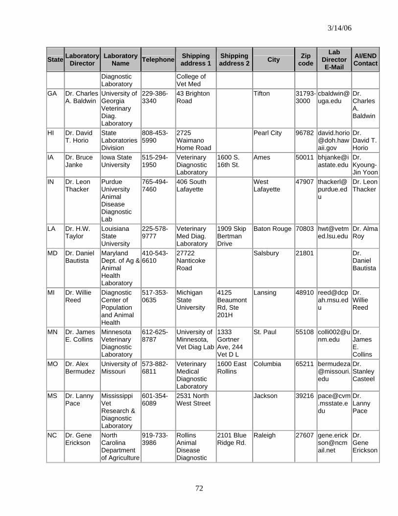
3/14/06
72
State Laboratory Director
Laboratory Name Telephone Shipping
address 1 Shipping address 2 City Zip
code Lab
Director E-Mail
AI/END Contact
Diagnostic Laboratory
College of Vet Med
GA Dr. Charles A. Baldwin
University of Georgia Veterinary Diag. Laboratory
229-386-3340
43 Brighton Road
Tifton 31793-3000
Dr. Charles A. Baldwin
HI Dr. David T. Horio
State Laboratories Division
808-453-5990
2725 Waimano Home Road
Pearl City 96782 [email protected]
Dr. David T. Horio
IA Dr. Bruce Janke
Iowa State University
515-294-1950
Veterinary Diagnostic Laboratory
1600 S. 16th St.
Ames 50011 [email protected]
Dr. Kyoung-Jin Yoon
IN Dr. Leon Thacker
Purdue University Animal Disease Diagnostic Lab
765-494-7460
406 South Lafayette
West Lafayette
47907 [email protected]
Dr. Leon Thacker
LA Dr. H.W. Taylor
Louisiana State University
225-578-9777
Veterinary Med Diag. Laboratory
1909 Skip Bertman Drive
Baton Rouge 70803 [email protected]
Dr. Alma Roy
MD Dr. Daniel Bautista
Maryland Dept. of Ag & Animal Health Laboratory
410-543-6610
27722 Nanticoke Road
Salsbury 21801 Dr. Daniel Bautista
MI Dr. Willie Reed
Diagnostic Center of Population and Animal Health
517-353-0635
Michigan State University
4125 Beaumont Rd, Ste 201H
Lansing 48910 [email protected]
Dr. Willie Reed
MN Dr. James E. Collins
Minnesota Veterinary Diagnostic Laboratory
612-625-8787
University of Minnesota, Vet Diag Lab
1333 Gortner Ave, 244 Vet D L
St. Paul 55108 [email protected]
Dr. James E. Collins
MO Dr. Alex Bermudez
University of Missouri
573-882-6811
Veterinary Medical Diagnostic Laboratory
1600 East Rollins
Columbia 65211 [email protected]
Dr. Stanley Casteel
MS Dr. Lanny Pace
Mississippi Vet Research & Diagnostic Laboratory
601-354-6089
2531 North West Street
Jackson 39216 [email protected]
Dr. Lanny Pace
NC Dr. Gene Erickson
North Carolina Department of Agriculture
919-733-3986
Rollins Animal Disease Diagnostic
2101 Blue Ridge Rd.
Raleigh 27607 [email protected]
Dr. Gene Erickson

3/14/06
73
State Laboratory Director
Laboratory Name Telephone Shipping
address 1 Shipping address 2 City Zip
code Lab
Director E-Mail
AI/END Contact
Lab NE Dr. David
Steffen Veterinary Diagnostic Center
402-472-1434
University of Nebraska
137 VDC UNL
Lincoln 68583-0907
Dr. David Steffen
NJ Dr. Robert Eisner
New Jersey Dept of Ag, Division of Animal Health
609-984-2293
State Diagnostic Lab, H & A Building
Rm 201 John Fitch Plaza, P.O. Box 330
Trenton 08625 [email protected]
Dr. Robert Eisner
NM Dr. Flint Taylor
New Mexico Department of Agriculture
505-841-2576
Veterinary Diagnostic Services
700 Camino de Salud, NE
Albuquerque 87106 [email protected]
Dr. David Mills
NV Dr. Anette Rink
Nevada Animal Disease Laboratory
775-668-1182
Nevada Department of Agriculture
350 Capitol Hill Ave.
Reno 89502-2923
Dr. Anette Rink
NY Dr. Alfonso Torres
Animal Health Diagnostic Center
607-253-4136
Cornell University, College of Vet. Med.
S3 110 Schurman Hall, Upper Tower Rd.
Ithaca 14853 [email protected]
Dr. Sung Kim
OH Dr. Beverly Byrum
Ohio Department of Agriculture
614-728-6220
Animal Disease Diagnostic Laboratory
8995 E. Main Street, Building 6
Reynoldsburg 43068 [email protected]
Dr. Beverly Byrum
OK Dr. Bill J. Johnson
Oklahoma Animal Disease Diagnostic Laboratory
405-744-6623
Oklahoma State Univ., College of Vet. Med.
Farm Road & Ridge Road
Stillwater 74078 [email protected]
Dr. W. C. Edwards
OR Dr. Jerry Heidel
Oregon State Veterinary Diagnostic Lab
541-737-3261
Oregon State Univ., College of Vet. Med.
30th & Washington
Corvallis 97331 [email protected]
Dr. Jerry Heidel
PA Dr. Helen Acland
Pennsylvania State Vet Diagnostic Laboratory
717-787-8808
2305 N. Cameron Street
Harrisburg 17110 [email protected]
Dr. Deepanker Tewari
PA University of Pennsylvania
610-925-6210
Lab of Large Animal Pathology & Toxicology
New Bolton Center, 382 West Street Rd
Kennett Square
19348-1692
Dr. Sherrill Davison
SC Dr. Pamela Parnell
Clemson Veterinary Diagnostic Center
803-788-2260
500 Clemson Road
Columbia 29229 [email protected]
Dr. Pamela Parnell
TX Dr. Lelve Gayle
Texas Vet Medical Diagnostic Laboratory
979-845-9000
1 Sippel Road
Drawer 3040
College Station
77843 [email protected]
Dr. Lelve Gayle

3/14/06
74
State Laboratory Director
Laboratory Name Telephone Shipping
address 1 Shipping address 2 City Zip
code Lab
Director E-Mail
AI/END Contact
UT Dr. Tom Baldwin
Utah Veterinary Diagnostic Laboratory
435-797-1895
950 E. 1400 North
Logan 84322-5700
Dr. Tom Baldwin
VA Dr. David W. Brown
Virginia Dept of Agriculture and Animal Health Lab
540-434-3897
116 Reservoir
Harrisonburg 22801 Dr. David Brown
WA Dr. Terry McElwain
Washington Animal Disease Diagnostic Laboratory
509-335-9696
Bustad Hall Room 155-N
Pullman 99164 [email protected]
Dr. Terry McElwain
WA Avian Health and Food Safety Laboratory
253-445-4537
7613 Pioneer Way E.
Puyallup 98371-4919
Dr. A. S. Dhillon
WI Dr. Leslie Dierauf
USGS National Wildlife Health Center
608-270-2400
6006 Schroeder Road
Madison 53711 [email protected]
Dr. Leslie Dierauf
WI Dr. Kathy Kurth and Dr. Pete Vanderloo
Wisconsin Veterinary Diagnostic Laboratory
608-262-5432
Wisconsin Department of Agriculture
6101 Mineral Point Road
Madison 53705 [email protected]
Dr. Kathy Kurth and Dr. Pete Vanderloo
WV West Virginia Dept of Agriculture
304-558-2214
1900 Boulevard, East
Charleston 25305-0172
Dr. Jewell Plumley
a This list represents the National Animal Health Laboratory Network (NAHLN) labs certified as of 3/14/06 to conduct avian influenza screening. This list will be updated as new labs become certified. For the latest list of certified laboratories, please contact [email protected]

3/14/06
75
Attachment 12 Real-Time Reverse Transcriptase-Polymerase Chain
Reaction for the Detection of Type A Influenza and the Avian H5 and H7 Ha Subtypes
In Tracheal and Cloacal Samples
Cepheid Smart Cycler Protocol
I. MATERIALS...............................................................................................................................74
General recommendations regarding reagents.........................................................74 General...........................................................................................................................74 RNA extraction..............................................................................................................74 Real-time RT-PCR.........................................................................................................74 Primer and probe sequences......................................................................................75 Primer and probe handling and dilution….................................................................75 Suppliers.......................................................................................................................76
II. METHODS.................................................................................................................................77
Note on sample types and RNA extraction methods...................................................77 RNA extraction with the Qiagen RNeasy kit: Centrifuge method...............................77 RNA extraction with the Qiagen RNeasy kit: Vacuum manifold method...................78 RNA Extraction with Trizol reagent...............................................................................78 RNA handling and storage.............................................................................................79 Real-time RT-PCR for type A influenza and the avian H5 and H7 HA subtypes.......79
III. ANALYSIS OF RESULTS........................................................................................................81
Recommendations for evaluating fluorograms...........................................................81
IV. APPENDIX.............................................................................................................................84
Troubleshooting...........................................................................................................84 Cross-contamination prevention ...............................................................................84
Real-time PCR basics..................................................................................................85 Prepared by: Erica Spackman, Ph.D. Southeast Poultry Research Lab U.S. Dept. of Agriculture, Agricultural Research Service 934 College Station Rd. Athens, GA 30605 706-546-3617 [email protected] Updated March 15, 2005

3/14/06
76
I. MATERIALS Mention of trade names or commercial products in this procedure is solely for the purpose of providing specific information and does not imply recommendation or endorsement by the U.S. Department of Agriculture.
General recommendations regarding reagents This assay was optimized using the using the Qiagen RNeasy kit and Qiagen one-step RT-PCR kit, therefore these reagents are recommended for uniformity, if available. Additionally, it is highly recommended that the PCR primers and probe be highly purified (i.e. HPLC purified).
General Reagent grade H2O (nuclease free) Pipetors and tips for volumes between 1µl and 1ml 1.5 ml microcentrifuge tubes Microcentrifuge TE buffer pH 8.3 (Promega #V6231 or V6232) (optional) RNA Extraction Qiagen RNeasy Mini Kit (Qiagen #74104 or #74106) 100% Ethanol 70% Ethanol (in nuclease free water) QiaVac 24 vacuum manifold (Optional) 2-Mercaptoethanol (BME) (Sigma #M-6250)
Trizol LS reagent (Invitrogen #10296028, 200ml) Chloroform (Sigma #C-2432) Isopropanol (Sigma #I-9516) Glycogen 5mg/ml (Ambion #9510)
Real-time RT-PCR Qiagen one step RT-PCR Kit (Qiagen #210210 or #210212) Hydrolysis probes (IDT, Idaho Tech/Biochem or Qiagen-Operon) (Table 1) Primers (IDT, Idaho Tech/Biochem or Qiagen-Operon) (Table 1) 25 mM MgCl2 (Promega #A3511 or #A3513) RNase Inhibitor (Promega #N2511 or # N2515) Positive control RNA Nuclease free H2O 25µl Smart Cycler tubes (Cepheid #900-0022 or 900-0003)

3/14/06
77
Table 1. Influenza real-time RT-PCR probe and primer sequences. Protocols for H7 subtype Eurasian, H7 subtype South American strains, the H6 subtype and the H9 subtype strains are available as supplementary protocols.
Specificity Sequence M+25 5’ Primer
5’-AGA TGA GTC TTC TAA CCG AGG TCG-3’ Type A influenza-Matrix gene M+64 Probe 5’-FAM-TCA GGC CCC CTC AAA GCC GA-BHQ1-3’
M-124 3’Primer
5’-TGC AAA AAC ATC TTC AAG TCT CTG-3’
H7+ 1244 5’ Primer
5’-ATT GGA CAC GAG ACG CAA TG-3’
H7Subtype-North American strains
H7+1281 Probe
5’-FAM-TAA TGC TGA GCT GTT GGT GGC-BHQ1-3’
H7-1342 3’Primer
5’-TTC TGA GTC CGC AAG ATC TAT TG-3’
H5+1637 Probe
5’-FAM-TCA ACA GTG GCG AGT TCC CTA GCA-BHQ1-3’ H5 subtype-Any
straina H5-1685 3’Primer
5’-AGA CCA GCT AYC ATG ATT gC-3’
H5 subytpe-North American strains
H5+1456 NA 5’ Primer
5’-ACG TAT GAC TAT CCA CAA TAC TCA-3’
H5 subtype-Eurasian strains
H5+1456 EA 5’ Primer
5’-ACG TAT GAC TAC CCG CAG TAT TCA-3’
a. H5 strain specificity is determined by the 5’ primer. Use only one primer in the reaction.
Note on subtype determination: Due to the high level of sequence variation within each HA subtype, a negative RRT-PCR result for a specific subtype does not exclude the possibility that that subtype is present.
Primer and probe handling and dilution Lyophilized primers and probes must be centrifuged briefly, to ensure that the DNA pellet is at the bottom of the tube, before they are opened and reconstituted. TE buffer should be used for the initial reconstitution of lyophilized primers and probes (Idaho Tech sends TE with probes and primers and it is commercially available). Concentrated stock solutions should be stored at -20°C. Primer stock solutions should be 200µM (200pmol/µl), probes should be 120µM (120pmol/µl). Quantitation information will be supplied for each oligo (primers and probes are DNA oligos) by the manufacturer.
An example of calculation for oligo reconstitution:
You have 17786 pmol of oligo (will be on oligo information sheet from manufacturer).
Need 200pmol/µl for stock concedntration.
Divide pmol of oligo by the pmol/µl needed or: 17786 pmol = 88.9µl 200 pmol/µl
3/14/06
78
For 200pmol/µl resuspend the pellet in 89µl of TE or nuclease free H2O. The calculation for the probe is the same, except divide the number of probe pmol by 120pmol/µl. Mix gently by tapping the tube and allow the oligo to resuspend for about 10 minutes before use.
Working stocks of primers should be 20pmol/ul (20µM) and working stocks of probes should be 6µM. Dilute the primers 1:10 and dilute the probe 1:20 in nuclease free H2O (do not use TE buffer) for the working stocks. Working stocks should be stored at 4°C. The probes are stable at this concentration at 4° C for approximately 1 month. It may be useful to make up several aliquots (5-6) of working stocks of primers and probes which are a volume that can be used in about one month. Store the unused aliquots at -20°C.
Note: the probes are light sensitive; store them in amber tubes if available, and minimize their exposure to light.
Additional information on fluorescent probe handling and storage can found at: www.idahotech.com, www.operon.com and www.idtdna.com.
Suppliers Biosearch Technologies, Inc. Promega 81 Digital Drive 2800 Woods Hollow Rd Novato, CA 94949-5750 Madison, WI 53711-5399 1.800.436.6631 1-800-356-9526 WWW.Biosearchtech.com www.promega.com
Idaho Technology/Biochem IDT 390 Wakara Way 1710 Commercial Park Salt Lake City, UT 84108 Coralville, IA 52241 1-800-735-6544 1-800-328-2661 www.idahotech.com www.idtdna.com
Qiagen Inc./Operon 28159 Avenue Stanford Valencia, CA 91355 1-800-426-8157 www.qiagen.com www.operon.com

3/14/06
79
II. METHODS
NOTE ON SAMPLE TYPES AND RNA EXTRACTION METHODS The types of samples collected and the processing of those samples varies by species. The optimal sample types and processing methods for many species are given below.
Table 2. Sample types and optimal processing methods.
Recommended Processing Species/ Type Specimen Method Notes
Gallinaceous Poultry Tracheal swab RNeasy RNA Virus primarily replicates in (chickens, turkeys, extraction, then RRT- the respiratory tract (LPAI) quail) PCR
Waterfowl/ducks Cloacal Swab Trizol Reagent RNA extraction, then RRT-PCR
Virus primarily replicates in the intestinal tract. RNA extraction method must be modified for cloacal
samples
Any species Tissue samples RNA extraction with For HPAI viruses high Trizol Reagent, then levels of virus may be in RRT-PCR tissues. Environmental Virus isolation, RRT- RRT-PCR can detect samples (Swab) PCR not inactivated virus recommended
REVISED RNA EXTRACTION PROTOCOL FOR TRACHEAL SWABS (7/03)
RNA Extraction with Qiagen RNeasy Kit- Centrifuge Method Notes: • Adaptation of kit for fluid samples from manufacturer.
• All kit supplied buffers and reagents should be prepared in accordance with the kit instructions.
• Use only RNA grade reagents and supplies 1. Vortex the sample (cloacal or tracheal swabs in BHI or other media) for 3-5 seconds and
withdraw 500µl and place in a 1.5 ml microcentrifuge tube. 2. Add 500 µl of RLT buffer. Close the tube and vortex the sample for 5 seconds. 3. Add 500 µl of RNA grade 70% ethanol to the tube and mix. Centrifuge the sample for 5
minutes at ~5KXg to pellet any debris. 4. Add 750µl of the supernatant from step 3 to the RNeasy column and centrifuge for 15
seconds at ~12 KXg, empty the flow through from the collection tube and repeat (all of the sample/RLT/70% ethanol mix should be applied to the column).
5. Add 700µl RW1 buffer to the RNeasy column and centrifuge for 15 seconds at ~12 KXg
and place the column in a clean collection tube (the collection tube with RW1 flow through may be discarded and replaced with a fresh collection tube).

3/14/06
80
6. Add 500µl RPE buffer to the RNeasy column and centrifuge for 15 seconds at ~12 KXg, empty the flow through from the collection tube.
7. Repeat step 6 for a total of 2 washes with RPE buffer. 8. Centrifuge the empty RNeasy column an extra 2 minutes at ~14 KXg and discard the
collection tube. 9. Place the RNeasy column in an elution tube (or 1.5ml microfuge tube) and add 50 µl
nuclease free H2O to the column. Incubate at room temperature for 1 minute. Elute RNA by centrifuging for 1 minute at ~14KXg. Discard RNeasy column.
RNA Extraction with Qiagen RNeasy Kit- QiaVac 24 Vacuum Manifold Method Notes:
• Adaptation of the RNeasy kit for fluid samples from manufacturer. • All kit supplied buffers and reagents should be prepared in accordance with the kit
instructions. • RNeasy column lids should be open whenever vacuum is being applied. • Use only RNA grade reagents and supplies
1. Vortex the sample (cloacal or tracheal swabs in BHI or other media) for 3-5 seconds and
withdraw 500 µl and place in a 1.5 ml microcentrifuge tube. 2. Add 500 µl of RLT buffer. Close the tube and vortex the sample for 5 seconds. 3. Add 500 µl of RNA grade 70% ethanol to the tube and mix. Centrifuge the sample for 5
minutes at ~5KXg to pellet any debris. 4. Place the appropriate number of RNeasy columns in the luer locks of the vacuum
manifold, cover any empty positions with the luer caps supplied with the vacuum manifold.
5. Apply vacuum and add the entire sample/RLT/ethanol mixture to an RNeasy column for
each sample. 6. Wash by applying 700µl RW1 buffer to each column. 7. Wash again by applying 500µl RPE buffer to the column and repeat for a total of 2
washes with buffer RPE. 8. Shut off the vacuum and place each RNeasy column in a 2ml collection tube. Centrifuge
the column for 2 minutes at ~14 KXg and discard the collection tube. 9. Place each column in an elution tube (or 1.5ml microfuge tube) and add 50 µl nuclease
free H2O and incubate at room temperature 1 minute. Elute RNA by centrifuging for 1 minute at ~14KXg.
10. Use 8µl per PCR reaction. Store at -70°C for long term storage.
3/14/06
81
RNA EXTRACTION FROM CLOACAL SWABS OR TISSUE WITH TRIZOL REAGENT
1. Sample Prparation: a. Cloacal Swabs: Vortex vigorously for 7-10 seconds. Centrifuge for 5 min. at
12,000Xg.Extract RNA from the supernatant. b. Tissues: Make a 10% homogenate of tissue in PBS. Centrifuge for 10 min. at
12,000Xg. Extract RNA from the supernatant. 2. Add 250µl of the supernatant from the sample prepared as described in step 1, to 750µl
of Trizol LS reagent. Vortex. Pulse spin to remove liquid from the tube lid. 3. Add 200µl 100% chloroform to the sample/Trizol homogenate. Vortex for 15 sec.
Incubate at room temperature for 7 min. 4. Centrifuge at 12,000 x g for 15 min at room temperature. 5. Transfer 400-450µl of the upper aqueous layer to a separate microcentrifuge tube
marked with sample number. Caution: The transfer of organic phase material with the aqueous layer will inhibit the PCR reaction. Add 500µl of 100% isopropanol. Add carrier to the isopropanol to aid precipitation i.e. glycogen: 1µl of 5mg/ml stock (may be added prior to addition of the aqueous phase from the trizol). Invert tube several times to mix. Incubate at room temperature for 10 min.
6. Centrifuge at 10,000 x g for 10 min at 4 C. 7. Decant liquid. Care should be taken to assure that the RNA pellet is not disturbed. Add
1.0 ml of 70% or 80% ethanol. Mix gently. 8. Centrifuge at 10,000 X g for 5 min at 4 C. 9. Decant ethanol. Invert tube on a clean tissue wipe and allow to air dry for 10 min. or until
all visible signs of moisture are gone. It is important not to let the RNA pellet over-dry, as this will decrease its solubility.
10. Hydrate pellet in 100µl of RNase free water and allow to sit at 4 C for 1 hr to overnight. RNA Handling and Storage The RNA sample may be stored at 4°C for < 1 week, storage for longer than one week should be at –70°C. Always wear gloves when handling RNA and use only RNase or nuclease free materials and reagents with RNA. Additional RNA handling and storage information can be found in: Sambrook, J. and Russell, D. Molecular Cloning, A Laboratory Manual, Cold Spring Harbor Laboratory Press, Cold Spring Harbor, New York, 2001.
Procedure for Real-time RT-PCR for type A Influenza (MA gene), the H5, H6, H7 and H9 HA subtypes This procedure was designed for the Cephied Smart-Cycler (Cepheid, Sunnyvale, CA). A protocol for the Roche Light Cycler and Idaho Tech R.A.P.I.D. is available. The original reference and validation study for this assay is: Spackman, et. al.; Journal of Clinical Microbiology 40:3256-3260, 2002.
Information on setting-up and programming the Smart Cycler can be found in the Smart Cycler user’s manual. The conditions for the influenza primers and probes on the Smart Cycler are shown in tables 2 and 3. The RT step is the same for all primer and probe sets, these conditions
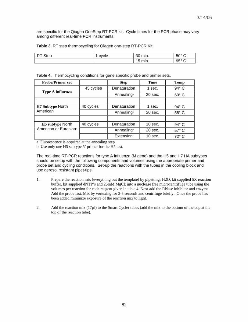
3/14/06
82
are specific for the Qiagen OneStep RT-PCR kit. Cycle times for the PCR phase may vary among different real-time PCR instruments.
Table 3. RT step thermocycling for Qiagen one-step RT-PCR Kit.
RT Step 1 cycle 30 min. 50° C 15 min. 95° C
Table 4. Thermocycling conditions for gene specific probe and primer sets.
Probe/Primer set Step Time Temp 45 cycles Denaturation 1 sec. 94° C
Type A influenza Annealinga 20 sec. 60° C
40 cycles Denaturation 1 sec. 94° C H7 Subtype North
American Annealinga 20 sec. 58° C
40 cycles Denaturation 10 sec. 94° C Annealinga 20 sec. 57° C
H5 subtype North American or Eurasianb
Extension 10 sec. 72° C a. Fluorescence is acquired at the annealing step. b. Use only one H5 subtype 5’ primer for the H5 test. The real-time RT-PCR reactions for type A influenza (M gene) and the H5 and H7 HA subtypes should be setup with the following components and volumes using the appropriate primer and probe set and cycling conditions. Set-up the reactions with the tubes in the cooling block and use aerosol resistant pipet-tips.
1. Prepare the reaction mix (everything but the template) by pipetting: H2O, kit supplied 5X reaction buffer, kit supplied dNTP’s and 25mM MgCl2 into a nuclease free microcentrifuge tube using the volumes per reaction for each reagent given in table 4. Next add the RNase inhibitor and enzyme. Add the probe last. Mix by vortexing for 3-5 seconds and centrifuge briefly. Once the probe has been added minimize exposure of the reaction mix to light.
2. Add the reaction mix (17µl) to the Smart Cycler tubes (add the mix to the bottom of the cup at the
top of the reaction tube).

3/14/06
83
Table 5. Real-time RT-PCR reaction mix volumes and conditions for type A influenza (M gene), H5 and H7 HA subtypes.
Volume Per Final Reaction Concentration
H2O 6.95µl 5X 5 1X
25mM MgCl2 1.25 3.75 mM Enzyme Mix 1
Forward Primer 0.5 10 pmol Reverse Primer 0.5 10 pmol
dNTP’s 0.8 320 µM ea. dNTP Probe 0.5 0.12 µM
Rnase Inhibitor 0.5 13 units MM per rxn 17 Template 8 Total 25µl
3. Add the template to the smart cycler tubes (8µl per reaction). Note: The template for the
positive controls is in vitro transcribed RNA from the appropriate gene and the template for the negative controls is H2O.
4. Centrifuge the reaction tubes briefly in the Smart Cycler centrifuge and run the real-time
RT-PCR with the conditions described in tables 2 and 3 depending on the probe and primer set used. Note the RT step is the same for all probe and primer sets (the RT step is specific for the Qiagen one-step RT-PCR kit).
III. ANALYSIS OF RESULTS
Positive results on the Smart Cycler may be determined by the Smart Cycler software (shown on the results table in the Smart Cycler software) and are generally reliable; however results should be manually confirmed by examination of the fluorogram.
On the Smart Cycler the default minimum increase in fluorescence for a sample to be classified as positive by the software is 30 units. Because this is an arbitrary threshold, any samples which have an increase in fluorescence between 20 and 40 should be considered suspect and should be re-tested with the type A influenza (M gne) assay and/or subtype specific assays. In general, any questionable samples should be retested. If results of the second test are unsatisfactory additional sampling from the flock or premises should be considered if possible.
Recommendations for evaluating fluorograms
Evaluation of the fluorogram with the following conditions may be helpful in determining results manually:
All reactions with default settings.
Remove all reactions with greater than 100 units increase in fluoresce from the graph (this changes the scale, making it easier to identify weak negatives). (Figures 1a and 1b).
If there are samples which have a “V” shaped fluorescence trace incrementally lower the “background maximum cycles” (analysis settings table) to approximately 2 cycles below the cycle number where the base of the “V” is (Figures 2a and 2b).

3/14/06
84
Figure 1a. Example of a fluorogram from samples run on the Smart Cycler. All samples shown. Background subtraction is on. All analysis criteria are set to the default values. Note that scale is from 0 to 1000 fluorescence units (Y axis), making it difficult to evaluate weak positive samples.
Figure 1b. Same fluorogram as figure 1a, however all samples which increased greater than 100units in fluorescence were removed from the graph. Note that the scale is from 0 to 120 fluorescence units (Y axis) making it easier to recognize weak positives.
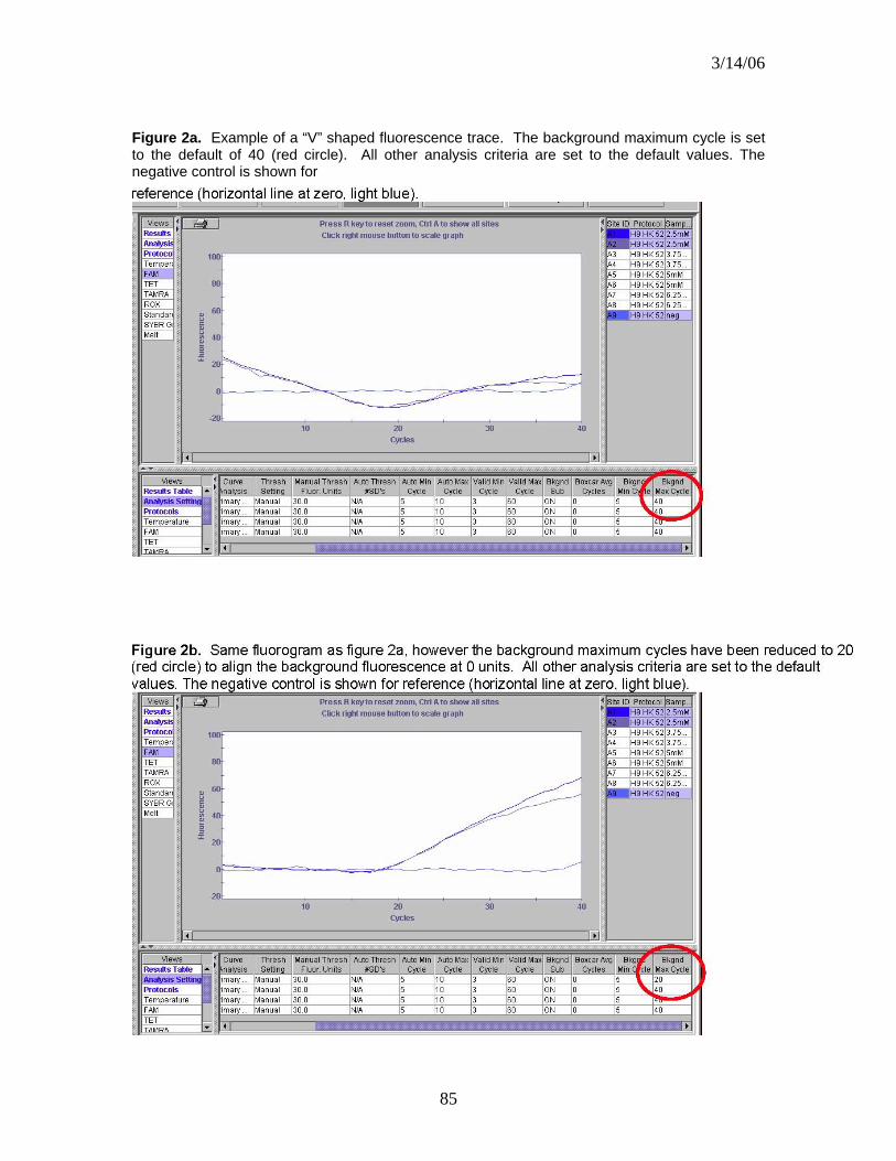
3/14/06
85
Figure 2a. Example of a “V” shaped fluorescence trace. The background maximum cycle is set to the default of 40 (red circle). All other analysis criteria are set to the default values. The negative control is shown for

3/14/06
86
IV. APPENDIX
Troubleshooting
• Positive controls are negative: • Control template has degraded. • Probe may be old and the fluorescence may be dead. • The enzymes may be inactivated. • Incorrect thermal-cycling program or fluorescence acquisition (wrong setting or
wrong channel being viewed).
• Negative controls are positive: • There may be cross contamination among the samples. • There may be non-specific probe degradation, use fresh probe and primers.
• Background level too high or too low (should be approximately 100-200 units): • The probe concentration may be wrong. • The probe may be degraded or too old if the background level is too low.
• Warning message in sample status on results screen. • Probe concentration too high (may cause ‘railing’; a sharp decrease in
fluorescence after a steady increase).
Cross-contamination prevention
Due to the high sensitivity of RT-PCR based assays cross-contamination is an important issue. The following guidelines will help to prevent contamination of PCR samples in the lab:
Use aerosol resistant pipet tips Centrifuge all reagents prior to use, especially freeze-dried materials Preparation of samples in a biosafety cabinet Use of separate areas (separate biosafety cabinets for RNA extraction and RT-PCR
reaction preparation). Minimizing sample handling Change gloves often

3/14/06
87
Real-time PCR Basics
The general principle of real-time PCR is the same as standard PCR, however the reaction product can be monitored in real-time with a fluorogenic probe. There are several types of probes for real-time PCR: hydrolysis probes, hybridization probes and molecular beacons. This assay utilizes hydrolysis probes.
In the hydrolysis probe system, a DNA probe which binds the PCR product and which has a fluorogenic reporter dye on one end and a quencher dye on the other end, is added to the PCR reaction (figure 4). As the target PCR product increases the probe binds the amplicons and reporter dye is cleaved from the 5’ end of the probe by taq polymerase (due to 5’ exonuclease activity). As the reporter is cleaved from more and more probe molecules the fluorescence signal from the increases. The fluorescence signal is monitored every cycle, revealing increases in the PCR product as it occurs.
Additional information about Real-time PCR, primers and probes can be found at www.operon.com and www.idtdna.com.
a. Reporter Quencher
b. Reporter Quencher
Figure 4. Hydrolysis probe mechanism. a. The probe (---) binds the PCR product (-) during amplification. b. The polymerase ( ) runs into the probe during synthesis of the PCR product. c. Taq polymerase cleaves the reporter dye from the probe, increasing the detectable fluorescence of the reporter dye.

3/14/06
88
ATTACHMENT 13
Proposed Highly pathogenic H5N1 avian influenza Data Integration, Data Management and Spatial Modeling
Data Standards The Wildlife Disease Information Node (WDIN) system will accommodate a common set of data standards as developed by the Interagency HPAI Early Detection Working Group. It is expected that these standards will be compliant with those under consideration for use in the National Animal Health Laboratory Network (NAHLN). The needs for standards utilized in other systems (e.g. USGS Bird Banding Laboratory) will also be addressed. The WDIN will provide a data schema of required fields and elements that will be used through the system, as defined by the Working Group and collaborators in the data management field.
Data Security Security for the system will be achieved through various checkpoints throughout the application. There will be several access roles each user can be assigned or revoked (“data entry”, “data edit”, “data verification”, and “data administrator”). Users can be granted any number of the roles available. Without sufficient access, the user will be rejected from entering that portion of the system. Depending on access, a user’s roles may include: (1) Data entry; (2) Data edit; (3) Data verification, and (4) Data administrator. Data sharing will be achieved alongside the security measures. Each user/institution will have the ability to grant or revoke access to their data in agreed upon levels of access, and this access will be determined by the WDIN in collaboration with the Working Group. WDIN is envisioning low and high level access roles that can be granted to partner institutions.
Data System Environment These are the existing components of the system proposed for HPAI data management:
• Java J2EE environment 1.4.2 (HTML, javascript, JSP) for the web application;
• Microsoft SQL (database) to house the entered data; • Apache 2 (web server); • Tomcat 5; (application server); • ESRI ArcIMS 9 (web-enabled mapping); • ESRI ArcSDE (spatial data engine component on top of MS SQL).

3/14/06
89
To keep the system up-to-date and fully functional, additional components, such as Rhapsody/Chameleon or another HL7 messaging software system will be needed to accommodate the transfer and receipt of HL7 messages.
Data Entry Because of the multiple agencies and groups involved in sampling, there may be different procedures for field data capture and diversity of abilities and mechanisms for entering these data into an electronic system. The data management platform must accommodate these differences, and allow data entry to proceed in an efficient manner. WDIN is exploring the following options for data entry. Some or all of these may be implemented depending on user needs: (1) Direct web access; (2) File transfer; (3) Optical Mark-Read data forms; or (4) Handheld/PDA. WDIN will work with users whose preferences for data entry may change over time.
Data Access and Mapping Based on the security protocols described above, and within the access guidelines determined by the Interagency Working Group, through the web portal, partners will be able to view all data that has been entered in a number of ways. Data can be browsed in entirety, or filtered by various parameters (e.g. species, sex, location). Standardized reports for individual partners, as well as grouped data will also be available. If permission has been obtained, subsets of raw data could also be downloaded. Through the use of an interactive mapping tool (ArcIMS), maps will be available both on-line and printable. These maps are created on demand and can show whatever data fields the user desires, overlaid on a wide range of backgrounds, such as roads, political boundaries, species populations, topography, etc.
Spatial Analysis and Modeling Once surveillance data has been collected and mapped, they can be used in spatial analysis both to assess the progress of the surveillance effort, and if HPAI is detected, observe the course of the disease and potentially model its spread, providing guidance for operational staff undertaking control and eradication measures. As the proposed WDIN Interagency Data Management System already contains a Geographic Information System (GIS) component, this process can be easily instituted. WDIN will work closely with the USDA APHIS Centers for Epidemiology and Animal Health (CEAH) to integrate GIS surveillance, mapping and modeling tools for application to HPAI analysis and response.

Appendix B: Bird Capture Equipment
• Mist nets 1. Avinet, Inc., P. O. Box 1103, Dryden, NY 13053-1103, (888) 284-6387,
Email: [email protected] Web site: www.avinet.com • 38 mm mesh-polyester (sparrows to jays and small shorebirds) • 38 mm Mesh – canopy nets • 60 mm Mesh– (small to medium shorebirds, robin-sized birds) • 100 mm Mesh – (small hawks-medium shorebirds) • 127 mm Mesh (Hawks, ducks)
2. HotFoot America, P.O. Box 1339, Sausalito, CA, 94966
Phone: 415-789-5135, 800-533-8421 Fax: 415-789-0564 Email: [email protected] site: www.hotfoot.com
• Net poles 1. Avinet (see above for contact information)
• Net bags 1. Avinet (see above for contact information)
• Q Net: (waterfowl, pigeons, shorebirds, raptors, vultures)
1. Fuhrman Diversified, 2912 Bayport, Seabrook, TX 77586-1501, (281) 474-1388, Contact RC Carver, Email: [email protected]
• Walk-in decoy traps: small birds (i.e. sparrows, pigeons, cardinals
or starling size birds) 1. Bird-B-Gone, Inc., Mission Viejo, CA, (800) 392-6915 www.birdbgone.com 2. Fly-Bye Inc. 13611 NE 126th Pl., #200, Kirkland, WA 98034
Phone: 800-820-1980, 425-820-8496 Email: [email protected] site: www.flybye.com
• Snare traps: raptors, eagles, kites, vultures, sparrows 1. Brad Wood, PO Box 874, Rainer, Washington 98576, (800) 446-5080 www.northwoodsfalconry.com
• Padded leg holds traps: cranes, pelicans, cormorants, storks
1. Oneida Victor Inc., PO Box 32398, Euclid, OH 44132, (216) 761-9010 www.nwtrappers.com
• Nest traps: gulls or ground nesting birds
1. Spike Construction, 16347 Stoneledge Dr., Parker, CO 80134, (303) 941-4202, Contact: Jim Spykstra.
Version 1.0 – July 17, 2006 Bird Capture Equipment: Page B1

Appendix B: Bird Capture Equipment
• Cannon nets
1. Coda Enterprises, 1038 E. Norwood, Mesa, AZ 85203, (480) 964-0155, Fax: (480) 461-1574, www.codaenterprises.com
Version 1.0 – July 17, 2006 Bird Capture Equipment: Page B2
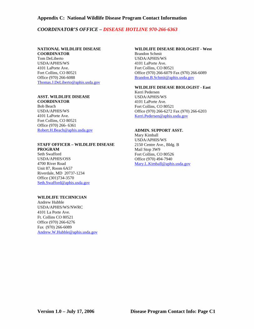
Appendix C: National Wildlife Disease Program Contact Information
Version 1.0 – July 17, 2006 Disease Program Contact Info: Page C1
COORDINATOR’S OFFICE – DISEASE HOTLINE 970-266-6363 NATIONAL WILDLIFE DISEASE COORDINATOR Tom DeLiberto USDA/APHIS/WS 4101 LaPorte Ave. Fort Collins, CO 80521 Office (970) 266-6088 [email protected] ASST. WILDLIFE DISEASE COORDINATOR Bob Beach USDA/APHIS/WS 4101 LaPorte Ave. Fort Collins, CO 80521 Office (970) 266- 6361 [email protected] STAFF OFFICER – WILDLIFE DISEASE PROGRAM Seth Swafford USDA/APHIS/OSS 4700 River Road Unit 87, Room 6A57 Riverdale, MD 20737-1234 Office (301)734-3570 [email protected] WILDLIFE TECHNICIAN Andrew Hubble USDA/APHIS/WS/NWRC 4101 La Porte Ave. Ft. Collins CO 80521 Office (970) 266-6276 Fax (970) 266-6089 [email protected]
WILDLIFE DISEASE BIOLOGIST - West Brandon Schmit USDA/APHIS/WS 4101 LaPorte Ave. Fort Collins, CO 80521 Office (970) 266-6079 Fax (970) 266-6089 [email protected] WILDLIFE DISEASE BIOLOGIST - East Kerri Pedersen USDA/APHIS/WS 4101 LaPorte Ave. Fort Collins, CO 80521 Office (970) 266-6272 Fax (970) 266-6203 [email protected] ADMIN. SUPPORT ASST. Mary Kimball USDA/APHIS/WS 2150 Centre Ave., Bldg. B Mail Stop 3W9 Fort Collins, CO 80526 Office (970) 494-7940 [email protected]
Appendix C: National Wildlife Disease Program Contact Information
Version 1.0 – July 17, 2006 Disease Program Contact Info: Page C2
WESTERN REGION ARIZONA Mark Lutman USDA/APHIS/WS 8836 N. 23rd Ave., Suite 2 Phoenix, AZ 85021 • (602) 870-2081 [email protected] CALIFORNIA Gerald Wiscomb USDA/APHIS/WS 3419A Arden Way Sacramento, CA 95825 • (916) 979-2675 [email protected] COLORADO Todd Felix USDA/APHIS/WS 12345 W. Alameda Pkwy., Ste. 204 Lakewood, CO 80228 • (303) 236-5820 [email protected] NEBRASKA Dallas Virchow USDA/APHIS/WS 5940 S. 58TH St. P. O. Box 81866 Lincoln, NE 68501-1866 • (402) 434-2343 [email protected] NORTH DAKOTA Ryan Powers USDA/APHIS/WS 2110 Miriam Cir., Ste. A Bismarck, ND 58501-2502 (701) 250-4405 [email protected]
OKLAHOMA Michael Marlow USDA/APHIS/WS 2800 N. Lincoln Blvd. Oklahoma City, OK 73105-4298 (405) 522-5453 [email protected] OREGON Justin Stevenson USDA/APHIS/WS 3413 Del Webb Ave. Salem, OR 97303 • (503) 399-5814 [email protected] TEXAS Janean Romines Mail Code: 1956 Dept. of State Health Services Zoonosis Control 1100 w. 49th St. Austin, TX 78756 (512) 458-7111 x 6421 [email protected] WYOMING Mike Pipas USDA/APHIS/WS 6731 W. Coal Rd. P. O. Box 59 Casper, WY 82602 • (307) 261- 5336 [email protected]

Appendix C: National Wildlife Disease Program Contact Information
Version 1.0 – July 17, 2006 Disease Program Contact Info: Page C3
EASTERN REGION ALABAMA Ashley Lovell USDA/APHIS/WS 1445 Federal Dr. Room 222 Montgomery, AL 36107 (334) 240-7217 [email protected] GEORGIA Darrell Kavanaugh USDA/APHIS/WS School of Forest Resources University of Georgia 200 Phoenix Rd. Athens, GA 30602 (706) 546-5637 [email protected] ILLINOIS Jane Weiskittel USDA/APHIS/WS 2869 Via Verde Dr. Springfield, IL 62703-4325 (217) 241-6700 [email protected] INDIANA Joe Caudell USDA/APHIS/WS Purdue University, Smith Hall 901 W. State Street West Lafayette, IN 47907-2089 (765) 496-3913 [email protected] MAINE Kirk Shively USDA/APHIS/WS Capital West Business Center 81 Leighton Rd., Suite 12 Augusta, ME 04330 (207) 622-8263 [email protected]
MARYLAND Daniel Emanueli USDA/APHIS/WS 1568 Whitehall Rd. Annapolis, MD 21401 (410) 349-8055 [email protected] MICHIGAN David Marks USDA/APHIS/WS 2803 Jolly Road, Suite 100 Okemos, MI 48864 (517) 336-1928 [email protected] MISSOURI Thomas Hutton USDA/APHIS/WS PO Box 180 2901 W. Truman Blvd. Jefferson City, MO 65102 (573) 522-4115 x3422 [email protected] NEW JERSEY Beth Jones USDA/APHIS/WS 140-C Locust Grove Rd. Pittstown, NJ 08867-4049 (908) 735-5654 [email protected] NEW YORK Justin Gansowski USDA/APHIS/WS 1930 Route 9 Castleton, NY 12033-9653 (518) 477-4837 [email protected]

Appendix C: National Wildlife Disease Program Contact Information
Version 1.0 – July 17, 2006 Disease Program Contact Info: Page C4
NORTH CAROLINA Carl Betsill USDA/APHIS/WS 6213-E Angus Drive Raleigh, NC 27617 (919) 786-4480 x230 [email protected] SOUTH CAROLINA Sheldon Owen USDA/APHIS/WS 400 Northeast Drive, Suite L Columbia, SC 29203-5182 (803) 786-5418 [email protected] TENNESSEE Joseph Clint McFee USDA/APHIS/WS 537 Myatt Drive Madison, TN 37115 (615) 736-5506 [email protected] WISCONSIN Barb Bodenstein USDA/APHIS/WS 732 Lois Drive Sun Prairie, WI 53590-1100 (608) 837-2727 [email protected]
Appendix D: USDA APHIS Directive 6800.1
United States Department of Agriculture Marketing and Regulatory Programs Animal and Plant Health Inspection Service
Directive APHIS 6800.1 5/10/06 ENSURING THE PROTECTION OF EMPLOYEES INVOLVED IN HIGHLY PATHOGENIC AVIAN INFLUENZA CONTROL AND ERADICATION ACTIVITIES 1. PURPOSE This Directive specifies APHIS policy to ensure the safety of employees engaged in highly pathogenic avian influenza (HPAI) control and eradication activities. The policy is based on the degree of risk known to be associated with various levels and types of exposures to HPAI viruses and should be considered complementary to avian disease control and eradication strategies as determined by State government, industry, or the United States Department of Agriculture (USDA). 2. AUTHORITIES a. Occupational Safety and Health Act of 1970, Section 5(a)(1), the General Duty Clause of the Act: “each employer shall furnish to each of his employees employment and a place of employment which are free from recognized hazards that are causing or are likely to cause death or serious physical harm to his employees.” b. 29 Code of Federal Regulations (CFR) 1910.120, 1910.132, 1910.134, and 1910.1030, Occupational Safety and Health Administration (OSHA) General Industry Regulations. 3. BACKGROUND Avian influenza (AI) is a contagious viral infection or disease of many avian species including poultry, wild and exotic birds, ratites, shorebirds, and migratory waterfowl. HPAI is seen primarily in poultry (rarely in other birds) and is characterized by severe depression, a decrease in egg production, high mortality, edema, hemorrhage, and necrosis. Birds that are infected with avian influenza virus can shed virus in saliva, nasal secretions, and feces. Contact with feces or respiratory secretions is important in the transmission of infection among poultry. Avian influenza viruses may be defined as highly pathogenic based either on mortality rates in chickens following intravenous inoculations or on the amino acid sequence at the hemagglutinin cleavage site. Only those results confirmed as HPAI by the National Veterinary Services Laboratory (NVSL) in Ames, Iowa will be considered highly pathogenic. Distribution: APHIS Originating Office: ESD-SHEWB Although HPAI viruses rarely infect humans, since 1997, instances of human infection have occurred outside the United States resulting in serious illness and even death. Transmission to humans is mainly thought to be caused by direct contact with infected poultry. The modality of transmission is not known, but could include virus entering a person’s mouth, nose, eyes, or lungs via aerosolization and inhalation into the lungs, or
Version 1.0 – July 17, 2006 Safety Directive: Page D1
Appendix D: USDA APHIS Directive 6800.1
by ingestion of contaminated material. Additionally, it is possible that infection could result from contact with virus-contaminated surfaces followed by self-inoculation of the virus in the eyes, nose, or mouth. This Directive is based on what are currently deemed optimal precautions to protect individuals from infection and illness while they are involved in the response to an HPAI outbreak, and to guard against the subsequent risk of viral reassortment (i.e., mixing of genes from human and avian viruses) if a human does become infected. Employees involved in HPAI control and eradication activities on known affected or potentially affected premises are at increased risk for exposure to the HPAI virus because those employees frequently have prolonged and direct contact with infected birds or contaminated surfaces in an enclosed setting. 4. POLICY a. APHIS employees involved in activities to control and eradicate any HPAI virus among poultry in the United States or abroad must read, understand, and follow Attachment 1 entitled: “APHIS Guidance for Protecting Workers Against Highly Pathogenic Avian Influenza.” This document was adapted from the Occupational Safety and Health Administration (OSHA) publications “Guidance for Protecting Workers Against Avian Flu” (http://www.osha.gov/dsg/guidance/avian-flu.html) and “Avian Influenza–Protecting Poultry Workers at Risk” (http://www.osha.gov/dts/shib/shib121304.html). b. Employees also must review the Centers for Disease Control and Prevention’s interim guidance documents regarding protection of employees involved in controlling and eradicating avian influenza in U.S. poultry. These guidance documents, “Interim Recommendations for Persons with Possible Exposure to Avian Influenza During Outbreaks Among Poultry in the United States” and “Interim Guidance for Protection of Persons Involved in U.S. Avian Influenza Outbreak Disease Control and Eradication Activities” are available online at http://www.cdc.gov/flu/avian/professional/possible-exposure.htm and http://www.cdc.gov/flu/avian/professional/protect-guid.htm, respectively. c. To mitigate the risk of exposure or infection, all employees involved in such activities must follow the precautions specified in Attachment 1. Among other topics, the Attachment includes recommendations about personal protective equipment, vaccination with the seasonal influenza vaccine, administration of antiviral drugs for prophylaxis, surveillance and monitoring of workers, and evaluation of workers who develop a febrile respiratory illness within 7 days of their last exposure to infected birds or contaminated surfaces. d. All employees involved in an HPAI response must understand and comply with this Directive. e. Any required negotiations with appropriate bargaining unit exclusive representatives will be conducted. 5. INQUIRIES a. Questions about this Directive or the specific instructions detailed in Attachment 1 should be directed to the Safety, Health, and Employee Wellness Branch (SHEWB), Employee Services Division, Marketing and Regulatory
Version 1.0 – July 17, 2006 Safety Directive: Page D2

Appendix D: USDA APHIS Directive 6800.1
Programs Business Services. SHEWB can be reached during regular business hours Monday-Friday (8 AM to 5 PM Eastern Time) at 301-734-6116. b. This Directive is available on the Internet at www.aphis.usda.gov/library Wm. R. DeHaven Administrator Attachment 1 APHIS Directive 6800.1 5/10/06 ANIMAL AND PLANT HEALTH INSPECTION SERVICE GUIDANCE FOR PROTECTING WORKERS AGAINST HIGHLY PATHOGENIC AVIAN INFLUENZA GUIDANCE FOR PROTECTING POULTRY WORKERS AT RISK Highly pathogenic avian influenza (HPAI) is a highly contagious disease of poultry. Despite the uncertainties, poultry experts agree that immediate culling of infected and exposed birds is the first line of defense to both reduce further losses in the agricultural sector and to protect human health. However, culling must be carried out in a way that protects workers from exposures to highly pathogenic avian influenza viruses and therefore reduce the likelihood of infection, illness or viral reassortment. Exposure to infected poultry, their feces, or respiratory secretions, or contact with potentially contaminated surfaces can result in transmission of the virus to humans. Human infection with avian influenza, however, is a rare occurrence. Although there is evidence of limited person-to person spread of the HPAI virus infection, sustained and efficient human-to-human transmission has not been identified. The following summarizes recommendations for protecting at-risk workers developed by the Centers for Disease Control and Prevention (CDC), the World Health Organization, and the Occupational Safety and Health Administration. Employees involved in HPAI control and eradication activities must take these precautions. 1. All persons who have been in contact with poultry, their feces or respiratory secretions, or contact with potentially contaminated surfaces must wash their hands frequently. Hand hygiene also must be performed immediately after gloves are removed and must consist of washing with soap and water for at least 15-20 seconds or using other standard handdisinfection procedures as specified by State government, industry, or United States Department of Agriculture (USDA) outbreak-response guidelines. 2. All workers involved in the culling, transport, or disposal of HPAI virus-infected poultry must not eat, drink, or smoke while performing these duties and must be provided with the following appropriate personal protective equipment: a. Protective clothing capable of being disinfected or discarded, preferably coveralls (plus an impermeable apron) or surgical gowns with long cuffed sleeves (plus an impermeable apron). b. Gloves capable of being disinfected or discarded; gloves must be carefully removed and discarded or disinfected and hands should be thoroughly washed when possible or disinfected using an alcohol-based handcleaner or 10%
Version 1.0 – July 17, 2006 Safety Directive: Page D3
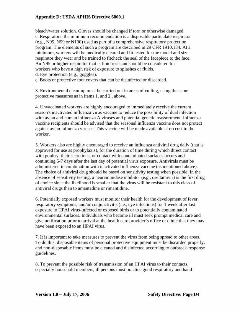
Appendix D: USDA APHIS Directive 6800.1
bleach/water solution. Gloves should be changed if torn or otherwise damaged. c. Respirators: the minimum recommendation is a disposable particulate respirator (e.g., N95, N99 or N100) used as part of a comprehensive respiratory protection program. The elements of such a program are described in 29 CFR 1910.134. At a minimum, workers will be medically cleared and fit tested for the model and size respirator they wear and be trained to fitcheck the seal of the facepiece to the face. An N95 or higher respirator that is fluid resistant should be considered for workers who have a high risk of exposure to splashes or fluids. d. Eye protection (e.g., goggles). e. Boots or protective foot covers that can be disinfected or discarded. 3. Environmental clean-up must be carried out in areas of culling, using the same protective measures as in items 1. and 2., above. 4. Unvaccinated workers are highly encouraged to immediately receive the current season's inactivated influenza virus vaccine to reduce the possibility of dual infection with avian and human influenza A viruses and potential genetic reassortment. Influenza vaccine recipients should be advised that the seasonal influenza vaccine does not protect against avian influenza viruses. This vaccine will be made available at no cost to the worker. 5. Workers also are highly encouraged to receive an influenza antiviral drug daily (that is approved for use as prophylaxis), for the duration of time during which direct contact with poultry, their secretions, or contact with contaminated surfaces occurs and continuing 5-7 days after the last day of potential virus exposure. Antivirals must be administered in combination with inactivated influenza vaccine (as mentioned above). The choice of antiviral drug should be based on sensitivity testing when possible. In the absence of sensitivity testing, a neuraminidase inhibitor (e.g., oseltamivir) is the first drug of choice since the likelihood is smaller that the virus will be resistant to this class of antiviral drugs than to amantadine or rimantidine. 6. Potentially exposed workers must monitor their health for the development of fever, respiratory symptoms, and/or conjunctivitis (i.e., eye infections) for 1 week after last exposure to HPAI virus-infected or exposed birds or to potentially contaminated environmental surfaces. Individuals who become ill must seek prompt medical care and give notification prior to arrival at the health care provider’s office or clinic that they may have been exposed to an HPAI virus. 7. It is important to take measures to prevent the virus from being spread to other areas. To do this, disposable items of personal protective equipment must be discarded properly, and non-disposable items must be cleaned and disinfected according to outbreak-response guidelines. 8. To prevent the possible risk of transmission of an HPAI virus to their contacts, especially household members, ill persons must practice good respiratory and hand
Version 1.0 – July 17, 2006 Safety Directive: Page D4
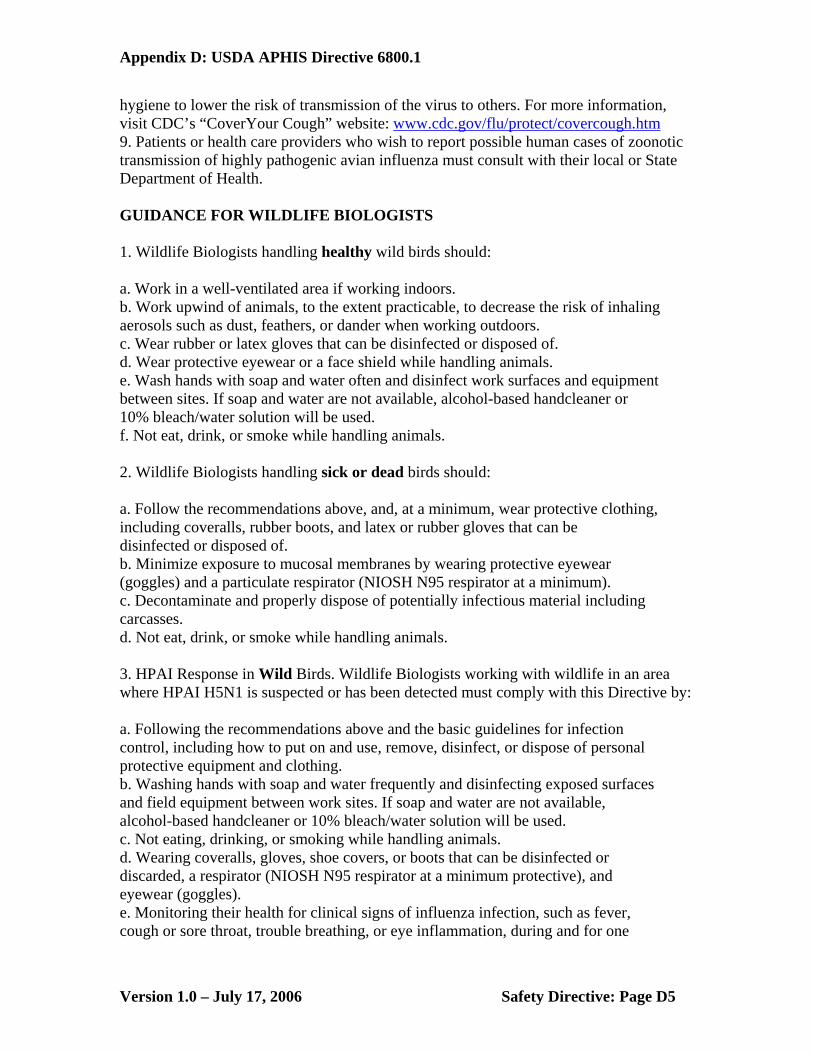
Appendix D: USDA APHIS Directive 6800.1
hygiene to lower the risk of transmission of the virus to others. For more information, visit CDC’s “CoverYour Cough” website: www.cdc.gov/flu/protect/covercough.htm 9. Patients or health care providers who wish to report possible human cases of zoonotic transmission of highly pathogenic avian influenza must consult with their local or State Department of Health. GUIDANCE FOR WILDLIFE BIOLOGISTS 1. Wildlife Biologists handling healthy wild birds should: a. Work in a well-ventilated area if working indoors. b. Work upwind of animals, to the extent practicable, to decrease the risk of inhaling aerosols such as dust, feathers, or dander when working outdoors. c. Wear rubber or latex gloves that can be disinfected or disposed of. d. Wear protective eyewear or a face shield while handling animals. e. Wash hands with soap and water often and disinfect work surfaces and equipment between sites. If soap and water are not available, alcohol-based handcleaner or 10% bleach/water solution will be used. f. Not eat, drink, or smoke while handling animals. 2. Wildlife Biologists handling sick or dead birds should: a. Follow the recommendations above, and, at a minimum, wear protective clothing, including coveralls, rubber boots, and latex or rubber gloves that can be disinfected or disposed of. b. Minimize exposure to mucosal membranes by wearing protective eyewear (goggles) and a particulate respirator (NIOSH N95 respirator at a minimum). c. Decontaminate and properly dispose of potentially infectious material including carcasses. d. Not eat, drink, or smoke while handling animals. 3. HPAI Response in Wild Birds. Wildlife Biologists working with wildlife in an area where HPAI H5N1 is suspected or has been detected must comply with this Directive by: a. Following the recommendations above and the basic guidelines for infection control, including how to put on and use, remove, disinfect, or dispose of personal protective equipment and clothing. b. Washing hands with soap and water frequently and disinfecting exposed surfaces and field equipment between work sites. If soap and water are not available, alcohol-based handcleaner or 10% bleach/water solution will be used. c. Not eating, drinking, or smoking while handling animals. d. Wearing coveralls, gloves, shoe covers, or boots that can be disinfected or discarded, a respirator (NIOSH N95 respirator at a minimum protective), and eyewear (goggles). e. Monitoring their health for clinical signs of influenza infection, such as fever, cough or sore throat, trouble breathing, or eye inflammation, during and for one
Version 1.0 – July 17, 2006 Safety Directive: Page D5

Appendix D: USDA APHIS Directive 6800.1
week after, their last exposure to potentially HPAI virus-infected or exposed birds. f. Contacting their healthcare provider if they develop fever, flu-like symptoms, or conjunctivitis (eye inflammation). Inform the provider prior to arrival that they have potentially been exposed to HPAI. Additional information about HPAI H5N1 can be found at the following web link: USGA National Wildlife Health Center: http://www.nwhc.usgs.gov/disease_information/avian_influenza/index.jsp GUIDANCE FOR VETERINARY LABORATORY WORKERS Highly pathogenic avian influenza A viruses are classified as “select agents” and must be handled under Biosafety Level (BSL) 3 enhanced or BSL 3-Agriculture laboratory standards. These include controlled access, double door entry with change room and shower out, use of respirators when working with specimens outside a biological safety cabinet, and decontamination of all waste. Laboratories working on these viruses must be USDA-approved. Clinical specimens from suspect HPAI virus cases may be tested by polymerase chain reaction (PCR) assays using standard BSL 2 work practices in a Class II biological safety cabinet. In addition, commercial antigen detection testing can be conducted under BSL 2 levels to test for influenza viruses. All employers processing biologic specimens suspected of being infected with the HPAI virus must ensure that their employees comply with all provisions of 29 CFR 1910.1030 for employee protection against bloodborne pathogens, including the reporting of accidental exposure to avian influenza virus. Any accidental exposure must be reported to an immediate supervisor or employee health department. Additional Sources of Information on Avian Influenza Centers for Disease Control and Prevention, http://www.cdc.gov/flu/avian/index.htm U.S. Department of Agriculture, Animal and Plant Health Inspection Service, http://www.aphis.usda.gov/lpa/issues/ai_us/ai_us.html World Health Organization, "Avian Influenza – Fact Sheet" http://www.who.int/csr/disease/avian_influenza/en/ World Health Organization, "Avian Influenza Frequently Asked Questions" http://www.who.int/csr/disease/avian_influenza/avian_faqs/en/ APHIS Medical Surveillance Service Form 29 and How to Complete http://www.aphis.usda.gov/mrpbs/forms/aphisforms.html
Version 1.0 – July 17, 2006 Safety Directive: Page D6

Appendix E: Sample AI Surveillance Field Data Sheet Appendix E: Sample AI Surveillance Field Data Sheet
Version 1.0 – July 17, 2006 Sample AI Surveillance Field Data Sheet: Page E1
Version 1.0 – July 17, 2006 Sample AI Surveillance Field Data Sheet: Page E1

Appendix F: Species Codes
COMMON NAME ALPHA CODE
COMMON NAME ALPHA
CODE
COMMON NAME ALPHA CODE
Aleutian Cackling/Canada Goose
ACGO Gray-cheeked Thrush GCTH Red-necked Phalarope
RNPH
American Green-winged Teal
AGWT Greater Frigatebird GRFR Red-necked Stint RNST
American Wigeon AMWI Greater Scaup GRSC Red-tailed Tropicbird
RTTR
Atlantic Brant ATBR Greater Yellowlegs GRYE Red-throated Loon RTLO Arctic Loon ARLO Harlequin Duck HARD Rock Sandpiper ROSA Artic Tern ARTE Hawaiian Coot HACO Ross’s Goose ROGO Arctic Warbler ARWA Hawaiian Duck HAWD Ruddy Turnstone RUTU Baikal Teal BATE Hawaiian (Nene)
Goose HAGO Sanderling SAND
Barn Owl BNOW Hawaiian Hawk HWAH Sandhill Crane SACR Bar-tailed Godwit BARG Hawaiian Moorhen HAMO Semipalmated
Plover SEPL
Black Brant BLBR Herring Gull HERG Semipalmated Sandpiper
SESA
Black Duck (American) ABDU Horned Grebe HOGR Sharp-tailed Sandpiper
SHAS
Black Guillemot BLGU House Finch HOFI Sky Lark SKLA Black-crowned Night Heron
BCNH King Eider KIEI Snow Goose SNGO
Black Noddy BLNO Laysan Albatross LAAL Spectacled Eider SPEI Black Scoter BLSC Least Sandpiper LESA Spotted Sandpiper SPSA Black Turnstone BLTU Lesser Blackbacked
Gull LBBG Steller's Eider STEI
Black-bellied Plover BBPL Lesser Scaup LESC Surfbird SURF Black-necked Stilt BNST Lesser Yellowlegs LEYE Tufted Duck TUDU Bonaparte’s Gull BOGU Long-billed Dowitcher LBDO Tundra Swan TUSW Bristle-thighed Curlew BTCU Long-tailed Duck LTDU Wandering Tattler WATA Buff-breasted Sandpiper BBSA Mallard MALL Wedge-tailed
Shearwater WTSH
Cackling Goose CACG Marbled Godwit MAGO Western Meadowlark
WEME
Canada Goose CAGO Mute Swan MUSW Western Sandpiper WESA Canvasback CANV Northern Cardinal NOCA Whimbrel WHIM Cattle Egret CAEG Northern Mockingbird NOMO White-fronted Goose GWFG Common Eider COEI Northern Pintail NOPI Whooper Swan WHOS Common Goldeneye COGO Northern Shoveler NSHO Yellow Bittern YEBI Common Loon COLO Olomao (Molokai
Thrush) OLOM Yellow-billed Loon YBLO
Common Pochard COPO Omao (Hawaiian Thrush)
OMAO Yellow Wagtail YWAG
Common Tern COTE Pacific Golden-Plover PAGP Dunlin DUNL Pacific Loon PALO Emperor Goose EMGO Pectoral Sandpiper PESA Eurasian Tree Sparrow ETSP Peregrine Falcon PEFA Eurasian Wigeon EUWI Philippine Turtle Dove PHTD
Version 1.0 – July 17, 2006 Species Codes: Page F1
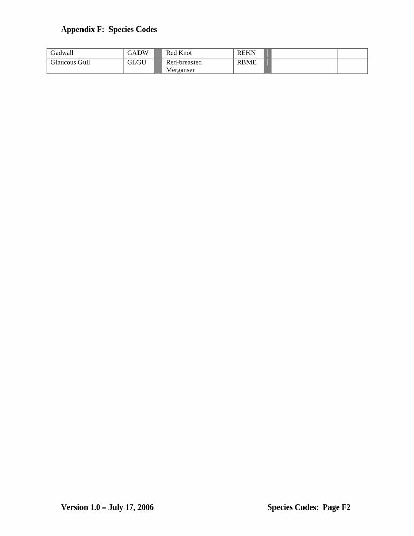
Appendix F: Species Codes
Gadwall GADW Red Knot REKN Glaucous Gull GLGU Red-breasted
Merganser RBME
Version 1.0 – July 17, 2006 Species Codes: Page F2
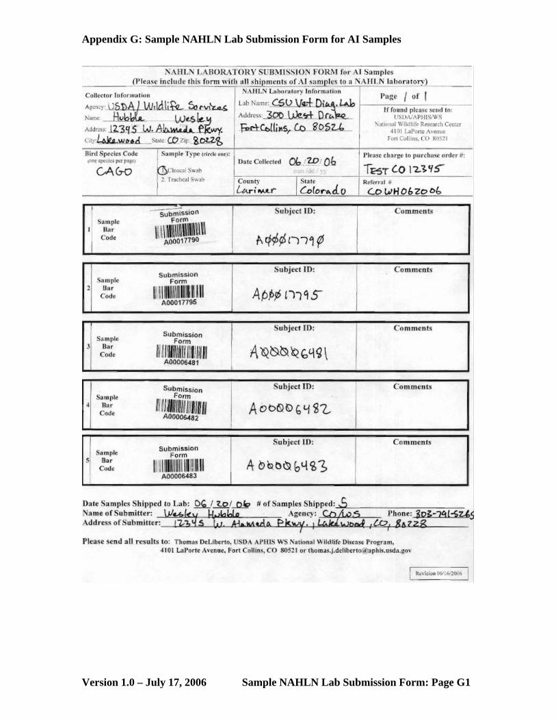
Appendix G: Sample NAHLN Lab Submission Form for AI Samples
Version 1.0 – July 17, 2006 Sample NAHLN Lab Submission Form: Page G1

Appendix H: Laboratories Approved to Participate in AI Sample Testing
LABORATORIES APPROVED TO PARTICIPATE IN AI SAMPLE TESTING FROM WILDLIFE SERVICES
State Laboratory Name Laboratory Director
Lab Director E-Mail Telephone
AK State of Alaska Dept. of Environmental Conserv.
Dr. Tom Hathaway [email protected] (907) 375-8206
AL Thompson Bishop Sparks State Diagnostic Lab
Dr. Fred Hoerr [email protected] (334) 844-4987
AR Arkansas Livestock & Poultry Commission Lab
Dr. Konnie Plumlee
[email protected] (501) 907-2430
AZ Arizona Veterinary Diagnostic Laboratory
Dr.Greg Bradley [email protected] (520) 621-2356
CA California Animal Health & Food Safety Lab
Dr. Alex Ardans [email protected], [email protected]
(530) 752-8700
CO Colorado State University Veterinary Diag. Lab
Dr. Barbra Powers [email protected] (970) 297-1281
CT Department of Pathobiology & Veterinary Science Connecticut Veterinary Medical Diagnostic Laboratory
Dr. Herbert Van Kruiningen
[email protected] (860) 486-3738
DE University of Pennsylvania Poultry Lab
Dr. Sherrill Davison-Yeakel
(610) 444-5800
FL Kissimmee Diagnostic Laboratory Dr. Betty Miguel [email protected] (407) 846-5200 or (321) 697-1400
GA Athens Veterinary Diagnostic Laboratory
Dr. Doris Miller [email protected] (706) 542-5568
HI State Laboratories Division Dr. A. Christian Whelen
[email protected] (808) 453-6652
ID Utah Veterinary Diagnostic Laboratory
Dr. Tom Baldwin [email protected] (435)-797-1895
ID Washington Animal Disease Diagnostic Laboratory
Dr. Terry McElwain
[email protected] (509)-335-9696
IA Iowa State University Dr. Bruce Janke [email protected] (515)-294-1950
IL Illinois Department of Agriculture Galesburg Animal Disease Laboratory
Dr. Dale Webb [email protected] (309) 344-2451
IN Purdue University Animal Disease Diagnostic Lab
Dr. Leon Thacker [email protected] (765)-494-7440
KS Veterinary Diagnostic Center Dr. David Steffen [email protected] (402)-472-2952
KY Breathitt Veterinary Center Dr. Wade Northington
[email protected] (270) 886-3959
LA Louisiana Vet Med Diagnositc Laboratory
Dr. Alma Roy [email protected] (225)-578-9777
ME Department of Pathobiology & Veterinary Science Connecticut Veterinary Medical Diagnostic Laboratory
Dr. Herbert Van Kruiningen
[email protected] (860) 486-3738
MD Maryland Dept. of Ag & Animal Health Laboratory
Dr. Maurice Clarke [email protected] (410)-543-6610
Version 1.0 – July 17, 2006 Labs Approved to Participate in AI Testing: Page H1

Appendix H: Laboratories Approved to Participate in AI Sample Testing
MA Department of Pathobiology & Veterinary Science Connecticut Veterinary Medical Diagnostic Laboratory
Dr. Herbert Van Kruiningen
[email protected] (860) 486-3738
MI Diagnostic Center of Population and Animal Health
Dr. Willie Reed [email protected] (517)-353-1683
MN Minnesota Veterinary Diagnostic Laboratory
Dr. James E. Collins
[email protected] (612)-625-8787
MO University of Missouri Dr. Alex Bermudez [email protected] (573)-882-6811
MS Mississippi Vet Research & Diagnostic Laboratory
Dr. Lanny Pace [email protected] (601)-354-6089
NC North Carolina Department of Agriculture-Rollins Animal Disease Diagnostic Lab
Dr. David Marshall [email protected] (919)-733-3986
ND Minnesota Veterinary Diagnostic Laboratory
Dr. James E. Collins
[email protected] (612)-625-8787
NE Veterinary Diagnostic Center Dr. David Steffen [email protected] (402)-472-2952
NM New Mexico Department of Agriculture
Dr. Flint Taylor [email protected] (505)-841-2576
NH Maryland Dept. of Ag & Animal Health Laboratory
Dr. Maurice Clarke [email protected] (410)-543-6610
NH Department of Pathobiology & Veterinary Science Connecticut Veterinary Medical Diagnostic Laboratory
Dr. Herbert Van Kruiningen
[email protected] (860) 486-3738
NJ New Jersey Department of Ag. Division of Animal Health
Dr. Nancy Halpern [email protected] (609) 292-3965
NV Nevada Animal Disease Laboratory Dr. Anette Rink [email protected] (775)-668-1182
NY Animal Health Diagnostic Center Dr. Alfonso Torres [email protected] (607)-253-4136
OH Ohio Department of Agriculture Dr. Beverly Byrum [email protected] (614)-728-6220
OK Oklahoma Animal Disease Diagnostic Laboratory
Dr. Bill J. Johnson [email protected] (405)-744-6623
OR Oregon State Veterinary Diagnostic Lab
Dr. Jerry Heidel [email protected] (541)-737-3261
PA Pennsylvania Dept. of Ag Veterinary Laboratory
Dr. Helen Acland [email protected] (717)-787-8808
SC Clemson Veterinary Diagnostic Center
Dr. Pamela Parnell [email protected] (803)-788-2260
SD Animal Disease Research & Diagnostic Lab
Dr. David Zeman [email protected] (605) 688-5171
TN CE Kord Animal Disease Diagnostic Lab
Dr. Ronald B. Wilson
[email protected] (615) 837-5120
TX Texas Vet Medical Diagnostic Laboratory
Dr. Lelve Gayle [email protected] (979)-845-9000
UT Utah Veterinary Diagnostic Laboratory
Dr. Tom Baldwin [email protected] (435)-797-1895
VA Virginia Dept of Agriculture and Animal Health Lab
Dr. David W. Brown
[email protected] (540)-434-3897
Version 1.0 – July 17, 2006 Labs Approved to Participate in AI Testing: Page H2

Appendix H: Laboratories Approved to Participate in AI Sample Testing
VT Department of Pathobiology & Veterinary Science Connecticut Veterinary Medical Diagnostic Laboratory
Dr. Herbert Van Kruiningen
[email protected] (860) 486-3738
WA Washington Animal Disease Diagnostic Laboratory
Dr. Terry McElwain
[email protected] (509)-335-9696
WA Avian Health and Food Safety Laboratory
(253)-445-4537
WI Wisconsin Veterinary Diagnostic Laboratory
Dr.Kathy Kurth and Dr.Pete Vanderloo
[email protected] (608)-262-5432
WY Wyoming State Vet Laboratory Dr. Donal O'Toole (307) 742-6638
Version 1.0 – July 17, 2006 Labs Approved to Participate in AI Testing: Page H3

Appendix I: Sample Environmental Submission Form
Version 1.0 – July 17, 2006 Sample Environmental Submission Form: Page I1

Appendix J: Tools and Supplies for Sampling
• Sampling kit from NVSL (includes supplies for 40 samples) 1. Data sheets (10 included – enough for 50 samples) 2. Laboratory submission form (10 included – to
be used until automated data entry system is operational. This form also serves as the itemized list that must be included when diagnostic specimens are shipped to the lab)
3. Vials with transport media 4. Dacron swabs – large (40 included) (small
swabs available upon request) 5. Sample tube containing 1.5 mL of Brain Heart Infusion (BHI) media (40
included) 6. Bar codes – 40 sets of 4 duplicate bar codes (1 for data sheet, 1 for sample
tube, 1 for laboratory submission form, 1 extra) 7. Shipping box (supplies will be shipped within this container) 8. 40 section box 9. Secondary container (STP #740 and STP #741) 10. Absorbent material to include between vials and secondary containment
vessel. 11. Blue ice frozen cold packs (2) 12. UN3373 diagnostic specimens label
• 10 - 20 sample shipping cooler (should be purchased by collecting agency
for use when shipping < 20 samples). All supplies listed below can be purchased from SAFTPAK (www.saftpak.com). 1. Cooler (STP #309 or equivalent shipper) 2. Secondary containment bag for shipping diagnostic specimens (STP #710) 3. Blue ice frozen cold packs 4. UN3373 shipping labels
• Miscellaneous supplies
1. Ball-point pens 2. Scissors 3. Alcohol swabs 4. Paper towels 5. Trash bags 6. GPS units (set to WGS 84 datum and decimal degrees format) 7. Hand sanitizer 8. Bird identification guides 9. Laptop with mapping software or area maps 10. Example data sheet 11. Tape
Version 1.0 – July 17, 2006 Tools and Supplies: Page J1
Appendix K: Communication Protocol in the Event of a HPAI Finding
USDA/APHIS/ Wildlife Services Multi-hazard Emergency Communications
Standard Operating Procedure 1.0 Background
Development of a National Wildlife Disease Surveillance and Emergency Response System (SERS) within Wildlife Services (WS) is a critical component of APHIS’ mission to protect the health and value of American agriculture, natural resources, and human health and safety. As part of its strategic plan, APHIS is focusing on strengthening emergency preparedness and response, and managing issues related to the health of U.S. animal resources and conflicts with wildlife. One strategy to accomplish this is through APHIS’ commitment to enhance its current emergency response infrastructure as directed in Homeland Security Presidential Directive 8 (HSPD-8). Additionally, APHIS and its stakeholders have become increasingly aware of the need to manage and research wildlife diseases, particularly those that are transmissible to humans and domestic species. The SERS is the program within WS that has responsibility for developing and implementing WS’ multi-hazard emergency response capabilities. A complete overview of this program can be found in the SERS Business Plan. In Homeland Security Presidential Directive-5 (HSPD-5), the President called on the Secretary of Homeland Security to develop a national incident management system to provide a consistent nationwide approach for federal, state, tribal and local governments to work together to prepare for, prevent, respond to and recover from domestic incidents, regardless of cause, size or complexity. After close collaboration with state and local government officials and representatives from a wide range of public safety organizations, the Department of Homeland Security (DHS) issued the National Incident Management System (NIMS). It incorporates many existing best practices into a comprehensive national approach to domestic incident management, applicable at all jurisdictional levels and across all functional disciplines. The NIMS represents a core set of doctrine, principles, terminology, and organizational processes to enable effective, efficient and collaborative incident management at all levels. To provide the framework for interoperability and compatibility, the NIMS is based on a balance between flexibility and standardization. One of the most important 'best practices' that has been incorporated into the NIMS is the Incident Command System (ICS), a standard, on-scene, all-hazards
Version 1.0 – July 17, 2006 Communication Protocol: Page K1

Appendix K: Communication Protocol in the Event of a HPAI Finding
incident management system. The ICS has been established by the NIMS as the standardized incident organizational structure for the management of all incidents. The NIMS provides a consistent, flexible and adjustable national framework within which government and private entities at all levels can work together to manage domestic incidents, regardless of their cause, size, location or complexity. This flexibility applies across all phases of incident management: prevention, preparedness, response, recovery and mitigation. The NIMS provides a set of standardized organizational structures – including the ICS, Multi-Agency Coordination Systems and public information systems – as well as requirements for processes, procedures and systems to improve interoperability among jurisdictions and disciplines in various areas. The overwhelming majority of emergency incidents are handled on a daily basis by a single jurisdiction at the local level. However, there will be instances in which successful domestic incident management operations depend on the involvement of emergency responders from multiple jurisdictions, as well as personnel and equipment from other States and the Federal government. These instances require effective and efficient coordination across a broad spectrum of organizations and activities, and the success of emergency operations to address them depends on the ability to mobilize and effectively utilize multiple resources. These resources must come together in an organizational framework that is understood by everyone and must utilize a common plan, as specified through a process of incident action planning.
2.0 Objective
This standard operating procedure (SOP) outlines how WS will respond to requests for emergency assistance from within and outside the Program. The objective is to provide a framework that streamlines communications within WS during emergency situations. Efficient communications will ensure effective allocation of WS’ resources (e.g., funds, personnel, equipment) during emergency responses. This SOP will also provide an overview of WS’ communications structure for responding to emergency requests for assistance from other agencies. Although this SOP is required during emergency operations, it does not preclude communications through standard supervisory channels.
3.0 Definitions
3.1 “Emergency” means any occasion or instance when an ICS is established by an agency (i.e., local, State, Federal, Tribal), for which WS assistance is needed to supplement State, local or other Federal agency efforts and capabilities to save lives or protect resources, or to lessen or avert the threat of a catastrophe (e.g., foreign animal disease introduction, epizootic of an endemic disease, natural disaster, bioterrorist attack, oil spills).
Version 1.0 – July 17, 2006 Communication Protocol: Page K2

Appendix K: Communication Protocol in the Event of a HPAI Finding
3.2 “Extraordinary Emergency” means those instances when the U.S.
Secretary of Agriculture determines that because of the presence in the United States of a pest or disease of livestock and that the presence of the pest or disease threatens the livestock of the U.S., the Secretary may take measures in a State to control or limit the spread of such diseases or pests in accordance with the Farm Security and Rural Investment Act of 2002 (7 USC 7901), Title X, Subsection E (Animal Health Protection Act), Section 10407.
3.3 "Federal Agency" means any department, independent establishment,
Government Corporation, or other agency of the executive branch of the Federal Government, including the U.S. Postal Service, but shall not include the American National Red Cross.
3.4 “First Responder” means any WS’ wildlife disease biologist that is part of
the SERS. 3.5 “Local Agency” means any department, independent establishment,
Government Corporation, or other agency of the Local Government. 3.6 “Local Government” means the county, municipality, city, town,
township, public authority, school district, special district, intrastate district, council of governments (regardless of whether the council of governments is incorporated as a nonprofit corporation under State law), regional or interstate government entity, or agency or instrumentality of a local government; an Indian tribe or authorized tribal organization, or Alaska Native village or organization; and a rural community, unincorporated town or village, or other public entity, for which an application for assistance is made by a State or political subdivision of a State.
3.7 “Major Disaster” means any natural catastrophe (e.g., hurricane, tornado,
storm, high water, wind-driven water, tidal wave, tsunami, earthquake, volcanic eruption, landslide, mudslide, snowstorm, or drought), or, regardless of cause, any fire, flood, or explosion, in any part of the United States, which in the determination of the President causes damage of sufficient severity and magnitude to warrant major disaster assistance under Robert T. Stafford Disaster Relief and Emergency Response Act (42 USC 5122) to supplement the efforts and available resources of States, local governments, and disaster relief organizations in alleviating the damage, loss, hardship, or suffering caused thereby.
3.8 “Program” means U.S. Department of Agriculture, Animal and Plant
Health Inspection Service, Wildlife Services
Version 1.0 – July 17, 2006 Communication Protocol: Page K3
Appendix K: Communication Protocol in the Event of a HPAI Finding
3.9 “Second Responder” means all WS’ employees except wildlife disease biologists.
3.10 "State" means any State of the U.S., the District of Columbia, Puerto Rico,
the Virgin Islands, Guam, American Samoa, and the Commonwealth of the Northern Mariana Islands.
3.11 “State Agency” means any department, independent establishment,
Government Corporation, or other agency of the executive branch of the State Government.
3.12 "United States (U.S.)" means the fifty States, the District of Columbia,
Puerto Rico, the Virgin Islands, Guam, American Samoa, and the Commonwealth of the Northern Mariana Islands.
4.0 Roles and Responsibilities
4.1 State Directors
According to ICS procedure, decision making authority for emergency response is held at local levels. Off-site coordination and support functions serve the needs of on-site command functions. During emergency operations, the State Director responsible for WS operations in the area (or his/her designee), will be responsible for communicating with the Incident Commander and his/her staff. The State Director will supply the Incident Commander with local WS:
• capabilities, • available personnel and expertise • appropriate and available equipment: • required (but unavailable) equipment necessary to provide
requested support • Estimated schedule to complete request with local resources • Estimated cost to complete the requested services
If the State Director or Incident Commander determines that WS resources in the State are insufficient to meet the needs of the operation, they must contact the SERS Office either directly, or through the Regional or National Command Centers. It is the responsibility of SERS to evaluate and coordinate all requests to WS for emergency assistance, which require a multi-state, regional, or national response by the Program. However, on-site management of all WS resources remains under the direction of the State Director in coordination with the Command Center. The State Director is also responsible for providing SERS with periodic updates on WS’ activities in conjunction with the emergency response.
Version 1.0 – July 17, 2006 Communication Protocol: Page K4
Appendix K: Communication Protocol in the Event of a HPAI Finding
As discussed above, when local WS’ resources are sufficient to meet the request of the Incident Commander, it is not usually necessary for State Directors to contact SERS. However, for specific types of emergencies (i.e., major disasters, foreign animal disease introduction, epizootic of an endemic disease, bioterrorist attack, oil spills) State Directors must contact the SERS office and provide periodic updates to ensure that information on the event, and WS’ response, is communicated to appropriate personnel within the Program (e.g., Deputy Administrator, WS Management Team). This communication between the State Director and SERS for these specific emergencies will enable SERS to track the event and prepare for a multi-state, regional, or national response if and when it becomes necessary.
4.2 SERS Office
The primary functions of SERS during emergencies are to:
1. ensure efficient communication within WS, among other agencies, and with Regional and National Command Centers.
2. provide logistical support to on-site State Director(s) and Incident Command Center,
3. Coordinate a WS multi-state, regional, or national response to and emergency.
The SERS Office will be available on a 24 hours/day, 7 days/week, 365 days/year basis. A designated phone number (970-266-6363) has been established that will be routinely monitored by SERS personnel. Upon initial notification of a request for WS’ assistance in an emergency from a State Director, Regional Office, or another agency, the SERS Office will notify the Deputy Administrator’s Office. After consultation with the Deputy Administrator’s Office, the SERS Office will provide available information concerning the request to WS Management Team, other appropriate field offices, and the National Wildlife Research Center (NWRC). Within 24 hours of receiving the initial request for emergency assistance, the SERS Office will provide a briefing to the Deputy Administrator’s Office that explains and prioritizes the request. The briefing will contain information such as:
1. Nature of the emergency
a. Type i. Foreign animal disease introduction ii. Epizootic of an endemic disease
iii. Natural disaster iv. Bioterrorist attack
Version 1.0 – July 17, 2006 Communication Protocol: Page K5

Appendix K: Communication Protocol in the Event of a HPAI Finding
v. Oil spill vi. Other
b. Classification i. Major Disaster ii. Extraordinary Emergency iii. Other
c. SERS defined priority i. Low - Neither the time or issue elements are
considered critical ii. Moderate – Either the time or issue element is
considered critical iii. High - Both issue and time elements are considered
critical 2. Source of the request:
a. WS State Director/On-site Incident Command Center b. Regional/National Command Center c. Government Agency
3. Type of assistance being requested a. Technical Personnel b. Administrative Personnel c. Equipment/supplies d. Financial e. Other
4. Estimated impact on the WS Program a. Minimal - The State Director(s) and/or SERS is capable of
fulfilling the emergency request with local WS’ personnel and/or 1st responders, and the expected non-reimbursed demands are minimal.
b. Tolerable - The State Director(s) and/or SERS is capable of fulfilling the emergency request with local WS’ personnel and/or 1st responders; the expected non-reimbursed demands are moderate, but can be absorbed within the State(s) WS’ and/or SERS budget.
c. Extensive – The State Director(s) and/or SERS is incapable of fulfilling the emergency request with only local WS’ personnel and/or 1st responders, and the need for 2nd responders is required; the expected non-reimbursed demands are high and cannot be absorbed within the State(s) WS’ and/or SERS budget, or personnel expertise beyond the capabilities of local WS’ personnel and/or 1st responders is required.
5. Estimated Program resources required a. Personnel;
i. Number, expertise, skills b. Vehicles;
i. Number, type, etc.
Version 1.0 – July 17, 2006 Communication Protocol: Page K6

Appendix K: Communication Protocol in the Event of a HPAI Finding
c. Equipment & Supplies: i. Number & type
6. Estimated duration of assistance 7. Source of funding for provided assistance
a. WS Program funds b. Other Agency funds
8. WS response time 10. Proposed Immediate Response by the SERS Office
a. No action b. Deployment of 1st responders. c. Request to Regional Offices and NWRC for 2nd responders.
Once the SERS Office commits to utilize its resources for an emergency request, 1st responders will be mobilized and the Deputy Administrator’s Office, Management Team, and affected State Directors will be notified of their availability. If required, 1st responders will report to the State Director(s) at the incident scene and the Incident Command Center for duty assignments. The State Director(s) are responsible for establishing routine communication with the SERS Office, which will evaluate the information provided and develop routine briefing information for the Deputy Administrator’s Office and/or Management Team. At anytime prior to, or while in the process of responding to an emergency request, the SERS Office is asked to provide personnel in addition to 1st responders, the Regional Offices and NWRC will be notified. The Regional Offices and NWRC will work with their respective State Directors or Program Managers to provide a list of 2nd responders that have the required training and experience, and are available to assist in the response.
5.0 Summary of Communication Pathways
During an emergency response, it is critical to maintain a clearly defined pathway of communication within the Program. The State Director(s) is responsible for providing on-site information regarding an emergency that requires multi-state WS resources and/or those consisting of a major disaster, foreign animal disease introduction, epizootic of an endemic disease, bioterrorist attack, or oil spill to the SERS Office. For emergencies that do not meet these criteria, State Directors need not contact SERS, but must notify their respective Regional Offices as appropriate. The SERS office is primarily responsible for assimilating information from State Directors and Regional/National Command Centers, evaluating WS’ responses, and making recommendations for additional assistance to the Deputy Administrator’s Office and the Management Team. When requested by State
Version 1.0 – July 17, 2006 Communication Protocol: Page K7

Appendix K: Communication Protocol in the Event of a HPAI Finding
Director(s) and/or Incident Command Centers, the SERS Office will attempt to fulfill emergency requests with 1st responders. However, if insufficient numbers of 1st responders are available, technical expertise is required that is not available through 1st responders, or if logistical and financial constraints limit the use of 1st responders, SERS will request Regional Offices and/or NWRC to provide a list of available 2nd responders. The SERS Office will notify the Deputy Administrator’s Office and the WS Management Team prior to mobilizing 2nd responders. In emergencies requiring the use of WS’ aircraft, State Directors and Incident Commanders will coordinate with WS’ Aviation Operations Manager to ensure that any aerial response will be safe and effective. The Aviation Operations Manager is responsible for providing SERS with periodic updates on WS’ aviation activities during an emergency. Throughout an emergency response, the SERS Office is responsible for updating the Deputy Administrator’s Office, Management Team, and State Director’s on the progress of the response and provide other related information obtained through interagency communications. Updates from the SERS Office will be forwarded to other WS personnel through normal communication channels.
Version 1.0 – July 17, 2006 Communication Protocol: Page K8