aha/asa guideline guideline for the management of...
TRANSCRIPT

science is whyscience is why
AHA/ASA Guideline
Guideline for the Management of Patients with Unruptured Intracranial
AneurysmsA Guideline for Healthcare Professionals from the American Heart
Association/American Stroke Association
The American Academy of Neurology (AAN) affirms the value of this guideline as an educational tool for neurologists
Endorsed by the American Association of Neurological Surgeons (AANS) and Congress of Neurological Surgeons (CNS)
Endorsed by the Society of NeuroInterventional Surgery (SNIS)
science is why

science is whyscience is why
B. Gregory Thompson, MD, Chair; Robert D. Brown Jr., MD, MPH, FAHA, Co-Chair; Sepideh Amin-Hanjani, MD,
FAHA; Joseph P. Broderick, MD, FAHA; Kevin M. Cockcroft, MD, MSc, FAHA; E. Sander Connolly, Jr., MD,
FAHA; Gary R. Duckwiler, MD, FAHA; Catherine C. Harris, PhD, RN; Virginia J. Howard, PhD, MSPH, FAHA; S. Claiborne (Clay) Johnston, MD, PhD; Philip M. Meyers, MD, FAHA; Andrew Molyneuz, MD; Christopher S. Ogilvy,
MD; Andrew J. Ringer, MD; James Torner, PhD, MS, FAHA on behalf of the American Heart Association Stroke Council, Council on Cardiovascular and Stroke Nursing,
and Council on Epidemiology and Prevention.
Authors

science is whyscience is why
Michael T. Mullen, MD MSAssistant Professor
Department of NeurologyPerelman School of Medicine,
University of Pennsylvania
Slides Prepared by Members of the Stroke Professional Education Committee

science is whyscience is why
Applying classification of recommendations and levels of evidence

science is whyscience is why
I. IntroductionII. Risk factors for aneurysm development, growth and
ruptureIII. Clinical presentationIV. Diagnosis/imagingV. Screening in asymptomatic individualsVI. Natural history of unruptured aneurysmsVII. Surgical Clipping of unruptured aneurysmsVIII.Endovascular treatment of unruptured aneurysmsIX. Comparative efficacy of clipping vs. coilingX. Aneurysm follow-upXI. Conclusion
Outline

science is whyscience is why
I. Introduction
RightMCA Aneurysm
Incidentally discovered 6mm unruptured aneurysm arising from an MCA branchd
Courtesy Michael T. Mullen MD – University of Pennsylvania
science is whyscience is why
I. Introduction
• Unruptured intracranial aneurysms (UIA) are common, occurring in approximately 3.2% of the general population
• They are often discovered incidentally on cerebrovascular imaging.
• The VAST majority of UIA will not rupture
science is whyscience is why
It is estimated that out of 1 million adults in the general population with mean age of 50:
• 32,000 will harbor an UIA
• Only 80 (0.25%) would be expected to present with subarachnoid hemorrhage (SAH).
I. Introduction

science is whyscience is why
I. Introduction
• Despite the low risk of rupture, aneurysmal SAH is potentially catastrophic, due to high rates of morbidity and mortality.
• As a result, patients and physicians are often faced with the dilemma of whether to treat patients with UIA or manage them conservatively.
• The purpose of this statement is to provide guidance for healthcare providers and to serve as a framework for decision making when a UIA is discovered.
science is whyscience is why
II. Risk Factors for Aneurysm Development, Growth, and Rupture
Non-modifiable Risk Factors for Aneurysm Development
• Older age • Female sex• Family history of aneurysms/SAH• At risk disorders (<10% of UIA):
Polycystic Kidney Disease, Type IV Ehlers Danlos, Marfan’s Syndrome, Coarctation of the aorta, Bicuspid aortic valve, and Fibromuscular dysplasia, Microcephalic osteodysplastic primordial dwarfism, and intracranial arteriovenous malformation among others.

science is whyscience is why
II. Risk Factors for Aneurysm Development, Growth, and Rupture
Modifiable Risk Factors for Aneurysm Development:
• Smoking
• Hypertension
science is whyscience is why
II. Risk Factors for Aneurysm Development, Growth, and Rupture
Potential Risk Factors for Aneurysm Growth• Not well studied• May include:
– Female sex– Smoking– Hypertension– Excessive alcohol– Larger aneurysm size– Arterial branch related aneurysm

science is whyscience is why
II. Risk Factors for Aneurysm Development, Growth, and Rupture
Risk Factors for Aneurysm Rupture:• Aneurysm size
– Larger aneurysm = Increased Risk
• Aneurysm location– Anterior communicating artery and pericallosal may be over-
represented in the ruptured cohort
• Other potential risk factors (less well defined):– Aneurysm morphology, aneurysm growth, smoking,
hypertension, prior SAH, and family history of SAH.

science is whyscience is why
II. Risk Factors for Aneurysm Development, Growth, and Rupture
Class I Recommendations Class, Level of Evidence(LOE)
Given that smoking appears to increase risk of UIA formation, patients with UIA should be counseled regarding the importance of smoking cessation.
Class I, LOE B
Given that hypertension may play a role in growth and rupture of intracranial aneurysms, patients with UIA should monitor blood pressure and undergo treatment for hypertension.
Class I, LOE B
Aneurysmal growth may increase the risk of rupture and intermittent imaging studies to follow those UIAs managed conservatively should be considered.
Class I, LOE B

science is whyscience is why
UIA often identified:• After SAH from another aneurysm
• Incidentally during evaluation of unrelated neurologic symptoms
• Symptomatic from mass effect causing cranial nerve palsy
– May increase risk of rupture up to 4x– Sudden onset of CN III palsy may indicate expansion
and imminent risk of rupture
III. Clinical Presentation

science is whyscience is why
III. Clinical Presentation
Right Posterior Communicating Artery Aneurysm
• Presented with a 3rd Nerve Palsy from compression
• Suggests high risk of rupture
• Urgent treatment is indicated
Courtesy David K. Kung MD - University of Pennsylvania
science is whyscience is why
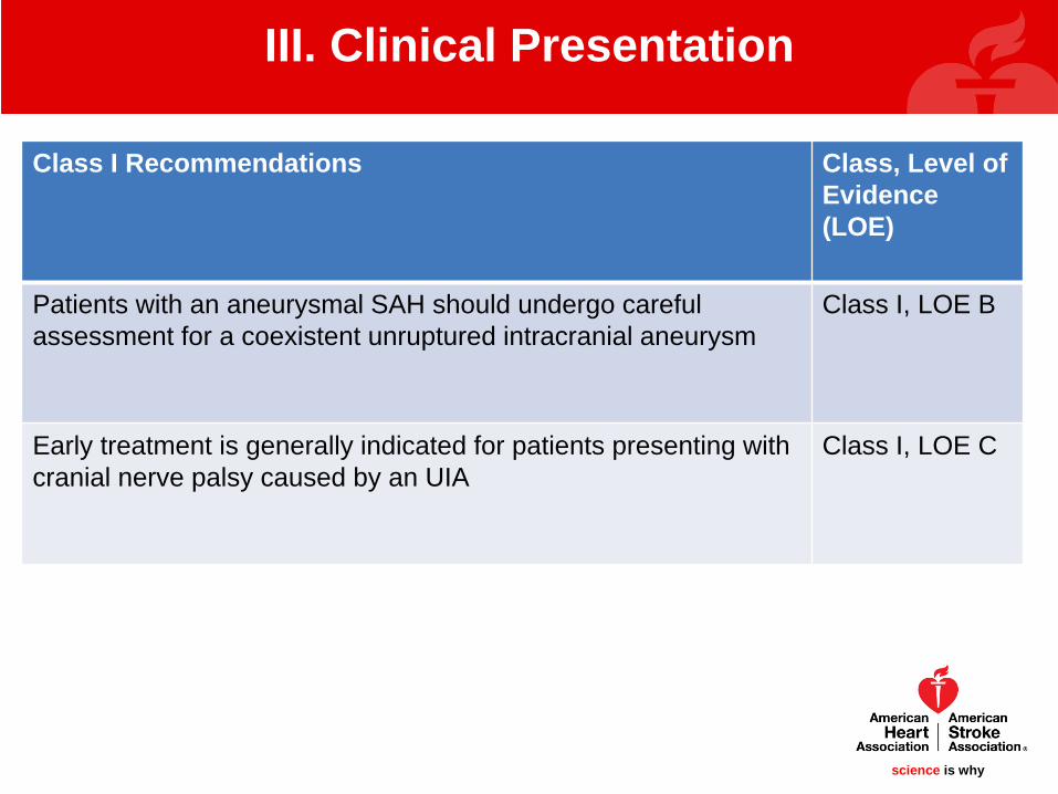
III. Clinical Presentation
Class I Recommendations Class, Level of Evidence(LOE)
Patients with an aneurysmal SAH should undergo careful assessment for a coexistent unruptured intracranial aneurysm
Class I, LOE B
Early treatment is generally indicated for patients presenting with cranial nerve palsy caused by an UIA
Class I, LOE C

science is whyscience is why
III. Clinical Presentation
Class II recommendations Class, Level of Evidence (LOE)
The effectiveness of the routine treatment of unruptured intracranial aneurysms for the prevention of ischemic cerebrovascular disease is uncertain.
Class IIb, LOE C

science is whyscience is why
Digital Subtraction Angiography• Gold standard for aneurysm diagnosis• Low risk of contrast and radiation exposure, cerebral infarction,
aneurysmal rupture, arterial injury– Permanent neurologic complication rate of 0.07%
CT Angiography• High sensitivity and specificity, especially for aneurysms >3mm• May not fully delineate details of vascular anatomy required for
treatment decisions• Risk of contrast and radiation exposure
MR Angiography• High sensitivity, especially for aneurysms >3mm• Can be done without contrast or radiation
IV. Diagnosis/Imaging

science is whyscience is why
IV. Diagnosis/Imaging
Follow-up imaging:• Untreated aneurysms should be followed intermittently
• Follow-up requirements for treated aneurysms are uncertain– Typically not performed if aneurysm is adequately clipped and
aneurysm is completely occluded– Typically performed 6-12 months after coiling
Further imaging depends on occlusion status and results of early follow-up imaging
• MRI is a reasonable option for follow-up of UIA given lack of radiation/contrast.

science is whyscience is why
IV. Diagnosis/Imaging
CT Angiography demonstrates a small UIA
Courtesy Michael T. Mullen MD – University of Pennsylvania

science is whyscience is why
IV. Diagnosis/Imaging
Class I Recommendations Class, Level of Evidence(LOE)
CTA and MRA are useful for detection and follow-up of UIA Class I, LOE B
Coiled aneurysms, especially those with wide neck or dome diameters or those that have residual filling should havefollow-up evaluation.
Class I, LOE B

science is whyscience is why
IV. Diagnosis/Imaging
Class II Recommendations Class, Level of Evidence (LOE)
DSA can be useful compared to noninvasive imaging for identification and evaluation of cerebral aneurysms if surgical or endovascular treatment Is being considered.
Class IIa, LOE B
DSA is reasonable as the most sensitive imaging for follow-up of treated aneurysms
Class IIa, LOE C
It is reasonable to perform MRA as an alternate for follow-up of treated aneurysms with DSA used as necessary when deciding on therapy
Class IIa, LOE C
The importance of surveillance imaging after endovascular treatment of UIAs lacking high risk features for recurrence remains unclear, but is probably indicated
Class IIa, LOE C
science is whyscience is why
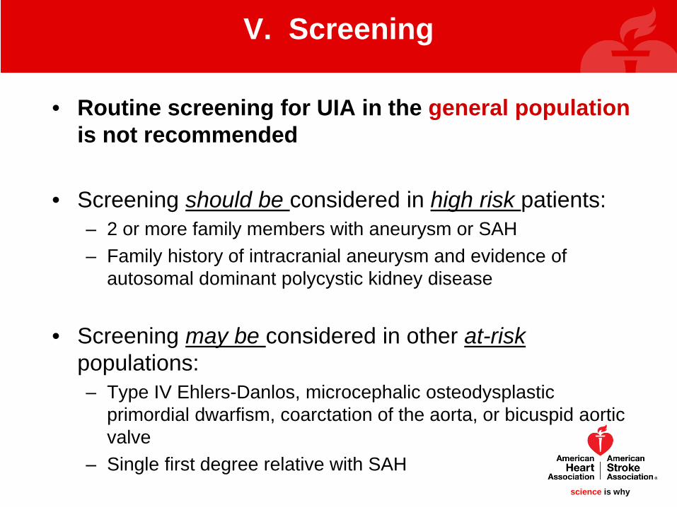
• Routine screening for UIA in the general population is not recommended
• Screening should be considered in high risk patients:– 2 or more family members with aneurysm or SAH– Family history of intracranial aneurysm and evidence of
autosomal dominant polycystic kidney disease
• Screening may be considered in other at-risk populations:– Type IV Ehlers-Danlos, microcephalic osteodysplastic
primordial dwarfism, coarctation of the aorta, or bicuspid aortic valve
– Single first degree relative with SAH
V. Screening
science is whyscience is why
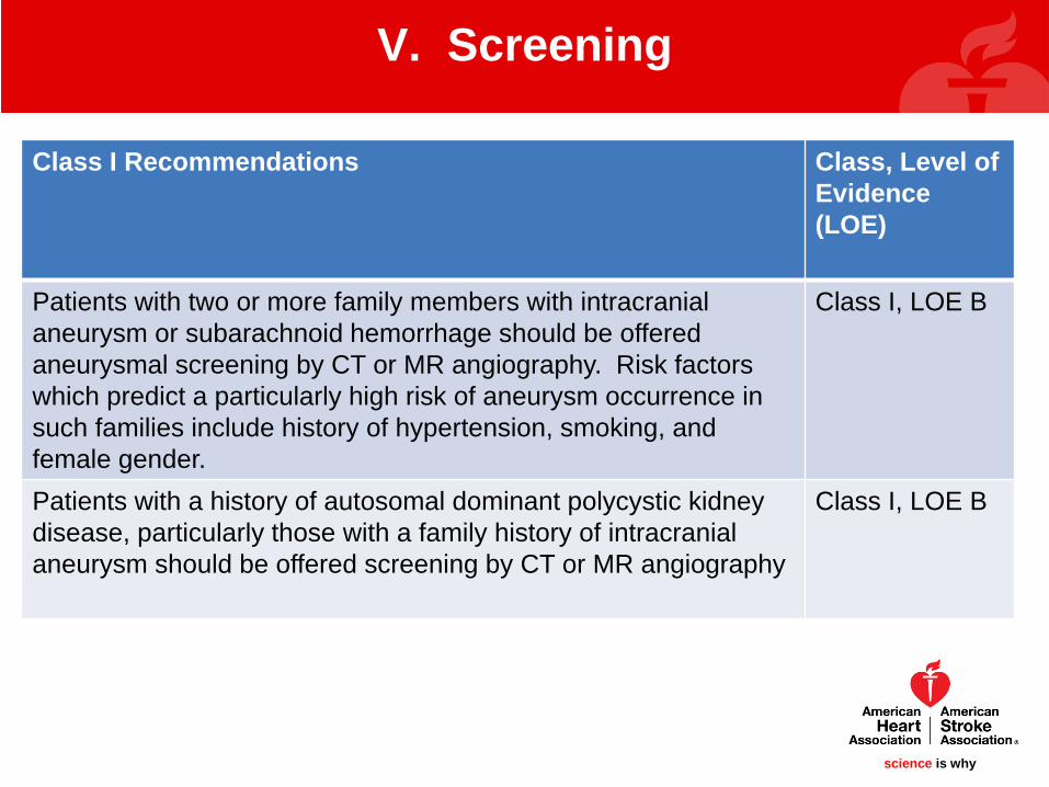
V. Screening
Class I Recommendations Class, Level of Evidence(LOE)
Patients with two or more family members with intracranial aneurysm or subarachnoid hemorrhage should be offered aneurysmal screening by CT or MR angiography. Risk factors which predict a particularly high risk of aneurysm occurrence in such families include history of hypertension, smoking, and female gender.
Class I, LOE B
Patients with a history of autosomal dominant polycystic kidney disease, particularly those with a family history of intracranial aneurysm should be offered screening by CT or MR angiography
Class I, LOE B

science is whyscience is why
V. Screening
Class II Recommendations Class, Level of Evidence (LOE)
It is reasonable to offer CT or MR angiography to patients with coarctation of the aorta and patients with microcephalic osteodysplastic primordial dwarfism.
Class IIa, LOE B

science is whyscience is why
Many studies have suggested that rupture risk is dependent on aneurysm size
Low rates of rupture in UIA <7-10mm
• Meta-analysis of 19 studies and 6556 UIA – Annual rupture rate for UIA <7mm was 0.4%– Most, 70%, of these subjects came from two international
studies (ISUIA)– Other data published since then have reported variable rupture
rates, from 0% to 1%
VI. Natural History of UIA
science is whyscience is why
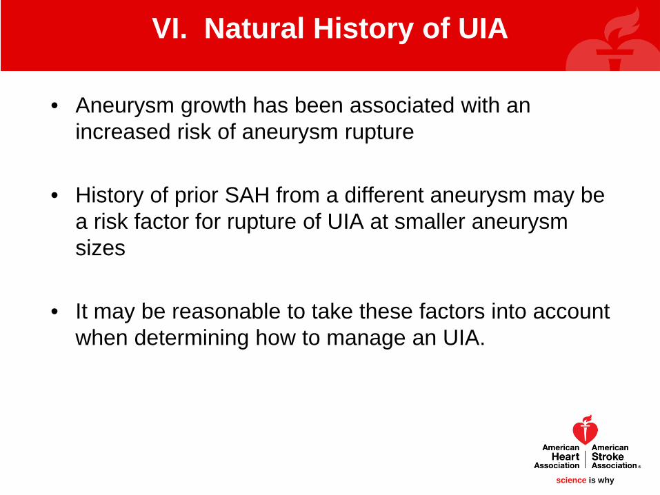
• Aneurysm growth has been associated with an increased risk of aneurysm rupture
• History of prior SAH from a different aneurysm may be a risk factor for rupture of UIA at smaller aneurysm sizes
• It may be reasonable to take these factors into account when determining how to manage an UIA.
VI. Natural History of UIA

science is whyscience is why
VI. Natural History of UIA
History of SAH from a different aneurysm may be a risk factor for rupture of other small UIA
Courtesy David K Kung MD – University of Pennsylvania

science is whyscience is why
VI. Natural History of UIA
Class I Recommendations Class, Level of Evidence(LOE)
Aneurysms with documented enlargement during follow-up should be offered treatment
Class I, LOE B

science is whyscience is why
VI. Natural History of UIA
Class II Recommendations Class, Level of Evidence (LOE)
Prior history of aneurysmal subarachnoid hemorrhage may be considered to be an independent risk factor for future hemorrhage secondary to a different, small, unruptured aneurysm
Class IIb, LOE B
Treatment of UIA in patients with a family history of intracranial aneurysm is reasonable even in aneurysms at smaller sizes
Class IIa, LOE B

science is whyscience is why
• Meta-analysis of 60 studied published between 1990-2011 found that up to 1 year after surgery:– Mortality rate of 1.7% – Morbidity, defined by lack of independence, was 5%– Cognitive dysfunction common– 85% of studies were poor quality based on STROBE criteria
• A single large prospective study (ISUIA) of 1917 patients found:– Mortality rate 2.3%– Morbidity (modified Rankin score>2 and/or impaired
cognition) in 12.1% – Cerebral infarction in 11%, hemorrhage in 4%
VII. Surgical Clipping of UIA
science is whyscience is why
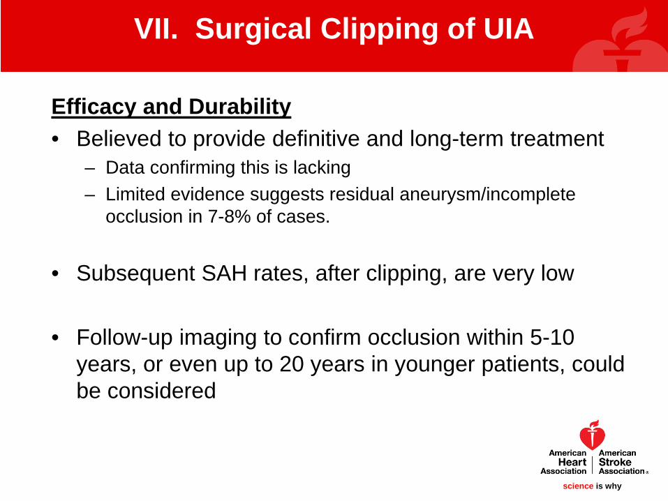
Efficacy and Durability• Believed to provide definitive and long-term treatment
– Data confirming this is lacking– Limited evidence suggests residual aneurysm/incomplete
occlusion in 7-8% of cases.
• Subsequent SAH rates, after clipping, are very low
• Follow-up imaging to confirm occlusion within 5-10 years, or even up to 20 years in younger patients, could be considered
VII. Surgical Clipping of UIA

science is whyscience is why
Risk Factors for Poor Outcome after Clipping
• Lesion: aneurysm size>10mm, posterior circulation location
• Patient: age>50, compressive symptoms, prior ischemic stroke.
• Surgeon/Hospital: surgeon inexperience, hospital volume <20 cases per year
VII. Surgical Clipping of UIA

science is why
VII. Surgical Clipping of UIA
Left PICA Aneurysm
Clip in Position Clipping completed
Courtesy David K Kung MD – University of Pennsylvania

science is whyscience is why
VII. Surgical Clipping of UIA
VII. Surgical Clipping of Unruptured Aneurysms
Class I Recommendations Class, Level of Evidence(LOE)
Patient age, family history of cerebral aneurysm rupture and aneurysm location, morphology, size, and documented growth should be taken into account when considering surgical clipping of an UIA.
Class I, LOE B
Imaging post surgical interventions, to document aneurysm obliteration, is recommended given the differential risk of growth and hemorrhage for completely versus incompletely obliterated aneurysms.
Class I, LOE B
Surgical treatment of UIA is recommended to be performed at higher volume centers (e.g. performing >20 cases annually).
Class I, LOE B

science is whyscience is why
VII. Surgical Clipping of UIA
VII. Surgical Clipping of Unruptured Aneurysms
Class II Recommendations Class, Level of Evidence(LOE)
Long-term follow-up imaging may be considered after surgical clipping given the combined risk of aneurysm recurrence and de novo aneurysm formation. Long-term follow-up may be particularly important for those aneurysms that are incompletely obliterated during initial treatment.
Class IIb, LOEB
The use of specialized intraoperative tools and techniques for avoiding vessel compromise or residual aneurysms may be considered to reduce the adverse outcomes with operative management of UIA.
Class IIb, LOE C
science is whyscience is why
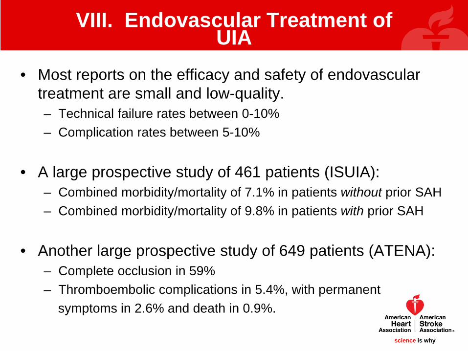
• Most reports on the efficacy and safety of endovascular treatment are small and low-quality.– Technical failure rates between 0-10%– Complication rates between 5-10%
• A large prospective study of 461 patients (ISUIA):– Combined morbidity/mortality of 7.1% in patients without prior SAH– Combined morbidity/mortality of 9.8% in patients with prior SAH
• Another large prospective study of 649 patients (ATENA):– Complete occlusion in 59%– Thromboembolic complications in 5.4%, with permanent
symptoms in 2.6% and death in 0.9%.
VIII. Endovascular Treatment of UIA

science is whyscience is why
Durability of aneurysm occlusion may be problematic• Meta-analysis of 71 studies reported aneurysm regrowth or
recurrence in 24.4% at up to 3.2 years of follow-up.
• Additional techniques to improve aneurysm occlusion:
– Coated Coils NOT beneficial.
– Balloon remodeling and stent-assisted occlusion Unknown benefit
– Liquid embolic agent (primarily in large or giant aneurysms) Complete occlusion rate of 79%, serious AE in 26.8%
VIII. Endovascular Treatment of UIA

science is whyscience is why
VIII. Endovascular Treatment of UIA
9.4mm para-ophthalmic aneurysm pre-treatment
Post coil-embolization
Courtesy Timothy R. Miller MD – University of Maryland

science is whyscience is why
VIII. Endovascular Treatment of UIA
Stent-assisted coil embolization of a carotid terminus aneurysm
Courtesy David K Kung MD – University of Pennsylvania
science is whyscience is why
VIII. Endovascular Treatment of UIA
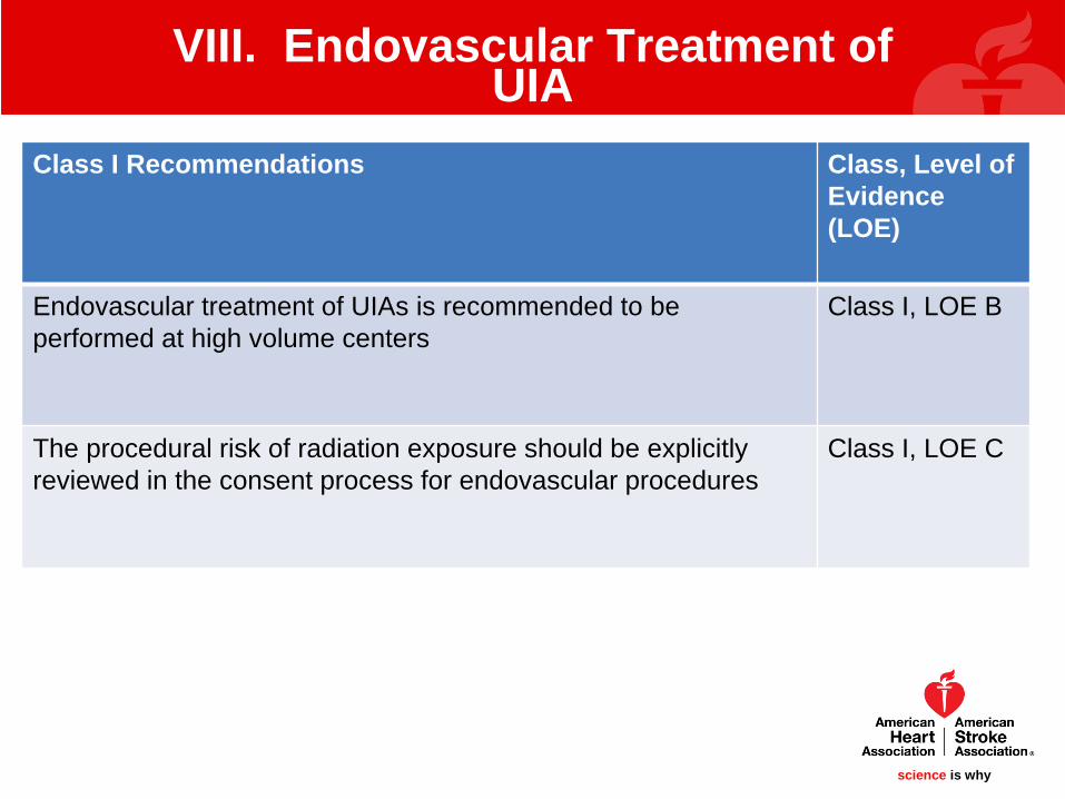
Class I Recommendations Class, Level of Evidence(LOE)
Endovascular treatment of UIAs is recommended to be performed at high volume centers
Class I, LOE B
The procedural risk of radiation exposure should be explicitly reviewed in the consent process for endovascular procedures
Class I, LOE C
science is whyscience is why
VIII. Endovascular Treatment of UIA
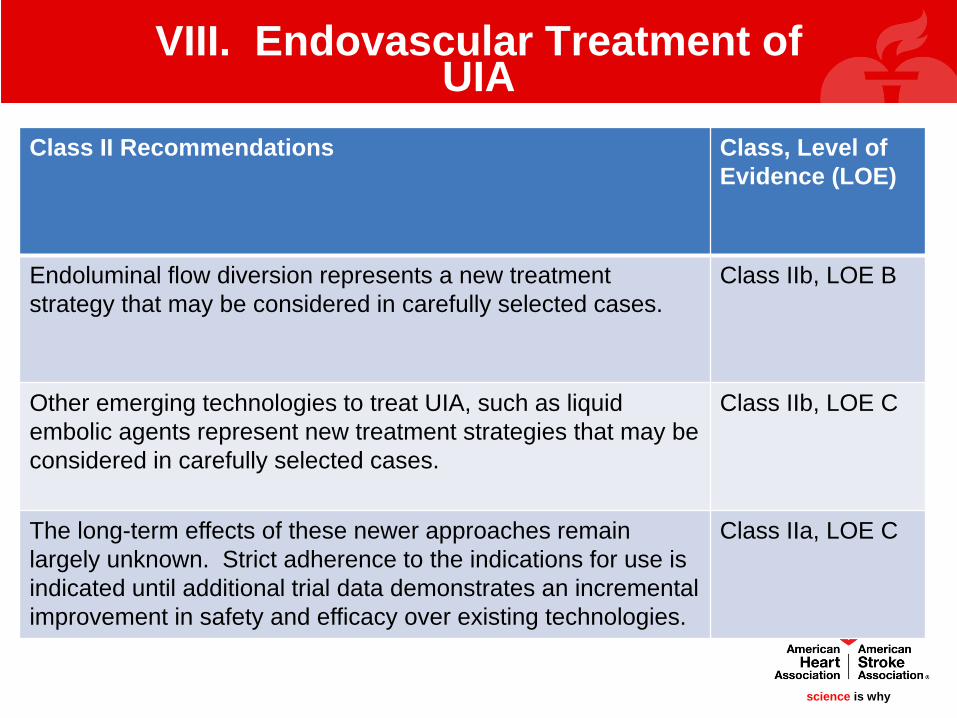
Class II Recommendations Class, Level of Evidence (LOE)
Endoluminal flow diversion represents a new treatment strategy that may be considered in carefully selected cases.
Class IIb, LOE B
Other emerging technologies to treat UIA, such as liquid embolic agents represent new treatment strategies that may be considered in carefully selected cases.
Class IIb, LOE C
The long-term effects of these newer approaches remain largely unknown. Strict adherence to the indications for use is indicated until additional trial data demonstrates an incremental improvement in safety and efficacy over existing technologies.
Class IIa, LOE C
science is whyscience is why
VIII. Endovascular Treatment of UIA
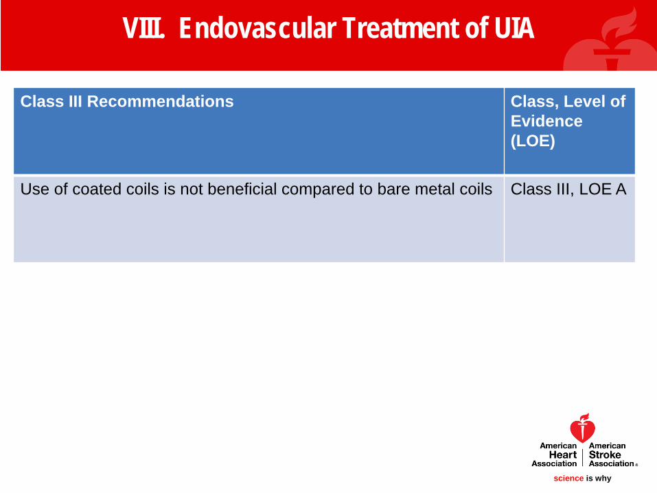
Class III Recommendations Class, Level of Evidence(LOE)
Use of coated coils is not beneficial compared to bare metal coils Class III, LOE A

science is whyscience is why
Compared to clipping, endovascular therapy has:
• Reduced morbidity/mortality and shorter length of stay– Even greater benefit in patients >60 years
• Lower rates of aneurysm occlusion and higher risk of recurrence
IX. Comparative Efficacy of Clipping vs. Coiling

science is whyscience is why
Special circumstances to consider:
• With current technology, microsurgical repair has an advantage for most middle cerebral artery aneurysms
• Endovascular treatment has an advantage for most basilar apex and vertebro-basilar aneurysms
IX. Comparative Efficacy of Clipping vs. Coiling
science is whyscience is why
IX. Comparative Efficacy of Clipping vs. Coiling
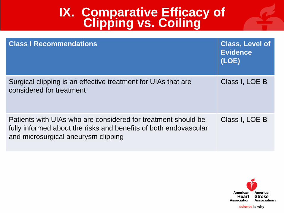
Class I Recommendations Class, Level of Evidence(LOE)
Surgical clipping is an effective treatment for UIAs that are considered for treatment
Class I, LOE B
Patients with UIAs who are considered for treatment should be fully informed about the risks and benefits of both endovascular and microsurgical aneurysm clipping
Class I, LOE B

science is whyscience is why
IX. Comparative Efficacy of Clipping vs. Coiling
Class II Recommendations Class, Level of Evidence (LOE)
Endovascular coiling is an effective treatment for selected UIAs that are considered for treatment
Class IIa, LOE B
Endovascular coiling is associated with a reduction in procedural morbidity and mortality over surgical clipping in selected cases but has an overall higher risk of recurrence.
Class IIb, LOE B
science is whyscience is why
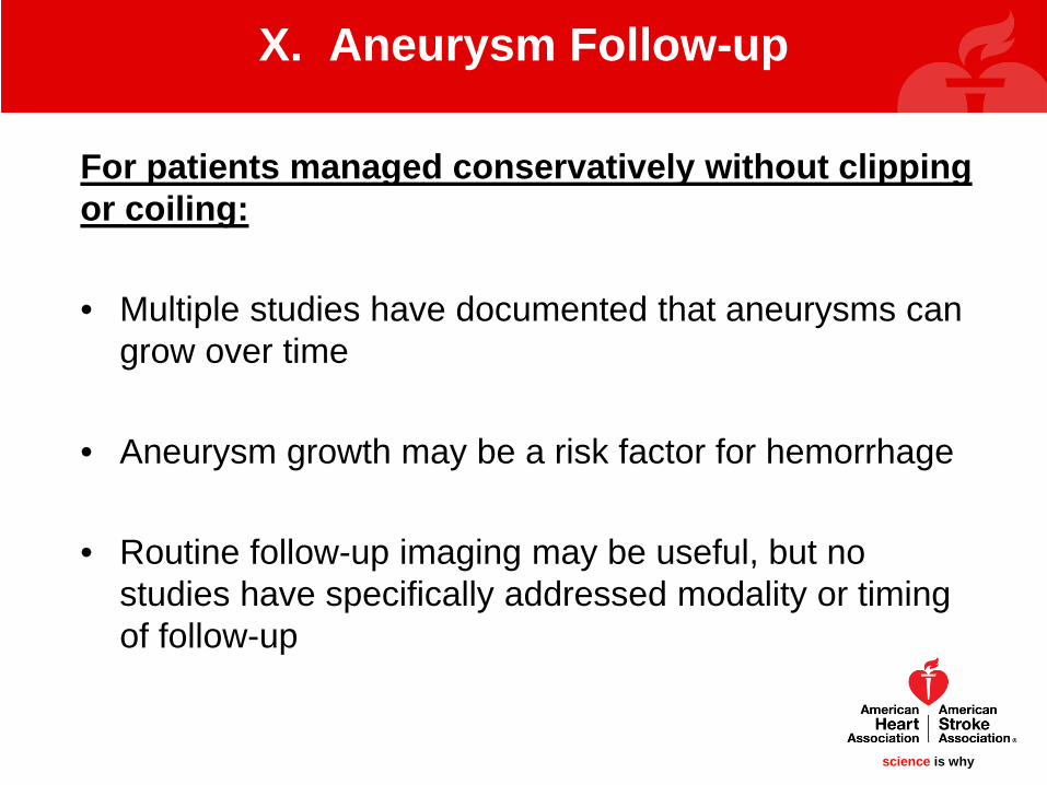
For patients managed conservatively without clipping or coiling:
• Multiple studies have documented that aneurysms can grow over time
• Aneurysm growth may be a risk factor for hemorrhage
• Routine follow-up imaging may be useful, but no studies have specifically addressed modality or timing of follow-up
X. Aneurysm Follow-up

science is whyscience is why
• Most studies indicate a preference for:– First follow-up imaging 6-12 months after initial discovery– Every 1-2 years once stability is documented.
• There may come a point in which the risks of intervention would be unacceptably high and follow-up imaging can be discontinued– Advanced age– Medical comorbidities that increase procedural risk or decrease
life expectancy
X. Aneurysm Follow-up

science is whyscience is why
• Both CTA and MRA have been utilized for aneurysm follow-up
• It is unknown if one modality is superior to another
• Because time of flight MRA does not require contrast and does not involve x-ray radiation, this may be the most appropriate first line method
X. Aneurysm Follow-up

science is whyscience is why
RightMCA Aneurysm
Time of Flight MRA may be ideal for routine follow-up of UIA because it does not require contrast and there is no
radiation exposure
X. Aneurysm Follow-up
Courtesy Michael Mullen MD – University of Pennsylvania

science is whyscience is why
X. Aneurysm Follow-up
Class I Recommendations Class, Level of Evidence(LOE)
For patients with unruptured intracranial aneurysms that are managed non-invasively without either surgical or endovascular intervention, radiographic follow-up with magnetic resonance angiography (MRA) or computed tomographic angiography (CTA) at regular intervals is indicated. The optimal interval and duration of recommended follow-up are uncertain.
Class I, LOE B

science is whyscience is why
X. Aneurysm Follow-up
Class II Recommendations Class, Level of Evidence (LOE)
For patients with UIA that are managed non-invasively without either surgical or endovascular intervention, a first follow-up study at 6-12 months after initial discovery, followed by subsequent yearly or every other year follow-up may be reasonable.
Class IIb, LOE C
For patients with UIA that are managed non-invasively and in whom there are no contra-indications to MR imaging, it may be reasonable to consider TOF MRA in favor of CTA for repeated long term follow-up.
Class IIb, LOE C

science is whyscience is why
• UIAs are common. SAH is a feared complication of UIA, but the vast majority of UIAs (>99%) will not rupture
• Modifiable risk factors for aneurysms are hypertension and cigarette smoking
• Larger aneurysm size portends a higher risk of rupture
• Growth of UIA is associated with rupture and couldbe an indication for repair
XI. Conclusion
science is whyscience is why
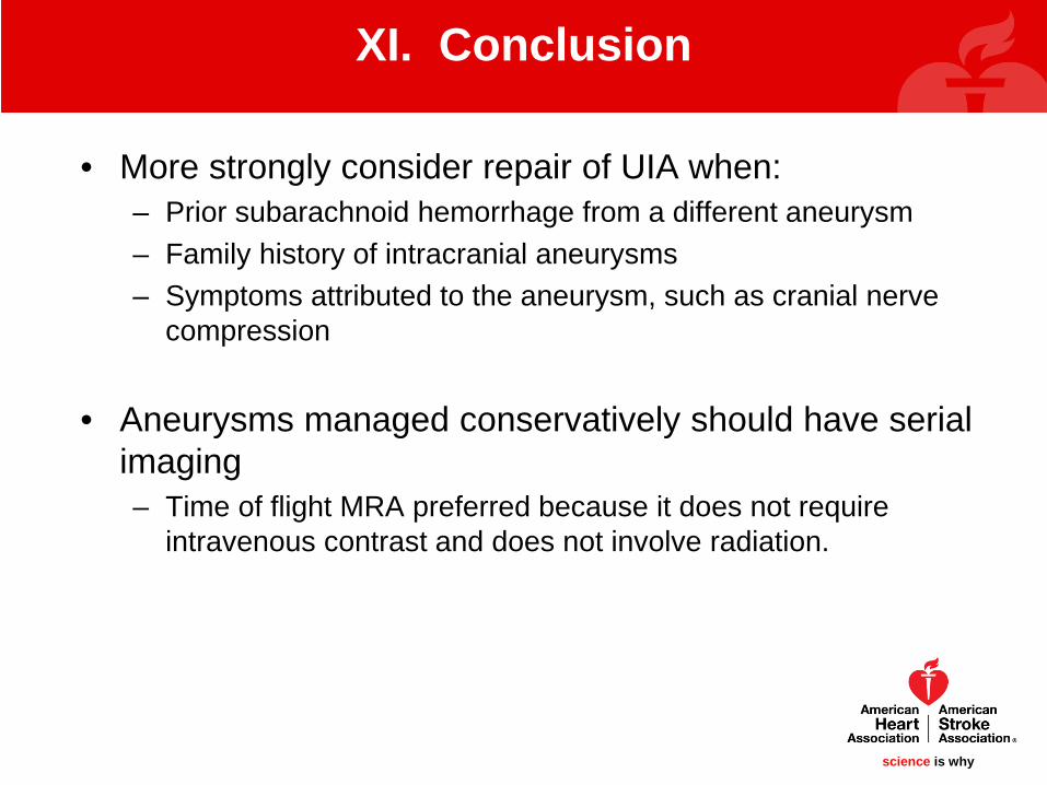
• More strongly consider repair of UIA when:– Prior subarachnoid hemorrhage from a different aneurysm– Family history of intracranial aneurysms– Symptoms attributed to the aneurysm, such as cranial nerve
compression
• Aneurysms managed conservatively should have serial imaging – Time of flight MRA preferred because it does not require
intravenous contrast and does not involve radiation.
XI. Conclusion
science is whyscience is why
• When treatment is elected digital subtraction angiography is indicated to:– Plan repair– Define whether the aneurysm has been completely occluded
post-procedure.
• It is uncertain if any populations should undergo routine screening for asymptomatic UIA. – Two populations to consider:
Autosomal dominant polycystic kidney disease Strong family history of aneurysm or subarachnoid hemorrhage.
XI. Conclusion
science is whyscience is why
• The results of UIA treatment appear to be inferior in low volume centers (<20 cases per year)
• Surgical repair is generally associated with:– higher rates of aneurysm obliteration – lower rates of recurrence– higher perioperative morbidity
• Early documentation of the degree of aneurysm obliteration is necessary following any repair to guide the frequency of further follow-up imaging
XI. Conclusion
science is whyscience is why
XI. Conclusion
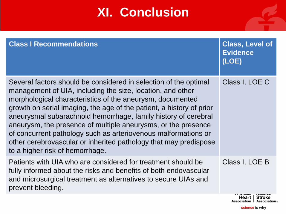
Class I Recommendations Class, Level of Evidence(LOE)
Several factors should be considered in selection of the optimal management of UIA, including the size, location, and other morphological characteristics of the aneurysm, documented growth on serial imaging, the age of the patient, a history of prior aneurysmal subarachnoid hemorrhage, family history of cerebral aneurysm, the presence of multiple aneurysms, or the presence of concurrent pathology such as arteriovenous malformations or other cerebrovascular or inherited pathology that may predispose to a higher risk of hemorrhage.
Class I, LOE C
Patients with UIA who are considered for treatment should be fully informed about the risks and benefits of both endovascular and microsurgical treatment as alternatives to secure UIAs and prevent bleeding.
Class I, LOE B

science is whyscience is why
XI. Conclusion
Class I Recommendations (Continued) Class, Level of Evidence(LOE)
The results of UIA treatment are inferior at low volume centers and hence treatment is recommended to be performed at higher volume centers.
Class I, LOE B

science is whyscience is why
XI. Conclusion
Class II Recommendations Class, Level of Evidence (LOE)
Data from prospective and retrospective studies from multiplenational and international investigations indicate that microsurgical clip ligation may confer more durable protection against aneurysm regrowth, but coil embolization may be superior to surgical clipping with respect to procedural morbidity and mortality, length of stay, and hospital costs, so it may be reasonable to choose endovascular therapy over surgical clipping in the treatment of select UIAs, particularly in cases where surgical morbidity is high, such as the basilar apex and in the elderly.
Class IIb, LOE B
The treatment risk of patients with UIAs is related to advancing age, medical co-morbidities, and aneurysm location and size, so in older patients (age>65) and those with associated medical morbidities with small asymptomatic UIAs with low hemorrhage risk by location, size, morphology, family history and other relevant factors, observation is a reasonable
Class IIa, LOE B

science is whyscience is why