agglutination reaction immunology tutorial mlt402 prepare by : 1) saad dahesh ali al-amri 2) salem...
TRANSCRIPT

Agglutination reactionimmunology Tutorial
MLT402PREPARE BY :
1) Saad Dahesh Ali Al-Amri 2) Salem Dahesh Ali Al-Amri 3) Saleh Al-Juhani
Mr. Mohamed AL-Khatatneh

Agglutination
Is the clumping of antibody–antigen complex.
Reaction occurs between insoluble
antigen and appropriate antibody.
The reaction will result in forming visible aggregates or agglutinate
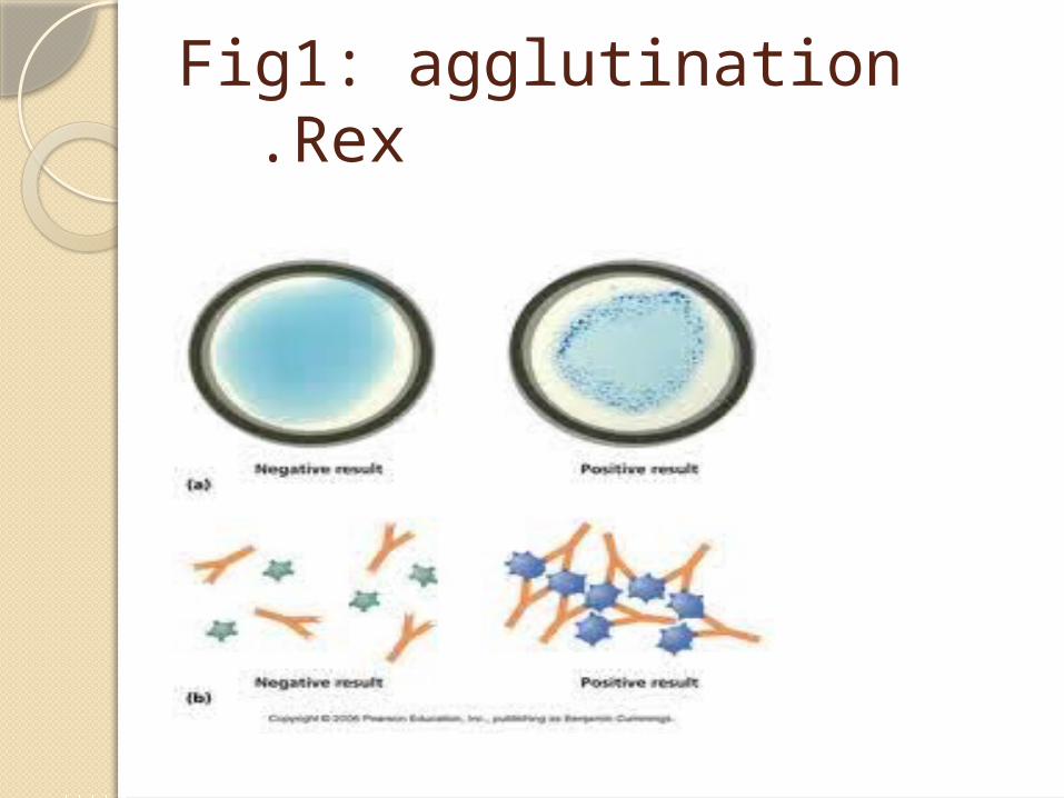
Fig1: agglutination Rex .
Stages of agglutination reactionPhase oneAntibody reacts with single
antigenic determinants on or close to particle surface.
It is a rapid reaction.
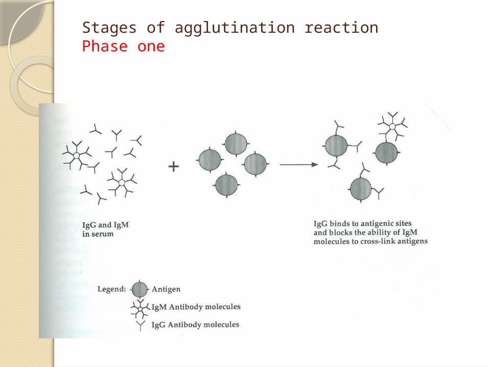
Stages of agglutination reactionPhase one
Stages of agglutination reaction Secondary phase
A single antibody molecule binds to antigenic determinants on adjacent particles.
The visible reaction occur under appropriate conditions
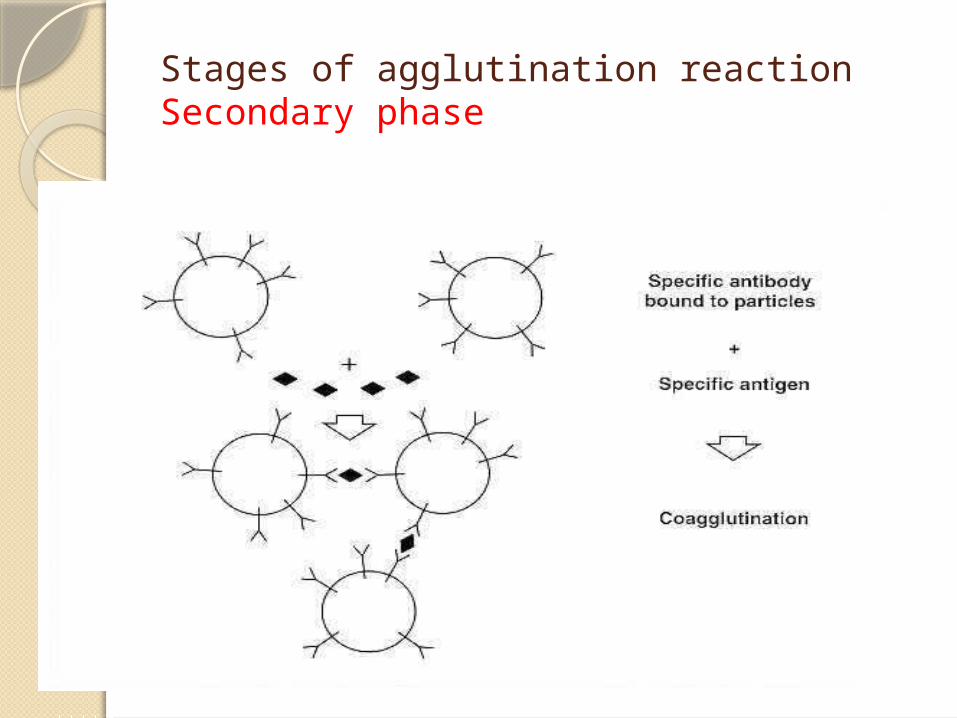
Stages of agglutination reaction Secondary phase
Factors affecting the agglutination reaction in vitro:
1. Antigen to antibody ratio: The ratio between antigen and antibody influences
the detection of antigen-antibody complexes.Antigen or antibody excess make invisible
reaction.◦ Prozone phenomenon (antibody excess):
There are too many antibodies.no agglutination appears (false-negative reactions).
◦Zone of equivalence: Antibodies and antigens are present in an optimum
ratio. This leads to cross-linkages between acells or particles,
so agglutination appers (positive reaction)
Post-zone phenomenon (Antigen excess):
There are too many antigens
These false-negative reactions can be detected by repeating the test at a higher dilution of sample, which reduces the antigen or antibody concentration into the range that produces visible agglutination.
Factors affecting the agglutination reaction in vitro. Cont:

2. Number of Antigen Sites:The more antigen sites on a cell
result in more cross-linkages between cells.
These cross-linkages result in more visible agglutination.
Factors affecting the agglutination reaction in vitro. Cont:

3. Size and Structure of the Antibody: Larger antibody causes more cross-
linkages between different cells.The larger antibodies (IgM) can reach between more antigen sites on different
cells and therefore causing stronger agglutination reactions.
4. Distance between cells: Centrifugation of the cells attempts to
bring the cells closer together, so enhance agglutination.
Factors affecting the agglutination reaction in vitro. Cont:
Classification of agglutination reactions
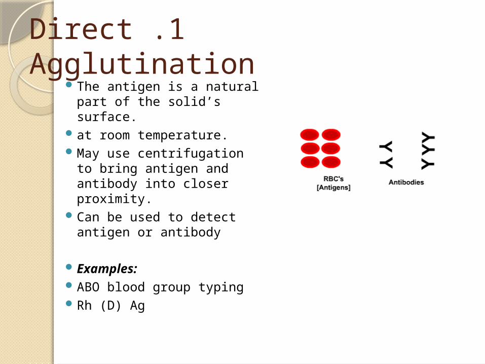
1 .Direct Agglutination The antigen is a natural
part of the solid’s surface. at room temperature. May use centrifugation to
bring antigen and antibody into closer proximity.
Can be used to detect antigen or antibody
Examples: ABO blood group typing Rh (D) Ag
2 .Passive Agglutination (indirect) Passive Agglutination is a very sensitive method for
antibody detection. RBC, bacterial cells or inert particles such latex can
be used as a carrier for antigens.
This makes the reaction more visible. latex -------------- latex agg. RBC ----------------passive heamagg.
If Antibody is attached to the particulate carrier called Reverse Passive Agglutination

Passive Agglutination. Con’tExample:
1) Rheumatoid factor detection.
2) C-Reactive Protein detection (CRP)

Passive Agglutination
Antigens on a carrier molecule, such as latex, combine with patient’s sample for antibody detection.
Reverse Passive Agglutination
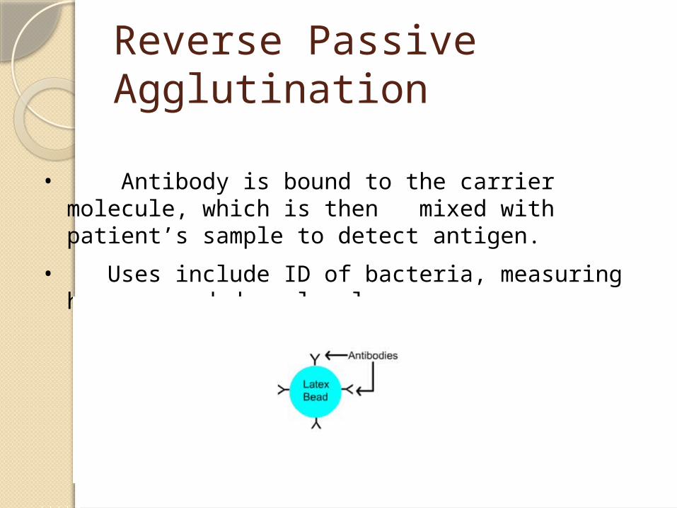
• Antibody is bound to the carrier molecule, which is then mixed with patient’s sample to detect antigen.
• Uses include ID of bacteria, measuring hormone and drug levels.

Agglutination reaction
Glass slides TubesMicrotiter plates
Advantages of agglutination methods
ease of performance.speed of performance, usually
requiring few minutes.high degree of sensitivity.
Disadvantages of agglutination methods
the reaction are only semiquantitative.
the occurrence of the prozone phenomenon, in which agglutination is inhibited by extreme antibody.
Limitations:1) The technique for shaking the tubes to
detect agglutination is critical. Harsh shaking may cause weak agglutinates.
2) Test should be performed regularly, 3) Dispensing incorrect quantities of diluent or
reagent or transferring more or less than the required amount of diluted serum will affect the outcome of this test .
False positive and false negative agglutination:
False positives w/ agglutination : 1) contaminated glassware 2) 2) overcentrifugation of cells 3) 3) autoagglutinationFalse negatives w/ agglutination : 1) improper condition for test:
specimen not prop prepared 2) expired/improperly stored reagents3) too much Ab or Ag

Materials required:
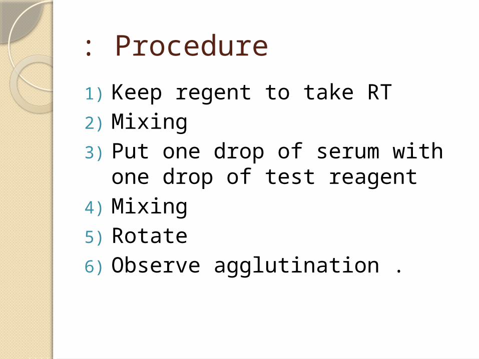
Procedure: 1) Keep regent to take RT2) Mixing3) Put one drop of serum with one
drop of test reagent 4) Mixing5) Rotate 6) Observe agglutination .

Factors Affecting Agglutination
.1Class of Antibody.
.2Charge of the Carrier Particle.
.3Number of Antigenic Sites.
.4Concentration Of Reaction.
.5Environmental Factor. amna alotiby 2008-2009
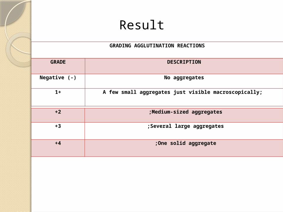
GRADING AGGLUTINATION REACTIONS
GRADE DESCRIPTION
Negative (-) No aggregates
1+ A few small aggregates just visible macroscopically;
2+ Medium-sized aggregates ;
3+ Several large aggregates ;
4+ One solid aggregate ;
Result
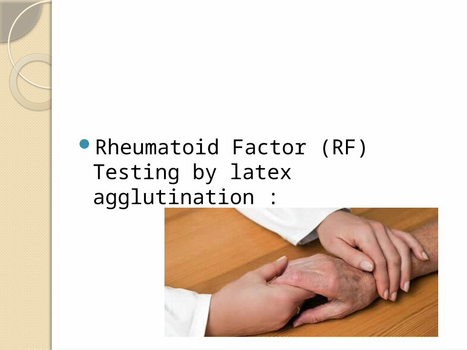
Rheumatoid Factor (RF) Testing by latex agglutination :
Introduction RF:
Rheumatoid arthritis (RA) is a chronic inflammatory disease affecting primarily the joints and tissues. For many years it has been known that several abnormal proteins circulate in the blood of patients with RA. These proteins, because disease, became known as rheumatoid factor (RF).
Principle RF test : Rheumatoid factor (RF) is an anti-antibody,
which in-vitro, is detected by its ability to agglutinate latex particles (or red blood cells) coated with human IgG. RF in patient sample, if present, will attach to the IgG coating the latex particles. Agglutination of the latex particles is a positive result indicating the presence of RF.

Materials :1. Rheumatoid factor test kit(s). 2. Patient and control serum
specimens. 3. Timer 4. Other materials as directed by
reagent product insert(s).
Procedure :See reagent product insert(s)
Interpretation &Expected Results :
Interpretation : Agglutination of latex particles is considered a positive reaction, indicating the presence of rheumatoid factor at detectable level..
Expected Results : Although the diagnosis of rheumatoid arthritis is based largely on clinical findings .
Limitations :1). RF is not detected in all patients
diagnosed with RA.2) Some products may produce
questionable results from hemolyzed, lipemic or contaminated specimens.
3) Avoid contamination of reagent or dropper.
ASO Latex Test Introduction : ASO Latex Test is a
rapid latex agglutination test for the qualitative and semi-quantitative determination of antistreptolysinO antibodies (ASO) in serum.
PRINCIPLES : The ASO Latex Test is a stabilised
buffered suspension of polystyrene latex particles that have been coated with Streptolysin O. When the latex reagent is mixed with a serum containing ASO, agglutination occurs. The sensitivity of the latex reagent will yield agglutination when the level of ASO is greater than 200 IU/ml, a level determined to be indicative of disease by epidemiological and clinical studies.
MATERIALS: 1. ASO Latex ( kit ) 2. ASO Positive Control 3. ASO Negative Control 4. Glass Reaction Slide. 5. Disposable wooden stick
PROCEDURE1) Take test reagents and samples to room
temperature. 2. Use pipette to take drop of sample into its
identified circle of the slide. Retain each pipette for mixing.
3. drop of positive and negative control into its identified circle.
4. Mix the ASO latex reagent by gently shaking. Add one drop of reagent to each control and sample.
5. Using the wooden stirrer thoroughly mix each sample with reagent within the full area of the circle. Discard the disposable stirrer.
6. Wait two (2) minutes and observe for agglutination under a high light.
7. Record results.

RESULTStest sample is considered to
contain ASO antibodies when agglutination is observed when compared to the result of the negative control.
LIMITATIONS OF THE PROCEDURESerum samples showing gross hemolysis,
lipemia should not be used .The test reaction must be read immediately
following the two (2) minute . Only serum specimens should be used. Do
not use plasma samples as they could cause non-specific agglutination of the latex.

Reference : MLAB 1235 Immunology/Serologyhttp://quizlet.com/7317754/immu
nology-agglutination-flash-cards/
. Rantz LD, DiCaprio JM, Randall E. Am .J. Med. Sci., 24, 1952. 2. Halbert,SP. Ann. N.Y. Acad. Sci., 103, 1027:1051; 1963