agglutination of stored erythrocytes by a human
TRANSCRIPT
Journal of Clinical InvestigationVol. 42, No. 11, 1963
AGGLUTINATION OF STORED ERYTHROCYTES BY A HUMANSERUM. CHARACTERIZATION OF THE SERUM FACTOR
AND ERYTHROCYTE CHANGES *
By F. L. OZER AND H. CHAPLIN, JR.
(From the Departments of Medicine and Preventive M11edicine, Washingtoni University Schoolof Medicine, St. Louis, Mo.)
(Submitted for publication May 8, 1963; accepted July 22, 1963)
There have been four previous reports of hu-man sera causing agglutination of stored but not offresh red cells (1-4). The sera are of special in-terest for two reasons. Since the agglutinins areactive against the patients' own stored cells aswell as against cells from other donors, one mustconsider whether such patients represent ex-amples of autoimmune disease. Of more im-mediate practical importance, the sera provide anew means for studying the red-cell storage de-fect, since the agglutinins detect in the erythro-cyte membrane a hitherto unrecognized altera-tion that progresses during in vitro storage con-currently with deterioration in cellular metabo-lism.
Characterization of the previously reportedstored-cell agglutinins has been limited. Thepresent communication describes detailed studiesof a serum causing strong agglutination of storedhuman red cells. The serum factor responsiblefor the agglutination was shown to be a gammamacroglobulin. Studies of red cells during stor-age in vitro indicate that development of agglutin-ability is closely related to the alterations inerythrocyte glucose metabolism occurring duringstorage. Also included are the results of sixteenstudies of erythrocyte survival in vivo that aredesigned to examine the relationship of agglutin-ability to nonviability of the stored cells.
METHODS
The patient was a 71-year-old woman whose chiefcomplaints were abdominal swelling and progressiveweakness. The principal positive physical findings werepallor and hepatosplenomegaly. Laboratory findings ofspecial interest were: hemoglobin, 9.7 g per 100 ml;reticulocytes, 4.2%o; serum bilirubin concentration, nor-
* Work supported by U. S. Public Health Service re-search grant G-2918 (C5) from the National CancerInstitute, Bethesda, Md.
mal; total serum protein concentration, 7 g per 100 ml(albumin, 2.7 g, globulin, 4.3 g). Survival in vivo ofCr51-labeled, f resh, compatible, red cells of normal do-nors was moderately severely reduced, judged by red-cell life-span (Cr51 tt, about 8 days) based on dailyblood sampling for 1 week. Exploratory laparotomyrevealed moderate hepatic enlargement, diffuse mesen-teric and retroperitoneal lymph-node enlargement, andmassive splenomegaly. Splenectomy was performed,and the patient's postoperative course was uneventful.Two units of blood were transfused before and two ad-ditional units during surgery. Although the patient wasimproved at the time of discharge and required no fur-ther transfusions, she died at home 7 months later ofundetermined cause; necroscopy was not performed.The spleen removed at operation weighed 2,050 g andshowed hyperplasia of the white pulp, with impairedfollicle formation; each Malpighian corpuscle was sur-rounded by a margin of hyperplastic lymphoid cells.Lymph nodes revealed intact architecture, but were filledout with lymphoid elements without follicle formation.A liver sample from biopsy was not remarkable, andaspirated bone marrow revealed only generalized hyper-plasia. A specific pathological diagnosis was not made.
Stndies of the serum factorThe patient's serum was obtained from clotted or de-
fibrinated blood; plasma was obtained in acid-citrate dex-trose anticoagulant solution.' Serum and plasma sam-ples were stored frozen at -20° C until used. Cold-water-precipitable protein was prepared by diluting thepatient's serum with 10 vol of cold, demineralized wa-ter; after three washings, the precipitate was dissolvedin isotonic phosphate buffered saline at pH 7.4. Addi-tional pathologic sera were obtained from twelve pa-tients with macroglobulinemia, two patients with 02Amyeloma, and one patient with nonspecific hyperglobu-linemia.2 Normal human macroglobulin was preparedfrom pooled plasma as described by Franklin and Stan-worth (5). Paper electrophoresis was performed in aSpinco model R cell, with barbital buffer at pH 8.6,ionic strength 0.1. Immunoelectrophoresis was by the
1 ACD, formula A, National Institutes of Health,Bethesda, Md.
2 Several of these sera were made available throughthe courtesy of Dr. H. G. Kunkel, the RockefellerInstitute, New York, N. Y.
1735
F. L. OZER AND H. CHAPLIN, JR.
method of Scheidegger as described by Wieme (6).Analytical ultracentrifugation was carried out at 59,000rpm at 20° C in a Spinco model E ultracentrifuge.Sucrose gradient density ultracentrifugation was per-formed as described by Kunkel (7). Column chroma-tography was carried out with DEAE cellulose 3 (1ml serum to 6 g DEAE cellulose) by stepwise elutionwith phosphate buffers-0.0175 M at pH 6.3, 0.1 M atpH 5.9, and 0.4 M phosphate containing 0.2 M sodiumchloride at pH 5.2-and with Sephadex G-200 4 (1 mlserum separated on a column 2.0 x 40 cm, with isotonicbuffered saline at pH 7.4 as the equilibrating and elut-ing solution). p-Hydroxymercuribenzoate5 (PMB),N-ethylmaleimide 5 (NEM), dl-penicillamine,5 glutathi-one 5 (GSH), oxidized glutathione5 (GSSG) and mer-captopyridoxine 6 were obtained from manufacturers.The compounds were added to serum (diluted 1/10 un-less otherwise indicated) after being dissolved in iso-tonic buffered saline. Incubation was at 370 C or roomtemperature for 1 to 2 hours. Where indicated, afterincubation, a sample of the treated serum was dialyzedfor 16 to 24 hours at 4° C against 1,000 vol of isotonicbuffered saline.For agglutination tests, dilutions of serum were made
in isotonic saline. The test cells were normal, group 0red cells that had been stored sterile at 40 C in thepresence of trisodium citrate, balanced oxalate, or acid-citrate detrose anticoagulants until they exhibited 4 + ag-glutination (i.e., > 90% of the cells agglutinated) whenexposed to the patient's serum diluted 1/100. One volumeof 5% red-cell suspension was well mixed on an opaqueglass plate with an equal volume of diluted serum. Theplate was gently rocked for exactly 3 minutes, and thereactions were recorded by gross inspection; on occa-sion, microscopic examination was also performed.
In vitro studies of erythrocyte changes
Red cells. Blood from normal donors and from 30patients with a variety of hematologic disorders wascollected in sterile bottles containing ACD solution, bal-anced sodium and potassium oxalate, trisodium citrate,heparin, or disodium EDTA anticoagulants. In addi-tion, red cells from 100 sterile clotted samples in blood-bank pilot tubing were examined, including one sample7from an I-negative donor (8). Most of the metabolicstudies were performed on red cells from a singlehealthy donor whose erythrocytes were known to ex-hibit normal glucose-6-phosphate dehydrogenase (G6PD)activity and normal proportions of A, and A2 hemo-globin. Animal bloods were obtained aseptically fromrats, rabbits, and dogs. Preparation of eluates fromred cells and red-cell ghosts by heating at 560 C wasperformed as described by Chaplin and Cassell (9). De-
3 Bio-Rad Laboratories, Richmond, Calif.4A. B. Pharmacia, Uppsala, Sweden.5 Sigma Chemical Co., St. Louis, Mo.6 Supplied by Merck & Co., Darmstad, Germany.7Provided through the courtesy of Dr. Richard
Rosenfield.
pletion of erythrocyte membrane lipids by treatmentwith alumina was carried out as described by Lovelock(10). Sickling was induced in fresh homozygous (SS)sickle-cell blood by equilibration with a 95% nitrogen:5% carbon dioxide gas mixture; reversal of sicklingwas accomplished by re-equilibration with a 97% oxy-gen: 3% nitrogen mixture as described by Anderson andChaplin (11). Fresh red cells were exposed to theenzymes trypsin, papain, ficin, and bromelin as describedby Mollison (12). Crude neuraminidase was added invarious concentrations to buffered saline suspensions ofthrice-washed fresh red cells and incubated at 37° C forup to 2 hours.
In metabolic studies, red cells from fresh blood col-lected aseptically in ACD anticoagulant were washedthree times in 10 vol of glucose-free phosphate-bufferedisotonic saline at pH 7.4, with removal of as much buffycoat as possible after each centrifugation. 2-Desoxy-D-glucose,5 Na arsenite 5 (as Na m-arsenite), Na arsenate 5
(as dibasic salt), Na monoiodoacetate, Na fluoride,5PMB,5 NEM,5 phlorizin,5 adenosine,5 adenine,5 inosine,5and methylene blue6 (MB) were obtained from manu-facturers. Adenosine and NEM were dissolved in di-lute HCl and neutralized to pH 7.4 by NaOH. Theother reagents were dissolved directly in buffered iso-tonic saline. The thrice-washed red cells were made toa 50% suspension in buffered saline containing the re-agents above, and the mixture was incubated in a Dub-noff metabolic shaker at 60 oscillations per minute and370 C for 1 to 2 hours. After incubation, the red cellswere washed once with 10 vol of buffered saline, and a5% suspension was immediately tested for agglutinationas previously described.
In vivo studies of erythrocyte changes
Transfusion studies were performed in thirteen vol-unteers. Six were in normal health; four were hemato-logically normal hospital patients; of the remaining threepatients, one had acute myelomonocytic leukemia, one,sickle-cell trait, and one, chronic myelocytic leukemia.Altogether, 9 U of outdated blood8 stored in ACDanticoagulant (mean length of storage, 30.5 days; range,22 to 38 days) were employed in 16 transfusion studiesto permit comparison of the survival in vivo of: a) un-treated stored blood, b) unagglutinable cells recoveredafter removal of cells agglutinated by macroglobulinemic(MG) serum, and c) unagglutinable cells recoveredfrom stored blood incubated with adenosine (3 mmolesper ml cells) for 1 hour either before or after the ad-dition of MG serum. Four of the 9 U were used forsingle studies of the survival of unagglutinable cells;the remaining 5 U were used for multiple studies of un-treated cells (controls) and unagglutinable cells re-covered after exposure to MG serum.
Separation of unagglutinable cells before transfusion.A 5% suspension of stored cells in saline was incubated
8 Obtained from the Barnes Hospital Blood Bank orfrom the St. Louis Chapter of the American NationalRed Cross.
1736
AGGLUTINATION OF STORED ERYTHROCYTES BY HUMAN SERUM
for 10 minutes at room temperature with an appropriateamount of sterile MG serum previously heated at 600 Cfor 10 hours to eliminate the risk of transmission of se-rum hepatitis. Separation of the unagglutinated cellsto be used for transfusion was accomplished either byaspirating the cells remaining in suspension after the ag-glutinates had been allowed to settle as sediment for 10minutes, or by removing the agglutinates by passagethrough a Millipore filter (pore size, 10 /)).9 The per-centage of the original cells remaining in the final sus-pension used for transfusion was estimated from dupli-cate measurements of oxyhemoglobin concentration andof turbidity of samples removed from the cell suspensionbefore addition of the MG serum, and again after sepa-ration of the agglutinates, with suitable correction fordilution by the suspending medium. Comparable "re-coveries" were obtained from both separation proce-dures, and averaged 50 to 60%o of the original cellspresent. The "loss" (40 to 50%9o) represents the com-bined losses from the agglutinates removed as well asresidual unagglutinated cells remaining on the filters,glassware, etc. About 30% of the cells in the storedblood was estimated to be removed as agglutinates.Microscopic examination of the cell suspensions em-ployed for transfusion revealed only occasional tinyagglutinates (never representing> 2% of the total cellspresent).
Red-cell survival in vivo was measured by the Cr`1method of Ebaugh, Emerson, and Ross (13). The la-beled red cells were washed in at least 10 vol of salinebefore inj ection, and measurement of radioactivity inthe suspending medium confirmed that more than 99%9of injected Cr`1 was erythrocyte bound. The recipient'sblood volume was measured by the simultaneous injec-tion of Evans' blue dye. Blood samples were obtained15, 30, and 45 minutes, and 1, 6, 24, and 48 hours afterinjection; additional samples were obtained at intervalsof 3 to 5 days over the ensuing 2 to 4 weeks in mostinstances. An estimate of the expected immediate 100%,survival was determined from the known volume of Cr"-labeled cells injected and the recipient's blood volume.Subsequent survival was determined from the radio-activity present per milliliter of whole blood and ex-pressed as a percentage of the expected 100% survival.Radioactivity in plasma separate from all samplesdrawn within the first hour after transfusion never ex-ceeded 1% of the activity present in the whole bloodsamples.
Relationship of agglutinability to chrontological red-cellage. Fe", 19.5 /Ac, incubated for 45 minutes at 370 C in25 ml of autogenous plasma, was injected intravenouslyinto a normal volunteer. Sterile blood samples werecollected in ACD anticoagulant 19, 22, 42, 67, 78, and110 days after injection and stored until about 30%o ofthe cells became agglutinable by MG serum. A portionof the stored sample was treated with MG serum, andthe agglutinates were removed by filtration. Duplicate
9Type OS solvent-resistant filters, Millipore FilterCorp., Bedford, Mass.
.4
0 .1 _
a4)Cm
'a .4 _
.2
0I/taI
1/10
TUBE NUMBER
A gglutinatingActivity
10TUBE NUMBER
20
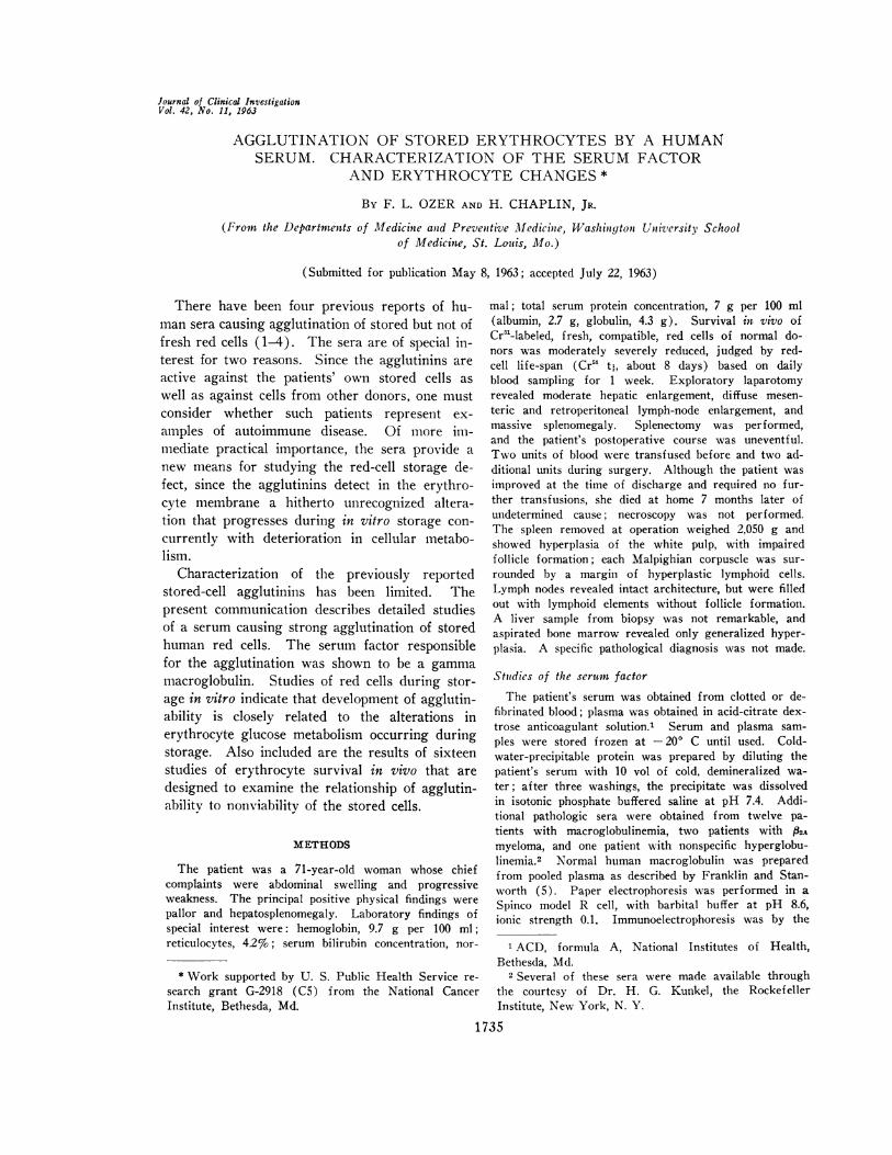
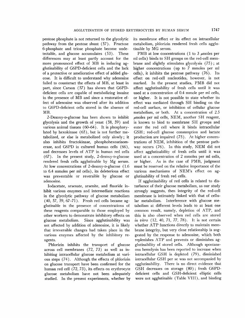
FIG. 1. SUCROSE GRADIENT DENSITY ULTRACENTRIFUGA-TION OF THE PATIENT S SERUM COMPARED WITH A NOR-MAL SERUM. The stored-cell agglutinin activity wasconcentrated in the sedimented macroglobulin fraction.
hemoglobin determinations were made on the originalblood sample and on the final suspension of unagglutinatedcells. Radioactivity in the two samples was measuredin a well-type scintillation counter and expressed ascounts per minute per gram of hemoglobin.
RESULTS
Characterization of the serum factor
Fresh red cells from normal donors were un-affected by the patient's serum, but stored redcells from the same donors were agglutinated inincreasing numbers with increasing time of stor-age. The agglutination reaction could be carriedout equally well over a range of pH from 5.6 to8.0 and of temperature from 40 to 370 C. Fourplus agglutination was demonstrable with the se-rum diluted 1/10,000, and positive agglutinationwas clearly definable up to dilutions of 1/100,000to 1/500,000. The ability of the serum to ag-glutinate stored cells withstood heating at 60° Cfor 10 hours, but was destroyed after 10 minutesat 800 C.
1737
F. L. OZER AND H. CHAPLIN, JR.
, 10,000 ,u 1,000-4 0-J 100-4 0 Ii
0.0175M 0.1 M 0.6M
E- 2.0 I-oItcli
50 100 150 200EFFLUENT VOLUME, ML
10,000
< 1,000
' 1000<
50 10 I 0
Ek 1.0 -
0
CD
05-
50 100 ISO 200EFFLUENT VOLUME, ML
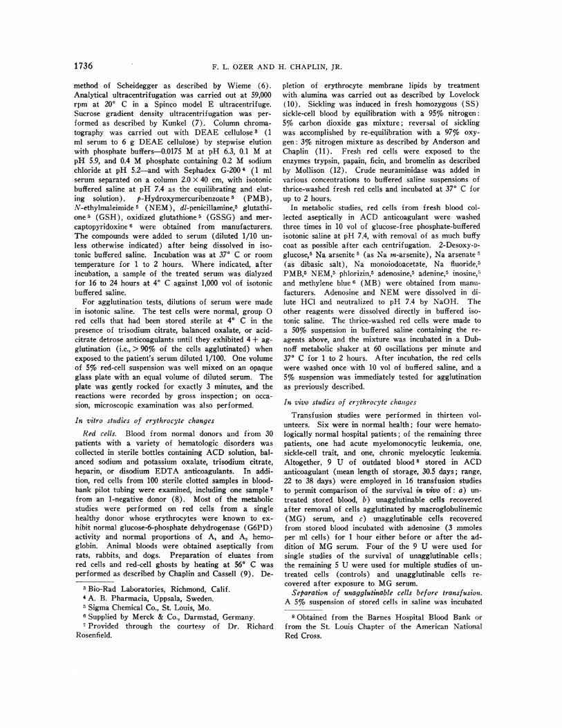
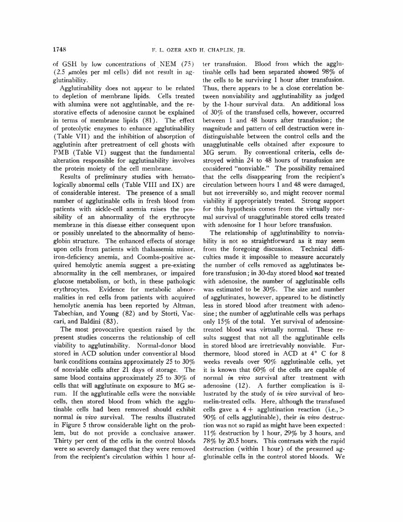
FIG. 2. DEAE-CELLULOSE (TOP) AND SEPHADLXG-200 (BOTTOM) CHROMATOGRAPHY OF THE PATIENT S
SERUM. Stored-cell agglutinin activity appeared in thefractions in which high-molecular-weight globulins areconcentrated.
Paper electrophoresis of the patient's serumshowed a dense homogeneous band in the gammaglobulin region. Immunoelectrophoresis demon-strated a heavy precipitin band in the gamma-1position. Analytical ultracentrifugation revealedthat 36.7%o of the total serum protein was ac-counted for by two macroglobulins, 32.6%o ex-hibiting an S20,, value of 18.8, and 4.1%o, one of28.8.
Protein precipitated by 1/10 dilution of serumwith distilled water possessed strong agglutinatingactivity when redissolved. The results of sucrosegradient density ultracentrifugation (Figure 1)demonstrated that the agglutinating activity ofwhole serum was concentrated in the macroglobu-lin fraction. Column chromatography on DEAEcellulose (14) and Sephadex G-200 (15) (Fig-ure 2) revealed the agglutinating activity con-centrated in the fractions richest in gamma macro-globulins. The 7 S20,, gamma globulin fractioneluted in the first DEAE-cellulose peak was in-cubated with stored cells in the presence and ab-
sence of added complement; no agglutination wasobserved, and subsequent exposure to antihumanglobulin serum revealed no evidence of "incom-plete" stored-cell agglutinating activity.
Treatment of a 1/10 dilution of serum withdisulfide-splitting agents-d,l-penicillamine (16)(10 mg per ml), mercaptopyridoxine (16) (10mg per ml), or GSH (17-21) (4 X 10⁻² M)-resulted in complete loss of agglutinating activityand disappearance of the macroglobulin peakspreviously -seen on ultracentrifugation. Reag-gregation of the protein subunits did not occurafter dialysis of the treated serum, nor was the ag-glutinating activity restored. Furthermore, storedred cells incubated with the treated serum eitherbefore or after dialysis failed to agglutinate uponthe addition of rabbit antihuman globulin serum,nor did they appear "blocked" when exposed tothe patient's untreated serum.
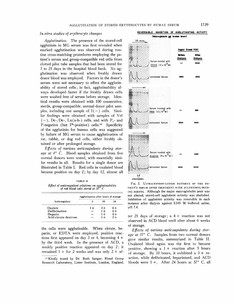
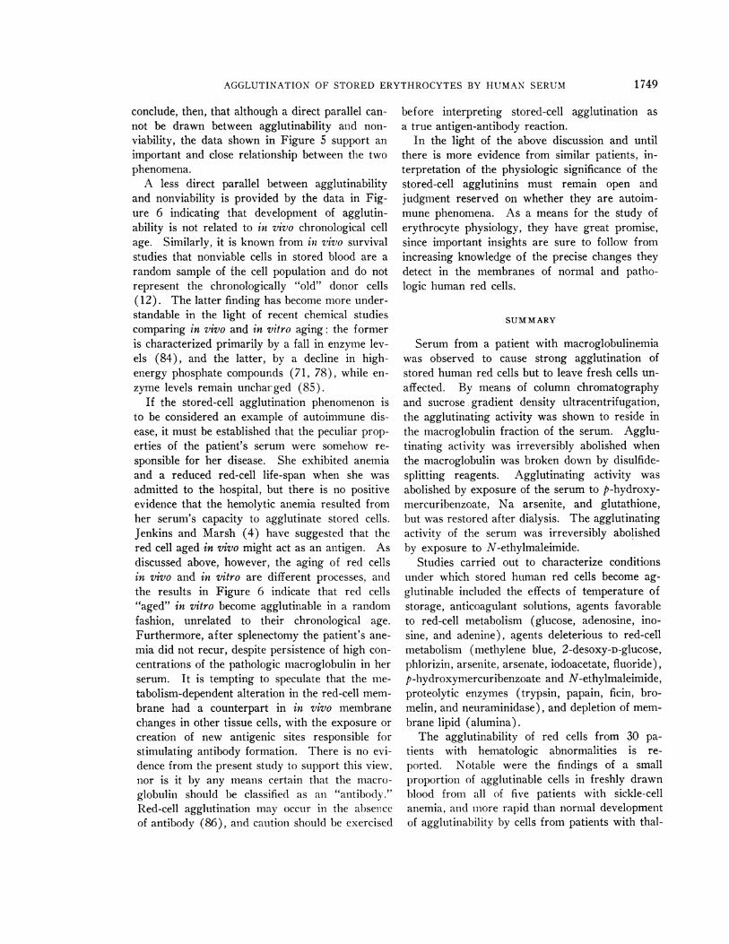
Sulfhydryl (SH)-binding agents such as PMB(22, 23), GSSG (20, 21), and Na arsenite (23-26)at concentrations of 2 to 5 X 10⁻² M abolished ag-glutinating activity. Ultracentrifugation patternsof the serum remained unchanged (Figure 3)except for loss of the 28.8 S20,w peak from theNa arsenite-treated sample. After dialysis, strongagglutinating activity was restored in all samples.NEM (27) inhibited the agglutinating activityonly at high concentrations (2 x 10⁻¹ M); the in-hibition was not reversible after dialysis.
TABLE I
Effect of anticoagulant solutions on agglutinabilityof red blood cells stored at 4° C
Agglutination after days of storage
Anticoagulant 0 2 6 12 21
Oxalate - 1+ 3+ 4+ 4+Citrate - - 1+ 3+ 4+Heparin - - 1+ 3+ 4+EDTA - - 1+ 1 -2+ 4+Acid-citrate dextrose - + 1 + 1 + 2 +
Ten of twelve MG sera, one of two /32A mye-loma sera, and a macroglobulin fraction preparedfrom pooled normal sera showed no agglutinationof stored cells. Weak agglutination of storedcells at serum dilutions of 1/10 or less was ob-served with samples from two patients with pri-mary macroglobulinemia and one patient with 82Amyeloma.
1 738
AGGLUTINATION OF STORED ERYTHROCYTES BY HUMAN SERUM
In vitro studies of erythrocyte changes
Agglutinationi. The presence of the stored-cellagglutinin in MG serum was first revealed whenmarked agglutination was observed during rou-tine cross-matching procedures employing the pa-tient's serum and group-compatible red cells fromclotted pilot tube samples that had been stored for5 to 21 days in the hospital blood bank. No ag-glutination was observed when freshly drawndonor blood was employed. Factors in the donor'sserum were not necessary to effect the agglutin-ability of stored cells; in fact, agglutinability al-ways developed faster if the freshly drawn cellswere washed free of serum before storage. Iden-tical results were obtained with 100 consecutive,sterile, group-compatible, normal-donor pilot sam-ples, including one sample of I(-) cells. Simi-lar findings were obtained with samples of Vel(-), Dc-/Dc-, Lu(a-b-) cells, and with P1- andP-negative (but Pk-positive) cells.10 Specificityof the agglutinin for human cells was suggestedby failure of MG serum to cause agglutination ofrat, rabbit, or dog red cells, either freshly ob-tained or after prolonged storage.
Effects of various anticoagulants during stor-age at 40 C. Blood samples obtained from fivenormal donors were tested, with essentially simi-lar results in all. Results for a single donor areillustrated in Table I. Red cells in oxalated bloodbecame positive on day 2; by day 12, almost all
TABLE II
Effect of anticoagulant solutions on agglutinabilityof red blood cells stored at 370 C
Agglutination after hours of storage
Anticoagulant 5 10 24
Oxalate 1+ 3+ 4+Defibrination - 1+ 4+Heparin - 1+ 4+Acid-citrate dextrose - 1 + 2+
the cells were agglutinable. When citrate, he-parin, or EDTA were employed, positive reac-
tions first appeared on day 3 or 4, becoming 4 +by the third week. In the presence of ACD, a
weakly positive reaction appeared on day 2; itremained 1 + for 2 weeks and was only 2 + af-
10 Kindly tested by Dr. Ruth Sanger, Blood GroupResearch Laboratory, Lister Institute, London, England.
REVERSIBLE INHIBITION OF AGGLUTINATING ACTIVITY
(Macroglobulin no broken down)35 rrins.
*V 4
Serum treated with
a /J_ of GSSG (2x10 M)
Untreated Serum
S~erum treated withP (2xI16M)
Untreated Serum
._t
A.
Serun treated withArsenitv(5x10OM
&-.f Untreated Serum
Agglut. Stored RBC
Before AfterOialysis Dialysis
- 44*
4+
**+ i
mocroglobs.
FIG. 3. ULTRACENTRIFUGATION PATTERNS OF THE PA-
TIENT'S SERUM AFTER TREATMENT WITH SULFHYDRYL-BIND-
ING AGENTS. Although the major macroglobulin peak wasnot altered, stored-cell agglutinin activity was abolished.Inhibition of agglutinin activity was reversible in eachinstance after dialysis against 0.145 M buffered saline,pH 7.4.
ter 21 days of storage; a 4 + reaction was notobserved in ACD blood until after about 6 weeksof storage.
Effects of various anticoagulants during stor-age at 37° C. Samples from two normal donorsgave similar results, summarized in Table II.Oxalated blood again was the first to becomepositive, showing a 1 + reaction after 5 hoursof storage. By 10 hours, it exhibited a 3 + re-action, while defibrinated, heparinized, and ACDbloods were 1 +. After 24 hours at 370 C. all
1739
F. L. OZER AND H. CHAPLIN, JR.
AGGLUTINATION OF STORED RBC BY MACROGLOBULIN
i~ ~1-2
X <i:,>X <; W~~~
&iz
*+
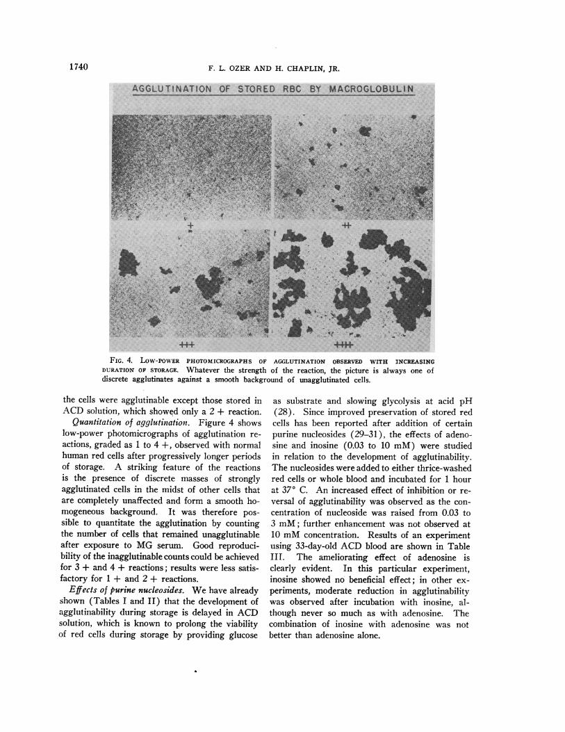
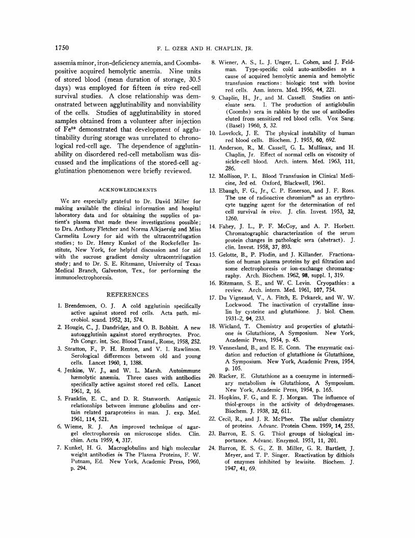
FIG. 4. LOW-POWER PHOTOMICROGRAPHS OF AGGLUTINATION OBSERVED WITH INCREASINGDURATION OF STORAGE. Whatever the strength of the reaction, the picture is always one ofdiscrete agglutinates against a smooth background of unagglutinated cells.
the cells were agglutinable except those stored inACD solution, which showed only a 2 + reaction.
Quantitation of agglutination. Figure 4 showslow-power photomicrographs of agglutination re-
actions, graded as 1 to 4 +, observed with normalhuman red cells after progressively longer periodsof storage. A striking feature of the reactionsis the presence of discrete masses of stronglyagglutinated cells in the midst of other cells thatare completely unaffected and form a smooth ho-mogeneous background. It was therefore pos-
sible to quantitate the agglutination by countingthe number of cells that remained unagglutinableafter exposure to MG serum. Good reproduci-bility of the inagglutinable counts could be achievedfor 3 + and 4 + reactions; results were less satis-factory for 1 + and 2 + reactions.
Effects of purine nucleosides. We have alreadyshown (Tables I and II) that the development ofagglutinability during storage is delayed in ACDsolution, which is known to prolong the viabilityof red cells during storage by providing glucose
as substrate and slowing glycolysis at acid pH(28). Since improved preservation of stored redcells has been reported after addition of certainpurine nucleosides (29-31), the effects of adeno-sine and inosine (0.03 to 10 mM) were studiedin relation to the development of agglutinability.The nucleosides were added to either thrice-washedred cells or whole blood and incubated for 1 hourat 370 C. An increased effect of inhibition or re-versal of agglutinability was observed as the con-centration of nucleoside was raised from 0.03 to3 mM; further enhancement was not observed at10 mM concentration. Results of an experimentusing 33-day-old ACD blood are shown in TableIII. The ameliorating effect of adenosine isclearly evident. In this particular experiment,inosine showed no beneficial effect; in other ex-
periments, moderate reduction in agglutinabilitywas observed after incubation with inosine, al-though never so much as with adenosine. Thecombination of inosine with adenosine was notbetter than adenosine alone.
1740
AGGLUTINATION OF STORED ERYTHROCYTES BY HUMAN SERUM
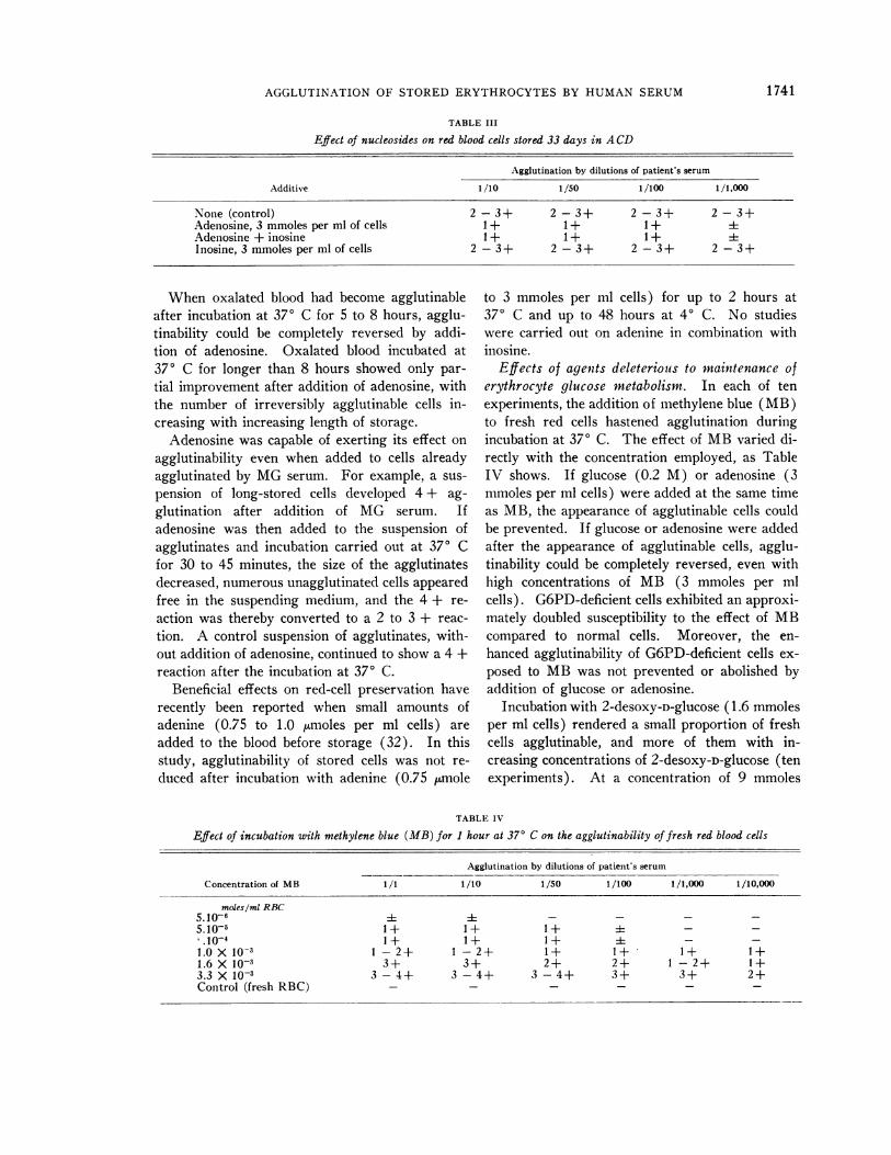
TABLE III
Effect of nucleosides on red blood cells stored 33 days in ACD
Agglutination by dilutions of patient's serum
Additive 1/10 1/50 1/100 1 /1,000
None (control) 2-3+ 2-3+ 2-3+ 2-3+Adenosine, 3 mmoles per ml of cells 1+ 1+ 1+ iAdenosine + inosine 1+ 1+ 1+ iInosine, 3 mmoles per ml of cells 2 - 3+ 2 - 3+ 2 - 3+ 2 - 3+
When oxalated blood had become agglutinableafter incubation at 370 C for 5 to 8 hours, agglu-tinability could be completely reversed by addi-tion of adenosine. Oxalated blood incubated at37° C for longer than 8 hours showed only par-
tial improvement after addition of adenosine, withthe number of irreversibly agglutinable cells in-creasing with increasing length of storage.
Adenosine was capable of exerting its effect on
agglutinability even when added to cells alreadyagglutinated by MG serum. For example, a sus-
pension of long-stored cells developed 4 + ag-
glutination after addition of MG serum. Ifadenosine was then added to the suspension ofagglutinates and incubation carried out at 37° Cfor 30 to 45 minutes, the size of the agglutinatesdecreased, numerous unagglutinated cells appearedfree in the suspending medium, and the 4 + re-
action was thereby converted to a 2 to 3 + reac-
tion. A control suspension of agglutinates, with-out addition of adenosine, continued to show a 4 +reaction after the incubation at 370 C.
Beneficial effects on red-cell preservation haverecently been reported when small amounts ofadenine (0.75 to 1.0 umoles per ml cells) are
added to the blood before storage (32). In thisstudy, agglutinability of stored cells was not re-
duced after incubation with adenine (0.75 Stmole
to 3 mmoles per ml cells) for up to 2 hours at370 C and up to 48 hours at 40 C. No studieswere carried out on adenine in combination withinosine.
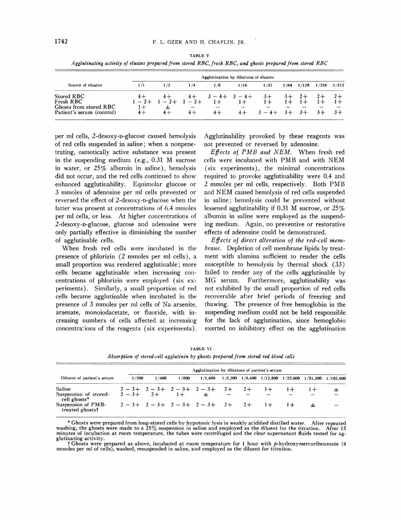
Effects of agents deleterious to maintenance oferythrocyte glucose metabolism. In each of tenexperiments, the addition of methylene blue (MB)to fresh red cells hastened agglutination duringincubation at 370 C. The effect of MB varied di-rectly with the concentration employed, as TableIV shows. If glucose (0.2 M) or adenosine (3mmoles per ml cells) were added at the same timeas MB, the appearance of agglutinable cells couldbe prevented. If glucose or adenosine were addedafter the appearance of agglutinable cells, agglu-tinability could be completely reversed, even withhigh concentrations of MB (3 mmoles per mlcells). G6PD-deficient cells exhibited an approxi-mately doubled susceptibility to the effect of MBcompared to normal cells. Moreover, the en-
hanced agglutinability of G6PD-deficient cells ex-
posed to MB was not prevented or abolished byaddition of glucose or adenosine.
Incubation with 2-desoxy-D-glucose (1.6 mmolesper ml cells) rendered a small proportion of freshcells agglutinable, and more of them with in-creasing concentrations of 2-desoxy-D-glucose (tenexperiments). At a concentration of 9 mmoles
TABLE IV
Effect of incubation with methylene blue (MB) for 1 hour at 370 C on the agglutinability of fresh red blood cells
Agglutination by dilutions of patient's serum
Concentration of MB 1/1 1/10 1/50 1/100 1/1,000 1/10,000
moles/ml RBC5.10-6 ±i - - - -
5.10-5 1+ 1+ 1+ ± - -' 10-4 I1+ 1+ 1+ - -1.0Xlo-3 1-2+ 1-2+ 1+ 1+ 1+ 1+1.6 X 10⁻³ 3+ 3+ 2+ 2+ 1-2+ 1+3.3 X 10⁻³ 3 - 4+ 3 - 4+ 3 -4+ 3+ 3+ 2+Control (fresh RBC) - - - - - -
1741
F. L. OZER AND H. CHAPLIN, JR.
TABLE V
Agglutinating activity of eluates prepared from stored RBC, fresh RBC, and ghosts prepared from stored RBC
Agglutination by dilutions of eluates
Source of eluates 1/1 1/2 1/4 1/8 1/16 1/32 1/64 1/128 1/256 1/512
Stored RBC 4+ 4+ 4+ 3-4+ 3-4+ 3+ 3+ 2+ 2+ 2+Fresh RBC 1 -2+ 1 -2+ 1 -2+ 1+ 1+ 1+ 1+ 1+ 1+ 1+Ghosts from stored RBC 1+ i - - - - -Patient's serum (control) 4+ 4+ 4+ 4+ 4+ 3 - 4+ 3+ 3+ 3+ 3+
per ml cells, 2-desoxy-D-glucose caused hemolysisof red cells suspended in saline; when a nonpene-
trating, osmotically active substance was presentin the suspending medium (e.g., 0.31 M sucrose
in water, or 25%o albumin in saline), hemolysisdid not occur, and the red cells continued to showenhanced agglutinability. Equimolar glucose or
3 mmoles of adenosine per ml cells prevented or
reversed the effect of 2-desoxy-D-glucose when thelatter was present at concentrations of 6.4 mmolesper ml cells, or less. At higher concentrations of2-desoxy-D-glucose, glucose and adenosine were
only partially effective in diminishing the numberof agglutinable cells.When fresh red cells were incubated in the
presence of phlorizin (2 mmoles per ml cells), a
small proportion was rendered agglutinable; more
cells became agglutinable when increasing con-
centrations of phlorizin were employed (six ex-
periments). Similarly, a small proportion of redcells became agglutinable when incubated in thepresence of 3 mmoles per ml cells of Na arsenite,arsenate, monoiodacetate, or fluoride, with in-creasing numbers of cells affected at increasingconcentralions of the reagents (six experiments).
Agglutinability provoked by these reagents was
not prevented or reversed by adenosine.Effects of PMB and NEM. When fresh red
cells were incubated with PMB and with NEM(six experiments), the minimal concentrationsrequired to provoke agglutinability were 0.4 and2 mmoles per ml cells, respectively. Both PMBand NEM caused hemolysis of red cells suspendedin saline; hemolysis could be prevented withoutlessened agglutinability if 0.31 M sucrose, or 25%oalbumin in saline were employed as the suspend-ing medium. Again, no preventive or restorativeeffects of adenosine could be demonstrated.
Effects of direct alteration of the red-cell mem-brane. Depletion of cell membrane lipids by treat-ment with alumina sufficient to render the cellssusceptible to hemolysis by thermal shock (33)failed to render any of the cells agglutinable byMG serum. Furthermore, agglutinability was
not exhibited by the small proportion of red cellsrecoverable after brief periods of freezing andthawing. The presence of free hemoglobin in thesuspending medium could not be held responsiblefor the lack of agglutination, since hemoglobinexerted no inhibitory effect on the agglutination
TABLE VI
Absorption of stored-cell agglutinin by ghosts prepared from stored red blood cells
Agglutination by dilutions of patient's serum
Diluent of patient's serum 1/200 1/400 1/800 1/1,600 1/3,200 1/6,400 1/12,800 1/25,600 1/51,200 1/102,400
Saline 2-3+ 2-3+ 2-3+ 2-3+ 2+ 2+ 1+ 1+ 1+ iSuspension of stored- 2-3+ 2+ 1+ i- - - - -
cell ghosts*Suspension ofPMB- 2-3+ 2-3+ 2-3+ 2-3+ 2+ 2+ 1+ 1+ +-
treated ghostst
* Ghosts were prepared from long-stored cells by hypotonic lysis in weakly acidified distilled water. After repeatedwashing, the ghosts were made to a 25% suspension in saline and employed as the diluent for the titration. After 15minutes of incubation at room temperature, the tubes were centrifuged and the clear supernatant fluids tested for ag-glutinating activity.
t Ghosts were prepared as above, incubated at room temperature for 1 hour with p-hydroxymercuribenzoate (4mmoles per ml of cells), washed, resuspended in saline, and employed as the diluent for titration.
1742
AGGLUTINATION OF STORED ERYTHROCYTES BY HUMAN SERUM
reaction when it was added to long-stored cellsexposed to MG serum.
That the agglutination phenoienon involvedactual binding of the agglutinin to the cell mem-
brane was demonstrated in each of three studiesof elution of the agglutinin by heating for 10 min-utes at 560 C. Table V illustrates the agglutinat-ing activity demonstrable in eluates prepared fromintact fresh and long-stored cells, and from ghostsprepared from long-stored cells previously ex-
posed to MG serum. Striking agglutinating ac-
tivity was present in the stored-cell eluate, andweak but undoubted activity in the eluate fromfresh cells, despite the complete absence of ag-
glutinates in the fresh-cell preparation. It is notpossible to say whether the weakly positive eluatefrom fresh cells implies the presence of reactivesites on the "fresh" red cell in vivo, since the timefor the in vitro sensitization and eluation proce-
dures could be sufficient to permit a minimal"storage" effect to become manifest. The very
low activity demonstrable in the eluate from thestored-cell ghosts suggests that preparation ofthe ghosts (by hypotonic lysis in weakly acidifiedwater) either liberated or inactivated the agglu-tinin. That ghosts retain their ability to reactwith the agglutinin was shown by four absorptionstudies; a typical result is illustrated in Table VI.The serum serially diluted in ghost suspensiondemonstrated diminished agglutinating activitycompared to serum diluted in saline alone. Pre-treatment of the ghosts with PMB inhibited theircapacity to absorb the agglutinin.
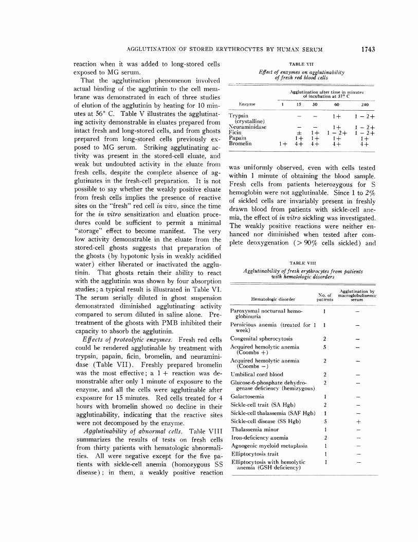
Effects of proteolytic enzymes. Fresh red cellscould be rendered agglutinable by treatment withtrypsin, papain, ficin, bromelin, and neuramini-dase (Table VII). Freshly prepared bromelinwas the most effective; a 1 + reaction was de-monstrable after only 1 minute of exposure to theenzyme, and all the cells were agglutinable afterexposure for 15 minutes. Red cells treated for 4hours with bromelin showed no decline in theiragglutinability, indicating that the reactive siteswere not decomposed by the enzyme.
Agglutinability of abnormal cells. Table VIIIsummarizes the results of tests on fresh cellsfrom thirty patients with hematologic abnormali-ties. All were negative except for the five pa-tients with sickle-cell anemia (homozygous SSdisease) ; in them, a weakly positive reaction
TABLE VII
Effect of enzymes on agglutinabilityoffresh red blood cells
Agglutination after time in minutesof incubation at 370 C
Enzyme 1 15 30 60 240
Trypsin - - 1+ 1 - 2+(crystalline)
Neuraminidase - - 1 + 1 - 2+Ficin ± 1+ 1 - 2+ 1 - 2+Papain 1+ 1+ 1+ 1+Bromelin 1+ 4+ 4+ 4+ 4+
was uniformly observed, even with cells testedwithin 1 minute of obtaining the blood sample.Fresh cells from patients heterozygous for Shemoglobin were not agglutinable. Since 1 to 2%oof sickled cells are invariably present in freshlydrawn blood from patients with sickle-cell ane-mia, the effect of in vitro sickling was investigated.The weakly positive reactions were neither en-hanced nor diminished when tested after com-plete deoxygenation (> 90% cells sickled) and
TABLE VIII
Agglutinability of fresh erythrocytes from patientswith hematologic disorders
Agglutination byNo. of macroglobulinemic
Hematologic disorder patients serum
Paroxysmal nocturnal hemo- 1 -
globinuriaPernicious anemia (treated for 1 1 -
week)Congenital spherocytosis 2 -
Acquired hemolytic anemia 5 -
(Coombs +)Acquired hemolytic anemia 2 -
(Coombs -)Umbilical cord blood 2 -
Glucose-6-phosphate dehydro- 2 -
genase deficiency (hemizygous)Galactosemia 1 -
Sickle-cell trait (SA Hgb) 2 -
Sickle-cell thalassemia (SAF Hgb) 1 -
Sickle-cell disease (SS Hgb) 5 +Thalassemia minor 1Iron-deficiency anemia 2Agnogenic myeloid metaplasia 1Elliptocytosis trait 1Elliptocytosis with hemolytic 1anemia (GSH deficiency)
1743
F. L. OZER AND H. CHAPLIN, JR.
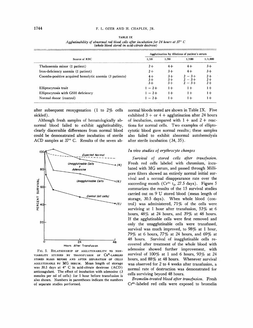
TABLE IX
Agglutinability of abnormal red blood cells after incubation for 24 hours at 370 C(whole blood stored in acid-citrate dextrose)
Agglutination by dilutions of patient's serum
Source of RBC 1/10 1/50 1/100 1/1,000
Thalassemia minor (1 patient) 2+ 4+ 4+ 3+Iron-deficiency anemia (1 patient) 2+ 3+ 4+ 3+Coombs-positive acquired hemolytic anemia (3 patients) 4+ 3+ 2 - 3 + 2+
3+ 3+ 2 -3+ 2+3+ 3+ 2 - 3+ 2+
Elliptocytosis trait 1 -2+ 1+ 1+ 1+Elliptocytosis with GSH deficiency 1 -2+ 1 + 1 + 1+Normal donor (control) 1 -2+ 1+ 1+ 1+
after subsequent reoxygenation (1 to 2%o cellssickled).Although fresh samples of hematologically ab-
normal blood failed to exhibit agglutinability,clearly discernible differences from normal bloodcould be demonstrated after incubation of sterileACD samples at 370 C. Results of the seven ab-
100
Unogglulinable Cells (4)80 a tAdenosine
Unoggl l Cl (6)
> 60 _
Control (o// cells)zr 40-_(5)w
20-
_ ~~~~~~~~~~~~~~~~~~~~~~~~II0 24 48
Hours After Transfusion
FIG. 5. RELATIONSHIP OF AGGLUTINABILITY TO NON-VIABILITY STUDIED BY TRANSFUSION OF CR51-LABELEDSTOR9D BLOOD BEFORE AND AFTER SEPARATION OF CELLSAGGLUTINABLE BY MG SERUM. Mean length of storagewas 30.5 days at 4° C in acid-citrate dextrose (ACD)anticoagulant. The effect of incubation with adenosine (3mmoles per ml of cells) for 1 hour before transfusion isalso shown. Numbers in parentheses indicate the numbersof separate studies performed.
normal bloods tested are shown in Table IX. Fiveexhibited 3 + or 4 + agglutination after 24 hoursof incubation, compared with 1 + and 2 + reac-tions for normal cells. Two examples of ellipto-cytotic blood gave normal results; these samplesalso failed to exhibit abnormal autohemolysisafter sterile incubation (34, 35).
In vivo studies of erythrocyte changes
Survival of stored cells after transfusion.Fresh red cells labeled with chromium, incu-bated with MG serum, and passed through Milli-pore filters showed an entirely normal initial sur-vival and a normal disappearance rate over thesucceeding month (Cr51 ti, 27.5 days). Figure 5summarizes the results of the 15 survival studiescarried out on 9 U stored blood (mean length ofstorage, 30.5 days). When whole blood (con-trol) was administered, 71% of the cells weresurviving at 1 hour after transfusion, 53%o at 6hours, 48% at 24 hours, and 39% at 48 hours.If the agglutinable cells were first removed andonly the unagglutinable cells were transfused,survival was much improved, to 98% at 1 hour,79% at 6 hours, 77% at 24 hours, and 69% at48 hours. Survival of inagglutinable cells re-covered after treatment of the whole blood withadenosine showed further improvement, withsurvival of 100%o at 1 and 6 hours, 93% at 24hours, and 88% at 48 hours. Whenever survivalwas observed for 2 to 4 weeks after transfusion, anormal rate of destruction was demonstrated forcells surviving beyond 48 hours.
Bromelin-treated blood after transfusion. FreshCr51-labeled red cells were exposed to bromelin
1 744
AGGLUTINATION OF STORED ERYTHROCYTES BY HUMAN SERUM
for 15 minutes, then washed four times in 100 volsaline. A sample tested with a 1/100 dilution ofMG serum revealed 4 + agglutination. The re-maining cells (not exposed to MG serum) wereinfused to the original donor. Eighty-nine percent were present in the circulation at 1 hourafter infusion, 71% at 3 hours, and 22% at 20.5hours.
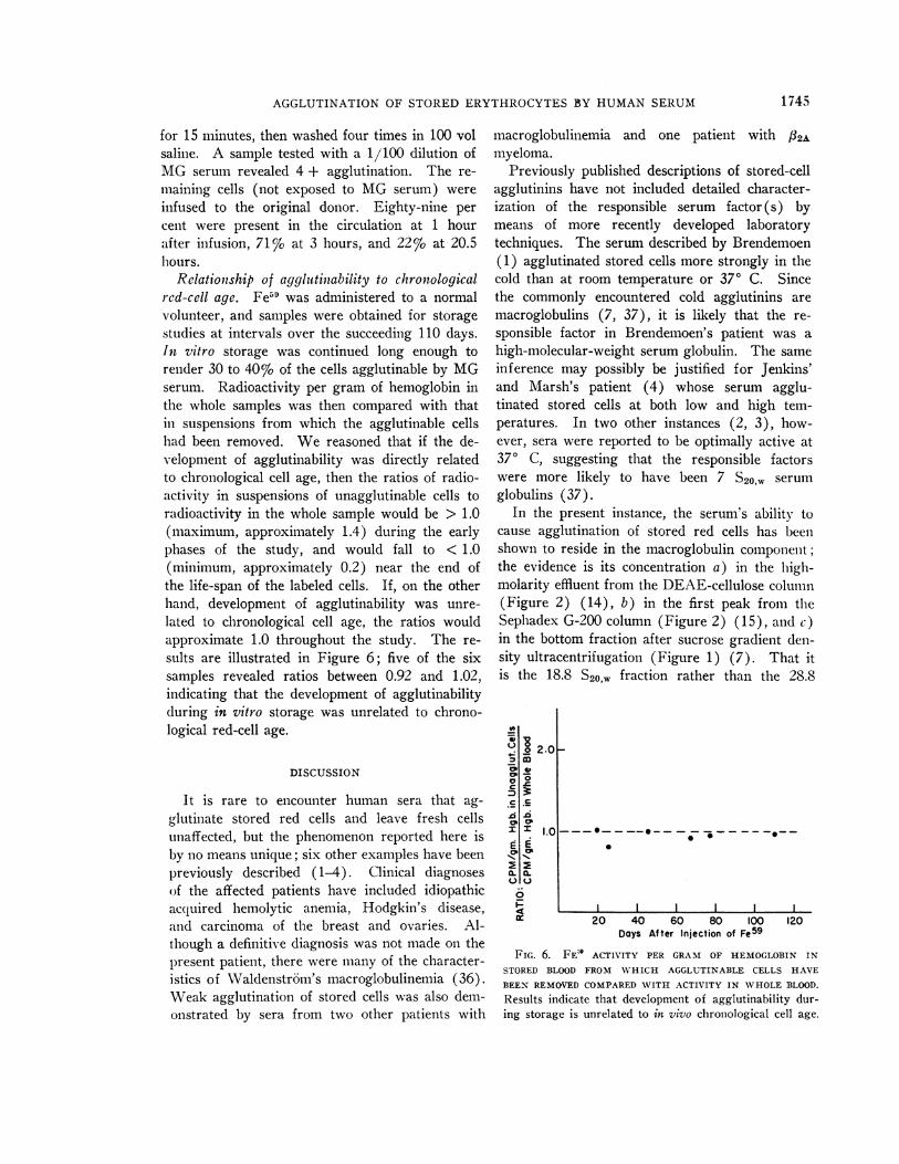
Relationship of aggldtinability to chronologicalrcd-cell age. Fe59 was administered to a normalvolunteer, and samples were obtained for storagestudies at intervals over the succeeding 110 days.In vitro storage was continued long enough torender 30 to 40% of the cells agglutinable by MGserum. Radioactivity per gram of hemoglobin inthe whole samples was then compared with thatin suspensions from which the agglutinable cellshad been removed. We reasoned that if the de-velopment of agglutinability was directly relatedto chronological cell age, then the ratios of radio-activity in suspensions of unagglutinable cells toradioactivity in the whole sample would be > 1.0(maximum, approximately 1.4) during the earlyphases of the study, and would fall to < 1.0(minimum, approximately 0.2) near the end ofthe life-span of the labeled cells. If, on the otherhand, development of agglutinability was unre-lated to chronological cell age, the ratios wouldapproximate 1.0 throughout the study. The re-sults are illustrated in Figure 6; five of the sixsamples revealed ratios between 0.92 and 1.02,indicating that the development of agglutinabilityduring in vitro storage was unrelated to chrono-logical red-cell age.
DISCUSSION
It is rare to encounter human sera that ag-glutinate stored red cells and leave fresh cellsunaffected, but the phenomenon reported here isby no means unique; six other examples have beenpreviously described (1-4). Clinical diagnosesof the affected patients have included idiopathicacquired hemolytic anemia, Hodgkin's disease,and carcinoma of the breast and ovaries. Al-though a definitive diagnosis was not made on thepresent patient, there were many of the character-istics of Waldenstrdm's macroglobulinemia (36).Weak agglutination of stored cells was also dem-onstrated by sera from two other patients with
macroglobulinemia and one patient with /32Amyeloma.
Previously published descriptions of stored-cellagglutinins have not included detailed character-ization of the responsible serum factor (s) bymeans of more recently developed laboratorytechniques. The serum described by Brendemoen(1) agglutinated stored cells more strongly in thecold than at room temperature or 370 C. Sincethe commonly encountered cold agglutinins aremacroglobulins (7, 37), it is likely that the re-sponsible factor in Brendemoen's patient was ahigh-molecular-weight serum globulin. The sameinference may possibly be justified for Jenkins'and Marsh's patient (4) whose serum agglu-tinated stored cells at both low and high tem-peratures. In two other instances (2, 3), how-ever, sera were reported to be optimally active at370 C, suggesting that the responsible factorswere more likely to have been 7 S20,w serumglobulins (37).
In the present instance, the serum's ability tocause agglutination of stored red cells has beenshown to reside in the macroglobulin componient;the evidence is its concentration a) in the high-molarity effluent from the DEAE-cellulose column(Figure 2) (14), b) in the first peak from theSephadex G-200 column (Figure 2) (15), and c)in the bottom fraction after sucrose gradient den-sity ultracentrifugation (Figure 1) (7). That itis the 18.8 S20,w fraction rather than the 28.8
, la
v 03
D Dcb00
a I
d 0x E2 2a. a.u
2.0H
1.0 ---0-- --- - - -_ _ _ . _ _ ._
0 *S
I I20 40 60 80 100
Days After Injection of Fe59120
FIG. 6. FE4' ACTIVITY PER GRAM OF HEMOGLOBIN IN
STORED BLOOD FROM WHICH AGGLUTINABLE CELLS HAVE
BEEN REMOVED COMPARED WITH ACTIVITY IN WHOLE BLOOD.
Results indicate that development of agglutinability dur-ing storage is unrelated to in vivo chronological cell age.
1 745
F. L. OZER AND H. CHAPLIN, JR.
S20,w fraction that is responsible for the agglu-tinating activity is indicated by the retention of ag-glutinating activity despite the loss of the 28.8 S2o,wpeak in the ultracentrifugation pattern of post-dialysis serum after treatment with Na arsenite(Figure 3). Integrity of the whole 18.8 S20,wmolecule appears to be necessary for maintenanceof the agglutinating property, since agglutinationno longer occurs when the molecule is broken downinto smaller subunits after treatment with disul-fide-splitting agents. Similar loss of activity afterS-S cleavage has been reported for a variety ofmacromolecular agglutinins by other workers(38, 39).The inhibition of agglutinating activity without
breakdown of the 18.8 S20,1 globulins after treat-ment with PMB, GSSG, Na arsenite, and NEMcannot be attributed to the SH-binding propertiesof these compounds. It is not definitely knownthat there are any free SH groups on intact or
pathological gamma macroglobulins. None ofthe reagents is entirely specific for thiol groups,and SH-binding would not be reversible aftersimple dialysis against buffered saline. No con-clusions can be drawn from the limited evidencein hand, and an interpretation of the importantinhibition phenomenon must await results of moredefinitive studies.The relation of the present macroglobulin to
normal macroglobulin remains obscure. Macro-globulin concentrated from a pool of normalplasma caused no agglutination of stored cells thatgave a 4 + reaction with the patient's serum.
The stored-cell agglutinin reaction can be distin-guished from anti-T agglutination (12), since itis not observed with normal sera and since the al-teration of the red-cell surface takes place duringstorage under sterile conditions.The results of the present investigation indicate
that the development of agglutinability is closelyrelated to the alterations in erythrocyte glucosemetabolism taking place during in vitro storage.The importance of metabolic factors was firstsuggested by the relationship of agglutinability tothe temperature of storage and to the character-istics of the anticoagulant solution (Tables I andII); agglutinability increased with conditions fa-voring depletion of glucose and consequent de-pletion of high-energy phosphate compounds.These observations were extended by studies em-
ploying purine nucleosides, notably adenosine,which have been shown to improve the viabilityof stored red cells (29-31) either a) by conversionat the red-cell membrane to ribose 5-phosphate,which can be further metabolized with resultantregeneration of ADP and ATP (40-47), or b)by providing the purine moiety to serve as sub-strate for ATP synthesis (30, 48, 49). Adenosinewas able to restore many agglutinable cells to theunagglutinable state (Table III) when added af-ter storage, and it delayed the development of ag-glutinability if present during storage. It wasparticularly remarkable that when adenosine wasadded to stored cells already agglutinated by MGserum, many of the cells detached themselvesfrom the formed agglutinates, presumably re-flecting a restorative effect on their metabolism.Under normal conditions, the red cell derives
its energy entirely from the metabolism of glu-cose, approximately 90%o via the glycolytic (Emb-den-Meyerhof) pathway and 10%o via the oxida-tive hexose monophosphate shunt (50, 51). Ifthe beneficial effects of adenosine on agglutin-ability of stored cells were related to restorationof cellular metabolism, increased agglutinabilityshould result from treatment with compounds in-terfering with cellular metabolism. MB providesa case in point, although its metabolic effects arenumerous and complex. It is known to stimulatethe hexose monophosphate shunt and to increaseglucose utilization and CO2 formation; in thepresence of MB, erythrocyte oxygen uptake mayincrease as much as twentyfold (52-54). Evenin the absence of glucose, increased uptake ofoxygen is observed that presumably results fromutilization of intracellular phosphate esters (51);the rate of breakdown of triose phosphate is in-creased, and lactate is oxidized to pyruvate (53,55). Washed red cells contain almost no glucoseand limited amounts of other substrates (56). Inthe presence of MB, these substrates will be rap-idly exhausted, which may explain the mechanismwhereby fresh red cells are rendered agglutinablein its presence. The interpretation is consistentwith the finding that substrate provided in theform of glucose or adenosine inhibited or reversedthe agglutination-inducing effect of MB.The effect of MB on G6PD-deficient cells dif-
fers from its effects on normal cells. Not onlydoes the hexose monophosphate shunt fail, but
1746
AGGLUTINATION OF STORED ERYTHROCYTES BY HUMAN SERUM
pentose phosphate is not returned to the glycolyticpathway from the pentose shunt (57). Fructose6-phosphate and triose phosphate become unde-tectable, and glucose accumulates (53). Thesedifferences may at least partly account for themore pronounced effect of MB in inducing ag-glutinability of G6PD-deficient cells and the lackof a protective or ameliorative effect of added glu-cose. It is difficult to understand why adenosinefailed to counteract the effects of MB, at least inpart, since Carson (57) has shown that G6PD-deficient cells are capable of metabolizing inosinein the presence of MB and since a restorative ef-fect of adenosine was observed after its additionto G6PD-deficient cells stored in the absence ofMB.
2-Desoxy-D-glucose has been shown to inhibitglycolysis and the growth of yeast (58, 59) andvarious animal tissues (60-64). It is phosphory-lated by hexokinase (65), but is not further me-tabolized, or else is metabolized only slowly; italso inhibits fructokinase, phosphohexoseisom-erase, and G6PD in cultured human cells (66),and decreases levels of ATP in human red cells(67). In the present study, 2-desoxy-D-glucoserendered fresh cells agglutinable by Mg serum.At low concentrations of 2-desoxy-D-glucose (1.6to 6.4 mmoles per ml cells), its deleterious effectwas preventable or reversible by glucose oradenosine.
Iodacetate, arsenate, arsenite, and fluoride in-hibit various enzymes and intermediate reactionsin the glycolytic pathway of glucose metabolism(40, 57, 59, 67-71). Fresh red cells became ag-glutinable in the presence of concentrations ofthese reagents comparable to those employed byother workers to demonstrate inhibitory effects onglucose metabolism. Since agglutinability wasnot affected by addition of adenosine, it is likelythat irreversible changes had taken place in thevarious enzymes affected by the inhibitory re-agents.
Phlorizin inhibits the transport of glucoseacross cell membranes (72, 73) as well as in-hibiting intracellular glucose metabolism at vari-ous steps (74). Although the effects of phlorizinon glucose transport have been confirmed for thehuman red cell (72, 73), its effects on erythrocyteglucose metabolism have not been adequatelystudied. In the present experiments, whether by
its membrane effect or its effect on intracellularmetabolism, phlorizin rendered fresh cells agglu-tinable by MG serum.PMB at low concentrations (1 to 3 umoles per
ml cells) binds to SH groups on the red-cell mem-brane and slightly stimulates glycolysis (75) ; athigher concentrations (up to 7 mmoles per mlcells), it inhibits the pentose pathway (76). Itseffect on red-cell nucleotides, however, is notmarked. In the present studies, PMB did notaffect agglutinability of fresh cells until it wasused at a concentration of 0.4 mmole per ml cells,or higher. It is not possible to state whether itseffect was mediated through SH binding on thered-cell surface, or inhibition of cellular glucosemetabolism, or both. At a concentration of 2.5,umoles per ml cells, NEM, another SH reagent,is known to bind to membrane SH groups andenter the red cell where it binds intracellularGSH; red-cell glucose consumption and lactateproduction are impaired (75). At higher concen-trations of NEM, inhibition of the pentose path-way occurs (76). In this study, NEM did notaffect agglutinability of fresh cells until it wasused at a concentration of 2 mmoles per ml cells,or higher. As in the case of PMB, judgmentmust be reserved on the relative importance of thevarious mechanisms of NEM's effect on ag-glutinability of fresh red cells.
If agglutinability of red cells is related to dis-turbance of their glucose metabolism, as our studystrongly suggests, then integrity of the red-cellmembrane is intimately linked with that of cellu-lar metabolism. Interference with glucose me-tabolism at different levels leads to at least onecommon result, namely, depletion of ATP, andthis is also observed when red cells are storedin vitro (12, 40, 71, 77, 78). It is not certainwhether ATP functions directly to maintain mem-brane integrity, but very close relationship is sug-gested by the response to adenosine, which bothreplenishes ATP and prevents or diminishes ag-glutinability of stored cells. Although spontane-ous hemolysis has been reported to increase whenintracellular GSH is depleted (79), diminishedintracellular GSH per se was not accompanied byagglutinability. There is no direct evidence thatGSH decreases on storage (80); fresh G6PD-deficient cells and GSH-deficient elliptic cellswere not agglutinable (Table VIII), and binding
1747
F. L. OZER AND H. CHAPLIN, JR.
of GSH by low concentrations of NEM (75)(2.5 /Lmoles per ml cells) did not result in ag-glutinability.
Agglutinability does not appear to be relatedto depletion of membrane lipids. Cells treatedwith alumina were not agglutinable, and the re-storative effects of adenosine cannot be explainedin terms of membrane lipids (81). The effectof proteolytic enzymes to enhance agglutinability(Table VII) and the inhibition of absorption ofagglutinin after pretreatment of cell ghosts withPMB (Table VI) suggest that the fundamentalalteration responsible for agglutinability involvesthe protein moiety of the cell membrane.
Results of preliminary studies with hemato-logically abnormal cells (Table VIII and IX) areof considerable interest. The presence of a smallnumber of agglutinable cells in fresh blood frompatients with sickle-cell anemia raises the pos-sibility of an abnormality of the erythrocytemembrane in this disease either consequent uponor possibly unrelated to the abnormality of hemo-globin structure. The enhanced effects of storageupon cells from patients with thalassemia minor,iron-deficiency anemia, and Coombs-positive ac-quired hemolytic anemia suggest a pre-existingabnormality in the cell membranes, or impairedglucose metabolism, or both, in these pathologicerythrocytes. Evidence for metabolic abnor-malities in red cells from patients with acquiredhemolytic anemia has been reported by Altman,Tabechian, and Young (82) and by Storti, Vac-cari, and Baldini (83).The most provocative question raised by the
present studies concerns the relationship of cellviability to agglutinability. Normal-donor bloodstored in ACD solution under conventional bloodbank conditions contains approximately 25 to 30%of nonviable cells after 21 days of storage. Thesame blood contains approximately 25 to 30% ofcells that will agglutinate on exposure to MG se-rum. If the agglutinable cells were the nonviablecells, then stored blood from which the agglu-tinable cells had been removed should exhibitnormal in vivo survival. The results illustratedin Figure 5 throw considerable light on the prob-lem, but do not provide a conclusive answer.Thirty per cent of the cells in the control bloodswere so severely damaged that they were removedfrom the recipient's circulation within 1 hour af-
ter transfusion. Blood from which the agglu-tinable cells had been separated showed 98% ofthe cells to be surviving 1 hour after transfusion.Thus, there appears to be a close correlation be-tween nonviability and agglutinability as judgedby the 1-hour survival data. An additional lossof 30% of the transfused cells, however, occurredbetween 1 and 48 hours after transfusion; themagnitude and pattern of cell destruction were in-distinguishable between the control cells and theunagglutinable cells obtained after exposure toMG serum. By conventional criteria, cells de-stroyed within 24 to 48 hours of transfusion areconsidered "nonviable." The possibility remainedthat the cells disappearing from the recipient'scirculation between hours 1 and 48 were damaged,but not irreversibly so, and might recover normalviability if appropriately treated. Strong supportfor this hypothesis comes from the virtually nor-mal survival of unagglutinable stored cells treatedwith adenosine for 1 hour before transfusion.The relationship of agglutinability to nonvia-
bility is not so straightforward as it may seemfrom the foregoing discussion. Technical diffi-culties made it impossible to measure accuratelythe number of cells removed as agglutinates be-fore transfusion; in 30-day stored blood not treatedwith adenosine, the number of agglutinable cellswas estimated to be 30%. The size and numberof agglutinates, however, appeared to be distinctlyless in stored blood after treatment with adeno-sine; the number of agglutinable cells was perhapsonly 15% of the total. Yet survival of adenosine-treated blood was virtually normal. These re-sults suggest that not all the agglutinable cellsin stored blood are irretrievably nonviable. Fur-thermore, blood stored in ACD at 40 C for 8weeks reveals over 90% agglutinable cells, yetit is known that 60% of the cells are capable ofnormal in vivo survival after treatment withadenosine (12). A further complication is il-lustrated by the study of in vivo survival of bro-melin-treated cells. Here, although the transfusedcells gave a 4 + agglutination reaction (i.e., >90% of cells agglutinable), their in vivo destruc-tion was not so rapid as might have been expected:11% destruction by 1 hour, 29% by 3 hours, and78%o by 20.5 hours. This contrasts with the rapiddestruction (within 1 hour) of the presumed ag-glutinable cells in the control stored bloods. We
1748
AGGLUTINATION OF STORED ERYTHROCYTES BY HUMAN SERUM
conclude, then, that although a direct parallel can-not be drawn between agglutinability and non-viability, the data shown in Figure 5 support animportant and close relationship between the twophenomena.A less direct parallel between agglutinability
and nonviability is provided by the data in Fig-ure 6 indicating that development of agglutin-ability is not related to in vivo chronological cellage. Similarly, it is known from inl vivo survivalstudies that nonviable cells in stored blood are arandom sample of the cell population and do notrepresent the chronologically "old" donor cells(12). The latter finding has become more under-standable in the light of recent chemical studiescomparing in vivo and in vitro aging: the formeris characterized primarily by a fall in enzyme lev-els (84), and the latter, by a decline in high-energy phosphate compounds (71, 78), while en-zyme levels remain uncharged (85).
If the stored-cell agglutination phenomenon isto be considered an example of autoimmune dis-ease, it must be established that the peculiar prop-erties of the patient's serum were somehow re-sponsible for her disease. She exhibited anemiaand a reduced red-cell life-span when she wasadmitted to the hospital, but there is no positiveevidence that the hemolytic anemia resulted fromher serum's capacity to agglutinate stored cells.Jenkins and Marsh (4) have suggested that thered cell aged in vivo might act as an antigen. Asdiscussed above, however, the aging of red cellsin vivo and in vitro are different processes, andthe results in Figure 6 indicate that red cells"aged" in vitro become agglutinable in a randomfashion, unrelated to their chronological age.Furthermore, after splenectomy the patient's ane-mia did not recur, despite persistence of high con-centrations of the pathologic macroglobulin in herserum. It is tempting to speculate that the me-tabolism-dependent alteration in the red-cell mem-brane had a counterpart in in vivo membranechanges in other tissue cells, with the exposure orcreation of new antigenic sites responsible forstimulating antibody formation. There is no evi-dence from the present study to support this view,nor is it by any means certain that the macro-globulin should be classified as an "antibody."Red-cell agglutination may occur in the absenceof antibody (86), and caution should be exercised
before interpreting stored-cell agglutination asa true antigen-antibody reaction.
In the light of the above discussion and untilthere is more evidence from similar patients, in-terpretation of the physiologic significance of thestored-cell agglutinins must remain open andjudgment reserved on whether they are autoim-mune phenomena. As a means for the study oferythrocyte physiology, they have great promise,since important insights are sure to follow fromincreasing knowledge of the precise changes theydetect in the membranes of normal and patho-logic human red cells.
SUMMARY
Serum from a patient with macroglobulinemiawas observed to cause strong agglutination ofstored human red cells but to leave fresh cells un-affected. By means of column chromatographyand sucrose gradient density ultracentrifugation,the agglutinating activity was shown to reside inthe macroglobulin fraction of the serum. Agglu-tinating activity was irreversibly abolished whenthe macroglobulin was broken down by disulfide-splitting reagents. Agglutinating activity wasabolished by exposure of the serum to p-hydroxy-mercuribenzoate, Na arsenite, and glutathione,but was restored after dialysis. The agglutinatingactivity of the serum was irreversibly abolishedby exposure to N-ethylmaleimide.
Studies carried out to characterize conditionsunder which stored human red cells become ag-glutinable included the effects of temperature ofstorage, anticoagulant solutions, agents favorableto red-cell metabolism (glucose, adenosine, ino-sine, and adenine), agents deleterious to red-cellmetabolism (methylene blue, 2-desoxy-D-gluCose,phlorizin, arsenite, arsenate, iodoacetate, fluoride),p-hydroxymercuribenzoate and N-ethylmaleimide,proteolytic enzymes (trypsin, papain, ficin, bro-melin, and neuraminidase), and depletion of mem-brane lipid (alumina).The agglutinability of red cells from 30 pa-
tients with hematologic abnormalities is re-ported. Notable were the findings of a smallproportion of agglutinable cells in freshly drawnblood from all of five patients with sickle-cellanemia, and more rapid than normal developmentof agglutinability by cells from patients with thal-
1 749
F. L. OZER AND H. CHAPLIN, JR.
assemia minor, iron-deficiency anemia, and Coombs-positive acquired hemolytic anemia. Nine unitsof stored blood (mean duration of storage, 30.5days) was employed for fifteen in vivo red-cellsurvival studies. A close relationship was dem-onstrated between agglutinability and nonviabilityof the cells. Studies of agglutinability in storedsamples obtained from a volunteer after injectionof Fe59 demonstrated that development of agglu-tinability during storage was unrelated to chrono-logical red-cell age. The dependence of agglutin-ability on disordered red-cell metabolism was dis-cussed and the implications of the stored-cell ag-
glutination phenomenon were briefly reviewed.
ACKNOWLEDGM ENTS
We are especially grateful to Dr. David Miller formaking available the clinical information and hospitallaboratory data and for obtaining the supplies of pa-
tient's plasma that made these investigations possible;to Drs. Anthony Fletcher and Norma Alkjaersig and MissCarmelita Lowry for aid with the ultracentrifugationstudies; to Dr. Henry Kunkel of the Rockefeller In-stitute, New York, for helpful discussion and for aidwith the sucrose gradient density ultracentrifugationstudy; and to Dr. S. E. Ritzmann, University of TexasMedical Branch, Galveston, Tex., for performing theimmunoelectrophoresis.
REFERENCES
1. Brendemoen, 0. J. A cold agglutinin specificallyactive against stored red cells. Acta path. mi-crobiol. scand. 1952, 31, 574.
2. Hougie, C., J. Dandridge, and 0. B. Bobbitt. A new
autoagglutinin against stored erythrocytes. Proc.7th Congr. int. Soc. Blood Transf., Rome, 1958, 252.
3. Stratton, F., P. H. Renton, and V. I. Rawlinson.Serological differences between old and young
cells. Lancet 1960, 1, 1388.4. Jenkins, W. J., and W. L. Marsh. Autoimmune
haemolytic anaemia. Three cases with antibodiesspecifically active against stored red cells. Lancet1961, 2, 16.
5. Franklin, E. C., and D. R. Stanworth. Antigenicrelationships between immune globulins and cer-
tain related paraproteins in man. J. exp. Med.1961, 114, 521.
6. Wieme, R. J. An improved technique of agar-
gel electrophoresis on microscope slides. Clin.chim. Acta 1959, 4, 317.
7. Kunkel, H. G. Macroglobulins and high molecularweight antibodies in The Plasma Proteins, F. W.Putnam, Ed. New York, Academic Press, 1960,p. 294.
8. Wiener, A. S., L. J. Unger, L. Cohen, and J. Feld-man. Type-specific cold auto-antibodies as acause of acquired hemolytic anemia and hemolytictransfusion reactions: biologic test with bovinered cells. Ann. intern. Med. 1956, 44, 221.
9. Chaplin, H., Jr., and M. Cassell. Studies on anti-eluate sera. I. The production of antiglobulin(Coombs) sera in rabbits by the use of antibodieseluted from sensitized red blood cells. Vox Sang.(Basel) 1960, 5, 32.
10. Lovelock, J. E. The physical instability of humanred blood cells. Biochem. J. 1955, 60, 692.
11. Anderson, R., M. Cassell, G. L. Mullinax, and H.Chaplin, Jr. Effect of normal cells on viscosity ofsickle-cell blood. Arch. intern. Med. 1963, 111,286.
12. Mollison, P. L. Blood Transfusion in Clinical Medi-cine, 3rd ed. Oxford, Blackwell, 1961.
13. Ebaugh, F. G., Jr., C. P. Emerson, and J. F. Ross.The use of radioactive chromium"1 as an erythro-cyte tagging agent for the determination of redcell survival in vivo. J. clin. Invest. 1953, 32,1260.
14. Fahey, J. L., P. F. McCoy, and A. P. Horbett.Chromatographic characterization of the serumprotein changes in pathologic sera (abstract). J.clin. Invest. 1958, 37, 893.
15. Gelotte, B., P. Flodin, and J. Killander. Fractiona-tion of human plasma proteins by gel filtration andsome electrophoresis or ion-exchange chromatog-raphy. Arch. Biochem. 1962, 98, suppl. 1, 319.
16. Ritzmann, S. E., and W. C. Levin. Cryopathies: areview. Arch. intern. Med. 1961, 107, 754.
17. Du Vigneaud, V., A. Fitch, E. Pekarek, and W. W.Lockwood. The inactivation of crystalline insu-lin by cysteine and glutathione. J. biol. Chem.1931-2, 94, 233.
18. Wieland, T. Chemistry and properties of glutathi-one in Glutathione, A Symposium. New York,Academic Press, 1954, p. 45.
19. Vennesland, B., and E. E. Conn. The enzymatic oxi-dation and reduction of glutathione in Glutathione,A Symposium. New York, Academic Press, 1954,p. 105.
20. Racker, E. Glutathione as a coenzyme in intermedi-ary metabolism in Glutathione, A Symposium.New York, Academic Press, 1954, p. 165.
21. Hopkins, F. G., and E. J. Morgan. The influence ofthiol-groups in the activity of dehydrogenases.Biochem. J. 1938, 32, 611.
22. Cecil, R., and J. R. McPhee. The sulfur chemistryof proteins. Advanc. Protein Chem. 1959, 14, 255.
23. Barron, E. S. G. Thiol groups of biological im-portance. Advanc. Enzymol. 1951, 11, 201.
24. Barron, E. S. G., Z. B. Miller, G. R. Bartlett, J.Meyer, and T. P. Singer. Reactivation by dithiolsof enzymes inhibited by lewisite. Biochem. J.1947, 41, 69.
1750
AGGLUTINATION OF STORED ERYTHROCYTES BY HUMAN SERUM
25. Peters, R. A., H. M. Sinclair, and R. H. S.Thompson. An analysis of the inhibition of py-ruvate oxidation by arsenicals in relation to theenzyme theory of vesication. Biochem. J. 1946, 40,516.
26. Stocken, L. A., and R. H. S. Thompson. Britishanti-lewisite. I. Arsenic derivatives of thiol pro-teins. Biochem. J. 1946, 40, 529.
27. Smyth, D. G., A. Nagamatsu, and J. S. Fruton.Some reactions of N-ethylmaleimide. J. Amer.chem. Soc. 1960, 82, 4600.
28. Loutit, J. F., and P. L. Mollison. Advantages of adisodium-citrate-glucose mixture as a blood pre-servative. Brit. Med. J. 1943, 2, 744.
29. Gabrio, B. W., D. M. Donohue, F. M. Huennekens,and C. A. Finch. Erythrocyte preservation. VII.Acid-citrate-dextrose-inosine (ACDI) as a pre-servative for blood during storage at 40 C. J.clin. Invest. 1956, 35, 657.
30. Mollison, P. L., and M. A. Robinson. Observationson the effects of purine nucleosides on red-cellpreservation. Brit. J. Haemat. 1959, 5, 331.
31. Prankerd, T. A. J. Revival of stored blood withguanosine and its successful transfusion. Lancet1956, 1, 469.
32. Simon, E. R., R. G. Chapman, and C. A. Finch.Adenine in red cell preservation. J. clin. Invest.1962, 41, 351.
33. Lovelock, J. E. Hemolysis by thermal shock. Brit.J. Haemat. 1955, 1, 117.
34. Dacie, J. V. Hemolytic anemias, Part I, 2nd ed.London, Churchill, 1960, p. 156.
35. De Gruchy, G. C., P. B. Loder, and I. V. Hennessy.Hemolysis and glycolytic metabolism in hereditaryelliptocytosis. Brit. J. Haemat. 1962, 8, 168.
36. Waldenstr6m, J. Incipient myelomatosis or "essen-tial" hyperglobulinemia with fibrinogenopenia-anew syndrome? Acta med. scand. 1944, 117, 216.
37. Dacie, J. V. Hemolytic Anemias, Part II, 2nd ed.London, Churchill, 1962, p. 499.
38. Deutsch, H. F., and P. C. Y. Chan. Human serumhemagglutinins. Fed. Proc. 1958, 17, 210.
39. Fudenberg, H. H., H. G. Kunkel, and E. C. Frank-lin. High molecular weight antibodies. Proc.7th Congr. int. Soc. Blood Transf., Rome, 1958,522.
40. Prankerd, T. A. J., and K. I. Altman. A study ofthe metabolism of phosphorous in mammalian redcells. Biochem. J. 1955, 58, 622.
41. Gabrio, B. W., M. Hennessey, J. Thomasson, andC. A. Finch. Erythrocyte preservation. IV. Invitro reversibility of the storage lesion. J. biol.Chem. 1955, 215, 357.
42. Lowy, B. A., E. R. Jaffe, G. A. Vanderhoff, L. Crook,and I. M. London. The metabolism of purine nu-cleosides by the human erythrocyte in vitro. J.biol. Chem. 1958, 230, 409.
43. Prankerd, T. A. J. Chemical changes in storedblood, with observations on the effects of adenosine.Biochem. J. 1956, 64, 209.
44. I-luennekins, F. M., E. Nurk, and B. W. Gabrio.Erythrocyte metabolism. I. Purine nucleosidephosphorylase. J. biol. Chem. 1956, 221, 971.
45. Rubinstein, D., S. Kasher, and 0. F. Denstedt. Stud-ies on the preservation of blood. IV. The influ-ence of adenosine on the glycolytic activity of theerythrocyte during storage at 40 C. Canad. J.Biochem. 1956, 34, 61.
46. Lowy, B. A., B. Ramot, and I. M. London. Adeno-sine triphosphate metabolism in the rabbit erythro-cyte in vivo and in vitro. Ann. N. Y. Acad. Sci.1958, 75, 148.
47. Nakao, M., T. Nakao, M. Tatibana, and H. Yoshi-kawa. Phosphorous metabolism in human eryth-rocytes. III. Regeneration of adenosine triphos-phate in long-stored erythrocyte by incubationwith inosine and adenine. J. Biochem. (Tokyo)1960, 47, 661.
48. Overgaard-Hansen, S., K. Jorgensen, and E. Prae-torius. Rephosphorylation produced by inosine andadenosine of adenosine monophosphate and adeno-sine diphosphate in human erythrocytes. Nature(Lond.) 1956, 179, 152.
49. Hughes-Jones, N. C., P. L. Mollison, and M. A.Robinson. Factors affecting the viability of eryth-rocytes stored in the frozen state. Proc. roy. Soc.B 1957, 147, 476.
50. Murphy, J. R. Erythrocyte metabolism. II. Glu-cose metabolism and pathways. J. Lab. clin. Med.1960, 55, 286.
51. Prankerd, T. A. J. The Red Cell. Oxford, Black-well, 1961, p. 61.
52. Harrop, G. A., Jr., and E. S. G. Barron. Studies onblood cell metabolism. I. The effect of methyleneblue and other dyes upon the oxygen consumptionof mammalian and avian erythrocytes. J. exp. Med.1928, 48, 207.
53. Fornaini, G., G. Leoncini, L. Luzzatto, and G. Segni.Glucose metabolism in human erythrocytes fromnormal and fava bean-sensitive subj ects. J. clin.Invest. 1962, 41, 1446.
54. Szeinberg, A., and P. A. Marks. Substances stimu-lating glucose catabolism by the oxidative reactionsof the pentose phosphate pathway in human eryth-rocytes. J. clin. Invest. 1961, 40, 914.
55. Wendel, W. B. Oxidation of lactic acid by dogerythrocytes. Proc. Soc. exp. Biol. (N. Y.) 1929,26, 865.
56. Bartlett, G. R. Methods for the isolation of glyco-lytic intermediates by column chromatographywith ion exchange resins. J. biol. Chem. 1959,234, 459.
57. Carson, P. E. Glucose-6-phosphate dehydrogenasedeficiency in hemolytic anemia. Fed. Proc. 1960,19, 995.
1751
F. L. OZER AND H. CHAPLIN, JR.
58. Cramer, F. B., and G. E. Woodward. 2-Deoxy-D-glucose as an antagonist of glucose in yeast fermen-tation. Franklin Inst. J. 1952, 253, 354.
59. Woodward, G. E. 2-Deoxy-D-glucose as an inhibitorin the aerobic glucose metabolism of yeast.Franklin Inst. J. 1952, 254, 553.
60. Woodward, G. E., and M. T. Hudson. The effect of2-deoxy-D-glucose on glycolysis and respiration oftumor and normal' tissues. Cancer Res. 1954, 14,599.
61. Ely, J. O., F. A. Tull, and J. A. Hard. The influ-ence of 2-deoxy-D-glucose on the growth of em-bryonic chicken-heart fibroblasts in tissue cul-tures. Franklin Inst. J. 1952, 253, 361.
62. Nirenberg, M. W., and J. F. Hogg. Inhibition ofanaerobic glycolysis in Ehrlich ascites tumor cellsby 2-deoxy-D-glucose. Cancer Res. 1958, 18, 518.
63. Tower, D. B. The effects of 2-deoxy-D-glucose onmetabolism of slices of cerebral cortex incubatedin vitro. J. Neurochem. 1958, 3, 185.
64. Ball, H. A., A. N. Wick, and C. Sanders. Influ-ence of glucose antimetabolites on the Walker tu-mor. Cancer Res. 1957, 17, 235.
65. Sols, A., and R. K. Crane. Substrate specificity ofbrain hexokinase. J. biol. Chem. 1954, 210, 581.
66. Barban, S., and H. 0. Schulze. The effects of2-deoxyglucose on the growth and metabolism ofcultured human cells. J. biol. Chem. 1961, 236,1887.
67. Bishop, C. Factors in the in vitro maintenance ofthe nucleotide pattern of whole human blood.Transfusion 1961, 1, 355.
68. Rapkine, L. Sulfhydryl groups and enzymic oxido-reduction. Biochem. J. 1938, 32, 1729.
69. Gourley, D. R. H. The role of adenosine triphos-phate in the transport of phosphate in the humanerythrocyte. Arch. Biochem. 1952, 40, 1.
70. Guest, G. M., and S. Rapaport. Organic acid-solu-ble phosphorus compounds of the blood. Physiol.Rev. 1941, 21, 410.
71. Bartlett, G. R., and H. N. Barnet. Changes in thephosphate compounds of the human red blood cellduring blood bank storage. J. clin. Invest. 1960,39, 56.
72. LeFevre, P. G. The evidence for active transport ofmonosaccharides across the red cell membrane.Symp. Soc. exp. Biol. 1954, 8, 118.
73. Wilbrandt, WV. Secretion and transport of non-electrolytes. Symp. Soc. exp. Biol. 1954, 8, 136.
74. Lotspeich, W. D. Phlorizin and cellular transportof glucose. Harvey Lect. 1960-1, 56, 63.
75. Jacob, H. S., and J. H. Jandl. Effects of sulfhydrylinhibition on red blood cells. I. Mechanism ofhemolysis. J. clin. Invest. 1962, 41, 779.
76. Sheets, R. F., and H. E. Hamilton. A reversibleeffect on the metabolism of human erythrocytes byp-chloromercuribenzoic acid and N-ethylmaleimide.J. Lab. clin. Med. 1958, 52, 138.
77. Maizels, M. Factors in the active transport of ca-tions. J. Physiol. (Lond.) 1951, 112, 59.
78. Bishop, C. Changes in the nucleotides of stored orincubated human blood. Transfusion 1961, 1, 349.
79. Fegler, G. Relationship between reduced glutathi-one content and spontaneous haemolysis in shedblood. Nature (Lond.) 1952, 170, 624.
80. Szeinberg, A., C. Sheba, and A. Adams. Storagestability of blood with unstable glutathione. Israelmed. J. 1958, 17, 265.
81. Buchanan, A. A. Lipid synthesis by human leuko-cytes in vitro. Biochem. J. 1960, 75, 315.
82. Altman, K. I., H. Tabechian, and L. E. Young.Some aspects of the metabolism of red blood cellsfrom patients with hemolytic anemias. Ann. N. Y.Acad. Sci. 1959, 75, 142.
83. Storti, E., F. Vaccari, and E. Baldini. Changes inred cells metabolism in presence of incompleteantibodies. Experientia (Basel) 1956, 12, 108.
84. Marks, P. A., A. B. Johnson, and E. Hirschberg.Effect of age on the enzyme activity in erythro-cytes. Proc. nat. Acad. Sci. (Wash.) 1958, 44,529.
85. Marks, P. A., A. B. Johnson, R. deBellis, and J.Banks. Stability of metabolism of erythrocytesstored in vitro. Fed. Proc. 1958, 17, 269.
86. Jandl, J. H., and R. L. Simmons. The agglutina-tion and sensitization of red cells by metallic ca-tions: interactions between multivalent metals andthe red-cell membrane. Brit. J. Haemat. 1957, 3,19.
1752