agenda icd-9-cm coordination and maintenance committee
TRANSCRIPT

1
CMS WILL NO LONGER BE PROVIDING PAPER COPIES OF HANDOUTS FOR THE MEETING. ELECTRONIC COPIES OF ALL MEETING MATERIALS WILL BE POSTED ON THE CMS WEBSITE PRIOR TO THE MEETING AT HTTP://WWW.CMS.HHS.GOV/ICD9PROVIDERDIAGNOSTICCODES/03_MEETINGS.ASP
DEPARTMENT OF HEALTH & HUMAN SERVICES
Centers for Medicare & Medicaid Services
7500 Security Boulevard
Baltimore, Maryland 21244-1850
Agenda
ICD-9-CM Coordination and Maintenance Committee
Department of Health and Human Services
Centers for Medicare & Medicaid Services
CMS Auditorium
7500 Security Boulevard
Baltimore, MD 21244-1850
ICD-9-CM Volume 3, Procedures
March 9 – March 10, 2011
Pat Brooks – Introductions and Committee overview
Co-Chairperson
March 9, 2011
9:00 AM – 5:30 PM ICD-9-CM Volume 3, Procedure presentations and public comments
Phone lines are available for participants who are unable to attend in person and who want
to listen to the proceedings. Participants on the phone lines will be in “listen only” mode and
will not be able to ask questions or provide comments. Phone participants should send any
procedure code comments in writing to [email protected] by April 1, 2011, the
deadline for comments. We will not be posting an audio or written transcript of this meeting.

2
External participants dial: 1-877-267-1577 Meeting ID: 9141
ICD-9-CM Topics
1. Cardiac Valve Replacement: Transcatheter Aortic. Ann B. Fagan
Transapical Aortic, and Pulmonary Craig R. Smith, MD
Pages 9-12 NY-Presbyterian Hospital
Doff B. McElhinney, MD
Dept. of Cardiology; Children‟s
Hospital, Boston, MA
2. PTCA/Atherectomy: Proposed revision of code 00.66 Ann B. Fagan
Pages 13-15 Jeffrey Chambers, MD
Cardiology Consultants, MN
3. Temporary Therapeutic Endovascular Ann B. Fagan
Occlusion of Vessel J. Neal Rutledge, MD
Pages 16-17 Austin Rad. Assoc.
4. Implanted Vascular Coils Ann B. Fagan
Pages 18-19 Michael J. Alexander, MD
Dept. of Neurosurgery
Cedars-Sinai Med Ctr, LA, CA
5. Implantation of an Anti-Microbial Envelope Pat Brooks
Pages 20-21 Daniel Lerner, MD
TyRx Pharma, Inc.
6. Implantable Ischemic Detection System Ann B. Fagan
Pages 22-23 Kevin Bentley
Angel Medical
7. Insertion of Aqueous Drainage Shunt Mady Hue
Pages 24-25
8. Four-Port Rechargeable Spinal Cord Neurostimulator Mady Hue
Pages 26-28 Marshall Bedder M.D.
Dir. of Interventional Pain
Pacific Medical Centers
Seattle, Washington
9. Cardiac Lead Extraction Pat Brooks
Pages 29-32 Laurence M. Epstein, MD
Brigham & Women‟s Hosp.

3
10. Oxidized Zirconium Ceramic Hip Bearing Surface Pat Brooks
Pages 33-34 Robert Bourne, MD
London Health Sciences Center
11. Sling Operation for Stress Incontinence Celeste Beauregard
Pages 35-36
12. Sleeve Gastrectomy Celeste Beauregard
Pages 37-38 Dan Duvall, MD, CMS
13. Electromagnetic Navigation Bronchoscopy Celeste Beauregard
Pages 39-40 Alexander Chen, MD
Interventional Pulmonologist
Washington Univ. School
of Medicine and Barnes
Jewish Hospital
14. Ultrasound –enhanced Thrombolysis Celeste Beauregard
Pages 41-43 Bradford A. Zakes
Cerevast Therapeutics
15. External Ventricular Drainage Mady Hue
Pages 44-46 Dan Duvall, MD, CMS
16. Embolization of Uterine Artery Mady Hue
Pages 47-48
17. Occlusion of Left Atrial Appendage Ann B. Fagan
Pages 49-50 Adam Evan Saltman, MD, PhD
Interboro Surgical Associates, NY
18. Left Atrial Appendage Exclusion Femoral/Epicardial Ann B. Fagan
Access Randall Lee, MD, PhD
Pages 51-52 UCSF, CA
19. Ultrasonic Wound Debridement Pat Brooks
Pages 53-54 Vickie R. Driver, MS, DPM,
FACFAS
Boston Univ. Medical Campus
20. Hydrosurgery/VersaJet Pat Brooks
Pages 55-56 Christopher Attinger, MD

4
Georgetown Univ. Hospital
21. Non-excisional Debridement Mady Hue
Page 57
22. Cerebral and Tissue Oximetry Celeste Beauregard
Page 58-59 Harvey L. Edmonds, Jr., Ph.D.
23. Ultrasound Accelerated Thrombolysis Celeste Beauregard
Pages 60-61 Douglas R. Hansmann, Ph.D
24. Addenda Mady Hue
Pages 62-64
ICD-10 Topics:
1. Code Freeze Pat Brooks, CMS
Page 65
2. Abbreviated Titles Pat Brooks, CMS
Page 66
3. V28.0 ICD-10 MS-DRGs Pat Brooks, CMS
Page 66
4. ICD-10-PCS Updates Pat Brooks, CMS
Pages 67-68 Rhonda Butler, 3M
ICD-10-PCS Code Requests
1. Bearing Surface of Total Ankle Replacement Pat Brooks
Page 69-71 Roger A. Mann, MD
Oakland Bone and Joint
Specialists
2. Interspinous Process Internal Fixation - Mady Hue
Dynamic stabilization vs. Static Distraction Reginald J. Davis, MD
Pages 72-73 Greater Balt. Neuro. Assoc.
3. Spinal Fusion Mady Hue
Page 74-75

5
4. Mesh (biological) in PCS Pat Brooks
Pages 76-78 Parag Bhanot, MD
Georgetown Univ. Hospital
5. Intra-operative Monitoring PCS Mady Hue
Page79
6. Cerebral and Tissue Oximetry Celeste Beauregard
Pages 80-83
Registering for the meeting:
Information on registering online to attend the meeting can be found at:
http://www.cms.hhs.gov/apps/events/
For questions about the registration process, please contact Mady Hue at 410-786-4510 or
Continuing Education Credits:
Continuing education credits may be awarded by the American Academy of Professional Coders
(AAPC) or the American Health Information Management Association (AHIMA) for
participation in CMS ICD-9-CM Coordination and Maintenance (C&M) Committee Meeting
Conference Calls or on-site Meetings.
Continuing Education Information for American Academy of Professional Coders (AAPC) If you have attended or are planning to attend a CMS ICD-9-CM Coordination and Maintenance
(C&M) Committee Meeting Conference Call or on-site Meeting, you should be aware that CMS
does not provide certificates of attendance for these. Instead, the AAPC will accept your e-
mailed confirmation and call or meeting description as proof of participation. Please retain a
copy of your e-mailed confirmation for these as the AAPC will request them for any conference
call or meeting you entered into your CEU Tracker if you are chosen for CEU verification.
Members are awarded one (1) CEU per hour of participation.
Continuing Education Information for American Health Information Management
Association (AHIMA) AHIMA credential-holders may claim 1 CEU per 60 minutes of attendance at an educational
program. Maintain documentation about the program for verification purposes in the event of an
audit. A program does not need to be pre-approved by AHIMA, nor does a CEU certificate need
to be provided, in order to claim AHIMA CEU credit. For detailed information about AHIMA's
CEU requirements, see the Recertification Guide on AHIMA's web site.

6
Please note: The statements above are standard language provided to CMS by the AAPC
and the AHIMA. If you have any questions concerning either statement, please contact the
respective organization, not CMS.
ICD-9-CM TIMELINE
A timeline of important dates in the ICD-9-CM process is described below:
March 9 – March 10 ICD-9-CM Coordination and Maintenance Committee
2011 meeting.
April 1, 2011 There will not be any new ICD-9-CM codes implemented on April
1, 2011 to capture new technology.
April 1, 2011 Deadline for receipt of public comments on proposed code
revisions discussed at the March 9-10, 2011 ICD-9-CM
Coordination and Maintenance Committee meetings for
implementation on October 1, 2011.
April 2011 Notice of Proposed Rulemaking to be published in the Federal
Register as mandated by Public Law 99-509. This notice will
include the final ICD-9-CM diagnosis and procedure codes for the
upcoming fiscal year. It will also include proposed revisions to the
DRG system on which the public may comment. The proposed
rule can be accessed at:
http://www.cms.hhs.gov/AcuteInpatientPPS/IPPS/list.asp
April 2011 Summary report of the Procedure part of the March 9, 2011 ICD-9-
CM Coordination and Maintenance Committee meeting will be
posted on CMS homepage as follows:
http://www.cms.hhs.gov/ICD9ProviderDiagnosticCodes
Summary report of the Diagnosis part of the March 10, 2011 ICD-
9-CM Coordination and Maintenance Committee meeting report
will be posted on NCHS homepage as follows:
http://www.cdc.gov/nchs/icd9.htm
June 2011 Final addendum posted on web pages as follows:
Diagnosis addendum at - http://www.cdc.gov/nchs/icd9.htm
Procedure addendum at –
http://www.cms.hhs.gov/ICD9ProviderDiagnosticCodes
7
July 15, 2011 Those members of the public requesting that topics be discussed at
the September 14 – 15, 2011 ICD-9-CM Coordination and
Maintenance Committee meeting must have their requests to CMS
for procedures and NCHS for diagnoses.
August 1, 2011 Hospital Inpatient Prospective Payment System final rule to be
published in the Federal Register as mandated by Public Law 99-
509. This rule will also include all the final codes to be
implemented on October 1, 2011.
This rule can be accessed at:
http://www.cms.hhs.gov/AcuteInpatientPPS/IPPS/list.asp
August 2011 Tentative agenda for the Procedure part of the September 14 – 15,
2011 ICD-9-CM Coordination and Maintenance Committee
meeting will be posted on CMS homepage at -
http://www.cms.hhs.gov/ICD9ProviderDiagnosticCodes
Tentative agenda for the Diagnosis part of the September 14 – 15,
2011 ICD-9-CM Coordination and Maintenance Committee
meeting will be posted on NCHS homepage at -
http://www.cdc.gov/nchs/icd9.htm
Federal Register notice for the September 14 –15, 2011 ICD-9-CM
Coordination and Maintenance Committee meeting will be
published. This will include the tentative agenda.
August 12, 2011 On-line registration opens for the September 14-15, 2011 ICD-
9-CM Coordination and Maintenance Committee meeting at:
http://www.cms.hhs.gov/apps/events
September 9, 2011 Because of increased security requirements, those wishing to
attend the September 14 - 15, 2011 ICD-9-CM Coordination and
Maintenance Committee meeting must register for the meeting
online at:
http://www.cms.hhs.gov/apps/events
Attendees must register online by September 9, 2011; failure to
do so may result in lack of access to the meeting.
September 14 –15, ICD-9-CM Coordination and Maintenance Committee
2011 meeting.

8
Those who wish to attend the ICD-9-CM Coordination and
Maintenance Committee meeting must have registered for the
meeting online by September 9, 2011. You must bring an official
form of picture identification (such as a drivers license) in order to
be admitted to the building.
October 2011 Summary report of the Procedure part of the September 14 – 15,
2011 ICD-9-CM Coordination and Maintenance Committee
meeting will be posted on CMS homepage as follows:
http://www.cms.hhs.gov/ICD9ProviderDiagnosticCodes
Summary report of the Diagnosis part of the September 14– 15,
2011 ICD-9-CM Coordination and Maintenance Committee
meeting report will be posted on NCHS homepage as follows:
http://www.cdc.gov/nchs/icd9.htm
October 1, 2011 New and revised ICD-9-CM codes go into effect along
with DRG changes. Final addendum posted on web pages as
follows:
Diagnosis addendum - http://www.cdc.gov/nchs/icd9.htm
Procedure addendum at -
http://www.cms.hhs.gov/ICD9ProviderDiagnosticCodes
October 07, 2011 Deadline for receipt of public comments on proposed code
revisions discussed at the September 14-15, 2011 ICD-9-CM
Coordination and Maintenance Committee meetings for
implementation on April 1, 2012.
November 2011 Any new ICD-9-CM codes required to capture new technology
that will be implemented on the following April 1 will be
announced. Information on any new codes to be implemented
April 1, 2012 will be posted on the following websites:
http://www.cms.hhs.gov/ICD9ProviderDiagnosticCodes
http://www.cdc.gov/nchs/icd9.htm
November 18, 2011 Deadline for receipt of public comments on proposed code
revisions discussed at the September 14-15, 2011 ICD-9-CM
Coordination and Maintenance Committee meetings for
implementation on October 1, 2012.

9
Cardiac Valve Replacement: Transcatheter Aortic, Transapical Aortic,
and Transcatheter Pulmonary
Issue: Recently developed transcatheter techniques allow certain heart valves to be replaced
without conventional open heart surgery. Current ICD-9-CM codes do not clearly describe this
method of valve replacement. Should new ICD-9-CM codes be established to distinctly identify
these procedures?
New Technology Application?
Yes.
Food and Drug Administration (FDA) Approval? The SAPIEN transcatheter aortic valve
began clinical trials in the United States in 2008. FDA submission was made in the fourth
quarter of 2010 and approval is anticipated in late 2011. The CoreValve® transcatheter aortic
valve is in the clinical trial process and other transcatheter aortic valve devices are also being
developed.
The Melody® transcatheter pulmonary valve was approved by the FDA under a Humanitarian
Device Exemption in January 2010.
Clinical Background: Conventionally, heart valves have been replaced via open heart surgery.
Following a median sternotomy, a pericardotomy is performed and the heart is opened. The
diseased native valve is surgically removed and the new valve is implanted in its place.
Valvotomy was once used as a less drastic alternative intervention to valve replacement but is
rarely performed today. Due to the risks associated with open heart surgery in an elderly
population with frequent major comorbid conditions, balloon valvuloplasty was introduced in the
1980's as a less invasive alternative to valve replacement. However, while it provides temporary
relief, balloon valvuloplasty has not been shown to provide long-term benefit.
Recently, transcatheter heart valve replacement has emerged as a treatment option. The native
valve is destroyed in situ and the new valve is implanted on top of its remains, replacing the native
valve's structure and function.
The transcatheter valve assembly is mounted on a specially designed catheter for delivery to the
implantation site. A multidisciplinary team of cardiologists and cardiac surgeons is usually
required to perform a transcatheter valve replacement. The procedure typically requires between
two to four hours to perform. Much of this OR time is associated with the high number of
catheter exchanges and extreme precision needed to deliver the new valve and seat it properly, to
ensure that it will take over full valve function immediately upon placement. Transcatheter
valve replacement is performed in a specially equipped cardiac catheterization laboratory or

10
hybrid operating suite, with a backup cardiopulmonary bypass machine and full cardiac surgical
team readily available should emergency surgical conversion be required.
Currently, transcatheter technique can be used to replace the aortic valve and the pulmonary
valve.
There are two approaches to transcatheter aortic valve replacement: endovascular and
transapical.
For aortic valve replacement using an endovascular approach, access is obtained through the
femoral artery at the groin. A balloon valvuloplasty catheter is then advanced through the aorta
and positioned over the diseased native aortic valve. Balloon valvuloplasty is performed. After
removal of the valvuloplasty catheter, the delivery catheter is positioned across the native valve
and the new bioprosthetic valve is expanded in place, crushing the native valve beneath it.
In the transapical approach, a small thoracotomy is made in the left 5th or 6th intercostal space.
The apex of the heart is identified at the base of the left ventricle and opened. Guidewires and
catheters are then advanced up through the left ventricle to reach the diseased native aortic valve.
Balloon valvuloplasty is performed. After the valvuloplasty catheter is removed, the delivery
catheter is positioned across the native valve and the new bioprosthetic valve is expanded in place,
crushing the native valve beneath it. The opening at the apex of the heart is surgically repaired on
the way out.
Transcatheter Pulmonary Valve Replacement: Transcatheter pulmonary valve replacement is
typically performed in patients with certain congenital heart anomalies, such as pulmonary
atresia, where a conduit can be constructed on the outside of the heart bypassing the diseased
pulmonary valve. In these instances the valve being replaced is not at its normal anatomic
location; the pulmonary valve is within the previously constructed right ventricle-to-pulmonary
artery conduit.
For pulmonary valve replacement, the approach is endovascular.
Access is typically obtained via the femoral vein at the groin. A catheter is then advanced into the
right ventricle. From here, it is advanced into the previously created conduit and positioned over
the existing valve within the conduit. Balloon valvuloplasty is performed, then the delivery
catheter is positioned across the diseased existing valve and the new bioprosthetic valve is
expanded in place, crushing the existing valve beneath it. When placed in pediatric patients, these
valves do not grow with the patient and must be replaced from time to time.

11
Coding Options Option 1: Do not create a new code or codes. Continue to assign procedure codes for valve
replacement according to advice given in the ICD-9-CM procedure Index.
Option 2: Create new codes that reflect the transcatheter approach with destruction of the
existing valve followed by its physical and functional replacement with a new bioprosthetic
valve.
Revise category title 35.0 Closed heart valvotomy or transcatheter replacement of heart valve
New code 35.05 Endovascular replacement of aortic valve
Implantation of transcatheter aortic valve
Replacement of aortic valve:
Transarterial approach
Transfemoral approach
New code 35.06 Transapical replacement of aortic valve
Implantation of transcatheter aortic valve
Replacement of aortic valve:
Intercostal approach
Transventricular approach
That via thoracotomy
New code 35.07 Endovascular replacement of pulmonary valve
Implantation of transcatheter pulmonary valve
Replacement of pulmonary valve:
Transfemoral approach
Transvenous approach
That within previously created right ventricle-to-pulmonary
artery conduit
Revise category title 35.2 Open replacement of heart valve
Add exclusion term Excludes transcatheter replacement of heart valve (35.05-
35.07)
35.96 Percutaneous balloon valvuloplasty
Balloon dilation of valve
Excludes: mitral valve repair with implant (35.97)
Add exclusion term that associated with transcatheter replacement of heart
valve (35.05-35.07) - omit code

12
CMS Recommendation CMS recommends Option 2, as shown above.
Interim Coding Assign transcatheter aortic valve replacement to code 35.21, Replacement of aortic valve with
tissue graft.
Assign transapical aortic valve replacement to code 35.21, Replacement of aortic valve with
tissue graft.
Assign transcatheter pulmonary valve replacement to code 35.25, Replacement of pulmonary
valve with tissue graft.

13
PTCA/Atherectomy: Proposed Revision of Code 00.66
Issue: Coronary angioplasty and coronary atherectomy procedures are not synonymous and
coronary angioplasty may be performed without an atherectomy. However, with both
procedures included under one code, it is impossible to capture statistical information on
utilization and outcomes. Should the codes be split out of existing code 00.66, Percutaneous
transluminal coronary angioplasty [PTCA] or coronary atherectomy?
New Technology Application? No.
Food & Drug Administration (FDA) Approval: Not applicable.
Background: Angioplasty: Percutaneous transluminal coronary angioplasty (PTCA) is a
minimally invasive catheter-based procedure to open up blocked coronary arteries, allowing
blood to circulate unobstructed to the heart muscle. Local anesthesia is injected into the groin
area and a needle is inserted into the femoral artery. A guide wire is placed through the needle
and the needle is removed. An introducer is then placed over the guide wire, after which the wire
is removed. A different sized guide wire is put in its place.
Next, a diagnostic catheter is advanced through the introducer over the guide wire, into the blood
vessel. This catheter is then guided to the aorta and the guide wire is removed. Once the catheter
is placed in the opening of one the coronary arteries, the physician injects dye and takes an x-ray.
If a treatable blockage is noted, the first catheter is exchanged for a guiding catheter. Once the
guiding catheter is in place, a guide wire is advanced across the area of stenosis/occlusion; a
balloon catheter is then advanced to the area of stenosis/occlusion. The balloon is inflated for a
few seconds to compress the plaque against the artery wall. Then the balloon is deflated.
The physician may repeat this a few times, each time pumping up the balloon a little more to
widen the passage for the blood to flow through. This treatment may be repeated at each area of
stenosis/occlusion in the coronary arteries. Once the compression has been performed, contrast
media is injected and an x-ray is taken to check for any change in the arteries. Following this, the
catheter is removed and the procedure is completed.
Following this procedure, an atherectomy or stent insertion may also be performed.
Atherectomy: Atherectomy is a minimally invasive catheter-based procedure to remove plaque
from arteries, and is useful in cases where the plaque is very hard due to calcification, plaque has
built up in a coronary artery bypass graft, or to remove other difficult blockages, thus opening up
blocked coronary arteries and allowing blood to circulate unobstructed to the heart muscle.
At the beginning of the procedure, medications to control blood pressure, dilate the coronary
arteries, and prevent blood clots are administered. The patient is awake but sedated. A needle is

14
inserted into an artery in the groin, leg, or arm. A guide wire is placed through the needle and the
needle is removed. An introducer is then placed over the guide wire, after which the wire is
removed.
Next, a diagnostic catheter is advanced through the introducer into the blood vessel. This catheter
is then advanced to the aorta root and maneuvered to the opening of one of the coronary arteries.
Once the catheter is placed in the opening of one the coronary arteries, the physician injects dye
and takes an x-ray.
If a treatable blockage is noted, a guide wire is advanced across the area of stenosis/occlusion
and an atherectomy catheter is advanced into the diseased arterial segment. On the tip of the
catheter is either a high-speed rotating device ("burr"), or a sharp blade. The burr grinds the
plaque into minute particles, while the blade shaves the plaque away. The cutting head is
positioned against the plaque and activated, and the plaque is ground up or suctioned out.
Multiple passes with the atherectomy catheter may be necessary to physically remove the plaque
from the artery. The atherectomy catheter may require periodic removal from the artery to
empty the collection chamber of plaque (e.g. directional atherectomy catheter). It is then
reintroduced into the artery and the process repeated until the desired amount of plaque is
removed. Depending on the size of the vessel, length and extent of disease, more than one
atherectomy device may be needed to open the entire diseased segment. Once the plaque has
been removed, contrast media is injected and an x-ray is taken to check for any change in the
arteries. Following this, the catheter is removed and the procedure is completed.
Following this procedure, a balloon angioplasty or stent insertion may also be performed.
The types of mechanical atherectomy are rotational, directional, and transluminal extraction.
Rotational atherectomy uses a high speed rotating shaver to grind up plaque. Directional
atherectomy was the first type approved, but is no longer commonly used; it scrapes plaque into
an opening in one side of the catheter. Transluminal extraction coronary atherectomy uses a
device that cuts plaque off vessel walls and vacuums it into a bottle. It is used to clear bypass
grafts. Excimer laser catheters may also be used to pulverize the plaque in a similar process
referred to as laser atherectomy or laser angioplasty.
Atherectomy is performed in a cardiac catheterization lab. It can be used instead of, or along
with, balloon angioplasty. Atherectomy is successful about 95% of the time. Plaque forms again
in 20-30% of patients.

15
Current Coding:
Both percutaneous transluminal coronary angioplasty [PTCA] and percutaneous transluminal
coronary atherectomy are coded to 00.66.
Coding Options:
Option 1: Make no change to this code. Continue coding both angioplasty and atherectomy
using ICD-9-CM procedure code 00.66, Percutaneous transluminal coronary angioplasty [PTCA]
or coronary atherectomy.
Option 2: Create a new code to describe atherectomy, as shown below:
17.5 Additional Cardiovascular Procedures
New code 17.53 Transluminal coronary atherectomy
Directional atherectomy
Excimer laser atherectomy
Rotational atherectomy
That by laser
That by percutaneous approach
That by transluminal extraction
Code also any transluminal coronary angioplasty (00.66)
00.6 Procedures on Blood Vessels
Revise title 00.66 Percutaneous transluminal coronary angioplasty [PTCA] or
coronary atherectomy
Add code also note Code also any: transluminal coronary atherectomy (17.53)
CMS Recommendation: Create a new code at 17.53, Transluminal coronary atherectomy, as described above.
Interim Coding:
Continue to use ICD-9-CM procedure code 00.66 to describe both percutaneous transluminal
coronary angioplasty [PTCA] and percutaneous transluminal coronary atherectomy.

16
Temporary Therapeutic Endovascular Occlusion of Vessel
Issue: There are no existing ICD-9-CM procedure codes that adequately classify procedures in
which the abdominal aorta is partially occluded via an endovascular balloon catheter. This
treatment is for patients with cerebral ischemia.
New Technology Application? No.
Food & Drug Administration (FDA) Approval: The FDA approved the CoAxia NeuroFlo™
Catheter device under the Humanitarian Device Exemption (HDE) program. According to the
FDA‟s approval letter, “This device is indicated for the treatment of cerebral ischemia resulting
from symptomatic vasospasm following aneurismal subarachnoid hemorrhage, secured by either
surgical or endovascular intervention for patients who have failed maximal medical
management.”
Background: According to the FDA web site, the NeuroFlo™ Catheter is a potential treatment
for victims of ischemic stroke who have not responded to other forms of treatment. The catheter
is inserted through the femoral artery and into the descending aorta where it uses balloons to
partially restrict blood flow, diverting flow from the lower extremities to the cerebral collaterals.
The assumption is that this may improve neurologic outcomes in these patients. Initial efficacy
studies are still pending.
Current Coding: This procedure is a minimally invasive form of temporary vascular occlusion,
the purpose of which is to prevent blood flow to an area of the body. There is no unique or
predecessor code that currently describes this procedure. Code 39.79, Other endovascular
procedures on other vessels, is currently the most appropriate place to identify this procedure.
Coding Options:
Option 1: Do not create a new code. Use code 39.79, Other endovascular procedures on other
vessels, to identify temporary therapeutic endovascular occlusion of vessel. Add this language to
the procedure Index and to the inclusion terms in the Tabular:
39.7 Endovascular Procedures on Vessel(s)
39.79 Other endovascular procedures on other vessels
Add inclusion term Temporary balloon catheter occlusion
Add inclusion term Temporary therapeutic occlusion
Option 2: Create a new code which specifically identifies this procedure.
39.7 Endovascular Procedures on Vessel(s)
New code 39.77 Temporary therapeutic endovascular occlusion of vessel
That by balloon catheter

17
CMS Recommendation:
CMS recommends creation of a new code as specified in Option 2, above. If this option is
selected, appropriate Tabular changes to existing code 39.79 would be made to exclude new code
39.77.
Interim Coding: Use interim code 39.79, Other endovascular procedures on other vessels.

18
Insertion of Multiple Coils for the Embolization or Occlusion
of Head or Neck Vessels
Issue: There are currently no ICD-9-CM procedure codes that identify the number of coils
inserted during an embolization or occlusion procedure in vessels of the head or neck. Should
codes be created to capture this level of detail?
New Technology Application? No
Food & Drug Administration (FDA) Approval: Not applicable.
Background: Aneurysms result when weakness in the wall of an artery causes ballooning or
out-pouching of blood vessels. Untreated, intracranial aneurysms may rupture leading to
hemorrhagic stroke. Ruptured brain aneurysms are devastating events that have an extremely
poor prognosis, with a one-year mortality rate of 50% and an additional 30% of patients
suffering permanent neurological and cognitive deficits. A recent study estimated the incidence
of unruptured intracranial aneurysms at approximately 23,000 per year in the United States.
Neurovascular coils for occluding intracranial aneurysms became available in the 1990s and are
used for treating both non-ruptured and ruptured aneurysms. For the treatment to be effective,
aneurysms are embolized with coils until dense packing is achieved. The distinguishing element
of the procedure is the number of coils that are inserted by the physician to achieve this dense
packing.
Once the location of the cerebral aneurysms is identified using angiography, the first coil is
advanced through the microcatheter into the aneurysm. This coil may be repeatedly withdrawn
and advanced to ensure proper positioning. The placement of the first coil is commonly referred
to as “framing the aneurysm.” A subsequent coil is selected, then prepared and advanced into the
microcatheter and then into the aneurysm. The coil may also be withdrawn and advanced to
ensure proper positioning. This process is continued with each subsequent coil until the
embolization is nearly complete. The second coil and each subsequent coil are often referred to
as “the filling coils.” Finally, to complete the embolization of the aneurysm, one or more softer
and shorter “finishing coils” are inserted.
Because the size and type of aneurysms varies considerably, the number of coils placed varies
considerably too. Depending on the size of the aneurysm, the number of coils placed may range
from 3 coils for simple aneurysms up to 70 coils for giant aneurysms. The placement of
additional coils adds greatly to the clinical complexity of the procedure, particularly in terms of
duration of the procedure, difficulty, and risk to the patient. The number of coils placed varies
with the aneurysm's length, width, and shape, e.g. berry, elongated or bilobed. In stratifying the
ranges of coils that can be used for embolization, aneurysms of lesser complexity may use 1 to
10 coils. Aneurysms of moderate complexity may use 11 to 25 coils. Aneurysms that use more
than 25 coils are of the highest complexity.

19
It is not currently possible to relate the number of coils placed to clinical outcomes, long-term
durability and effectiveness, and quality measures in the treatment of aneurysms because the
level of data collected, today, lacks this element of specificity. Information on the number of
coils is routinely available in the medical record. In addition to the procedure report, the implant
record and implant stickers provide a precise count of coils implanted.
Current Coding: There is currently no way to report the number of coils used to occlude an
aneurysm, therefore it is not coded.
Coding Options: Option 1: Do not create a code or series of codes to capture this information.
Option 2: Create a series of adjunct codes to identify the number of coils inserted into an
aneurysm.
00.9 Other procedures and interventions
New code 00.9x Insertion of one to 10 vascular coils
Code also any fluoroscopy or angiogram
New code 00.9x Insertion of 11 to 24 vascular coils
Code also any fluoroscopy or angiogram
New code 00.9x Insertion of 25 or more vascular coils
Code also any fluoroscopy or angiogram
39.75 Endovascular embolization or occlusion of vessel(s) of head or
neck using bare coils
Add code also note Code also the number of coils inserted (00.9x-00.9x)
39.76 Endovascular embolization or occlusion of vessel(s) of head or
neck using bioactive coils
Add code also note Code also the number of coils inserted (00.9x-00.9x)
CMS Recommendation: CMS recommends the adoption of Option 1; do not create a code or
codes to describe the number of coils inserted. There are many ways to capture the parameters
of the procedure (number of coils inserted, exact size or shape of the aneurysm, exact location of
the aneurysm, delivery system of the coils), and all are good from a study perspective. However
we question the value added to the data base. We encourage all comments regarding this
recommendation.
Interim Coding: Do not code.

20
Implantation of Antimicrobial Envelope
Issue: There is not a code that identifies whether or not an antimicrobial envelope is used with
the insertion of a pacemaker or defibrillator. The antimicrobial envelopes are used in an effort to
reduce post operative infections. The requestor states that a new ICD-9-CM procedure code
could be used to monitor the occurrence of post operative infections following the use of an
antimicrobial envelope.
New Technology Application? No.
Food and Drug Administration (FDA) Approval: AIGISRx™ Antibacterial Envelope,
manufactured by TYRX (Monmouth, New Jersey), received 510K clearance on January 16,
2008. This device is only intended to be used in conjunction with pacemakers and implantable
defibrillators.
Background: With the widespread use of cardiovascular implantable electronic devices
(CIED), such as pacemakers and implantable defibrillators, there has been an associated rise in
complications, including infection. Fabric pouches, into which the generators are placed prior to
insertion, have been used for several decades to reduce device migration while local and
systemic prophylactic antibiotics have been used to reduce post implantation infection. A
manufacturer has recently released a fabric pouch composed of fibers that contain embedded
antibiotics. The antibiotics are released over a number of days following implantation in order to
provide continuous local antibiotic activity in the immediate post surgical period. The
manufacturer states that an ICD-9-CM procedure code would permit assessment of clinical
outcomes in patients with and without an antimicrobial envelope.
Current Coding: Hospitals currently code the primary surgical procedure including the
insertion of a cardiac pacemaker, implantable cardioverter-defibrillator, or cardiac
resynchronization therapy device. No code is assigned to capture the use of supplies or adjuncts
used during surgery, such as a traditional fabric pouch that does not elute antibiotics. If a new
code were created for the use of the antibacterial envelope, coders would need to review the
operative report to look for the use of the device.
Coding Options:
Option 1: Do not create a new code for the use of this envelope during surgery. Continue to
code the insertion of the pacemaker or the defibrillator.
Option 2: Create a new code in subcategory 17.5, Additional cardiovascular procedures, as
follows.
New code 17.5x Implantation of anti-microbial envelope

21
CMS Recommendation: Option 1. Do not create a new code. The use of supplies such as this
envelope during part of a procedure is not commonly captured in ICD-9-CM codes.

22
Implantable Ischemic Detection System (IIDS)
Issue: The ICD-9-CM procedure codes do not have a unique code to identify an implantable
ischemic detection system for the detection of coronary plaque rupture and ischemia. Should a
distinct code be created?
New Technology Application?
No.
Food & Drug Administration (FDA) Approval:
The device is currently undergoing an IDE trial, with data anticipated for the second quarter of
2012. FDA clearance would likely be sought if at least one of the three efficacy endpoints is
met.
Background: The AngelMed-Guardian® implantable ischemic detection system (IIDS) is
designed to provide early detection and patient monitoring for ischemic events in ambulatory
patients. The purpose of the IIDS is to provide a means of detecting rapidly progressive and
significant ST shifts in EKGs and other cardiac irregularities, capture the related EEG data, and
alert the patient to seek medical attention when a threshold has been met or exceeded.
Components of the device: a programmable implantable monitoring device (IMD) that is
implanted in the same manner as a pacemaker, including a right ventricular lead and a lead
adapter, a pager-sized portable external alarm device, and a workstation used to program the
detection parameters and upload EEG information from the implant.
Procedure: the IMD is implanted under the skin in the left pectoral region in the same manner as
a single chamber pacemaker. It attaches to a bipolar pacemaker lead inserted transvenously and
is implanted into the apex of the right ventricle of the heart.
Current Coding: The implantable ischemic detection system is very similar to pacemaker
implantation; therefore many existing codes can be utilized for this implantation. See the interim
coding section of this background paper for specific codes already in existence.
Coding Options: Option 1: Do not create a unique code for description of the implantable ischemic detection
system. Implantation of this device is quite similar to implantation of a pacemaker, and ICD-9-
CM procedure coding already exists for that procedure.
Option 2: Create new codes describing the implantable ischemic detection system for the codes
that do not correspond to existing pacemaker codes, as follows:
17.5 Additional cardiovascular procedures

23
New code 17.54 Insertion or replacement of implantable cardiovascular monitoring system
Implantable ischemic detection system (IIDS)
Total system
New code 17.55 Insertion of cardiovascular implantable monitoring device (IMD)
Note: IMD refers to transceiver/battery component
Code also any insertion of lead(s) e.g. into ventricle (37.70, 37.71, 37.76)
New code 17.56 Revision or removal of implantable monitoring device (IMD)
Note: IMD refers to transceiver/battery component
Removal without replacement of implantable monitoring device
(IMD)
Repair of implantable monitoring device (IMD)
Code also any removal of leads without replacement (37.77)
CMS Recommendation: Option 1; do not create a new code or codes for this device. The device is currently in clinical
trials, and does not have FDA approval at this time.
Interim Coding: The implantable ischemic detection system is very similar to pacemaker implantation; therefore
these existing codes can be used to describe portions of the implantation of this device.
37.70, Initial insertion of lead [electrode], not otherwise specified
37.71, Initial insertion of transvenous lead [electrode] into ventricle
37.75, Revision of lead [electrode]
37.76, Replacement of transvenous atrial and/or ventricular lead(s) [electrode]
37.77, Removal of lead(s) [electrode] without replacement
37.79, Revision or relocation of cardiac device pocket
37.81, Initial insertion of single-chamber device, not specified as rate responsive.

24
Insertion of Aqueous Drainage Shunt
Issue: Currently, there is not a unique ICD-9-CM procedure code that identifies the insertion of
an aqueous drainage shunt. Israel‟s National Committee on ICD-9-CM coding is requesting the
creation of a new code to describe this technology. Should a new code be created?
New Technology Application? No.
FDA Approval: The Ex-PRESS™ Miniature (Mini) Glaucoma Shunt, manufactured by
Optonol Ltd, received FDA approval in 2002 and became commercially available in the United
States in 2003. In 2010, Optonol Ltd was acquired by Alcon.
Background: The Ex-PRESS™ Mini Glaucoma Shunt is designed to relieve intraocular
pressure in patients with glaucoma who have failed medical and surgical interventions (such as
trabeculectomy). It consists of a stainless steel tube the size of a grain of rice with a blunt needle-
shaped penetrating tip at one end and a flat, angled flange at the opposite end. Its purpose is to
capture aqueous fluid from the anterior chamber of the eye and transport the fluid to the distal
end and out of the device. From there the fluid moves into the subconjunctival space to form a
bleb for absorption into the lymph and blood vessels around the eye. The device is implanted
under a partial-thickness scleral flap. The procedure is considered minimally invasive and can be
performed under local or topical anesthesia. The device has reportedly fewer complications than
standard trabeculectomy (the gold standard for surgically treating glaucoma), is reversible and
can be used in combination with cataract surgery. The procedure is also felt to help improve
compliance because patients may require fewer glaucoma medications after the procedure.
Other aqueous drainage shunts have been in use for many years. Silicone tubes, referred to as
Glaucoma Drainage Devices, shunts or valves, can be inserted between various locations in the
anterior chamber and the potential sub-conjunctival space. These procedures also come under
the heading of Insertion of Aqueous Drainage Shunt.
Coding options:
Option 1. Do not create a new code. Continue to use existing code 12.69, Other fistulizing
procedure, to identify the insertion of an aqueous drainage shunt.
Option 2. Create a new code to describe the insertion of a sub-conjunctival aqueous drainage
shunt.
New code 12.67 Insertion of sub-conjunctival aqueous drainage shunt
Anterior chamber drainage device
Eye valve implant
Filtration canal shunt or device

25
CMS Recommendation: We recommend option 2 as shown above.
Interim coding advice: Continue to assign code 12.69, Other fistulizing procedure, to identify
the insertion of an aqueous drainage shunt.

26
Four-Port Spinal Cord Neurostimulator
Issue: Currently there is not a unique ICD-9-CM procedure code to identify a four-port spinal
cord neurostimulator. Should a new code be created?
New Technology Application? No.
FDA Approval: The four-port neurostimulator system will be commercially available, subject
to FDA approval, in the first or second quarter of 2012.
Background: Spinal cord stimulator (SCS) technology is used as a late or last resort therapy to
treat patients with intractable chronic pain who are unresponsive to other more conservative
treatments (e.g., pharmacologic therapy). SCS operates on a platform that includes electrode
array(s) that contain four to eight electrodes (or contact points) and an implantable pulse
generator (IPG). There are also external accessories that include a programmer to control the
stimulation settings and, in the instances where the IPG is rechargeable, an external charging
system to recharge the internal battery via radiofrequency signal. In 2004, this technology was
revolutionized further with the introduction of rechargeable neurostimulator generators to better
extend battery life in mitigating the amount of replacement surgeries compared to non-
rechargeable generators.
The theory behind SCS therapy is that pulsed electrical stimulation to the spinal nerves
innervating the area of pain will result in reduction in pain. The electrode arrays are implanted
so that the contacts on the array are near the spinal cord and the arrays are attached to the IPG.
The IPG contains one or more “ports” or “channels” through which the electrode arrays are
attached to the IPG. The earliest IPGs had a single port/channel while newer IPGs have two
ports/channels. Traditionally each port is connected to a single electrode array. However,
clinicians have found that it is sometimes necessary to implant more than two arrays in order to
provide effective SCS therapy. This occurs in patients with multiple areas of pain or difficult to
reach target areas.
Even with a two port IPG, it is impossible to have more than 16 active contact points. So, if it is
medically necessary to implant three or more arrays to achieve optimal pain control (i.e., to have
more than 16 contact points), clinicians have had to implant a second IPG. There are a number
of technical and clinical drawbacks to implantation of two IPGs in a single patient. Therefore, in
the last few years, clinicians have started to use “splitters” which are “Y”-extensions that allow
two arrays to be attached to a single port/channel of a dual port IPG. This means that up to four
arrays can be attached to a single port IPG. The limitation of splitters used in conjunction with
an IPG is that only a maximum of 16 contact points can be active/programmed no matter how
many arrays are attached to a dual port IPG. Unfortunately, the choice as to which contacts are
active must be made at the time of implantation. This choice is permanent because it is not
possible to make an active contact inactive or vice versa.

27
Technology
The new SCS system will include a rechargeable IPG with four ports/channels. This system will
allow placement of up to four independent electrode arrays to a single IPG without the use of
splitters. The availability of a four port/channel IPG is likely to replace the use of splitters
because the connections of the arrays to the IPG will be more robust and technically easier.
Importantly, the new rechargeable four port/channel IPG will likely be used in a different patient
population than the two port/channel IPGs. More specifically, a number of patients refractory to
other treatments have pain that migrates from one site to another (e.g., from the buttock area to
the lower leg or foot). In these patients, dual array IPGs are not optimal because of the variety of
places pain can migrate and a second IPG may need to be implanted. IPGs with four ports are
able to provide relief over a much wider area and an array can be easily added when necessary.
Following are a few clinical scenarios:
1) Patients with severe disabling low back pain with multiple locations along the vertebral
column to target for treatment;
2) Patients with bilateral back or lower extremity pain that currently requires two IPGs;
3) Patients with reflex sympathetic dystrophy syndrome (RSDS). Data indicate that SCS
may be more effective than conventional treatments (Kemler, et. Al. 2002);
4) Patients with new areas of pain that develop after the initial neurostimulator generator
implant without having to insert a second pulse generator.
Current Coding
The ICD-9-CM procedure codes currently used to report SCS therapy are:
86.94, Insertion or replacement of single array neurostimulator pulse generator, not
specified as rechargeable
86.95, Insertion or replacement of dual array neurostimulator pulse generator, not
specified as rechargeable
86.96, Insertion or replacement of other neurostimulator pulse generator
86.97, Insertion or replacement of single array rechargeable neurostimulator pulse
generator
86.98, Insertion or replacement of dual array rechargeable neurostimulator pulse
generator
Codes 86.95 and 86.98 can be (and are) used to report the insertion or replacement of a two
port/channel IPG when splitters are used to allow implantation of four arrays. However,
there is no code that specifically identifies an IPG with four ports/channels. According to the

28
requestor, the existing ICD-9-CM procedure codes do not appear to accurately identify the
insertion or replacement of a four port/channel rechargeable neurostimulator generator.
Coding Options
Option 1: Do not create a new code. Use existing code 86.96, Insertion or replacement of
other neurostimulator pulse generator, and add appropriate inclusion/exclusion term(s).
Option 2: Revise existing code 86.98, Insertion or replacement of dual array rechargeable
neurostimulator pulse generator.
Revise code title 86.98 Insertion or replacement of dual multiple array (two or more)
rechargeable neurostimulator pulse generator
Add inclusion term Rechargeable pulse generator (multiple array, multiple channel) for
spinal cord neurostimulator
Option 3: Create a new code to identify a multiple array (more than two) rechargeable
neurostimulator pulse generator. Add exclusion terms as appropriate.
New code 86.08 Insertion or replacement of multiple array (more than two) rechargeable
neurostimulator pulse generator.
CMS Recommendation: CMS recommends option 2 as stated above.
Interim coding advice: Continue to use existing code 86.98, Insertion or replacement of dual
array rechargeable neurostimulator pulse generator.

29
Cardiac Lead Extraction
Issue: The ICD-9-CM procedure coding system does not differentiate between complex cardiac
lead extractions versus simple lead extractions or lead abandonment (capping of leads). The
requestor stated that the more complex lead extraction procedures require the assistance of
specialized devices and utilization of additional time and resources to manage various clinical
and mechanical complications that arise with implanted pacemaker and defibrillator (ICD) leads.
A new procedure code was requested which would distinguish more resource-intensive complex
lead extraction procedures from simple lead revision or removal procedures that require no
special tools or resources. The requestor asserts that this added coding detail is necessary in order
to better classify complex lead extraction procedures in registries and databases for tracking and
monitoring outcomes and performance, identification of resource utilization, and for developing
a uniform definition in the literature.
New Technology Application? No.
Food & Drug Administration (FDA) Approval: Yes
Background: The field of cardiac rhythm management has advanced considerably over the past
two decades. Improvements in technology coincide with the rapidly increasing number of
patients that require the implantation of pacemaker and defibrillator systems. The integrity of
the implanted system is essential for appropriate device therapy. Unfortunately, a proportion of
recipients experience some type of device system-related complications requiring a complex lead
extraction and a replacement of a lead. In other simpler circumstances, physicians may choose to
remove existing leads without any special tools or resources. The leads can also be capped and
abandoned in the implant vein before adding a replacement lead. Resources required for complex
lead extraction versus lead abandonment are quite different.
Lead Extraction and Clinical Indications: Clinical indications for lead extraction include
infection, chronic pain, thrombosis or venous stenosis, and non-functional leads. As technology
continues to advance, it is critical to have the ability to identify complications and accurately
trace their origin. Implantable cardiac defibrillator (ICD) leads may require specialized
extraction equipment. Extraction tools can be of different types, and include locking stylets and
sheaths which vary in design and action.
The Procedure: Lead extraction can involve varying degrees of difficulty. The extraction can
be performed in a variety of settings including an operative suite. Lead extraction may involve
the use of one or more specialized tools to remove leads from the patient‟s major vein and/or
heart. An implanted lead resides in the venous system and heart where over time, scar tissue
encases the lead at several points and binds it to the anatomy, thereby preventing the lead from

30
simply sliding out upon attempted removal. Typical sites for scar tissue include the subclavian
vein, innominate vein, superior vena cava, right atrium, and right ventricular apex.
Commonly, a locking stylet is inserted through the inner lumen of a lead to enable a physician to
apply traction to the lead for removal. Often a lead extraction sheath is needed in addition to a
locking stylet to manipulate the scar tissue to free the lead.
Basic sheaths and more advanced rotating mechanical sheaths involve a mechanical cutting
action to dissect the binding scar tissue.
Powered sheaths involving laser energy or radiofrequency energy to assist the lead extraction
process by vaporizing or ablating the scar tissue.
Other specialized snaring tools and sheaths may be used to remove leads from locations other
than the implant vein, such as the femoral vein.
In contrast, lead abandonment in this scenario involves no additional preparation beyond a
standard defibrillator implant procedure. The malfunctioning lead is simply disconnected from
the generator, and the terminal pin is covered with a plastic cap before being sutured down
within the device pocket after the replacement lead is inserted.
Tracking Complications
Recording all complications is crucial for quality assessment and quality improvement.
Documentation of complications is made difficult by the fact that several procedures may be
performed on the patient in succession during the same or closely spaced hospitalizations.
Examples of potential complications related to extraction or removal include:
Major Complications
Death
Cardiac avulsion or tear requiring thoracotomy, pericardiocentesis, chest tube, or surgical
repair
Vascular avulsion or tear (requiring thoracotomy, pericardiocentesis, chest tube, or surgical
repair)
Pulmonary embolism requiring surgical intervention
Respiratory arrest or anesthesia related complication leading to prolongation of
hospitalization
Stroke
Pacing system related infection of a previously non-infected site
Minor Complications
Pericardial effusion not requiring pericardiocentesis or surgical intervention
Hemothorax not requiring a chest tube
Hematoma at the surgical site requiring reoperation for drainage
31
Arm swelling or thrombosis of implant veins resulting in medical intervention
Vascular repair near the implant site or venous entry site
Hemodynamically significant air embolism
Migrated lead fragment without sequelae
Blood transfusion related to blood loss during surgery
Pneumothorax requiring a chest tube
Pulmonary embolism not requiring surgical intervention
Current Coding:
The following are the existing codes and descriptors for cardiac lead removal:
00.52 Implantation or replacement of transvenous lead [electrode] into left ventricular
coronary venous system
37.76 Replacement of transvenous atrial and/or ventricular lead(s) [electrode]
37.77 Removal of lead(s) [electrode] without replacement
37.89 Revision or removal of pacemaker device
37.97 Replacement of automatic cardioverter/defibrillator lead(s) only
These codes are not differentiated as to the complexity of the patient management, procedural
approach, and need for special tools or techniques to assist with the lead removals.
Complications leading to the need for removal of the leads are captured through a separately
reported diagnosis code. Coders do not review the medical record to determine what types of
tools are needed or used to perform a specific procedure such as a lead removal. Introducing
such a new concept may pose problems for coders who rely on physician documentation of the
procedure performed.
Coding Options:
Option 1: Do not create new codes for complex lead extractions. ICD-9-CM does not
differentiate procedures based on tools that may be used to assist with the procedures. Continue
to use the existing codes.
Option 2:
Create new code 37.XX Complex transvenous cardiac lead extraction, atrial and/or
ventricular lead(s) [electrode] with device assistance (laser)
(mechanical) (radiofrequency)
37.76 Replacement of transvenous atrial and/or ventricular leads(s)
[electrode]
Add code also note Code also any: Complex transvenous cardiac lead extraction (37.XX)

32
00.52 Implantation or replacement of transvenous lead [electrode] into
left ventricular coronary venous system
Add code also note Code also any: Complex transvenous cardiac lead extraction (37.XX)
37.97 Replacement of automatic cardioverter/defibrillator lead(s) only
Add code also note Code also any: Complex transvenous cardiac lead extraction (37.XX)
With this option new code 37.XX is created. The main code descriptors for the existing codes
37.76, 00.52, and 37.97 are not changed. The addition of subterms for the existing codes would
provide instructional guidance for capturing complex lead extraction procedures by the use on
new code 37.XX.
CMS Recommendation: Option 1. Do not create a new code for complex lead extractions.
Interim Coding: Continue to use existing codes as follows
00.52 Implantation or replacement of transvenous lead [electrode] into left ventricular
coronary venous system
37.76 Replacement of transvenous atrial and/or ventricular lead(s) [electrode]
37.77 Removal of lead(s) [electrode] without replacement
37.89 Revision or removal of pacemaker device
37.97 Replacement of automatic cardioverter/defibrillator lead(s) only
33
Oxidized Zirconium Ceramic Hip Bearing Surface
Issue: There is not a unique code to capture the use of oxidized zirconium as part of the ceramic
bearing surfaces in total hip arthroplasties. Oxidized zirconium is technically a layer of zirconia
ceramic on a metal head and for hips is captured by code 00.77, Hip bearing surface, ceramic on
polyethylene.
New Technology application? No.
Food & Drug Administration (FDA) Approval: The oxidized Zirconium articulating surface
has been FDA approved since 1992 for hip joints and 1996 for knee joints.
Background: Advances in technology have produced a wide variety of bearing surfaces that
may coexist in a single category in ICD-9-CM. Most bearing surfaces are based upon solid
metal or solid ceramic surfaces abutting metal, ceramic or polyethylene surfaces. The choice of
surface composition is based on the relative characteristics of the surface: The oxidizing process
can cause certain metals to form a ceramic surface overlying the metal core, creating a metal
bearing with a unique ceramic surface. One example is an articulating surface which is an
oxidized biocompatible substrate with the toughness of metal and superior wear characteristics of
ceramic without the brittleness.
Metal‟s chief advantage is toughness and fracture resistance
Metal‟s chief disadvantage may be wear characteristics and ion shedding
Ceramic‟s chief advantage is superior wear characteristics
Ceramic‟s chief disadvantage is brittleness
Polyethylene is used to take advantages of articulation of a hard surface against a “soft”
surface.
Polyethylene is prone to shedding over time, when articulating against a course metal
surface, causing debris and implant loosening.
The oxidizing process of zirconium creates a distinct set of characteristics that is not identical to
those of other ceramics, particularly when the zirconium ceramic is created as a surface on a
metal foundation. According to the manufacturer, the zirconium oxide forms an articulating
surface that overcomes the disadvantages of metal and solid ceramic surfaces and is additionally
biocompatible.
Patient Population:
Products in this category would have application limited to those patients whose life expectancy
is presumably greater than the typical joint replacement patient. The exception to any age cohort
would be patients who exhibit metal allergies that would likely result in early revision. Patients
undergoing joint replacement with oxidized zirconium have the same risk profile as those who
present for joint replacement with the current bearing surfaces when adjusted for known and

34
unknown metal allergies. It is arguable that co-morbidities may be less frequent in these patients
due to a lower mean age at the time of implantation.
Several international registries including Australia separately track oxidized zirconium as a
bearing surface.
Outcomes:
Studies in the literature support the use of oxidized zirconium articulation surfaces in hip
arthroplasty and resurfacing. For example, a recent study showed that, at a minimum follow-up
of 2 years, clinical outcomes for THA procedures comparing oxidized zirconium articulation
surfaces and chromium-cobalt femoral heads that oxidized zirconium appears statistical superior.
Current Coding
Currently code 00.77, Hip bearing surface, ceramic-on-polyethylene, is assigned for hip
replacement patients using the oxidized zirconium ceramic bearing surface.
Coding Options: Option 1: Do not create a new code for oxidized zirconium ceramic bearing surface since it is
appropriately grouped with other ceramic bearing surfaces. Continue to assign code 00.77, Hip
bearing surface, ceramic-on-polyethylene, to capture this bearing surface. Add an inclusion term
to clarify the use of this code as follows.
00.77 Hip bearing surface, ceramic-on-polyethylene
Add inclusion term Hip bearing surface, oxidized zirconium-on-polyethylene
Option 2: Create a new code to capture the oxidized zirconium ceramic bearing surface in hip
replacements. Add excludes note under 00.77 to exclude this type of ceramic bearing surface.
New code 00.78 Hip bearing surface, oxidized zirconium
Oxidized zirconium ceramic on polyethylene
CMS Recommendation:
Option 1: Do not create a new code for this type of ceramic bearing surface. Continue
assigning code 00.77 for oxidized zirconium ceramic hip bearing surface. Add an inclusion term
to clarify this code assignment.
Interim Coding: Continue to assign code 00.77 for oxidized zirconium hip bearing surface.

35
Insertion of Sling / Tape for Correction of Urinary Stress Incontinence
Issue: There is not a specific procedure code for males or females to capture the minimally
invasive sling operation for urinary stress incontinence. A requestor recommends a new, specific
ICD-9-CM code for Insertion of Sling / Tape for Correction of Urinary Stress Incontinence for
both male and female, and a new specific ICD-9-CM code for Removal or Revision of sling or
tape for both male and female.
New Technology Application:
No.
Food & Drug Administration (FDA) Approval: Not applicable.
Background: Stress incontinence in women often results from the bladder losing support and
gradually dropping toward the vagina. In men this occurs in the setting of prior pelvic surgery
including post-TURP (transurethral resection of the prostate) and post-radical prostatectomy.
Traditional repair involves an open surgical procedure in which the bladder is stabilized by
adjacent muscle or bone. Minimally invasive procedures involve the use of artificial mesh tapes
or slings to stabilize the bladder and/or apply pressure to the urethra. Depending on the selected
approach, the mesh is typically inserted through a pair of suprapubic incisions (women) or
through small incisions in the perineum. The slings may be self-stabilizing or anchored by
sutures, and may be compressive or non-compressive, again depending on the specific mesh and
approach chosen by the surgeon. Since these procedures are minimally invasive, they are
generally performed as outpatients with or without an overnight stay, but certain patients may
require an inpatient setting.
Current Coding: Female procedure codes: 59.79, Other repair of urinary stress incontinence
70.95, Insertion of Synthetic Graft or Prosthesis.
Male procedure code: 59.79, Other repair of urinary stress incontinence
The following options were considered for sling operation for stress incontinence. These options
could be used for either males or females:
Option 1: Do not create a new code and continue to existing code as described in Current Coding
above.

36
Option 2: Create a new code under category under 58.9, Other operations on urethra and
periurethral tissue, to capture the insertion of sling / tape for correction of urinary stress
incontinence:
58.9 Other operations on urethra and periurethral tissue
New Code 58.94 Insertion of sling / tape for correction of urinary stress
incontinence
Create a new code under category 59.9, Other operations on urinary system, to capture
the removal or revision of sling / tape for correction of urinary stress incontinence.
59.9 Other operations on urinary system
New Code: 59.96 Removal or revision of sling / tape for correction of urinary
stress incontinence
Option 3: Create a new code under category under 59.7 to capture the insertion of sling /
tape for correction of urinary stress incontinence:
59.7 Other Repair of Urinary Stress Incontinence
New Code 59.73 Insertion of sling / tape for correction of urinary stress
incontinence
Create a new code under category 59.9, Other operations on urinary system, to
capture the removal or revision of sling / tape for correction of urinary stress incontinence.
59.9 Other operations on urinary system
New Code: 59.96 Removal or revision of sling / tape for correction of urinary
stress incontinence
CMS Recommendation: Option 3 as above
Interim Coding: In the interim, continue to use the female procedure codes: 59.79, Other repair
of urinary stress incontinence and 70.95, Insertion of synthetic graft or prosthesis and the male
procedure code: 59.79, Other repair of urinary stress incontinence.

37
Sleeve Gastrectomy
Issue: There is not a specific ICD-9-CM procedure code for sleeve gastrectomy. A requestor
recommends that a unique procedure code be created to identify these surgical procedures
through both open and laparoscopic approaches.
New Technology Application? Not applicable
Food and Drug Administration Approval: Not applicable
Background: The sleeve gastrectomy and laparoscopic banding gastroplasty are common
procedures used to treat obesity. Sleeve gastrectomy, also called vertical sleeve gastrectomy
(VSG) can be performed using an open or laparoscopic approach. It is a surgical procedure
where the left side of the stomach or the greater curvature is removed. The remaining portion of
the stomach is approximately the size and shape of a banana. This operation is less complex than
the gastric bypass or duodenal switch because „rerouting‟ or reconnecting of the intestines is not
performed. Unlike the laparoscopic banding procedure there is no implantation of an artificial
device inside the abdomen in a sleeve gastrectomy.
The sleeve gastrectomy can be performed as a definitive (one-stage) procedure, or as the first
part of a 2-stage operation, which is typically performed on obese patients with BMI of over
60%. In the 2-stage operation, the sleeve gastrectomy is performed first, allowing the patient to
lose significant weight prior to undergoing the second procedure months later – a gastric bypass
or duodenal switch.
Current Coding: Sleeve gastrectomy is currently captured under code 43.89, Other partial
gastrectomy.
Coding Options:
Option 1: Do not create a new ICD-9-CM procedure code and continue to capture under code
43.89, Other partial gastrectomy.
Option 2: Create two new procedure codes for the laparoscopic and open approaches as follows:
43.8 Other partial gastrectomy
New code 43.82 Laparoscopic vertical (sleeve) gastrectomy
Excludes: laparoscopic banding (44.95)
laparoscopic gastric restrictive procedure (44.95)

38
New code 43.83 Other vertical (sleeve) gastrectomy
Open vertical (sleeve) gastrectomy
43.89 Other partial gastrectomy
Partial gastrectomy with bypass gastrogastrostomy
Add exclusion term Excludes: laparoscopic sleeve gastrectomy (43.82)
vertical sleeve gastrectomy (43.83)
CMS Recommendation: We recommend option 2; create two new procedure codes, as
described above.
Interim Coding: In the interim, continue to use code 43.89, Other partial gastrectomy, for sleeve
gastrectomy procedures.

39
Electromagnetic Navigation Bronchoscopy
Issue: Current ICD-9-CM procedure codes do not describe a new procedure in which a solitary
pulmonary nodule (SPN) also referred to as a peripheral lung lesion, is visualized and accessed
through the patients‟ airway, using an electromagnetic tip tracked series of instruments, while the
patient continues to breathe normally.
New Technology Application: Yes. This may be a new technology and / or newly approved by
the FDA, but it is not a New Tech Application for CMS IPPS FY2012.
Food & Drug Administration (FDA) Approval: Yes. The FDA defines the product codes
associated with the Veran Medical Technologies Ig4 Spin Drive under Product Code of KTI
which identifies their Electromagnetic (EM) Tip Tracked steerable devices as an accessory to a
bronchoscope or as functioning as a bronchoscope.
Background of Technology and Procedure: Electromagnetic navigation is an aid to
bronchoscopy that allows the bronchoscopist to rapidly reach lesions in the periphery of the lung.
It involves the use of a sensor probe on an ultrathin bronchoscope. The patient is positioned on
an electromagnetic grid that tracks the position of the probe. Computer software then
superimposes that position on a previously acquired CT image of the lung. This is particularly
valuable in accessing solitary pulmonary nodules that are smaller and located more peripherally
than can reached by conventional bronchoscopy, and providing an additional alternative to
image-guided bronchoscopy using CT fluoroscopy and transthoracic approaches.
Current Coding: Per the requestor, the current ICD-9-CM procedure codes 33.22, Fiber-optic
bronchoscopy, and 33.23, Other bronchoscopy, or rigid bronchoscopy, do not define the
technique of the new procedure. The new device is flexible, steerable, and can always visually
track the exact location of the small solitary pulmonary nodule while the interventional
pulmonologist is advancing the device through the patient‟s trachea, bronchus and into the distal
sub-segmental branch of the bronchial tree. The new technique allows an easier access through
the patient‟s airway, to the central region and peripheral regions of the bronchial tree.
Coding Options:
Option 1. Do not create a new ICD-9-CM procedure code but continue to use existing ICD-9-
CM procedure codes and their respective excludes notes to capture the electromagnetic tip
tracked instrument 33.22 Fiber-optic bronchoscopy, or 33.27, Closed endoscopic biopsy of lung,
to identify this technology.
Additional index or tabular changes can be made to assist with this code assignment.
33.22 Fiber-optic bronchoscopy
Add inclusion term Electromagnetic tip tracked procedure

40
33.24 Closed [endoscopic] biopsy of bronchus
Bronchoscopy (fiberoptic) (rigid) with:
Add inclusion term electromagnetic navigation with biopsy
33.27 Closed endoscopic biopsy of lung
Revise inclusion term Fiber-optic (flexible) bronchoscopy with:
Add inclusion term electromagnetic navigation with biopsy
Revise inclusion term indent fluoroscopic guidance with biopsy
Option 2. Create new subcategory and five new codes.
New subcategory 33.8 Electromagnetic tip tracked procedures
New code 33.80 Electromagnetic tip tracked procedure of the bronchus or lung
New code 33.81 Electromagnetic tip tracked biopsy of bronchus
New code 33.82 Electromagnetic tip tracked biopsy of lung
New code 33.83 Electromagnetic tip tracked excision or destruction of lesion or
tissue of bronchus
New code 33.84 Electromagnetic tip tracked excision or destruction of lesion or
tissue of lung
Option 3. Create a new code in subcategory 32.0, Local excision or destruction of lesion or tissue
of bronchus.
32.0 Local excision or destruction of lesion or tissue of bronchus
New Code 32.07 Electromagnetic tip tracked excision or destruction of lesion or tissue of
bronchus
CMS Recommendation: Option 1; do not create a new code.
Interim Coding: Continue to use existing ICD-9-CM procedure codes and their respective
excludes notes to capture the electromagnetic tip tracked instrument 33.22 Fiber-optic
bronchoscopy, or 33.27 Closed Endoscopic Biopsy of Lung to identify this technology.
41
Ultrasound-enhanced Thrombolysis
Issue: Currently there is not a unique ICD-9-CM procedure code to specifically identify
ultrasound-augmented thrombolysis. Should a new code be created to describe this technology?
New Technology Application? No.
FDA Approval: The Company developing this device has currently filed an IND and is
preparing to run a PHASE III trial with the device described in this document as a follow-up to
the Phase II CLOTBUST study. This study is not yet underway.
Background: Ultrasound energy can be applied to mechanically disrupt or “lyse” blood clots in
the vasculature. When the acoustic energy from ultrasound is directed at the site of a blood clot,
a radiation force is created that produces micro-streaming of the blood fluids at the site of the
clot. It is believed that this effect enhances the penetration of endogenous or exogenous
thrombolytics such as tissue plasminogen activator (tPA or alteplase) into the clot itself, thus
accelerating the clot lysis process.
To administer this therapy, tPA is delivered intravenously as a continuous infusion while the
ultrasound is administered externally through the skull, representing a non-invasive, easy-to-use
therapy that can be rapidly employed by emergency room staff.
Ultrasonic Head frame
This head frame will employ multiple transducers to sequentially transmit therapeutic levels of
transcranial ultrasound energy. The device will be comprised of multiple transducers fixed into
three different arrays mounted on an adjustable head frame. The transducer arrays will be fixed
in place to take advantage of known acoustic windows on the human head and will administer
therapeutic ultrasound to the principal regions in which vessel occlusions of the brain are
generally known to occur.
In general, the dose of ultrasound energy imparted on the wearer will be equivalent to the dose
given by any currently marketed non-imaging approved transcranial Doppler TCD diagnostic
ultrasound devices, or less.
The device will consist of a head frame with integrated transducers, battery, and a small pre-
programmed microprocessor to control the unit. The device differs from conventional TCD
systems in that it features an array of ultrasound transducers over each of the three “acoustic
windows” of the skull, allowing ultrasound energy to be directed to the intracranial vascular
territories which are associated with ischemic strokes where the NIH stroke score is >10,
including right and left M1 and M2 segments of the middle cerebral artery and the posterior
basilar circulation. The head frame sequentially applies ultrasound to these regions.

42
Method of Employment
It is expected that when a stroke patient presents to the emergency room in hospitals equipped
with computed tomography (CT) or magnetic resonance imaging capability (MRI), the type of
stroke (hemorrhagic or ischemic) can be confirmed and, if ischemic, the approximate location of
the thrombus can be located or isolated to one of the three regions to be insonated by the
ultrasonic head frame. If the stroke is ischemic, the ER team will administer the tPA (if eligible),
place the head frame on the patient, press the “start” button, and the device will begin
sequentially insonating from each transducer.
When the 120 minute insonation/treatment cycle has been completed, the head frame is removed,
lightly cleaned, and placed on a base charging station for recharging.
Clinical Background
Tissue plasminogen activator has been shown to be an effective treatment in stroke when used
within certain guidelines. In the pivotal NINDS-rt-PA Stroke Study, treatment with intravenous
(IV) tPA resulted in a 11-13% absolute increase in favorable outcome after 3 months in
comparison to the control group. The rate of (sICH) was found to be 6.4% in that study.
Ultrasound (US)-enhanced thrombolysis is an approach to increasing the thrombolytic efficacy
of tPA. The goal is to increase rates of recanalization and favorable clinical outcome without an
increase in the symptomatic intracranial hemorrhage (sICH) rate.
Current Coding: ICD-9-CM procedure code 00.01, Therapeutic ultrasound of vessels of head
and neck, is the code used to capture this procedure.
Coding Options:
Option 1: Do not create a new ICD-9-CM procedure code. Use code 00.01, Therapeutic
ultrasound of vessels of head and neck to identify the ultrasound technology. Use code 99.10,
Injection or infusion of thrombolytic agent, to describe the intravenous delivery of thrombolytics
such as tissue plasminogen activator (tPA or altepase).
00.01 Therapeutic ultrasound of vessels of head and neck
Anti-restenotic ultrasound
Intravascular non-ablative ultrasound
Excludes:
Diagnostic ultrasound of:
eye (95.13)
head and neck (88.71)
That of inner ear (20.79)
Ultrasonic:
angioplasty of non-coronary vessel (39.50)
embolectomy (38.01, 38.02)
endarterectomy (38.11, 38.12)
thrombectomy (38.01, 38.02)

43
Option 2: Create a new ICD-9-CM procedure code describing the procedure of combining
transcranial Doppler (TCD) ultrasound with intravenous administration of tPA resulting in a
greater rate of recanalization of occluded vessels in ischemic stroke patients compared to patients
that received tPA therapy alone (p=0.002).
99.7 Therapeutic apheresis or other injection, administration, or infusion
of other therapeutic or prophylactic substance
New Code: 99.70 Intravenous delivery of thrombolytic agents with the use of
therapeutic ultrasound
99.10 Injection or infusion of thrombolytic agent
Add exclusion term: Excludes: intravenous delivery of thrombolytic agents with use of
therapeutic ultrasound (99.70)
CMS Recommendation: Option 1, do not create a new code.
Interim coding: In the interim, continue to use code 00.01, Therapeutic ultrasound of vessels of
head and neck, to capture this procedure. Code 99.10, Injection or infusion of thrombolytic
agent, can be reported to capture any use of thrombolytics.

44
External Ventricular Drainage
Issue: We received a request to clarify the ICD-9-CM volume 3 procedure Index and Tabular to
direct coders to the appropriate code assignment for procedures involving ventricular drainage by
the use of a catheter or shunt. Currently, procedure code 02.2, Ventriculostomy, and code 02.39,
Other operations to establish drainage of ventricle, describe these types of procedures. Should
new codes be created and/or revisions made to the existing codes to further clarify correct code
assignment?
New Technology application? No.
Food & Drug Administration (FDA) Approval? Not applicable.
Background: ICD-9 procedure codes evolved out of the common names for procedures in use
at the time the taxonomy was created. Since the taxonomy was descriptive rather than
prescriptive, inconsistencies, overlaps and connotations in contemporary usage were at times
reflected in the taxonomy. One such instance occurred with ventricular drainage procedures and
has led to ongoing questions regarding code assignment.
An -ostomy is a “cut” (tome from latin tomus) to create an ostium (“door”). As typically used,
code 02.2, Ventriculostomy, referred to an open procedure that created a passage between the
ventricle and another intracranial space, such as in a Third Ventriculostomy. Since a shunt is a
procedure that creates a bypass pathway for a flowing fluid, these were creating intracranial
shunts although this was not the preferred terminology. Code 02.2, Ventriculostomy, was
contrasted with code category 02.3, Extracranial Ventricular Shunt, in which a tube is inserted
into the ventricle and tunneled under the skin in order to shunt the CSF elsewhere in the body,
such as the abdomen. Since a catheter is a tube used to “let out” or “let down” fluids, the shunt
tube was a type of catheter but again was not referenced in this manner.
The other set of common procedures involved simply inserting tubes (catheterization) into the
ventricle, usually through a burr hole, to infuse drugs, measure pressures, or drain CSF. These
procedures were not characterized as ostomies nor have they appropriately been considered to be
shunts. Catheterization codes, including 01.28, Placement of intracerebral catheter(s) via burr
hole(s) and 01.26, Insertion of catheters into cranial cavity or tissue, have significant semantic
overlap but were also limited by common usage.
Against this backdrop the question arises of correct coding for a ventricular drain, in which a
catheter is passed through a small burr hole into the ventricle and fluid is allowed to drain into a
container outside the body.

45
Coding Options:
Option 1. Do not create new codes or revise the existing codes. Continue to use existing codes
02.2, Ventriculostomy, and codes from subcategory 02.3, Extracranial Ventricular Shunt, to
identify these drainage procedures.
Option 2. Create a new subcategory at 02.2 and create two new codes to distinguish between an
external ventricular drain and an intracranial shunt. Revise the code title to existing code 02.39
to help clarify that this code is assigned for extracranial shunt procedures.
New subcategory 02.2 Ventriculostomy
Delete inclusion term Anastomosis of ventricle to:
Delete inclusion term cervical subarachnoid space
Delete inclusion term cistern magna
Delete inclusion term Insertion of Holter Valve
Delete inclusion term Ventriculocisternal intubation
New code 02.21 Insertion or replacement of external ventricular drain
[EVD]
External ventricular drainage [EVD] setup
Replacement of external ventricular drain
Ventricular catheter placement for:
drainage of cerebrospinal fluid [CSF]
injection of medication or other substance
sampling of cerebrospinal fluid [CSF]
New code 02.22 Intracranial ventricular shunt or anastomosis
Anastomosis of ventricle to:
cervical subarachnoid space
cistern magna
Insertion of Holter valve into intracranial system
Shunt between two intracranial ventricles
That by endoscopy
Third ventriculostomy
Ventriculocisternostomy
02.3 Extracranial ventricular shunt
Includes: That with insertion of valve
Revise code title 02.39 Other operations to establish drainage of ventricle
Ventricular shunt to extracranial site NEC
Ventricle to bone marrow shunt
Delete inclusion term Ventricular shunt to extracranial site NEC

46
Revise code 02.42 by deleting an existing inclusion term that may cause confusion.
02.4 Revision, removal, and irrigation of ventricular shunt
Excludes: revision of distal catheter of ventricular shunt (54.95)
02.42 Replacement of ventricular shunt
Reinsertion of Holter valve
Delete inclusion term Replacement of ventricular catheter
Revision of ventriculoperitoneal shunt at
ventricular site
CMS Recommendation: Option 2 as shown above.
Interim coding: In the interim, continue to use existing codes 02.2, Ventriculostomy, and a code
from subcategory 02.3, Extracranial ventricular shunt, to identify these drainage procedures.
47
Embolization of Uterine Artery
Issue: There is not a unique ICD-9-CM procedure code to identify uterine artery embolization.
Should a new code be created to describe this procedure?
New Technology Application? No
FDA Approval? Not applicable
Current coding: There is confusion among coders on correct code assignment for uterine artery
embolization procedures. This issue has been addressed in AHA‟s Coding Clinic for ICD-9-CM
however; comments continue to be received regarding the advice provided to assign code 99.29,
Injection or infusion of other therapeutic or prophylactic substance, versus code 39.79, Other
endovascular procedures on other vessels. As neither one of these existing procedure codes
clearly describes that a uterine artery embolization was performed, should a new code be created
to uniquely identify this procedure?
Coding options:
Option 1. Do not create a new code. Continue to use existing code 99.29, Injection or infusion
of other therapeutic or prophylactic substance, to identify uterine artery embolization.
Option 2. Create a new code to specifically identify uterine artery embolization was performed.
The new code could be placed in the Operations on the Female Genital Organs section of the
code book.
New code 68.24 Uterine artery embolization [UAE]
Includes that with or without coils
39.79 Other endovascular procedures on other vessels
Add exclusion term Excludes: uterine artery embolization (68.24)
99.29 Injection or infusion of other therapeutic or prophylactic
substance
Add exclusion term Excludes: uterine artery embolization (68.24)
Option 3. Create two new codes that specifically identify uterine artery embolization with and
without coils. Add exclusion terms to codes 39.79 and 99.29.
New code 68.24 Uterine artery embolization [UAE] with coils
39.79 Other endovascular procedures on other vessels
Add exclusion term Excludes: uterine artery embolization with coils (68.24)
48
New code 68.25 Uterine artery embolization [UAE] without coils
99.29 Injection or infusion of other therapeutic or prophylactic
substance
Add exclusion term Excludes: uterine artery embolization without coils (68.25)
CMS Recommendation: Option 3, create two new codes as stated above.
Interim coding advice: Continue to use code 99.29, Injection or infusion of other therapeutic or
prophylactic substance, for uterine artery embolization procedures by injection of a substance.
Assign code 39.79, Other endovascular procedures on other vessels, for any uterine artery
embolization procedures using coils.
49
Open Left Atrial Appendage Occlusion with “U” Fastener Implant
Issue: The occlusion, isolation or removal of the left atrial appendage (LAA) has been used as
an alternative to oral anticoagulation therapy for patients with atrial fibrillation, and is standard
practice in the surgical maze procedure. The FDA has approved staple-based and linear pressure
clip devices, but there is no unique code which recognizes a silicone “U” fastener with
connectors. Should a unique procedure code be created to identify this device?
New Technology Application? No
Food & Drug Administration (FDA) Approval: The TigerPaw® System technology was
cleared for marketing by the FDA on October 29, 2010. The device may currently be in clinical
trials.
Background: Atrial fibrillation (AF) is the most common type of arrhythmia, defined as an
abnormality with the rate or rhythm of the heartbeat. In people with AF, it is common for blood
clots to form in the left atrial appendage, a small sac attached to the left atrium. These clots can
dislodge and travel directly to the brain where they obstruct blood flow, thus causing an ischemic
stroke. Currently, long term oral anticoagulation (OAC) treatment is the most effective means of
protection in patients with AF at high risk of stroke. However, as many as 20% of patients with
AF are contraindicated, ineligible, or intolerant to OAC therapy.
The LAAx, Inc TigerPaw® System technology consists of a delivery tool and an implantable
silicone fastener. The fastener consists of a series of evenly spaced individual connectors
embedded in a silicone housing which has a “U” shaped connector at one end of the delivery
jaws.
Patients having exclusion of the LAA via this device would be undergoing a concomitant open
cardiac surgical procedure. Direct visualization by the surgeon is required.
Coding Options: Option1: Do not create a new procedure code.
Option 2:
Use existing ICD-9-CM procedure code 37.36, Excision or destruction of left atrial appendage
(LAA), with revisions.
37.3 Pericardiectomy and excision of lesion of heart
Revise title 37.36 Excision, or destruction or occlusion of left atrial appendage
(LAA)
Add note: Note: includes procedures done concomitantly with other cardiovascular
procedures, or stand-alone LAA procedures
Add includes note That using a silicone “U” fastener

50
Revise code also note Code also any concomitant procedure performed, fluoroscopy (87.49) or
transesophageal echocardiography (TEE) (88.72)
CMS Recommendation: Option 2, as listed above.
Interim Coding: Use existing procedure code 37.36, Excision or destruction of left atrial
appendage (LAA).
51
Percutaneous Left Atrial Appendage (LAA) Exclusion with
Femoral and Epicardial Access
Issue: There is currently no specific code that uniquely identifies a stand-alone procedure for
percutaneous left atrial appendage (LAA) exclusion using epicardial and femoral access. The
existing procedure code was intended for LAA exclusion in conjunction with another (open)
cardiac procedure.
New Technology Application? No.
Background: Atrial fibrillation (AF) is the most common type of arrhythmia, defined as an
abnormality with the rate or rhythm of the heartbeat. In people with AF, it is common for blood
clots to form in the left atrial appendage, a small sac attached to the left atrium. These clots can
dislodge and travel directly to the brain where they obstruct blood flow, thus causing an ischemic
stroke. Currently, long term oral anticoagulation (OAC) treatment is the most effective means of
protection in patients with AF at high risk of stroke. As many as 20% of patients with AF are
contraindicated, ineligible, or intolerant to OAC therapy.
The other option for contraindicated patients is LAA exclusion at the time of a concomitant open
sternotomy surgical procedure. However, many patients who are ineligible for long-term OAC
and at high risk of stroke do not require a cardiac surgical procedure beyond LAA exclusion to
protect them from a potentially fatal thrombus escaping the LAA. Percutaneous suture exclusion
of the LAA is technology new option.
Following femoral access, a guide wire with a small magnet is placed in the LAA through the
transseptal access and an occlusion balloon is advanced over the guide wire to the LAA. Using
TEE, the pericardial access site is made and a guide cannula is placed into the pericardial sac
Another magnetic guide wire provides the delivery of the suture delivery device to the base
(opening) of the LAA. Once in position, the suture snare is closed. A drainage catheter through
the pericardial access is inserted into the pericardium for monitoring of pericardial or pleural
effusion.
Current Coding:
If the ICD-9-CM procedure Index is followed, the coding instructions are clear: Excision,
appendage, left atrial = 37.36. There is some reluctance to use this code as it has been felt to not
describe a stand-alone OR procedure and does not specify occlusion of LAA.
Coding Options:
Option 1: Do not create a new code or modify existing codes. Continue to use code 37.36 as
specified by the Index.
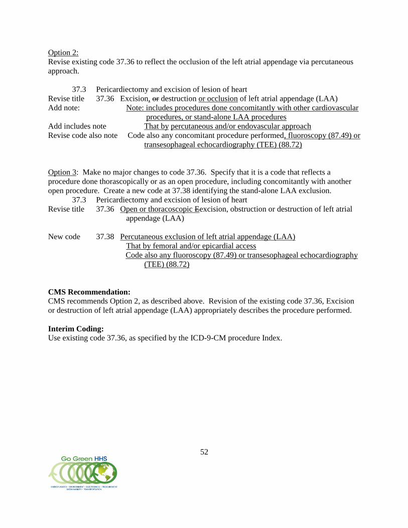
52
Option 2:
Revise existing code 37.36 to reflect the occlusion of the left atrial appendage via percutaneous
approach.
37.3 Pericardiectomy and excision of lesion of heart
Revise title 37.36 Excision, or destruction or occlusion of left atrial appendage (LAA)
Add note: Note: includes procedures done concomitantly with other cardiovascular
procedures, or stand-alone LAA procedures
Add includes note That by percutaneous and/or endovascular approach
Revise code also note Code also any concomitant procedure performed, fluoroscopy (87.49) or
transesophageal echocardiography (TEE) (88.72)
Option 3: Make no major changes to code 37.36. Specify that it is a code that reflects a
procedure done thorascopically or as an open procedure, including concomitantly with another
open procedure. Create a new code at 37.38 identifying the stand-alone LAA exclusion.
37.3 Pericardiectomy and excision of lesion of heart
Revise title 37.36 Open or thoracoscopic Eexcision, obstruction or destruction of left atrial
appendage (LAA)
New code 37.38 Percutaneous exclusion of left atrial appendage (LAA)
That by femoral and/or epicardial access
Code also any fluoroscopy (87.49) or transesophageal echocardiography
(TEE) (88.72)
CMS Recommendation:
CMS recommends Option 2, as described above. Revision of the existing code 37.36, Excision
or destruction of left atrial appendage (LAA) appropriately describes the procedure performed.
Interim Coding:
Use existing code 37.36, as specified by the ICD-9-CM procedure Index.

53
Ultrasonic Wound Debridement
Issue: Misonix Ultrasonic Wound Debridement System utilizes an ultrasonic metallic probe and
a water irrigation system to debride wounds. This procedure is currently captured by code 86.28,
Nonexcisional debridement of wound, infection, or burn. The manufacturer has requested that
indexing and tabular entries be changed so that this procedure would be captured by code 86.22,
Excisional debridement of wound, infection, or burn instead of code 86.28.
New Technology Application? No
Food & Drug Administration (FDA) approval: Yes, the Misonix SonicOne contact wound
debridement system has been cleared under 510K #: K050776.
Background: Contact ultrasonic wound debridement systems use ultrasonically energized
instruments and utilize water irrigation for tissue excision. The ultrasonic probe is brought into
direct contact with the tissue targeted for debridement. The probe‟s vibratory motion acts as a
micro jackhammer, creating local cavitational and hydrodynamic effects that dissect tissue at the
point of contact. The manufacturer states that the system is applicable for tissue excision outside
the wound margin, as well as within it. However the manufacturer also emphasizes that an
advantage of this system is that “viable tissue structures are more resistant to destruction” and
the system “flushes the wound of fibrin deposits…while preserving the granulation tissue.”
Advice in both the American Hospitals Association’s Coding Clinic for ICD-9-CM, as well as
indexing and tabular entries in ICD-9-CM for water scalpel (jet) procedures, leads to a code
assignment of 86.28. This Ultrasonic Wound Debridement System is also captured by code
86.28. The manufacturer disagrees with this code assignment and believes that this technology
performs an excisional debridement.
Coding options:
Option 1.
Continue capturing Misonix Ultrasonic Wound Debridement System and other ultrasonic
mechanical debridement systems through code 86.28, Nonexcisional debridement of wound,
infection, or burn.
Add an index entry and a tabular entry under code 86.28 for Ultrasonic mechanical debridement
to further clarify this code assignment. As indicated above, the process is not typically used to
excise block elements that can be available for examination but to pulverize (ablate) devitalized
tissue and debris into small particles for removal by the irrigation fluid. Additionally, although
this product does provide the capability of creating incisions (cuts) into vital tissue which is the
hallmark of excisional debridement, it is not designed, marketed or typically used to leverage
that ability.

54
Option 2.
Change the indexing and tabular entries so that ultrasonic mechanical debridement systems
would be assigned to code 86.22, Excisional debridement of wound, infection, or burn instead of
86.28, Nonexcisional debridement of wound, infection, or burn.
CMS Recommendation: Option 1, Continue capturing Misonix Ultrasonic Wound
Debridement System and other ultrasonic mechanical debridement systems through code 86.28,
Nonexcisional debridement of wound, infection, or burn. Add an index entry and a tabular entry
under code 86.28 for Ultrasonic mechanical debridement.
Interim coding: Capture the use of Ultrasonic Wound debridement through code 86.28,
Nonexcisional debridement of wound, infection, or burn.

55
Hydrosurgery/Versajet Debridement
Issue: Versajet™ Hydrosurgery System, is a high-pressure, high velocity water jet that assists in
the removal of devitalized tissue in the treatment of certain wounds. The System uses pressurized
streams of sterile fluid to ablate and remove tissue and foreign matter from wounds and to
remove material in a variety of surgical applications. The system depends on the use of repeated
passes of the hydrostatic handpiece over the surface of the wound in order to remove surface
elements in the manner of shaving or curettage. This procedure is currently captured in code
86.28, Nonexcisional debridement of wound, infection, or burn.
New Technology? No.
Food & Drug Administration (FDA) approval: The Versajet received 510(k) clearance on
June 22, 2001.
Background: Advice in both the American Hospitals Association’s Coding Clinic for ICD-9-
CM, as well as indexing and tabular entries in ICD-9-CM for water scalpel (jet) procedures,
leads to a code assignment of 86.28 as previously stated. The manufacturer disagrees with this
code assignment and believes that the VersaJet performs an excisional debridement. The
manufacturer states that other water jet procedures will not remove tissue outside the wound
margins and thus do not meet the definition of excisional debridement. However, they believe
that VersaJet differs from other water jet devices in that it does cut outside the margins of the
wound and meets the definition of “excisional” debridement as defined in the guidance. In order
to more clearly capture the debridement performed through the VersaJet Hydrosurgery System,
they have requested the creation of a new code.
The manufacturer states that VersaJet Hydrosurgery System uses pressurized streams of sterile
fluid to “cut, ablate and remove” tissue and foreign matter from wounds and “resects and
removes” material in a variety of surgical applications. The manufacturer states that the device
provides cutting, irrigation and evacuation in the same tool. The stream of fluid simultaneously
washes the tissue surface and removes foreign material from the wound or surgical site. The
stream of saline simultaneously washes the tissue surface and vacuums away foreign material,
including contamination and infected and necrotic tissue from the wound. The manufacturer
states that the fluid acts to ablate the surface of the tissue and propel the tissue and debris out of
the surgical site. The debris and fluid are directed immediately within the instrument into a
flexible tube, which carries the effluent to the drain or a collection canister.
The System employs two basic system components: the reusable power console unit and the
sterile, disposable pump cartridge, handpiece and tubing assembly.

56
Coding Options:
Option 1. Do not create a new code. Continue capturing the use of hydrosurgery for
debridement, including that by Versajet, under code 86.28.
Option 2. Create a new code for debridement performed using hydrosurgery as follows:
New Code: 86.20 Debridement of wound, infection or burn using hydrosurgery
CMS recommendation: Option 1. Do not create a new code. The process does not excise
block elements that can be available for examination but pulverizes (ablates) the tissue into small
particles for vacuum removal by Venturi effect. Additionally, the process is approved for the
removal of devitalized tissue and debris and is marketed for its ability to spare vital tissue. It
does not provide the capability of creating incisions (cuts) into vital tissue that are both the
hallmark and risk of excisional debridement. Continue capturing the use of hydrosurgery for
debridement, including that by Versajet, under code 86.28.
Interim coding: Capture the use of hydrosurgery for debridement, including that by Versajet,
under code 86.28, Nonexcisional debridement of wound, infection, or burn

57
Non-excisional Debridement
Issue: Currently, procedure code 86.28, Nonexcisional debridement of wound, infection, or
burn, is located within subcategory 86.2, Excision or destruction of lesion or tissue of skin and
subcutaneous tissue. A requestor believes it is inappropriate to report debridement of tissue
other than skin and subcutaneous tissue with a code located in the chapter for Operations on the
Integumentary System. The request is to create new codes to report non excisional debridement
of the deeper tissue layers (soft tissue, fascia, muscle and bone). Should new codes be created?
Coding options:
Option 1. Do not create new codes. Continue to use existing code 86.28, Nonexcisional
debridement of wound, infection, or burn, to identify all nonexcisional debridements.
Option 2. Create a new subcategory and codes to identify nonexcisional debridement of tendon,
muscle, deep fascia and bone as outlined below. Add exclusion notes to code 86.28.
New subcategory 17.8 Nonexcisional debridement of wound, infection, or burn of soft
tissue, fascia, muscle, or bone
Debridement NOS
Removal of devitalized tissue, necrosis, and slough by such
methods as:
brushing
irrigation (under pressure)
scrubbing
washing
Water scalpel (jet)
New code 17.81 Non excisional debridement of soft tissue
Tendon
New code 17.82 Non excisional debridement of deep fascia
New code 17.83 Non excisional debridement of muscle
New code 17.84 Non excisional debridement of bone
CMS Recommendation: Do not create new codes. Debridement of the deeper tissues through a
wound, infection or burn is part of the care of pathology in the integumentary system and is
appropriately coded by 86.28.
Interim coding advice: Continue to use existing code 86.28, Nonexcisional debridement of
wound, infection, or burn, to identify all nonexcisional debridements.

58
Cerebral and Somatic Oximetry
Issue: We received a request to create a unique ICD-9-CM procedure code for tissue oxygen
saturation monitoring using Near-infrared spectroscopy. Near-infrared spectroscopy (NIRS) is
currently captured in code 38.23, Intravascular spectroscopy, which has a table notation
“Includes near infrared (NIR) spectroscopy.” NIR spectroscopy is used as a noninvasive method
for the continuous, real-time monitoring of both cerebral and somatic tissue oxygenation in order
to detect regional hypoperfusion.
We received a request to create new ICD-9-CM codes for Tissue Oxygen Saturation Monitoring
using Near-Infrared Spectroscopy and Cerebral Oxygen Saturation Monitoring using Near-
Infrared Spectroscopy. The requestor states that the creation of a new ICD-9-CM procedure
code would allow reporting and tracking of the use of these procedures.
New Technology Application? No.
Food and Drug Administration (FDA) Approval: The Somanetics INVOS®
Cerebral/Somatic Oximeter first received FDA 510(k) clearance in May of 1996; in 2008 the
somatic oximeter was cleared for monitoring of oxygen saturation in tissues beneath the sensor
in individuals at risk for reduced-flow or no-flow ischemic states.
Background: NIRS is a noninvasive, optical method for continuous, real-time monitoring of
tissue oxygenation. The NIRS technique is based on the transmission and absorption of near
infrared (NIR) light (700 – 950 nm) as it passes through tissue. Oxygenated and unoxygenated
hemoglobin have different absorption characteristics, allowing the system to reflect the oxygen
saturation of the target tissue. Because near infrared light penetrates tissue relatively easily, the
sensor reflects the saturation of deep tissues rather than surface capillary beds. Variation in
penetration does not permit the device to report absolute levels; rather NIRS is used to track
trends in oxygenation levels once a baseline has been established at a particular site in a given
patient.
NIRS was first used to perform cerebral oximetry in cardiac surgery arena, but has subsequently
been used to monitor the adequacy of tissue perfusion on somatic sites such as muscle, kidneys
and the intestines, and to provide an early alert to unexpected hemodynamic changes.

59
Coding Options
Option 1: Do not create new coding structures for Tissue Oxygen Saturation Monitoring
Procedures using Near-Infrared Spectroscopy (NIRS). Continue capturing this through code
38.23, Intravascular spectroscopy.
Option 2: Create a new procedure code as follows:
New code XX.XY Tissue oxygen saturation monitoring using Near-Infrared Spectroscopy,
where the terminal digit, Y, is used to specify the target organ.
Option 3: Create new procedure codes within each system, e.g. 01.NN Cerebral Oxygen
Saturation Monitoring using Near-Infrared Spectroscopy
CMS Recommendation: Option 1. Do not create a new code.
Interim coding advice: Continue to use code 38.23, Intravascular spectroscopy.

60
Ultrasound Assisted Lysis of Intravascular Thrombus
Issue: There is currently no specific code which uniquely identifies the procedure of placement
and monitoring of the ultrasound assisted lysis catheter for arterial and venous thrombus.
New Technology Application? No.
Background: Traditional catheter-directed thrombolysis is a minimally invasive endovascular
procedure that dissolves blood clots in blood vessels to help improve blood flow and prevent
damage to tissues and organs. In a catheter-directed thrombolysis procedure, x-ray imaging is
used to help guide a catheter to the site of blood clots to deliver medication to dissolve the
blockage.
A new FDA approved catheter based system combines a drug delivery catheter with an
ultrasound core wire to augment drug-induced thrombolysis. The catheter has two components:
1) a drug delivery catheter and 2) an ultrasound core wire with small ultrasound transducers that
emit high frequency, low power sound waves into the clot. Ultrasound makes the clot more
porous and therefore able to absorb more lytic than clots that are not exposed to sound waves.
Percutaneous Mechanical Thrombectomy (PMT) uses other devices to break up the thrombus.
Ultrasound is not viewed by the manufacturer as ablative but as an adjunct to increase the
efficacy of the pharmaceutical lytic.
Ultrasound Assisted Lysis of Arterial or Venous Thrombus Procedure Description
The physician accesses the femoral artery or vein and performs an angiogram to identify the
anatomy and thrombus formation. A guidewire is placed through the access point and advanced
through the thrombus. The delivery catheter is placed over the guidewire into the thrombus.
Placement of the catheter is checked with fluoroscopy. The catheter is connected to the infusion
pump and lytic and ultrasound therapy is started.
The patient is returned to the cath lab for additional angiograms to monitor progress. If the
thrombus is still present after 24 hours of therapy, the clot is not receptive to CDT and another
option may be considered. Once the thrombus is dissolved and any obstruction treated, the
ultrasound assisted lysis catheter is removed from the patient.
Coding options
Option 1:
Do not create a new ICD-9-CM procedure code. Use codes in Category 00.0, Therapeutic
ultrasound, (Codes 00.01 – 00.09) to identify the ultrasound technology.
Then use code 99.10, Injection or infusion of thrombolytic agent, to describe the intravenous
delivery of thrombolytics such as tissue plasminogen activator (tPA or altepase).
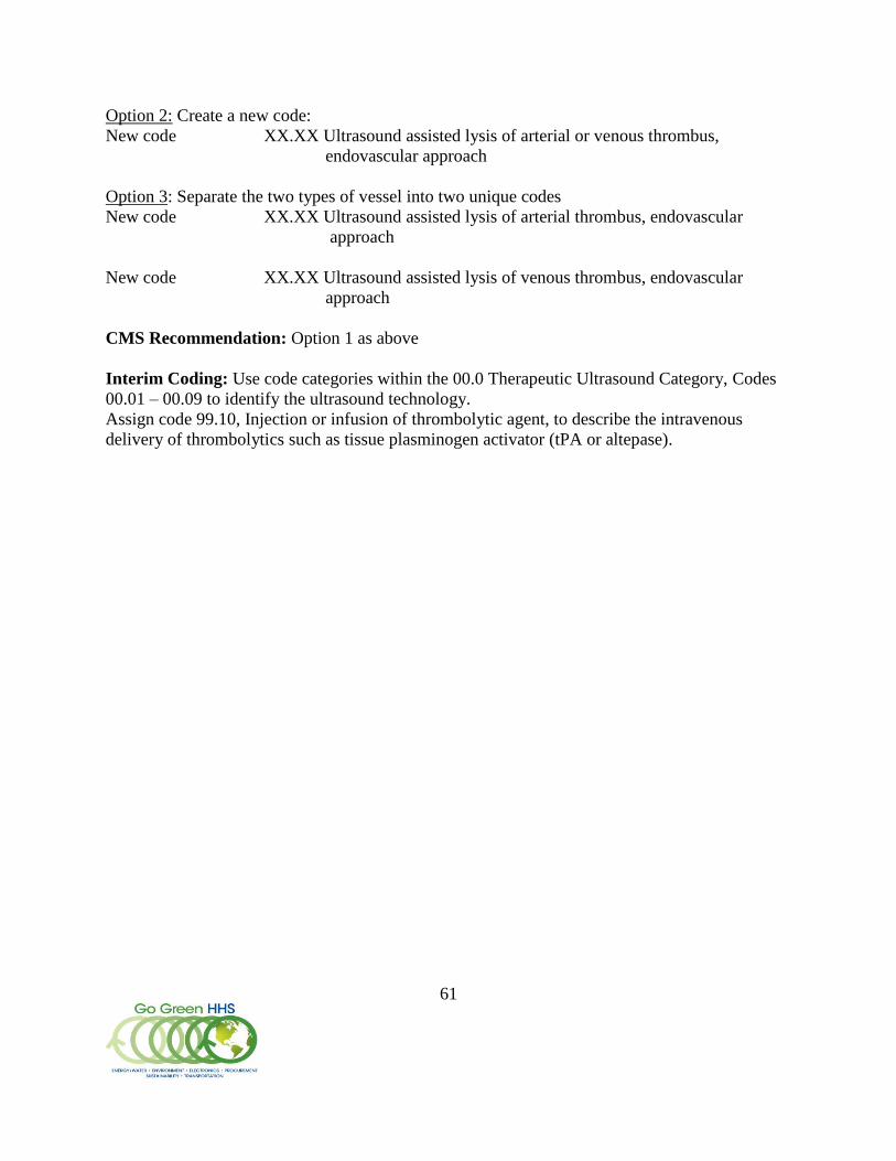
61
Option 2: Create a new code:
New code XX.XX Ultrasound assisted lysis of arterial or venous thrombus,
endovascular approach
Option 3: Separate the two types of vessel into two unique codes
New code XX.XX Ultrasound assisted lysis of arterial thrombus, endovascular
approach
New code XX.XX Ultrasound assisted lysis of venous thrombus, endovascular
approach
CMS Recommendation: Option 1 as above
Interim Coding: Use code categories within the 00.0 Therapeutic Ultrasound Category, Codes
00.01 – 00.09 to identify the ultrasound technology.
Assign code 99.10, Injection or infusion of thrombolytic agent, to describe the intravenous
delivery of thrombolytics such as tissue plasminogen activator (tPA or altepase).

62
Addenda Item
At the September 15, 2010 C&M Meeting, we discussed Endovascular Embolization with Head
or Neck Vessel Reconstruction – the Pipeline™ Embolization Device. This device mimics
devices and stents that are already on the market, and we had difficulty during the discussion of
differentiating this device from stents that are available for use in head or neck vessels. At the
time of the meeting, CMS did not have a clear recommendation regarding creation of a new code
or codes, and asked for input from the audience. The written responses we received were not
overwhelmingly in support of creation of a new code. Additionally, the Company has not sought
New Technology status from CMS for the 2012 fiscal year. At the time, the suggestion was
brought to the floor to add a new procedure code as follows:
New code 39.77 Endovascular embolization with head or neck vessel
vascular remodeling support
Embolization stent
Stent-like device
That for repair of aneurysm
CMS‟ Recommendation is Option 1; do not create a new procedure code to describe this device.
CMS‟ interim coding advice remains the same: continue to use procedure code 39.72,
Endovascular embolization or occlusion of head and neck vessels, to describe this procedure.
We are interested in hearing additional comments about this topic.

63
Proposed Addenda
Tabular
Revise code title 00.61 Percutaneous angioplasty or atherectomy of precerebral
(extracranial) vessel(s)
Revise code title 00.62 Percutaneous angioplasty or atherectomy of intracranial
intracerebral vessel(s)
Revise code also note Code also any:
Percutaneous insertion of intracranial intracerebral stent(s)
(00.65)
Revise code title 00.64 Percutaneous insertion of other precerebral (extracranial)
artery stent(s)
Revise code title 00.65 Percutaneous insertion of intracranial intracerebral
artery stent(s)
Revise code also note Code also any:
Percutaneous angioplasty or atherectomy of intracranial
intracerebral vessel(s) (00.62)
00.94 Intra-operative neurophysiologic monitoring
Add inclusion term That by:
Add inclusion term brainstem auditory evoked potentials [BAEP]
Add inclusion term electroencephalogram [EEG]
Add inclusion term electromyogram [EMG]
Add inclusion term motor evoked potentials [MEP]
Add inclusion term nerve conduction study
Add inclusion term somatosensory evoked potentials [SSEP]
Add inclusion term transcranial Doppler
Revise code title 13.65 Excision of secondary membrane [after cataract]
Capsulectomy
Capsulectomy Folio issue – inclusion term was in
title
88.7 Diagnostic ultrasound
Add exclusion term Excludes: that for intraoperative monitoring 00.94

64
89.19 Video and radio-telemetered electroencephalographic monitoring
Add exclusion term Excludes: intraoperative monitoring 00.94
93.08 Electromyography
Add exclusion term Excludes: that for intraoperative monitoring 00.94
Index
Angioplasty (laser) – see also Repair, blood vessel
percutaneous transluminal (balloon)
Revise subterm basilar 00.61
Add subterm precerebral 00.61
Add subterm intracerebral 00.62
Add term Cleft lift 86.21
Dopplergram, Doppler flow mapping – see also Ultrasonography
Add subterm intraoperative transcranial 00.94
Electroencephalogram (EEG) 89.14
monitoring (radiographic) (video) 89.19
Add subterm intraoperative 00.94
Infusion
Add subterm lymphocyte 99.09
Monitoring
electroencephalographic 89.19
Add subterm intraoperative 00.94
Revise subterm intraoperative anesthetic effect monitoring and titration 00.94
[89.14]
Revise subterm intraoperative
Revise subterm anesthetic effect monitoring and titration (IAEMT) 00.94
[89.14]
Add subterm neurophysiologic (BAEP) (brainstem auditory evoked
potentials) (EEG) (electroencephalogram) (electromyogram)
(EMG) (MEP) (motor evoked potentials) (nerve conduction
study) (somatosensory evoked potentials) (SSEP)
(transcranial Doppler) 00.94
Partial Code Freeze for ICD-9-CM and ICD-10 Finalized
65
The ICD-9-CM Coordination and Maintenance Committee will implement a partial
freeze of the ICD-9-CM and ICD-10 (ICD-10-CM and ICD-10-PCS) codes prior to the
implementation of ICD-10 on October 1, 2013. There was considerable support for this partial
freeze. The partial freeze will be implemented as follows:
The last regular, annual updates to both ICD-9-CM and ICD-10 code sets will be made
on October 1, 2011.
On October 1, 2012, there will be only limited code updates to both the ICD-9-CM and
ICD-10 code sets to capture new technologies and diseases as required by section 503(a)
of Pub. L. 108-173.
On October 1, 2013, there will be only limited code updates to ICD-10 code sets to
capture new technologies and diagnoses as required by section 503(a) of Pub. L. 108-173.
There will be no updates to ICD-9-CM, as it will no longer be used for reporting.
On October 1, 2014, regular updates to ICD-10 will begin.
The ICD-9-CM Coordination and Maintenance Committee will continue to meet twice a year
during the partial freeze. At these meetings, the public will be asked to comment on whether or
not requests for new diagnosis or procedure codes should be created based on the criteria of the
need to capture a new technology or disease. Any code requests that do not meet the criteria will
be evaluated for implementation within ICD-10 on and after October 1, 2014 once the partial
freeze has ended.
Codes discussed at the September 15 – 16, 2010 and March 9 – 10, 2011 ICD-9-CM
Coordination and Maintenance Committee meeting will be considered for implementation on
October 1, 2011, the last regular updates for ICD-9-CM and ICD-10. Code requests discussed at
the September 14 – 15, 2011 and additional meetings during the freeze will be evaluated for
either the limited updates to capture new technologies and diseases during the freeze period or
for implementation to ICD-10 on October 1, 2014. The public will be actively involved in
evaluating the merits of any such requests during the period of the partial freeze.

66
ICD-10-CM & ICD-10-PCS Abbreviated Titles
60 character abbreviated titles have been developed for the 2011 version of both ICD-10-CM and
ICD-10-PCS codes. The ICD-10 abbreviated titles for both ICD-10-CM and ICD-10-PCS have
been posted on the CMS website at
http://www.cms.gov/ICD10/11b1_2011_ICD10CM_and_GEMs.asp and
http://www.cms.gov/ICD10/11b_2011_ICD10PCS.asp
CMS will maintain and update these abbreviated titles each year. Users should feel free to
download and use these, and other ICD-10 files posted on the CMS website.
Version 28.0 ICD-10 MS-DRGs
The ICD-10 MS-DRGs v28 Definitions Manual (based on FY2011 MS-DRGs) is now posted on
the Centers for Medicare & Medicaid Services (CMS) website at
http://www.cms.gov/ICD10/17_ICD10_MS_DRG_Conversion_Project.asp in the “Related
Links Inside CMS” section. This update is part of the ICD-10 MS-DRG Conversion Project. In
the Conversion Project, CMS is using the General Equivalence Mappings (GEMs) to convert
CMS payment systems. CMS is sharing information learned from this project with other
organizations facing similar conversion projects. CMS has also posted the ICD-10 FY 2011
Medicare Code Editor. Please note that the ICD-10 MS-DRGs will be subject to formal
rulemaking.
The public is encouraged to review these files. A detailed presentation on this conversion
activity will take place at the September 14-15, 2011 ICD-9-CM Coordination and Maintenance
Committee Meeting.
2011 GEMs Update
The updated 2011 General Equivalence Mappings (GEMs) are posted for review and public
comment. The updated files contain all changes to date in response to public comment as
mandated by the Affordable Care Act, for the period ending November 11, 2010, as well as
additional ongoing internal review for accuracy and completeness.
Extensive public comment was received on the subject of the GEMs. Several organizations sent
general letters of support for the GEMs and asked for ongoing maintenance and increased
stakeholder input in improving and updating the GEMs. Providers, payers, vendors, independent
consultants and other individuals in the healthcare community submitted comments and
suggestions for improving the GEMs. Approximately 5,200 GEMs entries were the subject of
public comment. Roughly 250,000 total entries comprise the GEMs.
The public support and collaboration on the GEMs led to many improvements in the accuracy
and completeness of the GEMs as an ICD-10 transition resource. All comments and suggestions
were reviewed and considered. Any recommended change to an entry that met GEMs inclusion
criteria was incorporated into the updated 2011 files. Of the 5,200 comments submitted, roughly
one third of the recommended changes were either implemented for this update or had been
previously implemented in the September update, based on public comment received during the

67
past year. Approximately 850 recommended changes, or 16% of all comments received, were
new changes implemented for the 2011 GEMs, and approximately 900 additional recommended
changes, or 17% of all comments received, supported previous changes in the most recent
updated GEMs files (posted on the CMS website in September, 2010).
The 2011 ICD-10 GEM files for both ICD-10-CM and ICD-10-PCS have been posted on the
CMS website at http://www.cms.gov/ICD10/11b_2011_ICD10PCS.asp and
http://www.cms.gov/ICD10/11b1_2011_ICD10CM_and_GEMs.asp
ICD-10-PCS Device Key Being Developed
A PCS device key similar in organization and function to the PCS Body Part Key is under
development. The Device Key will help users choose the correct PCS device value for a given
clinical or industry device name. The resulting entries will be a public domain reference to
accompany the ICD-10-PCS tables. Entries will be available for lookup by both device value and
clinical or industry device name. Similar in function to “includes” notes in ICD-10-CM, the ICD-
10-PCS device key will be an official part of the classification system.
The draft device key will be posted with the FY2011 ICD-10-PCS update for public review and
comment prior to the September 14-15, 2011 ICD-9-CM Coordination and Maintenance
Committee meeting. Industry input will be solicited on this draft document. A detailed
presentation on the ICD-10-PCS device key and related reference material will take place at the
September 14-15, 2011 ICD-9-CM Coordination and Maintenance Committee Meeting.
New FY2012 ICD-10-PCS Update Schedule
The ICD-10-PCS annual update for FY2012 will follow the same update schedule as the
annual ICD-9-CM update for procedure codes. CMS is adopting this new ICD-10-PCS update
schedule in preparation for the move to ICD-10 on October 1, 2013. ICD-10-PCS changes
discussed at the September 15 – 16, 2010 and March 9 – 10, 2011 ICD-9-CM Coordination and
Maintenance Committee meeting will be considered for inclusion in the FY 2012 ICD-10-PCS
updates. The updated timeline will be as follows:
June 2011 The FY 2012 ICD-10-PCS final addenda will be posted on the ICD-10 CMS
website at http://www.cms.gov/ICD10/
October 2011 The FY 2012 ICD-10-PCS GEMs and the Procedure Reimbursement Mappings
will be posted on the ICD-10 CMS website at http://www.cms.gov/ICD10/
This new schedule of posting ICD-10-PCS final addenda in June and the updated ICD-10-PCS
GEMs and Reimbursement mappings in October will continue each year up to and after the
implementation of ICD-10.

68
Ankle, Hip and Knee Joint Replacement: Request for New Values in PCS Tables
The original proposal is to create codes that differentiate between cemented and uncemented
metal on polyethylene ankle replacement procedures. After consultation with the orthopedic
community, it was proposed that this information be included for hip and knee replacement
procedures as well. According to the original proposal, the current industry standard is to use
only metal on polyethylene implants for the ankle joint. Therefore, additional information
regarding the synthetic ankle joint does not need to be added to the PCS tables at this time.
Option 1: Do not create new PCS device values to differentiate between cemented and
uncemented synthetic joint replacement procedures.
Section: 0 Medical and Surgical
Body System: S Lower Joints
Operation: R Replacement: Putting in or on biological or synthetic material that physically
takes the place and /or function of all or a portion of a body part
Body Part Approach Device Qualifier
C Knee Joint, Right
D Knee Joint, Left
F Ankle Joint, Right
G Ankle Joint, Left
T Knee Joint, Femoral Surface, Right
U Knee Joint, Femoral Surface, Left
V Knee Joint, Tibial Surface, Right
W Knee Joint, Tibial Surface, Left
0 Open J Synthetic Substitute Z No Qualifier
Body Part Approach Device Qualifier
9 Hip Joint, Right
B Hip Joint, Left
0 Open J Synthetic Substitute 5 Metal on Polyethylene
6 Metal on Metal
7 Ceramic on Ceramic
8 Ceramic on Polyethylene
Z No Qualifier
Body Part Approach Device Qualifier
A Hip Joint, Acetabular Surface, Right
E Hip Joint, Acetabular Surface, Left
0 Open J Synthetic Substitute F Metal
G Ceramic
H Polyethylene
Z No Qualifier

69
Body Part Approach Device Qualifier
R Hip Joint, Femoral Surface, Right
S Hip Joint, Femoral Surface, Left
0 Open J Synthetic Substitute F Metal
G Ceramic
Z No Qualifier
Option 2: Add new device value to specify cemented implants for hip, knee and ankle joint
body parts in table 0SR (32 codes). Specify uncemented implants using qualifier J Synthetic
Substitute. This assumes that all implants are either cemented or uncemented.
Section: 0 Medical and Surgical
Body System: S Lower Joints
Operation: R Replacement: Putting in or on biological or synthetic material that physically
takes the place and /or function of all or a portion of a body part
Body Part Approach Device Qualifier
C Knee Joint, Right
D Knee Joint, Left
F Ankle Joint, Right
G Ankle Joint, Left
T Knee Joint, Femoral Surface, Right
U Knee Joint, Femoral Surface, Left
V Knee Joint, Tibial Surface, Right
W Knee Joint, Tibial Surface, Left
0 Open H Synthetic Substitute,
Cemented
J Synthetic Substitute
Z No Qualifier
Body Part Approach Device Qualifier
9 Hip Joint, Right
B Hip Joint, Left
0 Open H Synthetic Substitute, Cemented
J Synthetic Substitute
5 Metal on Polyethylene
6 Metal on Metal
7 Ceramic on Ceramic
8 Ceramic on Polyethylene
Z No Qualifier
Body Part Approach Device Qualifier
A Hip Joint, Acetabular Surface,
Right
E Hip Joint, Acetabular Surface,
Left
0 Open H Synthetic Substitute,
Cemented
J Synthetic Substitute
F Metal
G Ceramic
H Polyethylene
Z No Qualifier
Body Part Approach Device Qualifier
R Hip Joint, Femoral Surface,
Right
S Hip Joint, Femoral Surface,
Left
0 Open H Synthetic Substitute,
Cemented
J Synthetic Substitute
F Metal
G Ceramic
Z No Qualifier

70
Option 3: Add two new device values, one specifying cemented implants and one specifying
uncemented implants (64 codes). This allows an “unspecified” option if the documentation is
insufficient to determine whether the synthetic joint substitute was cemented or uncemented.
Section: 0 Medical and Surgical
Body System: S Lower Joints
Operation: R Replacement: Putting in or on biological or synthetic material that physically
takes the place and /or function of all or a portion of a body part
Body Part Approach Device Qualifier
C Knee Joint, Right
D Knee Joint, Left
F Ankle Joint, Right
G Ankle Joint, Left
T Knee Joint, Femoral Surface, Right
U Knee Joint, Femoral Surface, Left
V Knee Joint, Tibial Surface, Right
W Knee Joint, Tibial Surface, Left
0 Open G Synthetic Substitute,
Uncemented
H Synthetic Substitute,
Cemented
J Synthetic Substitute
Z No Qualifier
Body Part Approach Device Qualifier
9 Hip Joint,
Right
B Hip Joint, Left
0 Open G Synthetic Substitute,
Uncemented
H Synthetic Substitute, Cemented
J Synthetic Substitute
5 Metal on Polyethylene
6 Metal on Metal
7 Ceramic on Ceramic
8 Ceramic on
Polyethylene
Z No Qualifier
Body Part Approach Device Qualifier
A Hip Joint, Acetabular Surface,
Right
E Hip Joint, Acetabular Surface,
Left
0 Open G Synthetic Substitute,
Uncemented
H Synthetic Substitute,
Cemented
J Synthetic Substitute
F Metal
G Ceramic
H Polyethylene
Z No Qualifier
Body Part Approach Device Qualifier
R Hip Joint, Femoral Surface,
Right
S Hip Joint, Femoral Surface,
Left
0 Open G Synthetic Substitute,
Uncemented
H Synthetic Substitute,
Cemented
J Synthetic Substitute
F Metal
G Ceramic
Z No
Qualifier

71
CMS recommendation: Option 3.
Interspinous Process Internal Fixation Procedures: Request for Adding New Values in PCS Tables The proposal is to add PCS values to the PCS Insertion tables that will distinguish between
dynamic stabilization and static distraction interspinous process internal fixation devices of the
spine. PCS has standardized values in the Insertion tables for Upper and Lower Joints body
systems for insertion of interspinous process devices, without differentiating between dynamic
stabilization and static distraction devices. To extend the level of detail available for the root
operation Insertion to differentiate the procedures described above requires new qualifier values
in the PCS tables as specified in the proposal.
Option 1: Current PCS coding, shown in example 0SH Lower Joints body system
Section: 0 Medical and Surgical
Body System: S Lower Joints
Operation: H Insertion: Putting in a nonbiological appliance that monitors, assists, performs,
or prevents a physiological function but does not physically take the place of a
body part
Body Part Approach Device Qualifier
0 Lumbar Vertebral Joint
3 Lumbosacral Joint
0 Open
3 Percutaneous
4 Percutaneous Endoscopic
4 Internal Fixation Device 2 Interspinous Process
3 Pedicle-Based Dynamic
Stabilization
Z No Qualifier
Option 2: Create new PCS values in tables 0RH and 0SH for vertebral joint body parts to
specify Interspinous Process, Dynamic Stabilization and Interspinous Process, Static Distraction
in the 7th
character. (12 codes)
Section: 0 Medical and Surgical
Body System: S Lower Joints
Operation: H Insertion: Putting in a nonbiological appliance that monitors, assists, performs,
or prevents a physiological function but does not physically take the place of a
body part

72
Body Part Approach Device Qualifier
0 Lumbar Vertebral
Joint
3 Lumbosacral Joint
0 Open
3 Percutaneous
4 Percutaneous Endoscopic
4 Internal Fixation
Device
2 Interspinous Process
3 Pedicle-Based
Dynamic Stabilization
9 Interspinous Process,
Dynamic Stabilization
B Interspinous Process,
Static Distraction
Z No Qualifier
Option 3: Revise existing PCS 7th
character qualifier value Interspinous Process in tables 0RH
and 0SH for vertebral joint body parts to specify Interspinous Process, Dynamic Stabilization.
Create new 7th
character qualifier value PCS value in tables 0RH and 0SH for vertebral joint
body parts to specify Interspinous Process, Static Distraction. (6 codes)
Section: 0 Medical and Surgical
Body System: S Lower Joints
Operation: H Insertion: Putting in a nonbiological appliance that monitors, assists, performs,
or prevents a physiological function but does not physically take the place of a
body part
Body Part Approach Device Qualifier
0 Lumbar Vertebral Joint
3 Lumbosacral Joint
0 Open
3 Percutaneous
4 Percutaneous Endoscopic
4 Internal Fixation
Device
2 Interspinous Process,
Dynamic Stabilization
3 Pedicle-Based Dynamic
Stabilization
9 Interspinous Process,
Static Distraction
Z No Qualifier
CMS Recommendation: CMS does not have a formal recommendation at this time. We would
like to hear comments from the audience regarding documentation for this level of detail.
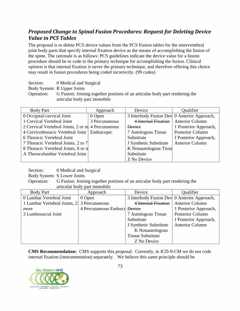
73
Proposed Change to Spinal Fusion Procedures: Request for Deleting Device Value in PCS Tables The proposal is to delete PCS device values from the PCS Fusion tables for the intervertebral
joint body parts that specify internal fixation device as the means of accomplishing the fusion of
the spine. The rationale is as follows: PCS guidelines indicate the device value for a fusion
procedure should be to code to the primary technique for accomplishing the fusion. Clinical
opinion is that internal fixation is never the primary technique, and therefore offering this choice
may result in fusion procedures being coded incorrectly. (99 codes)
Section: 0 Medical and Surgical
Body System: R Upper Joints
Operation: G Fusion: Joining together portions of an articular body part rendering the
articular body part immobile
Body Part Approach Device Qualifier
0 Occipital-cervical Joint
1 Cervical Vertebral Joint
2 Cervical Vertebral Joints, 2 or more
4 Cervicothoracic Vertebral Joint
6 Thoracic Vertebral Joint
7 Thoracic Vertebral Joints, 2 to 7
8 Thoracic Vertebral Joints, 8 or more
A Thoracolumbar Vertebral Joint
0 Open
3 Percutaneous
4 Percutaneous
Endoscopic
3 Interbody Fusion Device
4 Internal Fixation
Device
7 Autologous Tissue
Substitute
J Synthetic Substitute
K Nonautologous Tissue
Substitute
Z No Device
0 Anterior Approach,
Anterior Column
1 Posterior Approach,
Posterior Column
J Posterior Approach,
Anterior Column
Section: 0 Medical and Surgical
Body System: S Lower Joints
Operation: G Fusion: Joining together portions of an articular body part rendering the
articular body part immobile
Body Part Approach Device Qualifier
0 Lumbar Vertebral Joint
1 Lumbar Vertebral Joints, 2 or
more
3 Lumbosacral Joint
0 Open
3 Percutaneous
4 Percutaneous Endoscopic
3 Interbody Fusion Device
4 Internal Fixation
Device
7 Autologous Tissue
Substitute
J Synthetic Substitute
K Nonautologous
Tissue Substitute
Z No Device
0 Anterior Approach,
Anterior Column
1 Posterior Approach,
Posterior Column
J Posterior Approach,
Anterior Column
CMS Recommendation: CMS supports this proposal. Currently, in ICD-9-CM we do not code
internal fixation (instrumentation) separately. We believe this same principle should be

74
followed in ICD-10-PCS, thereby making it appropriate to delete the Internal Fixation Device
option from the PCS code tables.

75
Implantable Meshes—Request for New Detail in ICD-10-PCS Tables The proposal is to create PCS values that differentiate between nonautologous tissue of human
origin and nonautologous tissue of non-human origin for the root operation Supplement in the
Anatomical Regions, Urinary and Female Reproductive body systems.
Reconstruction of soft tissue defects can be performed by suture but, because it reduces tension
and decreases the rate of defect recurrence, mesh is used to reconstruct larger defects, for
reinforcement and to strengthen tissue.
Mesh is either synthetic or biologic. Synthetic patches and plugs are manufactured from
materials such as polypropylene and PTFE. They are easy to use but have a higher risk of
complications like infection and adhesions. Biologic mesh is composed of nonautologous tissue.
It tends to be used in inpatient cases where patients have aggravating factors such as obesity,
heavy smoking, contaminated surgical site, and loss of fascia. Nonautologous mesh is bio-
engineered to remove cells which can cause rejection, rendering a collagen-rich matrix. This
promotes tissue regeneration and revascularization, reducing the risk of complications.
Nonautologous mesh is either of human origin using tissue derived from cadavers , eg.
Alloderm, or of non-human origin (zooplastic) using bovine or porcine tissue, eg. Permacol.
There are currently no significant data sources which enable clinical analysis of the differences
in patient outcomes, effectiveness and adverse events between nonautologous mesh of human
and non-human origin.
PCS has standardized values for distinguishing between nonautologous tissue of human origin
and nonautologous tissue of non-human origin, as shown in this excerpt from table 02U, root
operation Supplement for the Heart and Great Vessels body system.
Section: 0 Medical and Surgical
Body System: 2 Heart and Great Vessels
Operation: U Supplement: Putting in or on biological or synthetic material that physically
reinforces and/or augments the function of a portion of a body part
Body Part Approach Device Qualifier
5 Atrial Septum
6 Atrium, Right
7 Atrium, Left
0 Open
3 Percutaneous
4 Percutaneous
Endoscopic
7 Autologous Tissue
Substitute
8 Zooplastic Tissue
J Synthetic Substitute
K Nonautologous
Tissue Substitute
Z No Qualifier
To extend the level of detail available for the root operation Supplement to additional body
systems requires only adding the existing device value Zooplastic Tissue to the body systems and

76
root operations specified in the proposal. Example shown is an excerpt from table 0YU, the
Lower Extremities Anatomical Regions.
Option 1: Current PCS coding
Section: 0 Medical and Surgical
Body System: Y Anatomical Regions, Lower Extremities
Operation: U Supplement: Putting in or on biological or synthetic material that physically
reinforces and/or augments the function of a portion of a body part
Body Part Approach Device Qualifier
5 Inguinal Region,
Right
6 Inguinal Region,
Left
7 Femoral Region,
Right
8 Femoral Region,
Left
A Inguinal Region,
Bilateral
E Femoral Region,
Bilateral
0 Open
4 Percutaneous
Endoscopic
7 Autologous Tissue
Substitute
J Synthetic Substitute
K Nonautologous
Tissue Substitute
Z No Qualifier
Option 2: Extend existing device value Zooplastic Tissue to root operation Supplement tables in
body systems T Urinary, U Female Reproductive, W General Anatomical Regions, and Y Lower
Extremities Anatomical Regions. (144 new codes, for all body parts in tables 0TU, 0UU, 0WU
and 0YU)
Section: 0 Medical and Surgical
Body System: Y Anatomical Regions, Lower Extremities
Operation: U Supplement: Putting in or on biological or synthetic material that physically
reinforces and/or augments the function of a portion of a body part
Body Part Approach Device Qualifier
5 Inguinal Region,
Right
6 Inguinal Region,
Left
7 Femoral Region,
Right
8 Femoral Region,
Left
0 Open
4 Percutaneous
Endoscopic
7 Autologous Tissue
Substitute
8 Zooplastic Tissue
J Synthetic Substitute
K Nonautologous
Tissue Substitute
Z No Qualifier

77
Body Part Approach Device Qualifier
A Inguinal Region,
Bilateral
E Inguinal Region,
Bilateral
CMS recommendation: Option 2.

78
Intraoperative Nerve Measurement and Monitoring: Request for Extension of Existing Values in PCS Tables The proposal is to add PCS values to the PCS Measurement and Monitoring section that will
expand the body parts and approaches available for coding monitoring procedures of the nervous
system. The proposed change would add the body part value Peripheral Nervous and the
approach value Percutaneous to the PCS tables 4A0 and 4A1, as specified in the proposal.
Option 1: Current PCS coding, as shown in the root operation Monitoring table 4A1.
Section: 4 Measurement and Monitoring
Body System: A Physiological Systems
Operation: 1 Monitoring: Determining the level of a physiological or physical function
repetitively over a period of time
Body System Approach Function/Device Qualifier
0 Central Nervous 0 Open
X External
4 Electrical Activity G Intraoperative
Z No Qualifier
Option 2: Extend existing PCS values in table 4A0 and 4A1 to include the body part value
Peripheral Nervous and the approach value Percutaneous. (12 codes)
Section: 4 Measurement and Monitoring
Body System: A Physiological Systems
Operation: 1 Monitoring: Determining the level of a physiological or physical function
repetitively over a period of time
Body System Approach Function/Device Qualifier
0 Central Nervous
1 Peripheral Nervous
0 Open
3 Percutaneous
X External
4 Electrical Activity G Intraoperative
Z No Qualifier
CMS Recommendation: Option 2 as displayed above.

79
Regional Brain Oxygen Saturation Monitoring Using Near-Infrared Spectroscopy: Request for Extension of Existing Values in PCS Tables The proposal is to add PCS values to the PCS Monitoring tables that will distinguish invasive
intracranial oxygen saturation monitoring from non-invasive external monitoring of regional
brain oxygen saturation using near-infrared spectroscopy. PCS has standardized values in the
Measurement and Monitoring section for recording monitoring of oxygen saturation.
To extend the level of detail available for the root operation Monitoring to differentiate the
procedures specified above requires only adding the existing approach value External and the
existing qualifier value No Qualifier to the PCS table 4A1, as specified in the proposal.
Option 1: Current PCS coding
Section: 4 Measurement and Monitoring
Body System: A Physiological Systems
Operation: 1 Monitoring: Determining the level of a physiological or physical function
repetitively over a period of time
Body System Approach Function/Device Qualifier
0 Central Nervous 3 Percutaneous
7 Via Natural or
Artificial Opening
B Pressure
K Temperature
R Saturation
D Intracranial
Option 2: Extend existing PCS values in table 4A1 to specify External approach in the 5th
character and No Qualifier in the 7th
character, for external oxygen saturation monitoring as well
as pressure and temperature monitoring, for both the regional brain and intracranial sites. (6
codes)
Section: 4 Measurement and Monitoring
Body System: A Physiological Systems
Operation: 1 Monitoring: Determining the level of a physiological or physical function
repetitively over a period of time
Body System Approach Function/Device Qualifier
0 Central Nervous 3 Percutaneous
7 Via Natural or
Artificial Opening
X External
B Pressure
K Temperature
R Saturation
D Intracranial
Z No Qualifier
Option 3: Extend existing PCS values in table 4A1 to specify External approach in the 5th
character and No Qualifier in the 7th
character, only apply the 6th
character function Saturation, to
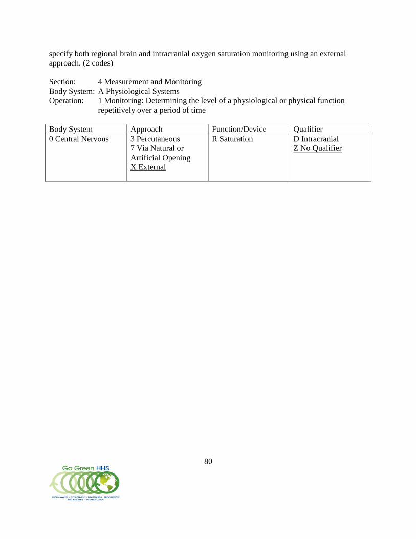
80
specify both regional brain and intracranial oxygen saturation monitoring using an external
approach. (2 codes)
Section: 4 Measurement and Monitoring
Body System: A Physiological Systems
Operation: 1 Monitoring: Determining the level of a physiological or physical function
repetitively over a period of time
Body System Approach Function/Device Qualifier
0 Central Nervous 3 Percutaneous
7 Via Natural or
Artificial Opening
X External
R Saturation D Intracranial
Z No Qualifier

81
Regional Somatic Saturation Monitoring Using Near-Infrared Spectroscopy: Request for New Values in PCS Tables The proposal is to add PCS values to the PCS Monitoring tables to create unique codes for
monitoring oxygen saturation in the soft tissue of various body regions using near-infrared
spectroscopy; namely, gastrointestinal, skin and breast, subcutaneous tissue, muscle, and kidney.
PCS has standardized values in the Measurement and Monitoring section for recording
monitoring of oxygen saturation, and several body system values corresponding to some of the
body regions specified in the proposal.
To extend the level of detail available for the root operation Monitoring to create unique codes
for the procedures specified in the proposal requires adding the existing approach value External
in the 5th
character and the existing function value Saturation in the 6th
character, as well as
creating new 4th
character physiological system values as needed.
Option 1: Do not add new PCS values to create unique codes.
Option 2: Extend existing PCS values in table 4A1 to specify External approach in the 5th
character and Saturation in the 6th
character, for non-invasive NIRS oxygen saturation
monitoring of existing body regions Gastrointestinal, Urinary, and Musculoskeletal. (5 codes)
Section: 4 Measurement and Monitoring
Body System: A Physiological Systems
Operation: 1 Monitoring: Determining the level of a physiological or physical function
repetitively over a period of time
Body System Approach Function/Device Qualifier
B Gastrointestinal
D Urinary
F Musculoskeletal
X External R Saturation Z No Qualifier
Create new PCS physiological system values for Skin and Breast, and for Subcutaneous Tissue,
and add existing PCS values in table 4A1 to specify External approach in the 5th
character and
Saturation in the 6th
character, for non-invasive NIRS oxygen saturation monitoring, for the
skin/breast and subcutaneous tissue body regions.
Section: 4 Measurement and Monitoring
Body System: A Physiological Systems
Operation: 1 Monitoring: Determining the level of a physiological or physical function
repetitively over a period of time

82
Body System Approach Function/Device Qualifier
K Skin and Breast
L Subcutaneous Tissue
X External R Saturation Z No Qualifier
Option 3: Create a single new physiological system value Anatomical Regions, and extend
existing PCS values in table 4A1 to specify External approach in the 5th
character and Saturation
in the 6th
character, for non-invasive NIRS oxygen saturation monitoring of all anatomical
regions other than the brain and circulatory system. (1 code)
Section: 4 Measurement and Monitoring
Body System: A Physiological Systems
Operation: 1 Monitoring: Determining the level of a physiological or physical function
repetitively over a period of time
Body System Approach Function/Device Qualifier
W Anatomical Regions,
General
X External R Saturation Z No Qualifier