age-related macular degeneration (amd); from pathogenesis
TRANSCRIPT

Yoko Ozawa, M.D., Ph.D.Department of Ophthalmology, Keio University School of Medicine;
35 Shinanomachi, Shinjuku-ku, Tokyo 160-8582, Japan.Tel: +81-3-3353-1211 / Fax: +81-3-3359-8302 / E-mail: [email protected]
Anti-Aging Medicine 5 (9) : 87-92, 2008(c) Japanese Society of Anti-Aging Medicine
Review Article
Age-related macular degeneration (AMD); From pathogenesis and approved therapies to proposed treatments for prevention
87
Age-related macular degeneration (AMD) is one of the most severe vision-threatening diseases. Wet AMD, caused by choroidal neovascularization (CNV), progresses rapidly, while dry AMD, characterized by neural retinal atrophy followed by choroidal vascular atrophy, progresses slowly. In addition to systemic risk factors, such as a high body-mass index (BMI), smoking, hypertension, and atherosclerosis, light-induced local oxidative stress and inflammation promotes AMD. CNV can be induced by multiple pathways. The accumulation of lipofuscin, a product of the undigested outer segments of photoreceptor cells, causes chronic local inflammation. Extracellular lipoprotein deposits, which contain pro-inflammatory components, such as complement, can trigger local inflammation. A single nucleotide polymorphism (SNP) in some genes; i.e., complement factor H (CFH) and excision repair cross-complementing rodent repair deficiency complementation group 6 (ERCC6), which induce local inflammation, is a risk factor for AMD. Two treatments, photodynamic therapy (PDT) and anti-vascular endothelial growth factor (VEGF) therapy, have been approved worldwide to cause the regression of CNV. In addition, the suppression of CNV progression combined with retinal neural protection by anti-oxidative reagents such as lutein/zeaxanthin and/or docosahexaenoic acid / eicosapentaenoic acid (DHA/EPA) is a promising prospective therapeutic approach. This is the subject of an ongoing prospective, randomized, double-blind, multicenter clinical trial, the Age-Related Eye Disease Study 2 (AREDS2). In addition, molecular and biological analyses in animal models have provided data supporting this anti-oxidant therapy.
Age-related eye diseases include not only cataract and glaucoma, but also age-related macular degeneration (AMD), one of the most severe vision-threatening diseases. 1-3) Of the adults over 40 in the United States, more than 1.75 million (1.5%) are estimated to have advanced AMD, and 7.3 million (6%) are in the early stage of AMD. 4) Advanced AMD is reported to be most prevalent in Caucasians, who have less pigmentation in their uvea and skin, but growing numbers of Asians also suffer from this disease. One report states that 0.67% of Japanese adults over 50 have advanced AMD. 5) One reason for the increase in Japanese patients is thought to be the changes in lifestyle that increase the risk factors for this disease, as described below. The difference in the prevalence of AMD among races is also partly owing to genetic background; for example, some single nucleotide polymorphisms (SNPs) in complement factors are associated with AMD in Caucasians. 6,7)
AMD destroys the sharp, central vision necessary for common daily tasks, and it is one of the principal causes of registered legal blindness. Under the influences of risk factors such as a high body mass index (BMI) and smoking, 3,8-15) the accumulation of local oxidative stress and inflammatory reactions induced by light
Abstract
Introduction exposure are believed to cause AMD. These stimuli promote the generation of choroidal neovascularization (CNV), the primitive lesion of advanced AMD. Treatments for AMD, especially those promoting the regression of CNV, are in use worldwide. However, the visual outcome of these treatments is not fully satisfactory, since once damaged, the neural retina does not recover fully. Developing treatments for preventing CNV and the secondary neural retinal damage are now a focus of research interest.
Yoko Ozawa 1,2), Susumu Ishida 1,2), Kazuo Tsubota 2)
1) Laboratory of Retinal Cell Biology
2) Departments of Ophthalmology, Keio University School of Medicine
KEY WORDS: AMD, CNV, AREDS, antioxidant, retina
Received: Sep 30, 2008Accepted: Oct 01, 2008Published online: Nov 20, 2008

On the other hand, dry AMD occurs when the light-sensitive retinal neural cells in the macula slowly become atrophic. Epidemiological and pathological studies point to choroidal vascular atrophy as the causative event. Age-related changes in the RPE and Bruch’s membrane are associated with this condition.17) Patients first recognize blurred vision, and over time, as less of the macula functions, the central vision is gradually lost in the affected eye.
Developments of CNV
Long-standing oxidative stress and the resultant tissue inflammation underlie the development and progression of CNV. 18-20) Multiple pathways are probably involved in the establishment of CNV. Age-related RPE dysfunction induces the accumulation of lipofuscin, a product of the undigested outer segments of photoreceptor cells. The outer segments, where Rhodopsin is concentrated and light stimuli are converted into electrical pulses, are physiologically phagocytosed by the RPE after light stimulation. Some of the components are digested and recycled, but A2E, a pyridinium bis-retinoid derived from all-trans-retinal and phosphatidyl-ethanolamine, is toxic to the RPE when it accumulates. 21) A2E, a component of lipofuscin, is photooxidized by 430-nm irradiation and generates a singlet oxygen and a superoxide anion. A2E also acts as an efficient quencher of singlet oxygen, forming a mixture of oxygen-containing moieties, some of which are highly reactive. Thus, since the visual cycle provides A2E continuously, A2E can cause chronic local inflammation. Moreover, oxidized A2E triggers the low-grade activation of complement. This mechanism was demonstrated using the human adult RPE cell line, ARPE-19. 22) In ARPE-19 cells, the cleavage and activation of complement factor C3 was clearly upregulated by the irradiated and oxidized A2E, which can also contribute to the chronic inflammatory process. 22) A2E also induces inflammation by altering gene expression. Both the mRNA and protein levels of vascular endothelial growth factor (VEGF), a key regulator of CNV, is upregulated in ARPE-19 cells through the retinoic acid receptor α (RAR-α).23) Therefore, prolonged and excessive activation of the visual cycle may produce a high level of A2E and induce chronic inflammation locally, which can then lead to CNV development. Oxidative stress is highly associated with this process.
Symptoms and Pathogenesis of AMD
AMD is divided into two categories: rapidly progressing “wet” AMD and slow-onset “dry” AMD. Wet AMD occurs when abnormal blood vessels begin to grow under the macula and cause hemorrhage (Fig. 1) and/or plasma exudation behind the neural retina (Fig. 2). The macula is located in the center of the retina, where large numbers of photoreceptor cells are concentrated. It is indispensable for central vision, and therefore its health determines a person’s ability to read, drive, and see fine details. In wet AMD, abnormal vessels originate from the choroids, and this condition is called choroidal neovascularization (CNV). Severe damage to the macula occurs when the CNV spreads just below the neural retina grown through the disruptsion of the retinal pigment epithelium (RPE) and Bruch’s membrane, both of which lie between the neural retina and the choroid 16) (Fig. 3). Recent studies suggest that CNV is initially induced by chronic inflammation that can arise from multiple pathways, as described in the next section. Patients may become aware of a decrease in visual acuity and distortion of vision from early in the disease, when the underlying CNV elevates the macula. If exudative lesions spread to the macula, the symptoms become more severe and progress quickly. This is due to neural retinal damage caused not only by the leakage of extracellular fluid from the CNV but also from inflammatory cytokines released from the CNV and its environment. The anatomical changes that disturb the central vision include RPE detachment and neural retinal detachment due to the CNV and exudative fluid, macular edema due to chronic inflammation in the retina, and the spread of hard exudate in the neural retina.
Age-related macular degeneration (AMD)
88
Fig. 1. Fundus photograph of AMDWhite circle shows a normal macular region (A). In the case of AMD, there is hemorrhage (red), hard exudate (yellowish dots), and exudative retinal detachment (arrowheads) in the macular region (B).
Fig. 2. Exudative lesion in the maculaFluorescent angiography shows leakage from the putative CNV in the macular region (arrowheads in A). The corresponding fundus photograph is shown in B. Hemorrhage is observed in the macular region. CNV, choroidal neovascularization
Fig. 3. Cross-sectional imaging of the maculaCross-sectional imaging of the macula detected by optical coherence tomography (OCT). Normal macula (A). The RPE line is disrupted (arrowheads) and CNV invades and spreads under the neural retina in the case of AMD (B). RPE, retinal pigment epithelium; CNV, choroidal neovascularization
Drusen, an early hallmark of AMD, is the extracellular lipoprotein deposits that form at the interface between the RPE and Bruch’s membrane. Drusen is composed of pro-inflammatory components such as complement, C reactive protein, and advanced glycation end-product (AGE), and induces the macrophage infiltration that releases inflammatory cytokines. Thus, drusen contributes to the triggering of local inflammation. 17,18,24-27) Drusen is one of the main indicators of increased risk for developing CNV in Caucasians, 16) although it is only a moderate risk indicator for the Japanese (18%). 28) This may be owing to differences in the etiology of the AMD in different genetic backgrounds. Interestingly, epidemiologic data show a relationship between AMD and complements. A single nucleotide polymorphism (SNP) in the gene of complement factor H (CFH) is a risk factor for AMD in Caucasians. 6) Half the cases of AMD are linked to CFH variants. 29) Since CFH is a regulatory protein that suppresses formation of the C3 cleavage enzyme, its variation may cause the activation of complement and local inflammatory events behind the RPE to promote the development of CNV. Another SNP that is closely associated with AMD, excision repair cross-complementing rodent repair deficiency complementation group 6 (ERCC6), is known to function extensively in the repair of damaged DNA. 7) ERCC6 expression is high in the retinal cells of the macula in AMD eyes compared with the normal retina, as assessed by immunohistochemistry. More interestingly, individuals homozygous for both the ERCC6 and CFH risk alleles have a dramatically increased risk of developing the disease. Thus, a SNP-SNP interaction between ERCC6 and CFH may increase the AMD risk. Since ERCC6 functions in universal transcription as a component of the RNA pol I transcription complex, there may also be an independent pathway promoting AMD risk.
Hypertension and atherosclerosis are also reported risk factors for AMD. 10,12,30) Among the causative factors, the renin-angiotensin system (RAS) may be involved in exacerbating the generation of CNV. 31,32) In addition to circulatory RAS, which regulates blood pressure, there is tissue RAS. Tissue RAS components are expressed intraocularly, and both Angiotensin II and Angiotensin II type 1 and type 2 receptors (AT1R and AT2R) are expressed in the human CNV tissues obtained from patients with AMD. Further evidence for the involvement of the RAS comes from the finding that AT1R signaling is responsible for the development of laser-induced CNV in a mouse model. 32) This animal model does not completely reconstruct the human disease, but a similar mechanism may be involved in the development of human CNV. In the mouse model, AT1R blockade reduces the infiltration of macrophages in the choroid and the expression of several inflammatory cytokines, both of which lead to a reduction in the size of the laser-induced CNV. Thus, individuals whose systemic background increases the upregulation of RAS may also be more susceptible to the induction of tissue RAS in the eye, which may promote CNV development.
Treatments for AMD
Treatments for AMD have been long in coming. However, several treatment strategies have now been approved.
1. Photodynamic therapy (PDT)
Photodynamic therapy (PDT) with verteporfin is effective for the treatment of a variety of neoplastic pathologies. In this therapeutic approach, the photosensitizer verteporfin is activated by non-thermal laser light to close neovascular structures by a cytotoxic reaction that takes place in the endothelial cells 33,34). There is a risk that visual acuity may be decreased after PDT, due to subretinal hemorrhage caused by the disruption of the CNV network by the activated verteporfin. Moreover, this treatment may need to be repeated several times to limit exudation or expansion of the CNV. Because of these limitations, anti-VEGF therapy may be gaining favor (see below). However, PDT is significantly effective for treating polypoidal choroidal vasculopathy (PCV), one of the subtypes of AMD. 35-38) Since most PCV patients are in Asian countries and approximately half of the AMD patients in Japan have PCV, 39) this therapy will often still be applied, especially in Asian countries.
2. Anti-VEGF therapy
To cause the regression of the CNV induced by local VEGF, 24,40) anti-VEGF drugs such as pegaptanib (a VEGF neutralizing aptamer) and ranibizumab (an anti-VEGF neutralizing antibody) are presently considered the most effective approved therapy for AMD. These drugs are injected intraocularly every 4-6 weeks. Several prospective, randomized, double-blind, multicenter controlled clinical trials have shown the efficacy of each drug. 41-
46) Because this kind of therapy can only reduce the CNV and its exudative activity, the damage to the neural retina and the visual dysfunction do not always improve. Nonetheless, the visual outcome of patients who received the drug injections compared with those receiving sham injections was much better in every report, although the indication for treatment in each study was different. Some of the reports only compared the number of patients who lost fewer than 15 letters of visual acuity. With this criterion, pegaptanib inhibited the vision loss in 70% of the patients compared with 55% of the controls in the VISION study, 42) and ranibizumab rescued the loss in 95% of the patients compared with 65% of the controls in the MARINA Study. 44) Furthermore, with ranibizumab, the mean improvement from baseline in the visual acuity scores was evident 7 days after the first injection, whereas the mean visual acuity in the sham injection group declined steadily over time with each monthly assessment. 44) Improvement in visual acuity is seldom observed in AMD, since once damaged, the neural retina loses its function. However, if the indication to start this therapy is adequate, it may lead to a gratifying outcome. The difference in function between pegaptanib and ranibizumab is in the isoforms of VEGF that are suppressed by these drugs. The former inhibits only the VEGF165 isoform, while the latter inhibits all VEGF isoforms. Since the VEGF165 isoform is the main pathogenic factor, 47) pegaptanib may act more specifically to cause the CNV to regress. Although ranibizumab seems to be more effective, serious adverse events, myocardial infarction and cerebral vascular event, were reported in a 2-year trial of this drug. 44) Recently, a new protocol combining the use of both drugs, ranibizumab for the initial drug burst and pegaptanib for longer-term maintenance, is under consideration.
89
Age-related macular degeneration (AMD)
nutrient questionnaire. 50) A positive association of DHA/EPA intake with a lowered risk of AMD was also reported. 50)
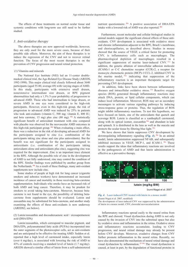
Furthermore, recent molecular and cellular biological studies in animal models support the significant clinical effects of these anti-oxidants. CNV development is associated with oxidative stress and chronic inflammation adjacent to the RPE, Bruch’s membrane, and choriocapillaries, as described above. Studies in mouse showed that the source of VEGF, a critical factor for promoting CNV, is inflammatory cells such as macrophages. The pharmacological depletion of macrophages resulted in a significant suppression of murine laser-induced CNV. 51) In addition, the genetic ablation of intercellular adhesion molecule (ICAM)-1 or C-C chemokine receptor (CCR)-2, a receptor for monocyte chemotactic protein (MCP)-1/CCL-2, inhibited CNV in the murine model, 52) indicating that suppression of the inflammatory reaction is an important therapeutic target for preventing CNV development. In addition, links have been shown between inflammatory diseases and intracellular oxidative stress. 53) Reactive oxygen species (ROS) generated through inflammatory reactions may attack DNA, protein, or lipid cellular components, which can induce local inflammation. Moreover, ROS may act as secondary messengers to activate various signaling pathways by inducing stress-response genes or proteins. Thus, the regulation of ROS might inhibit inflammatory reactions. To test this hypothesis, we have focused on lutein, one of the antioxidants that quench and scavenge ROS. Lutein is classified as a xanthophyll carotenoid, along with its optical isomer, zeaxanthin 54) and abundant in kale and spinach. It is distributed in the lens as well as the macula and protects the ocular tissue by filtering blue light. 55) We have shown that lutein suppresses CNV development by downregulating inflammatory signaling (Fig. 4). 56) In an animal model, lutein administration suppressed NF-kB, and subsequently inhibited increases in VEGF, MCP-1, and ICAM-1. 56) These results support the ideas that inflammatory reactions are involved in the pathogenesis of AMD and that lutein administration is effective as a preventive therapy.
Inflammatory reactions spread easily to the neural retina from the RPE and choroid. Visual dysfunction during AMD is not only caused by the invasion of CNV into the subretinal space but also by oxidative stress and inflammation in the retina. Oxidative stress and inflammatory reactions accumulate, leading to CNV progression, and neural retinal damage may already be present before CNV develops. Moreover, exudative changes from CNV further promote inflammation in the neural retina. Recently, we have also demonstrated the mechanism of neural retinal damage and visual dysfunction by inflammation. 57-59) The visual dysfunction is caused, at least in part, by the degradation of functional proteins in
The effects of these treatments on normal ocular tissue and systemic conditions with long-term use still need to be further studied.
3. Anti-oxidative therapy
The above therapies are now approved worldwide; however, they are only used for the more severe cases, because of their possible side effects. Moreover, the aim of these therapies is to induce the regression of the CNV and not to recover retinal function. The focus of the most recent therapies is on the prevention of CNV progression and neural retinal protection.
(1) Vitamins and minerals
The National Eye Institute (NEI) led an 11-center double-masked clinical trial, the Age-Related Eye Disease Study (AREDS, 1992-1998). This major clinical trial closely followed about 3640 participants (aged 55-80, average 69) with varying stages of AMD. In this study, participants with extensive small drusen, nonextensive intermediate size drusen, or RPE pigment abnormalities had only a 1.3% 5-year probability of progression to advanced AMD. Those who had intermediate to large drusen, or severe AMD in one eye were considered to be high-risk participants. However, even in this high-risk group, the risk of progression to advanced AMD was reduced by 25% in patients who took anti-oxidants (vitamin C, 500 mg; vitamin E, 400 IU; and beta carotene, 15 mg) plus zinc (80 mg). 48) A statistically significant benefit of antioxidant treatment with zinc compared with placebo was observed for the neovascular AMD outcomes in this high-risk participant group. 48) When evaluating a main effect, there was a reduction in the risk of developing advanced AMD for the participants assigned to zinc (i.e. combination of the participants taking zinc alone and zinc plus antioxidants). But the benefit was not significant for the participants assigned to antioxidants (i.e. combination of the participants taking antioxidants alone and antioxidants plus zinc), suggesting zinc was required for the improvement. Zinc is an important trace element in the RPE. Although the possible role of zinc in the development of AMD is not fully understood, zinc may control the condition of the RPE. Similar findings were published by another group from the Netherlands. 49) As a result of these findings, many anti-oxidant supplements now include zinc. Some studies of people at high risk for lung cancer (cigarette smokers and asbestos workers) have demonstrated an increased incidence of cancer and mortality in those receiving beta-carotene supplementation. Individuals who smoke have an increased risk of both AMD and lung cancer. Therefore, it may be prudent for smokers to avoid taking beta-carotene. Moreover, because beta-carotene is not found in the eye, the possibility of deleting beta-carotene from supplements should be evaluated. Lutein and zeaxanthin may be substituted for beta-carotene, and another study examining the effects of these anti-oxidants is now underway (AREDS2, see below).
(2) Lutein/zeaxanthin and docosahexaenoic acid / eicosapentaenoic acid (DHA/EPA)
Lutein/zeaxanthin, which correspond to macular pigment, and DHA/EPA, polyunsaturated fatty acids (PUFAs) incorporated into the outer segments of the photoreceptor cells, act as anti-oxidants and are anticipated to be effective for treating AMD. Seddon et al. reported that a high level of carotenoid intake, especially lutein (over 6 mg/day), is associated with lowering the risk of AMD to 43% of controls receiving a standard level of lutein (1.7 mg/day). AREDS showed a similar effect of lutein from a self-administered
90
Fig. 4. Laser-induced CNV treated with lutein in a mouse model (Izumi-Nagai et al. 2007, modified) The development of laser-induced CNV was suppressed by the administration of lutein in a mouse model. CNV, choroidal neovascularization

01)
0 2)
3)0
4)
5)
6)
7)
8)
ReferencesBunce, C. & Wormald, R. Leading causes of certification for blindness and partial sight in England & Wales. BMC public health 6, 58 (2006). la Cour, M., Kiilgaard, J.F. & Nissen, M.H. Age-related macular degeneration: epidemiology and optimal treatment. Drugs & aging 19, 101-133 (2002). O'Shea, J.G. Age-related macular degeneration: a leading cause of blindness. The Medical journal of Australia 165, 561-564 (1996). Friedman, D.S., et al. Prevalence of age-related macular degeneration in the United States. Archives of ophthalmology 122, 564-572 (2004). Oshima, Y., et al. Prevalence of age related maculopathy in a representative Japanese population: the Hisayama study. The British journal of ophthalmology 85, 1153-1157 (2001). Klein, R.J., et al. Complement factor H polymorphism in age-related macular degeneration. Science (New York, N.Y 308, 385-389 (2005). Tuo, J., et al. Synergic effect of polymorphisms in ERCC6 5' flanking region and complement factor H on age-related macular degeneration predisposition. Proceedings of the National Academy of Sciences of the United States of America 103, 9256-9261 (2006). Clemons, T.E., Milton, R.C., Klein, R., Seddon, J.M. & Ferris, F.L., 3rd. Risk factors for the incidence of Advanced Age-Related Macular Degeneration in the Age-Related Eye Disease Study (AREDS) AREDS report no. 19. Ophthalmology 112, 533-539 (2005).
9)
10)
11)
12)
13)
14)
15)
16)
17)
Hawkins, B.S., Bird, A., Klein, R. & West, S.K. Epidemiology of age-related macular degeneration. Molecular vision 5, 26 (1999). Hyman, L. & Neborsky, R. Risk factors for age-related macular degeneration: an update. Current opinion in ophthalmology 13, 171-175 (2002). Klein, R. Overview of progress in the epidemiology of age-related macular degeneration. Ophthalmic epidemiology 14, 184-187 (2007). Klein, R., Peto, T., Bird, A. & Vannewkirk, M.R. The epidemiology of age-related macular degeneration. American journal of ophthalmology 137, 486-495 (2004). Nolan, J., et al. Macular pigment and percentage of body fat. Investigative ophthalmology & visual science 45, 3940-3950 (2004). Seddon, J.M., George, S., Rosner, B. & Klein, M.L. CFH gene variant, Y402H, and smoking, body mass index, environmental associations with advanced age-related macular degeneration. Human heredity 61, 157-165 (2006). Thornton, J., et al. Smoking and age-related macular degeneration: a review of association. Eye (London, England) 19, 935-944 (2005). Nowak, J.Z. Age-related macular degeneration (AMD): pathogenesis and therapy. Pharmacol Rep 58, 353-363 (2006). Penfold, P.L., Madigan, M.C., Gillies, M.C. & Provis, J.M. Immunological and aetiological aspects of macular degeneration. Progress in retinal and eye research 20, 385-414 (2001).
Table 1 CAge-related Eye disease Study 2 (AREDS 2)the neural retinal cells during inflammation. In the inflammatory retinas, an indispensable visual substance, Rhodopsin is excessively degraded through the ubiquitin-proteasome pathway, leading to visual dysfunction. 59) A similar mechanism decreases the amount of a synaptic vesicle protein, Synaptophysin, 57) suggesting that anti-inflammatory therapy is important for protecting visual function. Moreover, anti-oxidative therapy with lutein inhibited both the retinal inflammation and Rhodopsin reduction leading to visual function impairment (Sasaki, Ozawa et al in revision), also suggesting that anti-oxidative therapy may lead to retinal neuroprotection.
In the mouse model, EPA, a PUFA, is also effective for preventing the CNV development. 60) PUFAs are classified into two groups, ω-3 and ω-6, based on their chemical structures. In the ω-3 (or n-3) type, the first double bond is three carbons from the methyl end of the PUFA molecule. It is well known that supplemental fish oil, a rich source of ω-3 PUFAs, reduces elevated triglyceride levels. EPA is already clinically used as an anti-hyperlipidemic agent. In our study using the mouse laser-induced CNV model, EPA administration reduced the level of VEGF, MCP-1, and ICAM-1 in the choroid. The administration of EPA increased the total ω-3 PUFAs, including EPA and its downstream metabolite, DHA. This change was observed not only in the RPE and choroids, but also in the serum. In contrast, the total ω-6 PUFAs, including arachidonic acid and linoleic acid, which positively regulate inflammation, were significantly decreased. This finding also suggests that EPA prevents inflammation which promotes the progression of AMD. The current study in progress, AREDS2 (Table 1), is designed to determine whether lutein/zeaxanthin (10 mg/2 mg) and/or DHA/EPA; (350 mg/650 mg) can slow the progression of vision loss from AMD. Data will be assessed on approximately 4000 participants enrolled through nearly 100 clinical centers who are aged 50 - 85 and have AMD. The enrollment concluded in June 2008 and participants will be followed between five and six years.
Summary
The pathological mechanisms of AMD are being elucidated, which will lead to new strategies for treating this disease. Several kinds of treatments have already been established and are being used worldwide; however, their effects on visual function are not fully satisfactory. Recently, the treatment focus has moved to preventing the progression of AMD in its early stages. Epidemiological and biological analyses to verify the effectiveness of anti-oxidant therapy are currently underway. Further translational research is required to establish more safe and effective therapies for AMD.
91
1. A multi-center, randomized trial
2. Men and women between the ages of 50 and 85 years
3. Macular status; large drusen in both eyes or large drusen in one eye and advanced AMD in the fellow eye
4. Groups; (i) placebo (ii) lutein (10mg) and zeaxanthin (2mg) (iii) DHA (350 mg) and EPA (650 mg) (iv) (ii)+(iii)
5. Evaluation; preventive effect on AMD progression
92
18)
19)
20)
21)
22)
23)
24)
25)
26)
27)
28)
29)
30)
31)
32)
33)
34)
35)
36)
37)
38)
39)
40)
Hollyfield, J.G., et al. Oxidative damage-induced inflammation initiates age-related macular degeneration. Nature medicine 14, 194-198 (2008). Justilien, V., et al. SOD2 knockdown mouse model of early AMD. Investigative ophthalmology & visual science 48, 4407-4420 (2007). Zarbin, M.A. Current concepts in the pathogenesis of age-related macular degeneration. Archives of ophthalmology 122, 598-614 (2004). Wolf, G. Lipofuscin and macular degeneration. Nutrition reviews 61, 342-346 (2003). Zhou, J., Jang, Y.P., Kim, S.R. & Sparrow, J.R. Complement activation by photooxidation products of A2E, a lipofuscin constituent of the retinal pigment epithelium. Proceedings of the National Academy of Sciences of the United States of America 103, 16182-16187 (2006). Iriyama, A., et al. A2E, a pigment of the lipofuscin of retinal pigment epithelial cells, is an endogenous ligand for retinoic acid receptor. The Journal of biological chemistry 283, 11947-11953 (2008). Ambati, J., et al. An animal model of age-related macular degeneration in senescent Ccl-2- or Ccr-2-deficient mice. Nature medicine 9, 1390-1397 (2003). Anderson, D.H., Mullins, R.F., Hageman, G.S. & Johnson, L.V. A role for local inflammation in the formation of drusen in the aging eye. American journal of ophthalmology 134, 411-431 (2002). Kamei, M., et al. Scavenger receptors for oxidized lipoprotein in age-related macular degeneration. Investigative ophthalmology & visual science 48, 1801-1807 (2007). Yoshida, T., et al. The potential role of amyloid beta in the pathogenesis of age-related macular degeneration. The Journal of clinical investigation 115, 2793-2800 (2005). Uyama, M., et al. The second eye of Japanese patients with unilateral exudative age related macular degeneration. The British journal of ophthalmology 84, 1018-1023 (2000). Hageman, G.S., et al. A common haplotype in the complement regulatory gene factor H (HF1/CFH) predisposes individuals to age-related macular degeneration. Proceedings of the National Academy of Sciences of the United States of America 102, 7227-7232 (2005). Guymer, R.H. & Chong, E.W. Modifiable risk factors for age-related macular degeneration. The Medical journal of Australia 184, 455-458 (2006). Nagai, N., et al. Suppression of choroidal neovascularization by inhibiting angiotensin-converting enzyme: minimal role of bradykinin. Investigative ophthalmology & visual science 48, 2321-2326 (2007). Nagai, N., et al. Angiotensin II type 1 receptor-mediated inflammation is required for choroidal neovascularization. Arteriosclerosis, thrombosis, and vascular biology 26, 2252-2259 (2006). Kramer, M., et al. Liposomal benzoporphyrin derivative verteporfin photodynamic therapy. Selective treatment of choroidal neovascularization in monkeys. Ophthalmology 103, 427-438 (1996). Michels, S. & Schmidt-Erfurth, U. Photodynamic therapy with verteporfin: a new treatment in ophthalmology. Seminars in ophthalmology 16, 201-206 (2001). Akaza, E., Mori, R. & Yuzawa, M. Long-term results of photodynamic therapy of polypoidal choroidal vasculopathy. Retina (Philadelphia, Pa 28, 717-722 (2008). Eandi, C.M., Ober, M.D., Freund, K.B., Slakter, J.S. & Yannuzzi, L.A. Selective photodynamic therapy for neovascular age-related macular degeneration with polypoidal choroidal neovascularization. Retina (Philadelphia, Pa 27, 825-831 (2007). Kurashige, Y., et al. Two-Year Results of Photodynamic Therapy for Polypoidal Choroidal Vasculopathy. American journal of ophthalmology (2008). Wakabayashi, T., Gomi, F., Sawa, M., Tsujikawa, M. & Tano, Y. Marked vascular changes of polypoidal choroidal vasculopathy after photodynamic therapy. The British journal of ophthalmology 92, 936-940 (2008). Maruko, I., Iida, T., Saito, M., Nagayama, D. & Saito, K. Clinical characteristics of exudative age-related macular degeneration in Japanese patients. American journal of ophthalmology 144, 15-22 (2007). Husain, D., Ambati, B., Adamis, A.P. & Miller, J.W. Mechanisms of age-related macular degeneration. Ophthalmology clinics of North America 15, 87-91 (2002).
41)
42)
43)
44)
45)
46)
47)
48)
49)
50)
51)
52)
53)
54)
55)
56)
57)
58)
59)
60)
Chakravarthy, U., et al. Year 2 efficacy results of 2 randomized controlled clinical trials of pegaptanib for neovascular age-related macular degeneration. Ophthalmology 113, 1508 e1501-1525 (2006). Gragoudas, E.S., Adamis, A.P., Cunningham, E.T., Jr., Feinsod, M. & Guyer, D.R. Pegaptanib for neovascular age-related macular degeneration. The New England journal of medicine 351, 2805-2816 (2004). Ng, E.W. & Adamis, A.P. Anti-VEGF aptamer (pegaptanib) therapy for ocular vascular diseases. Annals of the New York Academy of Sciences 1082, 151-171 (2006). Rosenfeld, P.J., et al. Ranibizumab for neovascular age-related macular degeneration. The New England journal of medicine 355, 1419-1431 (2006). Rosenfeld, P.J., Rich, R.M. & Lalwani, G.A. Ranibizumab: Phase III clinical trial results. Ophthalmology clinics of North America 19, 361-372 (2006). Takeda, A.L., Colquitt, J., Clegg, A.J. & Jones, J. Pegaptanib and ranibizumab for neovascular age-related macular degeneration: a systematic review. The British journal of ophthalmology 91, 1177-1182 (2007). Ishida, S., et al. VEGF164-mediated inflammation is required for pathological, but not physiological, ischemia-induced retinal neovascularization. The Journal of experimental medicine 198, 483-489 (2003). AREDS. A randomized, placebo-controlled, clinical trial of high-dose supplementation with vitamins C and E, beta carotene, and zinc for age-related macular degeneration and vision loss: AREDS report no. 8. Archives of ophthalmology 119, 1417-1436 (2001). van Leeuwen, R., et al. Dietary intake of antioxidants and risk of age-related macular degeneration. Jama 294, 3101-3107 (2005). SanGiovanni, J.P., et al. The relationship of dietary carotenoid and vitamin A, E, and C intake with age-related macular degeneration in a case-control study: AREDS Report No. 22. Archives of ophthalmology 125, 1225-1232 (2007). Sakurai, E., Anand, A., Ambati, B.K., van Rooijen, N. & Ambati, J. Macrophage depletion inhibits experimental choroidal neovascularization. Investigative ophthalmology & visual science 44, 3578-3585 (2003). Sakurai, E., et al. Targeted disruption of the CD18 or ICAM-1 gene inhibits choroidal neovascularization. Investigative ophthalmology & visual science 44, 2743-2749 (2003). Valko, M., et al. Free radicals and antioxidants in normal physiological functions and human disease. The international journal of biochemistry & cell biology 39, 44-84 (2007). Alves-Rodrigues, A. & Shao, A. The science behind lutein. Toxicol Lett 150, 57-83 (2004). Bone, R.A. & Landrum, J.T. Distribution of macular pigment components, zeaxanthin and lutein, in human retina. Methods in enzymology 213, 360-366 (1992). Izumi-Nagai, K., et al. Macular pigment lutein is antiinflammatory in preventing choroidal neovascularization. Arteriosclerosis, thrombosis, and vascular biology 27, 2555-2562 (2007). Kurihara, T., et al. Angiotensin II type 1 receptor signaling contributes to synaptophysin degradation and neuronal dysfunction in the diabetic retina. Diabetes 57, 2191-2198 (2008). Kurihara, T., et al. Neuroprotective effects of angiotensin II type 1 receptor (AT1R) blocker, telmisartan, via modulating AT1R and AT2R signaling in retinal inflammation. Investigative ophthalmology & visual science 47, 5545-5552 (2006). Ozawa, Y., et al. Roles of STAT3/SOCS3 Pathway in Regulating the Visual Function and Ubiquitin-Proteasome-dependent Degradation of Rhodopsin during Retinal Inflammation. The Journal of biological chemistry 283, 24561-24570 (2008). Koto, T., et al. Eicosapentaenoic acid is anti-inflammatory in preventing choroidal neovascularization in mice. Investigative ophthalmology & visual science 48, 4328-4334 (2007).
Age-related macular degeneration (AMD)