age-related hyperkyphosis; its causes, consequences, and management
DESCRIPTION
Geriatric rehabilitationTRANSCRIPT

Age-Related Hyperkyphosis: Its Causes, Consequences, andManagement
Wendy B. Katzman, PT, DPTSc [Assistant Clinical Professor]1, Linda Wanek, PT, PhD[Professor]2, John A. Shepherd, PhD [Assistant Professor in Residence]3, and Deborah E.Sellmeyer, MD [Associate Professor]41Department of Physical Therapy and Rehabilitation Science, University of California, SanFrancisco, San Francisco, CA2Graduate Program in Physical Therapy, San Francisco State University, San Francisco, CA3Department of Radiology, University of California, San Francisco, San Francisco, CA4Division of Endocrinology, Johns Hopkins School of Medicine, Baltimore, MD
AbstractAge-related postural hyperkyphosis is an exaggerated anterior curvature of the thoracic spine,sometimes referred to as Dowager’s hump or gibbous deformity. This condition impairs mobility,2,31 and increases the risk of falls33 and fractures.26 The natural history of hyperkyphosis is notfirmly established. Hyperkyphosis may develop from either muscle weakness and degenerativedisc disease, leading to vertebral fractures and worsening hyperkyphosis, or from initial vertebralfractures that precipitate its development.
Keywordsaging/geriatrics; kyphosis; osteoporosis; postural relationships; thoracic spine
It is also possible that different individuals may develop the same magnitude ofhyperkyphosis from different processes, some from vertebral fractures and others frommuscle weakness, degenerative disc disease, or other genetically determined processes.Regardless, there are significant negative consequences of hyperkyphosis, and earlyintervention and treatment of hyperkyphosis could have important clinical and public healthbenefits.
Our objectives are to review the prevalence and natural history of hyperkyphosis, along withassociated health implications if left untreated. We will discuss evidence-based treatmentoptions and potential contraindications, and observations about the direction for future studyof hyperkyphosis.
DEFINITION AND PREVALENCEWhile a small amount of anterior curvature of the thoracic spine is normal and present dueto the shape of the vertebral bodies and intervertebral discs, a kyphosis angle greater than40°, which is the 95th percentile of normal for young adults, is defined as hyperkyphosis.15,62 In childhood and through the third decade of life the angle of kyphosis averages from
Address correspondence to Wendy B. Katzman, UCSF Box 0625, San Francisco, CA 94143-0625. [email protected].
NIH Public AccessAuthor ManuscriptJ Orthop Sports Phys Ther. Author manuscript; available in PMC 2011 June 1.
Published in final edited form as:J Orthop Sports Phys Ther. 2010 June ; 40(6): 352–360. doi:10.2519/jospt.2010.3099.
NIH
-PA Author Manuscript
NIH
-PA Author Manuscript
NIH
-PA Author Manuscript
20° to 29°.15 After 40 years of age, kyphosis angle begins to increase—more rapidly inwomen than men15—from a mean of 43° in women aged 55 to 60 years to a mean of 52° inwomen 76 to 80 years of age.12 Reports of prevalence and incidence of hyperkyphosis inolder adults vary from approximately 20% to 40% among both men and women.32,60 Askyphosis angle increases, physical performance and quality of life often declines, makingearly intervention for hyperkyphosis a priority.
MEASUREMENTThe gold-standard orthopaedic technique for assessment of thoracic kyphosis is standinglateral spine radiographs. In elderly persons, spinal radiographs may be taken in the supineposition for comfort. The Cobb’s angle of kyphosis is calculated from perpendicular linesdrawn on a standard thoracic spine radiograph: a line extends through the superior endplateof the vertebral body, marking the beginning of the thoracic curve (usually at T4), and theinferior endplate of the vertebral body, marking the end of the thoracic curve (usually atT12) (FIGURE 1).29 While this method is the gold-standard, it is limited by the need forradiography.
Acceptable alternatives are the Debrunner kyphometer and the flexicurve ruler.41 Bothmethods are performed standing. The kyphometer measures the angle of kyphosis, the armsof the protractor-like device are placed at the top and bottom of the thoracic curve, usuallyover the spinous processes of T2 and T3 superiorly, and T11 and T12 inferiorly (FIGURE2).41 The flexicurve ruler is a plastic, moldable device that is aligned over the C7 spinousprocess to the L5–S1 interspace; the ruler is molded to the curvature of the spine and thethoracic and lumbar curves are traced (FIGURE 3). The kyphosis index is calculated as thewidth divided by the length of the thoracic curve, multiplied by 100 (FIGURE 3).47 Akyphosis index value greater than 13 is defined as hyperkyphotic.40
Lundon et al41 compared the reliability of standing radiographic, kyphometer, andflexicurve methods of measuring kyphosis in a group of 24 postmenopausal women withosteoporosis. There was excellent intrarater and interrater reliability (intraclass correlationcoefficients [ICCs] = 0.87–0.92) for each method, indicating the strength of each instrumentfor measuring kyphosis.41 Kado et al29 compared the agreement between standingkyphometer and supine radiologic measure of Cobb’s angle of kyphosis in older women.While the overall agreement was acceptable (ICC = 0.68), the agreement between thekyphosis measurements greater or equal to 50° was poor (ICC = 0.44). Thus, while allmeasures can be used to reliably quantify kyphosis, the standing kyphometer method formeasuring a kyphotic spine may overestimate the degree of kyphosis compared with supineradiographs. However, the external methods do not involve radiographic exposure and areinexpensive and easy to use in the clinical setting.
Other clinical measures are sometimes used to quantify hyperkyphotic posture. Standingmeasurements of tragus to the wall or occiput to the wall, and supine measurement of thenumber of 1.5-cm blocks needed to support the head have been described2,33; however,reliability of these methods has not been investigated and there are no studies comparingthese measures to the gold-standard radiograph.
CLINICAL CONSEQUENCES OF HYPERKYPHOSISFunctional Limitations
Excessive kyphosis has detrimental effects on physical performance, the ability to performactivities of daily living, and overall quality of life.2,52,60 Women with hyperkyphoticposture demonstrate difficulty rising from a chair repeatedly without using their arms,2,31
Katzman et al. Page 2
J Orthop Sports Phys Ther. Author manuscript; available in PMC 2011 June 1.
NIH
-PA Author Manuscript
NIH
-PA Author Manuscript
NIH
-PA Author Manuscript

significantly poorer balance and slower gait velocity, wider base of support with stance andgait, and decreased stair-climbing speed2—impairments that have been associated withincreased risk for falls. In addition, osteoporotic women with hyperkyphosis have increasedpostural sway compared to those with normal posture.42
Hyperkyphosis is also associated with self-reported decline in physical functioning. Womenwith hyperkyphosis report greater difficulty reaching and performing heavy housework andscore lower on the basic activities of daily living scale compared with their peers.2,10,52,60
Musculoskeletal AlterationsAs kyphosis increases, there are concomitant alterations in the normal sagittal planealignment that may cause pain and risk of dysfunction in the shoulder and pelvic girdle, andcervical, thoracic, and lumbar spine. Forward head posture, scapula protraction, reducedlumbar lordosis, and decreased standing height are often associated with hyperkyphosis. 2These postural changes increase the flexion bias around the hip and shoulder joints that caninterfere with normal joint mechanics and movement patterns.
Hyperkyphosis is a significant risk factor for future vertebral and extremity fractures.12,13,26
Older women with hyperkyphosis have a 70% increased risk of future fracture, independentof age or prior fracture, and the risk for fracture increases as hyperkyphosis progresses.26
Quality of LifeWomen with hyperkyphosis report more physical difficulty, more adaptations to their lives,and greater generalized fears than women without hyperkyphosis.44 Additionally,community-dwelling men and women aged 65 years and older with hyperkyphosis reportpoorer satisfaction with subjective health, family relationships, economic conditions, andtheir lives in general.60
MortalityHyperkyphotic posture has been associated with increased mortality, with higher mortalityrates associated with the severity of kyphosis.32 Reduced vital capacity is associated withhyperkyphosis, and severe hyperkyphosis is predictive of pulmonary death amongcommunity-dwelling women.28,38 Women in the highest quartile of kyphosis were morelikely to die of pulmonary death compared with those in the lower quartiles of kyphosis.28
Two recent cohort studies confirm these adverse health effects of hyperkyphosis even afteradjusting for vertebral fractures and bone mineral density.30,32
RISK FACTORSThe causes of hyperkyphosis have yet to be fully elucidated. However, multiplemusculoskeletal, neuromuscular, and sensory impairments are significant predictors of age-related hyperkyphosis.
Vertebral FracturesKyphosis increases with the number of vertebral fractures and is more strongly related tothoracic fractures than lumbar fractures.12 Hyperkyphosis is most prominent in women withmultiple thoracic anterior wedge fractures.12 Women without vertebral fractures, who havegreater degrees of kyphosis, are more likely to experience a subsequent vertebral fracture. 26
Biomechanical models of stress loading on the spine suggest that forces applied to theosteoporotic spine during daily living can cause vertebral wedging and compressionfractures.5,37 The severity of wedging increases as bone mineral density decreases, resulting
Katzman et al. Page 3
J Orthop Sports Phys Ther. Author manuscript; available in PMC 2011 June 1.
NIH
-PA Author Manuscript
NIH
-PA Author Manuscript
NIH
-PA Author Manuscript
in greater numbers of vertebral compression fractures and a further cascade of increasinghyperkyphosis.16,21,46
Degenerative Disc DiseaseMany people consider vertebral fractures to be the underlying cause of age-relatedhyperkyphosis, although studies of older adults report only approximately 40% of men andwomen with the most severe hyperkyphosis have vertebral compression or wedge fractures.53 A common radiographic finding associated with hyperkyphosis among older adults isdegenerative disc disease.16,43,53 In a study of healthy women aged 39 to 91 years, therewas a significant correlation between anterior disc height and kyphosis angle (r = −0.34, P<.001)43; as the anterior disc height decreased, the angle of kyphosis increased. Others havereported that the majority of older adults 50 to 96 years of age with hyperkyphosis haddegenerative disc disease and no evidence of vertebral fractures or osteoporosis,53
suggesting that hyperkyphosis doesn’t predict fractures or osteoporosis. However, a strongassociation between vertebral body anterior- to-posterior height ratio and kyphosis anglesuggests that it is the combined influence of both degenerative disc disease and anteriorvertebral deformities that accounts for significant variation in kyphosis.16,53
Muscle WeaknessSeveral studies confirm that hyperkyphosis is associated with spinal extensor muscleweakness.27,56,57 In healthy postmenopausal women, strength of the spinal extensor musclesis inversely associated with kyphosis (r = −0.30, P = .019).27,56 There is also an inverserelationship between grip and ankle strength and kyphosis,2 suggesting that age-relatedhyperkyphosis may be part of a larger geriatric syndrome associated with adverse healthoutcomes that negatively impact physical function.6,9
Decreased MobilityDecreased spinal extension mobility occurs with aging, interfering with the ability to standerect and maintain normal postural alignment.22 Cadaver studies suggest that calcificationand ossification of the anterior longitudinal ligament in the thoracic region might contributeto increased Cobb’s angle of kyphosis.4 Furthermore, shorter pectoral and hip flexormuscles are linked to severe hyperkyphosis, although it is not known whether the shortmuscles pull the shoulders and hips anteriorly, or whether the kyphotic posture results inshorter anterior musculature. 2 There are likely other contributing muscular, ligamentous,connective tissue, and joint impairments that have not been identified.
Sensory DeficitsAge-related deficits in the somatosensory, visual, and vestibular systems likely contribute tothe loss of upright postural control. With a loss of proprioceptive and vibratory input fromthe joints in the lower extremities in elderly adults compared with young adults,14 theperception of erect vertical alignment becomes impaired. 14,25 Similar declines occur in thevisual system with aging,54 and primary age-related diseases in the eyes, including cataractsand macular degeneration, exacerbate decline in visual acuity. Head pitch position wasfound to be greater during locomotion for normal elderly compared to young adults,23 andincreased even further among older adults wearing bifocals during stair descent.20
Additionally, age-related sensory loss in the vestibular system24 increases the reliance onalready declining visual and somatosensory cues, and can further impact upright posturalalignment.
Katzman et al. Page 4
J Orthop Sports Phys Ther. Author manuscript; available in PMC 2011 June 1.
NIH
-PA Author Manuscript
NIH
-PA Author Manuscript
NIH
-PA Author Manuscript
TREATMENT OF HYPERKYPHOSISThere is a lack of efficacious medical interventions for hyperkyphosis. Physical therapyshould be a first-line approach, particularly because many of the causes of hyperkyphosisare of musculoskeletal origin. Recognition and treatment of hyperkyphosis could contributeto reduced risk of falls, fractures, and functional limitations. Several physical therapyinterventions aimed at reducing hyperkyphosis are currently available (TABLE 1).
Medicines and SurgeryMany men and women with prevalent hyperkyphosis are treated with osteoporosisantiresorptive or bone-building medications because they have low bone density or spinefractures. While osteoporosis treatment helps to prevent incident spine fractures, nomedications have been shown to improve hyperkyphosis. Vertebroplasty and kyphoplastyare surgical procedures primarily used to treat refractory pain following vertebral fracture,and they have been shown to reduce kyphosis angle in select patient populations only.8,61
However, evidence suggests that physical disability and pain relief may be improved aftervertebroplasty and kyphoplasty compared to medical management but only within the first 3months after intervention.45 Furthermore, recent evidence from 2 randomized controlledtrials suggests that clinical improvement in physical disability and pain is similar amongpatients undergoing vertebroplasty, compared to sham procedure for painful vertebralfractures, at 1-month and 6-month follow- up.7,35 High-quality randomized trials with long-term follow-up are needed to investigate benefits of these procedures on subsequentvertebral fractures. No studies have investigated the effects on kyphosis of combinedtreatment with medications, surgical interventions, and physical therapy interventions.
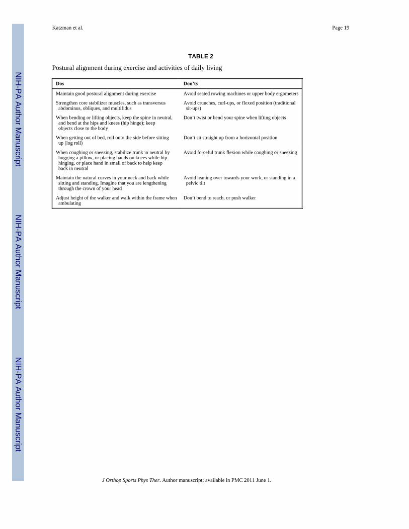
Exercise: Indications and ContraindicationsSeminal research by Sinaki et al59 suggests that the forces applied to the spine duringexercise can alter the occurrence of subsequent vertebral compression fractures in womenwith prior fracture. In this study, 68% of the women who performed flexion exercisesdeveloped a subsequent fracture within the following 6 months, compared with only 16% ofthose who performed extension exercises, suggesting that flexion exercises increase fracturerisk.59 In addition, the conceptual models of spinal loading suggest that flexion stress on thespine increases the risk for fractures when the underlying bone strength is impaired5 andmay partially explain why older women with hyperkyphosis have an increased risk of futurefracture independent of age or prior fracture.26 Hence, it is important to train individualswith age-related hyperkyphosis to avoid flexion stresses on the spine during exercise andactivities of daily living (TABLE 2), regardless of whether they have had a prior fracture.Furthermore, training using trunk stabilization should avoid curl-up exercises to reduceflexion bias on the spine.
In a randomized trial of prone trunk extension exercises in 60 healthy postmenopausalwomen, the angle of kyphosis and back extension strength improved among women with themost severe kyphosis and significant weakness of the spinal extensor muscles at baseline,suggesting that hyperkyphosis may be modified by spinal extensor muscle strengtheningexercises.27 Subjects in the intervention group performed 10 repetitions of prone trunkextension exercises 5 times a week for a year while wearing a weighted backpack (FIGURE4).27 At the 10-year follow-up, the number of vertebral compression fractures wassignificantly lower in the intervention group compared to controls, regardless of kyphosis orstrength, even though the intervention was not continued in the intervening time.57
In a randomized controlled trial among 118 men and women 60 years and older withkyphosis greater or equal to 40°, participation in modified classical yoga 3 days a week for
Katzman et al. Page 5
J Orthop Sports Phys Ther. Author manuscript; available in PMC 2011 June 1.
NIH
-PA Author Manuscript
NIH
-PA Author Manuscript
NIH
-PA Author Manuscript
24 weeks resulted in a 5% improvement in kyphosis index (P = .004), and 4.4%improvement in kyphosis angle measured from the flexicurve (P = .006).17 The interventiondid not result in statistically significant improvement in kyphometer angle, measuredphysical performance, or self-assessed health-related quality of life (each P>.1).17 The yogaintervention was limited to poses that included stretching into shoulder flexion, quadrupedalternate arm/leg lift, prone trunk extension, and standing lunges with shoulder flexion.17
In an uncontrolled trial of a multidimensional exercise intervention among 21 older womenwith kyphosis greater or equal to 50°, kyphosis improved 11% after 3 months of exercise.36
The exercise intervention was designed to target multiple strength, range-of-motion, andsensory impairments associated with kyphosis, and included prone and quadruped spinalextension strengthening with weights, lower trapezius and transversus abdominusstrengthening, spine mobility, shoulder and hip stretching, and postural alignment trainingtwice a week for 12 weeks in a group setting.36 Participants maintained gains in spinalextension strength and physical performance, and demonstrated additional improvements inmeasured kyphosis 1 year after completing the 12-week exercise program with no furtherintervention in the interim. These results present evidence that targeted exercises that reducehyperkyphosis provide long-term benefits.48
In an investigation among 81 women, aged 50 to 59 years, participants were instructed toperform spinal extension strengthening exercises 3 times per week for 1 year.1 Only 15 ofthese women complied with the exercises 3 times a week and 20 did not do any of theexercises. The group of 15 women who were compliant were compared to the group of 20who were not compliant.1 Kyphosis and forward head posture were significantly reducedamong the compliant exercise group compared with the noncompliant group.1
Renno et al50 employed respiratory muscle exercises combined with back extensor musclestrengthening and aerobic exercises in a study of 14 women with osteoporosis. They foundthat respiratory pressures improved 12% to 23%, exercise tolerance increased 13%, andthoracic curvature was reduced 5%.50 While it is not clear whether reducing hyperkyphosis,respiratory muscle exercises, or aerobic exercise training explains the improved respiratorypressures and exercise tolerance, this study suggests the importance of addressing lungcapacity and breathing exercises in this population.
Manual Therapy/MobilizationThree case reports suggest that myofascial, spinal, and scapular mobilization techniquesimprove postural alignment in patients with hyperkyphosis.11,39,51 Physical therapistsreported reduced kyphosis after soft tissue myofascial,11 neurodevelopmental, spinal, andscapular mobilization, 51 and active therapeutic movement techniques.39 These techniqueshave not been subjected to rigorous evaluation in clinical trials.
Therapeutic exercise, such as self-mobilization lying supine on a foam roller, has been usedsuccessfully in a multidimensional exercise program that reduced kyphosis amonghyperkyphotic women.36 This type of self-mobilization technique may be appropriatelyapplied in this population.
BracingIn a randomized controlled trial with 62 community-dwelling older women withosteoporosis and kyphosis greater or equal to 60°, wearing a Spinomed (Medi, Whitsett,NC) spinal orthosis 2 hours a day for 6 months resulted in an 11% decrease in kyphosisangle, improved standing height, increased spinal extensor strength, and decreased posturalsway.49 Although the orthosis appeared to be beneficial, passive bracing does not provide
Katzman et al. Page 6
J Orthop Sports Phys Ther. Author manuscript; available in PMC 2011 June 1.
NIH
-PA Author Manuscript
NIH
-PA Author Manuscript
NIH
-PA Author Manuscript

the beneficial effects of exercise on bone.63 While not yet studied, bracing used incombination with therapeutic exercises may provide additional beneficial effect.
The spinal weighted kyphosis orthosis is another bracing alternative for hyperkyphosis(FIGURE 5).55 This lightweight vest device reportedly improves balance and reduces painamong osteoporotic hyperkyphotic women.55
TapingTherapeutic taping may also reduce kyphosis angle according to preliminary research in 15women with osteoporotic vertebral fractures; those with the greatest initial kyphosis had thegreatest reduction in kyphosis with taping (FIGURE 6).19 Taping during 3 individual 40-second static standing tasks reduced kyphosis angle immediately after the tasks, comparedwith sham taping or no taping.19
FUTURE RESEARCHExisting evidence supports the use of exercise, bracing, and taping interventions to reducehyperkyphosis, improve quality of life, and reduce risk for future fractures for men andwomen. Additional research, especially large, well-controlled randomized clinical trials arerequired to confirm the optimal type, duration, and long-term effects of interventions. Theeffects of combined treatments of bracing or taping with exercise, or medications, surgicalinterventions, and exercise, warrant further study. Further work is needed to determinewhether reducing hyperkyphosis is associated with improved physical performance.Research is also needed to determine the threshold of hyperkyphosis associated withfunctional impairments. This information could be used to develop screening guidelines thatwould assist clinicians to time interventions. Prevention strategies for hyperkyphosis requiretesting to determine whether appropriately timed interventions might prevent age-relatedhyperkyphosis and reduce the associated cascade of fractures and functional impairments.While at this time evidence is lacking to support manual therapy techniques to reducehyperkyphosis, case reports suggest that appropriately applied manual treatments may havea place in a comprehensive treatment approach.
CONCLUSIONKyphosis is common in older individuals, increases risk for fracture and mortality, and isassociated with impaired physical performance, health, and quality of life. Screening forhyperkyphosis could be easily implemented in the clinical setting and the evidence to datesuggests that relatively simple, available, and inexpensive conservative interventions mayhave a beneficial effect. Further research and, particularly, large, well-controlledrandomized clinical trials are needed to develop optimal strategies to treat hyperkyphosisand prevent its serious associated complications.
AcknowledgmentsThe authors would like to thank Alyssa Herrera-Set, Christine Jacobsen, Tanya Leibovici, and Laura Miller fortheir assistance with research, editing and photography, and Amy Markowitz for manuscript editing.
The authors would like to acknowledge the UCSF-Kaiser Building Interdisciplinary Research Careers in Women’sHealth Program, NICHD/ORWH support, NICHD grant number 5K12 HD052163.
Katzman et al. Page 7
J Orthop Sports Phys Ther. Author manuscript; available in PMC 2011 June 1.
NIH
-PA Author Manuscript
NIH
-PA Author Manuscript
NIH
-PA Author Manuscript
REFERENCES1. Ball JM, Cagle P, Johnson BE, Lucasey C, Lukert BP. Spinal extension exercises prevent natural
progression of kyphosis. Osteoporos Int 2009;20:481–489. http://dx.doi.org/10.1007/s00198-008-0690-3. [PubMed: 18661090]
2. Balzini L, Vannucchi L, Benvenuti F, et al. Clinical characteristics of flexed posture in elderlywomen. J Am Geriatr Soc 2003;51:1419–1426. [PubMed: 14511162]
3. Benedetti MG, Berti L, Presti C, Frizziero A, Giannini S. Effects of an adapted physical activityprogram in a group of elderly subjects with flexed posture: clinical and instrumental assessment. JNeuroeng Rehabil 2008;5:32. http://dx.doi.org/10.1186/1743-0003-5-32. [PubMed: 19032751]
4. Birnbaum K, Siebert CH, Hinkelmann J, Prescher A, Niethard FU. Correction of kyphotic deformitybefore and after transection of the anterior longitudinal ligament--a cadaver study. Arch OrthopTrauma Surg 2001;121:142–147. [PubMed: 11262779]
5. Bouxsein ML, Melton LJ, Riggs BL, et al. Age- and sex-specific differences in the factor of risk forvertebral fracture: a population-based study using QCT. J Bone Miner Res 2006;21:1475–1482.[PubMed: 16939406]
6. Brocklehurst JC, Robertson D, James-Groom P. Skeletal deformities in the elderly and their effecton postural sway. J Am Geriatr Soc 1982;30:534–538. [PubMed: 7096856]
7. Buchbinder R, Osborne RH, Ebeling PR, et al. A randomized trial of vertebroplasty for painfulosteoporotic vertebral fractures. N Engl J Med 2009;361:557–568. http://dx.doi.org/10.1056/NEJMoa0900429. [PubMed: 19657121]
8. Cho DY, Lee WY, Sheu PC. Treatment of thoracolumbar burst fractures with polymethylmethacrylate vertebroplasty and short-segment pedicle screw fixation. Neurosurgery 2003;53:1354–1360. discussion 1360-1351. [PubMed: 14633301]
9. Chow RK, Harrison JE. Relationship of kyphosis to physical fitness and bone mass on post-menopausal women. Am J Phys Med 1987;66:219–227. [PubMed: 3434624]
10. Cortet B, Houvenagel E, Puisieux F, Roches E, Garnier P, Delcambre B. Spinal curvatures andquality of life in women with vertebral fractures secondary to osteoporosis. Spine (Phila Pa 1976)1999;24:1921–1925. [PubMed: 10515017]
11. Davis CM. Myofascial release as complementary in physical therapy for two elderly patients withosteoporosis and kyphoscoliosis-two case studies. J Geriatr Phys Ther 2002;25:33.
12. Ensrud KE, Black DM, Harris F, Ettinger B, Cummings SR. Correlates of kyphosis in olderwomen. The Fracture Intervention Trial Research Group. J Am Geriatr Soc 1997;45:682–687.[PubMed: 9180660]
13. Ettinger B, Black DM, Palermo L, Nevitt MC, Melnikoff S, Cummings SR. Kyphosis in olderwomen and its relation to back pain, disability and osteopenia: the study of osteoporotic fractures.Osteoporos Int 1994;4:55–60. [PubMed: 8148573]
14. Ferrucci L, Bandinelli S, Cavazzini C, et al. Neurological examination findings to predictlimitations in mobility and falls in older persons without a history of neurological disease. Am JMed 2004;116:807–815. [PubMed: 15178496]
15. Fon GT, Pitt MJ, Thies AC Jr. Thoracic kyphosis: range in normal subjects. AJR Am J Roentgenol1980;134:979–983. [PubMed: 6768276]
16. Goh S, Price RI, Leedman PJ, Singer KP. The relative influence of vertebral body andintervertebral disc shape on thoracic kyphosis. Clin Biomech (Bristol, Avon) 1999;14:439–448.
17. Greendale GA, Huang MH, Karlamangla AS, Seeger L, Crawford S. Yoga decreases kyphosis insenior women and men with adult-onset hyperkyphosis: results of a randomized controlled trial. JAm Geriatr Soc 2009;57:1569–1579. http://dx.doi.org/10.1111/j.1532-5415.2009.02391.x.[PubMed: 19682114]
18. Greendale GA, McDivit A, Carpenter A, Seeger L, Huang MH. Yoga for women withhyperkyphosis: results of a pilot study. Am J Public Health 2002;92:1611–1614. [PubMed:12356608]
19. Greig AM, Bennell KL, Briggs AM, Hodges PW. Postural taping decreases thoracic kyphosis butdoes not influence trunk muscle electromyographic activity or balance in women withosteoporosis. Man Ther 2008;13:249–257. [PubMed: 17433756]
Katzman et al. Page 8
J Orthop Sports Phys Ther. Author manuscript; available in PMC 2011 June 1.
NIH
-PA Author Manuscript
NIH
-PA Author Manuscript
NIH
-PA Author Manuscript

20. Hamel, K. Head and trunk kinematics during stair descent. International Society of Biomechanics,XVIIIth Congress; Switzerland; Zurich. 2001.
21. Harrison DE, Cailliet R, Harrison DD, Janik TJ, Holland B. Reliability of centroid, Cobb, andHarrison posterior tangent methods: which to choose for analysis of thoracic kyphosis. Spine(Phila Pa 1976) 2001;26:E227–E234. [PubMed: 11389406]
22. Hinman MR. Comparison of thoracic kyphosis and postural stiffness in younger and older women.Spine J 2004;4:413–417. http://dx.doi.org/10.1016/j.spinee.2004.01.002. [PubMed: 15246302]
23. Hirasaki E, Kubo T, Nozawa S, Matano S, Matsunaga T. Analysis of head and body movements ofelderly people during locomotion. Acta Otolaryngol Suppl 1993;501:25–30. [PubMed: 8447221]
24. Horak FB, Diener HC, Nashner LM. Influence of central set on human postural responses. JNeurophysiol 1989;62:841–853. [PubMed: 2809706]
25. Horak FB, Shupert CL, Mirka A. Components of postural dyscontrol in the elderly: a review.Neurobiol Aging 1989;10:727–738. [PubMed: 2697808]
26. Huang MH, Barrett-Connor E, Greendale GA, Kado DM. Hyperkyphotic posture and risk of futureosteoporotic fractures: the Rancho Bernardo study. J Bone Miner Res 2006;21:419–423. http://dx.doi.org/10.1359/JBMR.051201. [PubMed: 16491290]
27. Itoi E, Sinaki M. Effect of back-strengthening exercise on posture in healthy women 49 to 65 yearsof age. Mayo Clin Proc 1994;69:1054–1059. [PubMed: 7967758]
28. Kado DM, Browner WS, Palermo L, Nevitt MC, Genant HK, Cummings SR. Vertebral fracturesand mortality in older women: a prospective study. Study of Osteoporotic Fractures ResearchGroup. Arch Intern Med 1999;159:1215–1220. [PubMed: 10371229]
29. Kado DM, Christianson L, Palermo L, Smith-Bindman R, Cummings SR, Greendale GA.Comparing a supine radiologic versus standing clinical measurement of kyphosis in older women:the Fracture Intervention Trial. Spine (Phila Pa 1976) 2006;31:463–467. http://dx.doi.org/10.1097/01.brs.0000200131.01313.a9. [PubMed: 16481959]
30. Kado DM, Duong T, Stone KL, et al. Incident vertebral fractures and mortality in older women: aprospective study. Osteoporos Int 2003;14:589–594. http://dx.doi.org/10.1007/s00198-003-1412-5. [PubMed: 12827222]
31. Kado DM, Huang MH, Barrett-Connor E, Greendale GA. Hyperkyphotic posture and poorphysical functional ability in older community-dwelling men and women: the Rancho Bernardostudy. J Gerontol A Biol Sci Med Sci 2005;60:633–637. [PubMed: 15972617]
32. Kado DM, Huang MH, Karlamangla AS, Barrett-Connor E, Greendale GA. Hyperkyphotic posturepredicts mortality in older community-dwelling men and women: a prospective study. J AmGeriatr Soc 2004;52:1662–1667. http://dx.doi.org/10.1111/j.1532-5415.2004.52458.x. [PubMed:15450042]
33. Kado DM, Huang MH, Nguyen CB, Barrett-Connor E, Greendale GA. Hyperkyphotic posture andrisk of injurious falls in older persons: the Rancho Bernardo Study. J Gerontol A Biol Sci Med Sci2007;62:652–657. [PubMed: 17595423]
34. Kado DM, Prenovost K, Crandall C. Narrative review: hyperkyphosis in older persons. Ann InternMed 2007;147:330–338. [PubMed: 17785488]
35. Kallmes DF, Comstock BA, Heagerty PJ, et al. A randomized trial of vertebroplasty forosteoporotic spinal fractures. N Engl J Med 2009;361:569–579. http://dx.doi.org/10.1056/NEJMoa0900563. [PubMed: 19657122]
36. Katzman WB, Sellmeyer DE, Stewart AL, Wanek L, Hamel KA. Changes in flexed posture,musculoskeletal impairments, and physical performance after group exercise incommunitydwelling older women. Arch Phys Med Rehabil 2007;88:192–199. http://dx.doi.org/10.1016/j.apmr.2006.10.033. [PubMed: 17270517]
37. Keller TS, Harrison DE, Colloca CJ, Harrison DD, Janik TJ. Prediction of osteoporotic spinaldeformity. Spine (Phila Pa 1976) 2003;28:455–462. http://dx.doi.org/10.1097/01.BRS.0000048651.92777.30. [PubMed: 12616157]
38. Leech JA, Dulberg C, Kellie S, Pattee L, Gay J. Relationship of lung function to severity ofosteoporosis in women. Am Rev Respir Dis 1990;141:68–71. [PubMed: 2297189]
Katzman et al. Page 9
J Orthop Sports Phys Ther. Author manuscript; available in PMC 2011 June 1.
NIH
-PA Author Manuscript
NIH
-PA Author Manuscript
NIH
-PA Author Manuscript
39. Lewis C, Erhard R, Drysdale G. Kyphoscoliosis improvement while treating a patient for adhesivecapsulitis using the active therapeutic movement version 2. J Manipulative Physiol Ther2008;31:715–722. http://dx.doi.org/10.1016/j.jmpt.2008.10.003. [PubMed: 19028254]
40. Lindsey, C.; Bookstein, NA. Instructional Video: Kypholordosis Measurement Using the FlexibleCurve. APTA Section on Geriatrics; 2007.
41. Lundon KM, Li AM, Bibershtein S. Interrater and intrarater reliability in the measurement ofkyphosis in postmenopausal women with osteoporosis. Spine (Phila Pa 1976) 1998;23:1978–1985.[PubMed: 9779531]
42. Lynn SG, Sinaki M, Westerlind KC. Balance characteristics of persons with osteoporosis. ArchPhys Med Rehabil 1997;78:273–277. [PubMed: 9084349]
43. Manns RA, Haddaway MJ, McCall IW, Cassar Pullicino V, Davie MW. The relative contributionof disc and vertebral morphometry to the angle of kyphosis in asymptomatic subjects. Clin Radiol1996;51:258–262. [PubMed: 8617037]
44. Martin AR, Sornay-Rendu E, Chandler JM, Duboeuf F, Girman CJ, Delmas PD. The impact ofosteoporosis on quality-of-life: the OFELY cohort. Bone 2002;31:32–36. [PubMed: 12110409]
45. McGirt MJ, Parker SL, Wolinsky JP, Witham TF, Bydon A, Gokaslan ZL. Vertebroplasty andkyphoplasty for the treatment of vertebral compression fractures: an evidenced-based review of theliterature. Spine J 2009;9:501–508. http://dx.doi.org/10.1016/j.spinee.2009.01.003. [PubMed:19251485]
46. Milne JS, Lauder IJ. The relationship of kyphosis to the shape of vertebral bodies. Ann Hum Biol1976;3:173–179. [PubMed: 1275439]
47. Milne JS, Williamson J. A longitudinal study of kyphosis in older people. Age Ageing1983;12:225–233. [PubMed: 6624608]
48. Pawlowsky SB, Hamel KA, Katzman WB. Stability of kyphosis, strength, and physicalperformance gains 1 year after a group exercise program in community-dwelling hyperkyphoticolder women. Arch Phys Med Rehabil 2009;90:358–361. http://dx.doi.org/10.1016/j.apmr.2008.07.016. [PubMed: 19236993]
49. Pfeifer M, Begerow B, Minne HW. Effects of a new spinal orthosis on posture, trunk strength, andquality of life in women with postmenopausal osteoporosis: a randomized trial. Am J Phys MedRehabil 2004;83:177–186. [PubMed: 15043351]
50. Renno A, Granito RN, Driusso P, Costa D, Oishi J. Effects of an exercise program on respiratoryfunction, posture, and on quality of life in osteoporotic women: a pilot study. Physiotherapy2005;91:113–118.
51. Roehrig SM. Use of neurodevelopmental treatment techniques in a client with kyphosis: a casereport. Physiother Theory Pract 2006;22:337–343. http://dx.doi.org/10.1080/09593980601023713.[PubMed: 17166824]
52. Ryan SD, Fried LP. The impact of kyphosis on daily functioning. J Am Geriatr Soc 1997;45:1479–1486. [PubMed: 9400558]
53. Schneider DL, von Muhlen D, Barrett-Connor E, Sartoris DJ. Kyphosis does not equal vertebralfractures: the Rancho Bernardo study. J Rheumatol 2004;31:747–752. [PubMed: 15088302]
54. Shumway-Cook, A.; Woollacott, MH. Motor Control: Theory and Practical Applications.Baltimore, MD: Lippincott Williams and Wilkins; 1995.
55. Sinaki M, Brey RH, Hughes CA, Larson DR, Kaufman KR. Significant reduction in risk of fallsand back pain in osteoporotic-kyphotic women through a Spinal Proprioceptive ExtensionExercise Dynamic (SPEED) program. Mayo Clin Proc 2005;80:849–855. [PubMed: 16007888]
56. Sinaki M, Itoi E, Rogers JW, Bergstralh EJ, Wahner HW. Correlation of back extensor strengthwith thoracic kyphosis and lumbar lordosis in estrogen-deficient women. Am J Phys Med Rehabil1996;75:370–374. [PubMed: 8873705]
57. Sinaki M, Itoi E, Wahner HW, et al. Stronger back muscles reduce the incidence of vertebralfractures: a prospective 10-year follow-up of postmenopausal women. Bone 2002;30:836–841.[PubMed: 12052450]
58. Sinaki M, Lynn SG. Reducing the risk of falls through proprioceptive dynamic posture training inosteoporotic women with kyphotic posturing: a randomized pilot study. Am J Phys Med Rehabil2002;81:241–246. [PubMed: 11953540]
Katzman et al. Page 10
J Orthop Sports Phys Ther. Author manuscript; available in PMC 2011 June 1.
NIH
-PA Author Manuscript
NIH
-PA Author Manuscript
NIH
-PA Author Manuscript
59. Sinaki M, Mikkelsen BA. Postmenopausal spinal osteoporosis: flexion versus extension exercises.Arch Phys Med Rehabil 1984;65:593–596. [PubMed: 6487063]
60. Takahashi T, Ishida K, Hirose D, et al. Trunk deformity is associated with a reduction in outdooractivities of daily living and life satisfaction in community-dwelling older people. Osteoporos Int2005;16:273–279. [PubMed: 15235766]
61. Teng MM, Wei CJ, Wei LC, et al. Kyphosis correction and height restoration effects ofpercutaneous vertebroplasty. AJNR Am J Neuroradiol 2003;24:1893–1900. [PubMed: 14561624]
62. Voutsinas SA, MacEwen GD. Sagittal profiles of the spine. Clin Orthop Relat Res 1986:235–242.[PubMed: 3757369]
63. Wolff I, van Croonenborg JJ, Kemper HC, Kostense PJ, Twisk JW. The effect of exercise trainingprograms on bone mass: a metaanalysis of published controlled trials in preand postmenopausalwomen. Osteoporos Int 1999;9:1–12. [PubMed: 10367023]
Katzman et al. Page 11
J Orthop Sports Phys Ther. Author manuscript; available in PMC 2011 June 1.
NIH
-PA Author Manuscript
NIH
-PA Author Manuscript
NIH
-PA Author Manuscript

FIGURE 1.Cobb’s angle of kyphosis, calculated from a lateral radiograph. (A) Draw the first line (linea) through the superior end plate of T3, and a second line (line b) that is perpendicular toline a. (B) Draw a third line (line c) through the inferior endplate of T12, and a fourth line(line d) that is perpendicular to line c. Cobb’s angle of kyphosis is the measured angle at theintersection of lines b and d. Diagram from Kado DM, Prenovost K, Crandall C. NarrativeReview: Hyperkyphosis in Older Persons. Ann Int Med. 2007;147:330–338, withpermissions from Ann Int Med.33
Katzman et al. Page 12
J Orthop Sports Phys Ther. Author manuscript; available in PMC 2011 June 1.
NIH
-PA Author Manuscript
NIH
-PA Author Manuscript
NIH
-PA Author Manuscript
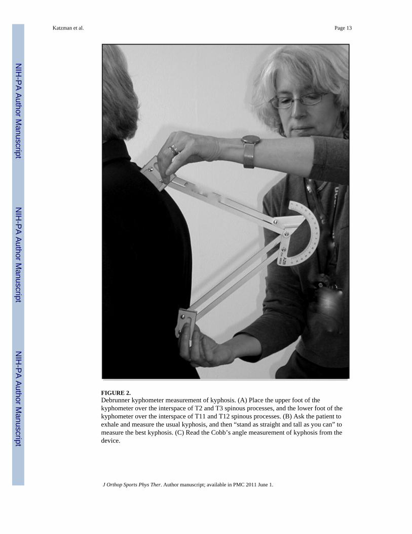
FIGURE 2.Debrunner kyphometer measurement of kyphosis. (A) Place the upper foot of thekyphometer over the interspace of T2 and T3 spinous processes, and the lower foot of thekyphometer over the interspace of T11 and T12 spinous processes. (B) Ask the patient toexhale and measure the usual kyphosis, and then “stand as straight and tall as you can” tomeasure the best kyphosis. (C) Read the Cobb’s angle measurement of kyphosis from thedevice.
Katzman et al. Page 13
J Orthop Sports Phys Ther. Author manuscript; available in PMC 2011 June 1.
NIH
-PA Author Manuscript
NIH
-PA Author Manuscript
NIH
-PA Author Manuscript

FIGURE 3.Flexicurve ruler measurement of kyphosis. (A) Mark the C7 spinous process and the L5–S1interspace on the patient’s skin with a grease pencil. (B) Place the superior end of the rulerat C7 and the inferior end over the lumbar spine, molding the ruler to the curves of thethoracic and lumbar spine. (C) Mark the level of the C7 spinous process and the L5–S1interspace on the ruler. (D) Carefully transfer the molded ruler to tracing paper, with the C7spinous process and the L5-S1 interspace marks aligned along a vertical line. (E) Trace thethoracic and lumbar curvatures from the ruler onto the paper, drawing a horizontal line fromthe vertical line to the apex of the thoracic curve. (F) Measure thoracic width (TW) andthoracic length (TL); calculate kyphosis index (KI): (TW/TL) × 100. (G) Lumbar width(LW) and lumbar length (LL) can also be measured. Photograph and diagram used withpermission from Carleen Lindsey, PT, MSc, GCS, and the Section on Geriatrics, APTA.38
Katzman et al. Page 14
J Orthop Sports Phys Ther. Author manuscript; available in PMC 2011 June 1.
NIH
-PA Author Manuscript
NIH
-PA Author Manuscript
NIH
-PA Author Manuscript
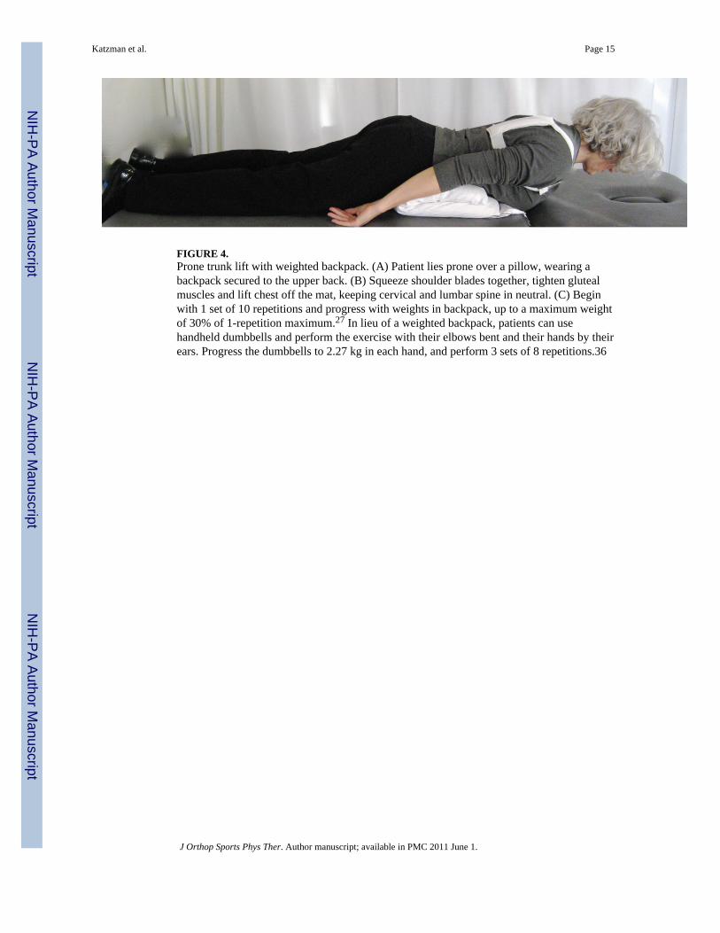
FIGURE 4.Prone trunk lift with weighted backpack. (A) Patient lies prone over a pillow, wearing abackpack secured to the upper back. (B) Squeeze shoulder blades together, tighten glutealmuscles and lift chest off the mat, keeping cervical and lumbar spine in neutral. (C) Beginwith 1 set of 10 repetitions and progress with weights in backpack, up to a maximum weightof 30% of 1-repetition maximum.27 In lieu of a weighted backpack, patients can usehandheld dumbbells and perform the exercise with their elbows bent and their hands by theirears. Progress the dumbbells to 2.27 kg in each hand, and perform 3 sets of 8 repetitions.36
Katzman et al. Page 15
J Orthop Sports Phys Ther. Author manuscript; available in PMC 2011 June 1.
NIH
-PA Author Manuscript
NIH
-PA Author Manuscript
NIH
-PA Author Manuscript
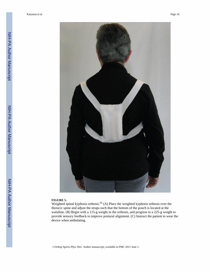
FIGURE 5.Weighted spinal kyphosis orthosis.55 (A) Place the weighted kyphosis orthosis over thethoracic spine and adjust the straps such that the bottom of the pouch is located at thewaistline. (B) Begin with a 115-g weight in the orthosis, and progress to a 225-g weight toprovide sensory feedback to improve postural alignment. (C) Instruct the patient to wear thedevice when ambulating.
Katzman et al. Page 16
J Orthop Sports Phys Ther. Author manuscript; available in PMC 2011 June 1.
NIH
-PA Author Manuscript
NIH
-PA Author Manuscript
NIH
-PA Author Manuscript
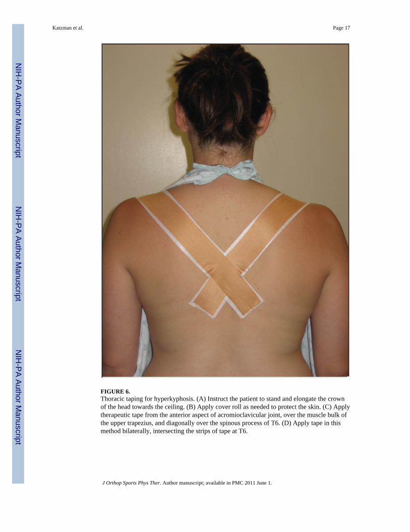
FIGURE 6.Thoracic taping for hyperkyphosis. (A) Instruct the patient to stand and elongate the crownof the head towards the ceiling. (B) Apply cover roll as needed to protect the skin. (C) Applytherapeutic tape from the anterior aspect of acromioclavicular joint, over the muscle bulk ofthe upper trapezius, and diagonally over the spinous process of T6. (D) Apply tape in thismethod bilaterally, intersecting the strips of tape at T6.
Katzman et al. Page 17
J Orthop Sports Phys Ther. Author manuscript; available in PMC 2011 June 1.
NIH
-PA Author Manuscript
NIH
-PA Author Manuscript
NIH
-PA Author Manuscript
NIH
-PA Author Manuscript
NIH
-PA Author Manuscript
NIH
-PA Author Manuscript
Katzman et al. Page 18
TABLE 1
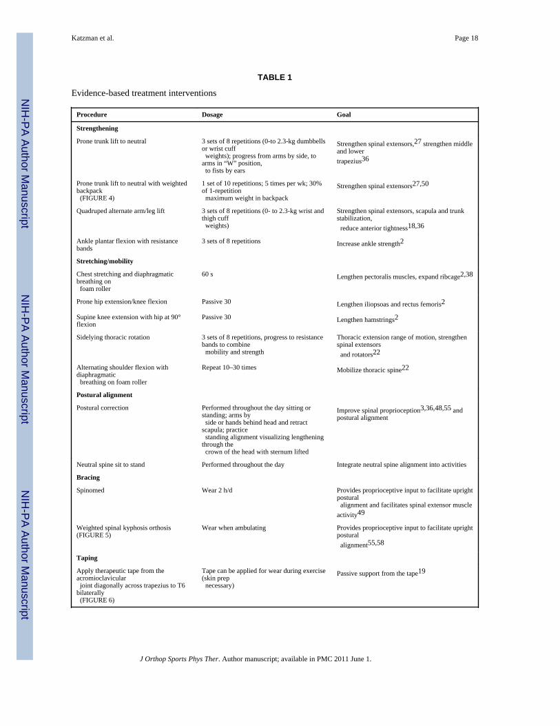
Evidence-based treatment interventions
Procedure Dosage Goal
Strengthening
Prone trunk lift to neutral 3 sets of 8 repetitions (0-to 2.3-kg dumbbellsor wrist cuff weights); progress from arms by side, toarms in “W” position, to fists by ears
Strengthen spinal extensors,27 strengthen middleand lowertrapezius36
Prone trunk lift to neutral with weightedbackpack (FIGURE 4)
1 set of 10 repetitions; 5 times per wk; 30%of 1-repetition maximum weight in backpack
Strengthen spinal extensors27,50
Quadruped alternate arm/leg lift 3 sets of 8 repetitions (0- to 2.3-kg wrist andthigh cuff weights)
Strengthen spinal extensors, scapula and trunkstabilization, reduce anterior tightness18,36
Ankle plantar flexion with resistancebands
3 sets of 8 repetitions Increase ankle strength2
Stretching/mobility
Chest stretching and diaphragmaticbreathing on foam roller
60 s Lengthen pectoralis muscles, expand ribcage2,38
Prone hip extension/knee flexion Passive 30 Lengthen iliopsoas and rectus femoris2
Supine knee extension with hip at 90°flexion
Passive 30 Lengthen hamstrings2
Sidelying thoracic rotation 3 sets of 8 repetitions, progress to resistancebands to combine mobility and strength
Thoracic extension range of motion, strengthenspinal extensors and rotators22
Alternating shoulder flexion withdiaphragmatic breathing on foam roller
Repeat 10–30 times Mobilize thoracic spine22
Postural alignment
Postural correction Performed throughout the day sitting orstanding; arms by side or hands behind head and retractscapula; practice standing alignment visualizing lengtheningthrough the crown of the head with sternum lifted
Improve spinal proprioception3,36,48,55 andpostural alignment
Neutral spine sit to stand Performed throughout the day Integrate neutral spine alignment into activities
Bracing
Spinomed Wear 2 h/d Provides proprioceptive input to facilitate uprightpostural alignment and facilitates spinal extensor muscleactivity49
Weighted spinal kyphosis orthosis(FIGURE 5)
Wear when ambulating Provides proprioceptive input to facilitate uprightpostural alignment55,58
Taping
Apply therapeutic tape from theacromioclavicular joint diagonally across trapezius to T6bilaterally (FIGURE 6)
Tape can be applied for wear during exercise(skin prep necessary)
Passive support from the tape19
J Orthop Sports Phys Ther. Author manuscript; available in PMC 2011 June 1.
NIH
-PA Author Manuscript
NIH
-PA Author Manuscript
NIH
-PA Author Manuscript
Katzman et al. Page 19
TABLE 2
Postural alignment during exercise and activities of daily living
Dos Don’ts
Maintain good postural alignment during exercise Avoid seated rowing machines or upper body ergometers
Strengthen core stabilizer muscles, such as transversus abdominus, obliques, and multifidus
Avoid crunches, curl-ups, or flexed position (traditional sit-ups)
When bending or lifting objects, keep the spine in neutral, and bend at the hips and knees (hip hinge); keep objects close to the body
Don’t twist or bend your spine when lifting objects
When getting out of bed, roll onto the side before sitting up (log roll)
Don’t sit straight up from a horizontal position
When coughing or sneezing, stabilize trunk in neutral by hugging a pillow, or placing hands on knees while hip hinging, or place hand in small of back to help keep back in neutral
Avoid forceful trunk flexion while coughing or sneezing
Maintain the natural curves in your neck and back while sitting and standing. Imagine that you are lengthening through the crown of your head
Avoid leaning over towards your work, or standing in a pelvic tilt
Adjust height of the walker and walk within the frame when ambulating
Don’t bend to reach, or push walker
J Orthop Sports Phys Ther. Author manuscript; available in PMC 2011 June 1.