age-dependent pattern of cerebellar susceptibility to bilirubin
TRANSCRIPT

© 2014. Published by The Company of Biologists Ltd | Disease Models & Mechanisms (2014) 7, 1057-1068 doi:10.1242/dmm.016535
1057
ABSTRACTNeonatal jaundice is caused by high levels of unconjugated bilirubin.It is usually a temporary condition caused by delayed induction ofUGT1A1, which conjugates bilirubin in the liver. To reduce bilirubinlevels, affected babies are exposed to phototherapy (PT), whichconverts toxic bilirubin into water-soluble photoisomers that arereadily excreted out. However, in some cases uncontrolledhyperbilirubinemia leads to neurotoxicity. To study the mechanismsof bilirubin-induced neurological damage (BIND) in vivo, wegenerated a mouse model lacking the Ugt1a1 protein and,consequently, mutant mice developed jaundice as early as 36 hoursafter birth. The mutation was transferred into two geneticbackgrounds (C57BL/6 and FVB/NJ). We exposed mutant mice to PTfor different periods and analyzed the resulting phenotypes from themolecular, histological and behavioral points of view. Severity of BINDwas associated with genetic background, with 50% survival ofC57BL/6-Ugt1−/− mutant mice at postnatal day 5 (P5), and of FVB/NJ-Ugt1−/− mice at P11. Life-long exposure to PT prevented cerebellararchitecture alterations and rescued neuronal damage in FVB/NJ-Ugt1−/− but not in C57BL/6-Ugt1−/− mice. Survival of FVB/NJ-Ugt1−/−
mice was directly related to the extent of PT treatment. PT treatmentof FVB/NJ-Ugt1−/− mice from P0 to P8 did not prevent bilirubin-induced reduction in dendritic arborization and spine density ofPurkinje cells. Moreover, PT treatment from P8 to P20 did not rescueBIND accumulated up to P8. However, PT treatment administered inthe time-window P0-P15 was sufficient to obtain full rescue ofcerebellar damage and motor impairment in FVB/NJ-Ugt1−/− mice.The possibility to modulate the severity of the phenotype by PTmakes FVB/NJ-Ugt1−/− mice an excellent and versatile model to studybilirubin neurotoxicity, the role of modifier genes, alternative therapiesand cerebellar development during high bilirubin conditions.
KEY WORDS: Neonatal jaundice, Ugt1, Phototherapy, BIND, Mousemodel
INTRODUCTIONNeonatal jaundice occurs in more than 60% of normal newbornsduring their first week of life (Bhutani and Johnson, 2009a; Bhutaniand Johnson, 2009b; Karen et al., 2009) as a result of excessiveunconjugated bilirubin (UCB) formation and transient inability of
RESEARCH ARTICLE
1International Centre for Genetic Engineering and Biotechnology (ICGEB), 34149Trieste, Italy. 2Basic Research and Integrative Neuroscience (BRAIN) Centre forNeuroscience, Department of Life Sciences, University of Trieste, 34127 Trieste,Italy.
*Author for correspondence ([email protected])
This is an Open Access article distributed under the terms of the Creative CommonsAttribution License (http://creativecommons.org/licenses/by/3.0), which permits unrestricteduse, distribution and reproduction in any medium provided that the original work is properlyattributed.
Received 11 April 2014; Accepted 9 July 2014
the neonatal liver to clear bilirubin rapidly enough from the bloodowing to delayed expression of UGT1A1, encoding UDP-glucuronosyltransferase 1a1 (UGT1A1). In fact, the key event inbilirubin detoxification is the glucuronidation reaction, which isaccomplished by UGT1A1 in the liver (Bosma et al., 1994). Severejaundice is also the hallmark of Crigler-Najjar syndrome type I(CNSI), in which mutations in the UGT1A1 gene result in thecomplete and life-long inactivity of the UGT1A1 enzyme.
Modest elevations of plasma UCB in newborns and adults areconsidered beneficial owing to the antioxidant properties of bilirubin(Doré and Snyder, 1999; Doré et al., 1999). The work on Gilbert’ssyndrome increasingly demonstrates the long-term beneficial effectsof low UCB plasma concentrations in averting numerous adultmaladies, including risk for certain cancers and atherosclerosis(Bulmer et al., 2013; Erlinger et al., 2014; Horsfall et al., 2012;Novotny and Vítek, 2003; Schwertner and Vítek, 2008; Vítek et al.,2002). However, severe hyperbilirubinemia is toxic to thedeveloping central nervous system (Ostrow et al., 2004; Ostrow etal., 2003). Prolonged and uncontrolled high levels of UCB lead tobilirubin encephalopathy (BE) and subsequently kernicterus. Indeveloping countries, up to 35% of newborns with kernicterus die,and some of the survivors suffer from permanent neurologicalsequelae (Kaplan et al., 2011).
Bilirubin targets specific brain regions, such as the basal ganglia,cochlear, oculomotor nuclei and cerebellum (Lauer and Spector, 2011;Watchko, 2006b). The spectrum of neurological deficits is severe andcan include movement disorders such as athetoid dystonic cerebralpalsy and oculomotor palsies, sensorineural deafness, auditorydysfunctions, and dental dysplasia. Despite the severely disablingcerebral palsy and hearing deficits, most individuals suffering frombilirubin encephalopathy have normal cognitive capacities. There isevidence that even moderate neonatal jaundice can result in subtleneuronal damage, with delayed motor development and minimallyimpaired cognitive functions that are unapparent during the neonatalperiod but lead to development or neurological impairment thatbecomes evident later in life (Chen and Kang, 1995; Oh et al., 2003).
High levels of UCB are conventionally treated by phototherapy(PT). Light energy (emission range 400-525 nm, peak emission 450-460 nm) is absorbed by UCB as it circulates in skin capillaries,resulting in the conversion of insoluble bilirubin into water-solublephotoisomers that are eliminated into the bile without the need ofliver conjugation (Maisels and McDonagh, 2008). PT is generallyvery effective in preventing transient hyperbilirubinemia in healthyneonates, because the hepatic conjugation system rapidly matures.However, in some cases high UCB levels concomitant with otherrisk factors, such as prematurity, hemolysis, sepsis, dehydration,early hospital discharge or lack of family training towards jaundice,can lead to serious neurological outcomes and ultimately death(Kaplan and Hammerman, 2005; Watchko and Tiribelli, 2013). Inaddition, individuals with CNSI, who need lifelong PT treatment forup to 14 hours per day, respond temporarily to PT and are at
Age-dependent pattern of cerebellar susceptibility to bilirubinneurotoxicity in vivo in miceGiulia Bortolussi1, Gabriele Baj2, Simone Vodret1, Giulia Viviani1, Tamara Bittolo2 and Andrés F. Muro1,*
Dis
ease
Mod
els
& M
echa
nism
s

1058
constant risk of developing brain damage unless liver transplantationis performed (Fagiuoli et al., 2013).
With the aim of studying the mechanisms of bilirubin toxicity invivo, we developed a mouse model bearing a null mutation in theUgt1 gene, resulting in early neonatal lethality due to bilirubintoxicity (Bortolussi et al., 2012). These mutant animals developsevere jaundice soon after birth and die within 11 days, showingsignificant cerebellar alterations and the major features of severeneonatal jaundice (Bortolussi et al., 2012). We previously showedthat PT treatment of these mutant mice extended their mediansurvival from 5 to 19 days, but it was not sufficient to rescue theirlethal phenotype (Bortolussi et al., 2012).
In the present work we transferred the Ugt1 genetic mutation intothe FVB/NJ and C57BL/6 mouse strains, and analyzed theneurodevelopmental effects of bilirubin toxicity in these lethalmurine models of neonatal jaundice. We showed that susceptibilityto bilirubin damage and, consequently, survival of mutant animalsis strain-specific, with a significant increase in the survival ofFVB/NJ-Ugt1−/− compared with C57BL/6-Ugt1−/− mice, thecerebellum being the most affected organ in both mutant strains.
Cerebellar susceptibility to bilirubin resulted in the alteration of itsarchitecture, with the important degeneration of Purkinje cell (PC)dendritic arbor, which was prevented by age-dependent PTtreatment. We identified the critical window of neuronalsusceptibility to bilirubin toxicity and analyzed how high bilirubinlevels affect brain development during the neonatal period. Weshowed that FVB/NJ-Ugt1−/− mutant mice were fully rescued whenPT treatment was administered from birth to 15 days of age.
One of the advantages of the presented model resides in thepossibility to modulate phenotype severity by applying PT fordifferent periods, allowing the study of physiological andbiochemical implications of bilirubin toxicity at the desireddevelopmental stage.
RESULTSPhenotype severity is associated with the geneticbackgroundWe have previously reported the generation of a mouse model ofneonatal severe hyperbilirubinemia bearing a targeted mutation inthe Ugt1 gene, which resembles the human CNSI from both thegenetic and phenotypic points of view (Bortolussi et al., 2012). Toreduce variability in the phenotype analysis, a more defined geneticbackground was obtained by backcrossing Ugt1 mutant mice formore than nine generations to wild-type (WT) C57BL/6 mice,reaching at least 99.8% of C57BL/6 genetic background. Owing tothe lack of Ugt1a1 activity, homozygous mutant mice developedhyperbilirubinemia within 36 hours after birth, as evident by theyellow staining of their skin (Fig. 1A), as already reported(Bortolussi et al., 2012). In this more defined genetic background,we observed a steeper shape of the mortality curve than foundpreviously (Bortolussi et al., 2012), with 50% mortality at postnatalday 5 (P5) (Fig. 1B). To study the effects of Ugt1 deficiency inanother genetic background, we transferred the mutation into theFVB/NJ strain, reaching at least 99.8% of FVB/NJ geneticbackground.
As in the C57BL/6 background, FVB/NJ-Ugt1−/− mice werevisibly jaundiced as early as 36 hours after birth (Fig. 1C).Interestingly, FVB/NJ-Ugt1−/− mice showed a less severe phenotypethan C57BL/6-Ugt1−/− mice, with 50% survival at 11 days after birth(P11; Fig. 1B). Prior to death, mutant mice showed features ofbilirubin encephalopathy such as lethargy, dystonia and seizures(supplementary material Movie 1).
Determination of plasma total bilirubin (TB) showed that levelsin C57BL/6-Ugt1−/− mice were significantly higher than in FVB/NJ-Ugt1−/− mice at both time points analyzed (Fig. 1D; two-wayANOVA, interaction time-genotype, NS; time, P≤0.05, genotype,P≤0.0005; Bonferroni’s post-hoc comparison test: P≤0.05 andP≤0.01 C57BL/6-Ugt1−/− versus FVB/NJ-Ugt1−/−, at P2 and P5,respectively). Moreover, we observed that total plasma bilirubin ofFVB/NJ-Ugt1−/− mice significantly increased with time, reachingplasma bilirubin levels of C57BL/6-Ugt1−/− levels in later timepoints (Fig. 1E; two-way ANOVA, interaction time-genotype,P≤0.0001; time, P≤0.0001, genotype, P≤0.0001; see alsosupplementary material Table S1).
To exclude the possibility that the differences in plasma TB andmortality could be associated with increased lysis of red blood cellsin the C57BL/6 strain, we determined the erythrocyte osmoticfragility from both strains in WT P5 pups and adults. The neonatalperiod is known to have a high level of erythrocyte hemolysis(Pearson, 1967), but our data show that there were no differencesbetween the strains in the NaCl concentration required for 50%hemolysis (C50), both in pups and adults (Fig. 1F,G, respectively).
RESEARCH ARTICLE Disease Models & Mechanisms (2014) doi:10.1242/dmm.016535
TRANSLATIONAL IMPACTClinical issueNeonatal jaundice is caused by high serum levels of unconjugatedbilirubin, a yellow pigment that is the breakdown product of hemoglobin.It is more common and severe in preterm than in term babies, and isusually a temporary condition caused by delayed induction of UDP-glucuronosyltransferase 1a1 (Ugt1a1), which conjugates bilirubin forexcretion. The condition can be treated with phototherapy, whichconverts bilirubin into a water-soluble form. Left untreated, severeneonatal hyperbilirubinemia, which is also the hallmark of Crigler-Najjarsyndrome type I (a genetic disorder caused by mutations in the UGT1A1gene), can damage the nervous system, leading first to bilirubinencephalopathy and then to kernicterus (‘yellow’ staining of the braintissue due to accumulation of bilirubin). About 70% of newborns whodevelop kernicterus die within a few days and survivors often havepermanent neurological deficits. However, the extent of neurologicaldamage caused by similar levels of unconjugated bilirubin varies widely,which makes it hard to assess the risk threshold of hyperbilirubinemiaor to develop intervention guidelines.
ResultsThe mechanism of bilirubin neurotoxicity remains poorly understoodbecause of the limitations of existing cellular and animal models. In thisstudy, the authors developed a mouse model bearing a null mutation inthe Ugt1 gene that results in early neonatal lethality due tohyperbilirubinemia. They investigated bilirubin neurotoxicity in twogenetic backgrounds and showed that susceptibility to bilirubin damageand the survival of mutant animals is strain-specific, a result thatunderscores the importance of modifier genes in the modulation ofbilirubin toxicity. Using phototherapy at different times after birth, theauthors identified the critical window of neuronal susceptibility to bilirubintoxicity and showed that the neurotoxic effects of bilirubin depend on thedevelopmental stage of the cerebellum. Finally, they reported thatphototherapy from birth to day 15 is sufficient to rescue mutant mice fromcerebellar damage and motor impairment.
Implications and future directionsThe results obtained in the mouse model developed here closely mirrorthe effects of hyperbilirubinemia in preterm infants. Thus, this new modelwill help the characterization of the molecular mechanisms of bilirubintoxicity during this critical period of human development. Moreover, theability to modulate the phenotype severity by applying phototherapy fordifferent periods makes this model a valuable and versatile setting inwhich to study bilirubin toxicity at different developmental stages, theeffects of modifier genes, and the effectiveness of new therapeuticapproaches.
Dis
ease
Mod
els
& M
echa
nism
s
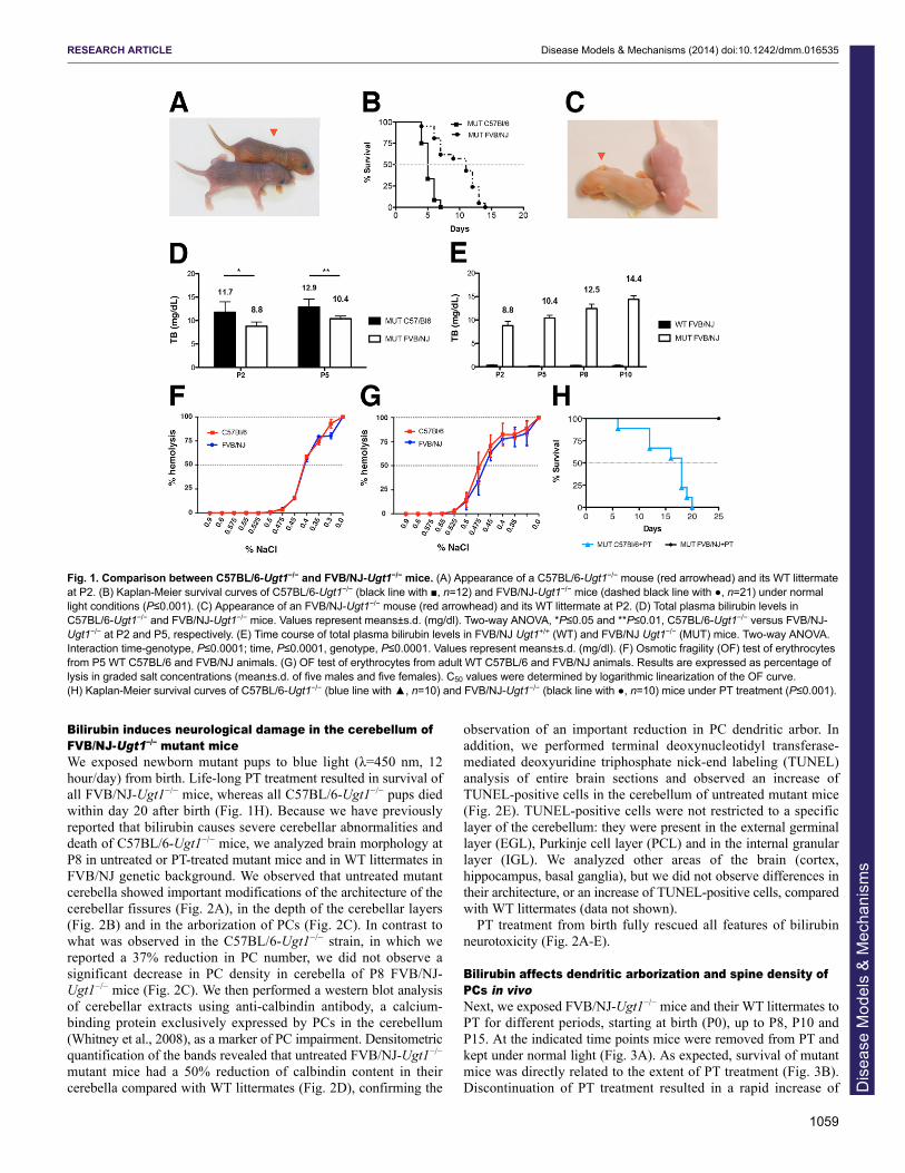
Bilirubin induces neurological damage in the cerebellum ofFVB/NJ-Ugt1−/− mutant miceWe exposed newborn mutant pups to blue light (λ=450 nm, 12hour/day) from birth. Life-long PT treatment resulted in survival ofall FVB/NJ-Ugt1−/− mice, whereas all C57BL/6-Ugt1−/− pups diedwithin day 20 after birth (Fig. 1H). Because we have previouslyreported that bilirubin causes severe cerebellar abnormalities anddeath of C57BL/6-Ugt1−/− mice, we analyzed brain morphology atP8 in untreated or PT-treated mutant mice and in WT littermates inFVB/NJ genetic background. We observed that untreated mutantcerebella showed important modifications of the architecture of thecerebellar fissures (Fig. 2A), in the depth of the cerebellar layers(Fig. 2B) and in the arborization of PCs (Fig. 2C). In contrast towhat was observed in the C57BL/6-Ugt1−/− strain, in which wereported a 37% reduction in PC number, we did not observe asignificant decrease in PC density in cerebella of P8 FVB/NJ-Ugt1−/− mice (Fig. 2C). We then performed a western blot analysisof cerebellar extracts using anti-calbindin antibody, a calcium-binding protein exclusively expressed by PCs in the cerebellum(Whitney et al., 2008), as a marker of PC impairment. Densitometricquantification of the bands revealed that untreated FVB/NJ-Ugt1−/−
mutant mice had a 50% reduction of calbindin content in theircerebella compared with WT littermates (Fig. 2D), confirming the
observation of an important reduction in PC dendritic arbor. Inaddition, we performed terminal deoxynucleotidyl transferase-mediated deoxyuridine triphosphate nick-end labeling (TUNEL)analysis of entire brain sections and observed an increase ofTUNEL-positive cells in the cerebellum of untreated mutant mice(Fig. 2E). TUNEL-positive cells were not restricted to a specificlayer of the cerebellum: they were present in the external germinallayer (EGL), Purkinje cell layer (PCL) and in the internal granularlayer (IGL). We analyzed other areas of the brain (cortex,hippocampus, basal ganglia), but we did not observe differences intheir architecture, or an increase of TUNEL-positive cells, comparedwith WT littermates (data not shown).
PT treatment from birth fully rescued all features of bilirubinneurotoxicity (Fig. 2A-E).
Bilirubin affects dendritic arborization and spine density ofPCs in vivoNext, we exposed FVB/NJ-Ugt1−/− mice and their WT littermates toPT for different periods, starting at birth (P0), up to P8, P10 andP15. At the indicated time points mice were removed from PT andkept under normal light (Fig. 3A). As expected, survival of mutantmice was directly related to the extent of PT treatment (Fig. 3B).Discontinuation of PT treatment resulted in a rapid increase of
1059
RESEARCH ARTICLE Disease Models & Mechanisms (2014) doi:10.1242/dmm.016535
Fig. 1. Comparison between C57BL/6-Ugt1−/− and FVB/NJ-Ugt1−/− mice. (A) Appearance of a C57BL/6-Ugt1−/− mouse (red arrowhead) and its WT littermateat P2. (B) Kaplan-Meier survival curves of C57BL/6-Ugt1−/− (black line with ■, n=12) and FVB/NJ-Ugt1−/− mice (dashed black line with ●, n=21) under normallight conditions (P≤0.001). (C) Appearance of an FVB/NJ-Ugt1−/− mouse (red arrowhead) and its WT littermate at P2. (D) Total plasma bilirubin levels inC57BL/6-Ugt1−/− and FVB/NJ-Ugt1−/− mice. Values represent means±s.d. (mg/dl). Two-way ANOVA, *P≤0.05 and **P≤0.01, C57BL/6-Ugt1−/− versus FVB/NJ-Ugt1−/− at P2 and P5, respectively. (E) Time course of total plasma bilirubin levels in FVB/NJ Ugt1+/+ (WT) and FVB/NJ Ugt1−/− (MUT) mice. Two-way ANOVA.Interaction time-genotype, P≤0.0001; time, P≤0.0001, genotype, P≤0.0001. Values represent means±s.d. (mg/dl). (F) Osmotic fragility (OF) test of erythrocytesfrom P5 WT C57BL/6 and FVB/NJ animals. (G) OF test of erythrocytes from adult WT C57BL/6 and FVB/NJ animals. Results are expressed as percentage oflysis in graded salt concentrations (mean±s.d. of five males and five females). C50 values were determined by logarithmic linearization of the OF curve.(H) Kaplan-Meier survival curves of C57BL/6-Ugt1−/− (blue line with ▲, n=10) and FVB/NJ-Ugt1−/− (black line with ●, n=10) mice under PT treatment (P≤0.001).
Dis
ease
Mod
els
& M
echa
nism
s

1060
bilirubin levels, reaching a plateau value of 14-16 mg/dl 3 days afterPT removal (Fig. 3C,D), whereas, during PT treatment, mutant micehad TB levels of 3.9±0.4 mg/dl (Fig. 3D), well below thoseconferring the risk of developing neurological damage (Bortolussiet al., 2012). We analyzed cerebellar morphology at P15 and did notobserve significant alterations of cerebellar fissures after thedifferent treatments (Fig. 3E). Moreover, at this stage, cerebellarlayer thickness was comparable among all treatments and was notstatistically different from WT littermates (Fig. 3F). In addition, thePC number and calbindin content (quantified by densitometricanalysis of the western blot bands) was comparable among alltreatments and was not statistically different from WT littermates(Fig. 3G and supplementary material Fig. S1). These resultsindicated that PT treatment for 8 days after birth was sufficient toavoid major cerebellar abnormalities. However, calbindin stainingof cerebella showed an apparent impairment of the PC dendriticarbor in P0-P8 PT-treated mutant mice, when determined at P15(Fig. 3G).
Owing to the high density and complexity of dendritic arbors inthe PC layer, it was not possible to quantify the extent of PCimpairment directly in anti-calbindin-immunostained sections.
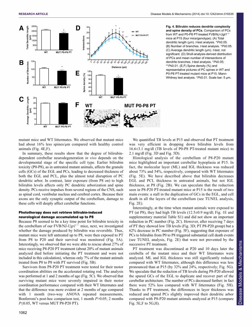
Thus, we assessed morphological changes in individual PCs byGolgi staining in P0-P8 PT-treated mutant mice at P15 andcompared them with WT littermates (Fig. 4). Golgi staining is avery powerful technique that allows the analysis of morphologicalfeatures such as dendritic arborization and spines of individualneurons in the brain (Ranjan and Mallick, 2010). After Golgistaining, ten single PCs with a complete dendritic arbor wereselected from each animal (four animals/genotype). Wereconstructed the dendritic arbor of each PC and measured threeparameters: total dendritic length, number of branching points andaverage length of each branch. The total length of the PC dendriticarbor and the number of branching points from P0-P8 PT-treatedmutant mice were reduced 16% and 17%, respectively, comparedwith WT littermates (Fig. 4A,B). In contrast, the average length ofeach branch (calculated as the ratio between total dendritic lengthand the number of branching points) was not reduced by bilirubin(Fig. 4C), suggesting that the entire dendritic arbor of PCs in P0-P8 PT-treated mutant mice was shorter in length owing to areduction in the branching number, maintaining the same distancebetween successive branches. To more accurately assess subtledamage produced by bilirubin in P0-P8 PT-treated mutant mice,
RESEARCH ARTICLE Disease Models & Mechanisms (2014) doi:10.1242/dmm.016535
Fig. 2. Characterization of P8 cerebellarlesions in FVB/NJ-Ugt1−/− mice with andwithout PT treatment. (A) Nissl staining of 8-day-old (P8) WT, untreated mutant and PT-treated mutant mice. Boxed areas indicatefields shown in panel B. IV, VIa, VIb and IXbindicate the cerebellar fissures. Scale bar:200 μm. (B) High-magnification images ofcerebellar layers from P8 mice and, in the rightpanel, layer depth quantification (fourmice/genotype). EGL, external germinal layer;PCL, Purkinje cell layer; IGL, internal granularlayer. Scale bar: 100 μm. Error bars, s.d.(C) Representative fluorescentimmunohistochemistry and quantification of PCnumber (cells/mm) from P8 WT, untreatedmutant and PT-treated mutant mice. PCs werestained with anti-calbindin1 antibody (green)and nuclei with Hoechst staining (blue). Scalebar: 50 μm. Quantification of the PC number(cells/mm) is represented in the right panel(four mice/genotype). Error bars, s.d. One-wayANOVA test not significant. (D) Western blotanalysis of total cerebellum protein extractusing an anti-calbindin1 antibody in P8 WT,untreated mutant and PT-treated mutant mice.Tubulin was used as a loading control. Lowerpanel: densitometric quantification of thebands. (E) TUNEL analysis. Positive cells areshown as brown dots; negative cells arecounterstained with methyl green. Scale bar:100 μm. One-way ANOVA test. *P≤0.05;**P≤0.01; ***P≤0.001.
Dis
ease
Mod
els
& M
echa
nism
s

we performed the Sholl analysis of PCs, a quantitative method formorphometric neuronal studies (Sholl, 1953). Sholl analysisrevealed that PCs from P0-P8 PT-treated mutant mice had areduced dendritic complexity compared with WT controls. The
reduction was particularly evident at a distance between 70 to110 μm from the soma (Fig. 4D).
Moreover, to deeply characterize subtle bilirubin-induceddamage, we measured spine density in PCs of P0-P8 PT-treated
1061
RESEARCH ARTICLE Disease Models & Mechanisms (2014) doi:10.1242/dmm.016535
Fig. 3. Efficacy of PT treatment in FVB/NJ-Ugt1−/− mice. (A) Schematic representation of PT treatment. FVB/NJ-Ugt1−/− mice and their WT littermates wereexposed to PT (blue arrows) for different periods, starting at birth (P0) up to P8, P10, P12 and P15. At the indicated time points, mice were removed from PTand kept under normal light (yellow arrows). (B) Kaplan-Meier survival curves of FVB/NJ-Ugt1−/− mice under normal light conditions (red line with ●, n=21),treated with PT for 8 days (light blue line with ▲, n=13), 10 days (blue line with ■, n=10) and 15 days (dark blue line with �, n=11) (P≤0.001). (C) Total plasmabilirubin (TB) levels at P15 of P0-P8, P0-P10, P0-P12 and P0-P15 PT-treated FVB/NJ-Ugt1−/− mutant mice (n=14, 5, 4 and 6, respectively) and WT littermates(n=9). One-way ANOVA test with Bonferroni’s post-hoc comparison tests; ns, not significant; *P≤0.05, ***P≤0.001. (D) Total plasma bilirubin levels in WT andFVB/NJ-Ugt1−/− mice. Values represent means±s.d. Values in parenthesis indicate number of samples analyzed. (E) Nissl staining of 15-day-old (P15) WT andPT-treated mutant mice. Boxed areas indicate fields shown in panel F. (F) High-magnification images of cerebellar layers from P15 mice and, in the right panel,layer depth quantification (four mice/treatment group). Scale bar: 100 μm. Error bars, s.d. One-way ANOVA test not significant. (G) Representative fluorescentimmunohistochemistry and quantification of PC number (cells/mm) from P15 WT and PT-treated mutant mice. PCs were stained with anti-calbindin1 antibody(green) and nuclei with Hoechst staining (blue). Scale bar: 50 μm. Quantification of the PC number (cells/mm) is represented in the right panel (fourmice/treatment group). Error bars, s.d. One-way ANOVA test with Bonferroni’s post-hoc comparison tests, not significant.
Dis
ease
Mod
els
& M
echa
nism
s
1062
mutant mice and WT littermates. We observed that mutant micehad about 16% less spines/μm compared with healthy controlanimals (Fig. 4E,F).
In summary, these results show that the degree of bilirubin-dependent cerebellar neurodegeneration in vivo depends on thedevelopmental stage of the specific cell type. Earlier bilirubintoxicity (P0-P8), as in untreated mutant animals, affects the granulecells (GCs) of the EGL and PCs, leading to decreased thickness ofboth the EGL and PCL, plus the almost total disruption of PCdendritic arbor. In contrast, later exposure (from P8 on) to highbilirubin levels affects only PC dendritic arborization and spinedensity. PCs receive impulses from several regions of the CNS, suchas spinal cord, vestibular nucleus and cerebral cortex. Because theiraxons are the only synaptic output of the cerebellum, damage tothese cells will deeply affect cerebellar functions.
Phototherapy does not retrieve bilirubin-inducedneurological damage accumulated up to P8Because P8 seemed to be a key time point for bilirubin toxicity inthe cerebellum of our FVB/NJ-Ugt1−/− mice, next, we investigatedwhether the damage produced by bilirubin was reversible. Thus,mutant mice were left untreated up to P8, were then exposed to PTfrom P8 to P20 and their survival was monitored (Fig. 5A).Interestingly, we observed that we were able to rescue about 27% ofmice receiving P8-P20 PT treatment (about 20% of mutant animalsanalyzed died before initiating the PT treatment and were notincluded in this calculation), whereas only 7% of the mutant animalstreated from P0 to P8 with PT survived (Fig. 5B).
Survivors from P8-P20 PT treatment were tested for their motorcoordination abilities on the accelerated rotating rod. The analysiswas performed at 1 and 2 months of age (Fig. 5C). We observed thatsurviving mutant mice were severely impaired in their motorcoordination performance compared with their WT littermates andthat the difference was more evident at 2 months of age comparedwith 1 month (two-way ANOVA repeated measurements,Bonferroni’s post-hoc comparison test, 1 month P>0.05; 2 monthsP≤0.05, WT versus MUT P8-P20 PT).
We quantified TB levels at P15 and observed that PT treatmentwas very efficient in dropping down bilirubin levels from16.6±3.1 mg/dl (TB levels of P0-P8 PT-treated mutant mice) to2.1 mg/dl (Fig. 3D and Fig. 5D).
Histological analysis of the cerebellum of P8-P20 mutant mice highlighted an important cerebellar hypoplasia at P15. Infact, the molecular layer (ML) and IGL thickness was reducedabout 73% and 54%, respectively, compared with WT littermates(Fig. 5E). We have described above that bilirubin decreases EGL and PCL thickness in untreated animals, but not IGLthickness, at P8 (Fig. 2B). We can speculate that the reduction seen in P8-P20 PT-treated mutant mice at P15 is the result of twomain events: a stall in the duplication of GCs in the EGL, and celldeath in all the layers of the cerebellum (see TUNEL analysis,Fig. 2E).
Interestingly, at the time when mutant animals were exposed toPT (at P8), they had high TB levels (12.5±0.9 mg/dl; Fig. 1E andsupplementary material Table S1) and did not show an importantreduction in PC number (Fig. 2C). However, after receiving 7 daysof PT they showed low TB levels (Fig. 3D; PT P8-P20 group) but a62% decrease in PC number (Fig. 5F), suggesting that exposure ofPCs to bilirubin from P0 to P8 triggered substantial cell death events(see TUNEL analysis, Fig. 2E) that were not prevented by thesuccessive PT treatment.
PT treatment was discontinued at P20 and 10 days later thecerebella of the mutant mice and their WT littermates wereanalyzed. ML and IGL thickness was still significantly reducedcompared with WT littermates, although this difference was lesspronounced than at P15 (by 32% and 24%, respectively; Fig. 5G).We speculate that the reduction of TB levels during P8-P20 allowedthe spared GCs of the EGL to duplicate and recover part of thecerebellar architecture. The number of PCs decreased further; in factthere were 52% less compared with WT littermates (Fig. 5H).Thanks to PT treatment, the differences in layer thickness wasreduced and spared PCs slightly improved their dendritic arborcompared with P8-P20 mutant animals analyzed at P15 (compareFig. 5E,F to 5G,H).
RESEARCH ARTICLE Disease Models & Mechanisms (2014) doi:10.1242/dmm.016535
Fig. 4. Bilirubin reduces dendrite complexityand spine density of PCs. Comparison of PCsfrom WT and P0-P8 PT-treated FVB/NJ-Ugt1−/−
mice at P15 (four mice/genotype). (A) Totaldendritic length (μm). t-test analysis. *P≤0.05.(B) Number of branches. t-test analysis. *P≤0.05.(C) Average dendritic length (μm). t-test, notsignificant. (D) Sholl-analysis-derived distributionof PCs and mean number of intersections ofdendrite branches. t-test analysis. *P≤0.05;**P≤0.01. (E,F) Spine density (%) andrepresentative pictures of PC spines in WT andP0-P8 PT-treated mutant mice at P15. Mann-Whitney test analysis. *P≤0.01. Scale bar: 5 μm.
Dis
ease
Mod
els
& M
echa
nism
s

PT treatment for only 15 days is sufficient to fully rescuesurvival, and histological and behavioral features of FVB/NJ-Ugt1−/− miceWhen mutant mice were exposed to PT from P0 to P15, weobserved 100% survival of treated mice (Fig. 3B). To determinewhether 15 days of PT was sufficient to fully rescue mutant mice,we performed a more detailed histological, functional and molecularanalysis. We observed that continuous PT treatment of mutant micefor 15 days prevented all morphological and functional deficitsproduced by bilirubin neurotoxicity, such as alterations in cerebellarmorphology (Fig. 6A), and differences in layer thickness (Fig. 6B)and in the number and arborization of PCs (Fig. 6C). In addition, weobserved that P0-P15 PT-treated mutant mice did not show any
obvious motor coordination impairment as assessed by theaccelerating rotarod test at any of the time points analyzed (Fig. 6D).
DISCUSSIONNeonatal hyperbilirubinemia is the most common cause of hospitalreadmission in the first week of life (75%) (Escobar et al., 2005).There is a growing concern in the pediatric field regardingbilirubin neurotoxicity during the neonatal period because highbilirubin levels are life threatening and if neglected can lead topermanent brain disabilities and ultimately death. Moreover,infants show a huge variability in the extent of neurologicaldamage caused by similar UCB levels, limiting the capacity toassess risk threshold for neurotoxicity and common intervention
1063
RESEARCH ARTICLE Disease Models & Mechanisms (2014) doi:10.1242/dmm.016535
Fig. 5. Characterization of P15 cerebellar lesions in P8-P20 PT-treated FVB/NJ-Ugt1−/− mice. (A) Schematic representation of PT (blue arrows) treatment.(B) Kaplan-Meier survival curves of FVB/NJ-Ugt1−/− mice treated with PT from P0 to P8 (light blue line with ●, n=13) and from P8 to P20 (dark blue line with ■,n=18). (C) Time course of motor coordination performance on rotarod of WT and P8-P20 PT-treated mutant mice (n=4/genotype). Two-way ANOVA test,interaction time-genotype, not significant (NS); time, NS; genotype, P≤0.01; Bonferroni’s post-hoc comparison tests: NS and P≤0.05 WT versus FVB/NJ-Ugt1−/−, at 1 and 2 months, respectively). *P≤0.01. WT, Ugt1 wild-type allele; MUT, Ugt1 mutant allele. (D) Total plasma bilirubin levels of P0-P8 and P8-P20PT-treated mutant mice at P15 (n=14 and 4, respectively). t-test analysis. ***P≤0.0001. (E) High-magnification images of cerebellar layers from P15 mice and,in the right panel, layer depth quantification (four mice/genotype). ML, molecular layer; IGL, internal granular layer. Scale bar: 100 μm. Error bars=s.d. t-testanalysis. ***P≤0.0001. (F) Representative fluorescent immunohistochemistry and quantification of PC number (cells/mm) from P15 mice. PCs were stainedwith anti-calbindin1 antibody (green) and nuclei with Hoechst staining (blue). Scale bar: 50 μm. Quantification of the PCs number (cells/mm) is represented inthe right panel (four mice/genotype). Error bars=s.d. t-test analysis. ***P≤0.0001. (G) High-magnification images of cerebellar layers from P30 mice and, in theright panel, layer depth quantification (four mice/genotype). Scale bar: 100 μm. Error bars=s.d. t-test analysis. **P≤0.001. (H) Representative fluorescentimmunohistochemistry and quantification of PC number (cells/mm) from P30 mice. PCs were stained with anti-calbindin1 antibody (green) and nuclei withHoechst staining (blue). Scale bar: 50 μm. Quantification of the PC number (cells/mm) is represented in the right panel (four mice/genotype). Error bars=s.d. t-test analysis. ***P≤0.0001.
Dis
ease
Mod
els
& M
echa
nism
s

1064
guidelines (Shapiro, 2003; Watchko and Jeffrey Maisels, 2010;Wennberg et al., 2006). During the past decades numerousprogresses in understanding the mechanism by which bilirubincauses neurological damage have been made (Watchko andTiribelli, 2013). However, the molecular and cellular mechanismsof bilirubin toxicity are still poorly defined owing to limitations ofthe available animal and cellular models.
To study bilirubin toxicity in vivo, we generated a mutant mousemodel of neonatal hyperbilirubinemia lacking Ugt1a1 enzymeactivity (Bortolussi et al., 2012). Mutant mice develop severehyperbilirubinemia as early as 36 hours after birth, severeneurological damage and early lethality, recapitulating all majorfeatures of severe neonatal jaundice and bilirubin-inducedneurological damage (BIND).
Strain-specific bilirubin susceptibilityWe characterized the lethal effects of Ugt1 deficiency in two mousegenetic backgrounds, C57BL/6 and FVB/NJ, showing importantdifferences in phenotype severity between the strains. In fact, mutantpups in the C57BL/6 genetic background had a median survival of 5days (Bortolussi et al., 2012), whereas those in the FVB/NJ geneticbackground survived longer, with a median survival of 11 days. Thedifferences in severity between the strains seem to be associated withthe increased plasma bilirubin values in the C57BL/6 mice but not toa difference in erythrocyte fragility. We have shown that thisdifference is not associated with increased hemolysis and bilirubinproduction in the C57BL/6 strain, despite this strain having higher redcell count (10.3×10⁶ versus 9.2×10⁶, for C57BL/6 and FVB/NJ,respectively; http://phenome.jax.org) and hematocrit values (50.2 and43.4, for C57BL/6 and FVB/NJ, respectively; http://phenome.jax.org).
Other genetically engineered mouse models, such as EGFreceptor, retinoblastoma-related p130, fibronectin and Tgfb1knockout (KO) strains, also showed that differences in the geneticbackground cause considerable variations in the phenotype(Doetschman, 2009; George et al., 1997; LeCouter et al., 1998a;LeCouter et al., 1998b; Threadgill et al., 1995). These importantdifferences in phenotype severity are not yet fully elucidated, but
seem to be associated with the modulatory effects of modifier genes(Astrof et al., 2007; Doetschman, 2009). Here, we have shown thatthe less severe phenotype of FVB/NJ-Ugt1−/− mice correlates withthe reduced cerebellar morphological defects, as compared withC57BL/6-Ugt1−/− mice. In fact, C57BL/6 mutant mice showed asignificant reduction both in the number of PCs and depth ofcerebellar layers, whereas the number of PCs in FVB/NJ-Ugt1−/−
mice was not affected and the differences in cerebellar layers wereless pronounced. A more detailed analysis showed that, in the FVBmodel, PC dendritic arborization is affected, similarly to thatobserved in C57BL/6-Ugt1−/− pups (Bortolussi et al., 2012).
Interestingly, the mouse models characterized in this study havethe same genetic mutation present in the hyperbilirubinemic Gunnrat (Bortolussi et al., 2012; Iyanagi et al., 1989), but displayed amuch stronger phenotype. In fact, all mutant mice died a few daysafter birth due to bilirubin toxicity. Over time, different colonies ofGunn rats were used to study bilirubin toxicity, with phenotypesranging from complete survival of untreated animals, with cerebellarabnormalities and hearing impairment, but reaching adulthood andreproducing (Chowdhury et al., 1993), to partial lethality (Keino andKashiwamata, 1989; Keino et al., 1985-1986). Early studies in theGunn rats (Johnson et al., 1959) reported plasma bilirubin levelssimilar to those found in the FVB/NJ-Ugt1−/− mouse strain, althoughmortality was low and difficult to interpret owing to intercurrentinfections. A more severe phenotype of the Gunn rats was obtainedby treatment, at different postnatal days, with sulfadimetoxine,showing histological abnormalities in the cerebellar layers and PCs(Conlee and Shapiro, 1997; Schutta and Johnson, 1967; Schutta andJohnson, 1969; Schutta and Johnson, 1971) similar to thoseobserved in our untreated mutant mouse strains. Other importantdifferences are also observed when Ugt1 mutant mice and the Gunnrats are exposed to PT: a single 24-hour dose of PT is enough torescue Gunn rat lethality and prevent hypoplasia in the cerebellum,with treatment at day 7 being most effective (Keino andKashiwamata, 1989) and the critical period for bilirubin-inducedcerebellar hypoplasia being between P6 and P10 (Conlee andShapiro, 1997; Keino and Kashiwamata, 1989; Sawasaki et al.,
RESEARCH ARTICLE Disease Models & Mechanisms (2014) doi:10.1242/dmm.016535
Fig. 6. Characterization of P30 cerebellar lesions in FVB/NJ-Ugt1−/− mice exposed for 15 days to PT. (A) Nissl staining of 30-day-old WT and P0-P15 PT-treated mutant mice. Boxed areas indicate fields shown in B. (B) High-magnification images of cerebellar layers from P30 mice and, in the right panel, layerdepth quantification (four mice/genotype). Scale bar: 100 μm. Error bars, s.d. t-test analysis, not significant. ML, molecular layer; IGL, internal granular layer.(C) Representative fluorescent immunohistochemistry and quantification of PC number (cells/mm) from P15 mice. PCs were stained with anti-calbindin1antibody (green) and nuclei with Hoechst staining (blue). Scale bar: 50 μm. Quantification of the PC number (cells/mm) is represented in the right panel (fourmice/genotype). Error bars, s.d. t-test analysis, not significant. (D) Time course of motor coordination performance on rotarod of WT and P0-P15 PT-treatedmutant mice at P30 and P60. Error bars=s.d. Two-way ANOVA repeated measurements, WT versus MUT P0-P15 PT, at P30 and P60, not significant.
Dis
ease
Mod
els
& M
echa
nism
s

1976). In contrast, continuous PT is required in FVB/NJ-Ugt1−/−
mice to rescue lethality, whereas this treatment is not sufficient toprevent death of C57BL/6 mutant animals.
Modulation of phenotype severityWe have presented here a novel and versatile model to studybilirubin toxicity. One of the advantages resides in the possibility tomodulate phenotype severity by applying PT for different periods,allowing the study of physiological and biochemical implications ofbilirubin toxicity at the desired developmental stage. For example,we studied the effects of bilirubin toxicity in mutant FVB/NJ-Ugt1−/− mice treated with PT for 8 days. In these mice, despite theabsence of gross cerebellar abnormalities, and a similar number ofPCs and calbindin levels compared with WT mice, almost 90% ofmutant mice did not survive. A more detailed analysis showed asignificant reduction in dendritic arborization and in spine density,suggesting that these defects could contribute to neuronalabnormalities and death. When we extended PT treatment foranother 2 days (P0-P10 PT), we observed a less severe condition,which resulted in about 50% survival.
We anticipate that these experimental conditions are valuablewhen the effect of a specific gene under study or pharmacologicaltreatment is masked by the severity of hyperbilirubinemia, acondition that underestimates the therapeutic outcome or molecular
partner contribution. Again, the readout of the experiment will be assimple as the increase of the survival rate.
The window of neuronal susceptibilityIt is known that the cerebellum is particularly vulnerable to insultsbecause of its very rapid growth during peri- and postnataldevelopment (Biran et al., 2012; Fonnum and Lock, 2000). Uponaccomplishment of the early cerebellar patterning in which twogerminal compartments (dorsal rhombic lip and the ventricular zoneof the fourth ventricle) are generated, the cerebellar developmentexperiences two main phases: (1) migration and proliferation ofrhombotic lip progenitors to populate the external germinal layer;and (2) migration and differentiation of the PCs to populate the PCL(Roussel and Hatten, 2011).
Shortly after birth, GC progenitors undergo a strong andprolonged phase of clonal expansion in the EGL that lasts up toP20. The proliferation and migration of GCs is regulated by PCgrowth factors. After several cycles of duplication, GCs located inthe external part of the EGL (facing the PCL) stop duplicating andmigrate radially inward to their final destination in the IGL.During their migration into the IGL, GCs extend their T-shapedaxons orthogonal to their migration path to generate the parallelfibers. Once in the IGL they will end their maturation process andreceive input from the mossy fibers of the cortico-pontocerebellar
1065
RESEARCH ARTICLE Disease Models & Mechanisms (2014) doi:10.1242/dmm.016535
Fig. 7. Model of bilirubin toxicity during postnatal cerebellar development in the FVB/NJ-Ugt1−/− mouse model. Bilirubin toxicity during the neonatalperiod produces different outcomes depending on the developmental period of exposure. The window of high toxicity is between P0 and P8. The main eventsoccurring in the cerebellum during this period are granule cell (GC) proliferation and migration, and Purkinje cells (PCs) extend their dendritic arbor. Bilirubincauses early neonatal lethality and substantial cerebellar damage. Later exposures (after P8) increase the number of spared animals and produce subtledamages, such as reduction of PC dendritic arbor and spine density. After P15, bilirubin does not cause lethality and evident cerebellar abnormalities. Thecomparison with human cerebellar development revealed that the critical window of toxicity is between the 24th and 38th gestational week (gw), also referred as the very preterm and preterm period, respectively. It is known that very preterm and preterm infants have a higher risk of developing bilirubin-inducedneurological damage (BIND). The risk of developing BIND is critically reduced in term infant. pnw, postnatal week. Modified from Biran et al. (Biran et al., 2012). D
isea
se M
odel
s &
Mec
hani
sms

1066
neurons, the main input of the cerebellum. Concomitant with theonset of GC parallel fibers, PCs differentiate by completing theirdendritogenesis.
Rodents and humans share a high degree of conservation ofcerebellar anatomy and function, although the timing is slightlydifferent (Biran et al., 2012). As described above, in rodents theproliferation of the EGL and the formation of the IGL occurpostnatally, whereas the same phase in humans occurs between the24th gestational week and birth (Fig. 7). Similarly, the main part ofPC differentiation happens prenatally in humans, but postnatally inrodents. Thus, it is not surprising to note that cerebellarabnormalities in babies are a common complication of verypremature and premature births (24-28 and 28-32 gestational weeks,respectively). Survivors frequently show a broad range of disabilitiesconnected to cerebellar development, such as hypotonia, fine motorcoordination failures, ataxia, impaired motor coordinationsequencing and cerebral palsy (Haldipur et al., 2011; Limperopouloset al., 2005; Volpe, 2009).
Preterm gestation is one of the most prevalent known risk factorsfor the development of severe hyperbilirubinemia and kernicterus(for a detailed review, see Watchko, 2006b); in fact,hyperbilirubinemia in preterm infants is more frequent and severeand its course is more prolonged then in term infants (Bhutani andJohnson, 2006). Treatment guidelines indicate that infants at higherrisk to develop bilirubin-induced neurological sequelae are thoseborn between the 35th and 37th gestational weeks (Watchko,2006a). The spectrum of bilirubin-induced neurological disabilitiesalso include movement disorders such as dystonia, athetosis,cerebral palsy and occasionally spasticity, which are well-knownmarkers of basal ganglia, cerebellum and brainstem damage(Shapiro et al., 2006).
The results obtained in our mouse model mirror what has beenreported so far in preterm and term infants. In fact, exposure ofmutant mice to high levels of bilirubin during the first 8 days afterbirth (corresponding to preterm period in humans) causes severeneurological damage and a high mortality rate; at later time points(after P8, corresponding to term and late term infants), we reporteda gradual decrease in the mortality rate, accompanied by less severeneurological outcomes (Fig. 7). It is well known that bilirubinneurotoxic effects can still be reversible if bilirubin levels arepromptly reduced, avoiding permanent damage (Hansen et al., 2009;Zuccoli et al., 2011), although a range of outcomes suggests inter-individual variations in vulnerability (Hankø et al., 2001). Inpatients, the likelihood of reversing advanced stages depends on theimplementation of rapid and effective treatments, such as intensivePT (Hansen, 2011) and exchange transfusion (Keenan et al., 1985;Roussel and Hatten, 2011).
In our FVB/NJ-Ugt1−/− mice, the reversibility threshold seems tobe before P8: when PT was applied from P8 and mutant miceanalyzed at P15 (Fig. 5), a compromised motor-coordinationfunction was observed, together with reduced depth of cerebellarlayers, PC number and dendritic arborization. These damages werenot reversed in spite of low plasma bilirubin values from P8 to P20as the result of PT treatment. Such a critical period suggests that, in preterm infants, the identification and treatment ofhyperbilirubinemia must be prompt and very efficient.
To summarize, we have shown here the consequences of the Ugt1null mutation in two mouse genetic backgrounds, with C57BL/6-Ugt1−/− mice showing a more severe phenotype than FVB/NJ-Ugt1−/− ones. We showed that PT treatment was effective in theprevention of brain damage and resulted in the complete rescue ofthe lethal phenotype only in the FVB/NJ-Ugt1−/− animals. It emerges
that susceptibility to bilirubin damage and, consequently, survival ofmutant animals seems to be strain- and species-specific,underscoring the importance of modifier genes in the modulation ofbilirubin toxicity.
The better characterization of the molecular mechanisms ofbilirubin toxicity in the two mouse strains could result in theidentification of novel genes involved in the modulation of bilirubin-induced neurological damage. Moreover, the capability tomanipulate the mouse genome will allow the generation of mousemodels of genes potentially involved in bilirubin metabolism thatare expected to improve the comprehension of the mechanisms ofUCB toxicity and protection. In view of these observations,FVB/NJ-Ugt1−/− mice represent an excellent and versatile model tostudy bilirubin toxicity, the role of modifier genes and theeffectiveness of new therapeutic approaches.
MATERIALS AND METHODSAnimalsUgt1 mutant mice in the C57BL/6 background have been generatedpreviously (Bortolussi et al., 2012). WT littermates were used as a control.Mice were housed and handled according to institutional guidelines, andexperimental procedures approved by the International Centre for GeneticEngineering and Biotechnology (ICGEB) board, with full respect to the EUDirective 2010/63/EU for animal experimentation. Animals used in thisstudy were at least 99.8% C57BL/6 or FVB/NJ genetic background,obtained after more than nine backcrosses with C57BL/6 and FVB/NJ mice,respectively. Mice were kept in a temperature-controlled environment with12/12 hours light/dark cycle. They received a standard chow diet and waterad libitum.
Phototherapy treatmentNewborns were exposed to blue fluorescent light (20 μW/cm2/nm, PhilipsTL 20W/52 lamps; Philips, Amsterdam, The Netherlands) for 12 hours/day(synchronized with the light period of the light/dark cycle) up to theindicated postnatal day and then maintained under normal light conditions.Intensity of the blue lamps was monitored monthly with an Olympic MarkII Bili-Meter (Olympic Medical, Port Angeles, WA). The distance from thelamps to the bottom of the cases was ~27 cm.
Bilirubin measurementsBlood samples were collected at different time points in mutant and WTlittermates by decapitation or cardiac puncture in EDTA-collecting tubes.Total bilirubin (TB) determination in plasma was performed using Directand Total Bilirubin Reagent kit (BQ Kits, San Diego, CA) adapting themethod to use minimal volumes (10 μl of plasma). The adaptation of themethod consisted in the reduction of the volumes (all), in order to use lesssample volume. The original proportions were maintained. Threecommercial bilirubin reference standards (Control Serum I, Control SerumII and Bilirubin Calibrator, Diazyme Laboratories, Poway, CA) wereincluded in each set of analysis as quality control. Absorbance values at 560nm were obtained by using a multiplate reader (Perkin Elmer Envision PlateReader, Walthman, MA).
Erythrocyte osmotic fragility testThe erythrocyte osmotic fragility (OF) test was performed as previouslydescribed (Muro et al., 2000) from freshly drawn blood. Five WT C57BL/6and FVB/NJ animals per gender and time point were analyzed. Theexperiment was repeated three times with similar values. Results areexpressed as percentage of lysis in graded salt concentrations (mean±s.d. offive males and five females). C50 values were determined by logarithmiclinearization of the OF curve.
Rotarod analysisThe coordination and balance ability on a rotating cylinder was assessed at1 and 2 months of age with an accelerating apparatus as previouslydescribed (Bortolussi et al., 2012).
RESEARCH ARTICLE Disease Models & Mechanisms (2014) doi:10.1242/dmm.016535
Dis
ease
Mod
els
& M
echa
nism
s

Cerebellar histologyBrains were removed from the skulls and divided into two hemispheres: onewas subjected to brain histology, whereas the other was subjected to proteinextraction.
Brains from each genotype were fixed with 4% PFA in PBS overnight at4°C. After cryoprotection in 20% sucrose, 0.02% sodium azide in PBS,specimens were frozen in cryostat embedding medium (Bio-optica) and 14-μm sagittal sections were obtained in a cryostat. Nissl staining wasperformed as previously described (Bortolussi et al., 2012). Forimmunofluorescence, 14-μm sagittal sections were blocked for 2 hours atroom temperature (RT) with 2.5% BSA in PBS 0.3% Triton X-100. Afterblocking, specimens were incubated with the primary antibody for 2 hoursat RT in blocking solution with anti-calbindin (Synaptic Systems,Goettingen, Germany). After 3×5-minute washes with blocking solution,specimens were incubated with secondary antibody (Alexa Fluor 488;Invitrogen Carlsbad, CA) for 2 hours at RT. Nuclei were visualized byaddition of Hoechst (10 μg/ml, Invitrogen) for 5 minutes after secondaryantibody solution.
TUNEL assay was performed using the In Situ Cell Death Detection Kit,POD according to manufacturer’s instructions (Roche). Methyl green wasused as counterstaining.
Nissl-stained and TUNEL slides were mounted in Eukitt (Fluka, St Louis,MO), whereas immunostained slides were mounted in Mowiol 4-88 (Sigma-Aldrich).
Images were acquired on a Nikon Eclipse E-800 epifluorescentmicroscope with a charge-coupled device camera (DMX 1200F; NikonAmstelveen, The Netherlands). Digital images were collected using ACT-1(Nikon) software.
Analysis of the layer thickness was performed on Nissl-stained sectionsby measuring the layer depth (μm) as previously described (Bortolussi etal., 2012). PC number was calculated by counting calbindin-positive cellsin vermis sections along the entire cerebellum perimeter and expressed aslinear density (cell/mm) as previously described (Bortolussi et al., 2012).
Golgi staining was performed as described by Ranjan and Mallick(Ranjan and Mallick, 2010) with minor modifications to adapt the methodto stain PCs of 15-day-old mice. Brains (n=4 mice for each group) weredivided into the two hemispheres along the midline and, after a wash inPBS, they were submerged in the Golgi solution containing 5% of potassiumdichromate, 5% of mercuric chloride and 5% of potassium chromate.Specimens were kept in darkness for 72 hours at 37°C. Golgi solution wasreplaced every day to increase the staining. Later, brains were washed indistillated water and sliced in a vibratome (Campden Instruments, MA752motorised advance vibroslice) in 200-μm sagittal sections. Sections werethen treated according to the Ranjan and Mallick protocol, dehydrated andmounted in Eukitt.
Ten PCs for each genotype/treatment were analyzed and a series of stackimages were collected at 40× magnification. Stacks were flattened withImageJ software to allow the quantification of specific parameters. Totaldendritic length, number of branches, average of dendrite length and Shollanalysis were quantified using NeuroStudio software (Icahn School ofMedicine at Mount Sinai, NY). Sholl analysis was performed withconcentric circles of 10 μm. The complexity of the dentritic arbor is directlyrelated to the number of times the dendrites cross the concentric circles(Sholl, 1953).
For spine density, ten segments of 50-μm long apical dendrites wereanalyzed for each animal (four animals/treatment) as previously described(Baj et al., 2014).
The genotype of the animals and the treatment were unknown to theoperator. A second operator analyzed the data. Measurements were averagedfor each animal. The results are expressed as mean±s.d. for each genotype.
Preparation of total protein extracts and western blot analysisCerebella were dissected and homogenized in RIPA buffer (150 mM NaCl,1% NP-40, 0.5% DOC, 0.1% SDS, 50 mM Tris HCl pH 8, 2× proteaseinhibitors) and analyzed by western blot analysis as described previously(Bortolussi et al., 2012). Primary antibodies used were as follows: anti-calbindin (Synaptic Systems, Goettingen, Germany) and anti-β-tubulin mAbE7 (Developmental Studies Hybridoma Bank, Iowa City, IA).
StatisticsResults are expressed as mean±s.d. The Prism package (GraphPad Software,La Jolla, CA) was used to analyze the data. Values of P≤0.05 wereconsidered statistically significant. Depending on the experimental design,we used Student’s t-test, one-way ANOVA or two-way ANOVA, withBonferroni’s post-hoc comparison tests, as indicated in the legends to thefigures and text.
AcknowledgementsThe authors thank Prof. E. Tongiorgi for the microscope facility resources and forcritical reading of the manuscript; and M. Sturnega and S. Artico for help withanimal care.
Competing interestsThe authors declare no competing financial interests.
Author contributionsG. Bortolussi designed and performed all the experiments, analyzed the data andwrote the manuscript. G. Baj performed the Golgi staining, analyzed the data, andprepared and discussed the manuscript. S.V. helped in the collection of thehistological data and carefully discussed the manuscript. G.V. performed theerythrocyte fragility test. T.B. helped in the Golgi protocol setting. A.F.M. conceivedthe project, designed the experiments, overlooked data analysis, provided financialsupport and wrote the manuscript.
FundingThis work was supported by Telethon (GGP10051), by the Friuli-Venezia GiuliaRegional Grant and by Beneficentia Stiftung to A.F.M.; and by AXA Research Fundto G. Bortolussi.
Supplementary materialSupplementary material available online athttp://dmm.biologists.org/lookup/suppl/doi:10.1242/dmm.016535/-/DC1
ReferencesAstrof, S., Kirby, A., Lindblad-Toh, K., Daly, M. and Hynes, R. O. (2007). Heart
development in fibronectin-null mice is governed by a genetic modifier onchromosome four. Mech. Dev. 124, 551-558.
Baj, G., Patrizio, A., Montalbano, A., Sciancalepore, M. and Tongiorgi, E. (2014).Developmental and maintenance defects in Rett syndrome neurons identified by anew mouse staging system in vitro. Front Cell Neurosci 8, 18.
Bhutani, V. K. and Johnson, L. (2006). Kernicterus in late preterm infants cared for asterm healthy infants. Semin. Perinatol. 30, 89-97.
Bhutani, V. K. and Johnson, L. (2009a). Kernicterus in the 21st century: frequentlyasked questions. J. Perinatol. 29 Suppl. 1, S20-S24.
Bhutani, V. K. and Johnson, L. (2009b). A proposal to prevent severe neonatalhyperbilirubinemia and kernicterus. J. Perinatol. 29 Suppl. 1, S61-S67.
Biran, V., Verney, C. and Ferriero, D. M. (2012). Perinatal cerebellar injury in humanand animal models. Neurol. Res. Int. 2012, 858929.
Bortolussi, G., Zentilin, L., Baj, G., Giraudi, P., Bellarosa, C., Giacca, M., Tiribelli,C. and Muro, A. F. (2012). Rescue of bilirubin-induced neonatal lethality in a mousemodel of Crigler-Najjar syndrome type I by AAV9-mediated gene transfer. FASEB J.26, 1052-1063.
Bosma, P. J., Seppen, J., Goldhoorn, B., Bakker, C., Oude Elferink, R. P.,Chowdhury, J. R., Chowdhury, N. R. and Jansen, P. L. (1994). Bilirubin UDP-glucuronosyltransferase 1 is the only relevant bilirubin glucuronidating isoform inman. J. Biol. Chem. 269, 17960-17964.
Bulmer, A. C., Verkade, H. J. and Wagner, K. H. (2013). Bilirubin and beyond: areview of lipid status in Gilbert’s syndrome and its relevance to cardiovasculardisease protection. Prog. Lipid Res. 52, 193-205.
Chen, Y. J. and Kang, W. M. (1995). Effects of bilirubin on visual evoked potentials interm infants. Eur. J. Pediatr. 154, 662-666.
Chowdhury, J. R., Kondapalli, R. and Chowdhury, N. R. (1993). Gunn rat: a modelfor inherited deficiency of bilirubin glucuronidation. Adv. Vet. Sci. Comp. Med. 37,149-173.
Conlee, J. W. and Shapiro, S. M. (1997). Development of cerebellar hypoplasia injaundiced Gunn rats: a quantitative light microscopic analysis. Acta Neuropathol. 93,450-460.
Doetschman, T. (2009). Influence of genetic background on genetically engineeredmouse phenotypes. Methods Mol. Biol. 530, 423-433.
Doré, S. and Snyder, S. H. (1999). Neuroprotective action of bilirubin against oxidativestress in primary hippocampal cultures. Ann. N. Y. Acad. Sci. 890, 167-172.
Doré, S., Takahashi, M., Ferris, C. D., Zakhary, R., Hester, L. D., Guastella, D. andSnyder, S. H. (1999). Bilirubin, formed by activation of heme oxygenase-2, protectsneurons against oxidative stress injury. Proc. Natl. Acad. Sci. USA 96, 2445-2450.
Erlinger, S., Arias, I. M. and Dhumeaux, D. (2014). Inherited disorders of bilirubintransport and conjugation: new insights into molecular mechanisms andconsequences. Gastroenterology 146, 1625-1638.
1067
RESEARCH ARTICLE Disease Models & Mechanisms (2014) doi:10.1242/dmm.016535
Dis
ease
Mod
els
& M
echa
nism
s
1068
Escobar, G. J., Greene, J. D., Hulac, P., Kincannon, E., Bischoff, K., Gardner, M.N., Armstrong, M. A. and France, E. K. (2005). Rehospitalisation after birthhospitalisation: patterns among infants of all gestations. Arch. Dis. Child. 90, 125-131.
Fagiuoli, S., Daina, E., D’Antiga, L., Colledan, M. and Remuzzi, G. (2013).Monogenic diseases that can be cured by liver transplantation. J. Hepatol. 59, 595-612.
Fonnum, F. and Lock, E. A. (2000). Cerebellum as a target for toxic substances.Toxicol. Lett. 112-113, 9-16.
George, E. L., Baldwin, H. S. and Hynes, R. O. (1997). Fibronectins are essential forheart and blood vessel morphogenesis but are dispensable for initial specification ofprecursor cells. Blood 90, 3073-3081.
Haldipur, P., Bharti, U., Alberti, C., Sarkar, C., Gulati, G., Iyengar, S., Gressens, P.and Mani, S. (2011). Preterm delivery disrupts the developmental program of thecerebellum. PLoS ONE 6, e23449.
Hankø, E., Lindemann, R. and Hansen, T. W. (2001). Spectrum of outcome in infantswith extreme neonatal jaundice. Acta Paediatr. 90, 782-785.
Hansen, T. W. (2011). The role of phototherapy in the crash-cart approach to extremeneonatal jaundice. Semin. Perinatol. 35, 171-174.
Hansen, T. W., Nietsch, L., Norman, E., Bjerre, J. V., Hascoet, J. M., Mreihil, K. andEbbesen, F. (2009). Reversibility of acute intermediate phase bilirubinencephalopathy. Acta Paediatr. 98, 1689-1694.
Horsfall, L. J., Nazareth, I. and Petersen, I. (2012). Cardiovascular events as afunction of serum bilirubin levels in a large, statin-treated cohort. Circulation 126,2556-2564.
Iyanagi, T., Watanabe, T. and Uchiyama, Y. (1989). The 3-methylcholanthrene-inducible UDP-glucuronosyltransferase deficiency in the hyperbilirubinemic rat(Gunn rat) is caused by a -1 frameshift mutation. J. Biol. Chem. 264, 21302-21307.
Johnson, L., Sarmiento, F., Blanc, W. A. and Day, R. (1959). Kernicterus in rats withan inherited deficiency of glucuronyl transferase. AMA J. Dis. Child. 97, 591-608.
Kaplan, M. and Hammerman, C. (2005). Bilirubin and the genome: the hereditarybasis of unconjugated neonatal hyperbilirubinemia. Curr. Pharmacogenomics 3, 21-42.
Kaplan, M., Bromiker, R. and Hammerman, C. (2011). Severe neonatalhyperbilirubinemia and kernicterus: are these still problems in the third millennium?Neonatology 100, 354-362.
Karen, T., Bucher, H. U. and Fauchère, J. C. (2009). Comparison of a newtranscutaneous bilirubinometer (Bilimed) with serum bilirubin measurements inpreterm and full-term infants. BMC Pediatr. 9, 70.
Keenan, W. J., Novak, K. K., Sutherland, J. M., Bryla, D. A. and Fetterly, K. L.(1985). Morbidity and mortality associated with exchange transfusion. Pediatrics 75,417-421.
Keino, H. and Kashiwamata, S. (1989). Critical period of bilirubin-induced cerebellarhypoplasia in a new Sprague-Dawley strain of jaundiced Gunn rats. Neurosci. Res.6, 209-215.
Keino, H., Sato, H., Semba, R., Aono, S., Aoki, E. and Kashiwamata, S. (1985-1986). Mode of prevention by phototherapy of cerebellar hypoplasia in a newSprague-Dawley strain of jaundiced Gunn rats. Pediatr. Neurosci. 12, 145-150.
Lauer, B. J. and Spector, N. D. (2011). Hyperbilirubinemia in the newborn. Pediatr.Rev. 32, 341-349.
LeCouter, J. E., Kablar, B., Hardy, W. R., Ying, C., Megeney, L. A., May, L. L. andRudnicki, M. A. (1998a). Strain-dependent myeloid hyperplasia, growth deficiency,and accelerated cell cycle in mice lacking the Rb-related p107 gene. Mol. Cell. Biol.18, 7455-7465.
LeCouter, J. E., Kablar, B., Whyte, P. F., Ying, C. and Rudnicki, M. A. (1998b).Strain-dependent embryonic lethality in mice lacking the retinoblastoma-related p130gene. Development 125, 4669-4679.
Limperopoulos, C., Soul, J. S., Gauvreau, K., Huppi, P. S., Warfield, S. K., Bassan,H., Robertson, R. L., Volpe, J. J. and du Plessis, A. J. (2005). Late gestationcerebellar growth is rapid and impeded by premature birth. Pediatrics 115, 688-695.
Maisels, M. J. and McDonagh, A. F. (2008). Phototherapy for neonatal jaundice. N.Engl. J. Med. 358, 920-928.
Muro, A. F., Marro, M. L., Gajović, S., Porro, F., Luzzatto, L. and Baralle, F. E.(2000). Mild spherocytic hereditary elliptocytosis and altered levels of alpha- andgamma-adducins in beta-adducin-deficient mice. Blood 95, 3978-3985.
Novotný, L. and Vítek, L. (2003). Inverse relationship between serum bilirubin andatherosclerosis in men: a meta-analysis of published studies. Exp. Biol. Med.(Maywood) 228, 568-571.
Oh, W., Tyson, J. E., Fanaroff, A. A., Vohr, B. R., Perritt, R., Stoll, B. J.,Ehrenkranz, R. A., Carlo, W. A., Shankaran, S., Poole, K. et al.; NationalInstitute of Child Health and Human Development Neonatal Research Network(2003). Association between peak serum bilirubin and neurodevelopmentaloutcomes in extremely low birth weight infants. Pediatrics 112, 773-779.
Ostrow, J. D., Pascolo, L., Shapiro, S. M. and Tiribelli, C. (2003). New concepts inbilirubin encephalopathy. Eur. J. Clin. Invest. 33, 988-997.
Ostrow, J. D., Pascolo, L., Brites, D. and Tiribelli, C. (2004). Molecular basis ofbilirubin-induced neurotoxicity. Trends Mol. Med. 10, 65-70.
Pearson, H. A. (1967). Life-span of the fetal red blood cell. J. Pediatr. 70, 166-171. Ranjan, A. and Mallick, B. N. (2010). A modified method for consistent and reliable
Golgi-cox staining in significantly reduced time. Front Neurol 1, 157. Roussel, M. F. and Hatten, M. E. (2011). Cerebellum development and
medulloblastoma. Curr. Top. Dev. Biol. 94, 235-282. Sawasaki, Y., Yamada, N. and Nakajima, H. (1976). Developmental features of
cerebellar hypoplasia and brain bilirubin levels in a mutant (Gunn) rat with hereditaryhyperbilirubinaemia. J. Neurochem. 27, 577-583.
Schutta, H. S. and Johnson, L. (1967). Bilirubin encephalopathy in the Gunn rat: a fine structure study of the cerebellar cortex. J. Neuropathol. Exp. Neurol. 26, 377-396.
Schutta, H. S. and Johnson, L. (1969). Clinical signs and morphologic abnormalitiesin Gunn rats treated with sulfadimethoxine. J. Pediatr. 75, 1070-1079.
Schutta, H. S. and Johnson, L. (1971). Electron microscopic observations on acutebilirubin encephalopathy in Gunn rats induced by sulfadimethoxine. Lab. Invest. 24,82-89.
Schwertner, H. A. and Vítek, L. (2008). Gilbert syndrome, UGT1A1*28 allele, andcardiovascular disease risk: possible protective effects and therapeutic applicationsof bilirubin. Atherosclerosis 198, 1-11.
Shapiro, S. M. (2003). Bilirubin toxicity in the developing nervous system. Pediatr.Neurol. 29, 410-421.
Shapiro, S. M., Bhutani, V. K. and Johnson, L. (2006). Hyperbilirubinemia andkernicterus. Clin. Perinatol. 33, 387-410.
Sholl, D. A. (1953). Dendritic organization in the neurons of the visual and motorcortices of the cat. J. Anat. 87, 387-406.
Threadgill, D. W., Dlugosz, A. A., Hansen, L. A., Tennenbaum, T., Lichti, U., Yee,D., LaMantia, C., Mourton, T., Herrup, K., Harris, R. C. et al. (1995). Targeteddisruption of mouse EGF receptor: effect of genetic background on mutantphenotype. Science 269, 230-234.
Vítek, L., Jirsa, M., Brodanová, M., Kalab, M., Marecek, Z., Danzig, V., Novotný, L.and Kotal, P. (2002). Gilbert syndrome and ischemic heart disease: a protectiveeffect of elevated bilirubin levels. Atherosclerosis 160, 449-456.
Volpe, J. J. (2009). Cerebellum of the premature infant: rapidly developing, vulnerable,clinically important. J. Child Neurol. 24, 1085-1104.
Watchko, J. F. (2006a). Hyperbilirubinemia and bilirubin toxicity in the late preterminfant. Clin. Perinatol. 33, 839-852, abstract ix.
Watchko, J. F. (2006b). Kernicterus and the molecular mechanisms of bilirubin-induced CNS injury in newborns. Neuromolecular Med. 8, 513-530.
Watchko, J. F. and Jeffrey Maisels, M. (2010). Enduring controversies in themanagement of hyperbilirubinemia in preterm neonates. Semin. Fetal Neonatal Med.15, 136-140.
Watchko, J. F. and Tiribelli, C. (2013). Bilirubin-induced neurologic damage –mechanisms and management approaches. N. Engl. J. Med. 369, 2021-2030.
Wennberg, R. P., Ahlfors, C. E., Bhutani, V. K., Johnson, L. H. and Shapiro, S. M.(2006). Toward understanding kernicterus: a challenge to improve the managementof jaundiced newborns. Pediatrics 117, 474-485.
Whitney, E. R., Kemper, T. L., Rosene, D. L., Bauman, M. L. and Blatt, G. J. (2008).Calbindin-D28k is a more reliable marker of human Purkinje cells than standardNissl stains: a stereological experiment. J. Neurosci. Methods 168, 42-47.
Zuccoli, G., Pipitone, N., Haldipur, A., Brown, R. D., Jr, Hunder, G. and Salvarani,C. (2011). Imaging findings in primary central nervous system vasculitis. Clin. Exp.Rheumatol. 29 Suppl. 64, S104-S109.
RESEARCH ARTICLE Disease Models & Mechanisms (2014) doi:10.1242/dmm.016535
Dis
ease
Mod
els
& M
echa
nism
s