aflatoxins - recent advances and future prospects
DESCRIPTION
aflatoksini i sigurnost hraneTRANSCRIPT

AFLATOXINS - RECENTADVANCES AND FUTURE
PROSPECTS
Edited by Mehdi Razzaghi-Abyaneh

Aflatoxins - Recent Advances and Future Prospectshttp://dx.doi.org/10.5772/2500Edited by Mehdi Razzaghi-Abyaneh
ContributorsAntonello Santini, Alberto Ritieni, N K S Gowda, Gianfranco Giraudi, Laura Anfossi, Claudio Baggiani, CristinaGiovannoli, Robert Lawrence Brown, Zhi-Yuan Chen, Abebe Menkir, Eva Guadalupe Guadalupe Lizarraga-Paulin,Susana Patricia Patricia Miranda-Castro, Irineo Torres-Pacheco, Ernesto Moreno-Martinez, Alma Virginia Lara-Sagahón,S. Godfrey Bbosa, Masoomeh Shams-Ghahfarokhi, Mehdi Razzaghi-Abyaneh, Sanaz Kalantari, Amos Alakonya, EthelMonda, Ayhan Filazi, Ufuk Tansel Sireli, Ariane Pacheco, Carlos Oliveira, Carlos Corassin, Fernanda Bovo, AlessandraJager, K.R.N. Reddy, Luis Miguel Contreras-Medina, Carlos Duarte-Galván, Arturo Fernández-Jaramillo, Rafael Muñoz-Huerta, Jesús Roberto Millán-Almaraz, Suthep Ruangwises, Tahereh Ziglari, Abdolamir Allameh, Michael Kew, CurtisJolly, Vivian Feddern, Anildo Cunha Jr., Giniani Carla Dors, Fernando Tavernari, Everton Krabbe, Gerson N.Scheuermann
Published by InTechJaneza Trdine 9, 51000 Rijeka, Croatia
Copyright © 2013 InTechAll chapters are Open Access distributed under the Creative Commons Attribution 3.0 license, which allows users todownload, copy and build upon published articles even for commercial purposes, as long as the author and publisherare properly credited, which ensures maximum dissemination and a wider impact of our publications. After this workhas been published by InTech, authors have the right to republish it, in whole or part, in any publication of which theyare the author, and to make other personal use of the work. Any republication, referencing or personal use of thework must explicitly identify the original source.
NoticeStatements and opinions expressed in the chapters are these of the individual contributors and not necessarily thoseof the editors or publisher. No responsibility is accepted for the accuracy of information contained in the publishedchapters. The publisher assumes no responsibility for any damage or injury to persons or property arising out of theuse of any materials, instructions, methods or ideas contained in the book.
Publishing Process Manager Iva SimcicTechnical Editor InTech DTP teamCover InTech Design team
First published January, 2013Printed in Croatia
A free online edition of this book is available at www.intechopen.comAdditional hard copies can be obtained from [email protected]
Aflatoxins - Recent Advances and Future Prospects, Edited by Mehdi Razzaghi-Abyanehp. cm.ISBN 978-953-51-0904-4

free online editions of InTechBooks and Journals can be found atwww.intechopen.com


Contents
Preface IX
Section 1 Molecular Genetics and Management Strategies 1
Chapter 1 Development of Maize Host Resistance toAflatoxigenic Fungi 3Robert L. Brown, Deepak Bhatnagar, Thomas E. Cleveland, Zhi-YuanChen and Abebe Menkir
Chapter 2 Terrestrial Bacteria from Agricultural Soils: Versatile Weaponsagainst Aflatoxigenic Fungi 23Masoomeh Shams-Ghahfarokhi, Sanaz Kalantari and MehdiRazzaghi-Abyaneh
Chapter 3 A New Approach in Aflatoxin Management in Africa: TargetingAflatoxin/Sterigmatocystin Biosynthesis in Aspergillus Speciesby RNA Silencing Technique 41Amos Emitati Alakonya and Ethel Oranga Monda
Chapter 4 Recent Trends in Microbiological Decontamination ofAflatoxins in Foodstuffs 59Carlos Augusto Fernandes Oliveira, Fernanda Bovo, CarlosHumberto Corassin, Alessandra Vincenzi Jager and KasaRavindranadha Reddy
Chapter 5 Novel Methods for Preventing and Controlling Aflatoxins inFood: A Worldwide Daily Challenge 93Eva Guadalupe Lizárraga-Paulín, Susana Patricia Miranda-Castro,Ernesto Moreno-Martínez, Irineo Torres-Pacheco and Alma VirginiaLara-Sagahón

Chapter 6 Recent Advances for Control, Counteraction and Ameliorationof Potential Aflatoxins in Animal Feeds 129N.K.S. Gowda, H.V.L.N. Swamy and P. Mahajan
Section 2 Food and Agriculture 141
Chapter 7 Occurrence of Aflatoxins in Food 143Ayhan Filazi and Ufuk Tansel Sireli
Chapter 8 Aflatoxins Importance on Animal Nutrition 171Vivian Feddern, Giniani C. Dors, Fernando de C. Tavernari, HeleniceMazzuco, Anildo Cunha, Everton L. Krabbe and Gerson N.Scheuermann
Chapter 9 Aflatoxin in Fish Flour from the Amazon Region 197Ariane M. Kluczkovski and Augusto Kluczkovski Junior
Chapter 10 Occurrence of Aflatoxin M1 in Raw and Pasteurized Goat Milkin Thailand 207Suthep Ruangwises, Piyawat Saipan and Nongluck Ruangwises
Section 3 Chemico-Biological Interactions and Human Health 221
Chapter 11 Synergistic Interaction Between Aflatoxin and Hepatitis B Virusin Hepatocarcinogenesis 223Michael C. Kew
Chapter 12 Review of the Biological and Health Effects of Aflatoxins onBody Organs and Body Systems 239Godfrey S. Bbosa, David Kitya, A. Lubega, Jasper Ogwal-Okeng ,William W. Anokbonggo and David B. Kyegombe
Chapter 13 The Significance of Glutathione Conjugation in AflatoxinMetabolism 267Tahereh Ziglari and Abdolamir Allameh
Section 4 Detection and Analysis 287
Chapter 14 Characteristics of Mycotoxin Analysis Tools for Tomorrow 289Luis Miguel Contreras-Medina, Alejandro Espinosa-Calderon, CarlosDuarte-Galvan, Arturo Alfonso Fernandez-Jaramillo, Rafael
ContentsVI

Francisco Muñoz-Huerta, Jesus Roberto Millan-Almaraz, RamonGerardo Guevara-Gonzalez and Irineo Torres-Pacheco
Chapter 15 Lateral Flow Immunoassays for Aflatoxins B and G and forAflatoxin M1 315Laura Anfossi, Claudio Baggiani, Cristina Giovannoli and GianfrancoGiraudi
Section 5 Risk Assessment, Economics and Trade 341
Chapter 16 Aflatoxins: Risk, Exposure and Remediation 343Antonello Santini and Alberto Ritieni
Chapter 17 Aflatoxin and Peanut Production Risk and Net Incomes 377Cynthia Bley N’Dede, Curtis M. Jolly, Davo Simplice Vodouhe andPauline E. Jolly
Contents VII


Preface
Aflatoxins are a group of polyketide mycotoxins that are produced during fungal develop‐ment as secondary metabolites mainly by members of the fungal genus Aspergillus. Con‐tamination of food, feed and agricultural commodities by aflatoxins impose an enormouseconomic concern, as these chemicals are highly carcinogenic, they can directly influence thestructure of DNA, they can lead to fetal misdevelopment and miscarriages, and are knownto suppress immune systems. In a global context, aflatoxin contamination is considered aperennial concern between the 35N and 35S latitude where developing countries are mainlysituated. With expanding these boundaries, aflatoxins more and more become a problem incountries that previously did not have to worry about aflatoxin contamination.
Nowadays, aflatoxins research is one of the most exciting and rapidly developing areas ofmicrobial toxins with applications in many disciplines from medicine to agriculture. Al‐though aflatoxins have been a subject of several studies and reviews, but this monographtouches on fresh territory at the cutting edge of research into aflatoxins by a group of ex‐perts in the field. Broadly divided into five sections and 17 chapters, this book highlights re‐cent advances in aflatoxin research from epidemiology to diagnostic and control measures,biocontrol approaches, modern analytical techniques, economic concerns and underlyingmechanisms of contamination processes. This book will update readers on several cutting-edge aspects of aflatoxins research bring together up-to-date information for mycologists,toxicologists, microbiologists, agriculture scientists, plant pathologists and pharmacologists,who may be interest to understanding of the impact, significance and recent advances with‐in the field of aflatoxins with a focus on control strategies.
I would like to sincere gratitude all expert scientists who actively contributed in the book aschapter editors, Ms. Romana Vukelic and Ms. Iva Simcic; publishing process managers andInTech Open Access Publisher for providing the opportunity for publishing the book.
Mehdi Razzaghi-AbyanehAssociate professor and head
Department of MycologyPasteur Institute of Iran
Tehran, IRAN


Section 1
Molecular Genetics and Management Strategies


Chapter 1
Development of Maize Host Resistance toAflatoxigenic Fungi
Robert L. Brown, Deepak Bhatnagar,Thomas E. Cleveland, Zhi-Yuan Chen andAbebe Menkir
Additional information is available at the end of the chapter
http://dx.doi.org/10.5772/54654
1. Introduction
Aflatoxins, the toxic and highly carcinogenic secondary metabolites of Aspergillus flavusand A.parasiticus are the most widely investigated of all mycotoxins because of their cen‐tral role in establishing the significance of mycotoxins in animal diseases, and the regula‐tion of their presence in food [1, 2]. Aflatoxins pose serious health hazards to humansand domestic animals, because they frequently contaminate agricultural commodities [3,4]. Presently, numerous countries have established or proposed regulations for control‐ling aflatoxins in food and feeds [5]; the US Food and Drug Administration (FDA) haslimits of 20 ppb, total aflatoxins, on interstate commerce of food and feed, and 0.5 ppbof aflatoxin M1 on the sale of milk. However, many countries, especially in the develop‐ing world, experience contamination of domestic-grown commodities at alarmingly great‐er levels than does the U.S. Evidence of this was shown in a study that revealed astrong association between exposure to aflatoxin and both stunting (a reflection of chron‐ic malnutrition) and being underweight (a reflection of acute malnutrition) in West Afri‐can children [6]. Also, a 2004 outbreak of acute aflatoxicosis in Kenya, due to theingestion of contaminated maize, resulted in 125 deaths [7].
Recognition of the need to control aflatoxin contamination of food and feed grains has elicit‐ed responses outlining various approaches from researchers to eliminate aflatoxins frommaize and other susceptible crops. The approach to enhance host resistance through breed‐ing gained renewed attention following the discovery of natural resistance to A. flavus infec‐tion and aflatoxin production in Maize [8-12]. While several resistant maize genotypes have
© 2013 Brown et al.; licensee InTech. This is an open access article distributed under the terms of the CreativeCommons Attribution License (http://creativecommons.org/licenses/by/3.0), which permits unrestricted use,distribution, and reproduction in any medium, provided the original work is properly cited.

been identified through field screening, there is always a need to continually identify andutilize additional sources of maize genotypes with aflatoxin-resistance.
An important contribution to the identification/investigation of kernel aflatoxin-resistancehas been the development of a rapid laboratory screening assay. The kernel screening assay(KSA), was developed and used to study resistance to aflatoxin production in GT-MAS:gkkernels [13, 14]. The KSA is designed to address the fact that aflatoxin buildup occurs in ma‐ture and not developing kernels. Although, other agronomic factors (e.g. husk tightness) areknown to affect genetic resistance to aflatoxin accumulation in the field, the KSA measuresseed-based genetic resistance. The seed, of course, is the primary target of aflatoxigenic fun‐gi, and is the edible portion of the crop. Therefore, seed-based resistance represents the coreobjective of maize host resistance. Towards this aim, the KSA has demonstrated proficiencyin separating susceptible from resistant seed [13, 14]. This assay has several advantages, ascompared to traditional field screening techniques [14]: 1) it can be performed and repeatedseveral times throughout the year and outside of the growing season; 2) it requires few ker‐nels; 3) it can detect/identify different kernel resistance mechanisms; 4) it can dispute or con‐firm field evaluations (identify escapes); and 5) correlations between laboratory findingsand inoculations in the field have been demonstrated. The KSA can, therefore, be a valuablecomplement to standard breeding practices for preliminary evaluation of germplasm. How‐ever, field trials are necessary for the final confirmation of resistance.
2. Discovery of aflatoxin-resistance
2.1. Traditional screening techniques
Screening maize for resistance to kernel infection by Aspergillus flavus or for resistance toaflatoxin production is a more difficult task than most disease screening. Successful screen‐ing in the past had been hindered [15] by the lack of 1) a resistant control; 2) inoculationmethods that yield infection/aflatoxin levels high enough to differentiate among genotypes(natural infection is undependable); 3) repeatability across different locations and years;and, 4) rapid and inexpensive methods for assessment of fungal infection and aflatoxin lev‐els. Several inoculation methods, including the pinbar inoculation technique (for inoculatingkernels through husks), the silk inoculation technique, and infesting corn ears with insectlarvae infected with A. flavus conidia have been tried with varying degrees of success [9, 16].These methods can each be useful, however, clarity must exist as to the actual resistancetrait to be measured (e.g. husk tightness; silk traits; the kernel pericarp barrier; woundedkernel resistance), before an appropriate technique can be employed. Silk inoculation, how‐ever, (possibly more dependent upon the plant’s physiological stage and/or environmentalconditions) has proven to be the most inconsistent of the inoculation methods [17].
Plating kernels to determine the frequency of kernel infection and examining kernels foremission of a bright greenish-yellow fluorescence (BGYF) are methods that have been usedfor assessing A. flavus infection [15]. While both methods can indicate the presence of A. fla‐vus in seed, neither can provide the kind of accurate quantitative or tissue-localization data
Aflatoxins - Recent Advances and Future Prospects4

useful for effective resistance breeding. Several protocols have been developed and used forseparation and relatively accurate quantification of aflatoxins [18].
2.2. Early identification of resistant maize lines
Two resistant inbreds (Mp420 and Mp313E) were discovered and tested in field trials at dif‐ferent locations and released as sources of resistant germplasm [11, 19]. The pinbar inocula‐tion technique was one of the methods employed in the initial trials, and contributedtowards the separation of resistant from susceptible lines [11]. Several other inbreds, demon‐strating resistance to aflatoxin contamination in Illinois field trials (employing a modifiedpinbar technique) also were discovered [12]. Another source of resistance discovered wasthe maize breeding population, GT-MAS:gk. This population was derived from visibly clas‐sified segregating kernels, obtained from a single fungus-infected hybrid ear [10]. It testedresistant in trials conducted over a five year period, where a kernel knife inoculation techni‐que was employed.
These discoveries of resistant germplasm may have been facilitated by the use of inocula‐tion techniques capable of repeatedly providing high infection/aflatoxin levels for geno‐type separation to occur. While these maize lines do not generally possess commerciallyacceptable agronomic traits, they may be invaluable sources of resistance genes, and assuch, provide a basis for the rapid development of host resistance strategies to eliminateaflatoxin contamination.
3. Investigations of resistance mechanisms/traits in maize lines
3.1. Molecular genetic investigations of aflatoxin-resistant lines
Chromosome regions associated with resistance to A. flavus and inhibition of aflatoxin pro‐duction in maize have been identified through Restriction Fragment Length Polymorphism(RFLP) analysis in three “resistant” lines (R001, LB31, and Tex6) in an Illinois breeding pro‐gram, after mapping populations were developed using B73 and/or Mo17 elite inbreds asthe “susceptible” parents [20, 21]. Chromosome regions associated with inhibition of aflatox‐in in studies considering all 3 resistant lines demonstrated that there are some regions incommon. Regions on chromosome arms 2L, 3L, 4S, and 8S may prove promising for improv‐ing resistance through marker assisted breeding into commercial lines [21]. In some cases,chromosomal regions were associated with resistance to Aspergillus ear rot and not aflatoxininhibition, and vice versa, whereas others were found to be associated with both traits. Thissuggests that these two traits may be at least partially under separate genetic control. QTLstudies involving other populations have identified chromosome regions associated withlow aflatoxin accumulation.
In a study involving 2 populations from Tex6 x B73, conducted in 1996 and 1997, promisingQTLs for low aflatoxin were detected in bins 3.05-6, 4.07-8, 5.01-2, 5.05-5, and 10.05-10.07[22]. Environment strongly influenced detection of QTLs for lower toxin in different years;
Development of Maize Host Resistance to Aflatoxigenic Fungihttp://dx.doi.org/10.5772/54654
5

QTLs for lower aflatoxin were attributed to both parental sources. In a study involving across between B73 and resistant inbred Oh516, QTL associated with reduced aflatoxin wereidentified on chromosomes 2, 3 and 7 (bins 2.01 to 2.03, 2.08, 3.08, and 7.06) [23]. QTLs con‐tributing resistance to aflatoxin accumulation were also identified using a population creat‐ed by B73 and resistant inbred Mp313E, on chromosome 4 of Mp313E [24]. This confirmedthe findings of an earlier study involving Mp313E and susceptible Va35 [25]. Another QTLin this study, which has similar effects to that on chromosome 4, was identified on chromo‐some 2 [24]. A recent study to identify aflatoxin-resistance QTL and linked markers formarker-assisted breeding was conducted using a population developed from Mp717, anaflatoxin-resistant maize inbred, and NC300, a susceptible inbred adapted to the southernU.S. QTL were identified on all chromosomes, except 4, 6, and 9; individual QTL accountedfor up to 11% of phenotypic variance in aflatoxin accumulation [26]. Lastly, in a study ofpopulation of F2:3 families developed from resistant Mp715 and a southern-adapted suscep‐tible, T173, QTL with phenotypic effects up to 18.5% were identified in multiple years onchromosomes 1, 3, 5, and 10 [27].
A number of genes corresponding to resistance-associated proteins (RAPs), that were identi‐fied in proteomics studies (see section 3.5.1 below) have been mapped to chromosomal loca‐tion using the genetic sequence of B73 now available online (http://archive.maizesequence.org/index.html) [28]. Using the DNA sequence of the RAPs andblasting them against the B73 sequence allowed us to place each gene into a virtual bin, al‐lowing us to pinpoint the chromosomal location to which each gene maps. The chromo‐somes involved include the above-mentioned chromosomes 1, 2, 3, 7, 8 and 10, some in binsclosely located to those described above. Another study also mapped RAPs to bins on theabove-chromosomes as well as chromosomes 4 and 9 [29].
3.2. Kernel pericarp wax
Kernel pericarp wax of maize breeding population GT-MAS:gk has been associated with re‐sistance to Aspergillus flavus infection /aflatoxin production. Previously, kernel wax of GT-MAS:gk was compared to that of 3 susceptible genotypes. Thin layer chromatography (TLC)of wax from these genotypes showed a band unique to GT-MAS:gk and a band unique tothe three susceptible lines [30]. GT-MAS:gk kernel wax also was shown to inhibit A. flavusgrowth. A later investigation compared GT-MAS:gk wax resistance-associated traits to thatof twelve susceptible maize genotypes [31]. TLC results of wax from these lines confirmedfindings of the previous investigation, demonstrating both the unique GT-MAS:gk TLCband and the unique ‘susceptible’ band. Gas chromatography/mass spectroscopy (GC/MS)analysis of the whole wax component showed a higher percentage of phenol-like com‐pounds in the resistant genotype than in the susceptibles. Alkylresorcinol content was dra‐matically higher in GT-MAS:gk wax than in susceptible lines. An alkylresorcinol, 5-methylresorcinol, also inhibited in vitro growth of A. flavus. Further research is needed for aclear identification of the component(s) responsible for kernel wax resistance and to deter‐mine its expression level in other maize lines.
Aflatoxins - Recent Advances and Future Prospects6
3.3. Two levels of resistance
The KSA employs a very simple and inexpensive apparatus involving bioassay trays, petridishes, vial caps as seed containers, and chromatography paper for holding moisture [14].Kernels screened by the KSA are maintained in 100% humidity, at a temperature favoring A.flavus (31° C) growth and aflatoxin production, and are usually incubated for seven days.Aflatoxin data from KSA experiments can be obtained two to three weeks after experimentsare initiated. KSA experiments confirmed GT-MAS:gk resistance to aflatoxin production anddemonstrated that it is maintained even when the pericarp barrier, in otherwise viable ker‐nels, is breached [13]. Penetration through the pericarp barrier was achieved by woundingthe kernel with a hypodermic needle down to the endosperm, prior to inoculation. Wound‐ing facilitates differentiation between different resistance mechanisms in operation, and themanipulation of aflatoxin levels in kernels for comparison with other traits (e.g. fungalgrowth; protein induction). The results of this study indicate the presence of two levels ofresistance: at the pericarp and at the subpericarp level. The former was supported by theabove-studies which demonstrated a role for pericarp waxes in kernel resistance [30], andhighlighted quantitative and qualitative differences in pericarp wax between GT-MAS:gkand susceptible genotypes [31, 32].
3.4. Comparing fungal growth to toxin production
When selected resistant Illinois maize inbreds (MI82, CI2, and T115) were examined by theKSA, modified to include an A. flavus GUS transformant (a strain genetically engineeredwith a gene construct consisting of a β-glucuronidase reporter gene linked to an A. flavusbeta-tubulin gene promoter for monitoring fungal growth) [14], kernel resistance to fungalinfection in nonwounded and wounded kernels was demonstrated both visually and quan‐titatively, as was a positive relationship between the degree of fungal infection and aflatoxinlevels [14, 33]. This made it possible assess fungal infection levels and to determine if a cor‐relation exists between infection and aflatoxin levels in the same kernels. A. flavus GUStransformants with the reporter gene linked to an aflatoxin biosynthetic pathway gene couldalso provide a way to indirectly measure aflatoxin levels [34-36], based on the extent of theexpression of the pathway gene.
Recently, It was demonstrated, using the KSA and an F. moniliforme strain, geneticallytransformed with a GUS reporter gene linked to an A. flavus β-tubulin gene promoter,that the aflatoxin-resistant genotype, GT-MAS:gk, inhibits growth of F. moniliforme aswell [37]. This indicates that some resistance mechanisms may be generic for ear rotting/mycotoxigenic fungi.
A more recent use of reporter genes was performed on cotton using a green fluorescent pro‐tein reporter; a GFP-expressing A. flavus strain to successfully monitor fungal growth, modeof entry, colonization of cottonseeds, and production of aflatoxins [38]. This strain providesfor an easy, potentially non-destructive, rapid and economical assay which can be done inreal time, and may constitute an advance over GUS transformants.
Development of Maize Host Resistance to Aflatoxigenic Fungihttp://dx.doi.org/10.5772/54654
7

3.5. Resistance-associated proteins
Developing resistance to fungal infection in wounded as well as intact kernels would go along way toward solving the aflatoxin problem [17]. Studies demonstrating subpericarp(wounded-kernel) resistance in maize kernels have led to research for identification of sub‐pericarp resistance mechanisms. Examinations of kernel proteins of several genotypes re‐vealed differences between genotypes resistant and susceptible to aflatoxin contamination[39]. Imbibed susceptible kernels, for example, showed decreased aflatoxin levels and con‐tained germination-induced ribosome inactivating protein (RIP) and zeamatin [40]. Bothzeamatin and RIP have been shown to inhibit A. flavus growth in vitro [40]. In another study,two kernel proteins were identified from a resistant corn inbred (Tex6) which may contrib‐ute to resistance to aflatoxin contamination [41]. One protein, 28 kDa in size, inhibited A. fla‐vus growth, while a second, over 100 kDa in size, primarily inhibited toxin formation. Whena commercial corn hybrid was inoculated with aflatoxin and nonaflatoxin-producing strainsof A. flavus at milk stage, one induced chitinase and one ß-1,3-glucanase isoform was detect‐ed in maturing infected kernels, while another isoform was detected in maturing uninfectedkernels [42].
In another investigation, an examination of kernel protein profiles of 13 maize genotypes re‐vealed that a 14 kDa trypsin inhibitor protein (TI) is present at relatively high concentrationsin seven resistant maize lines, but at low concentrations or is absent in six susceptible lines[43]. The mode of action of TI against fungal growth may be partially due to its inhibition offungal -amylase, limiting A. flavus access to simple sugars [44] required not only for fungalgrowth, but also for toxin production [45]. TI also demonstrated antifungal activity againstother mycotoxigenic species [46]. The identification of these proteins may provide markersfor plant breeders, and may facilitate the cloning and introduction of antifungal genesthrough genetic engineering into other aflatoxin-susceptible crops.
An investigation into maize kernel resistance [47] determined that both constitutive and in‐duced proteins are required for resistance to aflatoxin production. It also showed that onemajor difference between resistant and susceptible genotypes is that resistant lines constitu‐tively express higher levels of antifungal proteins compared to susceptible lines. The realfunction of these high levels of constitutive antifungal proteins may be to delay fungal inva‐sion, and consequent aflatoxin formation, until other antifungal proteins can be synthesizedto form an active defense system.
3.5.1. Proteomic analysis
Two-dimensional (2-D) gel electrophoresis, which sorts proteins according to two independ‐ent properties, isoelectric points and then molecular weights, has been recognized for anumber of years as a powerful biochemical separation technique. Improvements in map res‐olution and reproducibility [48, 49], rapid analysis of proteins, analytical soft ware and com‐puters, and the acquisition of genomic data for a number of organisms has given rise toanother application of 2-D electrophoresis: proteome analysis. Proteome analysis or “proteo‐mics” is the analysis of the protein complement of a genome [50, 51]. This involves the sys‐tematic separation, identification, and quantification of many proteins simultaneously. 2-D
Aflatoxins - Recent Advances and Future Prospects8

electrophoresis is also unique in its ability to detect post- and cotranslational modifications,which cannot be predicted from the genome sequence.
Through proteome analysis and the subtractive approach, it may be possible to identify im‐portant protein markers associated with resistance, as well as genes encoding these proteins.This could facilitate marker-assisted breeding and/or genetic engineering efforts. Endo‐sperm and embryo proteins from several resistant and susceptible genotypes have beencompared using large format 2-D gel electrophoresis, and over a dozen such protein spots,either unique or 5-fold upregulated in resistant maize lines (Mp420 and Mp313E), have beenidentified, isolated from preparative 2-D gels and analyzed using ESI-MS/MS after in-gel di‐gestion with trypsin [52, 53]. These proteins, all constitutively expressed, can be grouped in‐to three categories based on their peptide sequence homology: (1) storage proteins, such asglobulins and late embryogenesis abundant proteins; (2) stress-responsive proteins, such asaldose reductase, a glyoxalase I protein and a 16.9 kDa heat shock protein, and (3) antifungalproteins, including the above-described TI.
During the screening of progeny developed through the IITA-USDA/ARS collaborativeproject, near-isogenic lines from the same backcross differing significantly in aflatoxin accu‐mulation were identified, and proteome analysis of these lines is being conducted [54]. In‐vestigating corn lines from the same cross with contrasting reaction to A. flavus shouldenhance the identification of RAPs clearly without the confounding effect of differences inthe genetic backgrounds of the lines.
Heretofore, most RAPs identified have had antifungal activities. However, increased tem‐peratures and drought, which often occur together, are major factors associated with afla‐toxin contamination of maize kernels [55]. It has also been found that drought stressimposed during grain filling reduces dry matter accumulation in kernels [55]. This oftenleads to cracks in the seed and provides an easy entry site to fungi and insects. Possession ofunique or of higher levels of hydrophilic storage or stress-related proteins, such as the afore‐mentioned, may put resistant lines in an advantageous position over susceptible genotypesin the ability to synthesize proteins and defend against pathogens under stress conditions.Further studies including physiological and biochemical characterization, genetic mapping,plant transformation using RAP genes, and marker-assisted breeding should clarify theroles of stress-related RAPs in kernel resistance. RNAi gene silencing experiments involvingRAPs may also contribute valuable information. [54].
3.5.2. Further characterization of RAPs
A literature review of the RAPs identified above indicates that storage and stress-relatedproteins may play important roles in enhancing stress tolerance of host plants. The expres‐sion of storage protein GLB1 and LEA3 has been reported to be stress-responsive and ABA-dependant [56]. Transgenic rice overexpressing a barley LEA3 protein HVA1 showedsignificantly increased tolerance to water deficit and salinity [57]. The role of GLX I in stress-tolerance was first highlighted in an earlier study using transgenic tobacco plants overex‐pressing a Brassica juncea glyoxalase I [58]. The substrate for glyoxalase I, methylglyoxal, is apotent cytotoxic compound produced spontaneously in all organisms under physiological
Development of Maize Host Resistance to Aflatoxigenic Fungihttp://dx.doi.org/10.5772/54654
9

conditions from glycolysis and photosynthesis intermediates, glyceraldehydes-3-phosphateand dihydroxyacetone phosphate. Methylglyoxal is an aflatoxin inducer even at low concen‐trations; experimental evidence indicates that induction is through upregulation of aflatoxinbiosynthetic pathway transcripts including the AFLR regulatory gene [59]. Therefore, glyox‐alase I may be directly affecting resistance by removing its aflatoxin-inducing substrate,methylglyoxal. PER1, a 1-cys peroxiredoxin antioxidant identified in a proteomics investiga‐tion [60], was demonstrated to be an abundant peroxidase, and may play a role in the re‐moval of reactive oxygen species. The PER1 protein overexpressed in Escherichia colidemonstrated peroxidase activity in vitro. It is possibly involved in removing reactive oxy‐gen species produced when maize is under stress conditions [60]. Another RAP that hasbeen characterized further is the pathogenesis-related protein 10 (PR10). It showed high ho‐mology to PR10 from rice (85.6% identical) and sorghum (81.4% identical). It also shares51.9% identity to intracellular pathogenesis-related proteins from lily (AAF21625) and as‐paragus (CAA10720), and low homology to a RNase from ginseng [61]. The PR10 overex‐pressed in E. coli exhibited ribonucleolytic and antifungal activities. In addition, an increasein the antifungal activity against A. flavus growth was observed in the leaf extracts of trans‐genic tobacco plants expressing maize PR10 gene compared to the control leaf extract [61].This evidence suggests that PR10 plays a role in kernel resistance by inhibiting fungalgrowth of A. flavus. Further, its expression during kernel development was induced in theresistant line GT-MAS:gk, but not in susceptible Mo17 in response to fungal inoculation [61].Recently, a new PR10 homologue was identified from maize (PR10.1) [62]. PR10 was ex‐pressed at higher levels in all tissues compared to PR10.1, however, purified PR10.1 overex‐pressed in E. coli possessed 8-fold higher specific RNase activity than PR10 [62]. Thishomologue may also play a role in resistance. Evidence supporting a role for PR10 in hostresistance is also accumulating in other plants. A barley PR10 gene was found to be specifi‐cally induced in resistant cultivars upon infection by Rhynchosporium secalis, but not in near-isogenic susceptible plants [63]. In cowpea, a PR10 homolog was specifically up-regulated inresistant epidermal cells inoculated with the rust fungus Uromyces vignae Barclay [64]. APR10 transcript was also induced in rice during infection by Magnaporthe grisea [65].
To directly demonstrate whether selected RAPs play a key role in host resistance againstA. flavus infection, an RNA interference (RNAi) vector to silence the expression of endog‐enous RAP genes (such as PR10, GLX I and TI) in maize through genetic engineeringwas constructed [59, 66]. The degree of silencing using RNAi constructs is greater thanthat obtained using either co-suppression or antisense constructs, especially when an in‐tron is included [67]. Interference of double-stranded RNA with expression of specificgenes has been widely described [68, 69]. Although the mechanism is still not well un‐derstood, RNAi provides an extremely powerful tool to study functions of unknowngenes in many organisms. This posttranscriptional gene silencing (PTGS) is a sequence-specific RNA degradation process triggered by a dsRNA, which propagates systemicallythroughout the plant, leading to the degradation of homologous RNA encoded by en‐dogenous genes, and transgenes. Both particle bombardment and Agrobacterium-mediatedtransformation methods were used to introduce the RNAi vectors into immature maizeembryos. The former was used to provide a quick assessment of the efficacy of the
Aflatoxins - Recent Advances and Future Prospects10

RNAi vector in gene silencing. The latter, which can produce transgenic materials withfewer copies of foreign genes and is easier to regenerate, was chosen for generatingtransgenic kernels for evaluation of changes in aflatoxin-resistance. It was demonstratedusing callus clones from particle bombardment that PR10 expression was reduced by anaverage of over 90% after the introduction of the RNAi vector [66]. The transgenic ker‐nels also showed a significant increase in susceptibility to A. flavus infection and aflatox‐in production. The data from this RNAi study clearly demonstrated a direct role forPR10 in maize host resistance to A. flavus infection and aflatoxin contamination [66].RNAi vectors to silence other RAP genes, such as GLX I and TI, have also been con‐structed, and introduced into immature maize embryos through both bombardment andAgrobacterium infection [70]. It will be very interesting to see the effect of silencing theexpression of these genes in the transgenic kernels on host resistance to A. flavus infec‐tion and aflatoxin production.
ZmCORp, a protein with a sequence similar to cold-regulated protein and identified in theabove-proteomic studies, was shown to exhibit lectin-like hemagglutination activity againstfungal conidia and sheep erythrocytes [71]. When tested against A. flavus, ZmCORp inhibit‐ed germination of conidia by 80% and decreased mycelial growth by 50%, when germinatedconidia were incubated with the protein. Quantitative real-time RT-PCR revealed ZmCORpto be expressed 50% more in kernels of a resistant maize line versus a susceptible.
ZmTIp, a 10 kDa trypsin inhibitor, had an impact on A. flavus growth, but not as great as thepreviously-mentioned 14 kDa TI [72].
3.5.3. Proteomic studies of rachis and silk tissue
A study was conducted to investigate the proteome of rachis tissue, maternal tissue thatsupplies nutrients to the kernels [75]. An interesting finding in this study is that after infec‐tion by A. flavus, rachis tissue of aflatoxin-resistant genotypes did not up-regulate PR pro‐teins as these were already high in controls where they had strongly and constitutivelyaccumulated during maturation. However, rachis tissue of aflatoxin-susceptible lines didnot accumulate PR proteins to such an extent during maturation, but increased them in re‐sponse to fungal infection. Given the relationship of the rachis to kernels, these results con‐firm findings of a previous investigation [47], which demonstrated levels of proteins inresistant versus susceptible kernels was a primary factor that determined kernel genetic re‐sistance to aflatoxin contamination. Another study was conducted to identify proteins inmaize silks that may be contributing to resistance against A. flavus infection/colonization[76]. Antifungal bioassays were performed using silk extracts from two aflatoxin-resistantand two–susceptible inbred lines. Silk extracts from resistant inbreds showed greater anti-fungal activity compared to susceptible inbreds. Comparative proteomic analysis of the tworesistant and susceptible inbreds led to the identification of antifungal proteins includingthree chitinases that were differentially-expressed in resistant lines. When tested for chiti‐nase activity, silk proteins from extracts of resistant lines also showed significantly higherchitinase activity than that from susceptible lines. Differential expression of chitinases in
Development of Maize Host Resistance to Aflatoxigenic Fungihttp://dx.doi.org/10.5772/54654
11

maize resistant and susceptible inbred silks suggests that these proteins may contribute toresistance.
3.5.4. Transcriptomic analyses
To investigate gene expression in response to A. flavus’ infection and to more thoroughlyidentify factors potentially involved in the regulation of RAP genes, a transcriptomic profilewas conducted on maize kernels of two inbred lines that were genetically closely-related[73]. Similar work had previously been performed using Tex6 as the resistant line and B73 asthe susceptible [74], however, in the study using closely-related lines, imbibed mature ker‐nels were used (for the first time) and proved to be a quicker and easier approach than tradi‐tional approaches. The involvement of certain stress-related and antifungal genes previouslyshown to be associated with constitutive resistance was demonstrated here; a kinase-bind‐ing protein, Xa21 was highly up-regulated in the resistant line compared to the susceptible,both constitutively and in the inducible state.
4. Current efforts to develop resistant lines
4.1. Closely-related lines
Recently, the screening of progeny generated through a collaborative breeding program be‐tween IITA-Nigeria (International Institute of Tropical Agriculture) and the Southern Re‐gional Research Center of USDA-ARS in Center (SRRC) of USDA-ARS in New Orleansfacilitated the identification of closely-related lines from the same backcross differing signifi‐cantly in aflatoxin accumulation, and proteome analysis of these lines is being conducted[77, 78]. Investigating corn lines sharing close genetic backgrounds should enhance the iden‐tification of RAPs without the confounding effects experienced with lines of diverse geneticbackgrounds. The IITA-SRRC collaboration has attempted to combine resistance traits ofU.S. resistant inbred lines with those of African lines, originally selected for resistance to earrot diseases and for potential aflatoxin-resistance (via KSA) [77, 78]. Five elite tropical inbredlines from IITA adapted to the Savanna and mid-altitude ecological zones of West and Cen‐tral Africa were crossed with four U.S. resistant maize lines in Ibadan, Nigeria. The five Af‐rican lines were originally selected for their resistance to ear rot caused by Aspergillus,Botrydiplodia, Diplodia, Fusarium, and Macropomina [77, 78]. The F1 crosses were backcrossedto their respective U.S. inbred lines and self-pollinated thereafter. The resulting lines wereselected through the S4 generation for resistance to foliar diseases and desirable agronomiccharacteristics under conditions of severe natural infection in their respective areas of adap‐tation. Promising S5 lines were screened with the KSA (Table 1). In total, five pairs of close‐ly-related lines were shown to be significantly different in aflatoxin resistance, while sharingas high as 97% genetic similarity [79]. Using these lines in proteomic comparisons to identifyRAPs has advantages: (1) gel comparisons and analyses become easier; and (2) protein dif‐ferences between resistant and susceptible lines as low as twofold can be identified withconfidence. In addition, the likelihood of identifying proteins that are directly involved in
Aflatoxins - Recent Advances and Future Prospects12

host resistance is increased. In a preliminary proteomics comparison of constitutive proteindifferences between those African closely-related lines, a new category of resistance-associ‐ated proteins (putative regulatory proteins) was identified, including a serine/threonine pro‐tein kinase and a translation initiation factor 5A [29, 79]. The genes encoding these tworesistance associated regulatory proteins are being cloned and their potential role in host re‐sistance to A. flavus infection and aflatoxin production will be further investigated. Conduct‐ing proteomic analyses using lines from this program not only enhances chances ofidentifying genes important to resistance, but may have immediate practical value. The II‐TA-SRRC collaboration has registered and released six inbred lines with aflatoxin-resistancein good agronomic backgrounds, which also demonstrate good levels of resistance to south‐ern corn blight and southern corn rust [80]. Resistance field trials for these lines on U.S. soilis being conducted; the ability to use resistance in these lines commercially will depend onhaving identified excellent markers, since seed companies desire insurance against thetransfer of undesirable traits into their elite genetic backgrounds. The fact that this resistanceis coming from good genetic backgrounds is also a safeguard against the transfer of undesir‐able traits.
Entry Aflatoxin B1 (ppb)
Susceptible control 10197 a
22* 1693 b
19 1284 bc
28 1605 bcd
27 1025 bcd
21 1072 bcd
26 793 bcde
20 574 cde
24 399 cde
GT-MAS:gk 338 de
25* 228 e
23 197 e
Resistant control 76 e
Table 1. KSA screening of IITA-SRRC maize breeding materials which identified 2 closely related lines (87.5% geneticsimilarity), #22 and #25, from parental cross (GT-MASgk x Ku1414SR) x GT-MAS:gk; these contrast significantly inaflatoxin accumulation. Values followed by the same letter are not significantly different by the least significantdifference test (P = 0.05).
Development of Maize Host Resistance to Aflatoxigenic Fungihttp://dx.doi.org/10.5772/54654
13

4.2. Recent breeding efforts
Recent breeding efforts towards the development of aflatoxin-resistant maize lines has re‐sulted in a number of germplasm releases including the above-mentioned IITA-SRRC in‐breds. In 2008, TZAR 101-106, derived from a combination of African and southern-adaptedU.S. lines are being field-tested in different parts of the Southern U.S. (Figure 1) [80]. Thesehave also exhibited resistance to lodging and common foliar diseases. GT-603 was releasedin 2011, after having been derived from GT-MAS:gk [81], while Mp-718 and Mp-719 werereleased as southern adapted resistant lines which are both shorter and earlier than previousMp lines [82, 83]. These lines are also being tested as inbreds and in hybrid combinations inthe southern U.S. [83].
Figure 1. Inoculation of maize ears with Aspergillus flavus spores using a ‘side needle’ wound technique for field eval‐uations of TZAR lines developed through IITA-SRRC program.
5. Conclusion
The host resistance approach to eliminating aflatoxin contamination of maize has beenadvanced forward by the identification/development of maize lines with resistance toaflatoxin accumulation. However, to fully exploit the resistance discovered in these lines,markers must be identified to transfer resistance to commercially useful backgrounds.Towards this goal numerous investigations have been undertaken to discover the factorsthat contribute to resistance, laying the basis for exploiting these discoveries as well.
Aflatoxins - Recent Advances and Future Prospects14

These investigations include QTL analyses to locate regions of chromosomes associatedwith the resistant phenotype, and the discovery of kernel resistance-related traits. Wenow know that there are two levels of resistance in kernels, pericarp and subpericarp.Also, there is a two-phased kernel resistance response to fungal attack: constitutive at thetime of fungal attack and that which is induced by the attack. Thus far, it’s been demon‐strated that natural resistance mechanisms discovered are antifungal in nature as op‐posed to inhibiting the aflatoxin biosynthetic pathway.
One of the most important discoveries, thus far, has been that of resistance-associated pro‐teins or RAPs. Due to the significance of the constitutive response, constitutive RAPs wereinvestigated first, although induced proteins are being studied as well. Investigations of oth‐er tissues such as rachis and silks begin to provide a more complete picture of the maize re‐sistance response to aflatoxigenic fungi. RAP characterization studies provide greaterevidence that these proteins are important to resistance, although clearly, more investiga‐tions are needed. Looking at data collectively that’s been obtained from different types ofstudies may enhance the identification of markers for breeding. A good example of this maybe the supporting evidence provided by QTL data to proteomic and RAP characterizationdata suggesting the involvement of 14 kDa TI, water stress inducible protein, zeamatin, heatshock, cold-regulated, glyoxalase I, cupin-domain and PR10 proteins in aflatoxin-resistance.It will be interesting to determine if this marker discovery approach can lead to the success‐ful transfer of a multigene-based and quantitative phenomenon such as aflatoxin-resistanceto commercially-useful genetic backgrounds.
Acknowledgements
Research discussed in this review received support from the USAID Linkage Program-IITA,Nigeria, and the USDA-ARS Office of International Research Programs (OIRP) -USAID Col‐laborative Support Program.
Author details
Robert L. Brown1*, Deepak Bhatnagar1, Thomas E. Cleveland1, Zhi-Yuan Chen2 andAbebe Menkir3
*Address all correspondence to: [email protected]
1 USDA-ARS, Southern Regional Research Center, New Orleans, LA, USA
2 Department of Plant Pathology and Crop Physiology, Louisiana State University Agricul‐tural Center, Baton Rouge, LA, USA
3 International Institute of Tropical Agriculture, Ibadan, Nigeria
Development of Maize Host Resistance to Aflatoxigenic Fungihttp://dx.doi.org/10.5772/54654
15

References
[1] Brown RL., Bhatnagar D., Cleveland TE., Cary JW. Recent Advances in PreventingMycotoxin Contamination. In: Sinha KK., Bhatnagar D. (eds) Mycotoxins in Agricul‐ture and Food Safety, Marcel Dekker: New York, NY, USA; 1998. p351-379.
[2] Dorner JW., Cole RJ., Wicklow DT. Aflatoxin Reduction in Maize through Field Ap‐plication of Competitive Fungi. Journal of Food Protection 1999; 62 650–656.
[3] CAST. Aflatoxins and Other Mycotoxins: An Agricultural Perspective CAST ReportNo. 80; Council for Agricultural Science and Technology: Ames, IA; 1979.
[4] Diener UL., Cole RJ., Sanders TH., Payne GA., Lee LS. Epidemiology of AflatoxinFormation by Aspergillus flavus. Annual Review of Phytopathology 1987; 25 249–270.
[5] Haumann F. Eradicating Mycotoxins in Food and Feeds. Inform 1995; 6 248–256.
[6] Gong YY., Cardwell K., Hounsa A., Egal S., Turner PC., Hall AJ.,Wild CP. DietaryAflatoxin Exposure and Impaired Growth in Young Children from Benin and Togo:cross sectional study. British Medical Journal 2002; 325 20–21.
[7] Probst C., Njapau H., Cotty PJ. Outbreak of an Acute Aflatoxicosis in Kenya in 2004:Identification of the Causal Agent. Applied and Environmental Microbiology 2007;73 2762–2764.
[8] Gardner CAC., Darrah LL., Zuber MS., Wallin JR. Genetic Control of Aflatoxin Pro‐duction in Maize. Plant Disease 1987;71 426–429.
[9] King SB., Scott GE. Screening Maize Single Crosses for Resistance to Preharvest In‐fection of Kernels by Aspergillus flavus. Phytopathology 1982;72 942.
[10] Widstrom NW., McMillian WW., Wilson DM. Segregation for Resistance to AflatoxinContamination Among Seeds on an Ear of Hybrid Maize. Crop Science 1987;27 961–963.
[11] Scott GE., Zummo N. Sources of Resistance in Maize to Kernel Infection by Aspergil‐lus flavus in the Field. Crop Science 1988;28 505–507.
[12] Campbell KW., White DG. (1995). Evaluation of Maize Genotypes for Resistance toAspergillus Ear Rot, Kernel Infection, and Aflatoxin Production. Plant Disease1995;79 1039–1045.
[13] Brown RL., Cotty PJ., Cleveland TE., Widstrom NW. (1993). Living Maize EmbryoInfluences Accumulation of Aflatoxin in Maize Kernels. Journal of Food Protection1993; 56 967–971.
[14] Brown RL., Cleveland TE., Payne GA., Woloshuk CP., Campbell KW., White DG. De‐termination of Resistance to Aflatoxin Production in Maize Kernels and Detection ofFungal Colonization Using an Aspergillus flavus Transformant Expressing Escherichiacoli β-glucuronidase. Phytopathology 1995; 85 983–989.
Aflatoxins - Recent Advances and Future Prospects16

[15] King SB., Wallin JR. Methods for screening corn for resistance to kernel infection andaflatoxin production by Aspergillus flavus. In:Diener, UL., Asquith RL., Dickens JW.(eds) Aflatoxin and Aspergillus flavus in Corn (Southern Cooperative Bulletin 279).Auburn University, Alabama, 1983; p77.
[16] Tucker DH Jr., Trevathan LE., King SB., Scott GE. Effect of Four Inoculation Techni‐ques on Infection and Aflatoxin Concentration of Resistant and Susceptible Corn Hy‐brids Inoculated with Aspergillus flavus. Phytopathology 1986; 76 290-293.
[17] Payne GA. Aflatoxin in Maize. Critical Reviews in Plant Science 1992; 10 423-440.
[18] Shotwell O. Aflatoxin detection and determination in corn. In: Diener UL.,AsquithRL., Dickens JW. (eds) Aflatoxin and Aspergillus flavus in Corn (Southern CooperativeBulletin 279)., Auburn University, Alabama, 1983; p38.
[19] Windham GL, Williams WP. Aspergillus flavus Infection and Aflatoxin Accumulationin Resistant and Susceptible Maize Hybrids. Plant Disease 1998; 82 281-284.
[20] White DG., Rocheford TR., Kaufman B., Hamblin AM. Chromosome regions associ‐ated with resistance to Aspergillus flavus and inhibition of aflatoxin production inmaize. In: Proceedings of USDA-ARS Aflatoxin Elimination Workshop, Atlanta, GA,USA, 1995; p.8.
[21] White DG., Rocheford TR., Naidoo G., Paul C., Hamblin AM. Inheritance of molecu‐lar markers associated with, and breeding for resistance to Aspergillus Ear Rot andaflatoxin production in corn using Tex6. In: Proceedings of USDA-ARS AflatoxinElimination Workshop, St. Louis, MO, USA, 1998; pp. 4–6.
[22] Paul C., Naidoo G., Forbes A., Mikkilineni V., White D., Rocheford T. QuantitativeTrait Loci for Low Aflatoxin Production in Two Related Maize Populations. Theoreti‐cal and Applied Genetics 2003; 7 263–270.
[23] Busboom KN. White DG. Inheritance of Resistance to Aflatoxin Production and As‐pergillus Ear Rot of Corn from the Cross of Inbreds B73 and Oh516. Phytopathology2004; 94 1107–1115.
[24] Brooks TD., Williams WP., Windham GL., Wilcox MC., Abbas H. Quantitative TraitLoci Contributing Resistance to Aflatoxin Accumulation in Maize Inbred Mp313E.Crop Science 2005; 45 171–174.
[25] Davis GL., Windham GL., Williams WP. QTL for Aflatoxin Reduction in Maize.Maize Genetics Conference Abstracts 2000; 41 139.
[26] Warburton ML., Brooks TD., Krakowsky MD., Shan X., Windham GL., Williams WP.Identification and Mapping of New Sources of Resistance to Aflatoxin Accumulationin Maize. Crop Science 2009; 49 1403–1408.
[27] Warburton ML., Brooks TD., Windham GL., Williams WP. Identification of NovelQTL Contributing Resistance to Aflatoxin Accumulation in Maize. Molecular Breed‐ing 2011; 27 491-499.
Development of Maize Host Resistance to Aflatoxigenic Fungihttp://dx.doi.org/10.5772/54654
17

[28] Brown RL., Chen Z-Y., Luo M., Menkir A., Fakhoury A., Bhatnagar D. Discovery andCharacterization of Proteins Associated with Aflatoxin-Resistance: Building a Casefor Their Potential as Breeding Markers. Toxins 2010 2(4) 919-933.
[29] Chen Z-Y., Brown R L., Menkir A., Cleveland TE. Identification of Resistance-Associ‐ated Proteins in Closely-Rrelated Maize Lines Varying in Aflatoxin Accumulation.Molecular Breeding 2012; 30 53-68.
[30] Guo BZ., Russin JS., Cleveland TE., Brown RL., Widstrom NW. Wax and Cutin Lay‐ers in Maize Kernels Associated with Resistance to Aflatoxin Production by Aspergil‐lus flavus. Journal of Food Protection 1995; 58 296-300.
[31] Gembeh SV., Brown RL., Grimm C., Cleveland TE. Identification of Chemical Com‐ponents of Corn Kernel Pericarp Wax Associated with Resistance to Aspergillus flavusInfection and Aflatoxin Production. Journal of Agricultural and Food Chemistry2001; 49 4635-4641.
[32] Russin JS., Guo BZ., Tubajika KM., Brown RL., Cleveland TE., Widstrom NW. Com‐parison of Kernel Wax from Corn Genotypes Resistant or Susceptible to Aspergillusflavus. Phytopathology 1997; 87 529-533.
[33] Brown RL., Cleveland TE., Payne GA., Woloshuk CP., White DG. Growth of an As‐pergillus flavus Transformant Expressing Escherichia coli ß- glucuronidase in MaizeKernels Resistant to Aflatoxin Production. Journal of Food Protection 1997; 60 84-87.
[34] Brown RL., Brown-Jenco CS., Bhatnagar D., Payne GA. Construction and preliminaryevaluation of an Aspergillus flavus reporter gene construct as a potential tool forscreening aflatoxin resistance. Journal of Food Protection 2003a; 66 1927-1931.
[35] Brown-Jenco C S., Brown R L., Bhatnagar D., Payne G A. 1998, Use of an om‐tA(p)::GUS reporter construct to evaluate corn for resistance to aflatoxin accumula‐tion. Proceedings of the USDA-ARS Aflatoxin Elimination Workshop, St. Louis, 1998;pp. 23.
[36] Payne GA. 1997. Characterization of inhibitors from corn seeds and the use of a newreporter construct to select corn genotypes resistant to aflatoxin accumulation. Pro‐ceedings of the USDA-ARS Aflatoxin Elimination Workshop, Memphis, TN, 1997; pp66-67.
[37] Brown RL., Cleveland T E., Woloshuk CP., Payne GA., Bhatnagar D. Growth Inhibi‐tion of a Fusarium verticillioides GUS Strain in Corn Kernels of Aflatoxin -ResistantGenotypes. Applied Microbiology and Biotechnology 2001a; 57 708-711.
[38] Rajasekaran K., Cary JW., Cotty PJ., Cleveland TE. Development of a GFP-ExpressingAspergillus flavus Strain to Study Fungal Invasion, Colonization, and Resistance inCottonseed. Mycopathologia 2008; 165 89-97.
[39] Guo BZ., Brown RL., Lax AR., Cleveland TE., Russin JS., Widstrom NW. Protein Pro‐files and Antifungal Activities of Kernel Extracts from Maize Genotypes Resistantand Susceptible to Aspergillus flavus. Journal of Food Protection 1998; 61 98–102.
Aflatoxins - Recent Advances and Future Prospects18

[40] Guo BZ., Chen Z-Y., Brown RL., Lax AR., Cleveland TE., Russin JS., Mehta A.D., Seli‐trennikoff CP., Widstrom NW. Germination Induces Accumulation of Specific Pro‐teins and Antifungal Activities in Maize Kernels. Phytopathology 1997; 87 1174–1178.
[41] Huang Z., White DG., Payne GA. Maize Seed Proteins Inhibitory to Aspergillus flavusand Aflatoxin Biosynthesis. Phytopathology 1997; 87 622–627.
[42] Ji C., Norton RA., Wicklow DT., Dowd PE. Isoform Patterns of Chitinase and β-1,3-glucanase in Maturing Maize Kernels (Zea mays L.) Associated with Aspergillus flavusMilk Stage Infection. Journal of Agricultural and Food Chemistry 2000; 48 507–511.
[43] Chen Z-Y., Brown RL., Lax AR., Guo BZ., Cleveland TE., Russin JS. Resistance to As‐pergillus flavus in Maize Kernels is Associated with a 14 kDa Protein. Phytopathology1998; 88 276–281.
[44] Chen Z-Y., Brown RL., Russin JS., Lax AR., Cleveland TE (1999b). A Maize TrypsinInhibitor with Antifungal Activity Inhibits Aspergillus flavus α-amylase. Phytopathol‐ogy 1996b; 89 902–907.
[45] Woloshuk CP., Cavaletto JR., Cleveland TE. Inducers of Aflatoxin Biosynthesis fromColonized Maize Kernels are Generated by an Amylase Activity from Aspergillus fla‐vus. Phytopathology 1997; 87 164–169.
[46] Chen Z-Y., Brown RL., Lax AR., Cleveland TE., Russin JS. Inhibition Plant Pathogen‐ic Fungi by a Maize Trypsin Inhibitor Over-Expressed in Escherichia coli. Applied andEnvironmental Microbiology 1999a; 65 1320–1324.
[47] Chen ZY., Brown RL., Cleveland TE., Damann KE., Russin JS. Comparison of Con‐stitutive and Inducible Maize Kernel Proteins of Genotypes Resistant or Susceptibleto Aflatoxin Production. Journal of Food Protection 2001; 64 1785–1792.
[48] Gorg A., Postel W., Gunther S., Weser J., Improved Horizontal Two-DimensionalElectrophoresis with Hybrid Isoelectric Focusing in Immobilized pH Gradients in theFirst Dimension and Laying-On Transfer to the Second Dimension. Electrophoresis1985; 6 599-604.
[49] Gorg A., Postel W., Gunther S.,The Current State of Two-Dimensional Electrophore‐sis with Immobilized pH Gradients. Electrophoresis 1988; 9 531-546.
[50] Pennington SR., Wilkins MR., Hochstrasser DF., Dunn MJ. Proteome Analysis: FromProtein Characterization to Biological Function. Trends in Cell Biology 1997; 7168-173.
[51] Wilkins MR., Pasquali C., Appel RD., Ou K., Golaz O., Sanchez JC., Yan JX., GooleyAA., Hughes G., Humphrey-Smith I., Williams KL., Hochstrasser DF. From Proteinsto Proteomes: Large-Scale Protein Identification by Two-Dimensional Electrophoresisand Amino Acid Analysis. Biotechnology 1996; 14 61-65.
[52] Chen Z-Y., Brown RL., Damann KE., Cleveland TE. 2000. Proteomics analysis of ker‐nel embryo and endosperm proteins of corn genotypes resistant or susceptible to As‐
Development of Maize Host Resistance to Aflatoxigenic Fungihttp://dx.doi.org/10.5772/54654
19

pergillus flavus infection. In: Proceedings of the USDA-ARS Aflatoxin EliminationWorkshop, 2000; pp. 88.
[53] Chen Z-Y., Brown R.L., Damann K.E., Cleveland T.E. Identification of Unique or Ele‐vated Levels of Kernel Proteins in Aflatoxin-Resistant Maize Genotypes through Pro‐teome Analysis. Phytopathology 2002; 92 1084–1094.
[54] Brown RL., Chen Z-Y., Menkir A., Cleveland TE. Using biotechnology to enhancehost resistance to aflatoxin contamination of corn. African Journal of Biotechnology2003b; 2 557-562.
[55] Payne GA. Process of Contamination by Aflatoxin-Producing Fungi and Their Im‐pact on Crops. In: Sinha KK., Bhatnagar D. (eds), Mycotoxins in Agriculture andFood Safety. Marcel Dekker, New York, NY, USA, 1998; pp. 279-306.
[56] Thomann E.B., Sollinger J., White C., Rivin C.J. Accumulation of Group 3 Late Em‐bryogenesis Abundant Proteins in Zea mays Embryos. Plant Physiology 1992; 99 607–614.
[57] Xu D., Duan X., Wang B., Hong B., Ho THD., Wu R. Expression of a Late Embryo‐genesis Abundant Protein Gene HVA1, from Barley Confers Tolerance to Water Defi‐cit and Salt Stress in Transgenic Rice. Plant Physiology 1996; 110 249–257.
[58] Veena V., Reddy S., Sopory SK. Glyoxalase I from Brassica juncea: Molecular Clon‐ing, Regulation and Its Over-expression Confer Tolerance in Transgenic Tobacco un‐der Stress. Plant Journal 1999; 17 385–395.
[59] Chen ZY., Brown RL., Damann KE., Cleveland TE. Identification of a Maize KernelStress-Related Protein and Its Effect on Aflatoxin Accumulation. Phytopathology2004; 94 938–945.
[60] Chen ZY., Brown RL., Damann KE., Cleveland TE. Identification of Maize Kernel En‐dosperm Proteins Associated with Resistance to Aflatoxin Contamination by Asper‐gillus flavus. Phytopathology 2007; 97 1094–1103.
[61] Chen Z.Y., Brown RL., Rajasekaran K., Damann KE., Cleveland TE. Evidence for In‐volvement of a Pathogenesis-Related Protein in Maize Resistance to Aspergillus flavusInfection/Aflatoxin Production. Phytopathology 2006; 96 87–95.
[62] Xie YR., Chen Z-Y., Brown RL., Bhatnagar D. Expression and Functional Characteri‐zation of Two Pathogenesis-Related Protein10 Genes from Zea mays. Journal of PlantPhysiology 2010; 167 121–130.
[63] Steiner-Lange S., Fischer A., Boettcher A., Rouhara I., Liedgens H., Schmelzer E.,Knogge W. Differential Defense Reactions in Leaf Tissues of Barley in Response toinfection by Rhynchosporium secalis and to Treatment with a Fungal Avirulence GeneProduct. Molecular and Plant-Microbe Interactions 2003; 16 893–902.
[64] Mould M.J., Xu T., Barbara M., Iscove NN., Heath MC. cDNAs Generated from Indi‐vidual Eepidermal Cells Reveal that Differential Gene Expression Predicting Subse‐
Aflatoxins - Recent Advances and Future Prospects20

quent Resistance or Susceptibility to Rust Fungal Infection Occurs Prior to theFungus Entering the Cell Lumen. Molecular and Plant-Microbe Interactions 2003; 16835–845.
[65] McGee JD., Hamer JE., Hodges TK. Characterization of a PR-10 Pathogenesis-RelatedGene Family Induced in Rice during Infection with Magnaporthe grisea. Molecularand Plant-Microbe Interactions 2001; 14 877–886.
[66] Chen Z-Y., Brown RL., Damann KE., Cleveland TE. PR10 Expression in Maize and ItsEffect on Host Resistance against Aspergillus flavusIinfection/Aflatoxin Production.Molecular Plant Pathology 2010; 11 69–81.
[67] Wesley SV., Helliwell CA., Smith NA., Wang MB., Rouse DT., Liu Q., Gooding PS.,Singh SP., Abbott D., Stoutjesdijk PA. Robinson SP., Gleave AP., Green AG., Water‐house PM. Construct design for efficient, effective and high-throughput gene silenc‐ing in plants. Plant Journal 2001; 27 581–590.
[68] Fire A. Xu S., Montgomery MK., Kostas SA., Driver SE., Mello CC. Potent and Specif‐ic Genetic Interference by Double Stranded RNA in Caenorhabditis elegans. Nature1998; 391 806–811.
[69] Gura T. A silence that speaks volumes. Nature 2000; 404 804–808.
[70] Chen Z-Y., Damann KE. Louisiana State University, Baton Rouge, LA; Brown RL.,Cleveland TE. USDA-ARS-SRRC New Orleans, LA., Unpublished work. 2007.
[71] Baker R., Brown RL., Cleveland TE., Chen Z-Y., Fakhoury A. COR, a Maize Lectin-like Protein with Antifungal Activity against Aspergillus flavus. Journal of Food Pro‐tection 2009a; 72 120–127.
[72] Baker R., Brown RL., Cleveland TE., Chen Z-Y., Fakhoury A. ZmTI, a Maize TrypsinInhibitor with Limited Activity against Aspergillus flavus. Journal of Food Protection2009b; 72 85–188.
[73] Luo M., Brown R L., Chen Z-Y., Menkir A., Yu J., Bhatnagar D. Transcriptional Pro‐files Uncover Aspergillus flavus Induced Resistance in Maize Kernels. Toxins 2011; 3766-785.
[74] Luo M., Liu J., Lee RD., Scully BT., Guo B. Monitoring the Expression of Maize Genesin Developing Kernels under Drought Stress Using Oligo-Microarray. Journal of In‐tegrative Plant Biology 2010; 52(12) 1059-1074.
[75] Pechanova O., Pechan T., Williams, WP., Luthe DS. Proteomic Analysis of Maize Ra‐chis: Potential Roles of Constitutive and Induced Proteins in Resistance to Aspergil‐lus flavus Infection and Aflatoxin Contamination. Proteomics 2011; 11 114-127.
[76] Peethambaran B., Hawkins L., Windham GL., Williams WP., Luthe DS. AntifungalActivity of Maize Silk Proteins and Role of Chitinases in Aspergillus flavus Resistance.Toxin Reviews 2010; 29 27–39.
Development of Maize Host Resistance to Aflatoxigenic Fungihttp://dx.doi.org/10.5772/54654
21

[77] Brown RL., Chen Z-Y., Menkir A., Cleveland T.E., Cardwell K., Kling, J., White D.G.Resistance to Aflatoxin Accumulation in Kernels of Maize Inbreds Selected for EarRot Resistance in West and Central Africa. Journal of Food Protection 2001b; 64 396–400.
[78] Menkir A., Brown RL., Bandyopadhyay R.., Chen Z-Y., Cleveland TE. A USA-AfricaCollaborative Strategy for Identifying, Characterizing, and Developing Maize Germ‐plasm with Resistance to Aflatoxin Contamination. Mycopathologia 2006; 162 225–232.
[79] Chen ZY., Brown RL., Menkir A., Damann KE., Cleveland TE. Proteome Analysis ofNear Isogenic Maize Lines Differing in the Level of Resistance against Aspergillus fla‐vus Infection/Aflatoxin Production. Phytopathology 2005; 95 S19.
[80] Menkir A., Brown RL., Bandyopadhyay R., Cleveland TE. Registration of Six Tropi‐cal Maize Germplasm Lines with Resistance to Aflatoxin Contamination. Journal ofPlant Registrations 2008; 2 246–250.
[81] Guo BZ., Krakowsky MD., Ni X., Scully BT., Lee RD., Coy AE., Widstrom NW. Regis‐tration of Maize Inbred Line GT603. Journal of Plant Registrations 2011; 5(2) 211-214.
[82] Williams WP.,Windham GL. Registration of Mp718 and Mp719 Germplasm Lines ofMaize. Journal of Plant Registrations 2012; 6 200-202.
[83] Scully BT., Guo BZ., Ni X., Williams WP., Henry WB., Krakowsky MD., Brown RL.Development of Aflatoxin and Insect Resistant Corn Inbreds Adapted to the South‐ern U.S. Proceedings of the Corn Utilization and Technology Workshop, Abstract (inpress).
Aflatoxins - Recent Advances and Future Prospects22

Chapter 2
Terrestrial Bacteria from Agricultural Soils: VersatileWeapons against Aflatoxigenic Fungi
Masoomeh Shams-Ghahfarokhi, Sanaz Kalantariand Mehdi Razzaghi-Abyaneh
Additional information is available at the end of the chapter
http://dx.doi.org/10.5772/45918
1. Introduction
Invasion of food, feed and agricultural crops with mycotoxigenic fungi from the genera Asper‐gillus, Fusarium and Penicillium is an economic problem that is not yet under adequate con‐trol despite modern food production technologies and the wide range of preservationtechniques available (Bennett & Klich, 2003). A small number of characterized fungi are as im‐portant as the genus Aspergillus, a taxonomic group which encompasses members with patho‐genic, agricultural, industrial and pharmaceutical importance (Jamali et al., 2012). Nearly allfungi that produce aflatoxins, the most potent naturally occurring hepatocarcinogens, aremembers of the genus Aspergillus classified into the section Flavi. Among 22 closely related spe‐cies in Aspergillus section Flavi, the members frequently encountered in agricultural prod‐ucts i.e. Aspergillus flavus and A. parasiticus are responsible for the majority of aflatoxin (AF)contamination events, with A. flavus being by far the most common (Varga et al., 2011). Afla‐toxigenic fungi are common soil habitants all over the world and they frequently contami‐nate agricultural crops, such as peanuts, cottonseed, maize, and tree nuts (Bennett & Klich,2003; Hedayati et al., 2007; Razzaghi-Abyaneh et al., 2006; Sepahvand et al., 2011). The fun‐gal community structure composed of several players, species, strains, isolates and vegeta‐tive compatibility groups (VCGs), in the soil and on the crop determines the final AFconcentration (Jamali et al., 2012; Razzaghi-Abyaneh et al., 2006). The life cycle of A. flavus ina pistachio orchard is shown in Fig. 1. AF contamination of agricultural crops is a major con‐cern due to economical losses resulting from inferior crop quality reduced animal productiv‐ity and impacts on trade and public health. In a global context, AF contamination is aneverlasting concern between the 35N and 35S latitude. Most of the countries in the belt of con‐cern are developing countries and this makes the situation even worse because in those coun‐tries people frequently rely on highly susceptible crops for their daily nutrition and income.It has also been evident that AF more and more becomes a problem in countries that previous‐ly did not have to worry about AF contamination.
© 2013 Shams-Ghahfarokhi et al.; licensee InTech. This is an open access article distributed under the terms ofthe Creative Commons Attribution License (http://creativecommons.org/licenses/by/3.0), which permitsunrestricted use, distribution, and reproduction in any medium, provided the original work is properly cited.

Figure 1. The life cycle of A. flavus is shown in a pistachio orchard. Infection of fruits with air-borne conidia occursduring Spring/Summer, while the fungus will survive by resistant structures named "sclerotia" during Autumn/Winter.
To ensure global safety on food and feed supplies, extensive researches have been carried outto effectively control and manage AF contamination of crops. The strategies for preventing AFcontamination are generally divided into two categories including pre- and post-harvest con‐trols (Kabak et al., 2006). Pre-harvest control strategies include appropriate field managementpractices (crop rotation, irrigation, soil cultivation, etc.), enhancing host resistance (transgenicor genetically modified crops), biological (application of antagonistic fungi and bacteria) andchemical control (fungicides, insecticides). Respect to biocontrol approaches, the rapid expan‐sion in our knowledge about the role of microorganisms in inhibiting AF biosynthesis has en‐abled us to utilize them as potential AF biocontrol agents (Holmes et al., 2008; Raaijmakers etal., 2002). A large number of plants, mushrooms, bacteria, microalgae, fungi and actinomy‐cetes have now been screened for the ability to inhibit toxigenic fungal growth and/or AF pro‐duction (Alinezhad et al., 2011, Bagheri-Gavkosh et al., 2009; Ongena & Jacques, 2007;Razzaghi-Abyaneh & Shams-Ghahfarokhi, 2011; Razzaghi-Abyaneh et al., 2005, 2007, 2008,2009, 2010, 2011). Substantial efforts have been carried out in identifying organisms inhibitoryto AF biosynthesis through co-culture with aflatoxigenic fungi with the aim of finding poten‐tial biocontrol agents as well as novel inhibitory metabolites. The use of beneficial microorgan‐isms is one of the most promising methods to the development of environmentally friendlyalternatives to chemical pesticides in preventing the growth of aflatoxigenic fungi and subse‐quent AF contamination of susceptible crops. Among beneficial microorganisms, antagonis‐tic bacteria are in the first line of investigation because of a much greater diversity than that ofany other organism and possessing valuable pharmaceutically active molecules (Ongena &Jacques, 2007; Stein, 2005). Recent advances in analytical methods and enormous expanding ofnatural products libraries, cloning, and genetic engineering have provided a unique opportu‐nity for isolation and structural elucidation of novel bioactive antifungal compounds from bac‐terial communities all over the world. It has been reported that, on average, two or threeantibiotics derived from bacteria break into the market each year (Clark, 1996). Among an esti‐mated number of 1.5 million bacterial species exists on our planet, only a little portion (lessthan 1%) has been identified yet of which a more little have tested for bioactive antifungal me‐tabolites. Terrestrial bacteria are an interesting group of antagonistic microorganisms capable
Aflatoxins - Recent Advances and Future Prospects24

of efficiently inhibit toxigenic fungus growth and AF production. They mainly belong to thegenera Bacillus, Pseudomonas, Agrobacterium and Streptomyces which have worldwide distribu‐tion (Holmes et al., 2008; Ongena & Jacques, 2007; Razzaghi-Abyaneh et al., 2011; Stein, 2005).Metabolites from Bacillus subtilis (Fengycins A and B, plipastatins A and B, iturin A, mycosub‐tilin, bacillomycin D), Streptomyces spp. (dioctatin A, aflastatin A, blasticidin A), and Achromo‐bacter xylosoxidans [cyclo (L-leucyl-L-propyl)] are good examples of potent inhibitors of AFbiosynthesis in laboratory conditions, crop model systems and also in the field (For review, seeRazzaghi-Abyaneh et al., 2011). Since production of antifungal metabolites in bacteria is quitedependent to the strain and species, ongoing search on finding strange bacteria within the ex‐isting biodiversity to increase the chance of finding novel antifungals is currently done all overthe world (Ranjbarian et al., 2011; Stein, 2005).
This chapter highlights comprehensive data on antagonistic bacteria isolated from agricul‐tural soils of pistachio, peanuts and maize fields with an emphasis on their ability for inhib‐iting growth of aflatoxigenic fungi and AF production. We first describe how we can isolateand identify a large number of soil bacteria with antagonistic activity against toxigenic A.parasiticus by simple, efficient and low-cost screening methods. Next to be addressed will bea practical approach to isolation, purification and identification of antifungal metabolitesfrom antagonistic bacteria by a combination of traditional and recent advanced technologies.
2. Biological control: a powerful management strategy
Biological control is defined as i) a method of managing pests by using natural enemies ii) anecological method designed by man to lower a pest or parasite population to acceptable sub-clinical densities or iii) to keep parasite populations at a non-harmful level using natural liv‐ing antagonists (Baker, 1987). The history of biological control dates back to an outstandingsuccessful story, the biocontrol of the cottony-cushion scale (Icerya purchasi) on Citrus plant inCalifornia (Debach & Rosen, 1991). Biological control agents act against plant pathogensthrough different modes of action. Antagonistic interactions that can lead to biological controlinclude antibiosis, competition and hyperparasitism (Bloom et al., 2003; Bull et al., 2002; Cook,1993; Hoitink & Boehm, 1999). Competition occurs when two or more microorganisms re‐quire the same resources in excess of their supply. These resources can include space, nu‐trients, and oxygen. In a biological control system, the more efficient competitor, i.e., thebiological control agent out-competes the less efficient one, i.e., the pathogen. Antibiosis oc‐curs when antibiotics or toxic metabolites produced by one microorganism have direct inhibi‐tory effect on another. Hyperparasitism or predation results from biotrophic or necrotrophicinteractions that lead to parasitism of the plant pathogen by the biological control agent. Somemicroorganisms, particularly those in soil, can reduce damage from diseases by promotingplant growth or by inducing host resistance against a myriad of pathogens. Nowadays, atoxi‐genic A. flavus strains, biocompetitive bacteria and antagonistic yeasts has been effectivelyused to reduce AF contamination in field and laboratory conditions (Brown et al., 1991; Dorn‐er et al., 1998, 1999; Hua et al., 1999; Palumbo et al., 2006). Commercial products from atoxigen‐ic A. flavus under the names of AF36, AflaSafe and AflaGuard have been successfully used forbiocontrol of aflatoxigenic fungi in maize, peanuts, cottonseed and pistachio fields in South‐ern US, Northern Mexico, Nigeria and West Africa (Atehnkeng et al., 2008; Donner et al., 2010).
Terrestrial Bacteria from Agricultural Soils: Versatile Weapons against Aflatoxigenic Fungihttp://dx.doi.org/10.5772/45918
25

3. Biocompetitive bacteria from agricultural soil
Regard to biocompetitive bacteria, Bacillus subtilis was first introduced as an inhibitor ofgrowth and AF production of aflatoxigenic fungi by Kimura and Hirano (1988) and the ef‐fective compound, iturin A, had been patented for the control of AF in nuts and cereals(Kimura & Ono, 1988). Nowadays, ubiquitous inhabitants of agricultural soils i.e. the generaBacillus and Pseudomonas are widely recognized as effective biocontrol agents of aflatoxigen‐ic fungi. The broad host range, ability to form endospores and produce different biologicallyactive compounds with a broad spectrum of activity made these bacteria as potentially use‐ful biocontrol agents (Saharan & Nehra, 2011).
3.1. Soil sampling and bacterial isolation
One-hundred fifty soil samples were collected from pistachio, maize and peanut fields locat‐ed in different regions of Damghan, Sari and Astaneh cities during June-July 2009. Sam‐pling was done according to the latitude of each field. Each soil comprised from tensubsamples each of approximately 1000 mm3 which were obtained using a sterile trowel at10 m intervals. The subsamples were collected from the 50 mm top of the surface soil andthen mixed thoroughly in a Nylon bag. The samples were air-dried in sterile Petri-dishes andstored at 4°C before use.
For bacteria isolation, 3 g of each soil sample was added to 10 ml of sterile normal salinesolution (0.8 M), mixed vigorously by vortex for 2 min and centrifuge at 2500 rpm for 10min. The amount of 10 µl aliquots of each sample supernatant was spread on to GY (Glucose2%, Yeast extract 0.5%) agar and KB (King’s B) agar plates and incubated for 3 days at 28°C.Discrete bacterial colonies were selected every 12 h and their purity was insured after trans‐ferring to master GY plate by tooth pick spot technique as shown in Fig. 2.
Figure 2. Various bacterial colonies appeared on GY agar after 3 days cultivation of soil suspensions (A). Separationand purification of colonies by using pick spot technique on GY agar master plates (B).
Aflatoxins - Recent Advances and Future Prospects26

3.2. Screening for antifungal activity by visual agar plate assay
For selecting bacteria that inhibit either fungal growth or AF production, a visual agar plateassay was used as described by Hua et al. (1999) with some modifications. A 5 µl aliquot of aconidial suspension (200 conidia/µl) of a norsolorinic acid (NA)-accumulating mutant of As‐pergillus parasiticus NRRL 2999 was streaked on the center of a Potato dextrose agar (PDA)plate. A single streak of 10 µl aliquots of isolated bacteria grown overnight in 0.5X Trypticsoy agar (TSA; Difco, Becton Dickinson, Franklin Lakes, NJ) at 28°C was inoculated in pe‐ripheral lines in distance of 1.5 cm from central line by tooth pick. Screen plates were incu‐bated for 3-5 days at 28°C and assessed visually for antifungal phenotypes (Fig. 3).Antifungal activity was assessed by comparing the zone of fungal growth inhibition in fun‐gus co-cultured with bacteria as tests, in comparison with control plates which were inocu‐lated only with the fungus. The effect of bacteria on AF production was assessed from theunderside of the fungus where a decrease in the red pigment (NA) in the mycelium indicat‐ed inhibition of AF production by the bacterium (Fig. 3).
Figure 3. Visual agar plate assay shows screen identifying antagonistic bacteria with inhibitory activity against fungal(NA-accumulating mutant of A. parasiticus NRRL 2999) growth and/or NA accumulation (AF production):A) Controlfungal culture against distilled water on both sides of GY agar.B) Control fungal culture against distilled water (left)and an antagonistic bacterium for fungal growth (right).C) Antagonistic bacteria for fungal growth with very weakinhibitory activity on NA accumulation on both sides.D) Antagonistic bacteria for both fungal growth and NA accumu‐lation (left) and for only NA accumulation without affecting fungal growth (right).
Terrestrial Bacteria from Agricultural Soils: Versatile Weapons against Aflatoxigenic Fungihttp://dx.doi.org/10.5772/45918
27

Table 1 represents the results of antifungal phenotypes among soil bacteria isolated from pis‐tachio, peanuts and maize fields. Different phenotypes were identified in all soils includingNA and fungal growth inhibitors (type I), NA inhibitors (type II), growth inhibitors (type III)and finally non-inhibitors of NA and growth (type IV). The only exception were bacteria typeII which was not isolated from peanuts field soils. In all fields, a pattern of type IV > type I> type III > type II were obtained regard to the number of antagonistic bacteria isolated. Thephenotypes I and III are suitable candidates for biocontrol of AF-producing fungi in the field,while bacteria from type II are useful for elucidate AF biosynthesis pathway.
Fields of soil
sampling
Total
bacteria
Inhibitory
bacteria
Inhibition of
NA Fungal growth
Pistachio 290 37 + +
9 + −
22 − +
222 − −
Maize 227 49 + +
6 + −
13 − +
159 − −
Peanuts 87 19 + +
0 + −
16 − +
62 − −
Table 1. Visual agar plate assay of antifungal phenotypes among soil bacteria isolated from pistachio, maize andpeanuts field of Iran on PDA plates using a norsolorinic acid (NA) mutant of A. parasiticus NRRL 2999.
3.3. Identification of biocompetitive bacteria
The strongest antagonistic bacteria recognized from initial screening on PDA by visual agarplate assay were selected for identifying at genus and species level.
3.3.1. Biochemical identification
Selected bacteria were first determined to be either Gram-positive or Gram-negative usingpotassium hydroxide (Gregersen, 1978). Catalase and oxidase enzymatic activities were alsodetermined (Barrow & Feltham, 1993). Gram-positive isolates were identified using GP2 Mi‐croPlates (Biolog), whereas Gram-negative isolates were identified using GN2 MicroPlates(Biolog), according to the instructions of the manufacturer. Identification was based on thesimilarity index of carbon source utilization by each isolate relative to that of identified ref‐erence strains in the Biolog GP and GN databases.
Aflatoxins - Recent Advances and Future Prospects28

3.3.2. Molecular identification
Fig. 4 illustrates all the steps for molecular identification of antagonistic bacteria. Overnightbacterial cultures on LB medium at 30°C were streaked on TSA plates. Single colonies fromcultures grown on 0.5X TSA at 28°C were suspended in 2.0 ml sterile distilled water. Bacteri‐al cells were pelleted by centrifugation at 12,000 × g for 10 min. and resuspended in 0.1 mlsterile distilled water. Total DNA from bacteria was prepared from single colonies grown onTSA according to the QIAGEN instruction. The 16s rRNA gene fragment was amplified inPCR using 1 to 5 µl of each cell suspension as template and universal primers 27F (5´-AGAGTTTGATCMTGGCTCAG-3´) and 1525R (5´AAGGAGGTGWTCCARCC-3´) (Lane,1991). The PCRs were carried out using approximately 500 ng of total bacterial DNA, 10 µlof 10x PCR buffer, 8 µl of MgCl2 (25 mM), 10 µl of deoxynucleoside triphosphates (dNTPs)(2 mM each), 3.3 µl of each primer (20 µM), 0.5 µl of Taq polymerase (5 U/µl), and enoughMilli Q water so that the final volume of the mixture was 100 µl.
Figure 4. Molecular identification of antagonistic bacteria using PCR and DNA sequencing:A) PCR reaction tempera‐ture cycling; denaturing at 94°C, annealing at 55°C and extension at 72°C. Every cycle, DNA between primers is dupli‐cated.B) An agarose gel stained with ethidium bromide shows PCR amplified bacterial DNAs (lines 2 to 13 from left).DNA molecular marker (100 bp DNA ladder) is shown in line 1 from left.C) Electroherogram data of purified DNA frag‐ments of Pseudomonas fluorescens 82 which originated from sequence analysis by an ABI Prism Big Dye® Terminatorv3.1 Cycle Sequencing Kit (Applied Biosystems).
Terrestrial Bacteria from Agricultural Soils: Versatile Weapons against Aflatoxigenic Fungihttp://dx.doi.org/10.5772/45918
29

The PCR mixtures were denatured at 95°C for 5 min, which was followed by 35 cycles of94°C for 30s, 55°C for 30s, and 72°C for 90s and then a final extension at 72°C for 5 min. Am‐plification was checked for purity by electrophoresis on a 1.0% agarose gel. The bands of in‐terest were excised from the gel, and the DNA was purified using QIAquick PCRpurification columns (Qiagen, Inc., Valencia, CA). Purified DNA fragments were sequencedusing the same sets of primers that were used for amplification by an ABI Prism Big Dye®Terminator v3.1 Cycle Sequencing Kit (Applied Biosystems). Bacteria were identified basedon sequence similarities to homologous 16S rRNA gene fragments in the Ribosomal Data‐base Project database (Cole et al., 2005) (accessed at http://rdp.cme.msu.edu/index.jsp).
3.4. Antagonistic activity against aflatoxigenic A. parasiticus NRRL 2999
Cell free culture supernatants of inhibitory bacteria were used in an antagonistic assay sys‐tem. Table 2 represents the strongest antagonistic bacteria which were identified by a combi‐nation of biochemical and molecular methods in relation to their source of isolation.
Antagonistic
bacteria
Strain
numberField
% of
growth
inhibition
% of
AFB1
inhibition
Surfactant
production on
blood agar
P. aeruginosa 320 Maize 63.9 95.3 +
214 Maize 57.7 95.7 +
155 Maize 48.9 78.2 –
313 Peanuts 60.4 63.4 +
257 Maize 64.5 85.3 +
271 Maize 55.6 74.7 +
293 Pistachio 55.7 73.6 +
247 Maize 59.3 65.3 +
287 Maize 59.0 87.6 +
307 Peanuts 35.0 84.4 +
168 Maize 62.6 96.9 +
266 Maize 69.3 70.3 +
P. chlororaphis 236 Peanuts 15.3 65.9 –
P. fluorescens 82 Pistachio 72.7 91.1 –
B. subtilis 248 Maize 52.0 19.1 +
298 Pistachio 70.6 18.7 +
295 Pistachio 56.0 43.0 +
B. amyloliquefaciens 296 Maize 66.7 24.4 +
Table 2. Inhibitory effects of the strongest antagonistic bacteria selected from screening plates of visual agar plateassay on A. parasiticus NRRL 2999 growth and AF production in Potato dextrose broth. Control fungal culture had agrowth rate of 51.17 mg and an AFB1 amount of 697.78 ng/mg fungal dry weight.
Aflatoxins - Recent Advances and Future Prospects30

Identified bacteria (0.1 ml of bacterial inoculums containing Ca. 107 CFU/ml) were inoculat‐ed on 20 ml of PDB prepared in 100 ml capacity flasks and incubated for 48 h at 28°C in shak‐ing condition (100 rpm). Cell free supernatant fluids were prepared by centrifuging thecultures at 23990×g for 15 min. The supernatant was supplemented with PDB to compensatefor the consumption of nutrient by bacterial growth the pH of supernatant fluid was adjust‐ed to that of the original medium. Supernatant fluids were sterilized by filtration through a0.45 µm pore size nylon membrane. Five ml aliquots of sterilized bacterial supernatant wereaseptically dispensed in 25 ml Erlenmeyer flasks and inoculated with 0.1 ml of a spore suspen‐sion of A. parasiticus NRRL 2999 containing Ca 107 conidia/ml. Cultures were incubated for 96h at 28°C and analyzed for fungal growth and AF production. At the end of incubation peri‐od, fungal mycelia were separated from culture medium using filter paper. Mycelia dryweight was determined as an index of fungal growth by incubating a known weight of fun‐gal biomass at 80°C for 3 h and then until a constant weight was obtained. AF was extractedfrom the culture medium using chloroform. The chloroformic extracts were concentrated by arotary evaporator (EYELA N-1000, Japan) to dryness. Quantitation of AFB1 was carried out us‐ing HPLC (KNAUER D-14163 UV-VIS system, Germany) (Razzaghi-Abyaneh et al., 2007). Fif‐ty ml of each sample (chloroformic extract) were injected into the HPLC column (TSKgelODS-80TS; 4.6 mm ID × 150 mm, TOSOH BIOSCIENCE, Japan) and eluted at a flow rate of 1ml/min. by water-acetonitrile-methanol (60:25:15, v/v/v) as mobile phase. AFB1 was meas‐ured at wavelength of 365 nm. The elution time of the samples was compared with AFB1 stand‐ards and quantified on the basis of the ratio of the peak area of samples to those of thestandards. As shown in Table 2, secretory metabolites of all tested antagonistic bacteria includ‐ing Pseudomonas aeruginosa (12 isolates), Bacillus subtilis (3 isolates), and one isolate of eachPseudomonas chlororaphis, P. fluorescens and Bacillus amyloliquefaciens inhibited both A. parasiti‐cus growth and AFB1 production by different extents. Fungal growth was inhibited in therange of 15.3 to 72.7%, while AFB1 synthesis was suppressed by 18.7 to 96.9%. The highest in‐hibition of fungal growth and AFB1 production was related to P. fluorescens 82 and P. aerugino‐sa 168, respectively. In contrast to Pseudomonas, Bacillus species strongly inhibited fungalgrowth with a weak suppressive effect on AF production. All antagonistic bacteria except P.aeruginosa 155 from maize, P. chlororaphis 236 from peanuts and P. fluorescens 82 from pista‐chio were capable of producing surfactants as a part of their pathogenesis system (Table 2).
4. Purification of antifungal metabolites from soil bacteria: A practicalapproach
4.1. Culture conditions for metabolite production
As the first step for production of bioactive antifungals, different culture conditions includ‐ing medium, incubation time and aeration should be optimized. In order to initial purifica‐tion of inhibitory metabolites, the selected bacterium with strongest antifungal activity ininitial screening was cultured on suitable liquid media such as GY (2% glucose, 0.5% yeastextract), SCD (2% bacto dextrose, 20% potato infusion), PDB (potato dextrose broth) or evenKB (King´s B). The cultures were checked for optimal conditions of aeration (stationary cul‐tures to shaking at different rpm from 100 to 250), incubation times (for at least 1 to maxi‐
Terrestrial Bacteria from Agricultural Soils: Versatile Weapons against Aflatoxigenic Fungihttp://dx.doi.org/10.5772/45918
31

mum 7 days) and temperature (from 20 to 40°C). After culturing the bacterium at optimizedcondition, the whole culture as the main source of secretory metabolites was centrifuged at8,000 x g for 30 min at room temperature. The cell free culture filtrate was then sterilized byfiltration through a 0.22-µm-pore-size Millipore membrane (Millex-GV; Millipore) and keptat -20°C before use. The heat stability of the inhibitory metabolites can be examined by incu‐bating the bacterial culture filtrate at 60, 80 and 100°C for 120 min or autoclaving at 121°Cfor 15 min. The acid and alkaline stabilities of the inhibitory metabolites can be checked bychanging the pH of the culture medium to 1.5 and 11 by adding 1 M HCl or 1 M NaOH andincubating the solution at room temperature for 3 h.
4.2. Purification of antifungal metabolites
Consecutive steps of purification of bioactive metabolites from bacterial culture filtrate aresummarized in Fig. 5. As the first step, the inhibitory bacterium should be cultured at opti‐mized culture conditions from section 4.1. The next steps are Ion exchange column chroma‐tography on Diaion HP20 resin, preparative thin layer chromatography on silica gel 60F254
and finally HPLC purification of bioactive metabolites.
Figure 5. Sequential steps of purification of A. parasiticus growth inhibitory metabolites from bacterial culture fil‐trate:A) Stepwise elution of culture broth from a Diaion HP20 resin column using 40-100% aqueous MeOH. Fungalgrowth inhibition was reported for only 80% MeOH elution in microtiter agar plate assay (MPA).B) Further purificationof fungal growth inhibitory metabolites from active Diaion HP20 column fraction (80% MeOH from step A) by thinlayer chromatography (TLC). According to MPA result, section "b" was scrapped from TLC gel contained inhibitorycompounds and thus, it was selected for further study.C) Final purification of inhibitory metabolites from section "b"of TLC in step B by normal-phase HPLC. Among 6 separated peaks shown (P1 to P6), two peaks i.e. P2 and P3 showedfungal growth inhibition in MPA.
Aflatoxins - Recent Advances and Future Prospects32

4.2.1. Metabolite production at pre-optimized culture conditions
The selected bacterium with strongest antifungal activity was cultured in 1000 ml capacityflasks contained 250 ml GY as selected medium from section 4.1. The cultures were incubat‐ed at pre-optimized conditions (28°C for 5 days with shaking at 120 rpm). The whole culture(2 liters totally) was then centrifuged at 8,000 × g at room temperature for 30 min. The super‐natant was used for purification of the inhibitory metabolites.
4.2.2. Ion exchange column chromatography
A glass column (2.5 × 60.0 cm) was equilibrated with MeOH. Five hundred grams of DiaionHP20 resin was suspended in MeOH and then packed onto the glass column. After remov‐ing of MeOH, the column was equilibrated with distilled water. The culture broth of select‐ed bacterium (500 ml) was loaded onto the column. The resin was washed with 3 liters ofdistilled water, and the substances bound to the resin were then stepwise eluted by using 2liters each of 40, 60, 80, and 100% methanol (MeOH) in water. Each elution was concentratedto dryness with a rotary evaporator and dissolves in desirable amounts of 100% MeOH. The80% MeOH fraction which showed the highest growth and/or AF inhibitory activity againstNA-mutant of A. parasiticus NRRL 2999 in microtiter agar plate assay (MPA), was selectedfor further purification (Fig. 5A).
4.2.3. Preparative thin layer chromatography
The 80% MeOH fraction from section 4.2.2 (an approximate of 250 mg dry weight) was ap‐plied to Silica gel 60F254 TLC plate and then developed with a mixture of chloroform/metha‐nol/water (65:25:4, v/v/v) as mobile phase. Total area developed on the TLC plate wasdivided into at least 5 regions under 365 nm UV light, and the silica gel was scraped sepa‐rately from each region. The substances presented in the silica gel were extracted with ten-fold amounts of 100% MeOH. Each fraction was concentrated to dryness, dissolves in asmall amount of MeOH, and subjected to the MPA on 96-well microplates. The fraction "b"(75.6 mg dry weight) which contained the strongest inhibitory activity against fungalgrowth and/or AF production was selected for further purification (Fig. 5B).
4.2.4. High performance liquid chromatography (HPLC)
The fraction "b" from section 4.2.3 was finally purified by HPLC equipped with a Cosmosil5C18-AR column (4.6 × 150 mm; 5 µm). After injecting the sample, the column was washedwith MeOH/water (50:50, v/v) for 80 min. The flow rate was adjusted at 1.0 ml/min, and elu‐tion was monitored at 290 nm wavelength. The number of 6 separated peaks (P1 to P6) werecollected from the ODS column as shown in Fig. 5. Based on the MPA results, two peaks i.e. P2and P3 were able to inhibit fungal growth and pigment production by A. parasiticus NRRL 2999(Fig. 5C). These peaks were selected for further characterization by LC-MS and MALDI-TOF.
Terrestrial Bacteria from Agricultural Soils: Versatile Weapons against Aflatoxigenic Fungihttp://dx.doi.org/10.5772/45918
33

4.3. Structural elucidation of antifungal metabolites
With a combination of Liquid chromatography-Mass spectrometry (LC-MS) and Matrix-as‐sisted laser desorption/ionization (MALDI-TOF), we will be able to elucidate the chemicalstructure of a protein or peptide in a best way. LC-MS spectrum determines retention timeand an approximate mass of a purified compound, while complementary MALDI-TOF ena‐ble us to explain chemical formula and precise mass of the compound as the final step ofidentification. LC-MS and MALDI-TOF spectra of purified antifungal are shown in Fig. 6.
4.3.1. Liquid chromatography-Mass spectrometry (LC-MS)
The LC-MS system usually consists of a LC-10Avp separation module equipped with a SPD-M10Avp photodiode array detector and LC-MS2010A single quadruple mass spectrometerwith atmospheric pressure photo ionization (APPI) source. The probe can be operated in thepositive/negative mode under the condition of defined probe voltage, temperature of 300°C,CDL temperature of 200°C, nabulization gas (N2) flow 2.5 1/min, and scan range 900-1600m/z (sec/scan). The amount of 2 µl of each inhibitory peak purified from HPLC separationwas injected to an Ascentis C18 column (150 mm × 2.1 mm, 5 µm) and washed with MeOH(65% aqueous solution) acidified with 0.1% acetic acid in a flow rate of 0.2 ml/min. The col‐umn temperature should be maintained at 40°C during the operation. Approximate massand retention time of the compound were recorded at the end of analysis.
Figure 6. Liquid chromatography-Mass spectrometry (LC-MS) analysis of a HPLC purified inhibitory metabolite for A.parasiticus growth shows an approximate retention time of 17.0 min and a mass of 1042.0 m/z (A), while MALDI-TOFdata indicates a structural formula of C48H76N12O14 and an exact mass of 1042.5447 m/z (B).
Aflatoxins - Recent Advances and Future Prospects34

4.3.2. MALDI-TOF
Matrix-assisted laser desorption ionization-time of flight spectrometer (MALDI-TOF) is a softionization technique used in mass spectrometry, allowing the analysis of biomolecules (bio‐polymers such as DNA, proteins, peptides and sugars) and large organic molecules (poly‐mers, dendrimers and other macromolecules), which tend to be fragile and fragment whenionized by more conventional ionization methods. The MALDI-TOF is a two step process.First, desorption is triggered by a UV laser beam. Matrix material heavily absorbs UV laserlight, leading to the ablation of upper layer of the matrix material. The second step is ioniza‐tion which takes place in the hot plume. Aside from peptide mass fingerprinting and usefulapplication in identifying of microorganisms such as bacteria and fungi, MALDI-TOF is usedfor the rapid identification of proteins isolated by using gel electrophoresis: SDS-PAGE, sizeexclusion chromatography, affinity chromatography, strong/weak ion exchange, isotope cod‐ed protein labeling (ICPL), and two-dimensional gel electrophoresis. MALDI-TOF analysis ofinhibitory compounds with defined retention time and an approximate mass from LC-MS stepreveals valuable data about chemical formula and exact mass and provides finally identifica‐tion of the absolute configuration of the purified inhibitory bacterial metabolite (Fig. 6).
5. Concluding remarks and future prospective
AF contamination of food and feed remains a major risk for human and animal health all overthe world. Despite the long history of our knowledge about AF, little has been documented onhow we can virtually combat the global distress of AF contamination of crops and agricultur‐al commodities. AF-producing fungi can infect grains from pre-harvest conditions in the fieldthrough to post-harvest stages in the stores. Several pre- and post-harvest strategies have be‐ing tested to reduce risk of AF contamination. One of the management strategies being devel‐oped is biological control using various antagonistic microorganisms such as fungi, bacteria,and actinomycetes by a competitive exclusion mechanism. Biological control in conjunctionwith other management practices has potential to dramatically reduce AF contamination. Nat‐ural population of A. flavus consists of toxigenic strains that produce considerable amount ofAF and atoxigenic strains that lack the capacity to produce AF. Nowadays, introducing atoxi‐genic strains has been successfully used to compete and exclude toxigenic strains in the fieldthereby reducing AF production in contaminated crops. However, there are some importantlimitations from the type of vegetative compatibility groups which shows the progeny of thefungus for AF-producing ability to geographic limitations in selection of atoxigenic strains.Considerable tolerance of B. subtilis and P. chlororaphis to environmental stresses, their large ca‐pacity for producing diverse array of beneficial antifungal metabolites and their readily pro‐ducing by current fermentation technology make them promising tools for biocontrol ofaflatoxigenic fungi in practice. Bacterial population from the genera Bacillus and Pseudomonasidentified in pistachio, maize and peanut fields in the present study with potent antagonisticactivity against aflatoxigenic Aspergillus parasiticus can potentially be developed into new bio‐control agents for combating AF contamination of crops in the field. These bacteria must beevaluated for a set of selection criteria for further use in biocontrol field experiments. Inabili‐
Terrestrial Bacteria from Agricultural Soils: Versatile Weapons against Aflatoxigenic Fungihttp://dx.doi.org/10.5772/45918
35

ty to produce toxic substances for biological systems and propensity to multiply, colonize andsurvive are the most important selection criteria to make sure that the selected antagonisticbacterial strains are safe and applicable when they introduced in to the environment. This en‐deavor shows biological control holds promise of offering a long-term solution for coloniz‐ing crops with aflatoxigenic fungi and thereby reducing AF contamination in the field.
Acknowledgements
This work was supported financially by Pasteur Institute of Iran.
Author details
Masoomeh Shams-Ghahfarokhi1, Sanaz Kalantari2 and Mehdi Razzaghi-Abyaneh2*
*Address all correspondence to: [email protected], [email protected]
1 Department of Mycology, Faculty of Medical Sciences, Tarbiat Modares University, Teh‐ran 14115-331 Iran
2 Department of Mycology, Pasteur Institute of Iran, Tehran 13164 Iran
References
[1] Alinezhad, S., Kamalzadeh, A., Shams-Ghahfarokhi, M., Rezaee, M. B., Jaimand, K.,Kawachi, M., Zamani, Z., Tolouei, R., & Razzaghi-Abyaneh, M. (2011). Search for nov‐el antifungals from 49 indigenous medicinal plants: Foeniculum vulgare and Platycla‐dus orientalis as strong inhibitors of aflatoxin production by Aspergillus parasiticus.Annals of Microbiology, 61, 673-681.
[2] Atehnkeng, J., Ojiambo, P. S., Ikotun, T., Sikora, R. A., Cotty, P. J., & Bandyopadhyay,R. (2008). Evaluation of atoxigenic isolates of Aspergillus flavus as potential biocontrolagents for aflatoxin in maize. Food Additives & Contaminants: Part A: Chemistry, Analy‐sis, Control, Exposure & Risk Assessment, 25, 1264-1271.
[3] Bagheri-Gavkosh, M., Bigdeli, M., Shams-Ghahfarokhi, M., & Razzaghi-Abyaneh, M.(2009). Inhibitory effects of Ephedra major Host on Aspergillus parasiticus growth andaflatoxin production. Mycopathologia, 168, 249-255.
[4] Baker, K. F. (1987). Evolving concepts of biological control of plant pathogens. AnnualReview of Phytopathology, 25, 67-85.
Aflatoxins - Recent Advances and Future Prospects36

[5] Barrow, G. I., & Feltham, R. K. A. (1993). Cowan and Steel’s manual for the identifica‐tion of medical bacteria, 3rd ed. Cambridge University Press, Cambridge, UnitedKingdom.
[6] Bennett, J. W., & Klich, M. (2003). Mycotoxins. Clinical Microbiology Reviews, 16,497-516.
[7] Hedayati, M. T., Pasqualetto, A. C., Warn, P. A., Bowyer, P., & Denning, D. W. (2007).Aspergillus flavus: human pathogen, allergen and mycotoxin producer. Microbiology,153, 1677-1692.
[8] Bloom, B., Ehlers, R., Haukeland-Salinas, S., Hoddanen, H., & Jung, K. (2003). Biologi‐cal control agents: Safety and regulatory policy. BioControl, 48, 477-484.
[9] Brown, R. L., Cotty, P. J., & Cleveland, T. E. (1991). Reduction in aflatoxin content ofmaize by atoxigenic strains of Aspergillus flavus. Journal of Food Protection, 54, 623-626.
[10] Bull, C. T., Shetty, K. G., & Subbarao, K. V. (2002). Interactions between myxobacteria,plant pathogenic fungi and biocontrol agents. Plant Disease, 86, 889-896.
[11] Clark, A. M. (1996). Natural products as a resource for new drugs,. Pharmaceutical Re‐search, 13, 1133-1141.
[12] Cole, J. R., Chai, B., Farris, R. J., Wang, Q., Kulam, S. A., McGarrell, D. M., Garrity, G.M., & Tiedje, J. M. (2005). The Ribosomal Database Project (RDP-II): sequences andtools for high-throughput rRNA analysis. Nucleic Acids Research, 33, D294-296.
[13] Cook, R. J. (1993). Making greater use of introduced microorganisms for biologicalcontrol of plant pathogens. Annual Review of Phytopathology, 31, 53-80.
[14] DeBach, P., & Rosen, D. (1991). Biological control by natural enemies. Cambridge Uni‐versity Press, New York.
[15] Donner, M., Atehnkeng, J., Sikora, K. A., Bandyopadhyay, R., & Cotty, P. J. (2010). Mo‐lecular characterization of atoxigenic strains for biological control of aflatoxins in Ni‐geria. Food Additives & Contaminants: Part A: Chemistry, Analysis, Control, Exposure &Risk Assessment, 27, 576-590.
[16] Dorner, J. W., Cole, R. J., & Blankenship, P. D. (1998). Effect of inoculum rate of bio‐control agents on preharvest aflatoxin contamination of peanuts. Biological Control, 12,171-176.
[17] Dorner, J. W., Cole, R. J., & Wicklow, D. T. (1999). Aflatoxin reduction in corn throughfield application of competitive fungi. Journal of Food Protection, 62, 650-656.
[18] Gregersen, T. (1978). Rapid method for distinction of Gram-negative from Gram-posi‐tive bacteria. European Journal of Applied Microbiology and Biotechnology, 5, 123-127.
Terrestrial Bacteria from Agricultural Soils: Versatile Weapons against Aflatoxigenic Fungihttp://dx.doi.org/10.5772/45918
37

[19] Hoitink, H., & Boehm, M. (1999). Biocontrol within the context of soil microbial com‐munities: A substrate-dependent phenomenon. Annual Review of Phytopathology, 37,427-446.
[20] Holmes, R. A., Boston, R. S., & Payne, G. A. (2008). Diverse inhibitors of aflatoxin bio‐synthesis. Applied Microbiology and Biotechnology, 78, 559-572.
[21] Hua, S. S., Baker, J. L., & Flores-Espiritu, M. (1999). Interactions of saprophytic yeastswith a nor mutant of Aspergillus flavus. Applied and Environmental Microbiology, 65,2738-2740.
[22] Jamali, M., Ebrahimi, M. A., Karimipour, M., Shams-Ghahfarokhi, M., Dinparast-Dja‐did, N., Kalantari, S., Pilehvar-Soltanahmadi, Y., Amani, A., & Razzaghi-Abyaneh, M.(2012). An insight into the distribution, genetic diversity and mycotoxin production ofAspergillus section Flavi in soils of pistachio orchards. Folia Microbiologica, 57, 27-36.
[23] Kabak, B., Dobson, A. D. W., & Var, I. (2006). Strategies to prevent mycotoxin contami‐nation of food and animal feed: A review. Critical Reviews in Food Science and Nutrition,46, 593-619.
[24] Kimura, N., & Hirano, S. (1988). Inhibitory strains of Bacillus subtilis for growth andaflatoxin production of aflatoxigenic fungi. Journal of Agricultural and Biological Chemis‐try, 52, 1173-1179.
[25] Kimura, N., & Ono, M. (1988). Republique Française Patent No. 2644038.
[26] Lane, D. J. (1991). 16S/23S rRNA sequencing. Nucleic acid techniques in bacterial system‐atic, Stackebrandt, E. & Goodfellow, M. (ed.), 115-175, John Wiley & Sons, New York.
[27] Ongena, M., & Jacques, P. (2007). Bacillus lipopeptides: versatile weapons for plantdisease biocontrol. Trends in Microbiology, 16, 115-125.
[28] Palumbo, J. D., Baker, J. L., & Mahoney, N. E. (2006). Isolation of bacterial antagonistsof Aspergillus flavus from almonds. Microbial Ecology, 52, 45-52.
[29] Raaijmakers, J. M., Vlami, M., & de Souza, J. E. (2002). Antibiotic production by bacte‐rial biocontrol agents. Antonie van Leeuwenhoek, 81, 537-547.
[30] Ranjbarian, A. R., Shams-Ghahfarokhi, M., Kalantari, S., & Razzaghi-Abyaneh, M.(2011). Molecular identification of antagonistic bacteria from Tehran soils and evalua‐tion of their inhibitory activities toward pathogenic fungi. Iranian Journal of Microbiolo‐gy, 3, 140-146.
[31] Razzaghi-Abyaneh, M., Allameh, A., Tiraihi, T., Shams-Ghahfarokhi, M., & Ghorbani‐an, M. (2005). Morphological alterations in toxigenic Aspergillus parasiticus exposedto neem (Azadirachta indica) leaf and seed aqueous extracts. Mycopathologia, 159,565-570.
Aflatoxins - Recent Advances and Future Prospects38

[32] Razzaghi-Abyaneh, M., & Shams-Ghahfarokhi, M. (2011). Natural Inhibitors of Food-borne Fungi from Plants and Microorganisms. Natural Antimicrobials in Food Safety andQuality, Rai, M. & Chikindas, M. (ed.), 182-203, CABI International, UK.
[33] Razzaghi-Abyaneh, M., Shams-Ghahfarokhi, M., Allameh, A., Kazeroon-Shiri, A. M.,Ranjbar-Bahadori, S., Mirzahoseini, H., & Rezaee, M. B. (2006). A survey on distribu‐tion of Aspergillus section Flavi in corn field soils in Iran: Population patterns basedon aflatoxins, cyclopiazonic acid and sclerotia production. Mycopathologia, 161,183-192.
[34] Razzaghi-Abyaneh, M., Shams-Ghahfarokhi, M., Chang, P., & , K. (2011). Aflatoxins:Mechanisms of Inhibition by Antagonistic Plants and Microorganisms. Aflatoxins: Bio‐chemistry and Molecular Biology, Ramon G. Guevara-Gonzalez (ed.), 285-304, INTECHOpen Access Publisher, Croatia.
[35] Razzaghi-Abyaneh, M., Shams-Ghahfarokhi, M., Rezaee, M. B., Jaimand, S., Alinez‐had, S., Saberi, R., & Yoshinari, T. (2009). Chemical composition and antiaflatoxigenicactivity of Carum carvi L., Thymus vulgaris and Citrus aurantifolia essential oils. FoodControl, 20, 1018-1024.
[36] Razzaghi-Abyaneh, M., Shams-Ghahfarokhi, M., Rezaee, M. B., & Sakuda, S. (2010).Natural Aflatoxin Inhibitors from Medicinal Plants. Mycotoxins in Food, Feed and Bio‐weapons, Rai, M. & Varma, A. (ed.), 329-352, Springer-Verlag, Berlin, Heidelberg.
[37] Razzaghi-Abyaneh, M., Shams-Ghahfarokhi, M., Yoshinari, T., Rezaee, M. B., Nagasa‐wa, H., & Sakuda, S. (2008). Inhibitory effects of Satureja hortensis L. essential oil ongrowth and aflatoxin production by Aspergillus parasiticus. International Journal ofFood Microbiology, 123, 228-233.
[38] Razzaghi-Abyaneh, M., Yoshinari, T., Shams-Ghahfarokhi, M., Rezaee, M. B., Nagasa‐wa, H., & Sakuda, S. (2007). Dillapiol and apiol as specific inhibitors of the biosynthe‐sis of aflatoxin G1 in Aspergillus parasiticus. Bioscience, Biotechnology and Biochemistry,71, 2329-2332.
[39] Saharan, B. S., & Nehra, V. (2011). Plant growth promoting rhizobacteria: A critical re‐view. Life Sciences and Medicine Research, 21, 1-30.
[40] Sepahvand, A., Shams-Ghahfarokhi, M., Allameh, A., Jahanshiri, Z., Jamali, M., &Razzaghi-Abyaneh, M. (2011). A survey on distribution and toxigenicity of Aspergil‐lus flavus from indoor and outdoor hospital environments. Folia Microbiologica, 56,527-534.
[41] Stein, T. (2005). Bacillus subtilis antibiotics: structures, syntheses and specific func‐tions. Molecular Microbiology, 56, 845-857.
[42] Varga, J., Frisvad, J. C., & Samson, R. A. (2011). Two new aflatoxin producing speciesand an overview of Aspergillus section Flavi. Studies in Mycology, 69, 57-80.
Terrestrial Bacteria from Agricultural Soils: Versatile Weapons against Aflatoxigenic Fungihttp://dx.doi.org/10.5772/45918
39


Chapter 3
A New Approach in Aflatoxin Management in Africa:Targeting Aflatoxin/Sterigmatocystin Biosynthesis inAspergillus Species by RNA Silencing Technique
Amos Emitati Alakonya and Ethel Oranga Monda
Additional information is available at the end of the chapter
http://dx.doi.org/10.5772/51440
1. Introduction
Africa is faced with the challenge of merging its food crop production with its ever-increas‐ing population in order to ensure food security of its people. The effort to meet Africa’s fooddemand is however hampered by drought, crop diseases, insect pests, suitable storage facili‐ties for various agricultural products, markets, lack of fertilizers, flooding, suitable seeds forvarious agro-ecological zones and poor rural infrastructure. The most limiting aspect and al‐so ahealth concern is infestation of grains by fungal pathogens that also produce toxic fun‐gal metabolites called mycotoxins [77, 25]. Though the fungi produce various mycotoxins,aflatoxins are a major concern in Africa [77]. This is partly because of the conducive weatherfor their accumulation in Africa (wet and humid climates and dry regions), their lethality oningestion and widespread occurrence in maize (Zea mays) a main stable food crop grown inAfrica by small-scale farmers for local consumption [28, 7].What this implies is that the mainfungal genera and mycotoxin contaminant of maize in Africa is therefore Aspergillus spe‐cies and aflatoxins respectively. Aspergillus species and aflatoxins not only attract world‐wide attention but also are of great significance in Africa due to their negative impact onyield, human health, animal productivity and trade [54, 7, 77, 79, 28]. To exacerbate theproblem, Sub Sahara Africa (SSA) experiences high temperatures and high relative humiditythat predisposes many crops to fungal pathogens. In addition, majority of farmers in Africaare small scale hence rely on the consumption of homegrown crops. Therefore, irrespectiveof the quality considerations normally applied by some African governments to control afla‐toxin contamination in food supply, aflatoxicoses will frequently occur in the continent.
© 2013 Emitati Alakonya and Oranga Monda; licensee InTech. This is an open access article distributed underthe terms of the Creative Commons Attribution License (http://creativecommons.org/licenses/by/3.0), whichpermits unrestricted use, distribution, and reproduction in any medium, provided the original work isproperly cited.

The high temperatures and high relative humidity predisposes many crops to fungal andother pathogens. There is a significant correlation in aflatoxin levels in products after longstorage in Agro-ecological zones with wet and humid climates and dry regions [28]. Maizeis a staple food throughout the African continent but is highly colonized by Aspergillus spe‐cies that produce aflatoxins [7] and the fungal contamination is of great concern. Peanuts(Arachishypogaea) are also grown in many African countries by small-scale farmers for lo‐cal consumption and also export if food safety regulations are followed. Aflatoxin in pea‐nuts seeds hamper international trade and also adversely affects health of consumers [54].There should be reduction in food losses and maintenance of food quality. Due to malnutri‐tion, there are approximately over 5million deaths in children under the age of 5 years indeveloping countries every year and aflatoxin contamination is suspected to be a factor ininfant under-nutrition [38]. Some of the factors that contribute to aflatoxin contamination in‐clude; contact of product with soil during drying, high kernel moisture during storage, timeof harvest [33]. Aflatoxins are mainly classified in B1 B2 G1 G2 M1 M2 based on chromato‐graphic and fluorescent characteristics [42]. They occur in maize and other cereal crops, pea‐nuts, cotton and oil seed crops. When Dairy cattle feed on commodities contaminated withAflatoxin B1, the toxin is excreted in milk as aflatoxin M1 and can cause DNA damage, genemutation and chromosomal abnormalities. Aflatoxins particularly B1 is confirmed a poten‐tial carcinogen [32]. In Kenya Aflatoxin M1 has been reported in milk [37] and in Gambia,Aflatoxin M1 has been detected in breast milk [87]. This leads to maternal exposure of afla‐toxin M1 in breast milk to young children.
The failure of aflatoxin regulatory systems is therefore partly due to existing weather condi‐tions, poor harvesting, transportation, marketing and processing conditions that favour pro‐liferation of aflatoxin producing fungi [6, 62, 30, 4, 28, 67]. In addition, the Aspergillusspphave multiple infection courts that include;i) Mycelial growth on the silk kernels and cobs,ii) Kernel wounds created by insects and/or birds, iii) Soil debris and iv) Infected seed whichpredispose future maize crops to infection which makes it even harder to control [66, 50, 29].This review summarises the current work on aflatoxins and their management in Africa.Furthermore it presents an argument based on the current knowledge on host and parasitemacro and micromoleculartrafficking that suggeststhe possibility to circumvent the aflatox‐in problem by use of cross species RNA interference. The aim is to arm maize with mole‐cules that would shut down the aflatoxin biosynthesis upon infection with toxigenic fungihence thwarting aflatoxin accumulation.
2. Health effects associated with aflatoxins
Aspergillusflavus and Aspergillusparasiticus are of great concern due to production of afla‐toxins and millions of people in Africa are chronically exposed to aflatoxins due to feedingon contaminated food. The aflatoxin problem is most serious in tropical and subtropicalcountries due to favorable climatic conditions for Aspergillusflavus and Aspergillusparasiti‐cus. Human and animals are exposed to aflatoxin through diet [16, 7]. Animal feed is of con‐cern due to contaminated animal feeds. It is estimated that about 25,200 – 155,000 people
Aflatoxins - Recent Advances and Future Prospects42

worldwide have aflatoxin induced liver cancer. Of this population 40% occur in Africa [45].There are economic losses that result from contamination of crops and animal feeds withaflatoxins and also public health problems that result from ingestion of products contami‐nated with aflatoxins [54, 7]. In many developed countries, there are stringent governmentregulations on aflatoxins than any other mycotoxins with very low threshold for tolerance[20]. Maximum limit of contamination with aflatoxin in peanuts in Brazil and USA is20µg/kg while Canada and European Union have imposed a limit of 15µg/kg [24]. For ani‐mal feeds, European Commission has maximum level for aflatoxins in animal feeds at0.02mg/kg [21]. A number of African countries still have to put in place regulatory mecha‐nisms for aflatoxins. However, Kenya’s limit for aflatoxin in products for human consump‐tion is 20ppb [39]. The two general forms of effects of aflatoxins are acute and chronictoxicity.
a. Acute toxicity is caused by ingestion of large amount of aflatoxins from heavily conta‐minated food. This causes decreased liver function and could lead to blood clottingmechanism, jaundice, a decrease in serum proteins that are synthesized by the liver,edema, abdominal pain, vomiting and death of affected person. This was the case inKenya in 2004 where they were 317 cases and 125 deaths reported due to consumptionof maize contaminated with aflatoxins [17, 58] identified the S strain of Aspergillusfla‐vus as the causal agent of the outbreak. Epidemiological, clinical and experimentalstudies have indicated that exposure to large doses of aflatoxin causes acute toxicity butexposure to small doses for prolonged periods of time is carcinogenic. The liver is ad‐versely affected by aflatoxins that cause necrosis of liver cells and death [15].
b. Chronic toxicity is due to long time exposure to low aflatoxin concentration. The mainsymptoms are decreased growth rate that leads to stunted growth [26]. In Togo and Be‐nin, children who are underweight as a result of aflatoxins are also at higher risk forinfections and diarrhea [26]. Aflatoxin-albumin adducts (32.8pg/mg) were detected in99% of children between 9 months – 5 years. Exposure to aflatoxin in children increasesat weaning and this contributes to reduced growth [26]. Exposureof children to aflatox‐in can be through contaminated milk containing Aflatoxin M1 that is a metabolite ofAFB1. In domestic animals,aflatoxins cause lowered milk or egg production and im‐mune suppression that is caused by reactivity of aflatoxin with T-cell and a decrease invitamin K activities including decrease in phagocytic in macrophages. [61]. It has beenreported that there is a high risk among people with Hepatitis B and Hepatitis C carri‐ers to develop cancer due to consumption of food contaminated with aflatoxins [75].Aflatoxins have also been linked to immune suppression [70] and higher prevalence ofhepatocellular cancer has been reported in Africa [68].
3. Management strategies against aflatoxins
Aspergillus infection increase with high temperature, high humidity, insect damage and ni‐trogen deficiency. Temperature and humidity are therefore important in aflatoxin manage‐
A New Approach in Aflatoxin Management in Africa: Targeting Aflatoxin/Sterigmatocystin Biosynthesis in AspergillusSpecies by RNA Silencing Technique
http://dx.doi.org/10.5772/51440
43

ment. A. flavus and A. parasiticus are unable to grow or produce aflatoxin at water activityof less than 0.7 (relative humidity below 70% or temperature below 100C, however understress condition such as drought, aflatoxin contamination can be higher [17]. Various strat‐egies have been suggested in management of aflatoxins. The strategies should adhere to thefollowing: a) aflatoxin must be transformed to non-toxic products, b) fungal spores and my‐celia should be destroyed to prevent formation of new toxins, c) the food or feed materialshould retain its nutritive value and palatability, d) the physical properties of raw materialshould not change significantly d) it must be cost efficient [5, 16, 64].
The Physical and chemical treatment of contaminated commodities include detoxification ofaflatoxins using physical means such as removal of contaminated commodities or inactiva‐tion of the toxin in the commodity. These methods include mechanical sorting and separa‐tion, washing, density segregation, solvent extraction, irradiationand oxidation [5].However,efficiency of these techniques will depend on level of contamination. Furthermore,results obtained are often uncertain and relatively costly and could remove or destroy essen‐tial nutrients in feed [41]. Also some of the methods have disadvantages such as nutritionalloss, toxic, limited efficiency and high cost therefore limiting practical application. Variousnatural and synthetic agents could prevent growth of toxigenic fungi and formation of my‐cotoxins and these have been reviewed by Mahoneyet al. [47]. Chemical methods of deacti‐vating mycotoxins in feeds and also clay products that could be used in deactivatingmycotoxins have been extensively been reviewed by Kolosova and Stroka [41]. Managementstrategies can be divided into Pre-harvest and Post-harvest strategies.
4. Pre- harvest strategies
These include;
a. Good agricultural practices (GAP) that involve adequate fertilizer application and croprotation with non-host.
b. Management of insect pests that predispose crops to fungal infection through availabili‐ty of infection channels such as wounds and other entry points.
c. Optimal harvest time so that crops are not left in the field exposed to environmentalfactors that predispose crops to pathogen infection. Harvesting immediately after phys‐iological maturity is recommended since aflatoxin level can increase with delayed har‐vest interval [35].
d. Suitable management of crop residues as they harbor pathogens that are able to survivesaprophytically [5].
e. Management with fungicides has challenges due to environmental pollution and alsoemergence of resistant pathogen populations and also chemical residue in food prod‐ucts. Of fundamental valueare environmentally friendly strategies. Polysaccharides andglycoproteins particularly β-glucans from basidiomycetes Lentinulaedodes (edible
Aflatoxins - Recent Advances and Future Prospects44

mushroom) is known to promote health effects in animals and human and have abilityto inhibit aflatoxin biosynthesis by stimulating the antioxidant defence of the toxigenicfungus. Oxidative stress induced using paraquatenhanced the expression of β-glucansynthase gene and stimulated effect of β-glucans production that leads to a higher afla‐toxin inhibiting capacity. Efficient inhibition could be due to higher content of β-glu‐cans [60]. Utilization of microorganisms or their enzymatic metabolites to detoxifymycotoxins in food and feed has advantages such as mild reaction conditions, targetspecificity, efficiency and environmental friendly.
f. Resistant hybrids could be very promising but commercial hybrids are not alwaysavailable [1]. However, availability of resistant varieties is the best solution for farmersso long as they are available and affordable. Some high yielding yellow maize varietieswith good resistance to Aspergillus have been identified. This includes AO901-25 thathas a grain yield of 7115kg/h and low aflatoxin level (IITA). However there is still a lotto be done in order to consider consumer prevalence as most people in Africa haveprevalence to white maize. Furthermore, reduction in aflatoxin level is still required.Menkiret al. [51] registered tropical maize germplasm with resistance to aflatoxins.These varieties have been distributed to National programs for the development of lo‐cally adapted hybrids.
g. Biological control is use of one microorganism to control another microorganism suchas Pseudomonas strains [55]. It has been noted that Aspergillusflavus strains differ inaflatoxin production and this influences crop contamination. There are strains that pro‐duce a lot of aflatoxins and also produce numerous small sclerotia (<400µm). These arethe ‘S’ strains (toxigenic strain). Another strain the ‘L’ strain produces low aflatoxin lev‐els and a few large sclerotia that are about >400µm and are atoxigenic [18]. There iscompetitive exclusion when one strain competes to exclude another in the environment.This implies that a shift of strain profile from toxigenic to atoxigenic is a viable biologi‐cal control strategy. Atoxigenic strains of A. flavus from Nigeria have been combined asa bio-control product and registered as AflaSafe. It is used on sorghum as a carrier atthe rate of 10kg/ha applied 2-3 weeks before flowering. Native strains have been identi‐fied and are being used in African countries. In Diourbel (Senegal) peanuts treated withAflaSafe had aflatoxin level of 1.9ng/g while control had 29.7ng/g giving a reduction inaflatoxin level of 93%. In Ibadan (Nigeria), crops in treated plots had Aflatoxin level of11ppb while control had 42ppb giving 73% in reduction of aflatoxin. Stored productshad 105ppb in treated samples while untreated samples had 2408ppb giving a reduc‐tion of 96% in Aflatoxin level [7, 18]. Due to good performance of atoxigenic strains,peanut producers in Senegal and Gambia are willing to adopt competitive exclusiontechnology for aflatoxin control in peanuts.
5. Post-harvest management
Reduction of moisture in grains is very important. There are a number of technologies thatcould be used to dry maize fast. Such technologies have extensively been reviewed by Lut‐
A New Approach in Aflatoxin Management in Africa: Targeting Aflatoxin/Sterigmatocystin Biosynthesis in AspergillusSpecies by RNA Silencing Technique
http://dx.doi.org/10.5772/51440
45

fyet al., [46]. These technologies are expensive and most African farmers may not be able toacquire them. However some of the post-harvest strategies that could be used in Africa in‐clude the following: Rapid and proper drying of maize to moisture level of 13% or below.This will halt growth of fungi in the product. Products stored with high moisture increasegrowth of fungi in the stored product and this leads to increase of aflatoxin in the product[27]. Post-harvest insect control can prevent damage to maize. Clays such as Novasil couldbind to aflatoxin in animal feeds [36]. Other control strategies have been reviewed by Ker‐stin and Mutegi, [40]. Quality management systems for Hazard Analysis Critical ControlPoint (HACCP) should be employed for management of mycotoxins [65).
6. Cost effectiveness of aflatoxin reduction strategy in Africa
It is important to consider economic impacts of food contaminants such as aflatoxins as itimposes enormous socio-economic cost to human society. Wu and Khlangwiset [80] ana‐lyzed two potential aflatoxin control strategies in Africa, 1) pre-harvest control using atoxi‐genic strains of Aspergillusflavus competitively to exclude toxigenic strains in maize and 2)post-harvest intervention in a package to reduce aflatoxin contamination in peanuts inGuinea. Health benefit was gained from each intervention in terms of fewer aflatoxin-in‐duced cases compared to cost of implementing the intervention. Both interventions werefound to be cost-effective if applied widely in Africa. The monetary value of life saved andquality of life gained by reducing aflatoxin induced hepatocellular carcinoma exceeds thecost of either bio-control or post-harvest intervention package. The estimated cost-effective‐ness ratio (CER: gross domestic product multiplied by disability adjusted life years savedper unit cost) for bio-control in Nigerian maize ranged from 5.10 - 24.8 while estimated CERfor post-harvest intervention package in Guinea peanut ranged from 0.21 - 2.08. Any inter‐vention with a CER >1 is considered by world Health Organization (WHO) to be very costeffective while intervention with CER > 0.33 is considered cost effective [80]. The way for‐ward with toxigenic strains of Aspergillusflavusis therefore:-
1. Each African country should identify local non-toxigenic strains and develop a packagefor legal registration for use in aflatoxin management and develop capacity for manu‐facturing the strains.
2. There should be extensive awareness programmes in each country since some Africancountries exchange agricultural products across the border without strict control.Awareness of aflatoxin problem and management strategies should be extended toMedical Practitioners, religious leaders, herbalists and Private Sector.
3. Efficacy of non-toxigenic strains should be demonstrated through farmers Schools,Non-Governmental Organizations (NGO), extension staff, outreach programmes andWomen groups involved in agricultural services. This will enhance adoption by farm‐ers.
4. Government should provide incentives to resource poor farmers to access non-toxigenicstrains that should be available in small packages.
Aflatoxins - Recent Advances and Future Prospects46

5. There should be surveillance of aflatoxin testing in food and feed products
6. Government officials should be sensitized on aflatoxins and advantages of using localnon-toxigenic strains. This will assist in formulation of appropriate policies.
7. Current status of aflatoxins in Africa.
In 2010 the level of aflatoxin in maize stored by farmers in Kenya were found to be 1776ppbwhile in the markets the concentration was 1632ppb [49]. These levels are likely to causeacute toxicity if contaminated products are consumed. In 2011, 40% of samples that weretaken from farmers’ fields in Eastern and Western Kenya were found with aflatoxin level of>10ppb. In Mali between 2009- 2010 aflatoxin level in peanuts were found to be >10ppb in35-61 % of samples from farmers’ fields and 39-91% samples from farmers stores [73]. Pea‐nut paste in Mali had high aflatoxin level of >300ppb. Apparently the levels of aflatoxins inWest Africa have been quite high. Maize in Benin had 4,000ng/g, In Ghana aflatoxin level inpeanuts was reported to be 216ng/g while peanut paste had 3,278ng/g and peanut sauce943ng/g, cashew paste, 366ng/g. In Nigeria Peanut oil had 500ng/g while yam flour had7600ng/g [7]. This an indication that Ghana urgently needs intervention strategies to miti‐gate the aflatoxin challenges. In Kenya,aflatoxin M1 has been reported in milk [37]. Therehave been re-occurrence of outbreaks of acute aflatoxicoses in Eastern province that causesvarious deaths [57, 58].The S strain morphotype of A.flavus was identified as the cause ofaflatoxicoses in 2004 and 2006 [57]. Apparently the high incidence of S strain of A. flavush‐ighly correlated with acute aflatoxicosis in Eastern region of Kenya [56, 58, 57]. A simple testfor Aflatoxin in maize kernels is the Bright greenish-yellow fluorescence (BGYF) or the blacklight test. Kernels are viewed under UV lamp (365 nm) for characteristic BGYF. This indi‐cates a possible presence of aflatoxin producing fungi or mycotoxin itself [84] Laboratoriesin Africa should be able to perform these tests during surveillance survey.
8. RNA interference Strategy and its mechanisms
RNA interference (RNAi) refers to post-transcriptional gene silencing mediated by eitherdegradation or translation arrest. This mechanism was first discovered in plants wheretransgene and viral RNAs guide DNA methylation [74, 34, 52]. The process is a naturallyoccurring biological process that is highly conserved among multicellular organisms includ‐ing plants. The process is mediated by small interfering RNAs (siRNAs) that are producedfrom long dsRNA of exogenous or endogenous origin by an endonuclease (an enzyme)called a dicer. The resulting siRNAs are about 21-24 nucleotides long with 2 nucleotide sin‐gle stranded 3’ end overhangs on each strand. The siRNAs are then incorporated into a nu‐clease complex called the RNA-induced silencing complex (RISC), which then targets andcleaves mRNA that is complementary to the siRNA [86].
In plants, RNAi plays a role in cellular defense, protecting the cell from inappropriate ex‐pression of repetitive sequences, transposable elements and viral infections [43]. RNAi has
A New Approach in Aflatoxin Management in Africa: Targeting Aflatoxin/Sterigmatocystin Biosynthesis in AspergillusSpecies by RNA Silencing Technique
http://dx.doi.org/10.5772/51440
47

proved to have ability to regulate the expression of genes involved in a variety of cell proc‐esses such as proliferation, apoptosis and differentiation [2]. Moreover, it is thought to playa role in protecting the genome against damage caused by transposons [44]. More recently,these findings have been extended by the observations that siRNA-directed DNA methyla‐tion in plants is linked to histone modification [89]. In fission yeast, hetero-chromatin forma‐tion at centromere boundaries is associated with siRNAs [72].
Application of RNAi in crop improvement has been derived from targeted degradation ofgene products with significant homology to the introduced sequence. Successful utilizationof RNAi-based resistance effects rely on; (i) identification of a target gene (ii) dsRNA deliv‐ery, which includes in plantaexpression of dsRNA and (iii) delivery of sufficient amounts ofintact dsRNA. RNAi can therefore be an important tool for crop improvement given that theRNAi signal can be both local (cell-cell) and systemic (spread through vascular system) [8,71]. RNAi mediated silencing for agricultural traits offers the advantage of transmissionacross many cells and application in multigene family silencing.
9. Application of RNA interference in management of biotic challengesin agriculture
RNAi against crop parasites that include insects, nematodes, viruses and parasitic plants hasbeen demonstrated [3, 9, 10, 11, Day et al., 1991, 22, 31, 82, 83, 88]. For example, the cottonbollworm (Helicoverpaarmiger a; Lepidoptera) and western corn rootworm (Diabroticavir‐giferavirgiferaLeConte) where dsRNA directed against a gene encoding V-type ATPase A,demonstrated rapid knockdown of endogenous mRNA within 24 hours of ingestion. In ad‐dition, dsRNAs directed against three target genes (β-tubulin, V-ATPase A and V-ATPase E)in western corn rootworm effectively resulted in high larval mortality [9].
Root-knot nematodes (Meloidogynespp) cause significant crop losses in Africa with themost damaging ones being M. incognita, M. javanica, M. arenariaand M. hapla. Since thediscovery that RNAi is active in worms through oral uptake of dsRNA [22], intense studieson the control of parasitic nematodes through targeting essential parasite genes have beencarried out [31, 82]. Yadav [82] described almost complete resistance to Meloidogynespp in‐fection in transgenic tobacco. Geminiviruses, a major problem on crops in tropical and sub‐tropical countries have been targeted via RNAi [Asadet al., 2003, 10, 11, 19, 83, 88]. WithdsRNA and antisense RNA (as RNAi technologies), several regions of the viral genome canbe targeted by plants expressing fused viral siRNA or hairpin dsRNA sequences. Multipletargeting of the viral genome provides stable and durable resistance considering that the vi‐ral genome is highly recombinogenic [12].
Currently it is understood that transcripts can be trafficked from host to parasitic plants [59].Therefore when the RNAi transformed host plant is attacked by the parasite, the gene spe‐cific RNAi transcripts can be trafficked into the parasitic plant via the haustorial connectionleading to gene silencing. Some studies have proposed the targeting of KNOX genes whichare vital in plant development while others have suggested targeting genes that code for
Aflatoxins - Recent Advances and Future Prospects48

aquaporins that aid in loosening of the host plant cell wall during parasite infection [63].This ability to tap into native pathways has yielded crucial breakthrough in parasitic plantmanagement [23].
10. Prospects of applying RNAi in management of Aspergillus speciesand aflatoxins in grains
Early genetic studies have identifiedaflatoxin biosynthesis to becontrolled by a cluster ofaflatoxin and sterigmatocystin gene in an ~70kb region [85, 13, 69, 76]. A critical analysis ofthe pathway indicates that their exists three enzymes that catalyse the two rate-limitingsteps in aflatoxinbiosynthesis. Two enzymes stcJ and stcKcatalyse the first step that involvesthe conversion of Acetate and Malonyl-CoA into Hexanoyl-CoA. A further critical examina‐tion of the pathway identifies another enzyme stcA which catalyses the conversion of Hexa‐noyl-CoA to Norsolorinic acid. To addstrength to this observation, Brown et al.,[1996]reported that Aspergillus species with mutations in the stcJ and stcK grew normally butcould not produce aflatoxin and sterigmatocystin. In the same study addition of Hexanoicacid to growth media restored aflatoxin and sterigmatocystin production. Recent studieshave reported the trafficking of molecular cues between hosts and parasites including fungi.Among the molecules are small interfering RNA SiRNA. This targeted downregulation ofgene expression bySiRNA has been used to engineer crops against virueses, nematodes andparasitic plants in cross species version. The key steps for this strategy to succeed are;i) iden‐tifying a key gene to a process, ii) cloning the target sequence of the gene from the parasite,iii) making an RNAi construct with the target sequence of the parasite in sense and antisensedirection separated by an intron so as to allow formation of primary small intereferingRNAs(SiRNA) in host (maize), iv) transforming the host (maize) with the construct tailored forRNAi. In this case the rate-limiting steps in aflatoxin biosynthesis are known to be catalysedbystcJ, stcK and stcA [85,]. The strategy is therefore be to make an RNAi construct contain‐ing either combined partial or full sequences of the stcA, stcK and stcJ in sense and antisenseorientation and transform it into maize. Upon colonization with aflatoxigenic fungi in thefield, the primary SiRNA molecules will then cross from transgenic maize into Aspergil‐lussppfungi through the haustoria connection at infection. The siRNAs will then cleave thestcJ, stcK and stcA mRNAs into 20 to 28bp long double molecules hence downregulating orinhibiting aflatoxin and sterigmatocystin biosynthesis.The transformed aflatoxigenic speciesin the field will be unable to synthesizeaflatoxins both in field and storage. RNAi will suc‐ceed in this case because it can be both local (cell–cell) and systemic (spread through the vas‐cular system), hence all parts of the transgenic plant shall remain armed against aflatoxinbiosynthesis. The stcJ, stcK and stcA do not exist in maize hence their silencing will not af‐fect the crop.
A New Approach in Aflatoxin Management in Africa: Targeting Aflatoxin/Sterigmatocystin Biosynthesis in AspergillusSpecies by RNA Silencing Technique
http://dx.doi.org/10.5772/51440
49

11. Conclusion
Mycotoxins especially aflatoxins are believed to have caused harm to mankind since timeimmemorial. It is now almost 54 years after the discovery of the Turkeys X disease suspect‐ed to have been caused by aflatoxin contamination. Several major steps have been made to‐wards the understanding of the aflatoxin biosynthetic pathway and its related genes. Thisbook chapter not only emphasizes such work, but also focuses on Africa where due to thecomplex social economic dynamics, aflatoxins have greatly impacted negatively on the grainconsuming population. This work goes on to describe the biosynthetic control of aflatoxi‐nand further explores how AF/ST pathways could be altered via cross species RNA interfer‐ence of key steps. If adopted, together with other existing aflatoxin control methods webelieve researchers targeting mycotoxinswill realign their efforts in the development ofpractical methods for preventing not only aflatoxin contamination but alsoall the major my‐cotoxins in grains and nuts.
Acknowledgements
This work was funded by a grant from the Bill & Melinda Gates Foundation through theGrand Challenges Explorations initiative.
Author details
Amos Emitati Alakonya1* and Ethel Oranga Monda2
*Address all correspondence to: [email protected]
1 Institute of Biotechnology Research, Jomo Kenyatta University of Agriculture and Tech‐nology, P. O. Box 62000-00200, Nairobi
2 Department of Plant and Microbial Sciences, Kenyatta University, P. O Box 43844 -00100,Nairobi
References
[1] Abbas, H. K., Wilkinson, J. R., Zablotowicz, R. M., Accinelli, C., Abel, C. A., Bruns, H.A., & Weaver, M. A. (2009). Ecology of Aspergillusflavus, regulation of aflatoxin pro‐duction and management strategies to reduce aflatoxin contamination of corn. ToxinReviews, 28, 142-153.
Aflatoxins - Recent Advances and Future Prospects50

[2] Ambros, V. (2001). MicroRNAs: Tiny regulators with great potential. Cell, 107,823-826.
[3] Asad, S., Haris, W. A., Bashir, A., Zafar, Y., Malik, K. A., Malik, N. N., & Lichtenstein,C. P. (2003). Transgenic tobacco expressing geminiviral RNAs is resistant to the seri‐ous viral pathogen causing cotton leaf curl disease. Archives of Virology, 148,2341-2352.
[4] Avantaggio, G., Quaranta, F., Desidero, E., & Visconti, A. (2002). Fumonisin contami‐nation of maize hybrids visibly damaged by Sesamia. Journal of Science, Food and Agri‐culture, 83, 13-18.
[5] Awad, W. A., Ghareeb, K., Bhm, J., & Zentek, J. (2010). Decontamination and detoxi‐fication strategies for Fusariummycotoxindeoxynivalenol in animal feed and the ef‐fectiveness of microbial biodegradation. Food Addidives and Contaminants Part A, 27,510-520.
[6] Azziz-Baumgartner, E., Lindblade, K., Gieseker, K., Rogers, S., Kieszak, S., Njapau,H., Schleicher, R., Mc Coy, L., Misore, A., De Cock, K., Rubin, C., & Slutsker, L.(2005). AflatoxinInvestive Group. Case-Control Study of an Acute Aflatoxicosis Out‐break, Kenya, 2004. Environment Health Perspectives, 113, 1779-1783.
[7] Bandyopadhyay, R., & Cotty, P. (2011). Presentation at Annual Meeting of APS/IAPPS6-10August 2011, Honolulu, Hawaii.
[8] Bauchera, M., Montiesb, B., van Montagua, M., & Boerjana, W. (1998). Biosynthesisand genetic engineering of lignin. Critical Reviews in Plant Sciences, 17, 125-197.
[9] Baum, J. A. (2007). Control of coleopteran insect pests through RNA interference. Na‐ture Biotechnology, 25, 1322-1326.
[10] Bejarano, E. R., & Lichtenstein, C. P. (1994). Expression of TGMV antisense RNA intransgenic tobacco inhibits replication of BCTV but not ACMV geminiviruses. PlantMolecular Biology, 24, 241-248.
[11] Bendahmane, M., & Gronenborn, B. (1997). Engineering resistance against tomatoyellow leaf curl virus (TYLCV) using antisense RNA. Plant Molecular Biology, 33,351-357.
[12] Briddon, R. W., & Stanley, J. (2006). Subviral agents associated with plant single-stranded DNA viruses. Virology, 344, 198-210.
[13] Brown, D. W., Yu, J. H., Kel, Kar. H., Fernandes, M., Nesbitt, T. C., Keller, N. P.,Adams, T. H., & Leonard, T. J. (1996). Twenty five coagulated transcripts define Ster‐igmatocystin gene cluster in Aspergillusnidulans. Proceeding of National Academy ofScience USA., 93, 1418-1422.
[14] Center for Disease Control (CDC). (2004). Outbreak of aflatoxin poisoning- Eastern andCentral provinces, Kenya, January-July.Morbility and Mortality Weekly Report, 53,790-792.
A New Approach in Aflatoxin Management in Africa: Targeting Aflatoxin/Sterigmatocystin Biosynthesis in AspergillusSpecies by RNA Silencing Technique
http://dx.doi.org/10.5772/51440
51

[15] Chao, T. C., Maxwell, S. M., & Wong, S. Y. (1991). An outbreak of aflatoxicosis andboric acid poisoning in Malaysia: Aclinic pathological study. Journal of Pathology, 164,225-333.
[16] Chulze, S. N. (2010). Strategies to reduce mycotoxin levels in maize during storage: areview. Food Additives and Contaminants Part A , 27, 651-657.
[17] Codex Alimentarius Commission (CAC). (2005). Code of practice for the preventionand reduction of aflatoxin contamination in tree nuts. www.codexalimentarius.net/download/standand/10221/CXP_--_059e.pdf.
[18] Cotty, P. J., & Cardwell, K. F. (1999). Divergence of West African and North Ameri‐can communities of Aspergillus section Flavi. Applied and Environmental Microbiology,65, 2264-2266.
[19] Day, A. G., Bejarano, E. R., Buck, K. W., Burrell, M., & Lichtenstein, C. P. (1991). Ex‐pression of an antisense viral gene in transgenic tobacco confers resistance to theDNA virus tomato golden mosaic virus. Proceedings of National academy of Sciences ofUSA, 88, 6721-6725.
[20] Doster, M. A., Cotty, P. J., & Michailides, T. J. (2009). Description of a distinctive afla‐toxin-producing strain of Aspergillusnomius that produces submerged sclerotia. My‐copathologia, 168, 193-201.
[21] European Commission. (2003). Commission Direcctive 2003/100/EC of 31 October2003 ammendingAnnex 1 to Directive 2002/32/EC of the European Parliament and ofthe Council on undesirable substances in animal feed. Official Journal of the EuropeanUnion L, 285, 33-37.
[22] Fire, A., Xu, S., Montgomery, M. K., Kostas, S. A., Driver, S. E., Mello, C. C., & 199, .(1998). Potent and specific genetic interference by double-stranded RNA in Caeno‐rhabditiselegans. Nature, 391, 806-811.
[23] Fleming, A. (2006). Metabolic aspects of organogenesis in the shoot apical meristem.Journal of Experimental Botany, 57, 1863-1870.
[24] Fonseca, H. (2011). International legislation on mycotoxins. http://wwwmicotoxi‐nas.comb.br/Legis-i.pdf.
[25] Food Nutrition and Agriculture (FAO). (1991). Food for the Future. FAO 1.
[26] Gong, Y. Y., Hounsal, A., Egal, S., Turner, P. C., Sutcliffe, A. E., Hall, A. J., Cardwell,K. F., & Wild, C. P. (2004). Postweaning exposure to aflatoxin results in impairedchild growth. A longitudinal study in Benin, West Africa.Environmental Health Perspec‐tive, 112, 1334-1338.
[27] Hell, K., Fandohan, P., Bandyopadhyay, R., Cardwell, K., Kiewnick, S., Sikora, R., &Cotty, P. (2008). Pre- and post-harvest management of aflatoxin in maize. In Leslie, J.F.,Bandyopadhyay, R., Visconti, A., (eds) Mycotoxins: Detection Methods, Management, PublicHealth and Agricultural Trade, CABI Publishing.
Aflatoxins - Recent Advances and Future Prospects52

[28] Hell, K., Cardwell, K. F., Setamou, M., & Poehling, H. M. (2000). The influence ofstorage practices on aflatoxin contamination in maize in four agro-ecological zones ofBenin, West Africa. Journal of stored Products Research, 36, 365-382.
[29] Hesseltine, C. W., & Bothast, R. J. (1977). Mold development in ears of corn from tas‐seling to harvest. Mycologia, 69, 328-330.
[30] Holbrook, C. C., Guo, B. Z., Wilson, D. M., & and, Kvien. C. (2004). Effect of droughttolerance on preharvestaflatoxin contamination in peanut. Proceedings of the 4th Inter‐national Crop Science Congress Brisbane, Australia, 26 Sep-1 Oct 2004.
[31] Huang, G., Allen, R., Davis, E., Baum, T., Hussey, R., & 200, . (2006). Engineeringbroad root-knot resistance in transgenic plants by RNAi silencing of a conserved andessential root-knot nematode parasitism gene. Proceedings of the National Academyof Sciences, USA , 103, 14302-14306.
[32] IARC. (1993). Some naturally occurring substance: Food items and constituents.IARC monographs on evaluation of carcinogenic risks to humans [56].
[33] Jeurissen, S. M. F., Seyhan, F., Kandhai, M. C., Dekkers, S., Booij, C.J. H., Bos, P. M. J.,& vander-Klerx-Fels., H. J. (2011). An indicator based’traffic light’ model to pro-activelyassess the occurrence of mycotoxin in tree nuts.
[34] Jones, L., Ratcliff, F., & Baulcombe, D. C. (2001). RNA-directed transcriptional genesilencing in plants can be inherited independently of the RNA trigger and requiresMet1 for maintenance. Current Biology, 11, 747-757.
[35] Kaaya, A. N., & Kyamuhangire, W. (2006). The effect of storage time and agroecolog‐ical zone on mould incidence and aflatoxin contamination of maize from traders inUganda. International Journal of Food Microbiology, 110, 217-223.
[36] Kabak, B., & Dobson, A. D. (2009). Biological strategies to counteract the effects ofmycotoxins. Journal of Food Production, 72, 2006-2016.
[37] Kang’ethe, E. K., & Langa, K. A. (2009). Aflatoxin B1 and aflatoxin M1 contaminationof animal feeds and milk from urban Centers in Kenya. Africa Health Science, 9,218-226.
[38] Katerere, R. D., Shephard, G. S., & Faber, M. (2008). Infant malnutrition and chronicaflatoxicosis in Southern Africa. International Journal of Food Safety, Nutrition and Pub‐lic Health, 1, 127-136.
[39] Kenya Bureau of Standards. (1988). Dry-milled maize Products. Kenya Standard 05158Nairobi: Kenya Bureau of Standards.
[40] Kerstin, H., & Mutegi, C. (2011). Aflatoxin control and preventive strategies in keycrops of sub-Sahara Africa. African Journal of Microbiology Research, 5, 459-466.
[41] Kolosova, A., & Stroka, J. (2011). Substances for reduction of the contamination offeed by mycotoxins: a review. World Mycotoxin Journal, 4, 225-256.
A New Approach in Aflatoxin Management in Africa: Targeting Aflatoxin/Sterigmatocystin Biosynthesis in AspergillusSpecies by RNA Silencing Technique
http://dx.doi.org/10.5772/51440
53

[42] Lerda, D. (2010). Mycotoxin fact sheet. 3rd edition. JRC technical notes. IRRM, GeelBelgium
[43] Li, X., Song, Y., Century, K., Straight, S., Ronald, P., Dong, X., Lassner, M., & Zhang,Y. (2001). A fast neutron deletion mutagenesis-based reverse genetics system forplants. Plant Journal, 27, 235-42.
[44] Lippman, Z., & Martienssen, R. (2004). The role of RNA interference in heterochro‐matic silencing. Nature, 431, 364-370.
[45] Liu, Y., & Wu, F. (2010). Global Burden of Aflatoxin, A Risk Assessment.Aflatoxin-induced Hepatocellular Carcinoma. Environmental Health Perspectives, 118, 818-824.
[46] Lutfy, O. F., Mohd, Noor. S. B., Abbas, K. A., & Marhaban, M. H. (2008). Some con‐trol strategies in agricultural grain driers: A review. Journal of Food, Agriculture andEnvironment, 6, 74-85.
[47] Mahoney, N., Molyneux, R. J., Kim, J. H., Campbell, B. C., Waiss, A. C., & Gagerman,A. E. (2010). Aflatoxigenesis induced in Aspergillusflavus by oxidative stress and re‐duction by phenolic antioxidants from tree nuts. World Mycotoxin Journal, 3, 49-57.
[48] Mahuku, G., & Nzioki, H. S. (2011). Prevalence of aflatoxin along maize value chainsin Kenya. Report on workshop on prevalence and control of aflatoxin contamination alongmaize value chains organized by FAO and University of Nairobi, September 2011, Nairobi,Kenya.
[49] Mahuku, G., & Sila, H. (2011). http://www.slideshare.net/pchenevixtrench/preva‐lence-of-aflatoxin-along-the-maize-value-chain-in-kenya-10402612.
[50] Marsh, S. F., & Payne, G. A. (1984). Scaning EM studies on the colonization of dentcorn by Aspergillusflavus. Phytopathology ., 74, 557.
[51] Menkir, A., Brown, R. L., Bandyopadhyay, R., & Cleveland, T. E. (2008). Registrationof six tropical maize germplasm lines with resistance to aflatoxin contamination.Journal of Plant Registration, 2, 246-250.
[52] Mette, M. F., Aufsatz, W., van der Winden, J., Matzke, M.A., & Matzke, A. J. (2001).Transcriptional silencing and promoter methylation triggered by double-strandedRNA. EMBO Journal, 19, 5194-5201.
[53] Munkvold, G. P., & Hellminch, R. L. (2000). Genetically modified, insect resistantmaize, implications for management of ear and stalk diseases. Plant Health Program.http://www.planthealthprogress.org/current/reviews/maize/article.html.
[54] Nigam, S. N., Waliyar, F., Aruna, R., Reddy, S. V., Lava, Kumar. P., Craufurd, P. Q.,Diallo, A. T., Ntare, B. R., & Upadhyaya, H. D. (2009). Breeding Peanut for Resistanceto Aflatoxin Contamination at ICRISAT. Peanut Science, 36, 42-49.
[55] Palumbo, J. D., O’Keeffe, T. L., Kattan, A., Abbas, H. K., & Johnson, B. J. (2010). Inhib‐ition of Aspergillusflavus in soil by antagonistic Pseudomonas strains reduces thepotential for airborne spore dispersal. Phytopathology, 100, 532-538.
Aflatoxins - Recent Advances and Future Prospects54

[56] Probst, C., Bandyopadhyay, R., Price, L. E., & Cotty, P. J. (2011). Identification ofatoxigenicAspergillusflavus isolates to reduce aflatoxins contamination of maize inKenya. Plant Disease, 95, 212-218.
[57] Probst, C., Njapau, H., & Cotty, P. J. (2007). Outbreak of an acute Aflatoxicosis inKenya in 2004: Identification of the causal agent. Applied Environmental Microbiology,73, 2762-2764.
[58] Probst, C., Schulthess, H., & Cotty, P. J. (2010). Impact of Aspergillus sect.Flavi com‐munity structure on the development of lethal levels of aflatoxins in Kenyan maize(Zea mays). Journal of Applied Microbiology, 108, 600-610.
[59] Rakefet, Runo. S., Towsley, B., Machuka, J., & Sinha, N. (2008). Long-distance trans‐port of mRNA via parechyma cells and phloem across the host-parasite junction inCuscuta. New Phytologist, 179, 1133-1141.
[60] Reverberi, M., Zjalic, S., Ricelli, A., Di Meo, C., Scarpari, M., Fanelli, C., & Fabbri, A.A. (2011). Mushrooms versus fungi: natural compounds from Lentinulaedodes inhib‐it aflatoxin biosynthesis by Aspergillusparasiticus. World Mycotoxin Journal, 4,217-224.
[61] Robens, J. K., & Richard, J. L. (1992). Aflatoxins in animal and human health. Rev En‐vironmental Contamination and Toxicology, 127, 69-94.
[62] Robertson, A. (2005). Risk of aflatoxin contamination increases with hot and drygrowing conditions. Integrated Crop Management http://www.ipm.iastate.edu/ipm/icm/2005/9-19/aflatoxin.html., 185-186.
[63] Rushton, P. J., & Somssich, I. E. (1998). Transcriptional control of plant genes respon‐sive to pathogens. Current Opinion in Plant Biology, 1, 311-315.
[64] Rustom, I. Y. S. (1997). Aflatoxin in food and feed.Occurrence, legislation and inacti‐vation by physical methods. Food chemistry, 59, 57-67.
[65] Schamale, D., & Munkvold, G. (2011). Mycotoxin in crops: A threat to human anddomestic animal. healthhttp://www.apsnet.org/edcenter/intropp/topics/pages/managementstrategies.aspx. accessed 12/06 2012
[66] Scheidegger, K. A., & Payne, G. A. (2003). Unlocking the secrets behind secondarymetabolism: a review of Aspergillusflavus from pathogenicity to functional genom‐ics. Journal of Toxicology, 22, 423-459.
[67] St, Leger. R. J., Screen, S. E., & Shams-Pirzadeh, B. (2000). Lack of host specializationin Aspergillusflavus. Applied and Environmental Microbiology, 66, 320-324.
[68] Strosnider, H., Azziz-Baumgartner, E., Banziger, M., Bhat, R. V., Breiman, R., &Brune, M. N. (2006). Workgroup report: Public Health strategies for reducing aflatox‐in exposure in developing countries. Environmental Health Perspective, 114, 1898-1903.
[69] Trail, F., Mahanti, N., Rarick, M., Mehigh, R., Liang, S. H., Zhou, R., & Linz, J. E.(1995). Physical and transcriptional mapof an aflatoxin gene cluster in Aspergillus‐
A New Approach in Aflatoxin Management in Africa: Targeting Aflatoxin/Sterigmatocystin Biosynthesis in AspergillusSpecies by RNA Silencing Technique
http://dx.doi.org/10.5772/51440
55

parasiticus and functional disruption of a gene involved early in aflatoxin pathway.Applied Environmental Microbiology, 61, 2665-2673.
[70] Turner, P. C., Sylla, A., Gong, Y. Y., Diallo, M. S., Sutcliffe, A., E., , hall, A. J., & Wild,C. P. (2005). Reduction in exposure to carcinogenic aflatoxins by post-harvest inter‐vention measures in West Africa: a common-based intervention study. The Lancet,365, 1950-1956.
[71] Voinnet, O., & Baulcombe, D. C. (1997). Systemic signalling in gene silencing. Na‐ture , 389, 553-553.
[72] Volpe, T. A., Kidner, C., Hall, I. M., Teng, G., Grewal, S. I., & Martienssen, R. A.(2002). Regulation of heterochromatic silencing and histone H3 lysine-9 methylationby RNAi. Science, 297, 1833-1837.
[73] Waliyar, F., Kumar, L. P., Traore, A., Ntare, B. R., Diarra, , , B., & Kodio, O. (2008).Pre- and postharvest management of aflatoxin contamination in peanuts. Leslie et al.(eds) Mycotoxins: Detection methods, management, public health and agricultural trade.CAB International, Cromwell Press, Trowbridge, UK, 209-218.
[74] Wassenegger, M., Heimes, S., Riedel, L., & Sanger, H. L. (1994). RNA-directed de no‐vo methylation of genomic sequences in plants. Cell , 76, 567-576.
[75] William, J., Philips, T. D., Jolly, P. E., Stiles, J. K., Jolly, C. M., & Aggarwal, D. (2003).Human aflatoxicoses in developing countries: a review of toxicology exposure, po‐tential health consequences and interventions. American Journal of Clinical Nutrition,80, 1106-1122.
[76] Woloshuk, C. P., & Prieto, R. (1998). Genetic organization and fuction of the aflatoxin B1biosynthetic genes, FEMS Microbiology Letters, 160, 169-176.
[77] World Health Organization (WHO). (2006). Mycotoxins in African foods: implica‐tions to food safety and health. AFRO Food Safety Newsletter. World.
[78] Health Organization Food safety (FOS). (2006). www.afro.who.int/des.
[79] Wu, F., & 200, . (2006). Mycotoxin reduction in Bt corn: potential economic, health,and regulatory impacts. ISB News Report, September 2006.
[80] Wu, F., & Khlangwiset, M. (2010). Health economic impact and cost-effectiveness ofaflatoxin reduction strategies in Africa: case studies in bio-control and post-harvestinterventions. Food Additives and Contaminants, 27, 496-509.
[81] Xu, L., Xu, Y., Dong, A., Sun, Y., Pi, L., & Huang, H. (2003). Novel as1 and as2 defectsin leaf adaxial-abaxial polarity reveal the requirement for ASYMMETRIC LEAVES1and 2 and ERECTA functions in specifying leaf adaxial identity. Development, 130,4097-4107.
[82] Yadav, B. C. (2006). Host-generated double-stranded RNA induces RNAi in plant-parasitic nematodes and protects the host from infection. Molecular Biochemistry andParasitology, 148, 219-222.
Aflatoxins - Recent Advances and Future Prospects56

[83] Yang, Y., Sherwood, T. A., Patte, Hiebert. E., & Polston, J. E. (2004). Use of tomatoyellow leaf curl virus (TYLCV) Rep gene sequences to engineer TYLCV resistance intomato. Phytopathology, 94, 490-496.
[84] Yao, H., Hruska, Z., Kincaid, R., Brown, R., Cleveland, T., & Bhatnagar, D. (2010).Correlation and classification of single flourescencehyperspectral data with aflatoxinconcentration in corn kernels inoculated with Aspergillusflavus spores. Food Addi‐tives and Contaminants Part A, 27, 701-709.
[85] Yi, J., Chang, P. K., Cary, J. W., Wright, M., Bhatnagar, D., Cleveland, T. E., Payne, G.A., & Linz, J. E. (1995). Comparative mapping of Aflatoxin pathway gene clusters inAspergillusparasiticus and Aspergillusflavus. Applied and Environmental Microbiology,61, 2365-2371.
[86] Zamore, P. D., Tuschl, T., Sharp, P. A., & Bartel, D. P. (2000). RNAi: double-strandedRNA directs the ATP-dependent cleavage of mRNA at 21 to 23 nucleotide intervals.Cell, 101, 25-33.
[87] Zarba, A., Wild, C. P., Hall, A. J., Montesano, R., & Groopman, J. D. (1992). AflatoxinM1 in human milk from the Gambia West Africa, quantified by monoclonal antibodyimmunoaffinitychromatohraphy and HPLC. Carcinogenesis, 13, 891-894.
[88] Zhang, H., Shen, W. B., Zhang, W., & Xu, L. L. (2005). A rapid response of beta-amy‐lase to nitric oxide but not gibberellin in wheat seeds during the early stage of germi‐nation. Planta, 220, 708-16.
[89] Zilberman, D., Cao, X., & Jacobsen, S. E. (2003). ARGONAUTE4 control of locus spe‐cific siRNA accumulation and DNA and histone methylation. Science, 299, 716-719.
A New Approach in Aflatoxin Management in Africa: Targeting Aflatoxin/Sterigmatocystin Biosynthesis in AspergillusSpecies by RNA Silencing Technique
http://dx.doi.org/10.5772/51440
57


Chapter 4
Recent Trends in Microbiological Decontamination ofAflatoxins in Foodstuffs
Carlos Augusto Fernandes Oliveira, Fernanda Bovo,Carlos Humberto Corassin,Alessandra Vincenzi Jager andKasa Ravindranadha Reddy
Additional information is available at the end of the chapter
http://dx.doi.org/10.5772/51120
1. Introduction
Nowadays, about 100,000 fungi have already been identified. From these, more than 400may be considered potentially toxigenic, and about 5% are known to produce toxic com‐pounds or classes of compounds that cause adverse effects in animals and humans in sever‐al parts of the world [1]. These compounds, called mycotoxins, are secondary metabolites oflow molecular weight produced by mycelia or spores of filamentous fungi [2]. It is suggest‐ed that mycotoxin production is generally limited to a relatively small number of mold spe‐cies, and that toxin may be produced by the whole species or just one specific strain [3]. Themore complex the synthesis pathway of a mycotoxin, the lesser the number of mold speciesthat produce it.
The term “mycotoxin” originates from the Greek word "Mykes”, meaning fungus, and fromthe Latin word “Toxicum”, meaning poison or toxin [2]. Mycotoxins are classified as themost important chronic and noninfectious foodborne risk factor, more important than syn‐thetic contaminants, plant toxins, food additives, and pesticide residues. Both humans andanimals may show acute or chronic intoxication caused by mycotoxin ingestion, and thepathological condition that results from this ingestion is called mycotoxicosis [4]. Some fac‐tors affect the magnitude of toxicity in humans or animals, including the animal species,mechanism of action, metabolism and defense mechanisms [5].
About 400 types of mycotoxins have been already discovered, and they are generally divid‐ed into groups based on structural similarities and most important toxic effects [6]. From all
© 2013 Oliveira et al.; licensee InTech. This is an open access article distributed under the terms of theCreative Commons Attribution License (http://creativecommons.org/licenses/by/3.0), which permitsunrestricted use, distribution, and reproduction in any medium, provided the original work is properly cited.

mycotoxins that have been isolated, aflatoxin is one of the most well-known and widely dis‐tributed in foodstuffs, with proven and marked toxic properties. Aflatoxins are predomi‐nantly produced by Aspergillus flavus and A. parasiticus, but may also be produced by otherstrains, such as A. nomius, A. tamari, and A. pseudotamarii [7]. Contamination of foodstuffwith aflatoxigenic fungi may occur at any moment during production, harvesting, process‐ing, transportation, and storage [8]. The most different kinds of foods may be affected, suchas corn, peanuts, cotton seeds, rice, pistachio, almonds, chestnuts, Brazil nuts, and pumpkinseeds, as well as other oily seeds, such as sunflower and coconut [9].
Aflatoxins are distributed worldwide. Aspergillus species are able to grow in a wide varietyof substrates and under different environmental conditions. Toxin formation in agriculturalproducts occurs in hot and humid weather, and in inadequate or deficient storage facilities.The most important factors that influence growth and aflatoxin production are relative hu‐midity, ranging from 88 to 95% in most of the cases [8], and temperature, ranging from 36 to38 C for mold growth, and 25 to 27 C for maximum toxin production [10].
Other factors may also influence aflatoxin production: substrate composition, water activity,pH, atmosphere (concentration of oxygen and carbon dioxide), microbial competition, me‐chanical damage to the seeds, mold lineage, strain specificity and variation, instability oftoxigenic properties, plant stress, insect infestation, and use of fungicides or fertilizers [2, 5,11]. It is important to remember that aflatoxin contamination is cumulative, and the momentof harvesting and drying, and storage conditions may also play an important role in aflatox‐in production [12].
Concerns related to the negative impacts of aflatoxins on health led to the study of strategiesto prevent toxin formation in foodstuffs, as well as to eliminate, inactivate or reduce toxinbioavailability in contaminated products [13]. Contamination may be prevented by im‐proved agricultural practices, antifungal agents, genetic engineering, and control of storageconditions [2]. Bioavailability may be reduced by enterosorption, which is done by addingnutritionally inert adsorbent compounds to the diet. These compounds are mycotoxin se‐questrants, and prevent the toxin from being absorbed in the gastrointestinal tract of the ani‐mals, making its distribution to the target organs impossible [14]. This method has limitedpractical use, due to the safety of the adsorbent agents used, and the difficulty in applyingthem to human foods [15]. Elimination or inactivation, that is, decontamination, may be ach‐ieved by physical, chemical, and biological methods, which have to present the followingcharacteristics: complete inactivation; destruction or removal of the toxin; no production ortoxic residues in foods or no remainders of them; preservation of nutritional value and pal‐atability of the food; destruction of fungal spores and mycelia to prevent production or re-appearance of the toxin; no significant changes in the physical properties of the food; lowcost and ease of use [1,11].
Physical methods for mycotoxin decontamination involve procedures such as thermal inac‐tivation, ultraviolet light, ionizing radiation, or extraction with solvents. Chemical methodsare based on agents that break mycotoxin structure, such as chlorine treatment (sodium hy‐pochlorite or chlorine gas), oxidizing agents (hydrogen peroxide, ozone and sodium disul‐fide), or hydrolytic agents (acids, alkalis and ammonia). However, both chemical and
Aflatoxins - Recent Advances and Future Prospects60

physical methods have disadvantages, either because removal is not efficient, or because ofhigh costs or nutritional losses to the product [16,17]. Biological methods are based on theaction of microorganisms on mycotoxins. These microorganisms may be yeasts, filamentousfungi, bacteria, algae, among others, and their mechanisms of action is based on competitionby nutrients and space, interactions, and antibiosis, among others [18].
Biodegradation of aflatoxins by microorganisms offers an attractive alternative for the con‐trol or elimination of aflatoxins in foods and animal feed, preserving their quality and safety[19]. Besides, their use have a more "natural" appeal, given the ever-growing resistance ofthe consumer to chemical treatments [1]. Biological decontamination methods are beingwidely studied and may be a very promising choice, provided they show to be efficient,specific, cost-effective, and are environmentally friendly [20]. Among the types of microor‐ganisms available and that may be used to remove aflatoxins from a contaminated medi‐um, lactic acid bacteria (LAB) and yeasts are the most studied ones, showing the mostpromising results.
Therefore, the objective of this chapter was to present results of studies on microbiologicalmethods for aflatoxin decontamination, more specifically on the ability of LAB and yeasts todegrade or sequestrate this mycotoxin.
2. Toxicological Properties of Aflatoxins
Nowadays, there are 18 similar compounds called aflatoxins. However, the most importanttypes in terms of health and medical interest are identified based on their fluorescence un‐der ultraviolet light (B = Blue and G = Green), such as aflatoxin B1 (AFB1), B2 (AFB2), G1
(AFG1) and G2 (AFG2). From these compounds, AFB1 is the most prevalent and toxic one[21]. When AFB1 is ingested by domestic animals in contaminated feed or foodstuffs, such asby dairy cows, the toxin undergoes liver biotransformation and is converted into aflatoxinM1 (AFM1), becoming the hydroxilated form of AFB1, which is excreted in milk, tissues andbiological fluids of these animals [22-24]. It was reported that of all AFB1 ingested in feed,about 0.3% to 6.2% is transformed in AFM1 in milk and that there is a linear relationship be‐tween the concentration of AFM1 in milk and the concentration of AFB1 in contaminatedfeeds consumed by the animals [25,26].
Chronic exposure to low levels of aflatoxins represents a serious risk to economy, and main‐ly to health [21]. Economic losses are related to decreased efficiency in industrial or agricul‐tural production, with loss in quality, lower yield, and defective product [27]. It was alsoreported that in some states of the USA, economic losses to agriculture amount to 100 mil‐lion dollars [19]. On the other hand, these losses caused by mold contamination and myco‐toxins are greater than 1.6 billion dollars in the US, and African feeds lose about 670 billiondollars a year due to barriers to the trade of aflatoxin-contaminated foodstuffs [28].
As for human and animal health, biological effects of aflatoxins may be carcinogenic, muta‐genic, teratogenic, hepatotoxic, and immunosuppressive [29]. The International Agency for
Recent Trends in Microbiological Decontamination of Aflatoxins in Foodstuffshttp://dx.doi.org/10.5772/51120
61

Research on Cancer classifies AFB1 and AFM1 as Group 1 human carcinogens, even thoughAFM1 is about 10 times less carcinogenic than AFB1 [30]. All these aflatoxin effects are influ‐enced by variations according to the animal species, sex, age, nutritional status, and effectsof other chemical products, besides the dose of toxin and the length of exposure of the or‐ganism to it [31].
Aflatoxicosis is the poisoning caused by the ingestion of moderate to high levels of alfatoxinin contaminated foods. Acute aflatoxicosis causes quick and progressive jaundice, edema ofthe limbs, pain, vomiting, necrosis, cirrhosis and, in severe cases, acute liver failure anddeath, caused by the ingestion of about 10 to 20 mg of aflatoxin in adults. Aflatoxin LD50shows the following order of toxicity: AFB1> AFM1> AFG1> AFB2> AFG2 [4, 32]. Chronic afla‐toxicosis causes cancer, immunosuppression and other pathological conditions, having theliver as the primary target organ [4].
The greatest risk presented by aflatoxins for human beings is chronic exposure causing hep‐atocellular carcinoma, which may be made worse by hepatitis A virus [5]. It was also reportthat aflatoxins were found in the tissues of children affected by Reye syndrome (encephal‐opathy with serious lesions in liver and kidneys after influenza or chickenpox), and Kwa‐shiorkor (protein-energy malnutrition). Aflatoxicosis is considered, then, a contributingfactor to these diseases.
AFB1 is metabolized in the liver by the cytochrome P450 system, generating its most carcino‐genic metabolite, AFB1-8,9-epoxide (AFBO), or other less mutagenic forms, such as AFM1, Q1
or P1. There are several pathways for AFBO after it is metabolized, with one of them leadingto cancer, another to toxicity and another one, to excretion. AFBO exo-form easily binds tocell macromolecules, including genetic material such as DNA proteins, producing adducts.Formation of these DNA adducts leads to genetic mutations and cancer, and their excretionin the urine of infected people is not only a proof that humans have the necessary biochemi‐cal pathways for carcinogenesis, but also offers a reliable biomarker for AFB1 exposure [24].
Potential risk to human health caused by aflatoxins has led to surveillance programs for thetoxin in different raw materials, as well as regulations determined by almost every countryin the world [9]. A study carried out by the Food and Agriculture Organization of the Unit‐ed Nations (FAO) in 2002 pointed out that about 100 countries had specific regulations forthe presence of aflatoxin in foods, dairy products and animal feed, and that the total popula‐tion of these countries amounted to 90% of the world population. The same study showedthat regulations for aflatoxin are getting more diverse and detailed, including samplingmethods and methods of analysis [33].
In countries where a regulation for aflatoxin exists, tolerance levels for the total aflatoxin(sum of aflatoxins B1, B2, G1 and G2) ranges from 1 to 35 µg/kg for foods, with an average of10 g/kg; and from zero to 50 µg/kg for animal feed, with an average of 20 µg/kg. For AFM1
in milk, tolerance levels are between 0.05 and 0.5 µg/kg, with most countries adopting athreshold of 0.05 µg/kg [10].
Aflatoxins - Recent Advances and Future Prospects62

3. Decontamination of Aflatoxins by Lactic Acid Bacteria
LAB is a large group of genetically different bacteria that, besides producing lactic acid asthe main product of their metabolism, have similar characteristics: they are all gram-posi‐tive, non-sporoformers, non-motile, and catalase, and oxidase negative. They are, therefore,aerotolerant anaerobes. Besides, they mandatorily ferment sugars and tend to be nutritional‐ly fastidious, frequently requiring specific amino acids and B-complex vitamins as growthfactors [34]. Several LAB genera, such as Lactobacillus, Bifidobacterium and Lactococcus areknown for they ability to act as preserving agents in fermented foods, such as vegetables,cereals, dairy and meat products, actively inhibiting spoilage and growth of pathogenic bac‐teria, besides increasing shelf life and sensory properties of these foods [ 23].
Fermentation enables longer shelf life and improves sensory and nutritional properties ofthe product, as sugar fermentation lowers pH and inhibits growth of spoilage and patho‐genic microorganisms. Fermentation is also responsible for other reactions, such as proteinshydrolysis, improving texture and flavor; synthesis of aromatic components and texturizers,affecting the consistency of the product; and production of inhibitory components [35,36].This inhibition is, in part, caused by the final products of fermentation, such as lactic acid,diacetyl, acetaldehyde and acetic acid, which may accumulate in inhibitory concentrationsin certain foods and drinks. In other cases, inhibition may also be caused by secondary by-products of metabolism, such as hydrogen peroxide or bacteriocins [37].
Therefore, two aspects may be considered when LAB are used: fermentation and antibiosisability. In the first case, the starter culture added to the food acts on the substrate, causingadvantages to the food. In the second case, the starter culture has to inhibit the developmentof undesirable microorganisms that may spoil the product or be hazardous to human health.In reference [38], authors state that one of the effects that were identified in LAB was protec‐tion against toxins found in foods, such as heterocyclic amines, polycyclic aromatic hydro‐carbons, reactive oxygen species, and mycotoxins. In the latter case, studies havedemonstrated that LAB have the ability to inhibit aflatoxin biosynthesis, or that they havethe ability to remove mycotoxins from the medium, reducing their effects.
It should be emphasized that with increased interest in probiotic food production all overthe world, selection of LAB cultures with probiotic characteristics and greater ability to re‐move mycotoxins may help to reduce risk of exposure to these toxins in foodstuffs, which isa very promising line of research in mycotoxicology. Yeast and LAB strains have great abili‐ty to remove mycotoxins, and may be used as part of starter cultures in the fermentation offoods and drinks [39]. These microorganisms have, thus, ability to ferment and decontami‐nate the medium, and purified components of these strains may be used in small amountsas food additives without compromising the characteristics of the final product.
One of the first studies in this area was carried out in the 1960s, when these authors evaluat‐ed the ability of about 1,000 types of microorganisms to degrade aflatoxins [40]. Yeasts, fila‐mentous fungi, bacteria, actinomycetes, algae, and fungal spores were among the organismsstudied. From these, only the bacterium Flavobacterium aurantiacum B-184 (known today asNocardia corynebacterioides) was able to irreversibly remove aflatoxins from the solution.
Recent Trends in Microbiological Decontamination of Aflatoxins in Foodstuffshttp://dx.doi.org/10.5772/51120
63

After this study, many others followed. However, the most significant ones started to appearafter the 1990s. Table 1 presents the most relevant studies carried out with bacteria for aflatox‐in decontamination. The action of 7 different types of bacteria on AFB1 was evaluated and itwas found that some strains of Lactobacillus (L. rhamnosus GG and L. rhamnosus LC-705) wereable to efficiently remove most mycotoxin from the medium, up to about 80% [17]. In refer‐ence [27] authors analyzed 9 strains of Lactobacillus and achieved the same result, that L. rhamnosusGG and L. rhamnosus LC-705 were the most efficient strains in removing AFB1, with removalrates of 78.9% and 76.5%, respectively. Fifteen types of LAB, among them Lactobacillus andLactococcus, and 5 types of bifidobacteria, were studied and it was observed that removal ofAFB1 ranged from 5.6% to 59.7% [23]. Strains of Lactobacillus amylovorus (CSCC 5160 and CSCC5197) and L. rhamnosus LC 1/3 showed the best results: 59.7%, 57.8%, and 54.6%, respectively.It was also observed that different strains of bifidobacteria removed from 37% to 46% AFB1,and that Staphylococcus aureus and Escherichia coli removed 46% and 37%, respectively [22]. Itmay be observed that among a given genus, and even a given species, not all the strains showequivalent toxin removal rates. On the contrary, the ability to remove aflatoxin is a character‐istic of specific lineages, and efficiency varies widely [41].
Most assays on aflatoxin removal in the studies cited above were carried out in phosphate-buffered saline (PBS). In reference [42], besides testing the ability of 27 strains of Lactococcusspp. and 15 strains of Streptococcus spp. isolated from yogurt, raw milk, and Karish cheese toremove AFB1 in buffered solution, observed that Lactococcus L. lactis and Streptococcus ther‐mophilus presented the greatest rates of toxin removal (54.85% and 81.0%, respectively).They also tested the ability of viable and non-viable cells to remove AFB1 in different vegeta‐ble oils, and observed that viable L. lactis cells removed from 71% to 86.7% AFB1, whereasnon-viable cells removed 100% of the toxin in all the oils. Moreover, viable S. thermophiluscells removed from 66.5% to 91.5% of the toxin, and non-viable ones, from 81.7% to 96.8%.
AFB1 was added to yogurt and acidified milk in concentrations ranging from 1,000 to 1,400g/kg, and a reduction of AFB1 in yogurt (pH 4.0), ranging from 97.8% to 90% was ob‐tained [43]. Maximum decrease in AFB1 was observed during milk fermentation. As for milkacidified with citric, lactic, and acetic acid (pH 4.0) AFB1 reduction (concentration of 1,000µg/Kg) was 90%, 84% and 73%, respectively. The ability of probiotic bacteria (L. paracasei, L.casei, L. brevis and L. plantarum) and the yeast Saccharomyces cerevisiae to remove a sum ofaflatoxins (B1, B2, G1 and G2) during fermentation of dough made up of 50% barley flour,45% wheat flour, and 5% corn flour was evaluated [44]. They observed that after 6 hours offermentation, the amount of aflatoxin had decreased 18% and 33% for dough added of 4and 40 µg of aflatoxin, respectively, and after 24 hours, the amount of aflatoxin decreased27% and 50%, respectively.
Toxin polarity has an important role in the binding mechanism. The percentage of aflatoxinremoved by LAB decreases in the following order: AFB1> AFB2> AFG1> AFG2. This observa‐tion correlates with the decrease in the polarity of these toxins, and is consistent with hydro‐phobic reactions, which may also have a role in the binding mechanism [45]. AFM1 is lessefficiently removed than AFB1. However, scientific literature has few studies on the abilityof LAB to remove AFM1.
Aflatoxins - Recent Advances and Future Prospects64

In reference [46], authors examined the ability of 4 strains of Lactobacillus spp. and 2 strainsof Bifidobacterium spp. to remove AFM1 in PBS and reconstituted skim milk. In PBS, viablecells of 6 strains were able to remove from 10.22 to 26.65% AFM1 in solution, depending onthe level of contamination and the length of incubation, whereas non-viable cells removedfrom 14.04 to 28.97% of the toxin. In reconstituted skim milk incubated for 4 hours, 7.85 to25.94% AFM1 were removed by viable cells, and 12.85 to 27.31% for cells rendered non-via‐ble by heat treatment. These researchers concluded that the removal process was fast, withno differences between 0, 4, and 24 hours of contact, different from what was observed in[47] for strains of Lactobacillus spp., Lactobacillus spp. and Bifidobacterium spp., which showedremoval rates ranging from 0 to 14.6% after 24 hours of contact, and from 4.5 to 73.1% after96 hours of contact.
The ability of L. rhamnosus GG to remove AFM1 from reconstituted skim and whole milkwas investigated and it was observed rates of 18.8% and 26.0%, respectively [29]. The au‐thors concluded that the decrease in removal efficiency may be explained by the fact thatAFM1 is possibly not accessible in milk, that is, it is associated with casein, and the interfer‐ence of proteins in toxin removal may be the greatest responsible factor for the differencebetween skim milk and whole milk (approximately 10% lower), once powdered skim milkused in the study contained 37g of protein / 100 g, whereas protein content in powderedwhole milk was 25g /100g. In the same study, AFM1 removal in buffered solution (50.7%)was compared with AFB1 removal by the same bacterial strain in the same solution (75.3%).It was concluded that AFM1 removal was less effective possibly due to the presence of an -OH group in the molecule, increasing its polarity and making it less hydrophilic, what in‐creases the tendency of the molecule to be retained in aqueous solutions.
Some physical, chemical, and enzymatic treatments may increase the ability of LAB to bindto aflatoxin in the medium. In reference [48] authors studied the ability of L. rhamnosus GGto bind to AFB1, observing little difference between aflatoxin removal by heat-treated andacid-treated cells (85% and 91%, respectively), compared with viable bacterial cells (86%).The use of physical and chemical treatments (chloric acid, and heat treatment in autoclave orboiling at 100 °C) on L. rhamnosus GG and LC-705 caused a significant increase in AFB1 re‐moval, showing that metabolic degradation caused by viable bacterial cells may be ruled outas a possible mechanism of action [15-17].
Comparing the ability of viable and heat-treated bifidobacteria cells, it was observed that viablecells removed 4 to 56% AFB1 from the medium, whereas non-viable cells removed 12 to 82%[23]. Evaluating the influence of the inactivation treatment on the ability of 4 types of Lactoba‐cillus spp. to remove AFB1, it was observed that acid treatment (58.6 to 87.0%) and heat treatment(33.5 to 71.9%) increased the ability to remove the toxin, compared with viable cells in PBS(16.3 to 56.6%) [49]. On the other hand, alkali treatment (8.3 to 27.4%) and ethanol treatment(15.9 to 46.5%) decreased the amount of aflatoxin removed from the medium.
Removal of AFM1 with 8 LAB strains showed that heat-treated cells bound more efficiently(25.5 to 61.5%) to the toxin than viable bacterial cells (18.1 to 53.8%) [29]. In reference [50] itwas observed that heat-treated cells removed greater percentages of AFM1 (12.4% to 45.7%)in PBS compared with viable cells (5.6% to 33.5%), with no significant differences between 15
Recent Trends in Microbiological Decontamination of Aflatoxins in Foodstuffshttp://dx.doi.org/10.5772/51120
65

minutes or 24 hours of contact. Similar results were found in [51], because viable cells ofLactobacillus delbrueckii spp. bulgaricus CH-2 removed 29.42% AFM1 in PBS after 4 hours ofcontact at 37 C. These authors also analyzed the ability of Streptococcus thermophilus ST-36,observing that 18.70% AFM1 was removed from the medium. Until today, only one bacteri‐um, Flavobacterium aurantiacum NRRL B-184, was able to remove 100% of AFM1 from conta‐minated liquid medium, at a cell concentration of 5 x 10¹⁰ CFU/mL and 4 hours of contact [52].
In [53] authors observed that B. subtilis UTBSP1 presented significant removal of AFB1 froma medium contaminated with 2.5 µg/g (52.67% and 80.53%, after 24 and 48 hours, respec‐tively). After 72 and 96 hours, there was no significant increase in the amount of toxin re‐moved from the medium. Strains of B. subtilis were analyzed and it was concluded thatstrain ANSB060 was the one that best removed AFB1, AFM1, and AFG1 from the medium(81.5%, 60%, and 80.7%, respectively) [54]. Results of this study also demonstrated that afla‐toxin degradation is mainly observed in the supernatant culture, compared with cells or cellextracts. Besides, in assays that simulated the gastrointestinal environment (pH 2.0, and0.3% of biliary salts), viable cells of the same strain were able to survive for 24 hours of incu‐bation, and presented antimicrobial activity against E. coli, S. typhimurium, and S. aureus.
These examples show that both viable and non-viable cells are able to remove aflatoxin fromaqueous solutions. As non-viable cells are also able to remove the toxin, it is supposed thatcells are physically bound to the toxin, that is, components of the bacterial cell wall adhereto it, mainly polysaccharides and peptidoglycans, taking into account the possibility of a co‐valent bond or degradation caused by bacterial metabolism [1, 55, 56].
Both polysaccharides and peptidoglycans of the bacterial cell wall may be extremely affect‐ed by heat and acid treatment, once heat may denature proteins or form Maillard reactionproducts. Besides, acid treatment may break glycosidic bonds of polysaccharides, releasingmonomers that may be further broken into aldehydes, also degrading proteins to smallercomponents, such as peptides and amino acids. Thus, acid treatment may break the peptido‐glycan structure, compromising its structural integrity, that is, decreasing the thickness of thislayer, reducing cross links and increasing the size of the pores. These changes caused by thetreatments cited above enable AFB1 to bind to the bacterial cell wall and to the componentsof the plasmatic membrane that were not available when the bacterial cell was intact [27].
In reference [57] authors explained that the integrity of the bacterial cell wall is important inthe process of toxin removal by both viable and non-viable cells. In their study of AFB1, theyobserved that both the bacterial cell wall and its purified fragments were able to removeaflatoxin from the medium. However, when the cell wall was lost or destroyed (totally orpartially) by enzymatic treatment, there was a significant decrease in the ability to removethe toxin. It was observed, using atomic force microscopy, that the bond between AFB1 andLactobacillus casei Shirota produced structural changes that modified the surface of the bacte‐rial cell [58]. Before the toxin was bound to it, the surface was well-defined, smooth and ho‐mogenous, and after AFB1 adsorption, there were changes in shape. These changes wereprobably caused by the bond between the toxin and the surface of the cell wall, which be‐came very irregular and rough, with undefined edges. The authors suggest that changes inthe shape of teichoic acids are responsible for these alterations, once these molecules are
Aflatoxins - Recent Advances and Future Prospects66

found inside the cell wall in such a way that they produce no differences in the texture ofthe surface before the toxin was bound to it.
The ability of L. rhamnosus GG to bind to AFB1 was studied, observing that the addition ofurea - an anti-hydrophobic agent - to the medium, significantly decreased removal of thetoxin by non-viable cells, from 85-91% to 50-60%, showing that hydrophobic interactionshave a relevant role in the process [48]. Besides, addition of different concentrations of NaCland CaCl2 (from 0.01 to 1 M), and pH variations from 2.5 to 8.5 had practically no effect onAFB1 removal by the bacterium, suggesting that hydrogen bonds and electrostatic interac‐tions are not important in this process.
In the use of pronase E, lipase and periodate, treatment with periodate led to significant re‐duction in the ability to remove the toxin, both by viable and non-viable cells, once it oxidiz‐es the -OH cis groups in aldehyde and carboxylic acid groups, suggesting that the bondsinvolve predominantly bacterial polysaccharides. Treatment with pronase E caused thesame significant reduction in AFB1 removal, evidencing that proteins may also be involvedin the process. Thus, the fact that pronase E and periodate both have a significant reductionon AFB1 removal indicates that binding sites are made of protein. Treatment with lipase, onits turn, did not cause any significant reduction in AFB1, showing that lipids, such as lypo‐teichoic acid probably do not have a role in the process. Although the treatments decreasedAFB1 removal, it was still substantial in all cases, possibly showing the involvement of mul‐tiple components in the bond with mycotoxin [48].
However, not only the type of bacterial strain and the inactivation treatment used may influ‐ence formation and stability of the LAB/aflatoxin complex, but also of other factors, such asbacterial counts, specificity of the bacteria, pH, incubation temperature, addition of nu‐trients, and the solvents used, among others [23, 27, 48].
As for the number of bacterial cells in the medium, it has been concluded that there was asignificant decrease in the amount of AFM1 removed when cell counts changed from 107
CFU/mL (0 to 5.02%) to 108 CFU/mL (10.22 to 26.65%), indicating that bacterial counts arecritical factors in the removal of AFM1 by LAB [46]. In reference [59] authors observed thatno less than 5 x 10⁹ CFU/mL of Lactobacillus acidophilus or Bifidobacterium longum are necessa‐ry to remove only 13% AFB1 in about one hour.
In reference [17] authors reported that, for Lactobacillus rhamnosus (strains GG and LC705),minimum counts of 2 x 10⁹ CFU/mL were required to remove 50% AFB1, and greater remov‐al rates were obtained when LAB concentration was increased to 1010 CFU/mL. In this samestudy, the authors observed that the process depended on the temperature, once the effi‐ciency in aflatoxin removal was greater at 37 °C than at 4 and 25 °C. Besides, the authorsobserved that Gram-positive bacteria are better aflatoxin sequestrants than Gram-negativebacteria, with removal rates of 80% and 20%, respectively, suggesting the ability to removethe toxin depends on the structure of the cell wall. It has also been stated that aflatoxin con‐centration in the medium also influences adsorption rates, leading to the conclusion that thegreater its concentration in the medium, the greater the removal rate, both for viable andnon-viable cells [60].
Recent Trends in Microbiological Decontamination of Aflatoxins in Foodstuffshttp://dx.doi.org/10.5772/51120
67

Assays with AFB1 and L. rhamnosus GG and LC-705 at different incubation temperatureswas also carried out, but it was not observed significant differences in the stability of theLAB/AFB1 complex formed in the temperatures range between 4 °C and 37 °C [27]. WhenpH of the medium was changed from 2 to 10, a range that includes the pH switch in the gas‐trointestinal tract, only 10% AFB1 removed was released back into the solution, differentfrom what happened when organic solvents were used. In this case, almost all AFB1 thatwas removed by the bacterial strains was released back into the medium, providing extraevidence that the process is based on a non-covalent bond. In this study, the release efficien‐cy by solvents presented the following order: methanol < acetonitrile = benzene < chloro‐form, which does not coincide with the order of decreasing polarity. This may be explainedby the fact that AFB1 hydrophobicity is similar to that of the chloroform molecule. These re‐sults show once more that hydrophobic interactions have an important role in the bindingmechanism between LAB and the toxin.
The effect of washing on the stability of the LAB/aflatoxin complex was analyzed [47]. Theyobserved that after the first washing of bacterial pellets with PBS, the proportion of AFM1
released by the bacteria was 87.3% for Lactobacillus spp. strains; 85.7% for Lactococcus spp.strains, and 85.7% for strains of Bifidobacterium spp. They also observed that after the thirdwashing, practically all bacteria had released adsorbed AFM1 back into the medium (92.0 a100%). In reference [46] they concluded that AFM1 removal by bacteria was reversible, andthat small amounts of toxin were released back to the PBS solution (5.62 to 8.54%). This find‐ing is consistent with those observations of reference [27], who reported that L. rhamnosusGG, L. rhamnosus LC-705, and Lactobacillus casei Shirota released, respectively, 3.7%, 3.0%and 2.4% AFB1 back into the solution. Differently, in [23] authors showed that release ofAFB1 back into the solution in the first washing was 48.6%, 30.7% and 26.5% for L. amylovo‐rus (strains CSCC 5160 and CSCC 5197) and L. rhamnosus Lc 1/3, respectively. After 5 wash‐ings, AFB1 adsorbed by L. amylovorus CSCC 5160 was almost completely released (94.4%),whereas L. amylovorus CSCC 5197 and L. rhamnosus Lc 1/3 retained, respectively, only 17.4%and 32.2% AFB1 found in the original solution.
Thus, the LAB/aflatoxin complex seems to be unstable, once part of the aflatoxin, both forAFB1 and AFM1, is released from the complex after washing, and gradually returns to theaqueous solution. Therefore, the greater the number of washings, the greater the amount ofaflatoxin released back into the solution. This shows that the bond is not a strong one, sug‐gesting it is a weak non-covalent bond and an association with hydrophobic sites on the sur‐face of the bacteria [23, 48].
Different from this hypothesis, in reference [61], performing the same washings on a com‐plex between Flavobacterium aurantiacum and AFB1, authors observed that aflatoxin was notreleased into the aqueous solution. Analyzing the stability of the complex formed betweenAFB1 and 8 strains of Lactobacillus casei after the washings, it was demonstrated that the amountof aflatoxin released ranged from practically zero and 9.2% [13]. Possible explanations for thisvariation in aflatoxin release include the differences in binding sites found in the differentstrains, or more probably, that these biding sites are similar, but that they present minimaldifferences depending on the strain. Authors explained that lower rate of toxin release into
Aflatoxins - Recent Advances and Future Prospects68

the medium after the washings may be attributed to the interactions between aflatoxin mol‐ecules retained on the cell wall of a bacterium and molecules retained on the cell wall of theadjacent bacterium, forming a kind of reticulated matrix that prevents aflatoxin release. It hasalso been suggested that the greater the number of molecules that are removed by the bacte‐rial cells, the longer these molecules remain adsorbed on the cell surface [60].
The stability of the LAB/aflatoxin complex in a wide range of pH is an important factor inthe use of these microorganisms to remove aflatoxin from foods, once gastric release of thetoxin would have negative health implications. Therefore, the complex formed has to resistenvironmental stress caused by the gastrointestinal tract, such as low pH and presence ofbile. When the influence of the presence of bile on the LAB/aflatoxin complex was analyzed,it was observed that Lactobacillus casei removed more AFB1 when exposed to bile, suggestingthat this exposure causes changes in the structure and composition of the bacterial cell wall,probably inducing the formation of new biding sites for aflatoxin, or increasing the size ofthe sites available [13].
The ability of L. rhamnosus (strains GG and LC705) and Propionibacterium freudenreichii spp.shermanii JS to remove AFB1 from intestinal liquid medium extracted from the duodenum ofchicks was investigated, and it was observed that AFB1 concentration was reduced in 54% inonly 1 minute in the presence of L. rhamnosus GG, whereas it was reduced in only 44% in thepresence of L. rhamnosus LC705, and 36% in the presence of P. freudenreichii spp. shermanii JS[62]. The authors observed that the accumulation of AFB1 in the intestinal tissue was re‐duced in 74%, 63%, and 37%, respectively, for L. rhamnosus (strains GG and LC705) and P.freudenreichii spp. shermanii JS, showing that these bacteria may affect aflatoxin bioavailabili‐ty and be used to reduce its toxicity to humans and animals.
Rats treated with feed added of aflatoxin (3 mg/kg of feed) presented a significant de‐crease in the feed intake compared with the control group, different from the animals feddiets containing Lactobacillus casei and Lactobacillus reuteri (10 mL/kg of feed, with 1 x 10¹¹
CFU/mL) and aflatoxin [63]. The second group did not show reduced feed intake. Conse‐quently, animals treated with the diet containing only aflatoxin presented lower bodyweight, significant increase in serum levels of transaminase, alkaline phosphatase, choles‐terol, triglycerides, total lipids, creatinine, uric acid, and nitric oxide; and in lipid peroxi‐dation in the liver and kidneys, followed by a significant decrease in total antioxidantcapacity. Treatment with bacteria was able to induce a significant improvement in all bio‐chemical parameters and in the histological condition of the liver, with L. reuteri beingmore efficient than L. casei.
In Egypt, a pilot study investigated the effect of the addition of L. rhamnosus LC-705 and P.freudenreichii spp. shermanii JS in human diet on the levels of aflatoxin in feces samples. Itwas observed that from 11 of 20 volunteers, AFB1 ranged from 1.8 to 6 µg AFB1/kg feces, andafter two weeks of supplementation with probiotic bacteria, there was a significant reduc‐tion in the excretion rate, showing that these strains have the ability to influence the concen‐tration of AFB1 in feces [64].
Recent Trends in Microbiological Decontamination of Aflatoxins in Foodstuffshttp://dx.doi.org/10.5772/51120
69

Microorganism AF Bound
(%)
Conditions Ref.
L. rhamnosus GG B1
Viable cells
Freeze-dried cells
Heat-treated cells
L. rhamnosus LC-705
Viable cells
Freeze-dried cells
Heat-treated cells
L. gasseri
L. acidophilus
L. casei Shirota
E. coli
5 µg/mL 78.4
65
81
78.8
50
82
58.1
67.4
33.2
16.3
2 x 10¹⁰ cfu/mL, 0h, 37 °C, PBS
4h, 37 C, PBS
4h, 37 °C, PBS
2 x 10¹⁰ cfu/mL, 0h, 37 °C, PBS
4h, 37 °C, PBS
4h, 37 °C, PBS
2 x 10¹⁰ cfu/mL, 0h, 37 C, PBS
7 x 10⁹ cfu/mL, 0h, 37 °C, PBS
1 x 10¹⁰ cfu/mL, 0h, 37 C, PBS
5 x 10¹⁰ cfu/mL, 0h, 37 °C, PBS
[17]
L. paracasei, L. casei, L.
brevis, L. plantarum and
S. cerevisiae
B1, B2, G1,
G2
4 or 40
µg/kg
18-33
27-50
6h, 37 °C , barley flour (50%),
wheat flour (45%) and corn flour
(5%) mixed with water in 1:1.5
proportion
24h, 37 °C , barley flour (50%),
wheat flour (45%) and corn flour
(5%) mixed with water in 1:1.5
proportion
[44]
Lc. lactis ssp. cremoris
Lactobacillus delbrueckii
Lb. acidophilus
Lb. rhamnosus
Lb. plantarum
Lc. lactis ssp. lactis
Bifidobacterium lactis
Lb. helveticus
Lc. lactis ssp. cremoris
Lb. rhamnosus Lc
Lb. acidophilus
Lb. fermentum
Lb. johnsonii
Lb. rhamnosus
Lb. amylovorus
Lb. amylovorus
Bb. lactis
Bb. longum
Bb. animalis
Bb. lactis
B1
5 µg/mL
5.6
17.3
18.2
22.7
28.4
31.6
18.0
34.2
41.1
54.6
20.7
22.6
30.1
33.1
57.8
59.7
34.7
37.5
45.7
48.7
1 x 10¹⁰ cfu/mL, 24h, 37 °C, PBS [23]
Aflatoxins - Recent Advances and Future Prospects70
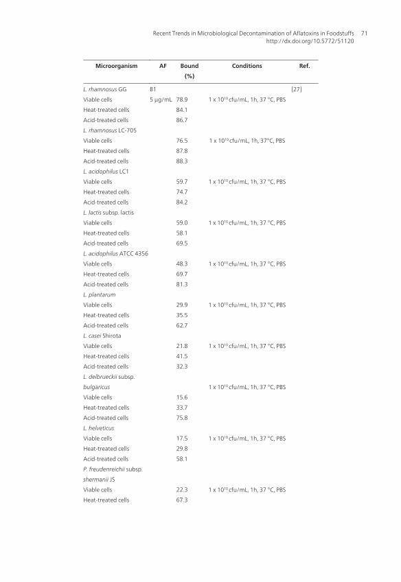
Microorganism AF Bound
(%)
Conditions Ref.
L. rhamnosus GG
Viable cells
Heat-treated cells
Acid-treated cells
L. rhamnosus LC-705
Viable cells
Heat-treated cells
Acid-treated cells
L. acidophilus LC1
Viable cells
Heat-treated cells
Acid-treated cells
L. lactis subsp. lactis
Viable cells
Heat-treated cells
Acid-treated cells
L. acidophilus ATCC 4356
Viable cells
Heat-treated cells
Acid-treated cells
L. plantarum
Viable cells
Heat-treated cells
Acid-treated cells
L. casei Shirota
Viable cells
Heat-treated cells
Acid-treated cells
L. delbrueckii subsp.
bulgaricus
Viable cells
Heat-treated cells
Acid-treated cells
L. helveticus
Viable cells
Heat-treated cells
Acid-treated cells
P. freudenreichii subsp.
shermanii JS
Viable cells
Heat-treated cells
B1
5 µg/mL
78.9
84.1
86.7
76.5
87.8
88.3
59.7
74.7
84.2
59.0
58.1
69.5
48.3
69.7
81.3
29.9
35.5
62.7
21.8
41.5
32.3
15.6
33.7
75.8
17.5
29.8
58.1
22.3
67.3
1 x 10¹⁰ cfu/mL, 1h, 37 °C, PBS
1 x 10¹⁰ cfu/mL, 1h, 37°C, PBS
1 x 10¹⁰ cfu/mL, 1h, 37 °C, PBS
1 x 10¹⁰ cfu/mL, 1h, 37 °C, PBS
1 x 10¹⁰ cfu/mL, 1h, 37 °C, PBS
1 x 10¹⁰ cfu/mL, 1h, 37 °C, PBS
1 x 10¹⁰ cfu/mL, 1h, 37 °C, PBS
1 x 10¹⁰ cfu/mL, 1h, 37 °C, PBS
1 x 10¹⁰ cfu/mL, 1h, 37 °C, PBS
1 x 10¹⁰ cfu/mL, 1h, 37 °C, PBS
[27]
Recent Trends in Microbiological Decontamination of Aflatoxins in Foodstuffshttp://dx.doi.org/10.5772/51120
71

Microorganism AF Bound
(%)
Conditions Ref.
Acid-treated cells
Lc. lactis subsp. cremoris
Viable cells
Heat-treated cells
Acid-treated cells
S. thermophilus
Viable cells
Heat-treated cells
Acid-treated cells
82.5
26.9
40.1
43.7
32.7
42.0
63.8
1 x 10¹⁰ cfu/mL, 1h, 37 °C, PBS
1 x 10¹⁰ cfu/mL, 1h, 37 °C, PBS
E. Coli
L. rhamnosus GG
S. aureus
Bifidobacterium sp. Bf6
B. adolescentis 14
B. bifidum BGN4
Bifidobacterium sp. CH4
B. longum JR20
Bifidobacterium sp. JO3
B1
2 µg/mL
37
37
46
25
31
46
37
37
41
30 min, 37 °C, PBS [22]
Lc. lactis
Living cells
Dead cells by boiling
Dead cells by autoclaving
S. thermophilus
Living cells
Dead cells by boiling
Dead cells by autoclaving
B1
0.5
µg/mL
54.8
86.7
82.3
71.0
81.0
100
80.0
81.0
91.5
90.7
66.5
100.0
96.8
81.7
96.0
83.0
107-108 cfu/mL,30 min, 37 °C, in:
PBS
maize oil
sunflower oil
soybean oil
PBS
maize, sunflower or soybean oil
PBS
PBS
maize oil
sunflower oil
soybean oil
PBS
maize oil
sunflower oil
soybean oil
PBS
[42]
Aflatoxins - Recent Advances and Future Prospects72

Microorganism AF Bound
(%)
Conditions Ref.
Yoghurt Culture B1
0.6mg/kg
1 mg/kg
1.4mg/kg
1 mg/kg
97
91
90
90
84
73
42 °C/3h, pH 4.0, overnight, milk
milk acidified with citric acid
milk acidified with latic acid
milk acidified with acetic acid
[43]
L. acidophilus NCC12
Living cells
Heated cells
L. acidophilus NCC36
Living cells
Heated cells
L. acidophilus NCC68
Living cells
Heated cells
B. bifidum Bb13
Living cells
Heated cells
B. bifidum NCC 381
Living cells
Heated cells
L. rhamnosus
Living cells
Heated cells
M1
5, 10 and
20
ng/mL
14.9-20.2
14.4-15.4
17.0-24.9
16.6-19.0
20.4-25.3
21.8-22.7
22.1-26.8
23.7-25.1
10.2-16.0
7.8-10.5
14.0-21.8
12.8-15.9
23.5-26.6
24.0-25.9
24.3-28.9
25.4-27.4
16.6-22.1
15.5-18.3
17.4-23.5
17.1-22.2
20.1-24.0
20.4-22.2
23.4-27.8
22.9-26.3
108 cfu/mL, 0, 4, 24 h, 37 °C, PBS
108 cfu/mL, 4h, 37 °C, milk
0, 4 , 24 h, 37 °C, PBS
4h, 37 °C, milk
108 cfu/mL, 0, 4, 24 h, 37 °C, PBS
108 cfu/mL, 4h, 37 °C, milk
0, 4, 24 h, 37 °C, PBS
4h, 37 °C, milk
108 cfu/mL, 0, 4, 24 h, 37 °C, PBS
108 cfu/mL, 4h, 37 °C, milk
0, 4, 24 h, 37 °C, PBS
4h, 37 °C, milk
108 cfu/mL, 0, 4, 24 h, 37 °C, PBS
108 cfu/mL, 4h, 37 °C, milk
0, 4,24 h, 37 °C, PBS
4h, 37 °C, milk
108 cfu/mL, 0, 4,24 h, 37 °C, PBS
108 cfu/mL, 4h, 37 °C, milk
0, 4 and 24 h, 37 °C, PBS
4h, 37 °C, milk
108 cfu/mL, 0, 4, 24 h, 37 °C, PBS
108 cfu/mL, 4h, 37 °C, milk
0, 4 and 24 h, 37 °C, PBS
4h, 37 °C, milk
[46]
Lactobacillus strains AFM1 9.4-73.1 96 h, 37 °C , PBS [47]
Recent Trends in Microbiological Decontamination of Aflatoxins in Foodstuffshttp://dx.doi.org/10.5772/51120
73

Microorganism AF Bound
(%)
Conditions Ref.
Lactococcus strains
Bifidobacterium strains
L. plantarum
B. adolescentes
Lactobacillus strains
Lactococcus strains
Bifidobacterium strains
L. bulgaricus
B. adolescentes
4.5-38.3
7.8-41.6
73
41.6
64-80.5
46.0-68.5
67.0-72.5
80.5
73
96 h, 37 °C , PBS
96 h, 37 °C , PBS
96 h, 37 °C , PBS
96 h, 37 °C , PBS
96 h, 37 °C ,milk
96 h, 37 °C , milk
96 h, 37 °C , milk
96 h, 37 °C , milk
96 h, 37 °C , milk
L. rhamnosus strain GG
(pre-cultured)
Viable cells
Heat-killed cells
L. rhamnosusstrain
LC-705
(pre-cultured)
Viable cells
Heat-killed cells
L. rhamnosus strain GG
(lyophilized)
Viable cells
Heat-killed cells
L. rhamnosus strain
LC-705
(lyophilized)
Viable cells
Heat-killed cells
L. lactis ssp. cremoris
strain ARH74
Viable cells
M1
0.15
µg/ml
50.7
18.8
26.0
57.8
26.6
36.6
46.3
69.6
27.4
51.6
63.6
30.1
53.8
56.2
45.7
57.4
40.4
5.3 x 10⁸, 15 - 16h, , 37 °C, in:
PBS
skim milk
full cream milk
PBS
skim milk
full cream milk
PBS
skim milk
full cream milk
PBS
skim milk
full cream milk
1.0 x 10¹⁰, 15-16h, 37 °C, PBS
1.0 x 10¹⁰, 15-16h, 37 °C, PBS
2.9 x 10⁸, 15-16h, , 37 °C, PBS
[29]
Aflatoxins - Recent Advances and Future Prospects74

Microorganism AF Bound
(%)
Conditions Ref.
Heat-killed cells
L. gasseri (ATCC 33323)
Viable cells
Heat-killed cells
L. acidophilus strain LA1
Viable cells
Heat-killed cells
L. rhamnosus strain 1/3
Viable cells
Heat-killed cells
38.9
30.8
61,5
18,3
25,5
18,1
39,9
3.9 x 10⁸, 15-16h, , 37 °C, PBS
1.7 x 10⁹, 15-16h, , 37 °C, PBS
3.9 x 10⁸, 15-16h, , 37 °C, PBS
L. rhamnosus strain GG
Pre-treatment:
Pronase E
Viable cells
Heat-treated cells
Acid-treated cells
Lipase
Viable cells
Heat-treated cells
Acid-treated cells
Phosphate Buffer
Viable cells
Heat-treated cells
Acid-treated cells
m-Periodater
Viable cells
Heat-treated cells
Acid-treated cells
Iodate
Viable cells
Heat-treated cells
Acid-treated cells
Urea
Viable cells
Heat-treated cells
Acid-treated cells
Water (Milli Q)
Viable cells
Heat-treated cells
Acid-treated cells
B1
5 µg/mL
66
72
85
76
74
89
86
85
91
60
49
36
83
84
80
64
60
50
76
83
84
1h, 37 °C, 5% CO2, PBS
Boiled for 1h, PBS
2 mol/L HCl, 1h, 37 °C, 5% CO2
1h, 37 °C, 5% CO2, PBS
Boiled for 1h, PBS
2 mol/L HCl, 1h, 37 °C, 5% CO2
1h, 37 °C, 5% CO2, PBS
Boiled for 1h, PBS
2 mol/L HCl, 1h, 37 °C, 5% CO2
1h, 37 °C, 5% CO2, PBS
Boiled for 1h, PBS
2 mol/L HCl, 1h, 37 °C, 5% CO2
1h, 37 °C, 5% CO2, PBS
Boiled for 1h, PBS
2 mol/L HCl, 1h, 37 °C, 5% CO2
1h, 37 °C, 5% CO2, PBS
Boiled for 1h, PBS
2 mol/L HCl, 1h, 37 °C, 5% CO2
1h, 37 °C, 5% CO2, PBS
Boiled for 1h, PBS
2 mol/L HCl, 1h, 37 °C, 5% CO2
[48]
Recent Trends in Microbiological Decontamination of Aflatoxins in Foodstuffshttp://dx.doi.org/10.5772/51120
75

Microorganism AF Bound
(%)
Conditions Ref.
L. acidophilus
Pre-treatment:
None
Heat
Ethanol
Acid
Alkaline
L. casi
None
Heat
Ethanol
Acid
Alkaline
L. helveticus
None
Heat
Ethanol
Acid
Alkaline
L. bulgaricus
None
Heat
Ethanol
Acid
Alkaline
B1
5 µg/mL
56.6
71.9
46.5
87.0
27.4
22.4
41.8
21.8
43.1
12.0
17.8
28.5
18.0
56.3
9.1
16.3
33.5
15.9
586
8.3
4h, 37 °C, PBS
[49]
L. plantarum
Viable cells
Heat-killed cells
E. avium
Viable cells
Heat-killed cells
P. pentosaceus
Viable cells
Heat-killed cells
L. gasseri
Viable cells
Heat-killed cells
L. bulgaricus
Viable cells
Heat-killed cells
M1
0.15
µg/mL
PBS
solution
0.5
µg/mL
skimmed
milk
5.6
8.1
7.4
6.6
8.7
7.8
21.4
22.8
30.2
33.5
1010 cfu/mL, 15 min, 37°C, in:
PBS
PBS
PBS
PBS
PBS
PBS
PBS
PBS
PBS
PBS
[50]
Aflatoxins - Recent Advances and Future Prospects76

Microorganism AF Bound
(%)
Conditions Ref.
L. rhamnosus
Viable cells
Heat-killed cells
B. lactis
Viable cells
Heat-killed cells
33.5
17.1
27.8
24.5
16.9
23.6
32.5
skimmed milk
PBS
PBS
skimmed milk
PBS
PBS
skimmed milk
L. delbrueckii subsp.
bulgaricus CH-2
S. thermophilus ST-36
M1
10 ng/mL
18.7
27.6
29.4
39.2
14.8
4h, 37 °C, PBS
4h, 42 °C, milk
4h, 37 °C, PBS
4h, 42 °C, milk
Yoghurt
[51]
F. aurantiacum NRRL
B-184
M1
10 µg/mL
100 5 x 10¹⁰ cfu/mL, 30 °C, 4h, PBS
and milk
[52]
B. subtilis UTBSP1
Viable cells
Cell Free Supernatant
B1
2.5
µg/mL
85.7
95
78.4
96h, 30 °C, nutrient broth culture
108 cfu/ml, 120 h, 30 °C, pistachio
nuts
120 h, 35 °C, nutrient broth
culture
[53]
B. subtilis ANSB060
“Inocula” suspension
Cell
Cell extract
Culture Supernatant
B1
G1
M1
(0.5
µg/mL)
B1
81.5
80.7
60
10.5
9.6
78.7
72h, 37 °C, Luria-Bertani medium
72h, 37 °C, PBS
[54]
L. rhamnosus strain GG
“In vivo”
“In vitro”
L. rhamnosus strain
LC-705
B1
3 µg/mL
51
92
80
1010cfu/mL:
1 min, duodenum of chicks
1 h, duodenum of chicks
37 °C, 1h , pH 7.3
[62]
Recent Trends in Microbiological Decontamination of Aflatoxins in Foodstuffshttp://dx.doi.org/10.5772/51120
77

Microorganism AF Bound
(%)
Conditions Ref.
“In vivo”
“In vitro”
P. freudenreichii subsp.
shermanii JS
“In vivo”
“In vitro”
36
71
77
37
82
22
1 min, duodenum of chicks
1 h, duodenum of chicks
37 °C, 1h , pH 7.3
1 min, duodenum of chicks
1 h, duodenum of chicks
37 °C, 1h , pH 7.3
Table 1. Aflatoxin binding / absorption by microorganisms. Note: PBS, Phosphate-Buffered Saline; cfu, colonyformingunit.
4. Decontamination of Aflatoxins by Yeasts
Yeasts are non-photosynthetic organisms with a separate nucleus and complex life cycle.They are larger than bacteria, normally spherical, non-motile, and reproduce by budding.Although their main function is alcoholic fermentation, these organisms are also capable ofproducing enzymes and vitamins. The primary substrates for yeasts are fermentable sugars,which are mainly transformed in ethanol, carbon dioxide, and biomass under oxygen-limit‐ed conditions. Under adequate oxygen supply, yeast produces carbon dioxide, water, andbiomass [65]. Saccharomyces cerevisiae (SC) is the most well-known and commercially impor‐tant species of yeast, and SC strains are widely used in the production of alcoholic drinksand in the baking industry.
As it occurs with LAB, SC cells have been studied to evaluate their ability to remove aflatox‐ins from contaminated media. The most important results obtained until now are summar‐ized in Table 2. Products based on SC (cell wall from baker and brewer yeasts, inactivatedbaker yeast, or alcohol yeast) was studied, and it was observed that in pH 3, 37 °C and 15minutes of contact, AFB1 removal ranged from 2.5% to 49.3%, depending on the concentra‐tion of the toxin in the medium, and on the yeast-based products used [66]. These authorsalso observed a decrease in toxin adsorption as the initial concentration increased, and con‐cluded that adsorption is not a linear phenomenon. Similar results with a SC strain andAFB1 concentration ranging from 1 to 20 µg/mL was also reported [56]. At the 1 µg/mL con‐centration, 69.1% AFB1 was removed; at 5 g/mL, removal rate was 41%; and at 20 µg/mL,34%. S. cerevisiae strains were isolated from animal feed, feces and intestines, and tested fortheir ability to tolerate gastrointestinal conditions and remove AFB1 from a contaminatedmedium [67]. These researchers observed that all strains isolated were able to survive in gas‐trointestinal conditions, and that the percentage of toxin removed ranged among SC strains(107 CFU/mL), and with AFB1 concentration used (16.4% to 82% of adsorption for 50 ng/mLAFB1; 21.3% to 48.7% for 100 ng/mL AFB1; and 20.2% to 65.5% for 500 ng/mL AFB1).
Aflatoxins - Recent Advances and Future Prospects78

The ability of SC (0.1%, 0.2%, and 0.3%) to adsorb AFB1 in contaminated corn (150, 300, 450and 800 µg/kg corn was analyzed [68]. The adsorption process showed an inversely propor‐tional relationship with the concentration, that is, the greater the AFB1 concentration in themedium, the lower the efficiency of AFB1 removal by SC (16% to 66% for 800 µg/kg AFB1 vs.40% to 93% for 150 µg/kg AFB1). The authors concluded, using densitogram analysis, thatthe adsorption process did not change the molecular structure of the mycotoxin, and thatthe decreased AFB1 adsorption rates observed as the toxin concentration increased may pos‐sibly be caused by saturation of the adsorption sites on the SC cell. Other factors, such aslength of incubation, pH, method of biomass purification, and methods of analysis, may alsoinfluence this process.
Immobilized SC cells (ATTC 9763) was investigated for their ability to remove AFB1 frompistachio seeds, and it was observed that the amount of toxin removed was dependent on itsconcentration in the medium (40% and 70% of removal for concentrations of 10 ng/mL and20 ng/mL AFB1, respectively) [69]. The authors also concluded that this ability to remove thetoxin was greater in SC exponential growth phase, and that the process was a quick one, be‐ing saturated after 3 hours of contact. Besides, the ability of SC cells to remove toxin wasincreased after treatment with acid (60% and 73% for 10 ng/mL and 20 ng/mL AFB1, respec‐tively) and heat (55% and 75%, respectively). In another study, authors also concluded thatthe treatment of SC cells with heat at 60 °C and 120 °C, and with chloric acid (2 mol/L) in‐creased their ability to remove AFB1 from the medium to 68.8%, 79.3%, and 72.1%, respec‐tively, against 38.7% when viable yeast cells were used [56].
Heat treatment may increase the permeability of the external layer of the cell wall due to thesuspension of some mannanes on the cell surface, leading to increased availability of previ‐ously hidden binding sites. Besides, countless physical-chemical changes take place on thecell wall during heat treatment, leading to more exposed binding sites. On the other hand,acid conditions may affect polysaccharides by releasing monomers, which are further frag‐mented in aldehydes after glycosidic bonds are broken. Continuous removal of aflatoxin,even after use of acid and heat treatments, confirms that yeast cell viability is not a signifi‐cant factor for the removal of aflatoxin from the medium [69].
During the fermentation of broiler feed using LAB (3 strains of Lactobacillus) and SC strainsresistant to gastric juices and bile, 55% AFB1 was removed when AFB1 concentration in themedium was 1 mg/kg, and 39% when concentration was 5 mg/kg AFB1, after 6 hours [70].This tendency for removal was maintained as incubation continued, and after 24 hours, theamount of AFB1 removed was 73% and 53%, respectively, for the two concentrations of thetoxin. The authors considered that, from a practical point of view, the most important factorwas the 6-hour fermentation period, once the passage of feed through the gastrointestinaltract of broilers lasts from 4 to 8 hours. In reference [71], authors analyzed the ability of SCto remove AFB1 from a contaminated medium at different pH values (3.0, 6.0, and 8.0), andobserved that the three strains analyzed showed great ability to remove the toxin (41.6% to94.5%), and that after washing, only a small amount of AFB1 was released back into the me‐dium. In vitro studies are not always good indications of the in vivo responses, as in vivo
Recent Trends in Microbiological Decontamination of Aflatoxins in Foodstuffshttp://dx.doi.org/10.5772/51120
79

studies are affected by physiological parameters, such as pH, peristaltic movements, andgastric and intestinal secretions.
In vivo studies using SC are not as rare as those with LAB, mainly in poultry science. Gener‐ally, SC is added to the feed as a growth promoter. However, the addition of yeasts has alsopresented beneficial effects against the exposure to AFB1. It was observed that the additionof 1% SC to feed contaminated with 5 g/g of AFB1 prevented loss of weight; liver and hearthyperplasia; and decreased serum albumin and total protein concentrations in broilers [72].The addition of SC in feed containing aflatoxin decreased the deleterious effects on feed in‐take, weight gain, and feed conversion in Japanese quails [73]. Compared with control ani‐mals, weight gain was 37% lower in birds fed a diet added only of aflatoxin, and was 15%greater than the control in the group that received feed containing aflatoxin and SC. The au‐thors concluded that the diet containing with only SC significantly improved all growth pa‐rameters investigated (about 40%), compared with the control group.
In a study with mice, it was observed that the addition of AFB1 to the diet (0.4 and 0.8mg/kg) caused a significant reduction in weight gain, and an increase of 85% (0.8 mg/kg) inthe rate of micronucleated normochromatic erythrocytes (MNE) after 3 weeks of ingestion,compared with the control group [68]. When diets containing AFB1 and SC (0.3%) were ad‐ministered, weight gain was twice greater than in diets that contained only the toxin, andthe rate of MNE increased only 46% (0.8 mg/kg) The authors stated that reduced bodyweight is one of the most common consequences of AFB1 ingestion, because the toxin altersthe activity of several digestive enzymes, giving rise to a malabsorption syndrome charac‐terized by steatorrhea, hypovitaminosis A and a decrease in the levels of bile, pancreatic li‐pase, trypsin, and amylase. Besides, biotransformation of AFB1 gives rise to severalmetabolites, particularly AFB1-8,9-epoxide, which may bind covalently to DNA and pro‐teins, changing enzymatic processes such as gluconeogenesis, Krebs cycle, and fatty acidsynthesis [74]. MNE rate is used to determine the genotoxicity of AFB1, because it quantifiesbroken chromosomes and whole chromosomes that are abnormally distributed to daughtercells, showing thus, that AFB1 is a potent mutagenic agent.
A diet containing 5 g/g of aflatoxin (82.06% AFB1, 12.98% AFB2, 2.84% AFG1, and 1.12%AFG2) by female quails (49 to 84 days of age) led to decreased egg production, feed intake,and feed conversion (31%, 28%, and 47%, respectively) [75]. However, addition of SC (2g/kg) significantly increased these parameters (16%, 4%, and 14%, respectively). They alsoobserved that the diet with aflatoxins caused a marked decrease in weight gain and eggweight, besides increasing animal mortality (39%, 7%, and 50%, respectively), whereas addi‐tion of SC reverted the negative effect on these parameters (65%, 8%, and 50%, respectively).The authors stated that these negative effects of aflatoxins in egg production, feed intake,and feed conversion may have been caused by anorexia, apathy, and inhibition of proteinsynthesis and lipogenesis. Besides, affected liver function and mechanisms of use of proteinand lipids may have affected performance criteria and the general health of the animals. Inreference [76] authors reported that the components of the cells wall of SC are able to adsorbmycotoxins, stimulate the immune system, and compete for binding sites in the enterocytes,inhibiting intestinal colonization by pathogens.
Aflatoxins - Recent Advances and Future Prospects80

SC cell wall is mainly made up of polysaccharides (80-90%), and its mechanical resistance isdue to an inside layer composed of β-D-glucans, which are formed by a complex network ofhighly polymerized β-(1,3)-D-glucans, branched off as β-(1,6)-D-glucans, that have a lowlevel of polymerization. This inside layer is firmly bound to the plasmatic membrane by lin‐ear chains of chitin, which have a significant role in the insolubility of the overall structureand packing of the branched β-D-glucans. Both chitin chains and β-D-glucans affect theplasticity of the cell wall. The external layer of the yeast cell wall is formed by mannopro‐teins, which have an important role in the exchanges with the external environment. Thiswhole structure is highly dynamic and may vary according to the yeast strain, phase of thecell cycle, and culture conditions, such as pH, temperature, oxygenation rate, nature of themedium, concentration and nature of the carbon source. Thus, these differences in the com‐position of the cell wall among yeast strains are related with their ability to bind to the my‐cotoxin [77].
Studies have shown that the components of SC cell wall, called oligomannanes, after esterifi‐cation, are able to bind more than 95% AFB1 [78]. Addition of 0.05% glucomannanes in thebasal diet improved broiler performance [79].
The possible binding mechanisms between yeast cell wall and mycotoxins were studied,and authors suggested that β-D-glucans are the components of the cell wall that are respon‐sible for forming the complex with the toxin, and that the reticular organization of β-D-glu‐cans and their distribution in β-(1,3)-D-glucans and β-(1,6)-D-glucans have an importantrole in the efficiency of the bond [77]. Besides, studies have shown that weak hydrogen andvan der Waals bonds are involved in the complex chemical connection between the myco‐toxins and β-D-glucans, a chemical interaction that is much more "adsorption” than “bond”.As for AFB1, they observed that the aromatic ring, the lactone and ketone groups of the po‐lar form of AFB1, or chemical bonds with glucose units in the single helix of the β-D-glucans,are what keep the toxin bound to the glucans.
It was demonstrated that yeast strains isolated from environments were animals are raisedare able to bind to AFB1 in saline solution (PBS, pH 7) [67]. These strains presented otherproperties that were beneficial to the host, such as the inhibition of pathogenic bacteria.Therefore, SC strains acted both as probiotics (co-aggregation and inhibition of pathogenicbacteria), and as mycotoxin adsorbents.
In reference [72], SC was able to reduce the deleterious effects of AFB1 in the diet of broilersand in [68] authors replicated these findings in rats. Protective effect against aflatoxins pro‐duced by yeasts was confirmed in rats. However, when yeast cells were inactivated by heat,they were inefficient [80] but when glucomannanes extracted from the cell wall of yeastswere used, there was an increase in the efficiency of the bond with AFB1, OTA and T-2 toxin[81-84], individually or in combination [75, 79, 85, 86]. The addition of SC in the diet reducedAFB1 toxic effects in chickens [72, 87]. The ability of SC to reduce AFB1 toxic effects in quailswas demonstrated, and this effect was apparently more efficient with the increase in inclu‐sion rates [88].
Recent Trends in Microbiological Decontamination of Aflatoxins in Foodstuffshttp://dx.doi.org/10.5772/51120
81

In [89] authors obtained a significant reduction in AFB1 concentration during beer produc‐tion, probably due to the bond between mycotoxins and SC cell. This hypothesis was sup‐ported by other studies [39, 90]. A 19% reduction in AFB1 during dough fermentation inbread production was observed [91].
Microorganism AF Bound
(%)
Conditions Ref.
S. cerevisiae B1
0.0058-
6.35 μg/mL
7.6-49.3
7.6-29
10-24
4-29
17-44
3-44
23-35
27-44
15 min, 37 °C:
YCW from brewer’s yeast
YCW from brewer’s yeast
Inactivated baker’s yeast
YCW from baker’s yeast
Inactivated baker’s yeast
YCW from baker’s yeast
YCW from baker’s yeast
Alcohol yeast
[66]
S. cerevisiae
Strain A18
Strain 26.1.11
Pre-treatment:
Heated cells 52°C
Strain A18
Strain 26.1.11
Heated cells 55 °C
Strain A18
Strain 26.1.11
Heated cells at 60 °C
Strain A18
Strain 26.1.11
Heat cells at 120 °C
Strain A18
Strain 26.1.11
2 mol/L HCl / 1h
Strain A18
Strain 26.1.11
B1
1 μg/mL
5 μg/mL
10 μg/mL
20 μg/mL
1 μg/mL
5 μg/mL
10 μg/mL
20 μg/mL
5 μg/mL
69.1
41
33
34.2
65.1
37.2
31
32.6
58.8
56.5
64.5
64
68.8
67
79.3
77.7
72.1
69.3
3h, 25 C, PBS
3h, 25 C, PBS
3h, 25 C, PBS
[56]
S. cerevisiae B1 (ng/mL) [67]
Aflatoxins - Recent Advances and Future Prospects82

Microorganism AF Bound
(%)
Conditions Ref.
Strain RC008
Strain RC009
Strain RC012
Strain RC016
50 ng/mL
100
500
50
100
500
50
100
500
50
100
500
67.6
43.5
38.2
16.4
21.3
31.8
29.6
20.6
20.2
82.0
48.7
65.5
107 cells/mL, 1h, 37 °C, PBS
S. cerevisiae
Yeast concentration:
0.1 %
0.2 %
0.3 %
B1
(µg/kg)
150
300
450
800
150
300
450
800
150
300
450
800
40
25
17
16
88
76
64
51
93
86
81
66
37 °C, 24 h, corn
37 °C, 24 h, corn
37 °C, 24 h, corn
[68]
S. cerevisiae ATTC 9763
Pre-treatment:
None
Acid treated cells
(2 mol/L / 90 min)
Heat-treated cells
(120 °C / 20 min)
B1
(ng/mL)
10
20
10
20
10
20
40
70
60
73
55
75
3 h, 25 °C, pistachio nuts
[69]
L. paracasei LOCK 0920,
L. brevis LOCK 0944, L.
plantarum LOCK 0945,
and S. cerevisiae LOCK
0140
B1
1 mg/kg
5 mg/kg
55
39
37 °C, 6h fermentation in broiler
feed
[70]
Recent Trends in Microbiological Decontamination of Aflatoxins in Foodstuffshttp://dx.doi.org/10.5772/51120
83

Microorganism AF Bound
(%)
Conditions Ref.
S. cerevisiae B1
1 µg /g
10 µg /g
86
72
12 °C, 8 days, brewing process
[89]
Table 2. Aflatoxin binding by yeasts. YCW, Yeast Cell Wall
5. Concluding Remarks
Considering the data from several studies carried out until now, it may be observed that mi‐croorganisms, among them lactic acid bacteria and yeasts, have a huge potential applicationin aflatoxin degradation in foodstuffs. However, new studies are necessary to identify bacte‐rial species with greater binding potential with aflatoxins, once there are differences in sen‐sitivity and selectivity, besides the influence of factors that are intrinsic and extrinsic to thebacteria in the decontamination process. After this step of choosing species with greater effi‐ciency has been overcome, new production technologies that are economically viable to beapplied to human and animal foods may be developed.
Several studies have demonstrated that the cell wall of SC and LAB and their componentsare responsible for binding with aflatoxins. However, the mechanisms by which this bondoccurs remain unclear. Cell walls with glucomannanes and manno-oligosaccharides havebeen pointed out as the responsible elements for AFB1 bond with yeasts. The great advant‐age in the commercial use of these microorganisms as binding agents is that these strainsare approved and already used in a wide range of fermented food products, being recog‐nized as safe. However, aflatoxin may be released from the cell-aflatoxin complex withchanges in the pH and temperature conditions. Therefore, further studies are necessary todetermine the behavior of yeasts in the different environmental conditions before they areused commercially.
Acknowledgements
The authors wish to acknowledge the Fundação de Amparo à Pesquisa do Estado de SãoPaulo (FAPESP) and the Conselho Nacional de Desenvolvimento Científico e Tecnológico(CNPq) of Brazil for financial support and fellowships.
Author details
Carlos Augusto Fernandes Oliveira1*, Fernanda Bovo1, Carlos Humberto Corassin1,Alessandra Vincenzi Jager1 and Kasa Ravindranadha Reddy2
Aflatoxins - Recent Advances and Future Prospects84

*Address all correspondence to: [email protected]
1 Faculty of Animal Science and Food Engineering, University of São Paulo, Brazil
2 Research and Development Department, SRI Biotech Laboratories Ltd, India
References
[1] Bata, A., & Lasztity, R. (1999). Detoxification of mycotoxin-contaminated food andfeed by microorganisms. Trends in Food Science & Technology, 10-223.
[2] Gonçalez, E., Pinto, M. M., & Felicio, J. D. (2001). Análise de micotoxinas no InstitutoBiológico de 1989 a 1999. Biológico, 63(1/2), 15-19.
[3] D’Mello, J. P. F., & Mac, Donald. A. M. C. (1997). Mycotoxins. Animal Feed Science andTechnology, 69-155.
[4] Nierman, W. C., Cleveland, T. E., Payne, G. A., Keller, N. P., Campbell, B. C., Ben‐nett, J. W., Guo, B., Yu, J., & Robens, J. F. (2008). Mycotoxin Production and Preven‐tion of Aflatoxin Contamination in Food and Feed. In: Goldman GH, Osmani SA.The Aspergilli: Genomics, Medical Aspects, Biotechnology, and Research Methods.Boca Raton: CRC Press , 457-472.
[5] Hussein, H. S., & Brasel, J. M. (2001). Toxicity, metabolism, and impact of mycotoxinson humans and animals. Toxicology, 167-101.
[6] Bhat, R., Rai, R. V., & Karim, A. A. (2010). Mycotoxins in Food and Feed: Present Sta‐tus and Future Concerns. Comprensive Reviews in Food Science and Food Safety, 9-57.
[7] Alberts, J. F., Engelbrecht, Y., Steyn, P. S., Holzapfel, W. H., & Van Zyl, W. H. (2006).Biological degradation of aflatoxin B1 by Rhodococcus erythropolis cultures. Interna‐tional Journal of Food Microbiology, 109-121.
[8] Park, D. L., & Liang, B. (1993). Perspectives on aflatoxin control for human food andanimal feed. Trends in Food Science & Technology, 41-334.
[9] Chu, F. S. (1991). Mycotoxins: food contamination, mechanism, carcinogenic poten‐tial and preventive measures. Mutation Research, 259-291.
[10] Abbas, H. K. (2005). Aflatoxin and food safety. Boca Raton, CRC Press.
[11] Magan, N., & Olsen, M. (2006). Mycotoxins in food: Detection and Control. Boca Ra‐ton, CRC Press.
[12] Prandini, A., Tansini, G., Sigolo, S., Filippi, L., Laporta, M., & Piva, G. (2009). On theoccurrence of aflatoxin M1 in milk and dairy products. Food and Chemical Toxicology,47-984.
Recent Trends in Microbiological Decontamination of Aflatoxins in Foodstuffshttp://dx.doi.org/10.5772/51120
85

[13] Hernandez-Mendoza, A., Garcia, H. S., & Steele, J. L. (2009). Screening of Lactobacil‐lus casei strains for their ability to bind aflatoxin B1. Food and Chemical Toxicology,47-1064.
[14] Gratz , S., Mykkänem, H. , & El-Nezami, H. (2005). Aflatoxin B1 binding by a mixtureof Lactobacillus and Propionibacterium: in vitro versus ex vivo. Journal of Food Protec‐tion, 68(11), 2470-2474.
[15] El -Nezami, H., Kankaanpaa, P., Salminen, S., & Ahokas, J. (1998). Physicochemicalalterations enhance the ability of dairy strains of lactic acid bacteria to remove afla‐toxin from contaminated media. Journal of Food Protection, 61(4), 466-468.
[16] Line, J. E., & Brackett, R. E. (1995). Factors affecting aflatoxin B1 removal by Flavo‐bacterium aurantiacum. Journal of Food Protection, 58(1), 91-94.
[17] El -Nezami, H., Kankaanpaa, P., Salminen, S., & Ahokas, J. (1998). Ability of dairystrains of lactic acid bacteria to bind a common food carcinogen, aflatoxin B1. Foodand Chemical Toxicology, 36-321.
[18] Fazeli, M. R., Hajimohammadali, M., Moshkani, A., Samadi, N., Jamalifar, H., Khosh‐ayand, M. R., et al. (2009). Aflatoxin B1 binding capacity of autochthonous strains oflactic acid bacteria. Journal of Food Protection, 72(1), 189-192.
[19] Alberts, J. F., Gelderblomb, W. C. A., Botha, A., & Van Zyl, W. H. (2009). Degradationof aflatoxin B1 by fungal laccase enzymes. International Journal of Food Microbiology,135-47.
[20] Wu, Q., Jezkova, A., Yuan, Z., Pavlikova, L., Dohnal, V., & Kuca, K. (2009). Biologicaldegradation of aflatoxins. Drug Metabolism Reviews, 41(1), 1-7.
[21] Oliveira, C. A. F., & Germano, P. M. L. (1997). Aflatoxinas: conceitos sobre mecanis‐mos de toxicidade e seu envolvimento na etiologia do câncer hepático celular. Revistade Saúde Pública, 31(4), 417-424.
[22] Oatley, J. T., Rarick, M. D., Ji, G. E., & Linz, J. E. (2000). Binding of aflatoxin B1 tobifidobacteria in vitro. Journal of Food Protection, 63(8), 1133-36.
[23] Peltonem, K., El-Nezami, H., Haskard, C., Ahokas, J., & Salminen, S. (2001). AflatoxinB1 binding by dairy strains of lactic acid bacteria and bifidobacteria. Journal of DairyScience, 84, 2152-2156.
[24] Murphy, P. A., Hendrich, S., Landgren, C., & Bryant, C. M. (2006). Food Mycotoxins:An Update. Journal of Food Science, 71(5), 51-65.
[25] Creppy, E. E. (2002). Update of survey, regulation and toxic effects of mycotoxins inEurope. Toxicology Letters, 127-19.
[26] Bakirci, I. (2001). A study on the occurrence of aflatoxin M1 in milk and milk prod‐ucts produced in Van province of Turkey. Food Control, 12-47.
Aflatoxins - Recent Advances and Future Prospects86

[27] Haskard, C. A., El -Nezami, H. S., Kankaanpaa, P. E., Salminen, S., & Ahokas, J. T.(2001). Surface binding of aflatoxin B1 by lactic acid bacteria. Applied and Environmen‐tal Microbiology, 67(7), 3086-3091.
[28] Oluwafemi, F., & Silva, F. A. (2009). Removal of aflatoxins by viable and heat-killedLactobacillus species isolated from fermented maize. Journal of Applied Biosciences.,16-871.
[29] Pierides, M., El -Nezami, H., Peltonem, K., Salminen, S., & Ahokas, J. (2000). Abilityof dairy strains of lactic acid bacteria to bind aflatoxin M1 in a food model. Journal ofFood Protection, 63(5), 645-650.
[30] IARC International Agency for Research on Cancer- World Health Organization.(2002). IARC Monograph on the Evaluation of Carcinogenic Risk to Humans., 82, 171.
[31] Mishra, H. N., & Das, C. (2003). A Review on biological control and metabolism ofaflatoxin. Critical Reviews in Food Science and Nutrition, 43(3), 245-264.
[32] Laciaková, A., Cicoòová, P., Máté, D., & Laciak, V. (2008). Aflatoxins and possibilitiesfor their biological detoxification. Medycyna Weterynaryjna, 64(3), 276-279.
[33] Van Egmond, H. P., & Jonker, M. A. (2004). Worldwide regulations on aflatoxins-The situation in 2002. Toxin Reviews, 23, 273-293.
[34] Walstra, P., Wouters, J. T. M., & Geurts, T. J. (2006). Dairy Science and Technology.Boca Raton, CRC Press.
[35] Salminen, S., Von, Wright. A., & Ouwehand, A. C. (2004). Lactic acid bacteria: micro‐biological and functional aspects. 3.ed. New York Marcel Dekker, Inc.
[36] Sybesma, W. F., & Hugenholtz, J. (2004). Food fermentation by lactic acid bacteria forthe prevention of cardiovascular disease. In: Arnoldi A. Functional Foods, Cardio‐vascular Disease and Diabetes. Boca Raton CRC Press
[37] Onilude, A. A., Fagade, O. E., Bello, M. M., & Fadahunsi, I. F. (2005). Inhibition ofaflatoxin-producing aspergilli by lactic acid bacteria isolates from indigenously fer‐mented cereal gruels. African Journal of Biotechnology, 4(12), 1404-1408.
[38] Fuchs, S., Sontag, G., Stidl, R., Ehrlich, V., Kundi, M., & Knasmüller, S. (2008). Detoxi‐fication of patulin and ochratoxin A, two abundant mycotoxins, by lactic acid bacte‐ria. Food and Chemical Toxicology, 46-1398.
[39] Shetty, P. H., & Jespersen, L. (2006). Saccharomyces cerevisiae and lactic acid bacteriaas potential mycotoxin decontaminating agents. Trends in Food Science & Technology,17-48.
[40] Ciegler, A., Lillehoj, E. B., Peterson, R. E., & Hall, H. H. (1966). Microbial detoxifica‐tion of aflatoxin. Applied Environmental Microbiology, 14(6), 934-939.
[41] El -Nezami, H., Mykkänen, H., Haskard, C., Salminen, S., & Salminen, E. (2004). Lac‐tic acid bacteria as a tool for enhancing food safety by removal of dietary toxins. In:
Recent Trends in Microbiological Decontamination of Aflatoxins in Foodstuffshttp://dx.doi.org/10.5772/51120
87

Salminen S, Von Wright A, Ouwehand A. Lactic Acid Bacteria: Microbiological andFunctional Aspects. 3.ed. Nova Iorque Marcel Dekker , 397-406.
[42] Shahin, A. A. M. (2007). Removal of aflatoxin B1 from contaminated liquid media bydairy lactic acid bacteria. International Journal of Agriculture and Biology, 9(1), 71-75.
[43] Rasic, J. L., Skrinjar, M., & Markov, S. (1991). Decrease of aflatoxins B1 in yoghurtand acidified milks. Mycopathologia, 113-117.
[44] Biernasiak, J., Piotrowska, M., & Libudzisz, Z. (2006). Detoxification of mycotoxinsby probiotic preparation for broiler chickens. Mycotoxin Research; , 22(4), 230-235.
[45] Kabak, B., & Dobson, A. D. W. (2009). Biological strategies to counteract the effects ofmycotoxins. Journal of Food Protection, 72(9), 2006-2016.
[46] Kabak, B., & Var, I. (2008). Factors affecting the removal of aflatoxin M1 from foodmodel by Lactobacillus and Bifidobacterium strains. Journal of Environmental Scienceand Health, Part B., 43-617.
[47] Elgerbi, A. M., Aidoo, K. E., Candlish, A. A. G., & Williams, A. G. (2006). Effects oflactic acid bacteria and bifidobacteria on levels of aflatoxin M1 in milk and phosphatebuffer. Milchwissenschaft, 61(2), 197-199.
[48] Haskard, C., Binnion, C., & Ahokas, J. (2000). Factors affecting the sequestration ofaflatoxin by Lactobacillus rhamnosus strain GG. Chemico-Biological Interactions,128-39.
[49] Azab, R. M., Tawakkol, W. M., Hamad, A. R. M., Abou-Elmagd, M. K., El -Agrab, H.M., & Refai, M. K. (2005). Detection and estimation of aflatoxin B1 in feeds and its bio‐degradation by bacteria and fungi. Egypt Journal of Natural Toxins, 2-39.
[50] Bovo, F., Corassin, C. H., Rosim, R. E., & Oliveira, C. A. F. (2012). Efficiency of lacticacid bacteria strains for decontamination of aflatoxin M1 in phosphate buffer salinesolution and in skimmed milk. Food and Bioprocess Technology.
[51] Sarimehmetoglu, B., & Küplülü, Ö. (2004). Binding ability of aflatoxin M1 to yoghurtbacteria. Ankara Üniversitesi Veteriner Fakültesi Dergisi; , 51-195.
[52] Lillehoj, E. B., Stubblefield, R. D., Shannon, G. M., & Shotwell, O. L. (1971). AflatoxinM1 removal from aqueous solutions by Flavobacterium aurantiacum. Mycopathologiaet mycologia applicata., 45-259.
[53] Farzaneh, M., Shi, Z. Q., Ghassempour, A., Sedaghat, N., Ahmadzadeh, M., Mirabol‐fathy, M., & Javan-Nikkhah, M. (2012). Aflatoxin B1 degradation by Bacillus subtilisUTBSP1 isolated from pistachio nuts of Iran. Food Control, 23-100.
[54] Gao, X., Ma, Q., Zhao, L., Lei, Y., Shan, Y., & Ji, C. (2011). Isolation of Bacillus subtilis:screening for aflatoxins B1, M1 and G1 detoxification. European Food Research and Tech‐nology, 232-957.
Aflatoxins - Recent Advances and Future Prospects88

[55] Lahtinen, S. J., Haskard, C. A., Ouwehand, A. C., Salminen, S. J., & Ahokas, J. T.(2004). Binding of aflatoxin B1 to cell wall components of Lactobacillus rhamnosusstrain GG. Food Additives and Contaminants, 21(2), 158-164.
[56] Shetty, P. H., Hald, B., & Jespersen, L. (2007). Surface binding of aflatoxin B1 by Sac‐charomyces cerevisiae strains with potential decontaminating abilities in indigenousfermented foods. International Journal of Food Microbiology, 113-41.
[57] Hernandez-Mendoza, A., Guzman-de-Peña, D., & Garcia, H. S. (2009). Key role of tei‐choic acids on aflatoxin B1 binding by probiotic bacteria. Journal of Applied Microbiolo‐gy, 107-395.
[58] Hernandez-Mendoza, A., Guzman-de-Peña, D., González-Córdova, A. F., Vallejo-Córdoba, B., & Garcia, H. S. (2010). In vivo assessment of the potential protective ef‐fect of Lactobacillus casei Shirota against aflatoxin B1. Dairy Science and Technology,90-729.
[59] Bolognani, F., Rummey, C. J., & Rowland, I. R. (1997). Influence of carcinogen bind‐ing by lactic acid-producing bacteria on tissue distribution and in vivo mutagenicityof dietary carcinogens. Food and Chemical Toxicology, 35-535.
[60] Lee, Y. K., El -Nezami, H., Haskard, C. A., Gratz, S., Puong, K. Y., Salminen, S., &Mykkänen, H. (2003). Kinetics of adsorption and desorption of aflatoxin B1 by viableand nonviable bacteria. Journal of Food Protection, 66(3), 426-430.
[61] D’Souza, D. H., & Brackett, R. E. (2001). Aflatoxin B1 degradation by Flavobacteriumaurantiacum in the presence of reducing conditions and seryl and sulfhydryl groupinhibitors. Journal of Food Protection, 64(2), 268-271.
[62] El -Nezami, H., Mykkänen, H., Kankaanpaa, P., Salminen, S., & Ahokas, J. (2000).Ability of Lactobacillus and Propionibacterium strains to remove aflatoxin B1 fromthe chicken duodenum. Journal of Food Protection, 63(4), 549-552.
[63] Hathout, A. S., Mohamed, S. R., El -Nekeety, A. A., Hassan, N. S., Aly, S. E., & Abdel-Wahhab, M. A. (2011). Ability of Lactobacillus casei and Lactobacillus reuteri to pro‐tect against oxidative stress in rats fed aflatoxins-contaminated diet. Toxicon, 58-179.
[64] El -Nezami, H. S., Mykkänen, H., Kankaanpää, P., Suomalainen, T., Ahokas, J. T., &Salminen, S. (2000). The ability of a mixture of Lactobacillus and Propionibacteriumto influence the faecal recovery of aflatoxins in healthy Egyptian volunteers: a pilotclinical study. Bioscience and Microflora, 19-41.
[65] Mc Kinney, R. E. (2004). Environmental pollution control microbiology: a fifty-yearperspective. Boca Raton, CRC Press.
[66] Joannis-Cassan, C., Tozlovanu, M., Hadjeba-Medjdoub, K., Ballet, N., & Pfohl-Lesz‐kowicz, A. (2011). Binding of zearalenone, aflatoxin B1, and ochratoxin A by yeast-based products: a method for quantification of adsorption performance. Journal ofFood Protection; , 74(7), 1175-1185.
Recent Trends in Microbiological Decontamination of Aflatoxins in Foodstuffshttp://dx.doi.org/10.5772/51120
89

[67] Armando, M. R., Dogi, C. A., Pizzolitto, R. P., Escobar, F., Peirano, M. S., Salvano, M.A., Sabini, L. I., Combina, M., Dalcero, A. M., & Cavaglieri, L. R. (2011). Saccharomy‐ces cerevisiae strains from animal environment with in vitro aflatoxin B1 bindingability and anti-pathogenic bacterial influence. World Mycotoxin Journal, 4(1), 59-68.
[68] Madrigal-Santillán, E., Madrigal-Bujaidar, E., Márquez-Márquez, R., & Reyes, A.(2006). Antigenotoxic effect of Saccharomyces cerevisiae on the damage produced inmice fed with aflatoxin B1 contaminated corn. Food and Chemical Toxicology, 44,2058-2063.
[69] Rahaie, S., Emam-Djomeh, Z., Razavi, S. H., & Mazaheri, M. (2010). Immobilized Sac‐charomyces cerevisiae as a potential aflatoxin decontaminating agent in pistachionuts. Brazilian Journal of Microbiology, 41-82.
[70] Slizewska, K., & Smulikowska, S. (2011). Detoxification of aflatoxin B1 and change inmicroflora pattern by probiotic in vitro fermentation of broiler feed. Journal of Animaland Feed Sciences;, 20-300.
[71] Dogi, C. A., Armando, R., Ludueña, R., Le Blanc, A. M., Rosa, C. A. R., Dalcero, A., &Cavaglieri, L. (2011). Saccharomyces cerevisiae strains retain their viability and afla‐toxin B1 binding ability under gastrointestinal conditions and improve ruminal fer‐mentation. Food Additives and Contamiantes, Part A., 28(12), 1705-1711.
[72] Stanley, V. G., Ojo, R., Woldesenbet, S., Hutchinson, D. H., & Kubena, L. F. (1993).The use of Saccharomyces cerevisiae to suppress the effects of aflatoxicosis in broilerchicks. Poultry Science, 72(10), 1867-1872.
[73] Parlat, S. S., Özcan, M., & Oguz, M. (2001). Biological suppression of aflatoxicosis inJapanese quail (Coturnix coturnix japonica) by dietary addition of yeast (Saccharo‐myces cerevisiae). Research in Veterinary Science, 7-207.
[74] Lesson, S., Diaz, G., & Summers, J. (1995). Poultry metabolic disorders and mycotox‐ins. Montreal (Canada), University Books.
[75] Yildiz, A. O., Parlat, S. S., & Yildirim, I. (2004). Effect of dietary addition of live yeast(Saccharomyces cerevisiae) on some performance parameters of adult Japanese quail(Coturnix coturnix japonica) induced by aflatoxicosis. Revue Médecine Vétérinaire,155(1), 38-41.
[76] Raju, M. V. L. N., & Devegowda, G. (2000). Influence of esterified-glucomannan onperformance and organ morphology, serum biochemistry and haematology in broil‐ers exposed to individual and combined mycotoxicosis (aflatoxin, ochratoxin and T-2toxin). British Poultry Science, 41(5), 640-650.
[77] Jouany, J. P., Yiannikouris, A., & Bertin, G. (2005). The chemical bonds between my‐cotoxins and cell wall components of Saccharomyces cerevisiae have been identified.Archiva Zootechnica, 8-26.
Aflatoxins - Recent Advances and Future Prospects90

[78] Devegowda, G., Aravind, B. I. R., & Morton, M. G. (1996). Saccharomyces cerevisiaeand mannanoligosaccharides to counteract aflatoxicoses in broilers. Proceedings ofAustralian Poultry Science Symposium, , 103-106.
[79] Aravind, K. L., Patil, V. S., Devegowda, G., Umakhanta, B., & Ganpule, S. P. (2003).Efficacy of esterified glucomannan to counteract mycotoxicosis in naturally contami‐nated feed on performance and serum biochemical, hematological parameters inbroilers. Poultry Science, 82-571.
[80] Baptista, A. S., Horii, J., Calori-domingues, M. A., Gloria, E. M., Salgado, J. M., & Vi‐zioli, M. R. (2002). Thermolysed and active yeast to reduce the toxicity of aflatoxin.ScientiaAgricola,, 59(2), 257-260.
[81] Bejaoui, H., Mathieu, F., Taillandier, P., & Lebrihi, A. (2004). Ochratoxin A removalin synthetic and natural grape juices by selected oenological Saccharomyces strains.Journal of Applied Microbiology,, 97-1038.
[82] Cecchini, F., Morassut, M., Moruno, E., & Di Stefano, R. (2007). Influence of yeaststrain on ochratoxin A content during fermentation of white and red must. Food Mi‐crobiology, 23-411.
[83] Angioni, A., Caboni, P., Garau, A., Farris, A., Orro, D., Budroni, M., & Cabras, P.(2007). In vitro interaction between ochratoxin A and different strains of Saccharo‐myces cerevisiae and Kloeckera apiculata. Journal of Agricultural and Food Chemistry,55, 2043-2048.
[84] Freimund, S., Sauter, M., & Rys, P. (2003). Efficient adsorption of the mycotoxinszearalenone and T-2 toxin on a modified yeast glucan. Journal of Environmental Scienceand Health Part B, 38-243.
[85] Yiannikouris, A., François, J., Poughon, L., Dussap, C. G., Bertin, G., Jeminet, G., &Jouany, J. P. (2004). Alkali extraction of β-D-gluvcans from Saccharomyces cerevisiaecell wall and study of their adsorptive properties toward zearalenone. Journal of Agri‐culture and Food Chemistry, 52-3666.
[86] Karaman, M., Basmacioglu, H., Ortatatli, M., & Oguz, H. (2005). Evaluation of the de‐toxifying effect of yeast glucomannan on aflatoxicosis in broilers as assessed by grossexamination and histopathology. British Poultry Science, 46-394.
[87] Santin, E., Paulillo, A. C., Nakagui, L. S. O., Alessi, A. C., Polveiro, W. J. C., & Maior‐ka, A. (2003). Evaluation of the efficacy of Saccharomyces cerevisiae cell wall to amel‐iorate the toxic effects of aflatoxin in broilers. International Journal of Poultry Science,2(6), 465-468.
[88] Eshak, M. G., Khalil, W. K. B., Hegazy, E. M., Ibrahim, M. F., Fadel, M., & Stino, K. R.D. (2010). Effect of SCE on reduction of aflatoxicosis, enhancement of growth per‐formance and expression of neural and gonadal genes in Japanese quail. Journal ofAmerican Science, 6-824.
Recent Trends in Microbiological Decontamination of Aflatoxins in Foodstuffshttp://dx.doi.org/10.5772/51120
91

[89] Chu, F. S., Chang, C. C., Ashoor, S. H., & Prentice, N. (1975). Stability of Aflatoxin B1
and Ochratoxin A in Brewing. Applied Microbiology, 29(3), 313-316.
[90] Celyk, K., Denly, M., & Savas, T. (2003). Reduction of toxic effects of aflatoxin by us‐
ing baker yeast (Saccharomyces cerevisiae): in growing broiler chicken diets. Revista
Brasileira de Zootecnia, 32-615.
[91] El -Banna, A. A., Lau, P. Y., & Scott, P. M. (1983). Fate of mycotoxins during process‐
ing of foodstuffs. II- Deoxynivalenol (vomitoxin) during making of Egyptian bread.
Journal of Food Protection, 46-484.
Aflatoxins - Recent Advances and Future Prospects92

Chapter 5
Novel Methods for Preventing and ControllingAflatoxins in Food: A Worldwide Daily Challenge
Eva Guadalupe Lizárraga-Paulín,Susana Patricia Miranda-Castro,Ernesto Moreno-Martínez,Irineo Torres-Pacheco andAlma Virginia Lara-Sagahón
Additional information is available at the end of the chapter
http://dx.doi.org/10.5772/50707
1. Introduction
Talking about Aflatoxins is not a new issue. Aflatoxins are a big problem that day by dayturns more important due to their implication in crop production, food quality and humanand animal health. Aflatoxins are also everywhere because those toxic secondary metabo‐lites are mycotoxins produced by a large number of Aspergillus species, being A. flavus, andA. parasiticus the main producers; nevertheless, species like A. nomius, A. pseudotamarii, A.parvisclerotigenus, A.bombycis, A. ochraceoroseus, A. rambellii, Emericella astellata and E. venezue‐lensis are aflatoxin generators too [1,2]. Since those toxins have been recognized as a signifi‐cant worldwide problem in 1960 (because of being isolated and identified as the causativetoxins in “Turkey-X-disease” after 100,000 turkeys died in England from liver acute necrosisand bile duct hyperplasia after consuming groundnuts infected with Aspergillus flavus) [3-5],researchers have studied lots of ways to fight against this threat; however, after more than ahalf century, aflatoxins are still a big problem that has not been easy to deal with, becausehumans are not able to manipulate essential factors that affect aflatoxin contamination likethe region weather, the crop genotype, the soil type, the minimum and maximum daily tem‐peratures and the daily net evaporation [5].
Aflatoxins (AF) affect almost everything we eat: cereals (maize, wheat and rice principally)and their derivates; oilseeds (cotton, peanut, rapeseed, coconut, sunflowers and others), cas‐sava, nuts, dry fruits, delicatessen products, spices, wines, legumes, fruits, milk and milk de‐
© 2013 Lizárraga-Paulín et al.; licensee InTech. This is an open access article distributed under the terms ofthe Creative Commons Attribution License (http://creativecommons.org/licenses/by/3.0), which permitsunrestricted use, distribution, and reproduction in any medium, provided the original work is properly cited.

rivates [6,7], and even chocolates [8]. In order to find a solution for this problem, someorganizations and institutions have purposed prevention strategies in order to reduce therisks given by this public problem especially in low-income countries, but those strategiesare not enough to give a real solution to this worldwide daily problem.
2. The global problem of AF in crops and food
The prevalence of AF in crops and livestock is a serious problem in many parts of the world,undermining public health and development efforts. AF are highly toxic, cancer causingfungal metabolites known to cause immune-system suppression, growth retardation, liverdisease, and death in both humans and domestic animals. According to the United NationsFood and Agriculture Organization (FAO), 25% of world food crops are affected, and coun‐tries that are situated between 40ºN and 40ºS are most at risk. Over 4.5 billion people in de‐veloping countries are at risk of chronic AF exposure [9]. Unless AF levels in crops andlivestock are effectively managed, international development efforts to achieve greater agri‐cultural development, food security and improve health will be undermined.
AF are very stable and persistent, so they are difficult to remove. Due to they are containedin many crops that are consumed by animals, AF have turned into a serious animal problemtoo. The most susceptible animals are rabbits, turkeys, chickens, pigs, cows and goats [10].AF can be transmitted from animals to human food (by eggs, meat and dairy) with the con‐sequent risk to human health.
Even non-mouldy foods or raw materials may contain AF. Spores can be transferred by in‐sects (especially flies, wasps and bees) or by birds to foods where the spores germinate, pro‐duce mycelium, and AF are excreted. Seeds can contain AF by infection of the egg-cells ofthe flowering plants. The spores of A. flavus and A. parasiticus can germinate on the stigmasurfaces of plants, then the germ tube penetrates to the developing embryo mimicking pol‐len germ tubes. The mycelium can establish an endotrophic relationship which is not harm‐ful for the healthy plant. However, if the plant is under drought stress, then significantlevels of AF may be produced in the plant tissue during growth in the field. Under thesecircumstances food commodities may already be contaminated at harvest and, although theconcentrations are never as high as those formed in stored commodities, they can be eco‐nomically significant [11, 12].
The danger of AF lies in their mode of action by inhibiting the incorporation of precursorsfor the synthesis of DNA, RNA and proteins; they also block the action of some enzymesthat are responsible for the synthesis of nucleic acids, causing centrilobular necrosis in theliver, polymorphonuclear infiltration and fatty degeneration. AF toxicity depends on thedose, the exposure degree, the age, the nutritional status of the animal and the possible syn‐ergic effects of the chemical agents to which they are exposed [13]. Some secondary metabo‐lites produced by Aspergillus species are harmful for animals too. That’s the case ofcyclopiazonic acid (CPA), which causes necrosis of liver or gastrointestinal tissue and ne‐crotic changes in skeletal muscle and kidney [14, 15].
Aflatoxins - Recent Advances and Future Prospects94

The economic impacts of AF contamination can vary greatly among affected food and feedcommodities. These differences include the severity of the contamination problem, the geo‐graphic range of AF problems, the types of AF control methods available, and which sectorsbear the burden of the cost of AF contamination. All of these factors affect whether AF con‐trol methods are adopted [16]. AF and mycotoxins in general have not been widely priori‐tized from a public health perspective in low-income countries. This is because knowledgeof mycotoxins and the full range and scale of their adverse health effects is incomplete andthe known risks are poorly communicated to governments in regions where the contamina‐tion is greatest [17]. Matters that have to be considered by government to avoid diseasesfrom aflatoxicosis are: an opportune and nonexpensive analytic detection, unifying world‐wide government regulations, deviation of AF-contaminated commodities from the foodsupply, improving research on the biosynthesis and molecular biology of AF, and designingnew control strategies for the abolition of AF contamination of food crops, inter alia [10].
3. How to prevent pre-harvest AF?
It was established in about 1970 that fungal contamination could start in the field before har‐vest [9]. Although the highest levels of AF are undoubtedly associated with post-harvestspoilage of food commodities stored under inappropriate conditions of water activity andtemperature, the aflatoxigenic fungi have more complex ecologies [12]. Factors that influ‐ence the incidence of fungal infection and subsequent toxin development include inverte‐brate vectors, grain damage, oxygen and carbon dioxide levels, inoculum load, substratecomposition, fungal infection levels, prevalence of toxigenic strains and microbiological in‐teractions. Insect damage on crops allows fungi to access in them, increasing the chances ofAF contamination, especially when loose-husked maize hybrids are used [18, 19].
Controlling or reducing infection by regulating the factors that increase the risk of AF con‐tamination in the field contributes extensively in managing AF. Management practices thatreduce the incidence of AF contamination in the field include timely planting, maintainingoptimal plant densities, proper plant nutrition, avoiding drought stress, controlling otherplant pathogens, weeds and insect pests and proper harvesting [20]. Pre-harvest measuresthat are efficient in reducing AF levels are the same as those that will enhance yields. Croprotation and management of crop residues also are important in controlling A. flavus infec‐tion in the field. Tillage practices, fertilizer application, weed control, late season rainfall, ir‐rigation, wind and pest vectors affect the source and level of fungal inoculum, maintaining adisease cycle in crops like maize [19, 21]. Lime application, use of farm yard manure and ce‐real crop residues as soil amendments have shown to be effective in reducing A. flavus con‐tamination as well as AF levels by 50-90%. Calcium, which is part of lime, thickens the cellwall and accelerates pod filling, while manure facilitates growth of microorganisms thatsuppress soil infections [21].
In order to minimize the levels of AF and mycotoxins in general, the National Institute ofAgricultural Technology of Argentina (INTA), recommends to make early plantings, to
Novel Methods for Preventing and Controlling Aflatoxins in Food: A Worldwide Daily Challengehttp://dx.doi.org/10.5772/50707
95

plant resistant genotypes, to do good farming practices, to avoid stress conditions, to mini‐mize insect damage, to harvest early in order to avoid delays, to avoid damaged kernels andto storage at less of 13% moisture in a clean, fresh and airy place with no insects [22]. Asmentioned before, it is important to avoid product moisture, high temperatures (between 25and 32°C) and high relative humidity in storage and seeds preservation. Weeds have to beremoved and crop rotation should be done routinely. Prior to the preparation of the ground,dead organic matter has to be disabled or burned; product mechanical damage has to beavoided; crops have to be collected at full maturity; storage places should be dry and theentry of water has not to be allowed; storage health standards have to be fulfilled (pallets,proper humidity levels, adequate ventilation and lighting, etc.), and periodic inspection ofthe stored product should be done [23].
To avoid risks to human and animal health, INTA also suggests to avoid feeding animalswith crops in poor condition (especially corn), not to use fractions of discarded corn fodder,and to make good manufacturing practices [22].
4. Traditional AF control methods
Since AF have been recognized as a significant worldwide problem, researches have pro‐posed some ways of detoxification. AF detoxification refers to those post-harvest treatmentsdirected to eliminate or diminish the toxic effects of toxins. Those strategies can be dividedinto three different groups: natural methods, physical methods and chemical methods,which are focused on destroying, modifying or adsorbing AF [24]. There is variety of toolssuch as post-harvest drying (which is economically accessible), adequate storage, shelling,dehulling, product sorting, early harvest, regionally adjusted planting dates, and insect con‐trol. However, even when storage conditions are generally good, AF frequently form priorto harvest while the crop is maturing and/or awaiting harvest, which can result in signifi‐cant losses [5].
4.1. Natural methods
The natural methods used to avoid AF are principally: seed cleaning, sorting and seed divi‐sion by screening and extrusion. Nevertheless, those techniques are neither practical nor ef‐ficient at all, and food micronutrients content get diminished [24]. Since 1989, the FAO hassupported some decontamination processes like the UK-Thai Project (UTP) System, whichshowed to reliably produce low AF-content maize during the rainy season. With the UTPsystem, maize is first field dried on the stalk for one to two weeks before harvesting to re‐duce moisture content to 20%. It is next shelled within 24 to 48 hours of harvest, and loadedinto a drier within 12 hours of shelling. Thus, within 48 hours, it is dried to 14% moisturecontent, with no part exceeding 15%. AF content is monitored rapidly by a special adapta‐tion of the bright greenish-yellow fluorescence (BGYF) test. Maize dried to 14% moisturecontent by the UTP system can be safely stored for a minimum of two months with no in‐crease in AF content [25].
Aflatoxins - Recent Advances and Future Prospects96

By the other hand, cleaning of stores before loading in the new harvests has been correlatedwith reduction in AF levels. Separating heavily damaged ears (those having greater than10% ear damage) also reduces AF levels in crops like maize. Wild hosts, which constitute amajor source of infestation for storage pests, should also be removed from the vicinity ofstores. For some crops like peanuts, the standard practice is drying of pods in the sun. Oftenpods are left in the field after uprooting for up to four weeks to partially dry prior to homedrying [19].
AF are unevenly distributed in a seed lot and may be concentrated in a very small percent‐age of the product. Sorting out of physically damaged and infected grains (known from col‐orations, odd shapes and size) from the intact commodity can result in 40-80% reduction inAF levels [19]. The advantage of this method is that it reduces toxin concentrations to safelevels without the production of toxin degradation products or any reduction in the nutri‐tional value of the food. This could be done manually or by using electronic sorters. Somestudies have also looked at the use of local plant products for the control of fungi mostlyproving their efficacy in-vitro but these products have not been sufficiently tested for theirefficiency in controlling AF in stored crops [19, 26].
4.2. Physical methods
Although natural methods are cost-effective, the fungal contamination in grains is often un‐avoidable, so there is the need to apply a suitable process to inactivate the toxin. Sorting canremove a major part of AF contaminated units, but levels in contaminated commodities mayalso be reduced through physical food processing procedures like dehulling (which reducesAF contamination by 92%), roasting, baking, frying, X-radiation, extrusion cooking and nix‐tamalization, being the last two the most studied because of their effectiveness [27-29].
Roasting, baking and frying are three common methods used in some low-income countries,and all of them involve heath. Nevertheless, the heat used as the only factor for the myco‐toxins destruction is ineffective because the temperatures reached during the detoxificationprocess affect vitamins and food proteins. In contrast, heath can be used for increase the re‐active capacity of some food molecules such as acids, alkalis and other chemical agents [30].
Radiation has also been used against AF. X-rays are capable of producing a high issuance ofenergy, which causes the breakdown of stable molecular structures. It has been establishedthat AFB1 and AFG1 are the most sensitive to X-rays [30, 31].
Extrusion cooking is a processing technology that involves pushing a granular food materialdown a heated barrel and through an orifice by a rotating, tight fitting Archimedean screw.The shear forces created by the rotating action of the screws, together with frictional, com‐pressive and pressure forces provide the necessary environment for rapidly cooking andtransforming the food into visco-elastic melt. Extrusion cooking is an efficient high tempera‐ture short time process, and it is used to produce a wide variety of foods and ingredients. Todestroy or inactivate AF, the extrusion cooking conditions need to be severe (high shear,high temperature, and the right pH) in order to provide the necessary environment in the
Novel Methods for Preventing and Controlling Aflatoxins in Food: A Worldwide Daily Challengehttp://dx.doi.org/10.5772/50707
97

barrel, but such treatments to destroy or inactivate AF in peanuts may affect essential nu‐trients and compromise the nutritional quality of the food product [32].
In 2011, Saalia & Philips reported that extrusion of artificially contaminated food degradeAF to varying degrees depending on the extrusion conditions without altering nutritionalquality. They extruded naturally contaminated peanut meal by varying the moisture (20, 28,35 g/ 100 g); pH (7.5, 9.5) and extruder die diameter (2.5, 3, 3.5, 4.0 mm). The highest AF re‐duction in naturally contaminated peanut meal was 59% at feed moisture content of 35g/100 g. Higher (91%) reduction was achieved in the artificially contaminated peanut mealat moisture of 20 g/100 g. In-vitro protein digestibility and Fluorodinitrobenzene (FDNB)-available lysine of the extrudates were not significantly different from non-extruded peanutmeal, and extrusion conditions for AF reduction did not adversely affect protein nutritionalquality. Extrusion conditions that reduced throughput in the single screw extruder promot‐ed greater AF reduction. Those conditions also marginally reduced the protein nutritionalquality of the extrudates. High moisture conditions provided extrudates with the least in-vitro protein digestibility and lowest available lysine. Decontamination of naturally conta‐minated peanuts using extrusion cooking was less successful (59%) than artificiallycontaminated peanut meal (91%) [32].
Nixtamalization (TNP) is an alkaline cooking process original from ancient Mexico which isapplied in corn tortillas. Alkalinity largely destroys AF in corn. TNP consists on the cookingof the grain in abundant water and lime (2–3 L of water/kg of maize processed, with 1–3%CaOH2) at boiling temperatures for 35–70 min, with a steeping period of 8–16 h. After thesteeping, the lime cooking solution (nejayote) is decanted, and the grain is thoroughly wash‐ed to leave the grain ready for milling to obtain the maize dough for making tortillas [33,34]. It has been shown that traditional nixtamalization is capable of destroying 85% of theAF present in maize, and 15% of AF remaining in mass does not retain its fluorescence prop‐erties, but can be recognized by the monoclonal antibodies used for recent studies detection[35]. Mendez-Albores and collaborators reported that traditional nixtamalization can reduceAF concentrations in 94% even in highly contaminated maize, being more effective than ex‐trusion cooking; nevertheless, this finding has been widely questioned because other au‐thors suggest that AF lactone rings, which are opened during nixtamalization alkalineprocess, can be closed when tortillas are acidified in stomach [34, 35]. It is important to men‐tion that some authors have reported nixtamalization as a chemical method [24].
4.3. Chemical methods
Chemical AF control methods are principally those which involve the use of chemical re‐agents for different purposes. Most investigators are looking for new sources of materials tocontrol spoilage caused by fungi in food. However, the application of synthetic preserva‐tives has led to a number of environmental and health problems because they are them‐selves carcinogenic, teratogenic, and highly toxic with long degradation periods [36, 37].
Insecticides and fumigants were the first chemicals to be used to deal with aflatoxigenic fun‐gi. The DOA Division of Plant Pathology and Microbiology screened since several decadesago, seven reagents in the laboratory for effectiveness in preventing or reducing AF contam‐
Aflatoxins - Recent Advances and Future Prospects98

ination of maize. Only three of the reagents were found to be effective: sodium bisulphite,ammonia, and propionic acid. Sodium bisulphite and ammonia treatments resulted in grainwith a strong residual odor; the ammonia treatment also produced darker grain. The mostpromising regent was the propionic acid-based fungicide formulation, which effectivelycontrolled both mould growth (A. flavus) and AF formation, while not adversely affectingthe physical quality of the grain [25]. Nowadays, the use of insecticides for this purpose hasbeen abandoned due to the toxic residues that they generate [19]. About fumigants, onlytwo were in common use in the last decade: methyl bromide and phosphine. Methyl bro‐mide has been identified as a major contributor to ozone depletion, which casts a doubt onits future use in pest control. There have been repeated indications that certain insects havedeveloped resistance to phosphine, so its use is now doubtful [30, 38]. It has also been re‐ported that propionic acid, sodium propionate, benzoic acid, ammonia, urea and citric acidare the best anti-fungal chemical compounds tested in feeds [39].
Organic solvents can be used to remove AF in food because mycotoxins have the physico‐chemical characteristic to be soluble in them. Combinations such as hexane-acetone-water orisopropanol-water, inter alia, have been reported to be effective mycotoxins draggers. Someacids such as hydrochloric acid, sulfuric acid and their derivatives have the capability to re‐act with the lactone groups of AFB1, AFG1, and with non-aromatic double bonds present inAF. Toxicologically, the addition reaction of the acids with the double bonds structures ap‐pears to be most effective in terms of detoxification because the reaction products are polarsubstances that can be eliminated in the urine. Alkalis like monoethylmethylamine, hydrox‐ide and calcium chloride, sodium hydroxide and ammonium carbonate, are reactive withthe lactone group of AF. Oxidant agents such as ozone, peroxides and permanganates in al‐kaline solutions are reactive with non-conjugated double bonds of AF. The ozonolysis reac‐tion leads to the creation of smaller molecules, but some of the obtained products could betoxic. The glycosylation reaction results in the creation of two hydroxyl groups that can sub‐sequently form hydrogen bonds; nevertheless although this mechanism is effective for AFdetoxification, it should be used in combination with polymers or silicates capable of ad‐sorbing physically AF through hydrogen bonds [30].
Adsorption of mycotoxin molecules has been studied recently. It can be done by differentinert chemicals, such as some complex indigestible carbohydrates (cellulose, polysacchar‐ides in the cell walls of yeast and bacteria like glucomannans, peptidoglycans and others),synthetic polymers (such as cholestyramine and polyvinylpyrrolidone), humic acid and veg‐etable fibers, and clays or synthetic silicates, which can sequester mycotoxins. The pyrroli‐done mechanism of action is due to both, physical adsorption effect and the bridgesestablishment of hydrogen and nitrogen in its structure [30, 40, 41]. The adsorptive capacityof the carbohydrate complexes in the yeast cell wall offers an interesting alternative to inor‐ganic adsorbing agents. Modifications in manufacturing techniques have enabled the pro‐duction of specifically modified yeast cell wall preparations with the ability to adsorb arange of mycotoxins. Several reports indicate the possibility of there being more than onetarget for mycotoxin binding in cell wall preparation. However, it is too early to interpretthe mechanistic aspects and more basic studies are needed on the interaction of individual
Novel Methods for Preventing and Controlling Aflatoxins in Food: A Worldwide Daily Challengehttp://dx.doi.org/10.5772/50707
99

mycotoxins with different components of S. cerevisiae cell wall. More studies are needed onthe chemistry of binding and stability of the complex, especially under the harsh conditionsof the gastrointestinal tract. Moreover, several studies suggest that yeasts or esterified gluco‐mannan products may not be effective in reducing AFM1 concentrations. Further in vivostudies are needed to confirm the effectiveness of yeasts and derivative products in sup‐pressing absorption of AF in ruminants. Results on the efficacy of synthetic polymers orvegetable fibers in sequestering mycotoxins are highly promising, although this field is stillin its infancy and further research is needed [40].
The aluminum silicates belong to clays, highlighting bentonite, sepiolite and zeolite. Thesecompounds possess a three-dimensional structure formed by the junction core of SiO4 tetra‐hedra, wherein some ions such as aluminum ions are intercalated. Nowadays, between of allthe chemical methods of detoxification, silicates are the most used because they don’t createwaste problems, they don’t destroy food vitamins and proteins, they don’t generate partialreactions, they don’t create toxic metabolites, and their prices are not elevated. Not only nat‐ural aluminum silicates but also Hydrated Sodium Calcium Aluminosilicates (HSCAS) areused, because the last ones have a greater adsorption capability because of being refinedproducts. In its structure, not only aluminum ions, but also calcium and sodium ions are in‐tercalated, increasing the distance between silicon ions and improving adsorption capacity.Since 1988 there are numerous publications that demonstrate the use of HSCAS as adsorb‐ents for mycotoxins, at in vivo and in vitro level [30, 41]. HSCAS clay can adsorb AFB1 withhigh affinity and high capacity in aqueous solutions (including milk) and in the meantime itcan markedly reduce the bioavailability of AF in poultry; it can greatly diminish the effectsof AF in young animals, i.e., rats, chicks, poults, ducklings, lambs, and pigs; and it can de‐crease the level of AFM1 in milk from lactating cows and goats [40].
5. Novel AF control methods
Although there are a lot of methods that have been practiced in order to fight against afla‐toxigenic fungi and their toxins, they have been criticized because of their low effectivenessor due to their contaminant nature as mentioned before. That is why in recent years re‐searchers have chosen new ways to deal with this threat involving microbiological and bio‐technological methods that are promising because of the good results that have beenobtained with them.
5.1. Microbiological methods
The use of microorganisms is a strategy that has been used recently. There have been report‐ed some processes such as the action that ruminal flora has over mycotoxins. It was foundthat it is capable of esterifying ochratoxin A, turning it into ochratoxin C. The isolated actionof bacteria and fungi such as Corynebacterium rubrum, Aspergillus niger, Trichoderma virideand Mucor ambiguus in the modification of the structure of AFB1 has been studied too [30].
Aflatoxins - Recent Advances and Future Prospects100

The most studied microbiological decontamination is the fermentation process, which isused during the production of bread from wheat kernels contaminated with deoxynivalenol.After fermentation, a reduction in toxins levels is observed, and this is attributed to fermen‐tation per se and to the thermal process to which the product is subjected. Decontaminationoccurs because yeast adsorb toxins [42]. Some reviews report that experiments of alcoholicfermentation by Saccharomyces cerevisiae with contaminated must with deoxynivalenol(DON) and zearalenone, showed results where after 7 to 9 days of fermentation the DONwas stable to the process, the initial content of zearalenone was converted to β-zearalenol (β-ZEL), and α-zearalenol; most of the metabolization of zearalenone occurred in the first andsecond days of fermentation, showing the instability of the toxin to this process [42]. Not on‐ly Saccharomyces cerevisiae but also some lactic bacteria and yeasts are used widely in foodfermentation because they have wall structures which are capable to adhere mycotoxins.Mycotoxins can be degraded by specific enzymes, as the case of ochratoxin A, which pepti‐dic group is attacked by proteases [30]. Other researches have shown good inhibition resultsin AF production using microorganisms such as Bacillus spp (98%), A. flavus (90%), A. para‐siticus (90%) and Trichoderma spp (75%) [42].
5.2. Biotechnological methods: Biological Control
Biotechnological methods are those in which biological systems or their derivates are usedin order to obtain better products. From among them, talking about AF control, we canhighlight the biological control, the use of natural extracts and essential oils and genetic en‐gineering to mention a few.
5.2.1. Biocompetition
An option to supplement, but not to supplant the traditional methods of AF control is bio‐logical control. Most AF biological control programs can truly be defined as biocompetitionsince they do not utilize parasites or diseases of the pest, but instead use atoxigenic Asper‐gillus species to competitively exclude toxigenic fungi [43]. Augmentative biological controlis as a pest management tactic that utilizes the deliberate introduction of living natural ene‐mies to low the population level of invasive pests. Biological control has been utilized formore than 100 years in efforts to control a wide number of agricultural pests including fun‐gi, insects and weeds [44]. Biocontrol strategies have been implemented to control AF con‐tamination in several important agricultural crops, such as peanut, cotton and corn [43, 45,46]. Some authors have reviewed some biological methods using bacteria, yeasts and fungias competitors for containment of A. flavus growth and/or toxin production [46, 47]. Naturalpopulation of fungi like A. flavus, consists of toxigenic strains that produce copious amountof AF and atoxigenic strains that lack the capacity to produce AF. In the competitive exclu‐sion mechanism, introduced atoxigenic strains out compete and exclude toxigenic strainsfrom colonizing grains thereby reducing AF production in contaminated grains [48]. The useof A. flavus atoxigenic strains (afla–) reduce AF contamination in many crops; nevertheless,the mechanism by which a non-aflatoxigenic strain interferes with AF accumulation of toxi‐genic strains has not been definitively elucidated [49, 50].
Novel Methods for Preventing and Controlling Aflatoxins in Food: A Worldwide Daily Challengehttp://dx.doi.org/10.5772/50707
101

Since the last decade of the past century, some yeasts and bacteria have shown to be effec‐tive on controlling fruits and vegetables postharvest diseases. In the early nineties, biologi‐cal control of grain fungi was studied only to a limited extent. Most of the studies had dealtmainly with the interaction between mycotoxigenic strains (mostly aflatoxigenic ones) andother fungi, occurring naturally on grains, grown in competition. A limited number of fungi(especially Aspergillus niger van Tieghem), yeasts and bacteria were found to inhibit, detoxi‐fy or metabolize AF; however, it was determined that their antagonistic effect was highlydependent on cultural and environmental conditions [51]. There has been found that theyeast Pichia guilliermondii is effective in controlling major citrus fruit rots [52]. Based in thosestudies, in 1993, Paster and collaborators evaluated the efficacy of Pichia guilliermondii Wick‐erham for the control of the common Aspergillus flavus storage fungus and the natural micro‐flora of soya beans, obtaining good results. The ability of Pichia guilliermondii to inhibitgrowth of grain microflora was studied using naturally contaminated soya beans and steri‐lized soya beans artificially inoculated with Aspergillus flavus. When A. flavus (at a spore con‐centration of 102 spores ml-1) and P. guilliermondii (at concentrations of 107 or 109 spores ml-1)were applied simultaneously to sterilized soya beans, fungal proliferation was inhibitedduring 16 days of storage. Application of yeast cells 3 days prior to fungal inoculation re‐sulted in decreased inhibitory activity. The inhibitory effect of the yeast was compared withthat of propionic acid using naturally infested soya beans at two levels of moisture content(11 and 16%). At both levels the yeast prevented fungal proliferation on the grain for a limit‐ed period, but propionic acid showed better fungistatic activity [51].
During 1994 and 1995, studies were conducted in the environmental control plot facility atthe National Peanut Research Laboratory in Georgia to determine the effect of different in‐oculum rates of biological control agents on preharvest AF contamination of Florunner pea‐nuts. Biocontrol agents were nontoxigenic color mutants of Aspergillus flavus and Aspergillusparasiticus that were grown on rice for use as soil inoculum. Those results were publishedthree years later [53]. Findings like these were the basis of further studies focused on the useof aflatoxigenic Aspergillus species that researchers are still investigating with more detail.
In recent years, some antagonists have been applied in biocontrol of postharvest diseases ofagricultural products. Naturally occurring populations of atoxigenic strains are consideredreservoirs from which to select strongest biocompetitors. The atoxigenic strains colonizingthe environment where crops are affected by repeated AF outbreaks should have adaptedto, and hence acquired, a superior fitness, for the relevant environment. Selecting biocontrolstrains is not straightforward, as it is difficult to assess fitness for the task without expensivefield trials. Reconstruction experiments have been generally performed under laboratoryconditions to investigate the biological mechanisms underlying the efficacy of atoxigenicstrains in preventing AF production and/or to give a preliminary indication of strain per‐formance when released in the field [54]. The mechanisms by which afla– strains interferewith AF accumulation has not yet been definitively established. The prevalent opinion isthat it depends on the competitive exclusion of AF producer (afla+) strains from the sub‐strate as a result of (a successful) physical displacement and competition for nutrients byafla– strains. However, different hypotheses may still be taken into consideration [55].
Aflatoxins - Recent Advances and Future Prospects102

Biological control is a promising approach for reducing both preharvest and postharvest AFcontamination. There are some studies that report reductions in AF that are achieved by ap‐plying nontoxigenic strains of A. flavus and A. parasiticus to soil around developing plants,especially in peanuts. When late-season drought conditions make peanuts susceptible to in‐vasion and growth by these fungi, the applied nontoxigenic strains competitively excludetoxigenic strains present in the soil and thereby reduce subsequent AF concentrations. Re‐ductions in AF contamination with the use of nontoxigenic strains, has also been demon‐strated in corn and cottonseed [56-59].
In 2003, Dorner and collaborators reported the results of a study that was conducted to eval‐uate the efficacy of three formulations of nontoxigenic strains of Aspergillus flavus and Asper‐gillus parasiticus to reduce preharvest AF contamination of peanuts during two years.Formulations included a solid-state fermented rice, fungal conidia encapsulated in an extru‐sion product termed Pesta and conidia encapsulated in pregelatinized corn flour granules.Analysis of soils for A. flavus and A. parasiticus showed that a large soil population of thenontoxigenic strains resulted from all formulations. In the first year, the percentage of ker‐nels infected by wild-type A. flavus and A. parasiticus was significantly reduced in plotstreated with rice and corn flour granules, but it was reduced only in the rice-treated plots inyear two. There were no significant differences in total infection of kernels by all strains ofA. flavus and A. parasiticus in either year. AF concentrations in peanuts were significantly re‐duced in year two by all formulation treatments with an average reduction of 92%. Reduc‐tions were also noted for all formulation treatments in year one (average 86%), but theywere not statistically significant because of wide variation in the AF concentrations in theuntreated controls. Each of the formulations tested, therefore, was effective in deliveringcompetitive levels of nontoxigenic strains of A. flavus and A. parasiticus to soil and in reduc‐ing subsequent AF contamination of peanuts [59]. The maize endophyte Acremonium zeae isantagonistic to kernel rotting and mycotoxin producing fungi Aspergillus flavus and Fusariumverticillioides in cultural tests for antagonism, and interferes with A. flavus infection and AFcontamination of preharvest maize kernels. In 2005, Wicklow, reported results of chemicalstudies of an organic extract from maize kernel fermentations of Acremonium zeae (NRRL13540), which displayed significant antifungal activity against Aspergillus flavus and F. verti‐cillioides, and revealed that the metabolites accounting for this activity were two newly re‐ported antibiotics pyrrocidines A and B. Pyrrocidines were detected in fermentation extractsfor 12 NRRL cultures of Acremonium zeae isolated from maize kernels harvested in differentplaces. Pyrrocidine B was detected in whole symptomatic maize kernels removed at harvestfrom ears of a commercial hybrid that were wound-inoculated in the milk stage with A. zeae(NRRL 13540) or (NRRL 13541). The pyrrocidines were first reported from the fermentationbroth of an unidentified filamentous fungus LL-Cyan426, isolated from a mixed Douglas Firhardwood forest on Crane Island Preserve, Washington, in 1993. Pyrrocidine A exhibitedpotent activity against most Gram-positive bacteria, including drug-resistant strains, andwas also active against the yeast Candida albicans. In an evaluation of cultural antagonismbetween 13 isolates of A. zeae in pairings with A. flavus (NRRL 6541) and F. verticillioides(NRRL 25457), A. zeae (NRRL 6415) and (NRRL 34556) produced the strongest reaction, in‐
Novel Methods for Preventing and Controlling Aflatoxins in Food: A Worldwide Daily Challengehttp://dx.doi.org/10.5772/50707
103

hibiting both organisms at a distance while continuing to grow through the resulting clearzone at an unchanged rate. [60].
In 2005, Bandyopadhyay reported a test of twenty-four atoxigenic A. flavus isolates underfield conditions in Nigeria to identify a few effective strains that could exclude toxigenicstrains. These atoxigenic strains were evaluated for a set of selection criteria to further nar‐row down the numbers to a few for further use in biocontrol field experiments. Good crite‐ria of selection will ensure that the candidate atoxigenic strains belong to unique vegetativecompatibility groups (for which testers have been developed) that are unable to producetoxigenic progenies in the natural environment. Propensity to multiply, colonize and sur‐vive are other selection criteria to make sure that few reapplications will be required oncethe atoxigenic strains are introduced in the environment [48].
In 2006, Palumbo and collaborators isolated bacteria from California almond orchard samplesto evaluate their potential antifungal activity against AF-producing Aspergillus flavus. Fungalpopulations from the same samples were examined to determine the incidence of aflatoxigenicAspergillus species. Antagonistic activities of the isolated bacterial strains were screened againsta neither nonaflatoxigenic nor mutant of A. flavus, which accumulates the pigmented AF pre‐cursor norsolorinic acid (NOR) under conditions conducive to AF production. 171 bacteria iso‐lated from almond flowers, immature nut fruits, and mature nut fruits showed inhibition of A.flavus growth and/or inhibition of NOR accumulation. Bacterial isolates were further charac‐terized for production of extracellular enzymes capable of hydrolyzing chitin or yeast cellwalls. Molecular and physiological identification of the bacterial strains indicated that the pre‐dominant genera isolated were Bacillus, Pseudomonas, Ralstonia, and Burkholderia, as well asseveral plant-associated enteric and nonenteric bacteria [61].
Chang & Hua in 2007, from screening subgroups of nonaflatoxigenic A. flavus, identified anA. flavus isolate, TX9-8, which competed well with three A. flavus isolates producing low, in‐termediate, and high levels of AF, respectively. TX9-8 has a defective polyketide synthasegene (pksA), which is necessary for AF biosynthesis. Co-inoculating TX9-8 at the same timewith large sclerotial (L strain) A. flavus isolates at a ratio of 1:1 or 1:10 (TX9-8:toxigenic) pre‐vented AF accumulation. The intervention of TX9-8 on small sclerotial (S strain) A. flavusisolates varied and depended on isolate and ratio of co-inoculation. At a ratio of 1:1 TX9-8prevented AF accumulation by A. flavus CA28 and reduced AF accumulation 10-fold by A.flavus CA43. No decrease in AF accumulation was apparent when TX9-8 was inoculated 24 hafter toxigenic L- or S strain A. flavus isolates started growing so the competitive effect likelyis due to TX9-8 outgrowing toxigenic A. flavus isolates [62].
In 2009, it was reported that Serratia plymuthica 5-6, isolatedfromthe rhizosphere of pea re‐duced dry rot of potato caused by Fusarium sambucinum [63]. In 2009, a new strain of Bacilluspumilus isolated from Korean soybean sauce showed strong antifungal activity against theAF-producing fungi A. flavus and A.parasiticus [64].
In 2010, a strain of marine Bacillus megaterium isolated from the Yellow Sea of East Chinawas evaluated by Kong and collaborators for its activity in reducing postharvest decay ofpeanut kernels caused by Aspergillus flavus in in vitro and in vivo tests, this, because microor‐
Aflatoxins - Recent Advances and Future Prospects104

ganisms are capable of producing many unique bioactive substances, and therefore could bea rich resource for antagonists [65]. The results showed that the concentrations of antagonisthad a significant effect on biocontrol effectiveness in vivo: when the concentration of thewashed bacteria cell suspension was used at 1×10⁹ CFU/ml, the percentage rate of rot ofpeanut kernels was 31.67%±2.89%, which was markedly lower than that treated with water(the control) after 7 days of incubation at 28 °C. The results also showed that unwashed cellculture of B. megaterium was as effective as the washed cell suspension, and better biocontrolwas obtained when longer incubation time of B. megaterium was applied. When the incuba‐tion time of B. megaterium was 60 h, the rate of decay declined to 41.67%±2.89%. Further‐more, relative to the expression of 18S rRNA, the mRNA abundances of aflR gene and aflSgene in the experiment group were 0.28±0.03 and 0.024±0.005 respectively, indicating thatthis strain of B. megaterium could significantly reduce the biosynthesis of AF and expressionof aflR gene and aflS gene [66].
In 2011, Degola and collaborators conducted a study in order to evaluate the potential of thedifferent atoxigenic A. flavus strains, colonizing the corn fields of the Po Valley, in reducingAF accumulation when grown in mixed cultures together with atoxigenic strains; addition‐ally, they developed a simple and inexpensive procedure that might be used to scale-up thescreening process and to increase knowledge on the mechanisms interfering with mycotoxinproduction during co- infection [54].
Farzaneh and collaborators reported in this year, an investigation in which Bacillus subtilisstrain UTBSP1 was isolated from pistachio nuts and studied for the degradation of AFB1.The results indicated B. subtilis UTBSP1 could considerably remediate AFB1 from nutrientbroth culture and pistachio nut by 85.66% and 95%, respectively. Cell free supernatant fluidcaused an apparent 78.39% decrease in AFB1 content. The optimal conditions for AFB1 deg‐radation by cell free supernatant appeared at 35 and 40°C, during 24 h. Furthermore, the re‐sults indicated that AFB1 degradation is enzymatic and responsible enzymes areextracellular and constitutively produced. They found that destructive AFB1 differed fromstandard AFB1 chemically, and lost a fluorescence property [67].
It was found that A. flavus K49 produces neither AFs nor cyclopiazonic acid (CPA) and iscurrently being tested in corn-growing fields in Mississippi. Its lack of production of AF andCPA results from single nucleotide mutations in the polyketide synthase gene and hybridpolyketide nonribosomal peptide synthase gene, respectively. Furthermore, based on singlenucleotide polymorphisms of the AF biosynthesis omtA gene and the CPA biosynthesisdmaT gene, it is known that K49, AF36 and TX9-8 form a biocontrol group, appear to be de‐rived from recombinants of typical large and small sclerotial morphotype strains [50].
Not only Aspergillus, but also other pathogens have been faced to biocontrol. For example,it is known that the plant pathogen Fusarium solani causes a disease root rot of common bean(Phaseolus vulgaris) resulting in great losses of yield in irrigated areas. Species of the genusTrichoderma have been used in the biological control of this pathogen as an alternative tochemical control. To gain new insights into the biocontrol mechanism used by Trichodermaharzianum against the phytopathogenic fungus, Fusarium solani, it was performed a tran‐scriptome analysis using expressed sequence tags (ESTs) and quantitative real-time PCR
Novel Methods for Preventing and Controlling Aflatoxins in Food: A Worldwide Daily Challengehttp://dx.doi.org/10.5772/50707
105

(RT-qPCR) approaches. A cDNA library from T. harzianum mycelium (isolate ALL42) grownon cell walls of F. solani (CWFS) was constructed and analyzed. A total of 2927 high qualitysequences were selected from 3845 and 37.7% were identified as unique genes. The GeneOntology analysis revealed that the majority of the annotated genes are involved in meta‐bolic processes (80.9%), followed by cellular process (73.7%). Genes that encode proteinswith potential role in biological control have been tested. RT-qPCR analysis showed thatnone of these genes were expressed when T. harzianum was challenged with itself. Thesegenes showed different patterns of expression during in vitro interaction between T. harzia‐num and F. solani [68].
It is a fact that several papers have been published about AFB1 reduction by some bacterialisolates. Lactic acid bacteria such as Lactobacillus, Bifidobacterium, Propionibacterium andLactococcus were found to be active in removing AFB1 primarily by the adhesion method.In addition, some bacteria such as Rhodococcus erythropolis, Bacillus sp., Stenotrophomonasmaltophilia, Mycobacterium fluoranthenivorans and Nocardia corynebacterioides were reported todegrade AFB1 [67].
5.2.2. Natural products and essential oils
Plants produce lots of secondary metabolites as part of their normal growth and develop‐ment in order to fight against environmental stress, pathogen attack or other adversities.One of the most important secondary metabolites are essential oils (EOs), which are extract‐ed from plants, commonly by a distillation process [69] and then used as natural additives indifferent foods to reduce the proliferation of microorganisms and their toxins productiondue to their antifungal, antiviral, antibacterial, antioxidant and anticarcinogenic properties[70-72]. They have received major consideration in regard to their relatively safe status andenrichment by a wide range of structurally different useful constituents [73]. Until 1989,more than 1340 plants were known to be potential sources of antimicrobial compounds,which are safe for the environment and consumers, and are useful to control postharvestdiseases, being an excellent alternative to reduce the use of synthetic chemicals in agricul‐ture. The majorities of the essential oils are classified as Generally Recognized As Safe(GRAS) and have low risk for developing resistance to pathogenic microorganisms [74, 75].
There is a large number of different groups of chemical compounds present in EOs, that iswhy antimicrobial activity is not attributable to one specific mechanism but to the existenceof several targets in the cell [76, 77]. There is a relationship between the chemical structuresof the most abundant compounds in the EOs and the anitimicrobial activity; minor compo‐nents have a critical part to play in antimicrobial activity, possibly by producing a synergiceffect between other components [78]. Not only EOs but also alkaloids, phenols, glycosides,steroids, coumarins and tannins have been found to have antimicrobial properties [79]. Gen‐erally, the extent of the inhibition of the oils could be attributed to the presence of an aro‐matic nucleus containing a polar functional group [80], being phenols the majority group.For example, in 2008, Bluma and Etcheverry, based in the principle that phenolics are secon‐dary metabolites synthesized via phenylpropanoid biosynthetic pathway which buildblocks for cell wall structures serving as defense against pathogens, found that phenolic
Aflatoxins - Recent Advances and Future Prospects106

compounds such as acetocyringone, syringaldehyde and sinapinic acid inhibit AFB1 biosyn‐thesis by A. flavus in PDA and reduce norsolinic acid production, because the presence ofphenolic OH groups are able to form hydrogen bonds with the active sites of target enzymesincreasing antimicrobial activity [69].
There is a wide list of natural products from the entire world (summarized in Table 1) usedin the last decade to diminish Aspergillus populations to counteract the effect of AFs in foodor to test fumigant activity in feed at specific inhibitory concentrations [81]. It has been dem‐onstrated that the antifungal capability of those EOs depend on the concentration in whichthey are applied and the conditions around them. In 2001, Varma and Dubey reported thatEOs from plants like Caesulia axillaris and Mentha arvensis have fumigant activity in the man‐agement of biodeterioration of stored wheat samples by A. flavus showing the same efficacyas postharvest fungicides used for this purpose [38]. In 2002, Soliman and Badeaa tested in‐hibitory activity of essential oils from 12 medicinal plants against A. flavus, A. parasiticus, A.ochraceus and Fusarium moniliforme, finding that the oils of thyme and cinnamon (at a 4500ppm concentration), marigold (42000 ppm), spearmint, basil and quyssum (3000 ppm) com‐pletely inhibit all the test fungi. Caraway was inhibitory at 2000 ppm against A. flavus, A.parasiticus and 3000 ppm against A. ochraceaus and F. moniliforme. A. flavus, A. ochraceus, A.parasiticus and F. moniliforme were completely inhibited by anise at 4500 ppm, being chamo‐mile and hazanbul essential oils just partially effective against the test toxigenic fungi [71].
NATURAL
PRODUCT
COMMON
NAME
PRINCIPAL
METABOLITE
PATHOGEN
INHIBITED
INHIBITORY
CONCENTRATION
REFERENCE
Achillea
fragrantissima
Qyssum Polyphenolic
compounds
A. flavus,
A. parasiticus,
A. ochraceus
3,000 ppm [71]
Agave asperrima
Maguey
Cenizo
Polyphenolic
compounds
A. flavus
A. parasiticus
< 2 mg ml-1 [15]
Agave striata
Maguey
Espadín
Polyphenolic
compounds
A. flavus
A. parasiticus
< 2 mg ml-1 [15]
Ageratum
conyzoides
Goatweed Precocene, Cumarine,
trans-Caryophyllene
A.flavus 0.10 µg ml-1 [91]
Azadirachta indica
A. Juss
Neem Aromatic compounds
A. parasiticus
"/ 10% (v/v) [81]
Caesulia axillaris
Pink Node
Flower
Aromatic compounds
A. flavus
nd [38]
Novel Methods for Preventing and Controlling Aflatoxins in Food: A Worldwide Daily Challengehttp://dx.doi.org/10.5772/50707
107

NATURAL
PRODUCT
COMMON
NAME
PRINCIPAL
METABOLITE
PATHOGEN
INHIBITED
INHIBITORY
CONCENTRATION
REFERENCE
Calendula
ofricinalis L.
Marigold Carfone A. flavus,
A. parasiticus,
A. ochraceus
< 2,000 ppm [71]
Carum carvi L. Caraway Carfone A. flavus,
A. parasiticus,
A. ochraceus
2,000 – 3,000 ppm [71]
Cicuta virosa L.
var. latisecta Celak
Umbelliferae γ-Terpinene p-
Cymene Cumin
Aldehyde
A. flavus 5 µl ml-1 [75]
Cinnamomum
cassia
Cassia Aromatic compounds
A. parasiticus
2.5 % (v/v) [79]
Cinnamomum
zeylanicum L.
Cinnamon Cinnamic aldehyde
O-methoxy-
cinnamaldehyde
Carfone
A. flavus,
A. parasiticus,
A. ochraceus
200 – 250 ppm,
< 500 ppm
[71, 83, 85]
Citrus limon
Lemon Limomene A. flavus 2, 000 ppm [13]
Cymbopogon
citratus
Lemongrass Citral,
geraniol,
eugenol,
α-pinene,
linalool
A.flavus 1 – 5%,
1,200 ppm
[81, 83]
Eucalyptus
globulus
Blue Gum 1,8-cineole A.flavus
A. parasiticus
nd [86]
Hedeoma
multiflora Benth
Mountain
Thyme
α-Terpinene
∂-Terpinene
ρ-Cimeno
o-Cimeno
Borneol
Thymol
Carvacrol
A. flavus,
A. parasiticus
2,000 – 3,000 µg g-1 [69]
Laurus
nobilis
Bay Leaf Aromatic compounds
A. parasiticus
1 – 5 % (v/v) [79]
Aflatoxins - Recent Advances and Future Prospects108

NATURAL
PRODUCT
COMMON
NAME
PRINCIPAL
METABOLITE
PATHOGEN
INHIBITED
INHIBITORY
CONCENTRATION
REFERENCE
Lippia turbinate
var. integrifolia
(griseb)
Poleo β-Cariofilene
α-Humulene
Camfene
Sabinene
A. flavus,
A. parasiticus,
2,000 – 3,000 µg g-1,
2500 μl l -1
[69, 95]
Mentha arvensis
Wild Mint Menthone
Menthol
A. flavus nd [38]
Mentha viridis Spearmint Menthone
Menthol
β-pinene α-pinene
A. flavus,
A. parasiticus,
A. ochraceus
3,000 ppm [71]
Ocimum basilicum Sweet Basil β-pinene α-pinene
Ocimene
Methyl Chavecol
A. parasiticus 5% (v/v) [71, 79]
Ocimum
basilicum L
Basil β-pinene α-pinene
Ocimene
Methyl Chavecol
A. flavus,
A. parasiticus,
A. ochraceus
3,000 ppm [71]
Ocimum
gratissimum
Clove Basil γ-terpinene
Methyl cinnamate
F.moniliforme,
A.flavus
A. fumigatus
800 ppm [83, 93]
Origanum vulgare
Oregano γ-terpinene
p-cimeme
Linalool
Cariophyllene
A. flavus 500 µg g-1,
100 – 2,000 ppm
[81, 85]
Pëumus boldus Boldo α-Pinene
β-Pinene
α-Terperpine ρ-
Cimene
Terpinen-4-ol
α-Terpinolene
A. flavus,
A. parasiticus
2,000 – 3,000 µg g-1,
2500 μl l -1
[69, 95]
Pimpinella anisum
L.
Anise Metilchavicol Anethol A. flavus,
A. parasiticus,
A. ochraceus
< 500 ppm [71]
Satureja hortensis
L.
Winter Savory Carvacrol Thymol
A. parasiticus
~0.5 mM [81, 87]
Novel Methods for Preventing and Controlling Aflatoxins in Food: A Worldwide Daily Challengehttp://dx.doi.org/10.5772/50707
109

NATURAL
PRODUCT
COMMON
NAME
PRINCIPAL
METABOLITE
PATHOGEN
INHIBITED
INHIBITORY
CONCENTRATION
REFERENCE
Syzygium
aromaticum
Clove Humulene
Cariophyllene
Eugenol
A. flavus,
A. parasiticus
1500 μl l -1 [95]
Thymus eriocalyx Avishan Thymol β-
phellandrene cis-
sabinene hydroxide
1,8-cineole β-pinene
A. parasiticus 250 ppm [84]
Thymus vulgaris L. Thyme β-pinene α-pinene
Thymol p- cymene
A. flavus,
A. parasiticus,
A. ochraceus
< 500 ppm,
1000 ppm
[71, 83]
Thymus X-porlock Thyme Thymol β-
phellandrene cis-
sabinene hydroxide
1,8-cineole β-pinene
A. parasiticus 250 ppm [84]
Trachyspermum
ammi (L.)
Ajowan Aromatic compounds A. flavus 1 g ml-1 [92]
Zingiber officinale
Ginger Polyphenolic
compounds
A.flavus 800 – 2,500 ppm [83]
Table 1. Metabolites obtained from some natural products which are used to diminish fungal populations and AFproduction (nd= no data).
EOs and other natural products have been tested not only against Aspergillus species butalso Fusarium species, which most of the times are developed in parallel. In 2003, Velluttiand collaborators reported the effect of cinnamon, clove, oregano, palmarose and lemon‐grass oils on fumonisin B1 growth and production by three different isolates of F. prolifera‐tum in irradiated maize grain at 0.995 and 0.950 aw and at 20 and 30°C. The five essentialoils inhibited growth of F. proliferatum isolates at 0.995 aw at both temperatures, while at0.950 aw only cinnamon, clove and oregano oils were effective in inhibiting growth of F. pro‐liferatum at 20°C and none of them at 30°C. Cinnamon, oregano and palmarose oils had sig‐nificant inhibitory effect on FB1 production by the three strains of F. proliferatum at 0.995 awand both temperatures, while clove and lemongrass oils had only significant inhibitory ef‐fect at 30°C [81]. In 2004, Nguefack and his group of researchers tested the inhibitory effectof EOs extracted from Cymbopogon citratus, Monodora myristica, Ocimum gratissimum, Thymusvulgaris and Zingiber officinale against F. moniliforme, being O. gratissimum, T. vulgaris and C.citratus the most effective over conidial germination and fungal growth at 800, 1000 and
Aflatoxins - Recent Advances and Future Prospects110

1200 ppm, respectively. Moderate activity was observed for the EO from Z. officinale be‐tween 800 and 2500 ppm, while the EO from M. myristica was less inhibitory. These effectsagainst food spoilage and mycotoxin producing fungi indicated the possible ability of eachEO as a food preservative [83].
In 2005, Sánchez and collaborators prepared ethanolic, methanolic and aqueous extracts offlowers from mexican Agave asperrima and Agave striata, in order to diminish growth andproduction of AF from A. flavus and A. parasiticus at in vitro and in vivo level. All extracts,but specifically the methanolic one, showed an effective inhibition growth (99%) [15]. In thesame year, Rasooli & Owlia extracted the EOS from Thymus eriocalyx and Thymus X-porlockin order to test antifungal activity against A. parasiticus growth and AF production. T. erioca‐lyx showed lethal effects at 250 ppm while T. X-porlock was lethal at 500 ppm [84].
EOs from common spices have been also investigated, that is the case of cinnamon (Cinna‐momum zeylanicum) and oregano (Origanum vulgare) which shows antifungal activity againstA. flavus at 2000 ppm and 1000 ppm respectively in a malt-agar medium and a fungistaticactivity at 100 ppm. [85]. Eucalyptus (Eucalyptus globules) is effective against the storage fun‐gi A. flavus and A. parasiticus [86]. Lemon EO (Citrus limon), applied in food AF-contaminat‐ed samples, results in a strong antiaflatoxigenic and antifungal substance, reducing AFconcentrations in food samples for broilers up to 73.6% [13]. Sweet basil (Ocimum basilicum),cassia (Cinnamomum cassia), coriander (Coriandrum sativum) and bay leaf (Laurus nobilis) at 1–5% (v/v) concentration were studied in palm kernel over the aflatoxigenic fungus A. parasiti‐cus CFR 223 and AF production. Sweet basil oil at optimal protective dosage of 5% (v/v) wasfungistatic on A. parasiticus; in contrast, oils of cassia and bay leaf stimulated the myceliagrowth of the fungus in vitro but reduced the AF concentration (AFB1+AFG1) of the fungusby 97.92% and 55.21% respectively, while coriander oil did not have any effect on both themycelia growth and AF content of the fungus. The combination of cassia and sweet basil oilsat half their optimal protective dosages (2.5% v/v) completely inhibited the growth of thefungus. It was found that the addition of whole and ground basil leaves markedly reducedAF contamination; however, 10% (w/w) of whole leaves was more effective as the reductionin AF was between 89.05% and 91% [79].
In 2008, Bluma and Etcheverry found that Pimpinella anisum L. (anise), Pëumus boldus Mol(boldus), Hedeoma multiflora Benth (mountain thyme), Syzygium aromaticum L. (clove), andLippia turbinate var. integrifolia (griseb) (poleo) had an inhibitory effect on Aspergillus sectionFlavi growth rate, and their efficacy depended mainly on the water activity and EOs concen‐tration. Boldus, poleo, and mountain thyme EOs completely inhibited AFB1 at 2000 and3000 µg g-1 [69]. Satureja hortensis L. has been also reported as a potent inhibitor of AFB1 andAFG1 produced by A. parasiticus at concentrations from 0.041 to 1.32 mM [87]. In 2009, Ku‐mar and collaborators found that Cymbopogon flexuosus EO and its components were effi‐cient in checking fungal growth and AF production, inhibiting absolutely inhibited thegrowth of A. flavus and AFB1 production at 1.3 µlml-1 and 1.0 µlml-1 respectively, due to theprincipal component: eugenol [88]. Razzaghi-Abyaneh and his investigation group foundthat Thymus vulgari and Citrus aurantifolia inhibit both A. parasiticus and AF production. TheEOs from Mentha spicata L., Foeniculum miller, Azadirachta indica A. Juss, Conium maculatum
Novel Methods for Preventing and Controlling Aflatoxins in Food: A Worldwide Daily Challengehttp://dx.doi.org/10.5772/50707
111

and Artemisia dracunculus only inhibited fungal growth, while Carum carvi L. effectively in‐hibited AF production without any obvious effect on fungal growth. Ferula gummosa, Citrussinensis, Mentha longifolia and Eucalyptus camaldulensis had no effect on A. parasiticus growthand AF production at all concentrations used [89]. There are other investigations of the po‐tential use of antifungal component eugenol for the reduction of AFB1. Komala and collabo‐rators reported some findings in stored sorghum grain due to fungal infestation of sorghumresults in deterioration of varied biochemical composition of the grain. In this study, threegenotypes (M35-1; C-43; LPJ) were inoculated with two highly toxigenic strains of Aspergil‐lus flavus with three different eugenol treatments in order to evaluate the AFB1 production.From this study it was found that at 8.025 mg/g concentration, eugenol completely inhibitedthe AFB1 production. The lowest amount of AFB1 was observed in genotype M35-1, where‐as higher amount AFB1 was observed in LPJ followed by C-43. In all sorghum genotypesthere was a significant positive correlation existing between protein content and AF pro‐duced, the r values being 0.789 and 0.653, respectively. Starch in three genotypes was foundto have a significant negative correlation with AF produced. The starch content decreasedwhereas the protein content in all sorghum varieties increased during infection [90].
Ageratum conyzoides EO is other specie that has been studied recently. It acts directly on themycelial growth and AFB1 production by A. flavus, inhibiting fungal growth to different ex‐tents depending on the concentration, and completely inhibiting AF production at concen‐trations above 0.10 µg/mL, because this EO acts affecting mainly the fungal mitochondria[91]. This EO acts similarly than Ajowan extract (Trachyspermum ammi L., which acts directlyover AFB1, AFB2 and AFG2 [92]. In 2011, it was found that Ocimum gratissimum EO acts anontoxic antimicrobial and antiaflatoxigenic agent against fungal and AF contamination ofspices infected with A. flavus isolated from Piper nigrum and Myristica fragrans respectivelyat 0.6 µl/ml and 0.5 µl/ml, as well as a shelf life enhancer in view of its antioxidant activity,playing a prominent role in the development of an ideal plant based food additive [93]. Itwas found too that EOs extracted from the fruits of Cicuta virosa L. var. latisecta Celak actsagainst A. flavus, A, oryzae, A. niger, and Alternaria alternata, having a strong inhibitory effecton spore production and germination in all tested fungi proportional to concentration. Theoil exhibited noticeable inhibition on dry mycelium weight and synthesis of AFB1 by A. fla‐vus, completely inhibiting AFB1 production at 4 µL/mL [75].
Because of the great results obtained with this kind of AFs biocontrol, researchers are stillinvestigating new natural products and their active compounds in order to deal with thosetoxins ad the fungi which produce them, and avoiding the use of fumigants that are toxic forplants and for plant consumers. In this year, EOs from plants like Zanthoxylum alatum Roxbhave been studied, because it has been proved that its two major constituents (linalool andmethyl cinnamate) inhibit the growth of a toxigenic strain of A. flavus (LHP-10) as well asAFB1 secretion at different concentrations. Zanthoxylum alatum Roxb EO has also showedstrong antioxidant activity with an IC50 value at 5.6 µl/ml [94]. EOs from boldo, clove, aniseand thyme are still studied against aflatoxigenic Aspergillus strains in specific cultures likepeanut-based medium, finding that those EOs have influence on lag phase, growth rate, andAFB1 accumulation [95]. The EO extracted from the bark of Cinnamomum jensenianum Hand.
Aflatoxins - Recent Advances and Future Prospects112

Mazz has been tested for antifungal activity against A. flavus. Mycelial growth and sporegermination was inhibited by the oil in a dose-dependent manner. The oil also exhibited anoticeable inhibition on the dry mycelium weight and the synthesis of AFB1 by A. flavus,completely restraining AFB1 production at 6 µl/ml. The possible mode of action of the oilagainst A. flavus is discussed based on changes in the mycelial ultrastructure [37]. Neverthe‐less, most research is needed in order to understand the mechanisms of action of the essen‐tial oils over aflatoxigenic fungi, turning them into potential sources for food preservation.
5.3. Genetic Engineering: Molecular biology and genetics proposals
The genome of plants has significant influence on fungal contamination and the subsequentbiosynthesis of mycotoxins, hence, the importance of developing new varieties through ge‐netic engineering, capable of withstanding the fungal attack or inhibiting toxin production.Several researchers have found some seed varieties with significant differences in regard tocontamination by Aspergillus flavus and its subsequent AF production. These differencesmay be due to different factors, and the plant genome can influence the expression of themycotoxin biosynthesis [95]. Various approaches have been suggested for genetic control ofpreharvest AF contamination including the development and use of crops with resistance toinsects and resistance to plant stress (especially for tolerance to drought and high tempera‐tures). Several sources of resistant germplasm have been identified and released for crop ge‐netic improvement [95]. Using a combination of genetic, genomic and proteomic approachesto elucidate crop defense mechanisms and their genetic regulation will significantly improvethe efficiency of genetic breeding for better crop cultivars [98].
One of the most important challenges in AFs genetic engineering has been the identificationof the genes that are present in aflatoxigenic strains but not in the non-toxigenic ones, in or‐der to design in the laboratory non-toxigenic strains by manipulating the genes of toxigenicstrains. The AF pathway genes are found to be clustered in the genome of A. flavus and A.parasiticus. These genes are expressed concurrently except for the regulatory gene aflR. Inthis gene cluster, a positive-acting regulatory gene, aflR, is located in the middle of the genecluster. Adjacent to aflR a divergently transcribed gene, aflS (aflJ), was also found to be in‐volved in the regulation of transcription. Other physically unrelated genes, such as laeA andveA, also have been shown to exhibit a “global” regulatory role on AF biosynthesis [98]; nev‐ertheless, although the basis of the toxigenic activity of AF are being well investigated, moreresearch is still needed in order to get more information about how to manipulate genes inthe different strains present in different crops and foods.
AF are synthesized by enzymes encoded within a large gene cluster. The initial step in thegeneration of the polyketide backbone of AF is proposed to involve polymerization of ace‐tate and nine malonate units (with a loss of CO2) by a polyketide synthetase in a manneranalogous to fatty acid biosynthesis. AF synthesis is controlled by different enzymes whichare expressed through gene expression processes. Genetic studies on AF biosynthesis in As‐pergillus flavus and Aspergillus parasiticus led to the cloning of 25 clustered genes within a 70kb DNA region responsible for the enzymatic conversions in the AF biosynthetic pathway.
Novel Methods for Preventing and Controlling Aflatoxins in Food: A Worldwide Daily Challengehttp://dx.doi.org/10.5772/50707
113

Regulatory elements such as aflR and aflS (aflJ) genes, nutritional and environmental factors,fungal developmental and sporulation were also found to affect AF formation [31].
Aflatoxigenic Aspergillus flavus isolates show four DNA fragments specific for aflR, nor-1, ver-1,and omt-A genes. Non-aflatoxigenic A. flavus strains give variable DNA banding pattern lack‐ing one, two, three or four of these genes. Recently, it has been found that some AF non-pro‐ducing A. flavus strains show a complete set of genes. Some studies suggest that 36.5% of non-aflatoxigenic A. flavus strains show DNA fragments that correspond to the complete set ofgenes (quadruplet pattern) as in aflatoxigenic A. flavus; 32% shows three DNA banding pat‐terns grouped in four profiles where nor-1, ver-1 and omt-A are the most frequent profile; 18.7%of non-aflatoxigenic A. flavus strains yield two DNA banding pattern whereas 12% of thestrains show one DNA banding pattern [99]. The aflR gene, encoding a 47 kDa sequence-specif‐ic zinc-finger DNA-binding protein is required for transcriptional activation of most, if not all,the structural genes of the AF gene cluster. Like other Gal4-type regulatory proteins that bindto palindromic sequences, functional AflR probably binds as a dimer. It binds to the palin‐dromic sequence 5'-TCGN5CGR-3' in the promoter regions of the structural genes. The AflR-binding motifs are found to be located from 80 to 600 bp, with the majority at the 100 to 200 bp,relative to the translation start site. AflR binds, in some cases, to a deviated sequence ratherthan the typical motif such as in the case of aflG (avnA). When there is more than one bindingmotif, only one of them is the preferred binding site such as in the case of aflC (pksA). Deletionof aflR in A. parasiticus abolishes the expression of other AF pathway genes. Overexpression ofaflR in A. flavus up-regulates AF pathway gene transcription and AF accumulation. AflR is spe‐cifically involved in the regulation of AF biosynthesis [98].
The aflS (aflJ) gene, although not demonstrating significant homology with any other encod‐ed proteins found in databases, is necessary for AF formation. In the A. parasiticus aflR trans‐formants, the production of AF pathway intermediates was significantly enhanced intransformants that contained an additional aflR plus aflS. Quantitative PCR showed that inthe aflS knockout mutants, the lack of aflS transcript is associated with 5- to 20-fold reduc‐tion of expression of some AF pathway genes such as aflC (pksA), aflD (nor-1), aflM (ver-1),and aflP (omtA). The mutants lost the ability to synthesize AF intermediates and no AFswere produced. However, deletion of aflS (aflJ) did not have a discernible effect on aflR tran‐scription, and vice versa. Overexpression of A. flavus aflS (aflJ) does not result in elevatedtranscription of aflM (ver-1), aflP (omtA), or aflR, but it appears to have some effect on aflC(pksA), aflD (nor-1), aflA (fas-1), and aflB (fas-2), which are required for the biosynthesis of theearly AF pathway intermediate, averantin [98, 100, 101].
The global regulatory gene, laeA (for lack of aflR expression), is well conserved in fungi asshown by its presence in the genomes of all fungi so far sequenced. LaeA is a nuclear pro‐tein which contains an S-adenosylmethionine (SAM) binding motif and activates transcrip‐tion of several other secondary metabolism gene clusters in addition to the AF cluster. It alsoregulates some genes not associated with secondary metabolite clusters, but this mechanismis not known yet. One proposed regulatory mechanism is that LaeA differentially methyl‐ates histone protein and it alters the chromatin structure for gene expression [98]. Recentanalyses of nonaflatoxigenic A. parasiticus sec- (for secondary metabolism negative) variants
Aflatoxins - Recent Advances and Future Prospects114

generated through serial transfer of mycelia of the sec+ parents show that laeA is expressedin both sec+ and sec- strains, suggesting that LaeA only exerts its effect on AF biosynthesis ata certain level and is independent of other regulatory pathways that are involved in fungaldevelopment [102].
The veA gene is initially found to be crucial for light-dependent conidiation. The light de‐pendence is abolished by a mutation (veA1) which allows conidiation of A. nidulans to occurin the dark. A comparison of the light effect on sterigmatocystin production by A. nidulansveA+ and veA1 strains showed that both strains produced sterigmatocystin but the highestamount was produced by the veA+ strain grown in darkness. However, veA-deleted A. flavusand A. parasiticus strains completely lost the ability to produce AF regardless of the illumi‐nation conditions [103, 104]. Under normal growth conditions, some A. flavus and all A. para‐siticus strains produce conidia in both dark and light conditions. VeA contains a bipartitenuclear localization signal (NLS) motif and its migration to the nucleus is light-dependentand requires the importing α carrier protein. In the dark VeA is located mainly in the nu‐cleus; under light it is located both in cytoplasm and nucleus. VeA has no recognizableDNA-binding sequences and likely exerts its effect on sterigmatosyctin and AF productionthrough proteinprotein interactions with other regulatory factors. Post- translational modifi‐cations such as phosphylation and dephosphorylation may modulate its activity. Lack ofVeA production in the veA-deleted A. flavus and A. parasiticus strains consequently abolishesAF production because a threshold concentration of nuclear VeA might be necessary to ini‐tiate AF biosynthesis [98, 104]. One of the approaches in the field of AF research with regardto proteomics is to study the AF resistance proteins in host plants such as corn. The investi‐gation on proteins associated with host resistance has been shown to be a possible strategyfor controlling AF contamination of plants [105, 106].
An important factor affecting the agricultural commodities is the drought stress. Pre-harvestcontamination of maize, peanuts and other products with AFs has been observed to be high‐er especially in the drought years, having devastating economical [106]. Guo and collabora‐tors reviewed the potential of genetics, genomics and proteomics in understanding therelationship between drought stress and preharvest AF contamination in agricultural prod‐ucts. Different proteomic approaches revealed that resistant lines have elevated levels ofstress-related proteins, antifungal and storage proteins in comparison to susceptible lines[95]. The use of proteomic tools has made possible to find different categories of resistanceassociated proteins which can be divided into 3 groups: stress-responsive proteins, storageproteins and antifungal proteins indicating that storage and stress-responsive proteins mayplay an important role in enhancing stress-tolerance of host plant [106, 107]. The use of pro‐teomics is still a new tool to understand plant resistance against fungal contamination, so itpromises to become an important field for understanding fungal genetic behavior.
5.4. Storage and packing technologies
As mentioned before, it is well known that AF contamination of foods increase with storageperiod. That is why proper selection of packaging materials is necessary to prevent absorp‐tion of moisture and AF formation which will influence the overall product quality and safe‐
Novel Methods for Preventing and Controlling Aflatoxins in Food: A Worldwide Daily Challengehttp://dx.doi.org/10.5772/50707
115

ty [19, 108]. Postharvest contamination of grain can also take place during transportation, sograins need to be well covered and/or aerated during transportation [19]. Storage prior andduring marketing has to be done in appropriate bagging, preferably sisal bags, because thiskind of material facilitates aeration in transit. The use of containers made from plant materi‐als (wood, bamboo, thatch) or mud placed on raised platforms and covered with thatch ormetal roofing sheet is another way to prevention. The stores should be constructed to pre‐vent insect and rodent infestation and to prevent moisture from getting into the grains.While new storage technologies such as the use of metal or cement bins by small-scale farm‐ers would serve better, their uptake has been slow due to their high cost. Many farmersnowadays store their grains in bags, especially polypropylene which are not airtight, butthere is evidence that this method facilitates fungal contamination and AF development [19,109, 110]. Presently there are efforts to market improved hermetic storage bags in Africa,based on triple bagging developed for cowpea which has been or is being tested for othercommodities [19].
Not only optimal storage plastic bagging and container materials have been proposed. Shak‐erardekani and Karim reported in 2012 a short communication in which they studied the ef‐fect of five different types of flexible packaging films (low density polyethylene (LDPE)which served as the control, food-grade polyvinyl chloride (PVC), nylon (LDPE/PA), polya‐mide/polypropylene (PA/PP) and polyethylene terephthalate (PET)) on the moisture and AFcontents of pistachio nuts during storage at room temperature (22–28 °C) and relative hu‐midity of 85–100%. Samples were analyzed at 0, 2, 4, 6, 8 and 10 months during the storageperiod. Results showed that there was an increase in moisture content with the increase instorage time of pistachio nuts. The increase in moisture content was associated with the AFlevel of pistachio nuts during storage time. All the packaging materials except LDPE de‐layed the moisture absorption and AF formation of the product. The most suitable packag‐ing materials for maintaining the quality and safety of pistachio nuts were PET filmsfollowed by nylon, PA/ PP and PVC. The shelf-life of pistachio showed to be extended from2 months (Control) to 5 months when PET was used as the packaging material [108].
In the market, there are some products that have been proved recently on grain shelf-life ex‐tension. This is the case of Mater-Bi® (MB), a bioplastic product composed of starch, poly‐caprolactone (e-caprolactone) and a minor amount of a natural plasticizer, being a reliableand readily adaptable product currently used for making shopping bags, biofillers, agricul‐tural films and a number of other commercial products [111]. Moreover, MB is completelybiodegradable, having a rate of breakdown similar to that of cellulose, having a highly fa‐vorable low environmental impact profile [112]. Based in MB properties and reviewing pre‐vious research that demonstrated that AF contamination in corn is reduced by fieldapplication of wheat grains pre-inoculated with the non-aflatoxigenic Aspergillus flavusstrain NRRL 30797, Accinelli and collaborators in 2009 conducted a series of laboratory stud‐ies on the reliability and efficiency of replacing wheat grains with the novel bioplastic for‐mulation Mater-Bi® to serve as a carrier matrix to formulate this fungus. Mater-Bi®granules were inoculated with a conidial suspension of NRRL 30797 to achieve a final celldensity of approximately log 7 conidia / granule. Incubation of 20-g soil samples receiving a
Aflatoxins - Recent Advances and Future Prospects116

single Mater-Bi® granule for 60-days resulted in log 4.2–5.3 propagules of A. flavus / g soil inmicrobiologically active and sterilized soil, respectively. Increasing the number of granuleshad no effect on the degree of soil colonization by the biocontrol fungus. In addition to themaintenance of rapid vegetative growth and colonization of soil samples, the bioplastic for‐mulation was highly stable, indicating that Mater-Bi® is a suitable substitute for biocontrolapplications of A. flavus NRRL 30797 [43].
Nowadays, the use of biopolymer covers on seeds has been a successful and economic bio‐control method. The most used is chitosan, a biopolymer which is found naturally in cellwalls of certain fungi, but which primary production source is the hydrolysis of chitin in al‐kaline medium at high temperatures [113]. Chitosan is known for its antifungal and antimi‐crobial properties, and it can be used in solution, films, spheres, hydrogels, nanoparticles,fibers and coatings, which makes it useful for a variety of applications in different areas[114]. Since the nineties, chitosan has been used to coat fruits and vegetables because of itsbactericidal and fungicidal properties, and its ability to form films favoring the preservationof products due to the modification of the internal atmosphere and reduced transpirationlosses. In addition, the coating gives the fruit more firmness and promotes the reduction ofmicrobial development [113, 115, 116]. Due to the success of the results obtained using chito‐san as a biocide, a large number of researchers all over the world have applied chitosan inseeds under storage conditions, reporting a favorable decrease on storage fungi even underhigh humidity conditions and thereby decreasing the amount of mycotoxins developed inthe grain [116, 117].
In 2011, Lizárraga-Paulín and collaborators reported their findings about the use of chitosanin maize against Aspergillus flavus and Fusarium moniliforme. The objective of this researchwas to determine the protective effect of chitosan in maize seedlings subjected to the fungimentioned above. In order to achieve the aim, after some quality tests, three groups of seedswere separately subjected to attacks by Aspergillus flavus and Fusarium moniliforme. A firstgroup was considered as a positive control, another was coated with chitosan solution and,a final group was mechanically damaged before application of the biopolymer. In the fifthweek of growth, leaf structures of the seedlings were planted in agar PDA in order to deter‐mine the presence of stressful-fungi. It was found that leaves from the seeds treated withchitosan developed no fungal burden, suggesting that chitosan acts as an activator of de‐fense mechanisms in maize seedlings, preventing infection by the pathogenic fungi andturning chitosan recovering into a good method to storage maize seeds under adverse con‐ditions [118]. More research is needed in order to determine if not only A. flavus and F. moni‐liforme but also AF and fumonisins development can be prevented since seed level.
6. Conclusions
The use of biotechnological methods is a promising tool based on the use of biological sys‐tems, living organisms or their derivatives, and focused not only on increasing agriculturalproducts quality, but also on the development of new approaches for fighting against AF
Novel Methods for Preventing and Controlling Aflatoxins in Food: A Worldwide Daily Challengehttp://dx.doi.org/10.5772/50707
117

and avoiding diseases caused by this threat. The use of new materials like biopolymers andbiodegradable plastics on crops seems to be more effective against toxins, and moreover,they have the capability to replace substances that are harmful for health, avoiding contami‐nation and offering the consumer better and uncontaminated products.
Acknowledgements
To PAPIIT program No. IT220411 “Estudio fitopatológico, bioquímico y molecular de la re‐spuesta contra estreses bióticos y abióticos en plántulas de maíz” for financial support.
Author details
Eva Guadalupe Lizárraga-Paulín1*, Susana Patricia Miranda-Castro2,Ernesto Moreno-Martínez2, Irineo Torres-Pacheco3 and Alma Virginia Lara-Sagahón1
*Address all correspondence to: [email protected]
1 Laboratorio de Biotecnología, Facultad de Estudios Superiores Cuautitlán, Campo 1, UN‐AM. Cuautitlán Izcalli, Estado de México, México
2 Unidad de Investigación en Granos y Semillas, Facultad de Estudios Superiores Cuauti‐tlán, CAT, UNAM. Cuautitlán Izcalli, Estado de México, México
3 CA Ingeniería de Biosistemas, División de Investigación y Posgrado, Facultad de Ingenie‐ría, Universidad Autónoma de Querétaro, Cerro de las Campanas s/n, 76010, Querétaro,Qro., México
References
[1] IARC. (2002). Some traditional herbal medicines, some mycotoxins, naphthalene andstyrene. 8 Summary of data reported and evaluation. IARC Monographs on the eval‐uation of the carcinogenic risk to humans. International Agency for Research on Cancer,Lyon, France.
[2] Frisvad, J. C., Smedsgaard, J., Larsen, T. O., & Samson, R. A. (2004). Mycotoxins,drugs and other extrolites produced by species in Penicillium subgenus Penicillium.Studies in Mycology, 49, 201-241.
[3] Asao, T., Buchi, G. H., Abdel-Kader, M. M., Chang, S. B., Wick, E. L., & Wogan, G. N.(1965). The structures of aflatoxins B1 and G1. Journal of the American Chemical Society,87-882.
Aflatoxins - Recent Advances and Future Prospects118

[4] D’Mello, J. P. F., & Macdonald, A. M. C. (1997). Mycotoxins. Animal Feed Science andTechnology, 69(1997), 155-166.
[5] Strosnider, H., Azziz-Baumgartner, E., Banziger, M., Bhat, R. V., Breiman, R., & Wil‐son, D. (2006). Public Health Strategies for Reducing Aflatoxin Exposure in Develop‐ing Countries: A Workgroup Report. Environmental Health Perspectives, 114(12),1898-1903.
[6] Gimeno. (2004). Aflatoxina M1 no leite. Riscos para a saúde pública, prevenção e con‐trolo Alimentação Animal. Revista de la Associação Portuguesa dos Industriais de Alimen‐tos Compostos para Animais, 49, 32-44.
[7] Wild, C. P., & Gong, Y. Y. (2010). Mycotoxins and human disease: A largely ignoredglobal health issue. Carcinogenesis, 31(1), 71-82.
[8] Copetti, M. V., Iamanaka, B. T., Pereira, J. L., Lemes, D. P., Nakano, F., & Taniwaki,M. H. (2012). Co-occurrence of ochratoxin A and aflatoxins in chocolate marketed inBrazil. Food Control, 26(1), 36-41.
[9] Williams, J. H., Phillips, T. D., Jolly, P. E., Stiles, J. K., Jolly, C. M., & Aggarwal, D.(2004). Human aflatoxicosis in developing countries: a review of toxicology, expo‐sure, potential health consequences, and interventions. American Journal of ClinicalNutrition, 80(5), 1106-1122.
[10] Lizárraga-Paulín, E. G., Moreno-Martínez, E., & Miranda-Castro, S. P. (2011). Afla‐toxins and Their Impact on Human and Animal Health: An Emerging Problem. Afla‐toxins- Biochemistry and Molecular Biology, Rijeka, InTech, 255-282.
[11] Hansen, E., & Jung, M. (1973). Control of Aflatoxins in the food industry. Pure andApplied Chemistry, 35-239.
[12] Moss, M.O. (2002). Risk assessment for aflatoxins in foodstuffs. International Biodeter‐ioration and Biodegradation, 50(3-4), 137-142.
[13] Bejarano, R. R. J., & Centeno, B. S. J. (2009). Extracto de Citrus limon para el controlde aflatoxinas y hongos aflatoxigénicos en alimentos concentrados para pollos de en‐gorde producidos en Venezuela. Revista de la Sociedad Venezolana de Microbiología,29(1), 57-61.
[14] Dorner, J. W., Cole, R. J., & Diener, U. L. (1984). The relationship of Aspergillus fla‐vus and Aspergillus parasiticus with reference to production of aflatoxins and cyclo‐piazonic acid. Mycopathologia, 87(1-2), 13-15.
[15] Sánchez, E., Heredia, N., & García, S. (2005). Inhibition of growth and mycotoxinproduction of Aspergillus flavus and Aspergillus parasiticus by extracts of Agavespecies. International Journal of Food Microbiology, 98(3), 271-279.
[16] Wu, F., Liu, Y., & Bhatnagar, D. (2008). Cost-effectiveness of aflatoxin control meth‐ods: Economic incentives. Toxin Reviews, 27-203.
Novel Methods for Preventing and Controlling Aflatoxins in Food: A Worldwide Daily Challengehttp://dx.doi.org/10.5772/50707
119

[17] Wild, C. P., & Gong, Y. Y. (2010). Mycotoxins and human disease: a largely ignoredglobal health issue. Carcinogenesis, 31(1), 71-82.
[18] Dowd, P.F. (2003). Insect management to facilitate preharvest mycotoxin manage‐ment. Journal of Toxicology- Toxins Reviews, 22(2-3), 327-350.
[19] Hell, K., & Mutegi, C. (2011). Aflatoxin control and prevention strategies in key cropsof Sub-Saharan Africa. African Journal of Microbiology Research, 5(5), 459-466.
[20] Bruns, H.A. (2003). Controlling aflatoxin and fumonisin in maize by crop manage‐ment. Journal of Toxicology- Toxin Reviews, 22(2-3), 153-173.
[21] Diener, U. L., Cole, R. J., Sanders, T. H., Payne, G. A., Lee, L. S., & Klich, M. A. (1987).Epidemiology of aflatoxin formation by Aspergillus flavus. Annual Review of Phytopa‐thology, 25-240.
[22] Iglesias, J., Presello, D. A., Fauguel, C. M., & Botta, G. L. (2008). Micotoxinas: Debe‐mos preocuparnos? Presented at the 3 Jornada de Actualización Técnica de Maíz organizedby INTA, AIANBA, MAIZAR, Pergamino, Buenos Aires, Argentina.
[23] Bolet, A. M. B., & Socarrás, S. M. M. (2005). Micotoxinas y cáncer. Revista Cubana deInvestigaciones Biomédicas, 24(1), 54-59.
[24] Denli, M., & Pérez, J. F. (2006). Contaminación por micotoxinas en los piensos: Efec‐tos, tratamiento y prevención. XXII Curso de especialización FEDNA, Barcelona, España.
[25] FAO. (1989). Control of aflatoxin in maize. In: Agriculture and Consumer Protection(ed.) Mycotoxin prevention and control in foodgrains. Rome:. FAO Corporate Docu‐ment Repository, http://www.fao.org/docrep/X5036E/X5036E00.htm.
[26] Hsieh, P. C., Mau, J. L., & Huang, S. H. (2001). Antimicrobial effect of various combi‐nations of plant extracts. Food Microbiology, 18(1), 35-43.
[27] Fandohan, P., Zoumenou, D., Hounhouigan, D. J., Marasas, W. F., Wingfield, M. J., &Hell, K. (2005). Fate of aflatoxins and fumonisins during the processing of maize intofood products in Benin. International Journal of Food Microbiology, 98(3), 249-259.
[28] Castells, M., Marin, S., Sanchis, V., & Ramos, A. J. (2005). Fate of mycotoxins in cere‐als during extrusion cooking: A review. Food Additives and Contaminants, 22(2),150-157.
[29] Siwela, A. H., Siwela, M., Matindi, G., Dube, S., & Nziramasanga, N. (2005). Decon‐tamination of aflatoxin-contaminated maize by dehulling. Journal of the Science of Foodand Agriculture, 85(15), 2535-2538.
[30] Borrell, J., & Gimeno, G. (2002). Micotoxinas en los alimentos: medidas de preven‐ción y detoxificación. Selecciones Avícolas, 1, 567-571.
[31] Allameh, A., Ziglari, T., & Rasooli, I. (2011). Phytoinhibition of Growth and AflatoxinBiosynthesis. in Toxigenic Fungi. In: Torres-Pacheco I. Aflatoxins- Detection, Measurementand Control, Rijeka, InTech, 283-305.
Aflatoxins - Recent Advances and Future Prospects120

[32] Saalia, F. K., & Phillips, R. D. (2011). Degradation of aflatoxins by extrusion cooking:Effects on nutritional quality of extrudates. LWT - Food Science and Technology, 44(6),1496-1501.
[33] Serna-Saldívar, S. O., Gómez, M. H., & Rooney, L. W. (1990). Technology, chemistryand nutritional value of alkaline cooked corn products. Pomeranz Y. (ed.) Advances inCereal Science and Technology, St. Paul: American Association of Cereal Chemists, 10,243-307.
[34] Méndez-Albores, J. A., Arámbula-Villa, G., Loarca-Piña, M. G., González-Hernández,J., Castaño-Tostado, E., & Moreno-Martínez, E. (2004). Aflatoxins’ fate during the nix‐tamalization of contaminated maize by two tortilla-making processes. Journal of Stor‐ed Products Research, 40(1), 87-94.
[35] Anguiano-Ruvalcaba, G. L., Vargas-Cortina, A. V., & Guzmán De Peña., D. (2005).Inactivación de aflatoxina B1 y aflatoxicol por nixtamalización tradicional del maíz ysu regeneración por acidificación de la masa. Salud Pública de México, 47(5), 369-375,http://dialnet.unirioja.es/servlet/articulo?codigo=1389007.
[36] Lingk, W. (1991). Health risk evaluation of pesticide contamination. Drinking water.Gesunde Pflangen, 43-21.
[37] Tian, J., Ban, X., Zeng, H., He, J., Huang, B., & Wang, Y. (2011). Chemical composi‐tion and antifungal activity of essential oil from Cicuta virosa L. var. latisecta Celak.International Journal of Food Microbiology, 145(2-3), 464-470.
[38] Varma, J., & Dubey, N. (2001). Efficacy of essential oils of Caesulia axillaris and Men‐tha arensis against some storage pests causing biodeterioration of food commodities.International Journal of Food Microbiology, 68(3), 207-210.
[39] Gowda, N. K. S., Malathi, V., & Suganthi, R. U. (2004). Effect of some chemical andherbal compounds on growth of Aspergillus parasiticus and aflatoxin production.Animal Feed Science and Technology, 116(3-4), 281-291.
[40] Boudergue, C., Burel, C., Drsgacci, S., et al. (2009). Review of mycotoxin-detoxifyingagents used as feed additives: mode of action, efficacy and feed / food safety. CFP/EFSA/FEEDAP/2009/01. Scientific Report submitted to EFSA, France.
[41] Kolosova, A., & Stroka, J. (2011). Substances for reduction of the contamination offeed by mycotoxins: A Review. World Mycotoxin Journal, 4(3), 225-256.
[42] Mallman, C. A., Rauber, R. H., & Giacomini, L. (2007). Factores de formación de lasmicotoxinas y sus formas de control. Engormix, http://www.engormix.com/MA-mico‐toxinas/foros/articulo-factores-formacion-micotoxinas-t13355/251-0 htm.
[43] Accinelli, C., Saccà, M. L., Abbas, H. K., Zablotowicz, R. M., & Wilkinson, J. R. (2009).Use of a granular bioplastic formulation for carrying conidia of a non-aflatoxigenicstrain of Aspergillus flavus. Bioresource Technology, 100(17), 3997-4004.
Novel Methods for Preventing and Controlling Aflatoxins in Food: A Worldwide Daily Challengehttp://dx.doi.org/10.5772/50707
121

[44] Stiling, P., & Cornelissen, T. (2005). What makes a successful biocontrol agent? Ameta-analysis of biological control agent performance. Biological Control, 34(3 SPEC.ISS), 236-246.
[45] Abbas, H. K., Zablotowicz, R., Bruns, H. A., & Abel, C. (2006). Biocontrol of aflatoxinin corn by inoculation with non-aflatoxigenic Aspergillus flavus isolates. BiocontrolScience and Technology, 16(5), 437-449.
[46] Dorner, J. W., Cole, R. J., & Blankenship, P. D. (1992). Use of a biocompetitive agentto control preharvest aflatoxins in drought stressed peanuts. Journal of Food Protection,55(11), 888-892.
[47] Yin, Y. N., Yan, L. Y., Jiang, J. H., & Z. H., Ma. (2008). Biological control of aflatoxincontamination of crops. Journal of Zhejiang University: Science B, 9(10), 787-792.
[48] Bandyopadhyay, R., Kiewnick, S., Atehnkeng, J., Donner, M., Cotty, P. J., & Hell, K.(2005). Biological control of aflatoxin contamination in maize in Africa. Presented inthe Conference on International Agricultural Research for Development, Tropentag.
[49] Huang, C., Jha, A., Sweany, R., Derobertis, C., & Damann, K. E. Jr. (2011). Intraspecif‐ic aflatoxin inhibition. Aspergillus flavus is thigmoregulated, independent of vegetativecompatibility group and is strain dependent. PLoS One, 6(8), e23470.
[50] Chang, P. K., Abbas, H. K., Weaver, M. A., Ehrlich, K. C., Scharfenstein, L. L., & Cot‐ty, P. J. (2012). Identification of genetic defects in the atoxigenic biocontrol strain As‐pergillus flavus K49 reveals the presence of a competitive recombinant group in fieldpopulations. International Journal of Food Microbiology, 154(3), 192-196.
[51] Paster, N., Drobyz, S., Chalutz, E., Mensaherov, M., Nitzan, T., & Wilson, C. L.(1993). Evaluation of the potential of the yeast Pichia guilliermondii as a biocontrolagent against Aspergillus flavus and fungi of stored soya beans. Mycological Research,97(10), 1201-1206.
[52] Chalutz, E., & Wilson, C. L. (1990). Postharvest biocontrol of green and blue moldand sour rot of citrus fruit by Debaryomyces hansenii. Plant Disease, 74-134.
[53] Dorner, J. W., Cole, R. J., & Blankenship, P. D. (1998). Effect of inoculum rate of bio‐logical control agents on preharvest aflatoxin contamination of peanuts. BiologicalControl, 12(3), 171-176.
[54] Degola, F., Berni, E., & Restivo, F. M. (2011). Laboratory tests for assessing efficacy ofatoxigenic Aspergillus flavus strains as biocontrol agents. International Journal of FoodMicrobiology, 146(3), 235-243.
[55] Mehl, H.L., & Cotty, P.J. (2010). Variation in competitive ability among isolates of As‐pergillus flavus from different vegetative compatibility groups during maize infec‐tion. Phytopathology, 100(2), 150-159.
Aflatoxins - Recent Advances and Future Prospects122

[56] Brown, R. L., Cotty, P. J., & Cleveland, T. E. (1991). Reduction in aflatoxin content ofmaize by atoxigenic strains of Aspergillus flavus. Journal of Food Protection, 54(8),623-626.
[57] Cotty, P.J. (1994). Influence of field application of an atoxigenic strain of Aspergillusflavus on the populations of A. flavus infecting cotton bolls and on the aflatoxin con‐tent of cottonseed. Phytopathology, 84(11), 1270-1277.
[58] Dorner, J. W., Cole, R. J., & Wicklow, D. T. (1999). Aflatoxin reduction in cornthrough field application of competitive fungi. Journal of Food Protection, 62(6),650-656.
[59] Dorner, J. W., Cole, R. J., Connick, W. J., Daigle, D. J., Mc Guire, M. R., & Shasha, B. S.(2003). Evaluation of biological control formulations to reduce aflatoxin contamina‐tion in peanuts. Biological Control, 26(3), 318-324.
[60] Wicklow, D. T., Roth, S., Deyrup, S. T., & Gloer, J. B. (2005). A protective endophyteof maize: Acremonium zeae antibiotics inhibitory to Aspergillus flavus and Fusari‐um verticillioides. Mycological Research, 109(5), 610-618.
[61] Palumbo, J. D., Baker, J. L., & Mahoney, N. E. (2006). Isolation of Bacterial Antago‐nists of Aspergillus flavus from Almonds. Microbial Ecology, 52(1), 45-52.
[62] Chang, P. K., & Hua, S. S. T. (2007). Nonaflatoxigenic Aspergillus flavus TX9-8 com‐petitively prevents aflatoxin accumulation by A. flavus isolates of large and smallsclerotial morphotypes. International Journal of Food Microbiology, 114(3), 275-279.
[63] Gould, M., Nelson, L. M., Waterer, D., & Hynes, R. K. (2008). Biocontrol of Fusariumsambucinum, dry rot of potato, by Serratia plymuthica 5-6. Biocontrol Science andTechnology, 18(10), 1005-1016.
[64] Cho, K. M., Math, R. K., Hong, S. Y., Asraful, S. M. D., Mandanna, D. K., Cho, J. J.,Yun, M. G., Kim, J. M., & Yun, H. D. (2009). Iturin produced by Bacillus pumilus HY1from Korean soybean sauce (kanjang) inhibits growth of aflatoxin producing fungi.Food Control, 20(4), 402-406.
[65] Blunt, J. W., Copp, B. R., Hu, W. P., Munro, M. H. G., Northcote, P. T., & Prinsep, M.R. (2008). Marine natural products. Natural Product Reports, 25(1), 35-94.
[66] Kong, Q., Shan, S., Liu, Q., Wang, X., & Yu, F. (2010). Biocontrol of Aspergillus flavuson peanut kernels by use of a strain of marine Bacillus megaterium. International Jour‐nal of Food Microbiology, 139(1-2), 31-35.
[67] Farzaneh, M., Shi, Z. Q., Ghassempour, A., Sedaghat, S., Ahmadzadeh, M., Mirabol‐fathy, M., & Javan-Nikkhah, M. (2012). Aflatoxin B1 degradation by Bacillus subtilisUTBSP1 isolated from pistachio nuts of Iran. Food Control, 23(1), 100-106.
[68] Steindorff, A. S., Silva, R. D. N., Coelho, A. S. G., Nagata, T., Noronha, E. F., & Ulhoa,C. J. (2012). Trichoderma harzianum expressed sequence tags for identification of
Novel Methods for Preventing and Controlling Aflatoxins in Food: A Worldwide Daily Challengehttp://dx.doi.org/10.5772/50707
123

genes with putative roles in mycoparasitism against Fusarium solani. Biological Con‐trol, 61(2), 134-140.
[69] Bluma, R. V., & Etcheverry, M. G. (2008). Application of essential oils in maize grain:Impact on Aspergillus section Flavi growth parameters and aflatoxin accumulation.Food Microbiology, 25(2), 324-334.
[70] Teissedre, P. L., & Waterhouse, A. L. (2000). Inhibition of Oxidation of Human Low-Density Lipoproteins by Phenolic Substances in Different Essential Oils Varieties.Journal of Agricultural and Food Chemistry, 48(9), 3801-3805.
[71] Soliman, K. M., & Badeaa, R. I. (2002). Effect of oil extracted from some medicinalplants on different mycotoxigenic fungi. Food and Chemical Toxicology, 40(11),1669-1675.
[72] Lang, G., & Buchbauer, G. (2012). A review on recent research results (2008-2010) onessential oils as antimicrobials and antifungals. Flavour and Fragance Journal A review,27(1), 13-39.
[73] Bruneton, J. (1995). Pharmacognosy. Phytochemistry of Medicinal Plants. Andover, UK:Intercept Limited Anodover. UK.
[74] Cardile, V., Russo, A., Formisano, C., Rigano, D., Senatore, F., Arnold, N. A., & Pioz‐zi, F. (2009). Essential oils of Salvia bracteata and Salvia rubifolia from Lebanon:Chemical composition, antimicrobial activity and inhibitory effect on human melano‐ma cells. Journal of Ethnopharmacology, 126(2), 265-272.
[75] Tian, J., Ban, X., Zeng, H., He, J., Huang, B., & Wang, Y. (2011). Chemical composi‐tion and antifungal activity of essential oil from Cicuta virosa L. var. latisecta Celak.International Journal of Food Microbiology, 145(2-3), 464-470.
[76] Skandamis, P. N., & Nychas, G. J. E. (2000). Development and evaluation of a modelpredicting to survival of Escherichia coli O157H7 NCTC 12900 in homemade egg‐plant salad at various temperatures, pHs, and oregano essential oils concentration.Applied and Environmental Microbiology, 66(4), 1646-1653.
[77] Carson, C. F., Mee, B. J., & Riley, T. V. (2002). Mechanism of action of Melaleuca al‐ternifolia (tea tree) oil on Staphyclococcus aureus determined by time-kill, lysis, leak‐age, and salt tolerance assay and electron microscopy. Antimicrobial Agents andChemotherapy, 46(6), 1914-1920.
[78] Burt, S. (2004). Essential oils: their antimicrobial properties and potential applicationsin foods-a review. International Journal of Food Microbiology, 94(3), 223-253.
[79] Atanda, O. O., Akpan, I., & Oluwafemi, F. (2007). The potential of some spice essen‐tial oils in the control of A. parasiticus CFR 223 and aflatoxin production. Food Con‐trol, 18(5), 601-607.
Aflatoxins - Recent Advances and Future Prospects124

[80] Farag, R. S., Daw, Z. Y., & Abo-Raya, S. H. (1989). Influence of some spice essentialoils on Aspergillus parasiticus growth and production of aflatoxins in a syntheticmedium. Journal of Food Science, 54(1), 74-76.
[81] Razzaghi-Abyaneh, M., Shams-Ghahfarokhi, M., Rezaee, M. B., & Sakuda, S. (2010).Natural Aflatoxin Inhibitors from Medicinal Plants. Rai M. and Varma A. (ed.) Myco‐toxins in Food, Feed and Bioweapons, Springer-Verlag Publication, 329-354.
[82] Velluti, A., Sanchis, V., Ramos, A. J., Egido, J., & Marín, S. (2003). Inhibitory effect ofcinnamon, clove, lemongrass, oregano and palmarose essential oils on growth andfumonisin B1 production by Fusarium proliferatum in maize grain. International Jour‐nal of Food Microbiology, 89(2-3), 145-154.
[83] Nguefack, J., Leth, V., Amvam Zollo, P. H., & Mathur, S. B. (2004). Evaluation of fiveessential oils from aromatic plants of Cameroon for controlling food spoilage andmycotoxin producing fungi. International Journal of Food Microbiology, 94(3), 329-334.
[84] Rasooli, I., & Owlia, P. Chemoprevention by thyme oils of Aspergillus parasiticusgrowth and aflatoxin production. 2005, Phytochemistry, 66(24), 2851-2856.
[85] García-Camarillo, E. A., Quezada-Viay, M. Y., Moreno-Lara, J., Sánchez-Hernández,G., Moreno-Martínez, E., & Pérez-Reyes, M. C. J. (2006). Actividad Antifúngica deAceites Esenciales de Canela (Cinnamomum zeylanicum Blume) y Orégano (Origa‐num vulgare L.) y su Efecto sobre la Producción de aflatoxinas en Nuez. Revista Mex‐icana de Fitopatología, 24-8.
[86] Vilela, G. R., de Almeida, G. S., D’Arce, M. A. B. R., Moraes, M. H. D., Brito, J. O., daSilva, M. F. D. G. F., Silva, S. C., de Stefano, P. S. M., Calori-Domingues, M. A., & daGloria, E. M. (2009). Activity of essential oil and its major compound, 1, 8-cineole,from Eucalyptus globulus Labill, against the storage fungi Aspergillus flavus Linkand Aspergillus parasiticus Speare. Journal of Stored Products Research, 45(2), 108-111.
[87] Razzaghi-Abyaneh, M., Shams-Ghahfarokhi, M., Yoshinari, T., Rezaee, M. B., Jai‐mand, K., Nagasawa, H., & Sakuda, S. (2008). Inhibitory effects of Satureja hortensisL. essential oil on growth and aflatoxin production by Aspergillus parasiticus. Inter‐national Journal of Food Microbiology, 123(3), 228-233.
[88] Kumar, A., Shukla, R., Singh, P., & Dubey, N. K. (2009). Biodeterioration of someherbal raw materials by storage fungi and aflatoxin and assessment of Cymbopogonflexuosus essential oil and its components as antifungal. International Biodeteriorationand Biodegradation, 63(6), 712-716.
[89] Razzaghi-Abyaneh, M., Shams-Ghahfarokhi, M., Rezaee, M. B., Jaimand, K., Alinez‐had, S., Saberi, R., & Yoshinari, T. (2009). Chemical composition and antiaflatoxigenicactivity of Carum carvi L., Thymus vulgaris and Citrus aurantifolia essential oils.Food Control, 20(11), 1018-1024.
Novel Methods for Preventing and Controlling Aflatoxins in Food: A Worldwide Daily Challengehttp://dx.doi.org/10.5772/50707
125

[90] Komala, V. V., Ratnavathi, C. V., Vijay, Kumar. B. S., & Das, I. K. (2012). Inhibition ofaflatoxin B1 production by an antifungal component, eugenol in stored sorghumgrains. Food Control, 26(1), 139-146.
[91] Nogueira, J. H. C., Gonçalez, E., Galleti, S. R., Facanali, R., Marques, M. O. M., & Fe‐lício, J.D. (2010). Ageratum conyzoides essential oil as aflatoxin suppressor of Asper‐gillus flavus. International Journal of Food Microbiology, 137(1), 55-60.
[92] Velazhahan, R., Vijayanandraj, S., Vijayasamundeeswari, A., Paranidharan, V., Sa‐miyappana, R., Iwamotob, T., Friebe, B., & Muthukrishnan, S. (2010). Detoxificationof aflatoxins by seed extracts of the medicinal plant, Trachyspermum ammi (L.) Spra‐gue ex Turrill- Structural analysis and biological toxicity of degradation product ofaflatoxin G1. Food Control, 21(5), 719-725.
[93] Prakash, B., Shukla, R., Singh, P., Kumar-Mishra, P., Dubey, N. K., & Nath-Khara‐war, R. (2011). Efficacy of chemically characterized Ocimum gratissimum L. essentialoil as an antioxidant and a safe plant based antimicrobial against fungal and aflatox‐in B1 contamination of spices. Food Research International, 44(1), 385-390.
[94] Prakash, B., Singh, P., Mishra, P. K., & Dubey, N. K. (2012). Safety assessment of Zan‐thoxylum alatum Roxb. essential oil, its antifungal, antiaflatoxin, antioxidant activityand efficacy as antimicrobial in preservation of Piper nigrum L. fruits. InternationalJournal of Food Microbiology, 153(1-2), 183-191.
[95] Passone, M. A., Girardi, N. S., Ferrand, C. A., & Etcheverry, M.A. (2012). Girardi N.S.,Ferrand C.A., Etcheverry M. 2012. Invitro evaluation of five essential oils as botanicalfungitoxicants for the protection of stored peanuts from Aspergillus flavus and A.parasiticus contamination. International Biodeterioration and Biodegradation, 70-82.
[96] Mallmann, C. A., Hummes, R. R., & Giacomini, L. (2007). Factores de formación delas micotoxinas y sus formas de control. Universidad Federal de Santa María, Brasil, 1-8.
[97] Guo, B., Chen, Z. Y., Dewey, L. R., & Scully, B. T. (2008). Drought stress and preharv‐est aflatoxin contamination in agricultural commodity: Genetics, genomics and pro‐teomics. Journal of Integrative Plant Biology, 50(10), 1281-1291.
[98] Yu, J., & Ehrlich, K. C. (2011). AF Biosynthetic Pathway and Pathway Genes. Guevara-González RG. (ed.) Aflatoxins - Biochemistry and Molecular Biology, Rijeka, InTech, 41-66.
[99] Criseo, G., Racco, C., & Romeo, O. (2008). High genetic variability in non-aflatoxigen‐ic A. flavus strains by using Quadruplex PCR-based assay. International Journal ofFood Microbiology, 125(3), 341-343.
[100] Chang, P. K., Ehrlich, K. C., Yu, J., Bhatnagar, D., & Cleveland, T. E. (1995). Increasedexpression of Aspergillus parasiticus aflR, encoding a sequence-specific DNA bind‐ing protein, relieves nitrate inhibition of aflatoxin biosynthesis. Applied and Environ‐mental Microbiology, 61(6), 2372-2377.
Aflatoxins - Recent Advances and Future Prospects126

[101] Du, W., Obrian, G. R., & Payne, G. A. (2007). Function and regulation of aflJ in theaccumulation of aflatoxin early pathway intermediate in Aspergillus flavus. Food Ad‐ditives and Contaminants, 24(10), 1043-1050.
[102] Kale, S. P., Cary, J. W., Hollis, N., Wilkinson, J. R., Bhatnagar, D., Yu, J., Cleveland, T.E., & Bennett, J. W. (2007). Analysis of aflatoxin regulatory factors in serial transfer-induced non-aflatoxigenic Aspergillus parasiticus. Food Additives and Contaminants,24(10), 1061-1069.
[103] Duran, R. M., Cary, J. W., & Calvo, A. M. (2007). Production of cyclopiazonic acid,aflatrem, and aflatoxin by Aspergillus flavus is regulated by veA, a gene necessaryfor sclerotial formation. Applied Microbiology and Biotechnology, 73(5), 1158-1168.
[104] Stinnett, S. M., Espeso, E. A., Cobeno, L., Araujo-Bazan, L., & Calvo, A. M. (2007). As‐pergillus nidulans VeA subcellular localization is dependent on the importin alphacarrier and on light. Molecular Microbiology, 63(1), 242-255.
[105] Brown, R. L., Chen, Z. Y., Warburton, M., Luo, M., Menkir, A., Fakhoury, A., & Bhat‐nagar, D. (2010). Discovery and characterization of proteins associated with aflatox‐in-resistance: Evaluating their potential as breeding markers. Toxins, 2(4), 919-933.
[106] Razzazi-Fazeli, E., Rizwan, M., Mayrhofer, C., & Nöbauer, . (2011). The Use of Pro‐teomics as a Novel Tool in Aflatoxin Research. Guevara-González RG. (ed.) Aflatoxins-Biochemistry and Molecular Biology, Rijeka, InTech, 127-148.
[107] Chen, Z. Y., Brown, R. L., Damann, K. E., & Cleveland, T. E. (2002). Identification ofunique or elevated levels of kernel proteins in aflatoxin-resistant maize genotypesthrough proteome analysis. Phytopathology, 92(10), 1084-1094.
[108] Shakerardekani, A., & Karim, R. (2012). Effect of different types of plastic packagingfilms on the moisture and aflatoxin contents of pistachio nuts during storage. Journalof Food Science and Technology In Press , 1-3.
[109] Hell, K., Cardwell, K. F., Setamou, M., & Poehling, H. M. (2000). The influence ofstorage practices on aflatoxin contamination in maize in four agro- ecological zonesof Benin, West Africa. Journal of Stored Products Research, 36(4), 365-382.
[110] Udoh, J. M., Cardwell, K. F., & Ikotun, T. (2000). Storage structures and aflatoxin con‐tent of maize in five agroecological zones of Nigeria. Journal of Stored Products Re‐search, 36(2), 187-201.
[111] Bastioli, C. (2001). Global status of the production of biobased packaging materials.Starch/Starke, 53(8), 351-355.
[112] Bastioli, C. (1998). Properties and applications of Mater-Bi starch-based materials.Polymer Degradation and Stability, 59(1-3), 263-272.
[113] Devlieghere, F., Vermeulen, A., & Debevere, J. (2004). Chitosan: antimicrobial activi‐ty, interactions with food components and applicability as a coating on fruit and veg‐etables. Food Microbiology ., 21(6), 703-714.
Novel Methods for Preventing and Controlling Aflatoxins in Food: A Worldwide Daily Challengehttp://dx.doi.org/10.5772/50707
127

[114] Miranda-Castro, S.P. (2000). La quitina y su potencial aplicación industrial. RevistaInvestigación y Desarrollo. Periodismo de Ciencia y tecnología, http://www.invdes.com.mx.
[115] Miranda-Castro, S.P. (2004). Películas compuestas de quitosano y esponjas para usosbiomédicos y alimentarios. (A Chitosan and/or porous solids film for Biomedicaland/or Food Uses, a process for the obtention thereof and use of the same). InstitutoMexicano de la Propiedad Industrial, México. File Number: PA/a/2004/001347, Folio PA/E/2004/007243.
[116] Miranda, S. P., Miranda, E., & Serrano, J. (2007). Chitosan and gamma irradiated chi‐tosan against Aspergillus flavus in maize. Asian Chitin Journal, 3-37.
[117] Rivero, D., Cruz, A., Martínez, B., Ramírez, M. A., Rodríguez, A. T., & Cárdenas, R.M. (2004). Efecto protector de la quitosana en semillas de arroz frente a Fusarium sp.Revista de Protección Vegetal, 19(2), 140-144.
[118] Lizárraga-Paulín, E. G., Torres-Pacheco, I., Moreno-Martínez, E., & Miranda-Castro,S. P. (2011). Protección contra estrés biótico inducida por quitosán en plántulas demaíz (Zea mays). Revista Mexicana de Ciencias Agrícolas, 2(6), 813-827.
Aflatoxins - Recent Advances and Future Prospects128

Chapter 6
Recent Advances for Control, Counteraction andAmelioration of Potential Aflatoxins in Animal Feeds
N.K.S. Gowda, H.V.L.N. Swamy and P. Mahajan
Additional information is available at the end of the chapter
http://dx.doi.org/10.5772/51779
1. Introduction
Mycotoxins are the toxic products of fungal metabolism occuring in a wide variety of com‐modities like animal feeds and human food products. Mycotoxins on ingestion can causehealth hazards both in livestock and human beings and hence there is a greater economicand public health implication. The severity of mycotoxin contamination is determined byenvironmental factors like excessive moisture in the field as well as in storage, hot and hu‐mid climate and insect infestation. Mycotoxin contamination of feed affects practically alllivestock but greater information is available on dairy cattle, poultry, and swine. In these an‐imals mycotoxins reduce production efficiency, impairs resistance to infection and compro‐mise reproduction. Economic losses due to mycotoxicosis are derived directly from livestockmorbidity, mortality and wastage of contaminated feed. On a global scale, it is estimatedthat around 25% of the world’s crops are affected by mycotoxins annually and in addition tothe above losses costs involved in monitoring the level of mycotoxins should also be consid‐ered. The recent mycotoxin surveys have indicated that the percent contamination is muchhigher than the perceived 25%. The mycotoxins that are of significance in animal feed are:Aflatoxins, Ochratoxins and Fusarial toxins (Fumonisins, Zearalenone, Trichothecenes in‐cluding Deoxynivalenol and T-2 toxin).
1.1. Aflatoxins and biological action
The aflatoxins are highly toxic and carcinogenic compounds produced by Aspergillus fungiat an optimum temperature of 25-320 C, moisture of greater than 12-16% and a relative hu‐midity of 85%. Commonly affected feeds are maize, groundnut cake, cottonseed cake andcopra cake and causes toxicity in poultry, cattle, sheep and swine. Animal consuming afla‐toxin contaminated feed display poor performance, reduced immunity, liver damage, kid‐
© 2013 Gowda et al.; licensee InTech. This is an open access article distributed under the terms of the CreativeCommons Attribution License (http://creativecommons.org/licenses/by/3.0), which permits unrestricted use,distribution, and reproduction in any medium, provided the original work is properly cited.

ney and intestinal haemorrhage and liver tumors. Among the afltoxins B1 is more prevalentand toxigenic. This is metabolized to Aflatoxin M1 in liver and is excreted in milk of dairycattle and also as residue in egg / meat.
Epoxide derivative of aflatoxin B1 binds with DNA and disrupts transcription and transla‐tion activities, thus initiating carcinogenesis. Oxidative nature of the toxic derivative releas‐es free radicals and cause cell damage (Fig.1). Advancement in molecular techniques likemicroarray and PCR has helped to understand the precise mechanism of action of aflatoxin.Recent gene expression studies have shown that down regulation of mitochondrial carnitinepalmitoyltransferase (CPT) system, down regulation of fatty acid metabolism pathway, up-regulation of cell proliferation pathway and down regulation of B cell activation are respec‐tively responsible for decreased body weight gain, fatty liver / increased liver weight,carcinoma and lowered immunity in birds fed aflatoxin. Supplementation of curcuminthrough turmeric powder ameliorated most of the ill effects induced by aflatoxin. Adverseeffects of aflatoxicosis are much severe when there is a concurrent contamination with othertoxins like ochratoxin and T-2 toxin.
1.2. Limits of aflatoxin
The presence of Aflatoxin M1 in food products meant for human consumption is not desira‐ble and the residual concentration should not exceed 0.5 ppb as per FDA regulations. Suchregulations are much more stringent in European Union where the level should not exceed0.05ppb. Aflatoxin B1 level of 20 ppb in the diet of dairy cattle is appropriate for reducing therisk of aflatoxin M1 in milk. In many countries there are strict guidelines for maximum toler‐able limits of aflatoxins, beyond which the commodity is unsafe and not accepted (Table 1).
Limits
Cattle 20 ppb, Broiler chicken 20 ppb, Finisher pig 200 ppb
Beef cattle 300 ppb, Layer poultry 100 ppb
India : 60 ppb (B1) for groundnut cake, 120 ppb (B1) for groundnut cake (export)
UK & Spain : Complete feeds 10-20 ppb(B1). Groundnut 50 ppb (B1)
Other feed ingredients 200 ppb (B1)
EEU : 500 ppb (B1) for feed ingredients ; France : 300 ppb (B1) for feed ingredients; Japan : 1000 ppb (B1) for raw
materials, 50 ppb (B1) for complete feeds of cattle, 20 ppb (B1) for complete feeds of pigs and poultry
USA : 300 ppb (B1) for cottonseed meal; 20 ppb (B1) for other feed ingredients, milk for human consumption 0.5
ppb.
Canada : 20 ppb (total aflatoxins) for livestock feeds
South Africa : 10 ppb(total), Australia : 15 ppb (B1) for groundnut
Table 1. Suggested limits for aflatoxin.
Aflatoxins - Recent Advances and Future Prospects130

2. Control and counteraction of aflatoxins
2.1. Preventive measures
Aflatoxins affect mainly liver and kidney and are also carcinogenic and mutagenic (Fig 1).Therefore effective control and detoxification measures need to be undertaken. Toxin pro‐ducing fungi may invade at pre-harvesting period, harvest-time, during post harvest han‐dling and in storage. According to the site and time of infestation, the fungi can be dividedinto three groups: (a) Field fungi (b) Storage fungi (c) Advanced deterioration fungi. Fieldfungi are generally plant pathogenic fungi; namely Fusarium. The storage fungi are Aspergil‐lus and Penicillium. The advanced deterioration fungi, normally do not infest intact grainsbut easily attack damaged grains and requires high moisture content, that include Aspergil‐lus clavatus, Aspergillus fumigatus.
Prevention and effective plan for reducing fungal growth and toxin production is very im‐portant. The recommended practices include 1. Development of fungal resistant varieties ofplants, 2. Suitable pre-harvest, harvest and post harvest techniques, 3. Store commodities atlow temperature as for as possible, 4. Use fungicides and preservatives against fungalgrowth and 5. Control of insect damage in grain storage with approved insecticides.
Figure 1. Mechanism of cell damage in mycotoxin toxicity.
(adopted from Joshua M Baughman and Vamsi K Mootha, 2006) [6]
The secondary prevention of fungal growth include limiting the growth of infested fungi byre-drying the product, removal of contaminated seeds. The tertiary measures could be toprevent the transfer of fungi and their health hazardous toxins into the food/feed and to the
Recent Advances for Control, Counteraction and Amelioration of Potential Aflatoxins in Animal Feedshttp://dx.doi.org/10.5772/51779
131

environment. This include complete destruction of the contaminated product or diversionfor fermentation to produce ethanol or detoxification / destruction of mycotoxins to the min‐imum level. Among the mycotoxins, aflatoxin is the most well-known and thoroughly stud‐ied and its prevention and control has been most successfully practiced in many countries.
2.2. Fungal growth inhibition
The inhibition of fungal growth can be achieved by physical, chemical and biological treat‐ments. After the crop is harvested, drying and proper storage and suitable transportation ofthe commodities are of prime importance. Factors contribute to the growth of fungi and tox‐in production includes high moisture content, humidity, warm temperature (25-40 °C), in‐sect infestation and grain damage.
2.2.1. Physical methods
• Drying seeds and commodities to the safe moisture level (< 9-11%).
• Maintenance of the container or store house at low temperature and humidity.
• Keep out insects and pests from the storage.
• Gamma-irradiation of large-scale commodities.
• Dilution of the contaminated feed with safe feed.
2.2.2. Chemical methods
• Use of fungicides (acetic acid, propionic acid, benzoic acid, citric acid and their sodiumsalts, copper sulfate): 0.2–0.4 % in feed.
• Use of fumigants – ammonia: 0.2-0.4%
• Addition of herbal extracts (garlic, onion, clove oil, turmeric powder, thyme) : 0.25-0.5%
2.2.3. Biological methods
Anti-fungal enzymes, chitinase and Beta -1,3 glucanase found in plant seeds, may act as de‐fense against pathogenic fungi as chitin and glucan are major polymeric components ofmany fungal cell walls. Such polysaccharides in fungal cell wall could be enzymatically hy‐drolysed into smaller products resulting in killing of mycelia or spore of fungi. It is foreseenthat seeds rich in such anti-fungal enzymes likely to resist the infestation of fungi. Use ofnon-toxigenic biocompetitive Aspergillus strains to out-compete the toxigenic isolates hasbeen found effective in reducing pre-harvest contamination with aflatoxin in peanut andcotton. However, the aflatoxin contamination process is so compelx that a combination ofapproaches will be required to eliminate toxin production.
Application of non-toxigenic strains of Aspergillus flavus and Aspergillus parasiticus to soil inmaize plots, favoured the reduction in colonization of toxigenic fungi in subsequent years.When the weather conditions were suitable for fungal growth and resulted in 65-80% de‐
Aflatoxins - Recent Advances and Future Prospects132

cline in aflatoxin production as compared to control. Inoculation of chitosan, Bacillus subtilisand Trichoderma harzianum to pre-harvest maize along with Aspergillus flavus inhibits aflatox‐in production. Many anti-fungal metabolites (cyclic dipeptides, phenylactic acid, caproicacid, reuterin, lactic acid, acetic acid, fungicin) have been isolated from different cultures oflactic acid bacteria. Aflastin A, an anti-microbial compound produced by Streptomyces Spp,.MRI 142 strain of bacteria is known to inhibit aflatoxin production by Aspergillus parasiticus.Iturin, an anti-fungal peptide produced by Bacillus subtilis had inhibitory effect an Aspergil‐lus parasiticus.
2.2.4. Plant breeding, genetic engineering and microarray
Genetic modification of mold susceptible plants holds some promise in ensuring food safe‐ty. This involves increasing production of compounds like anti-fungal proteins, hydroxamicacids, and phenolics that reduce fungal contamination. This may be accomplished by intro‐ducing a novel gene to express the target compound, or enhancing the expression of suchcompounds by the existing genes, thereby capitalizing on the plant’s own defense mecha‐nisms. Enzymes that catalyze production of anti-fungals could be targeted for their expres‐sion and such an approach is being actively pursued by researchers. Enhanced expression ofan alpha-amylase inhibitor in Aspergillus could result in reduced aflatoxin synthesis. Hybridvarieties of cereals with Bt (Bacillus thermophilus) genes have shown reduced aflatoxin pro‐duction, probably due to higher resistance of plants against pest and insects.
A cluster of genes are responsible for aflatoxin production through pathway-specific tran‐scriptional regulator. A total of 20 genes in the aflatoxin biosynthetic cluster and 3 addition‐al genes outside the aflatoxin biosynthetic cluster responsible for aflatoxin production havebeen identified. Identification of critical genes governing aflatoxin formation could lead touse of non-aflatoxigenic bio-competitive strains of Aspergillus flavus through use of gene dis‐ruption techniques. The advances in molecular biology could aid in early detection of myco‐toxin production in food/feed material. DNA-chip with microarray system containingoligonucleotide primers that are homologues to genes of several fungal species responsiblefor the expression of mycotoxins can be employed to forecast the mycotoxin production inadvance and accordingly critical anti-fungal strategies can be employed. Such PCR basedmolecular techniques are of value in assessing the potential for mycotoxin production. Thetime gap between expression of a set of genes and actual mycotoxin production is about 4-5days. This early forecasting of extent of mycotoxin production will help in adopting imme‐diate preventive anti-fungal measures.
2.3. Counteraction / Detoxification of aflatoxins
Aflatoxins in foods and feeds can be removed, inactivated or detoxified by physical, chemi‐cal and biological means. The treated products should be health safe from the chemicals andtheir essential nutritive value should not be deteriorated.
Recent Advances for Control, Counteraction and Amelioration of Potential Aflatoxins in Animal Feedshttp://dx.doi.org/10.5772/51779
133

2.3.1. Physical methods
Physically, aflatoxin contaminated seeds can be removed by hand picking or photoelectricdetecting machines, but this is labor intense and expensive. Heating and cooking underpressure can destroy nearly 70% aflatoxin. Dry roasting can reduce about 50-70% of aflatox‐in and sunlight drying of aflatoxin contaminated feed could reduce the toxin level by morethan 70%.
The addition of binding agents can reduce the bioavailability of these compounds in ani‐mals, and limit the presence of toxin residues in animal products. In case of aflatoxin B1
(AFB1), hydrated sodium calcium aluminosilicates (HSCAS) and phyllosilicates derivedfrom natural zeolites have a high affinity, both in vitro and in vivo. Zeolites, which are hy‐drated aluminosilicates of alkaline cations are able to adsorb AFB1. Bentonites have beenshown to be effective for the adsorption of AFB1. Other clays, such as kaolin, sepiolite andmontmorillonite, bind AFB1 but less effectively than HSCAS and bentonite. Activated char‐coal has mixed results against AFB1.
Although clays are effective against aflatoxins, caution should be exercised to make surethat their inclusion level is not too high and they are free from impurities such as dioxin.When the level of inclusion is very high, which is actually required for them to be effective,there are chances that these compounds can bind minerals and antibiotics like monensin.Some of the binders are not biodegradable and could pose environmental problem.
2.3.2. Chemical methods
A variety of chemical agents such as acids, bases (ammonia, caustic soda), oxidants (hydro‐gen peroxide, ozone, sodium hypochlorite), reducing agents (Bisulphites), chlorinatedagents and formaldehyde have been used to degrade mycotoxins in contaminated feeds par‐ticularly aflatoxins. However, these techniques are not totally safe, are expensive and notwell accepted by consumers.
2.3.3. Biological / microbiological methods
The biological decontamination of mycotoxins using yeast Saccharomyces cerevisiae and lacticacid bacteria has received much attention. Yeast and lactic acid bacterial cells are known tobind different toxins on the cell wall surface. This will be of immense value in reducing themycotoxin hazards (Table 2), and effective binding strains of these microbes could eventual‐ly be used to minimize aflatoxin exposure and improving overall health in animals.
To tackle the high inclusion levels of clays, cell walls of specific yeasts were studied for theirability to bind aflatoxins. The wealth of data to date has shown that beta-glucans (esterifiedglucomannans), specific sugars present in the inner cell wall of yeast, can bind aflatoxins.The levels of inclusion of yeast-based binders are much lower than clay-based binders.About 500 gm of glucomannans from yeast cell-wall have the same adsorption capacity as 8
Aflatoxins - Recent Advances and Future Prospects134

kg of clay. This binder reduces the AFM1 content of milk by 58% in cows given a diet conta‐minated with AFB1 at a concentration of 0.05% of dry mater.
Probiotic strain of Lactobacillus acidophilus CU028 has shown to bind aflatoxin. Probiotic fer‐mented milk containing Lactobacillus casei and Lactobacillus rhamnosus strains alone or incombination with chlorophyllin exhibited protective effect against aflatoxin B1- inducedhepatic damage. Acid treated lactic acid bacteria were able to bind high dosage of aflatoxinin gut conditions.
2.3.4. Biotransformation
Dual cultivation of Aspergillus niger, Mucor racemosus, Alternaria alternata, Rhizopus oryzae andBacillus stearothermophilus with toxigenic strain of Aspergillus flavus results in 70-80% degra‐dation of aflatoxins. Certain microbes are also able to metabolize mycotoxins (Corynebacteri‐um rubrum) in contaminated feed or to biotransform them(Rhizopius, Trichosporonmycotoxinivorans, Rhodotorula rubra, Geotrichum fermentans). However, these biological proc‐esses are generally slow and have a varied efficiency. Ruminants are considered to be rela‐tively resistant to aflatoxins, due to biodegrading and biotransforming ability of rumenmicrobes compared to monogastric animals. This would be a great asset in biological detoxi‐fication of aflatoxins and with the help of genetic engineering techniques, benefits of this canbe better realized.
Isolates
Number of aflatoxin B1 binding strains
Percentage of binding
<15 15-39 40-59 >60
Yeast
Saccharomyces cerevisiae 1 8 3 3
Condida krusei 4 5 1 1
Candida parapsilosis
Trichosporon mucoides 1 - - -
Candida catenulanta 1 - - -
Bacteria
Lactobacillus plantarum - - 4 1
Lactobacillus fermentum - - - 1
Pediococcus acidilactici - - 1 -
Table 2. Aflatoxin binding ability of different strains of yeast and bacteria.
Recent Advances for Control, Counteraction and Amelioration of Potential Aflatoxins in Animal Feedshttp://dx.doi.org/10.5772/51779
135

2.4. Dietary manipulations
2.4.1. Hepatotropic nutrients and anti-oxidants
Various nutritional strategies have been employed to alleviate the adverse effects of aflatox‐ins. Addition of specific amino acids like methionine in excess of their requirement protectthe chicks from growth depressing effects of AFB1, possibly through an increased rate of de‐toxification by glutathione, a sulfur amino acid metabolite. Supplementation of phenyl ala‐nine has shown to alleviate toxicity of ochratoxin. Addition of vegetable oil (safflower oil,olive oil) to aflatoxin contaminated feed improves the performance of chicks.
Aflatoxins cause toxicity through release of free radicals and lipid peroxidation. Hence, anti‐oxidants could aid in the overall detoxification process in liver and hence may help in allevi‐ation of aflatoxicosis. Butylated hydroxy toluene (BHT) is effective in preventing the adverseeffects of AFB1. Vitamin E and Selenium supplementation also has shown to overcome nega‐tive effects of aflatoxin. Of late, there is a growing interest in the use of phytochemicals (cur‐cumin, flavonoids, resveratrol, Allixin, polyphenolics) as antioxidants in increasing theactivity of antioxidant enzymes (SOD, catalase, glutathione peroxidase) and neutralizing thefree radicals, thus, ameliorating the mycotoxin toxicity.
3. Conclusion
Aflatoxins are common in nature, hence minimizing the contamination is not an a easy taskdue to the interaction of fungus with environment and feed material. This involves con‐stant attention during the entire process of grain harvest, storage, feed manufacturing andanimal production. Most effective methods (physical, chemical, biological, biotechnologi‐cal) to improve seed production, cultivation, harvest and storage need to be adopted. Useof binders and understanding their mechanism of action is the current concept and re‐search areas in the use of microbes for decontamination and biotransformation of aflatox‐ins is gaining momentum. Biotechnological intervention in terms of developing transgenicfungal resistant crops and biological control using non-toxigenic, competitive fungal spe‐cies holds a better promise in managing the problem of aflatoxicosis. Advancement in mo‐lecular techniques using fungal oligonucleotide probes with PCR based microarray analysiswould help in early forecasting / detection of potential aflatoxin production, suggesting forcritical control strategies.
Acknowledgements
The first author wishes to thank and acknowledge the technical and financial support ofM/S. Alltech Biotechnology Private Ltd., Bangalore in publishing this chapter. This companyis a leading manufacturer and marketer of natural feed supplements including mycotoxin
Aflatoxins - Recent Advances and Future Prospects136

binder. They are present in over 120 countries and operates on ACE Principle- providingsolutions to the animal industry which are friendly to animals, consumer and environment
Author details
N.K.S. Gowda1*, H.V.L.N. Swamy2 and P. Mahajan2
*Address all correspondence to: [email protected]
1 National Institute of Animal Nutrition and Physiology, Bangalore, India
2 Alltech Biotechnology Private Ltd., Bangalore , India
References
[1] Bakutis, B., Baliukoniene, V., & Paskevicius, A. (2005). Use of biological method fordetoxification of mycotoxins. Botanica Lithuania., 7, 123-129.
[2] Bhatnagar, D., Payne, G. A., Linz, J. E., & Cleveland, T. E. (1995). Molecular biologyto eliminate aflatoxin. Int. News Fats oils Related Mater., 6, 262-271.
[3] Byun, J. R., & Yoon, Y. H. (2003). Binding of aflatoxin G1, G2 and B2 by probiotic Lac‐tobacillus spp. Asian- Australasian Journal of Animal Science., 16(11), 1686-1689.
[4] Cleveland, T. E., Cary, J. W., Brown, R. L., Bhatnagar, D., Yu, J., Chang, P. K., Chlan,C. A., & Rajasekaran, K. (1997). Use of biotechnology to eliminate aflatoxin in pre‐harvest crops. Bulletin of the Institute for Comprehensive Agricultural Sciences., KinkiUniversity, 5, 75-90.
[5] Cuero, R. G., Duffus, E., Osuji, G., & Pettit, R. (1991). Aflatoxin control in preharvestmaize : effects of chitosan and two microbial agents. Journal of Agricultural Science.,117(2), 165-169.
[6] D’Mello, J. P. F., Placinta, C. M., & Macdonald, A. M. C. (1999). Fusarium mycotox‐ins : a review of global implications for animal health, welfare and productivity. Ani‐mal Feed Science and Technology, 80, 183-205.
[7] Devegowda, G., Raju, M. V. L. N., & Swamy, H. V. L. N. (1998). Mycotoxins in poul‐try feed: Methods of control and prevention. Poult. Punch., 6, 73-81.
[8] Dorner, T. W., Cole, R. J., & Wicklow, D. T. (1999). Aflatoxin reduction in cornthrough field application of competitive fungi. Journal of Food Protection, 62(6),650-656.
[9] Faraj, M. K., Smith, J. E., & Harran, G. (1993). Aflatoxin biodegradation :effects oftemperature and microbes. Mycotoxin Research., 97(11), 1388-1392.
Recent Advances for Control, Counteraction and Amelioration of Potential Aflatoxins in Animal Feedshttp://dx.doi.org/10.5772/51779
137

[10] Gowda, N. K. S., & Ledoux, D. R. (2008). Use of antioxidants in amelioration of my‐cotoxin toxicity: a reiview. Animal Nutrition and Feed Technology., 8(1), 1-11.
[11] Gowda, N. K. S., Ledoux, D. R., Rottinghaus, G. E., Bermudez, A. J., & Chen, Y. C.(2008). Efficacy of turmeric (Curcuma longa), containing a known level of curcumin,and a hydrated sodium calcium aluminosilicate to ameliorate the adverse effects ofaflatoxin in broiler chicks. Poultry Science, 87, 1125-1130.
[12] Gowda, N. K. S., Ledoux, D. R., Rottinghaus, G. E., Bermudez, A. J., & Chen, Y. C.(2009). Antioxidant efficacy of supplemental curcuminoids from turmeric (Curcumalonga L.) powder in broiler chicks fed diet containing aflatoxin B1. British Journal ofNutrition, 102, 1629-1634.
[13] Gowda, N. K. S., Malathi, V., & Suganthi, R. U. (2004). Effect of various chemical andherbal compounds on growth of Aspergillus parasiticus and aflatoxin production.Animal Feed Science and Technology., 116, 281-291.
[14] Gowda, N. K. S., Suganthi, R. U., Malathi, V., & Raghavendra, A. (2006). Efficacy ofheat treatment and sun drying of aflatoxin contaminated feed for reducing the harm‐ful biological effects in sheep. Animal Feed Science and Technology., 133, 167-175.
[15] Gowda, N. K. S., Suganthi, R. U., Malathi, V., & Raghavendra, A. (2004). Effect ofvarious chemical and herbal compounds on growth of Aspergillus parasiticus and afla‐toxin production. Animal Feed Science and Technology., 116, 281-291.
[16] Huwig, A., Freimund, S., Kappeli, O., & Dutler, H. (2001). Mycotoxin detoxificationof animal feed by different adsorbents. Toxicology Letters., 122, 1-12.
[17] Joshua, M., Baughman, K., & Vamsi, K. M. (2006). Buffering mitochondrial DNA var‐iation. Nature Genetics, 38(11), 1232-33.
[18] Khadem, A.a., Sharifi, S. D., Barati, M., & Borji, M. (2012). Evaluation of effectivenessof yeast, zeolite and active charcoal as aflatoxin absorbents in broiler diets. Global Vet‐erinaria., 8(4), 426-432.
[19] Kumar, M., Verma, V., Nagpal, R., Kumar, A., Behare, P. V., Singh, B., & Aggarwal,P. K. (2012). Anticarcinogenic effect of probiotic fermented milk and chlorophyllin onaflatoxin B1- induced liver carcinogenesis in rats. British Journal of Nutrition., 107,1006-1016.
[20] Manafi, M. Counteracting effect of high grade sodium bentonite during aflatoxicosisin broilers. Journal of Agricultural Science and Technology., 14(3), 539-547.
[21] Meronuck, R., & Concibido, V. (1997). Mycotoxins in feed. Feed Stuffs., 7(7), 142-148.
[22] Price, M. S., Yu, J., William, C., Nierman, H., Kim, S., Pritchard, B., Jacobes, C. A.,Bhatnagar, D., Cleveland, T. E., & Payne, G. A. (2006). The aflatoxin pathway regula‐torAflR induces gene transcription inside and outside of the aflatoxin biosyntheticcluster. FEMS Microbiology Letters., 255(2), 275-279.
Aflatoxins - Recent Advances and Future Prospects138

[23] Motameny, R., Sadeghi, A. A., Dehghan-Banadaky, M., Chamani, M., & Abolhassani,M. (2012). Effect of some acid treated bacteria on reduction of impure aflatoxin B1 inruminant gastrointestinal model. Journal of American Science., 7(12), 213-217.
[24] Munkvold, G. R. (2003). Cultural and genetic approaches to managing mycotoxins inmaize. Annual Review of Phytopathology., 41, 99-116.
[25] Murphy, P. A., Hendrich, S., Landgren, C., & Brynat, C. M. (2006). Food mycotoxins:An update. Journal of Food Science, 71(5), 51-65.
[26] Murphy, P. A., Hendrich, S., Landgren, C., & Bryant, C. M. (2006). Food mycotoxins:An update. Journal of Food Science., 71(5), R, 51-65.
[27] Oluwafemi, F., & Taiwo, V. O. (2003). Reversal of toxigenic effects of aflatoxin B1 oncockerels by alcoholic extract of African nutmeg, Monodora myristica. Journal of theScience of Food and Agriculture., 84, 333-340.
[28] Payne, G. A. (1998). Process of contamination by aflatoxin- producing fungi and theirimpact on crops. In:, Sinha K.K., Bhatngar, D., (eds). Mycotoxins in Agriculture and FoodSafety. New York, Dekker, 278-306.
[29] Viswanath, P. (2004). Mycotoxin regulations for foods : a global update. Journal ofFood Science and Technology., 41(2), 115-123.
[30] Raju, M. V. L. N., & Reddy, M. R. (2000). Prevention and control of mycotoxins inpoultry diets. In: Poultry seminar, 9 th March, 2000, Hyderabad, 61-81.
[31] Raju, M. V. L. N., & Devegowda, G. (2000). Influence of esterified glucomannan onperformance and organ morphology, serum biochemistry and hematology in broilersexposed to individual and combined mycotoxicosis(aflatoxin,ochratoxin and T-2 tox‐in). British Poultry Science., 41, 640-650.
[32] Shetty, P. H., & Jespersen, L. (2006). Saccharomyces cerevisiae and lactic acid bacteriaas protential mycotoxin decontaminating agents. Trends in Food Sci.Technol., 17, 48-55.
[33] Mabbett, T. (1999). The mycotoxin menace. Feeding Times., 4(3), 4-5.
[34] Upadhaya, S. D., Park, M. A., Jong, K., & Ha, . (2010). Mycotoxins and their biotrans‐formation in the rumen: a review. Asian-Australasian Journal of Animal Science., 23(9),1250-1260.
[35] Yarru, L. P., Settivari, R. S., Gowda, N. K. S., Antoniou, E., Ledoux, D. R., & Rotting‐haus, G. E. (2009). Effects of turmeric (Curcuma longa L) on the expression of hepaticgenes associated with biotransformation, anti-oxidant and immune systems in broil‐er chickens fed aflatoxin. Poultry Science, 88, 2620-2627.
[36] Yiannikouris, A., & Jouany, J. P. (2002). Mycotoxins in feed and their fate in animals:a review. Animal Research., 51, 81-99.
[37] Magnoli, A. P., Monge, M. P., Miazzo, R. D., Cavaglieri, L. R., Magnoli, C. E., Merkis,C. I., Cristofolini, A. L., Dalcero, A. M., & Chiacchiera, S. M. (2011). Effect of low lev‐
Recent Advances for Control, Counteraction and Amelioration of Potential Aflatoxins in Animal Feedshttp://dx.doi.org/10.5772/51779
139

els of aflatoxin B1 on performance, biochemical parameters, and aflatoxin B1 in broil‐er liver tissues in the presence of monensin and sodium bentonite. Poultry Science, 90,48-58.
[38] Yiannikouris, Alexandros, Gwénaëlle, Andre, Poughon, Laurent , Francüois, Jean ,Dussap, Claude-Gilles, Jeminet, Georges, Bertin, Gerard , & Jouan, Jean-Pierre .(2006). Chemical and conformational study of the interactions involved in mycotoxincomplexation with alpha-D-glucans. Biomacromolecules, 7, 1147-1155.
Aflatoxins - Recent Advances and Future Prospects140

Section 2
Food and Agriculture


Chapter 7
Occurrence of Aflatoxins in Food
Ayhan Filazi and Ufuk Tansel Sireli
Additional information is available at the end of the chapter
http://dx.doi.org/10.5772/51031
1. Introduction
Mycotoxins are natural contaminants in raw materials, food and feeds [1]. Aflatoxins aretoxic metabolites produced by different species of toxigenic fungi, called mycotoxins. Thediscovery of aflatoxins dates back to the year 1961 following the severe outbreak of turkey“X” disease, in the England, which resulted in the deaths of more than 100.000 turkeys andother farm animals. The cause of the disease was attributed to a feed using thin-layer chro‐matography (TLC) revealed that a series of fluorescent compounds, later termed aflatoxins,were responsible for the outbreak. The disease was linked to a peanut meal, incorporated inthe diet, contaminated with a toxin produced by the filamentous fungus Aspergillus flavus.Hence, the name aflatoxins, an acronym, has been formed from the following combination:the first letter, “A” for the genus Aspergillus, the next set of three letters, “FLA”, for the spe‐cies flavus, and the noun “TOXIN” meaning poison [2].
Aflatoxins (AFs) are difuranocoumarins produced primarily by two species of Aspergillusfungus which are especially found in areas with hot, humid climates. A. flavus is ubiquitous,favouring the aerial parts of plants (leaves, flowers) and produces B aflatoxins. A. parasiticusproduces both B and G aflatoxins, is more adapted to a soil environment and has more limit‐ed distribution [3]. A. bombysis, A. ochraceoroseus, A. nomius, and A. pseudotamari are also AFs-producing species, but are encountered less frequently. From the mycological perspective,there are qualitative and quantitative differences in the toxigenic abilities displayed by dif‐ferent strains within each aflatoxigenic species. For example, only about half of A. flavusstrains produce AFs-producing species more than 106 µg kg−1 [4].
Among the 18 different types of aflatoxins identified, the major members are aflatoxin B1(AFB1), B2 (AFB2), G1 (AFG1), G2 (AFG2), M1 (AFM1) and M2 (AFM2). AFB1 is normallypredominant in amount in cultures as well as in food products. Pure AFB1 is pale-white toyellow crystalline, odorless solid. AFs are soluble in methanol, chloroform, acetone, acetoni‐
© 2013 Filazi and Sireli; licensee InTech. This is an open access article distributed under the terms of theCreative Commons Attribution License (http://creativecommons.org/licenses/by/3.0), which permitsunrestricted use, distribution, and reproduction in any medium, provided the original work is properly cited.

trile. A. flavus typically produces AFB1 and AFB2, whereas A. parasiticus produce AFG1 andAFG2 as well as AFB1 and AFB2. Four other aflatoxins M1, M2, B2A, G2A which may beproduced in minor amounts were subsequently isolated from cultures of A. flavus and A.parasiticus. A number of closely related compounds namely aflatoxin GM1, parasiticol andaflatoxicol are also produced by A. flavus. The order of acute and chronic toxicity is AFB1 >AFG1 > AFB2 > AFG2, reflecting the role played by epoxidation of the 8,9-double bond andalso the greater potency associated with the cyclopentenone ring of the B series, when com‐pared with the six-membered lactone ring of the G series. AFM1 and AFM2 are hydroxylat‐ed forms of AFB1 and AFB2 [5]. AFM1 and AFM2 are major metabolites of AFB1 and AFB2in humans and animals and may be present in milk from animals fed on AFB1 and AFB2contaminated feed [6, 7]. AFM1 may be also present in egg [8], corn [9] and peanut [10, 11].
AFs interact with the basic metabolic pathways of the cell disrupting key enzyme process‐es including carbohydrate and lipid metabolism and protein synthesis [12]. The health ef‐fects of aflatoxins have been reviewed by a number of expert groups. Aflatoxins are amongthe most potent carcinogenic, teratogenic and mutagenic compounds in nature [13]. The In‐ternational Agency for Research on Cancer (IARC) has concluded that naturally occurringaflatoxins are carcinogenic to humans (group 1), with a role in etiology of liver cancer, no‐tably among subjects who are carriers of hepatitis B virus surface antigens. In experimen‐tal animals there was sufficient evidence for carcinogenicity of naturally occurring mixturesof aflatoxins and of AFB1, AFG1 and AFM1, limited evidence for AFB2 and inadequate evi‐dence for AFG2. The principal tumors were in the liver, although tumors were also foundat other sites including the kidney and colon. AFB1 is consistently genotoxic in vitro andin vivo [3].
The Joint Food and Agriculture Organization of the United Nations (FAO)/World HealthOrganization (WHO) Expert Committee on Food Additives (JECFA) estimated potency val‐ues for AFB1 from the epidemiological data. These corresponded to 0.3 cancers/year per100,000 population per ng aflatoxin/kg body weight (b.w). per day (uncertainty range:0.05-0.5) in hepatitis B virus antigen positive individuals and 0.01 cancers/year per 100,000population per ng aflatoxin/kg b.w. per day (uncertainty range: 0.002-0.03) in hepatitis B vi‐rus antigen negative individuals. AFM1 has been evaluated separately from AFB1 by theJECFA, because of its potential to be present in milk and milk products of livestock fed onaflatoxins contaminated feed [14]. The JECFA concluded that AFM1 should be presumed toinduce liver cancer in rodents by a similar mechanism to AFB1, and that estimates of the po‐tency of AFB1 can be used for determining the risk due to intake of AFM1, including thosefor populations with a high prevalence of carriers of hepatitis B virus. The carcinogenic po‐tency of AFM1 was estimated to be one-tenth that of AFB1, based on a comparative study inthe Fischer rat conducted by Cullen et al. [15].
Humans can be exposed to aflatoxins by the periodic consumption of contaminated food,contributing to an increase in nutritional deficiencies, immunosuppression and hepatocellu‐lar carcinoma. AFs have a wide occurrence in different kind of matrices, such as spices, cere‐als, oils, fruits, vegetables, milk, meat, etc. [5]. About 4.5 billion people, mostly indeveloping countries, are at risk of chronic exposure to aflatoxins from contaminated food
Aflatoxins - Recent Advances and Future Prospects144

crops [16]. Therefore, in order to avoid the toxicity, the levels of aflatoxins and similar toxiccompounds in foodstuffs have to be monitored closely, and to be kept under control contin‐uously. Otherwise, related health effects like acute and chronic intoxications, and evendeaths, will still be an issue [17].
In this review, we are presenting a report on the situation of aflatoxin contamination in foodand foodstuffs such as oilseeds (peanuts, almonds, pistachios and other tree nuts), cereals(wheat, barley, rice, corn, etc), spices, milk and dairy products and other foods of animal ori‐gin (meat, offal, eggs etc) in world. Incidence of contamination will classified as country andtype of food and discussed. Also, we are reviewing the scientific literature on aflatoxins infoods and how they affect animal and human health.
2. Occurrence of Aflatoxin in Oilseeds
Oilseed crops are primarily soybeans, sunflower seed, canola, rapeseed, safflower, flaxseed,mustard seed, peanuts and cottonseed, used for the production of cooking oils, proteinmeals for livestock, and industrial uses. These specific oilseeds are eligible for nonrecourseloans. Other oilseed crops include castor beans and sesame. After extraction of the oil theresidue is a valuable source of protein, especially for animal feeding stuffs, as in oil-seedcake or press cake. Oilseeds and their products are mainly consumed as snacks as well aspart of the ingredients of certain dishes in human daily diet [18].
According to many reports (Table 1), peanuts are the main susceptible products for aflatoxincontamination [19-21]. Tree nuts such as almonds, walnuts, and pistachios may be contami‐nated with aflatoxin, though at lower levels than for cottonseed and corn; however, theproblem is very significant to producers because: (1) the crop has a high unit value, and (2)much of the crop is sold to European markets that enforce limits significantly lower thanthose in some countries [22].
For over all sanitary precaution, the European Union has enacted in 1998, very severe afla‐toxin tolerance standards of 2 µg kg-1 AFB1 and 4 µg kg-1 total aflatoxins for nuts and cere‐als for human consumption and this has come into effect from January, 2001. Consumers inthe developed world are well aware of the carcinogenic effect of aflatoxins and will thusstay away from a product that has aflatoxin beyond the acceptance level. Exports of agricul‐tural products particularly groundnuts and other oilseeds from developing countries havedropped considerately in recent years resulting in major economic losses to producing coun‐tries as a result of this restriction. According to the World Bank estimate, the policy changeby the European Union will reduce by 64%, imports of cereals, dried fruits, oil seeds andnuts from nine African countries namely Chad, Egypt, Gambia, Mali, Nigeria, Senegal, SouthAfrica, Sudan and Zimbabwe and this will cost African countries about US $670 million intrade per year. However, the new rule of the EU has been criticized as being too strin‐gent. There is the need for mycotoxin surveillance because of its wide occurrence in conta‐minated commodities [23].
Occurrence of Aflatoxins in Foodhttp://dx.doi.org/10.5772/51031
145

Aflatoxin contamination of peanut, resulting from invasion by A. flavus or A. parasiticus, is amajor problem in semi-arid tropical regions where plants are primarily rain-fed. Light sandysoils, where peanut is typically cultivated, favor these fungi. While A. flavus infection of pea‐nuts does not affect yield, the fungus can produce high levels of aflatoxin in infected nuts,and these toxins can pose serious health risks to humans and animals [24]. The environmen‐tal conditions required to induce pre-harvest aflatoxin contamination of peanuts were stud‐ied. In the study, peanuts did not become contaminated with aflatoxins in the absence ofsevere and prolonged drought stress even when the frequency of infection by A. flavus andA. parasiticus was up to 80%. Also, larger, more mature peanut kernels required considera‐bly more drought stress to become contaminated than did smaller, immature kernels [25].Peanuts become infected with aflatoxigenic fungus when seed pods come into direct contactwith aflatoxigenic fungus in soil. These fungi can invade and produce toxins in peanut ker‐nels before harvest, during drying, and in storage [26].
Dried fruits can be contaminated with aflatoxins. Maximum permitted levels for total afla‐toxins in the European Union are 4 µg kg−1 in dried fruit intended for direct human con‐sumption. AFB1 was the most common mycotoxin encountered as a natural contaminant instored samples [25].
Food
Type
Country Contaminated/
Total examined
Aflatoxin Concentration
(ppb)
Method Reference
Peanut Argentina 2/50 AFB1
AFG1
435-625 for AFB1
83-625 for AFG1
TLC [19]
Peanut Botswana 94/120 Totala 12-329 ELISA [20]
Peanut China 2/16 AFB1+AFB2 1.96 (mean) UHPLC [21]
Peanut Malaysia 11/13 AFB1 1.47-15.33 ELISA [27]
Peanut China 15/65 Totala 0.03-28.39 HPLC [28]
Fresh
Peanuts
China 14/35 Totala 0.3-7.4 UHPLC-
MS/MS
[10]
Musty
Peanuts
China 5/5 Totalb 1.2-1482 UHPLC-
MS/MS
[10]
Peanut and
products
Trinidad 0/186 AFB1 NDc Charm II [29]
Peanut and
products
Brazil 41/80 Totala 43-1099 TLC [30]
Peanut
butter
China 41/50 AFB1 <LODd-68.51 HPLC [31]
Aflatoxins - Recent Advances and Future Prospects146

Peanut
butter
Sudan 120/120 AFB1 17-170 Fluorome
ter
[32]
Peanut
butters
China 31/33 Totale 0.7-96 UHPLC-
MS/MS
[10]
Pistachio Turkey 48/95 Totala 0.007-7.72 HPLC [33]
Pistachio Iran 3699/10068 AFB1 5.9 ± 41.7 (Mean) HPLC [34]
Hazelnut Egypt 18/20 Totala 25-175 TLC [35]
Walnut Egypt 15/20 Totala 15-25 TLC [35]
Walnut China 31/48 Totala 0.02-1.20 HPLC [28]
Hazelnut Turkey 43/51 Totala <0.625-10 ELISA [36]
Cacao
hazelnut
cream
Turkey 38/40 Totala <0.625-10 ELISA [36]
Unsorted, in-
shell
almonds
California,
USA
10/74 Totala 1-107 TLC [37]
Diced
almonds
California,
USA
13/27 Totala <LOD-119 TLC [37]
Sesame Malaysia 7/8 AFB1 0.54-1.82 ELISA [27]
Sesame
paste
China 37/100 AFB1 <LOD-20.45 HPLC [31]
Sunflower Malaysia 6/7 AFB1 1.14-5.33 ELISA [27]
Nuts Malaysia 2/7 AFB1 0.66-1.09 ELISA [27]
Pine nut China 2/12 Totala 0.19-0.25 HPLC [28]
Dried apricotIran 9/30 AFB1 "/0.2 HPLC [38]
Prune Iran 2/15 AFB1 "/0.2 HPLC [38]
a. Total: AFB1+ AFB2+ AFG1+ AFG2
b. Total AFs including AFM1 (ND-64.7 ppb) and AFM2 (ND-3.6 ppb)
c. ND: Not detected
d. LOD: Limit of detection
e. Total AFs including AFM1 (ND-4.2 ppb) and AFM2 (ND-1.8)
Table 1. Aflatoxins in oilseeds.
Occurrence of Aflatoxins in Foodhttp://dx.doi.org/10.5772/51031
147

3. Occurrence of Aflatoxin in Cereals
Cereals and its products are the main foods for human consumption throughout the world.The cereal grains belong to corn, rice, barley, wheat and sorghum are found susceptible toAFs accumulation by aflatoxigenic fungus. The problem of aflatoxins occurring naturally incereals, especially in rice and corn, has become troublesome because of changing agricultur‐al technology. The aflatoxin problem in cereals is not restricted to any geographic or climaticregion. Toxins are produced on cereals, both in the field and in storage; they involve boththe grain and the whole plant [39].
Our results showed that rice was significantly more heavily colonized by aflatoxin-produc‐ing Aspergillus spp. than other cereals, with overall aflatoxin levels being correspondinglyhigher. But this may be caused by the variations in cultivars used. Additionally, corn is thesecond susceptible after rice for aflatoxin accumulation by A. fungus (Table 2).
Rice and sorghum are the most important staple food crops in many countries. In thesecountries, the majority of rice is grown in the rainy season. During the rainy season, sundrying of rice, which is practiced by most farmers, may not reduce the moisture content ofgrains sufficiently to prevent growth of fungi. As a result, rice grains with a moisture con‐tent higher than the desired level (>14%) may enter the storage system. The harmful effectsof such fungal invasion are discoloration of the grain and/ or husk, loss in viability, loss ofquality, and toxin contamination. Sorghum is grown in harsh environments where othercrops do not grow well. Improvements in production, availability, storage, utilization, andconsumption of this food crop will significantly contribute to the household food securityand nutrition of the inhabitants of these areas. Sorghum is typically harvested as early aspossible so that fields can quickly be planted with another crop. Sometimes the sorghumharvest coincides with heavy rainfall, hurricanes, and floods, all of which promote infectionby mycotoxin-producing fungi [25].
As it is known, the consumption of large amounts of AFs contaminated food by starvingpeople can cause toxic hepatitis (jaundice) and death. Aflatoxin epidemics were reportedfrom India, in 1975 among the Bhils (the largest and most widely distributed tribal group inIndia), who had consumed corn heavily contaminated with A. flavus. The epidemic wascharacterized by jaundice, rapidly developing ascites, and portal hypertension. Approxi‐mately 400 persons were affected by the epidemic [40].
Food
Type
Country Contaminated/
Total examined
Aflatoxin Concentration
(ppb)
Method Reference
Rice India 814/1200 AFB1 0.1-308 ELISA [25]
Rice India 581/1511 AFB1 "/5 HPTLC [41]
Rice Iran 59/71 Totala 2.097-10.94 HPLC [42]
Rice Canada 99/199 AFB1 <0.002-7.1 HPLC [43]
Aflatoxins - Recent Advances and Future Prospects148

Rice Sweden 57/99 Totala 0.1-50.7 HPLC [44]
Rice China 23/74 Totala 0.15-3.88 HPLC [28]
Rice Japan 0/48 Totala NDb HPLC [45]
Rice Nigeria 21/21 Totala 27.7-371.9 HPLC [46]
Rice Turkey 56/100 Totala 0.05-21.4 ELISA [47]
Rice Tunisia 2/16 Totala 2.0-7.5 ELISA [48]
Rice Tunisia 0/11 Totala NDb HPLC [49]
Rice Malaysia 9/13 AFB1 0.68-3.79 ELISA [27]
Corn Tunisia 1/17 Totala 0.42 HPLC [49]
Corn Malaysia 6/8 AFB1 1.75-8.95 ELISA [27]
Corn Brazilian 82/214 AFB1 0.2-129 ELISA [50]
Corn China 4/18 Totala 5.67 (mean) UHPLC [21]
Corn China 52/84 Totala 0.02-1098 HPLC [28]
Corn Tunisia 9/21 Totala 2.9-12.5 ELISA [48]
Corn flour Morocco 16/20 AFB1 0.23-11.2 HPLC [51]
Wheat Ethiopia 5/120 AFB1 <1.0-12.3 HPLC [52]
Wheat Tunisia 15/51 Totala 4.0-12.9 ELISA [48]
Wheat Tunisia 10/46 Totala 0.15-18.6 HPLC [49]
Wheat Malaysia 9/14 AFB1 0.55-5.07 ELISA [27]
Wheat India 663/1646 AFB1 "/5 ELISA [41]
Wheat
flour
Morocco 3/17 AFB1 0.03-0.15 HPLC [51]
Sorghum Tunisia 36/49 Totala 0.4-25.8 HPLC [49]
Sorghum Ethiopia 5/82 AFB1 <1.0-25.9 HPLC [52]
Sorghum India 1173/1606 AFB1 0.01-263.98 ELISA [53]
Sorghum Tunisia 13/17 Totala 1.7-67.0 ELISA [48]
Barley Tunisia 2/46 Total 0.6-0.8 HPLC [49]
Barley Ethiopia 13/115 AFB1 <1.0-11.7 HPLC [52]
Barley Tunisia 11/25 Total 3.5-11.5 ELISA [48]
Oat Malaysia 5/10 AFB1 0.65-2.85 ELISA [27]
Cereals Ethiopia 31/352 AFB1 <LOD-26 HPLC [52]
a. Total: AFB1+ AFB2+ AFG1+ AFG2
b. ND: Not detected
Table 2. Aflatoxins in cereals.
Occurrence of Aflatoxins in Foodhttp://dx.doi.org/10.5772/51031
149

4. Occurrence of Aflatoxin in Spices
The popularity of hot peppers (Capsicum annuum L.), also known as chili peppers, as spicesor vegetables and for other uses increases every year. Powdered red pepper is one of the fa‐vorite spices in South Asia and is commonly used for flavoring, seasoning, and impartingaroma or coloring to foods. Hot peppers are the principal component of curry and chilipowder and can be used to make pepper sauce, red pepper, and paprika [54].
Spices are often contaminated with mycotoxins (Table 3). The climatic conditions prevailingin the tropics are especially favorable for mold contamination and mycotoxin production. Ofthe different mycotoxins, aflatoxin is the commonest contaminant in spices [55].
Peppers are very susceptible to aflatoxin contamination, which is affected by atmospherictemperature, humidity, insects, and drying and processing conditions. Mold contaminationcan occur in the field during crop production and during storage when conditions are favor‐able. Sun drying is a common postharvest practice in some countries, which involvesspreading peppers on soil in a single layer. Because of the drying processes are on the soil,some peppers are contaminated with fungus [54, 56].
Substantial aflatoxin contamination of ground red pepper has been reported from Ethiopia,where eight of 60 samples collected from markets, shops and storage facilities were contami‐nated with AFB1 in concentrations of 250-525 µg kg-1 [57]. In Turkey, 24% and 13% of sam‐ples of different pepper types contained 7.5-200 [58] and 1.1-97.5 µg kg-1 [56] total aflatoxins,respectively. Elshafie et al. [59] were surveyed one hundred and five samples of seven spices(cumin, cinnamon, clove, black pepper, cardamom, ginger, and coriander) for the mycofloraand AFs in the Sultanate of Oman. Twenty fungal species were isolated in which A. flavus,A. niger. Penicillium, Rhizopus, and Syncephalastrum racemosum were the most dominant. Ofthe seven spices studied, clove was found to be the least contaminated, while cumin was themost contaminated. None of the 15 selected samples of the spices contaminated by A. flavuswere found to contain AFs [59]. Cooking revealed that the aflatoxin content of spices did notdecrease [60].
Although the mold A. flavus grows well on the spices, the production of AFs is lower than incereals. This indicates that spices are not an ideal substrate for AF formation. It has beenshown that essential oils extracted from spices, e.g. cloves, can inhibit mold growth and AFproduction completely. According to some reports, fungal growth was weak on curcumin,black pepper and white pepper and no AF was detected in black or white pepper after 10days at 25 °C. According Martins et al. [61], the results of the survey indicate that there islittle evidence for significant AF contamination in spices. The majority of samples containedAFs at low levels and others were negative (cardamom, cloves, ginger and mustard). In themeat industry (sausage, dry cured ham, luncheon meat) and in confection of ethnic dishes,AF contamination of spices is probably not relevant as a direct health hazard [61].
Aflatoxins - Recent Advances and Future Prospects150

Food
Type
Country Contaminated/
Total examined
Aflatoxin Concentration
(ppb)
Method Reference
Pepper Turkey 12/90 Totala 1.1-97.5 TLC [56]
Pepper Turkey 12/49 Totala 7.5-200 TLC [58]
Pepper Malaysia 4/4 AFB1 0.65-2.1 ELISA [27]
Ground red
pepper
Turkey 33/164 Totala "/5 HPLC [33]
Ground red
pepper
Ethiopia 8/60 AFB1 250-525 ELISA [57]
Paprika Spain 8/17 Totala 1.8-50.4 HPLC [62]
Smoked
Paprika
Spain 4/4 Totala 22.3-83.7 HPLC [62]
Chilli Malaysia 8/8 AFB1 0.58-3.5 ELISA [27]
Chilli Spain 6/11 Totala 1.9-65.7 HPLC [62]
Cumin Malaysia 2/3 AFB1 1.89-4.64 ELISA [27]
Spices Hungary 23/91 AFB1 0.14-15.7 HPLC [55]
Spices Portuqual 34/79 AFB1 1.0-59.0 HPLC [61]
a. Total: AFB1+ AFB2+ AFG1+ AFG2
Table 3. Aflatoxins in spices.
5. Occurrence of Aflatoxin in Milk and Dairy Products
Most of AFB1 and AFB2 ingested by mammals are eliminated through urine and faeces,however a fraction is biotransformed in the liver and excreted together with milk in theform of AFM1 and AFM2, respectively. AFM1 could be detected in milk 12-24 h after thefirst AFB1 ingestion, reaching a high level after a few days. The ratio between AFB1 ingestedand AFM1 excreted has been estimated to be 1-3% [5].
The system responsible for the biotransformation of AFB1 basically has five mechanisms,represented by reactions of reduction, hydration, epoxidation, hydroxylation and ortho-de‐methylation. The aflatoxicol is produced by reduction of AFB1 by an NADPH-dependentcytoplasmic enzyme present in the soluble fraction of the liver. The toxicity of aflatoxicolis apparently much smaller than AFB1, but the conversion is reversible and the aflatoxi‐col can serve as a reservoir toxicity of AFB1 in the intracellular space, it can be convertedin this mycotoxin by microsomal dehydrogenase. The aflatoxicol can also be metabolizedto AFM1 and AFH1. The hydration process results in a metabolite AFB2a. This com‐pound has the main action the inhibition of enzymes, in the liver and other tissues, caus‐ing a reduction in proteic synthesis AFM1 and AFQ1 are results of hydroxylation reaction
Occurrence of Aflatoxins in Foodhttp://dx.doi.org/10.5772/51031
151

of AFB1. These compounds have a hydroxyl group, allowing their conjugation with glucur‐onic acid, sulfate and glutathione, making them very water-soluble substances that can beexcreted in the bile, urine and milk. Most of the aflatoxins are excreted between 72 to 96 hafter the exposure, with the liver and the kidney retaining the waste for a longer period com‐pare to other tissues [63].
A tolerable daily intake of 0.2 ng kg-1 b.w. for AFM1 was calculated by Kuiper-Goodman[64] and this toxin has been categorized by the International Agency for Research on Cancer(IARC) as a class 2B toxin, a possible human carcinogen. In the assessment of cancer risk, theinfants are more exposed to the risk because the milk is a major constituent of their diet. Itmust be also considered that young animals have been found to be more susceptible toAFB1 (and so probably AFM1) than adults. Therefore the presence of AFM1 in milk andmilk products is considered to be undesirable [65].
The carcinogenicity of AFM1 may be influenced by the duration and level of exposure. Ex‐posure is most likely to occur through the frequent consumption of milk and milk by-prod‐ucts (infant milk, cheese, butter, yoghurt). Several studies in different countries havereported high or low contamination levels of AFM1 in different categories of milk and dairysamples. These significantly variable AFM1 levels may be due to several influencing factorssuch as cheese manufacturing procedures and storage, types of cheese, conditions of cheeseripening, analytical methods and finally the geographical and seasonal effects [6].
The concentration of AFM1 is relatively increased in cheese samples because of its affinity toproteins. During cheese making, AFM1 can be decreased in cheese by increasing rennetingtemperature from 30 to 40°C, decreasing cutting size of curd and increasing press time from1 to 2 h, which causes more loss of AFM1 in the whey [66]. On a weight basis, however,AFM1 concentration in cheese actually increases. In soft cheese, it becomes 2.5 to 3.3 timeshigher and in hard cheese, 3.9 to 5.8 times higher than in the milk from which the cheeseswere made. Converting milk that may contain aflatoxin into a cheese, such as feta cheese,reduces the exposure of the consumer to this toxin. During pasteurization of milk, about90% or more of the AFM1 is retained in the milk but during cheese manufacturing, there is apartitioning of AFM1 between the cheese, whey, and brine. During cheese manufacturing,results on the distribution of AFM1 between curd and whey can be variable. This variabilityhas been associated with the type of cheese, the particular cheese-making process applied,the type and degree of milk contamination, and the analytical method employed. Lopez etal. [67] manufactured cheese using artificially AFM1 contaminated milk and found that thegreatest proportion of toxin (60%) was in whey, while 40% AFM1 remained in cheese. Someresearchers also reported that the greatest proportion of AFM1 was in the curd ranging be‐tween 66-80% [68]. About 37% of the AFM1 in milk is lost from the cheese into the whey,and another 30% diffuses from the cheese into brining solution during storage. Thus, theamount that would be ingested in a 30 g serving of cheese made from milk containing 500ng AFM1/L would be only 35 ng AFM1 compared to 125 ng AFM1 from a 250 g serving offluid milk. Thus, consumers in a region where there are high aflatoxin levels in milk wouldbe at less health risk if the milk is pasteurized and converted into a cheese such as feta orother white pickled cheese before it is delivered to the consumer [69]. Applebaum et al. [70]
Aflatoxins - Recent Advances and Future Prospects152

reported that AFM1 concentration in cheese was about four times higher than the cheesemilk. The increase in AFM1 concentration in cheese has been explained by the affinity ofAFM1 for casein.
The Commission of the European Communities established a limit for AFM1 of 50 ng kg-1
for milk and a variable limit for cheese, depending on concentration caused by drying proc‐ess or processing. Milk containing AFM1 concentrations above the action level must be dis‐carded, causing significant economic loss for the dairy producer. Similar regulations exist inmost developed countries.
In this Regulation the Commission stated that ‘‘even if AFM1 is regarded as a less danger‐ous genotoxic carcinogenic substance than AFB1, it is necessary to prevent the presence inmilk, and consequently in milk products, intended for human consumption and for youngchildren in particular’’. The Commission has also set a limit for AFB1 of 5 µg kg-1 for supple‐mentary feedstuffs for lactating dairy cattle. However this tolerance level is difficult to ob‐serve because the average daily individual intake in a herd should be limited to 40 µg AFB1per cow, in order to produce milk with less than 50 ng AFM1 per kg [65].
Many factors may affect the formation of aflatoxins in animal feeds. Geographic and climatechanges can affect the farm management practices and feed quality. These effects can lead tothe wide variations in AFM1 levels in milk (Table 4). The preserved fodder such as silageand hay might have been contaminated by aflatoxin producing fungi and the improper stor‐age led to aflatoxin production. The level of AFM1 in feed in rainy seasons is more than indry seasons. It can be also probable to use higher amounts of contaminated concentrates inthe cold months [71].
Food
Type
Country Contaminated/
Total examined
Aflatoxin Concentration
(ppb)
Method Reference
Raw Milka Italy 125/161 AFM1 <0.023 HPLC [72]
Raw Milka Greece 40/58 AFM1 0.005-0.055 HPLC [73]
Raw Milka North African35/49 AFM1 0.03-3.13 HPLC [74]
Raw Milka Italy ?/310 AFM1 0.002-0.09 HPLC [75]
Raw Milka Trinidad 13/212 AFM1 NMa Charm II [29]
Raw Milka Slovenia 0/60 AFM1 NDb HPLC [76]
Raw Milka Indonesia 65/113 AFM1 5-25 ELISA [77]
Raw Milka China 12/12 AFM1 0.16-0.5 ELISA [78]
Raw Milka Croatia NMa/61 (one
sample exceeded
limit EU)
AFM1 0.0006-0.059 ELISA [79]
Raw Milka Turkey 43/50 AFM1 <0.03 ELISA [80]
Occurrence of Aflatoxins in Foodhttp://dx.doi.org/10.5772/51031
153

Raw Milka Iran 60/60 AFM1 2.0-64.0 HPLC [81]
Raw Milka Pakistan 177/232 AFM1 0.002-1.9 ELISA [82]
Raw Milka Pakistan 63/120 AFM1 0.004-0.174 HPLC [83]
Raw Milka Syria 70/74 AFM1 0.02-0.69 ELISA [84]
Raw Milka South Korea 48/100 AFM1 0.002-0.08 HPLC [85]
Raw Milka Portugal 25/31 AFM1 <0.005-0.05 HPLC [86]
Raw Milka Iran 128/128 AFM1 0.031-0.113 ELISA [87]
Raw Milka Iran 117/140 AFM1 <0.01-0.10 ELISA [88]
Raw Milka Spain 3/92 AFM1 0.014-0.019 HPLC [89]
Buffalo raw
milk
Pakistan 153/360 AFM1 0.002-0.087 HPLC [83]
Pasteurized
milka
Greece 113/136 AFM1 0.005-0.05 HPLC [73]
Pasteurized
milka
Morrocco 47/54 AFM1 0.001-0.117 HPLC [51]
Pasteurized
milka
Brazil 7/10 AFM1 0.01-0.02 HPLC [90]
Pasteurized
milka
Iran 83/116 AFM1 0.006-0528 ELISA [91]
Pasteurized
milka
Iran 624/624 AFM1 0.045-0.08 ELISA [92]
Pasteurized
milka
Syria 10/10 AFM1 0.008-0.765 ELISA [84]
Pasteurized
milka
Iran 48/48 AFM1 0.01-0.10 ELISA [88]
Pasteurized
milka
Brazil 58/79 AFM1 0.05-0.24 HPLC [93]
Milka (Raw,
pasteurized
and powder)
Argentina 18/77 AFM1 0.01-0.03 ELISA [94]
UHT Milka Greece 14/17 AFM1 0.005-0.05 HPLC [73]
UHT Milka Turkey 75/129 AFM1 Max.0.54 ELISA [95]
UHT Milka Turkey 67/100 AFM1 0.01-0.63 ELISA [96]
UHT Milka Brazil 40/40 AFM1 0.010-0.5 HPLC [90]
Aflatoxins - Recent Advances and Future Prospects154

UHT Milka Turkey 50/50 AFM1 0.01-0.244 ELISA [7]
UHT Milka Iran 116/210 AFM1 0.012-0.249 ELISA [97]
UHT Milka Iran 68/109 AFM1 0.006-0.516 ELISA [91]
UHT Milka Brazil 53/60 AFM1 0.015-0.5 HPLC [93]
UHT Milka Iran 48/48 AFM1 0.01-0.10 ELISA [88]
UHT Milka Turkey 14/24 AFM1 <0.01-0.05 HPLC [98]
UHT-whole
milk
Portugal 17/18 AFM1 <0.005-0.059 HPLC [86]
UHT-semi
skimmed
milk
Portugal 20/22 AFM1 <0.005-0.061 HPLC [86]
UHT-
skimmed
milk
Portugal 23/30 AFM1 <0.005-0.02 HPLC [86]
UHT-
Pasteurized
milk
Japan 207/208 AFM1 0.001-0.029 HPLC [99]
Ewe’s milk Greece 19/27 AFM1 0.005-0.055 HPLC [73]
Ewe’s milk Greece 27/54 AFM1 <0.005-0.182 ELISA [100]
Ewe’s milk Syria 13/23 AFM1 0.006-0.634 ELISA [84]
Goat milk Greece 12/20 AFM1 0.005-0.05 HPLC [73]
Goat milk Syria 7/11 AFM1 0.008-0.054 ELISA [84]
Milk (ewe,
goat and
buffalo mix)
Italy 85/102 AFM1 0.05-0.25 ELISA [101]
Infant milk
food, Milk
based cereal,
weaning
food, infant
formula and
liquid milk
India 76/87 AFM1 0.063-1.012 ELISA [102]
Milk powder Brazil 72/75 AFM1 0.01-0.5 HPLC [90]
Milk powder China 15/15 AFM1 Max 0.54 ELISA [78]
Milk powder Syria 1/8 AFM1 0.012 ELISA [84]
Occurrence of Aflatoxins in Foodhttp://dx.doi.org/10.5772/51031
155

Milk powder South Korea 17/24 AFM1 0.083-0.342 HPLC [103]
Cheeseb Iran 66/80 AFM1 0.15-2.41 TLC [104]
Cheeseb China 4/4 AFM1 0.16-0.32 ELISA [78]
Cheeseb Lebanon 75/111 AFM1 0.056-0.315 ELISA [105]
Cheeseb Iran 30/50 AFM1 0.041-0.374 ELISA [106]
Cheeseb Brazil 39/58 AFM1 0.01-0.304 IAC/LC [107]
Cheeseb North African15/20 AFM1 0.11-0.52 HPLC [74]
Cheeseb Turkey 14/20 AFM1 <0.155 ELISA [80]
Cheeseb Turkey 10/200 AFM1 0.1-0.6 ELISA [108]
Cheeseb Iran 93/116 AFM1 0.052-0.745 ELISA [109]
Cheeseb Turkey 82/100 AFM1 <0.05-0.8 ELISA [110]
Cheeseb Turkey 36/127 AFM1 0.07-0.77 ELISA [111]
White brined
cheeseb
Turkey 31/50 AFM1 0.1-5.2 Fluorometri[112]
White brined
cheeseb
Turkey 159/193 AFM1 0.052-0.86 ELISA [113]
Herby
cheeseb
Turkey 52/60 AFM1 0.16-7.26 Fluorometri[112]
Cream
cheeseb
Turkey 44/49 AFM1 Max 0.25 ELISA [36]
Cream
cheeseb
Turkey 8/200 AFM1 0.1-0.7 ELISA [108]
Cream
cheeseb
Turkey 99/100 AFM1 0.01-4.1 ELISA [114]
Cream
cheeseb
Iran 68/94 AFM1 58.3-785.4 ELISA [109]
Kashar
cheeseb
Turkey 47/53 AFM1 "/0.25 ELISA [36]
Kashar
cheeseb
Turkey 12/200 AFM1 0.12-0.8 ELISA [108]
Kashar
cheese
Turkey 8/28 AFM1 <0.37 ELISA [80]
Kashar
cheeseb
Turkey 109/132 AFM1 0.05-0.69 ELISA [96]
Aflatoxins - Recent Advances and Future Prospects156

Kashar
cheeseb
Turkey 85/100 AFM1 0.05-0.80 ELISA [110]
Tulum
cheeseb
Turkey 16/20 AFM1 <0.378 ELISA [80]
Tulum
cheeseb
Turkey 81/100 AFM1 0.05-0.80 ELISA [110]
Ewe’s cheeseTurkey 14/50 AFM1 0.02-2.0 TLC [6]
Dairy drinks Brazil 10/12 AFM1 0.01-0.05 IAC/LC [107]
Milk
products
China 66/104 AFM1 Max 0.5 ELISA [78]
Butter Turkey 92/92 AFM1 0.01-7.0 ELISA [114]
Butter Turkey 25/27 AFM1 Max 0.1 ELISA [36]
Butter Turkey 66/80 AFM1 0.01-0.12 ELISA [115]
Yoghurt Brazil 49/65 AFM1 0.01-0.529 IAC/LC [107]
Yoghurt Italy 73/120 AFM1 <0.032 HPLC [72]
Yoghurt Turkey 68/104 AFM1 <0.1 ELISA [116]
Yoghurt South Korea 31/60 AFM1 0.017-0.124 HPLC [103]
Yoghurt Portugal 2/48 AFM1 0.043-0.045 HPLC [117]
Fruit yoghurtPortugal 16/48 AFM1 0.019-0.098 HPLC [117]
Fruit yogurt Turkey 7/21 AFM1 <0.1 ELISA [115]
Strained
yoghurt
Turkey 29/52 AFM1 <0.15 ELISA [116]
Yogurt
(whole fat)
Turkey 18/25 AFM1 <0.069 ELISA [80]
Yoghurt
(Semi fat)
Turkey 10/25 AFM1 <0.078 ELISA [80]
Infant
formula
South Korea 18/26 AFM1 0.032-0.132 HPLC [103]
Infant
formula
Iran 116/120 AFM1 0.001-0.014 ELISA [87]
Dairy dessert Turkey 26/50 AFM1 <0.08 ELISA [80]
a.Cow milk, b. Cow cheese
Table 4. Aflatoxins in milk and dairy products.
Occurrence of Aflatoxins in Foodhttp://dx.doi.org/10.5772/51031
157

6. Occurrence of Aflatoxin in Other Foods of Animal Origin
Meat refers to meat flesh, skeletal muscles, connective tissue or fat and others than meatflesh, including brain, heart, kidney, liver, pancreas, spleen, thymus, tongue and tripe that isused as food, excluding the bone and bone marrow and it contains high biological valueprotein and important micronutrients that are needed for good health throughout life. Resi‐dues of aflatoxins and their metabolites could be present in the meat, offal and eggs of ani‐mals receiving aflatoxin contaminated feeds (Table 5). In addition to the economic losses,aflatoxin in feeds could pose a risk to human health because of ingestion of aflatoxin con‐taining foods derived from the animals fed the toxin-contaminated diet [118].
Cytochrome P450 enzymes (CYP) (including CYP1A2, CYP3A4 and CYP2A6) in the liverand other tissues convert AFB1 to epoxides (AFB1-8,9-exo-epoxide, and AFB1-8,9-endo-ep‐oxide), and to AFM1, AFP1, AFQ1, and its reduced form aflatoxicol. Of the epoxides, theAFB1-8,9-exo-epoxide can form covalent bonds with DNA and serum albumin resulting inAFB1-N7-guanine and lysine adducts, respectively. Like AFB1, AFM1 can also be activatedto form AFM1-8,9-epoxide that binds to DNA resulting in AFM1-N7-guanine adducts.These guanine and lysine adducts have been noted to appear in urine. The metabolitesAFP1, AFQ1, and aflatoxicol are thought to be inactive and are excreted as such in urine, orin the form of glucuronyl conjugates from bile in feces [119].
When chicken exposed to AFB1 with contaminated rations, AFB1, AFM1, and aflatoxicolhave been detected in liver, kidneys, and thigh muscles. Besides these, AFB2a has also beendetected in livers of both broilers and layers on a ration contaminated with a mixture of afla‐toxins [120]. In laying hens the effects of exposure to AF are a dose-dependent decrease inegg production and egg quality with increased susceptibility to salmonellosis, candidiasis,and coccidiosis. AFs and some of their metabolites can be carried over from feed to eggs inratios ranging from 5,000:1 to 66,200:1 and even to 125,000:1, whereas in other trials nomeasurable residual AFB1 or its metabolites were found in eggs. These contrasting resultsmay be ascribed to the administration of naturally contaminated feeds containing differentAF with different levels of toxicity [8].
Wolzak et al. [121] have reported that tissue residues of aflatoxins were highest in kidney,gizzard, and liver (average concentration 3 µg kg-1 mass) when broilers were exposed for 4weeks to a mixture of AFB1 and AFB2. After 7 days of removal of the contaminated feed,aflatoxin residues could not be detected in above tissues. Hussain et al. [120] also indicatedthat the elimination of AFB1 in chicken increased during longer exposure to AFB1. They fedbroiler chicks on rations containing 0, 1.6, 3.2, and 6.4 µg AFB1/kg for 7, 14, or 28 day’s age.After 2 to 3 days of exposure, AFB1 could be detected in livers of the birds exposed to 1.6 µgAFB1/kg and higher dietary levels of the toxin. After cessation of toxin feeding, AFB1 resi‐dues decreased in livers and muscles of all the chicks, with lower levels at 10 days post-ces‐sation in the chicks exposed to higher toxin levels. They concluded that the residues of AFB1in tissues increase with increase in dietary concentration of the toxin but decrease with in‐crease in age (or after longer exposure) of broiler chicks. The elimination of AFB1 from tis‐sues was rapid in older birds than in younger birds [120].
Aflatoxins - Recent Advances and Future Prospects158

Poultry birds fed AF contaminated rations under experimental conditions resulted in thepresence of AF residues in their edible tissues like liver and muscles. Residues of AFB1 inliver of broiler and layer birds have been reported to vary from no detection to 3.0 µg kg-1 byfeeding 250-3310 µg kg-1 AFB1 for variable periods [118, 119]. The wide variations in the tis‐sue AF residue concentration suggested that these levels might be influenced by differentfactors including dietary AF levels, duration of administration, age, type of the birds etc.However, effect of such factors upon concentration of AFB1 residues in poultry meat (liverand muscles) and clearance of AFB1 from the body tissues after withdrawal of dietary AFhave not been adequately studied [120].
Dietary contamination of aflatoxins pose a big risk to human health including acute aflatoxi‐cosis, Hepatocellular carcinoma, hepatitis B virus infection, growth impairment in differentregions of the World particularly Asian and African countries [122]. European communityand many other countries have imposed 2 µg kg-1 AFB1 as maximum tolerance level in hu‐man food products. Birds fed Afs, following ingestion are rapidly metabolized into nontoxicsubstances in the body. A rapid decrease in AFB1 residues below the tolerance limits fromthe muscles and liver within 3 and 7 days of withdrawal of dietary AFB1 and that it may notbecome a significant human health risk. However, in areas with no regulatory limits onAFB1 levels of poultry feed, the secondary exposure to aflatoxins through consumption ofchicken liver and meat derived from the poultry fed AF contaminated feed may pose a riskto consumers health [123].
Food
Type
Country Contaminated/
Total examined
Aflatoxin Concentration
(ppb)
Method Reference
Chicken LiverThailand 248/450 AFB1 Mean 0.6092 HPLC [118]
Chicken
Muscle
Thailand 96/450 AFB1 Mean 0.0451 HPLC [118]
Meat Jordan 12/50 Totala 0.15-8.32 HPLC [124]
Fresh Fish Egypt 10/30 Totala 22-70.5 Florometric [125]
Salted fish Egypt 12/30 Totala 18.5-50 Florometric [125]
Smoked Fish Egypt 8/30 Totala 32-96 Florometric [125]
Egg Jordan 5/40 Totala 0.01-6.15 HPLC [124]
Processed
egg
Saudi
Arabia
0/25 Totala NDb ELISA [126]
Unwashed
egg
Saudi
Arabia
3/25 Totala 0.61-1.19 ELISA [126]
a. Total: AFB1+ AFB2+ AFG1+ AFG2
b. ND: Not detected
Table 5. Aflatoxins in other foods of animal origin.
Occurrence of Aflatoxins in Foodhttp://dx.doi.org/10.5772/51031
159

7. Conclusion
AFs are toxic secondary metabolites produced by Aspergillus fungus growing in susceptibleagricultural commodities. They can result in major economic losses and can negatively affectanimal and human health. This review has sought to summarize the possible AFs contami‐nation in a wide array of agricultural commodities worldwide. AFs contamination can occurboth in temperate and tropical regions of the World. Major food commodities affected arecereals, nuts, dried fruit, spices, oil seeds, dried peas and beans and fruit. Regulations formajor mycotoxins in commodities and food exist in at least 100 countries, most of which arefor aflatoxins, maximum tolerated levels differ greatly among countries [27].
Frequent analytical surveillance program by food control agencies is highly recommendedto control the incidence of aflatoxins contamination in food grains to ensure food safety andto protect consumer’s health [27]. Some analytical techniques such as thin-layer chromatog‐raphy (TLC), high performance liquid chromatography (HPLC), two-dimensional thin layerchromatography and enzyme-linked immunosorbent assay (ELISA) have been available forthe qualitative and quantitative analysis of AFs. Poor separation, unsatisfied accuracy andlow sensitivity limit the application of TLC. Although ELISA is a fast and sensitive methodfor AFs analysis is liquid chromatography combined with fluorescence detection, which hasbeen extensively studied in various food matrices. However, conventional approach byHPLC in a gradient reversed phase mode typically using columns with 6 µm particles oftencosts a lot of time to get a complete separation of the target compounds and additionally, inorder to improve detection limits of AFB1 and AFG1 a tedious pre- or post- column derivati‐zation must be done [10].
The inability to control and at times even predict AF production makes it a unique challengeto food safety. To avoid aflatoxin problem in food grains, farmers should improve the prac‐tice of drying seeds to the required moisture content immediately after harvest. They mustalso develop proper storage structures by spraying fungicides or some other chemicals to re‐duce Aspergilli and subsequent toxin accumulation on food grains under storage condi‐tions. Although prevention is the best control strategy, it is not always possible to preventall mycotoxin contamination. Optimal postharvest storage conditions will minimize con‐sumer exposure to AFs, but decontamination procedures may be needed in some cases. Oneapproach to managing the risks associated with AF contamination is use of an integratedsystem based on the Hazard Analysis and Critical Control Point (HACCP) approach. Thisapproach involves strategies for prevention, control, good manufacturing practices, andquality control at all stages of production, from the field to the final consumer [25]. Cheapand environmentally sustainable methods that can be applied pre or post-harvest to reducethe contamination of AFs are available. These methods include proper irrigation, choice ofgenetically resistant crop strains and bio-pesticide management which involves using a non-aflatoxigenic strain of Aspergillus that competitively excludes toxic strains. Other methodsinclude sorting and disposal of visibly moldy or damaged seeds, reducing the bioavailabili‐ty of aflatoxins using clay and chemo-protection [16].
Aflatoxins - Recent Advances and Future Prospects160

Author details
Ayhan Filazi1* and Ufuk Tansel Sireli2
*Address all correspondence to: [email protected]
1 Department of Pharmacology and Toxicology, Faculty of Veterinary Medicine, AnkaraUniversity, Turkey
2 Department of Food Hygiene and Technology, Faculty of Veterinary Medicine, AnkaraUniversity, Turkey
References
[1] Bosco, F., & Mollea, C. (2012). Mycotoxins in Food. In: Valdez B. (ed), Food IndustrialProcesses- Methods and Equipment, InTech, 169-200, Available from, http://www.inte‐chopen.com/books/food-industrial-processes-methods-and-equipment, (accessed 13May 2012).
[2] Rustom, I. Y. S. (1997). Aflatoxin in food and feed:occurrence, legistlation and inacti‐vation by physical methods. Food Chemistry, 59(1), 57-67.
[3] European Food Safety Authority (EFSA). (2007). Opinion of the scientific panel oncontaminants in the food chain on a request from the commission related to the po‐tential increase of consumer health risk by a possible increase of the existing maksi‐mum levels for aflatoxins in almonds, hazelnuts and pistachios and derivedproducts. The EFSA Journal, 446, 1-127.
[4] Turner, N. W., Subrahmanyam, S., & Piletsky, S. A. (2009). Analytical methods fordetermination of mycotoxins: A review. Analytica Chimica Acta, 632, 168-180.
[5] Dors, G. C., Caldas, S. S., Feddern, V., Bemvenuti, R. H., Hackbart, H. C. S., Souza, M.M., Oliveira, M. S., Garda-Buffon, J., Primel, E. G., & Badiale-Furlong, E. (2011). Afla‐toxins: Contamination, Analysis and Control. In:. Guevara-Gonzalez RG. (Ed), Afla‐toxins- Biochemistry cand Molcecular Biology, InTech, 415-438, Available from, http://cdn.intechopen.com/pdfs/20401/InTech-Aflatoxins_contamination_analy‐sis_and_control.pdf, (accessed 13 May 2012).
[6] Filazi, A., Ince, S., & Temamogullari, F. (2010). Survey of the occurrence of aflatoxinM1 in cheeses produced by dairy ewe’s milk in Urfa city, Turkey. Veterinary Journal ofAnkara University, 57(3), 197-199.
[7] Gundinc, U., & Filazi, A. (2009). Detection of Aflatoxin M1 Concentrations in UHTMilk Consumed in Turkey markets by ELISA. Pakistan Journal of Biological Sciences,12(8), 653-656.
Occurrence of Aflatoxins in Foodhttp://dx.doi.org/10.5772/51031
161

[8] Zaghini, A., Martelli, G., Roncada, P., Simioli, M., & Rizzi, L. (2005). Mannanoligosac‐charides and Aflatoxin B1 in Feed for Laying Hens: Effects on Egg Quality, Aflatox‐ins B1 and M1 Residues in Eggs, and Aflatoxin B1 Levels in Liver. Poultry Science,84(6), 825-832.
[9] Shotwell, O. L., Goulden, M. L., & Hesseltine, C. W. (1976). Aflatoxin M1. Occurrencein stored and freshly harvested corn. Journal of Agricultural and Food Chemistry, 24(3),683-684.
[10] Huang, B., Han, Z., Cai, Z., Wu, Y., & Ren, Y. (2010). Simultaneous determination ofaflatoxins B1, B2, G1, G2, M1 and M2 in peanuts and their derivative products by ul‐tra-high-performance liquid chromatography-tandem mass spectrometry. AnalyticaChimica Acta, 662(1), 62-68.
[11] Ren, Y., Zhang, Y., Shao, S., Cai, Z., Feng, L., Pan, H., & Wang, Z. (2007). Simultane‐ous determination of multi-component mycotoxin contaminants in foods and feedsby ultra-performance liquid chromatography tandem mass spectrometry. Journal ofChromatography A, 1143, 48-64.
[12] Quist, C. F., Bounous, D. I., Kilburn, J. V., Nettles, V. F., & Wyatt, R. D. (2000). TheEffect of dieteray aflatoxin on wild turkey poults. Journal of Wildlife Diseases, 36(3),436-444.
[13] Jackson, L. S., & Al-Taher, F. (2008). Factors Affecting Mycotoxin Production inFruits. In: Barkai-Golan R, Paster N. (Ed), Mycotoxins in Fruits and Vegetables, Aca‐demic Press is an imprint of Elsevier, 75-104.
[14] Food and Agriculture Organization of the United Nations/World Health Organiza‐tion (FAO/WHO). (1998). Evaluation of certain veterinary drug residues in food. For‐ty seventh report of the joint FAO/WHO Expert Committee on Food Additives (JECFA),World Health Organization Technical Report Series, 876, 1-85.
[15] Cullen, J. M., Ruebner, B. H., Hsieh, L. S., Hyde, D. M., & Hsieh, D. P. (1987). Carci‐nogenicity of Dietary Aflatoxin M1 in Male Fischer Rats Compared to Aflatoxin B1.Cancer Research, 47, 1913-1917.
[16] Shuaib, F. M. B., Ehiri, J., Abdullahi, A., Williams, J. H., & Jolly, P. E. (2010). Repro‐ductive health effects of aflatoxins: A review of the literature. Reproductive Toxicology,29, 262-270.
[17] Becer, U. K., & Filazi, A. (2010). Aflatoxins, Nitrates And Nitrites Analysis In TheCommercial Cat And Dog Foods. Fresenius Environmental Bulletin, 18(11), 2523-2527.
[18] Princen, L. H. (1983). New oilseed crops on the horizon. Economic Botany, 37(4),478-492.
[19] Fernandez Pinto, V., Patriarca, A., Locani, O., & Vaamonde, G. (2001). Natural co-oc‐currence of aflatoxin and cyclopiazonic acid in peanuts grown in Argentina. Food Ad‐ditives and Contaminants, 18(11), 1017-1020.
Aflatoxins - Recent Advances and Future Prospects162

[20] Mphande, F. A., Siame, B. A., & Taylor, J. E. (2004). Fungi, Aflatoxins, and Cyclopia‐zonic Acid Associated with Peanut Retailing in Botswana. Journal of Food Protection,67(1), 96-102.
[21] Fu, Z., Huang, X., & Mina, S. (2008). Rapid determination of aflatoxins in corn andpeanuts. Journal of Chromatography A, 1209, 271-274.
[22] Shephard, G. S. (2003). Aflatoxin and Food Safety: Recent African Perspectives. Jour‐nal of Toxicology, 22(2-3), 267-286.
[23] Negedu, A., Atawodi, S. E., Ameh, J. B., Umoh, V. J., & Tanko, H. Y. (2011). Economicand Health Perspectives of Mycotoxins: A Review. Continental Journal of BiomedicalSciences, 5(1), 5-26.
[24] Kumar, K. V. K., Basu, M. S., & Rajendran, T. P. (2008). Mycotoxin research and my‐coflora in some commercially important agricultural commodities. Crop Protection,27(6), 891-905.
[25] Reddy, K. R. N., Abbas, H. K., Abel, C. A., Shier, W. T., Oliveira, C. A. F., & Ragha‐vender, C. R. (2009). Mycotoxin contamination of commercially important agricultur‐al commodities. Toxin Reviews, 28(2-3), 154-168.
[26] Horn, B. W., Greene, R. L., & Dorner, J. W. (1995). Effect of corn and peanut cultiva‐tion on soil populations of Aspergillus flavus and A. parasiticus in southwesternGeorgia. Applied and Environmental Microbiology, 61(7), 2472-2475.
[27] Reddy, K. R. N., Raghavender, C. R., Salleh, B., Reddy, C. S., & Reddy, B. N. (2011).Potential of aflatoxin B1 production by Aspergillus flavus strains on commerciallyimportant food grains. International Journal of Food Science and Technology, 161-165.
[28] Wang, J., & Liu, X. M. (2007). Contamination of aflatoxins in different kinds of foodsin China. Biomedical and Envirionmental Sciences, 20(6), 483-487.
[29] Offiah, N., & Adesiyun, A. (2007). Occurrence of Aflatoxins in Peanuts, Milk, andAnimal Feed in Trinidad. Journal of Food Protection, 70(3), 771-775.
[30] Freitas, V. P. S., & Brigido, B. M. (1998). Occurrence of aflatoxins B1, B2, G1, and G2in peanuts and their products marketed in the region of Campinas, Brazil in 1995 and1996. Food Additives and Contaminants, 15(7), 807-811.
[31] Li, F. Q., Li, Y. W., Wang, Y. R., & Luo, X. Y. (2009). Natural occurrence of aflatoxinsin Chinese peanut butter and sesame paste. Journal of Agricultural and Food Chemistry,57(9), 3519-3524.
[32] Elshafie, S. Z. B., El Mubarak, A., El -Nagerabi, S. A. F., & Elshafie, A. E. (2011). Afla‐toxin B1 Contamination of Traditionally Processed Peanuts Butter for Human Con‐sumption in Sudan. Mycopathologia, 171, 435-439.
[33] Set, E., & Erkmen, O. (2010). The aflatoxin contamination of ground red pepper andpistachio nuts sold in Turkey. Food and Chemical Toxicology, 48, 2532-2537.
Occurrence of Aflatoxins in Foodhttp://dx.doi.org/10.5772/51031
163

[34] Cheraghali, A. M., Yazdanpanah, H., Doraki, N., Abouhossain, G., Hassibi, M., Ali-abadi, S., Aliakbarpoor, M., Amirahmadi, M., Askarian, A., Fallah, N., Hashemi, T.,Jalali, M., Kalantari, N., Khodadadi, E., Maddah, B., Mohit, R., Mohseny, M., Phaghi‐hy, Z., Rahmani, A., Setoodeh, L., Soleimany, E., & Zamanian, F. (2007). Incidence ofaflatoxins in Iran pistachio nuts. Food and Chemical Toxicology, 45(5), 812-816.
[35] Abdel-Hafez, A. I., & Saber, S. M. (1993). Mycoflora and mycotoxin of hazelnut (Cor‐ylus avellana L.) and walnut (Juglans regia L.) seeds in Egypt. Zentralblatt für Mikro‐biologie, 148(2), 137-147.
[36] Aycicek, H., Aksoy, A., & Saygi, S. (2005). Determination of aflatoxin levels in somedairy and food products which consumed in Ankara, Turkey. Food Control, 16,263-266.
[37] Schade, J. E., McGreevy, K., King, A. D., Mackey, B., & Fuller, G. (1975). Incidence ofAflatoxin in California Almonds. Applied and Environmental Microbiology, 29(1), 48-53.
[38] Janati, S. S. F., Beheshti, H. R., Asadi, M., Mihanparast, S., & Feizy, J. (2012). Prelimi‐nary survey of Aflatoxins and Ochratoxin A in dried fruits from Iran. Bulletin of Envi‐ronmental Contamination and Toxicology, 88(3), 391-395.
[39] Hesseltine, C. W. (1974). Natural occurrence of mycotoxin in cereals. Mycopathologia,53(1-4), 141-153.
[40] Krishnamachari, K. A. V. R., Bhat, R. V., Nagarajan, V., & Tilak, T. B. G. (1975). Hepa‐titis due to aflatoxicosis: An outbreak in Western India. Lancet, 1, 1061-1063.
[41] Toteja, G. S., Mukheriee, A., Diwakar, S., Singh, P., Saxena, B. N., Sinha, K. K., Sinha,A. K., Kumar, N., Nagaraja, K. V., Baj, G., Krishna Prasad, C. A., Vanchinathan, S.,Roy, R., & Sarkar, S. (2006). Aflatoxin B(1) contamination of parboiled rice samplescollected from different states of India: A multi-centre study. Food Additive and Con‐taminant, 23(4), 411-414.
[42] Mazaheri, M. (2009). Determination of aflatoxins in imported rice to Iran. Food andChemical Toxicology, 47(8), 2064-2066.
[43] Bansal, J., Pantazopoulos, P., Tam, J., Cavlovic, P., Kwong, K., Turcotte, A. M., Lau,B. P. Y., & Scott, P. M. (2011). Surveys of rice sold in Canada for aflatoxins, ochratox‐in A and fumonisins. Food Additives and Contaminants: Part A, 28(6), 767-774.
[44] Fredlund, E., Thim, A. M., Gidlund, A., Brostedt, S., Nyberg, M., & Olsen, M. (2009).Moulds and mycotoxins in rice from the Swedish retail market. Food Additives & Con‐taminants: Part A, 26(4), 527-533.
[45] Tanaka, K., Sago, Y., Zheng, Y., Nakagawa, H., & Kushiro, M. (2007). Mycotoxins inrice. International Journal of Food Microbiology, 119(1-2), 59-66.
[46] Makun, H. A., Dutton, M. F., Njobeh, P. B., Mwanza, M., & Kabiru, A. Y. (2011). Nat‐ural multi-occurrence of mycotoxins in rice from Niger State, Nigeria. Mycotoxin Re‐search, 27, 97-104.
Aflatoxins - Recent Advances and Future Prospects164

[47] Aydin, A., Aksu, H., & Gunsen, U. (2011). Mycotoxin levels and incidence of mouldin Turkish rice. Environmental Monitoring and Assessment, 178(1-4), 271-280.
[48] Ghali, R., Khlifa, K. H., Ghorbel, H., Maaroufi, K., & Hedili, A. (2008). Incidence ofaflatoxins, ochratoxin A and zearalenone in tunisian foods. Food Control, 19(9),921-924.
[49] Ghali, R., Khlifa, K. H., Ghorbel, H., Maaroufi, K., & Hedili, A. (2010). Aflatoxin de‐termination in commonly consumed foods in Tunisia. Journal of the Science of Food andAgriculture, 90(14), 2347-2351.
[50] Vargas, E. A., Preis, R. A., Castro, L., & Silva, C. M. G. (2001). Co-occurrence of afla‐toxins B1, B2, G1, G2, zearalenone and fumonisin B1 in Brazilian corn. Food Additivesand Contaminants, 18(11), 981-986.
[51] Zinedine, A., Gonzalez-Osnaya, L., Soriano, J. M., Molto, J. C., Idrissi, L., & Manes, J.(2007). Presence of aflatoxin M1 in pasteurized milk from Morocco. International Jour‐nal of Food Microbiology, 114, 25-29.
[52] Ayalew, A., Fehrmann, H., Lepschy, J., Beck, R., & Abate, D. (2006). Natural Occur‐rence of Mycotoxins in Staple Cereals from Ethiopia. Mycopathologia, 162(1), 57-63.
[53] Ratnavathi, C. V., Komala, V. V., Kumar, B. S. V., Das, I. K., & Patil, J. V. (2012). Natu‐ral occurrence of aflatoxin B1 in sorghum grown in different geographical regions ofIndia. Journal of the Science of Food and Agricultural, DOI: 10.1002/jsfa.5646.
[54] Iqbal, Q., Amjad, M., Asi, M. R., & Arino, A. (2011). Assessment of Hot Peppers forAflatoxin and Mold Proliferation during Storage. Journal of Food Protection, 74(5),830-835.
[55] Fazekas, B., Tar, A., & Kovacs, M. (2005). Aflatoxin and ochratoxin A content ofspices in Hungary. Food Additives and Contaminants, 22(9), 856-863.
[56] Erdogan, A. (2004). The aflatoxin contamination of some pepper types sold in Tur‐key. Chemosphere, 56, 321-325.
[57] Fufa, H., & Urga, K. (1996). Screening of aflatoxins in Shiro and ground red pepper inAddis Ababa. Ethiopian Medical Journal, 34, 243-249.
[58] Demircioglu, S., & Filazi, A. (2010). Detection of aflatoxin levels in red pepper pro‐duced in Turkey. Journal of Veterinary Medical Association, 81(2), 63-66.
[59] Elshafie, A. E., Al-Rashdi, T. A., Al-Bahry, S. N., & Bakheit, C. S. (2002). Fungi andaflatoxins associated with spices in the Sultanate of Oman. Mycopathologia, 155,155-160.
[60] MacDonald, S., & Castle, L. (1996). A UK retail survey of aflatoxins in herbs andspices and their fate during cooking. Food Additives and Contaminants, 13, 121-128.
[61] Martins, M. L., Martins, H. M., & Bernardo, F. (2001). Aflatoxins in spices marketedin Portugal. Food Additives and Contaminants, 18(4), 315-319.
Occurrence of Aflatoxins in Foodhttp://dx.doi.org/10.5772/51031
165

[62] Santos, L., Marin, S., Mateo, E. M., Gil-Serna, J., Valle-Algarra, F. M., Patino, B., & Ra‐mos, A. J. (2011). Mycobiota and co-occurrence of mycotoxins in Capsicum powder.International Journal of Food Microbiology, 151, 270-276.
[63] Heidtmann-Bemvenuti, R., Mendes, G. L., Scaglioni, P. T., Badiale-Furlong, E., &Souza-Soares, L. A. (2011). Biochemistry and metabolism of mycotoxins: A review.African Journal of Food Science, 5(16), 861-869.
[64] Kuiper-Goodman, T. (1990). Uncertainties in the risk assessment of three mycotoxins:aflatoxin, ochratoxin and zearalenone. Canadian Journal of Physiology and Pharmacolo‐gy, 68, 1017-1024.
[65] Prandini, A., Tansini, G., Sigolo, S., Filippi, L., Laporta, M., & Piva, G. (2009). On theoccurrence of aflatoxin M1 in milk and dairy products. Food and Chemical Toxicology,47, 984-991.
[66] Mohammadi, H., Alizadeh, M., Bari, M., Khosrowshahi, A., & Tajik, H. (2008). Mini‐mization of aflatoxinM1 content in Iranian white brine cheese. International Journal ofDairy Technology, 6(2), 141-145.
[67] Lopez, C., Ramos, L., Ramadan, S., Bulacio, L., & Perez, J. (2001). Distribution of afla‐toxin M1 in cheese obtained from milk artificially contaminated. International Journalof Food Microbiology, 211-5.
[68] Sengun, I. Y., Yaman, D. B., & Gonul, S. A. (2008). Mycotoxins and mould contamina‐tion in cheese: a review. World Mycotoxin Journal, 1(3), 291-298.
[69] Motawee, M. M., & McMahon, D. J. (2009). Fate of AflatoxinM1 during Manufactureand Storage of Feta Cheese. Journal of Food Science, 74(5), 42-45.
[70] Applebaum, R. S., Brackett, R. E., Wiseman, D. W., & Marth, E. H. (1982). Aflatoxins:toxicity to dairy cattle and occurrence in milk and milk products. Journal of Food Pro‐tection, 45, 752-777.
[71] Kamkar, A., Khaniki, G. R. J., & Alavi, S. A. (2011). Occurrence of aflatoxin M1 in rawmilk produced in Ardebil of Iran. Iranian Journal of Environmental Health Science andEngineering, 8(2), 123-128.
[72] Galvano, F., Galofaro, V., Ritieni, A., Bognanno, M., De Angelis, A., & Galvano, G.(2001). Survey of the occurrence of aflatoxin M1 in dairy products marketed in Italy:second year of observation. Food Additives and Contaminants, 18(7), 644-646.
[73] Roussi, V., Govaris, A., Varagouli, A., & Botsoglou, N. A. (2002). Occurrence of afla‐toxin M1 in raw and market milk commercialized in Greece. Food Additives and Con‐taminants, 19(9), 863-868.
[74] Elgerbi, A. M., Aidoo, K. E., Candlish, A. A. G., & Tester, R. F. (2004). Occurrence ofaflatoxin M1 in randomly selected North African milk and cheese samples. Food Ad‐ditives and Contaminants, 21(6), 592-597.
Aflatoxins - Recent Advances and Future Prospects166

[75] Nachtmann, C., Gallina, S., Rastelli, M., Ferro, G. L., & Decastelli, L. (2007). Regionalmonitoring plan regarding the presence of aflatoxin M1 in pasteurized and UHTmilk in Italy. Food Control, 18, 623-629.
[76] Torkar, G. K., & Venust, A. (2008). The presence of yeast, moulds and aflatoxin M1 inraw milk and cheese in Slovenia. Food Control, 19, 570-577.
[77] Nuryono, N., Agus, A., Wedhastri, S., Maryudani, Y. B., Setyabudi, F. M. C., Böhm,J., & Razzazi-Fazeli, E. (2009). Food Control, 20, 721-724.
[78] Pei, S. C., Zhang, Y. Y., Eremin, S. A., & Lee, W. J. (2009). Detection of aflatoxin M1 inMilk products from China by ELISA using monoclonal antibodies. Food Control, 20,1080-1085.
[79] Bilandzic, N., Vernina, I., & Solomun, B. (2010). Aflatoxin M1 in raw milk in Croatia.Food Control, 21, 1279-1281.
[80] Ertas, N., Gonulalan, Z., Yildirim, Y., & Karadal, F. (2011). A survey of concentrationof aflatoxin M1 in dairy products marketed in Turkey. Food Control, 22, 1956-1959.
[81] Sani, A. M., & Nikpooyan, H. (2012). Determination of aflatoxin M1 in milk by high-performance liquid chromatography in Mashhad (North east of Iran). Toxicology andIndustrial Health, DOI: 10.1177/0748233711434954.
[82] Sadia, A., Jabbar, M. A., Deng, Y., Hussain, E. A., Riffatd, S., Naveed, S., & Arif, M.(2012). A survey of aflatoxin M1 in milk and sweets of Punjab, Pakistan. Food Control,26, 235-240.
[83] Hussain, I., Anwar, J., Munawar, M. A., & Asi, M. R. (2008). Variation of levels ofaflatoxin M1 in raw milk from different localities in the central areas of Punjab, Paki‐stan. Food Control, 19, 1126-1129.
[84] Ghanem, I., & Orfi, M. (2009). Aflatoxin M1 in raw, pasteurized and powdered milkavailable in the Syrian market. Food Control, 20, 603-605.
[85] Lee, J. E., Kwak, B. M., Ahn, J. H., & Jeon, T. H. (2009). Occurrence of aflatoxin M1 inraw milk in South Korea using an immunoaffinity column and liquid chromatogra‐phy. Food Control, 20, 136-138.
[86] Martins, M. L., & Martins, H. M. (2000). Aflatoxin M1 in raw and ultra-high tempera‐ture-treated milk commercialized in Portugal. Food Additives and Contaminants,17(10), 871-874.
[87] Oveisi, M. R., Jannat, B., Sadeghi, N., Hajimahmoodi, M., & Nikzad, A. (2007). Pres‐ence of aflatoxin M1 in milk and infant milk products in Tehran, Iran. Food Control,18, 1216-1218.
[88] Rahimi, E., Shakerian, A., Jafariyan, M., Ebrahimi, M., & Riahi, M. (2009). Occurrenceof aflatoxin M1 in raw, pasteurized and UHT milk commercialized in Esfahan andShahr-e Kord, Iran. Food Sec., 1, 317-320.
Occurrence of Aflatoxins in Foodhttp://dx.doi.org/10.5772/51031
167

[89] Velasco, M. L. R., Delso, M. M. C., & Escudero, D. O. (2003). ELISA and HPLC deter‐mination of the occurrence of aflatoxin M1 in raw cow’s milk. Food Additives and Con‐taminants, 20(3), 276-280.
[90] Shundo, L., Navas, S. A., Lamardo, L. C. A., Ruvieri, V., & Sabino, M. (2009). Esti‐mate of aflatoxin M1 exposure in milk and occurrence in Brazil. Food Control, 2009,655-657.
[91] Fallah, A. A. (2010). Assessment of aflatoxin M1 contamination in pasteurized andUHT milk marketed in central part of Iran. Food and Chemical Toxicology, 48, 988-991.
[92] Alborzi, S., Pourabbas, B., Rashidi, M., & Astaneh, B. (2006). Aflatoxin M1 contami‐nation in pasteurized milk in Shiraz (south of Iran). Food Control, 17, 582-584.
[93] Garrido, N. S., Iha, M. H., Ortolani, M. R. S., & Favaro, R. M. D. (2003). Occurrence ofaflatoxins M1 and M2 in milk commercialized in Ribeira˜ o Preto-SP. Brazil Food Ad‐ditives and Contaminants, 20(1), 70-73.
[94] Lopez, C. E., Ramos, L. L., Ramadan, S. S., & Bulacio, L. C. (2003). Presence of aflatox‐in M1 in milk for human consumption in Argentina. Food Control, 14(1), 31-34.
[95] Unusan, N. (2006). Occurrence of aflatoxin M1 in UHT milk in Turkey. Food andChemical Toxicology, 44, 1897-1900.
[96] Tekinsen, K. K., & Eken, H. S. (2008). Aflatoxin M1 levels in UHT milk and kasharcheese consumed in Turkey. Food and Chemical Toxicology, 46, 3287-3289.
[97] Heshmati, A., & Milani, J. M. (2010). Contamination of UHT milk by aflatoxin M1 inIran. Food Control, 21, 19-22.
[98] Gurbay, A., Aydın, S., Girgin, G., Engin, A. B., & Sahin, G. (2006). Assessment of afla‐toxin M1 levels in milk in Ankara, Turkey. Food Control, 17, 1-4.
[99] Nakajima, M., Tabata, S., Akiyamax, H., Itoh, Y., Tanakak, T., Sunagawa, H., Tyonan,T., Yoshizawaxx, T., & Kumaga, S. (2004). Occurrence of aflatoxin M1 in domesticmilk in Japan during the winter season. Food Additives and Contaminants, 21(5),472-478.
[100] Grigoriadou, I. K., Eleftheriadou, A., Mouratidou, A. T., & Katikou, P. (2005). Deter‐mination of aflatoxin M1 in ewe’s milk samples and the produced curd and Fetacheese. Food Control, 16, 257-261.
[101] Anfossi, L., Baggiani, C., Giovannoli, C., D’Arco, G., Passini, C., & Giraudi, G. (2012).Occurrence of aflatoxin M1 in Italian cheese: Results of a survey conducted in 2010and correlation with manufacturing, production season, milking animals, and matu‐ration of cheese. Food Control, 25, 125-130.
[102] Rastogi, S., Dwivedi, P. D., Khanna, S. K., & Das, M. (2004). Detection of aflatoxin M1contamination in milk and infant milk products from Indian markets by ELISA. FoodControl, 15, 287-290.
Aflatoxins - Recent Advances and Future Prospects168

[103] Kim, E. K., Shon, D. H., Ryu, D., Park, J. W., Hwang, H. J., & Kim, Y. B. (2000). Occur‐rence of aflotoxin M1 in Korean dairy products determined by ELISA and HPLC.Food Additives and Contaminants, 17(1), 59-64.
[104] Kamkar, A. (2006). A Study on the occurrence of aflatoxin M1 in Iranian Feta cheese.Food Control, 17, 768-775.
[105] Elkak, A., Atat, O., & Abbas, M. (2012). Occurrence of aflatoxin M1 in cheese process‐ed and marketed in Lebanon. Food Control, 25, 140-143.
[106] Tavakoli, H. R., Riazipour, M., Kamkar, A., Shaldehi, H. R., & Nejad, A. S. M. (2012).Occurrence of aflatoxin M1 in white cheese samples from Tehran, Iran. Food Control,23, 293-295.
[107] Iha, M. H., Barbosa, C. B., Okada, I. A., & Trucksess, M. W. (2011). Occurrence of afla‐toxin M1 in dairy products in Brazil. Food Control, 22, 1971-1974.
[108] Yaroglu, T., Oruc, H. H., & Tayar, M. (2005). Aflatoxin M1 levels in cheese samplesfrom some provinces of Turkey. Food Control, 16, 883-885.
[109] Fallah, A. A., Jafari, T., Fallah, A., & Rahnama, M. (2009). Determination of aflatoxinM1 levels in Iranian white and cream cheese. Food and Chemical Toxicology, 47,1872-1875.
[110] Sarımehmetoglu, B., Kuplulu, O., & Celik, T. H. (2004). Detection of aflatoxin M1 incheese samples by ELISA. Food Control, 15, 45-49.
[111] Kav, K., Col, R., & Tekinsen, K. K. (2011). Detection of aflatoxin M1 levels by ELISAin white-brined Urfa cheese consumed in Turkey. Food Control, 22, 1883-1886.
[112] Tekinsen, K. K., & Tekinsen, O. C. (2005). Aflatoxin M1 in white pickle and Van otlu(herb) cheeses consumed in southeastern Turkey. Food Control, 16, 565-568.
[113] Ardic, M., Karakaya, Y., Atasever, M., & Adiguzel, G. (2009). Aflatoxin M1 levels ofTurkish white brined cheese. Food Control, 20, 196-199.
[114] Tekinsen, K. K., & Gurkan, U. (2008). Aflatoxin M1 levels in butter and cream cheeseconsumed in Turkey. Food Control, 19, 27-30.
[115] Aydemir, Atasever. M., Atasever, M., Oztorun, K., & Urcar, S. (2010). Determinationof aflatoxin M1 level in butter samples consumed in Erzurum, Turkey. The Journal ofthe Faculty of Veterinary Medicine, University of Kafkas, 16(Suppl-A), 159-162.
[116] Akkaya, L., Birdane, Y. O., Oguz, H., & Cemek, M. (2006). Occurrence of aflatoxınM1 in yogurt samples from Afyonkarahisar, Turkey. Bulletin of the Veterinary InstitutePulawy, 50, 517-519.
[117] Martins, M. L., & Marin, H. (2004). Aflatoxin M1 in yoghurts in Portugal. Internation‐al Journal of Food Microbiology, 91, 315-317.
Occurrence of Aflatoxins in Foodhttp://dx.doi.org/10.5772/51031
169

[118] Bintvihok, A., & Davitiyananda, D. (2002). Aflatoxins and their metabolites residuesin chicken tissues from 5 parts (10 provinces) of Thailand. Thai Journal of Health Re‐search, 16(1), 37-50.
[119] Yunus, A. W., Razzazi-Fazeli, E., & Bohm, J. (2011). Aflatoxin B1 in Affecting Broil‐er’s Performance, Immunity, and Gastrointestinal Tract: A Review of History andContemporary Issues. Toxins, 3, 566-590.
[120] Hussain, Z., Khan, M. Z., Khan, A., Javed, I., Saleemi, M. K., Mahmood, S., & Asi, M.R. (2010). Residues of aflatoxin B1 in broiler meat: Effect of age and dietary aflatoxinB1 levels. Food and Chemical Toxicology, 48, 3304-3307.
[121] Wolzak, A., Pearson, A. M., & Coleman, T. H. (1986). Aflatoxin carryover and clear‐ance from tissues of laying hens. Food and Chemical Toxicology, 24, 37-41.
[122] Wild, C. P., & Gong, Y. Y. (2010). Mycotoxins and human disease: a largely ignoredglobal health issue. Carcinogenesis, 31(1), 71-82.
[123] Micco, C., Miraglia, M., Onori, R., Brera, C., Mantovani, A., Ioppolo, A., & Stasolla,D. (1988). Long-term administration of low doses of mycotoxins to poultry. 1. Resi‐dues of aflatoxin B1 and its metabolites in broilers and laying hens. Food Additive andContaminant, 5, 303-308.
[124] Herzallah, S. M. (2009). Determination of aflatoxins in eggs, milk, meat and meatproducts using HPLC fluorescent and UV detectors. Food Chemistry, 114, 1141-1146.
[125] Hassan, A. A., Hassan, M. A., El Shafei, H. M., El Ahl, R. M. H. S., & El -Dayem, R. H.A. (2011). Detection of Aflatoxigenic Moulds Isolated From Fish and their Productsand its Public Health Significance. Nature and Science, 9(9), 106-114.
[126] Bahobail, A. A. S., Hassan, S. A., & El -Deeb, B. A. (2012). Microbial quality and con‐tent aflatoxins of commercially available eggs in Taif, Saudi Arabia. African Journal ofMicrobiology Research, 6(13), 3337-3342.
Aflatoxins - Recent Advances and Future Prospects170

Chapter 8
Aflatoxins Importance on Animal Nutrition
Vivian Feddern, Giniani C. Dors, Fernando de C. Tavernari,Helenice Mazzuco, Anildo Cunha Jr.,Everton L. Krabbe and Gerson N. Scheuermann
Additional information is available at the end of the chapter
http://dx.doi.org/10.5772/51952
1. Introduction
Mycotoxins are secondary metabolites produced by several fungal species, toxic to humans,animals and plants. Their ingestion, inhalation or dermal absorption may cause differentdiseases and even death. These compounds have been described for many decades, howev‐er in the beginning of the 1960s, they have been chemically characterized due to the discov‐ery of aflatoxins [1]. The word aflatoxin is the combination of 3 other words “a” forAspergillus genus, “fla” for the species flavus and toxin, meaning poison [2].
Aflatoxin is the mycotoxin generating the greatest losses and the highest management costsdue to its extremely high toxicity on a unit basis, and its long history of stringent regulation.The costs are inversely related to the regulatory level that must be met, and lower concentra‐tion allowances will increase the costs of crop management. Several effective ways for the man‐agement of mycotoxin contamination in agriculture have been stressed. One strategy tomanage mycotoxin contamination and decrease health risks and economic costs is to instructfood producers and handlers on how to minimize mycotoxin contamination, and to encouragethe adoption of process-based guidelines such as Good Agricultural Practices (GAPs) beforeharvest and good manufacturing practices (GMPs) after harvest. These actions would mini‐mize risk throughout the production, handling, and processing chain, and can complementproduct standards [3]. To control the presence of aflatoxins in foods, many countries establish‐ed maximum tolerated concentrations through legislation (Van Egmond, 1989a cited [4]).
This chapter focuses on properties of aflatoxins and their occurrence in feeds and animalproducts as meat, eggs, liver, kidneys and milk. Topics regarding mycotoxins absorbentsand legislation in feed ingredients and feeds are also covered.
© 2013 Feddern et al.; licensee InTech. This is an open access article distributed under the terms of theCreative Commons Attribution License (http://creativecommons.org/licenses/by/3.0), which permitsunrestricted use, distribution, and reproduction in any medium, provided the original work is properly cited.

2. Aflatoxin properties
All mycotoxins are low-molecular-weight natural products (i.e., small molecules) (Bennett,1987 cited [3]). The hyphal structure of filamentous fungi has evolved to utilize solid substratesefficiently by growing over surfaces and penetrating into solid matrices. Moulds are able to se‐crete enzymes to break down complex macromolecular compounds and utilize them forgrowth and metabolism. They can absorb low molecular weight nutrients, produce and se‐crete secondary metabolites, which are also relatively low molecular weight compounds butnot associated with the process of growth and primary metabolism (Bushell, 1989 cited by [4]).
Some aflatoxin derivatives are products of animal metabolism following ingestion of themould metabolites, which are divided into the B and G groups based on their blue orgreen fluorescence under UV light when absorbed to solid substrates. A. parasiticus is themost toxigenic species, the majority of strains producing both B and G toxins (Van Eg‐mond, 1989a cited by [4]).
Aflatoxins are crystalline substances, freely soluble in moderately polar solvents such aschloroform, methanol, dimethyl sulfoxide; they dissolve in water to the extent of 10-20 mg L-1.Some important physical and chemical properties of aflatoxins are given in Table 1 [5].
UV absorption max
(ε(L mol-1 cm-1)), methanol
Aflatoxin Molecular formula Molecular weight Melting
Point
265 nm 360-362 nm
B1 C17H12O6 312 268-269 12,400 21,800
B2 C17H14O6 314 286-289 12,100 24,000
G1 C17H12O7 328 244-246 9,600 17,700
G2 C17H14O7 330 237-240 8,200 17,100
Table 1. Physical and chemical properties of aflatoxins.
Due to the important structural diversity (Figure 1) of mycotoxins and the variations in theirmetabolism, it is difficult to edit general rules. Thus, each toxin and respective metaboliteshave to be investigated as a particular case. The chemical and physical properties of aflatox‐ins are described as follows [6]:
i. Description: Colorless to pale-yellow crystals. Intensely fluorescent in ultravioletlight, emitting blue (aflatoxins B1 and B2) or green (aflatoxin G1) and green–blue(aflatoxin G2) fluorescence, from which the designations B and G were derived, orblue–violet fluorescence (aflatoxin M1)
ii. Melting-points: see Table 1.
iii. Absorptionspectroscopy: see Table 1.
Aflatoxins - Recent Advances and Future Prospects172

iv. Solubility: Very slightly soluble in water (10–30 µg mL-1); insoluble in non-polar sol‐vents; freely soluble in moderately polar organic solvents (e.g., chloroform andmethanol) and especially in dimethyl sulfoxide
v. Stability: Unstable to ultraviolet light in the presence of oxygen, to pH extremes (<3, > 10) and to oxidizing agents
vi. Reactivity: The lactone ring is susceptible to alkaline hydrolysis. Aflatoxins are alsodegraded by reaction with ammonia or sodium hypochlorite.
Figure 1. Chemical structures of aflatoxins [7].
Aflatoxins are quite stable in many foods and are fairly resistant to degradation. The effec‐tiveness of some processes in reducing concentrations of aflatoxins in food can be affectedby many factors, such as the presence of protein, pH, temperature and length of treatment.Commercial processing of raw commodities using cleaning regimes including the removalof broken particles, milling and sorting can reduce aflatoxin concentration considerably [5].
Naturally occurring aflatoxins (as a group) and other 107 agents were evaluated as carcino‐genic to humans (Group 1). There is sufficient evidence in humans for the carcinogenicity ofaflatoxins, being liver cancer (hepatocellular carcinoma) the main effect. Aflatoxin M1, themetabolite of aflatoxin B1 found in milk of lactating mammals was classified in Group 2B aspossibly carcinogenic to humans [8]. Carcinogenicity of naturally occurring mixtures of afla‐toxins B1, G1 and M1 is also demonstrated in experimental animals. The intake of these toxinsover a long period of time in very low concentrations may be highly dangerous. These com‐
Aflatoxins Importance on Animal Nutritionhttp://dx.doi.org/10.5772/51952
173

pounds can enter the food chain, mainly, by ingestion through the diet of humans and ani‐mals (Miraglia et al., 1996 cited by [11]).
There is strong evidence that the carcinogenicity of aflatoxins operates by a genotoxic mech‐anism of action that involves metabolic activation to a genotoxic epoxide metabolite, forma‐tion of DNA adducts, and modification of the TP53 gene. In humans, hepatocellularcarcinomas from areas of high exposure to aflatoxins, up to 50% of tumors have been shownto harbor a specific point mutation in the TP53 tumor suppressor gene [9]. Table 2 showsaflatoxin main producing species and toxic effects, pointed by the International Agency forResearch on Cancer (IARC).
Aflatoxins Main producing species CAS No. Toxic effect
B1
A. flavus, A. parasiticus, A. nomius,
A. bombycis1162-65-8
Hepatotoxic, genotoxic, carcinogenic,
immunomodulation
B2
A. flavus, A. parasiticus, A. nomius,
A. bombycis7220-81-7 Limited evidence for carcinogenicity
G1
A. parasiticus, A. nomius, A.
bombycis1165-39-5 Sufficient evidence of carcinogenicity
G2
A. parasiticus, A. nomius, A.
bombycis7241-98-7 Inadequate evidence for carcinogenicity
Table 2. Main producing species and effects of aflatoxins [6,10].
AFB1 is the most potent carcinogenic substance naturally produced by Aspergillus species.Indeed, AFB1 is classified by IARC as Group 1 carcinogen [10]. This compound is certainlyacutely toxic to humans, is probably responsible for liver necrosis following chronic expo‐sure, and may be involved in the epidemiology of human liver cancer in some parts of theworld perhaps synergistically with hepatitis B virus (Van Egmond, 1989a cited by [4]).
After ingestion, aflatoxin B is metabolized by enzymes to generate a reactive 8,9-epoxidemetabolite that can be bound to DNA as well as to serum albumin forming aflatoxin-N-7guanine and lysine adducts, respectively. Covalent binding to DNA is considered to be acritical step in aflatoxin hepatocarcinogenesis [11].
Determination of these metabolites was solved by developing enzyme linked immunosorb‐ent assay (ELISA) methods (Vidyasagar et al., 1997 and Nayak, et al., 2001 cited by [11]). Thebiosynthesis of aflatoxins is induced by sugars. The induction is associated with the tran‐scriptional activation of the pathway genes and the pathway regulatory gene, aflR. The reg‐ulation of aflatoxin biosynthesis had been examined by manipulating the transcription ofaflR. Studies concerning this topic showed that constitutive overexpression of the pathwaytranscriptional regulatory gene aflR led to higher transcript accumulation of pathway genesand increased aflatoxin production (Flaherty and Payne, 1997 cited by [11]).
Aflatoxins - Recent Advances and Future Prospects174

Aflatoxins are metabolized in ruminants by the liver and are excreted in the bile. AFB1 in‐creases the apparent protein requirement of cattle. When significant quantities are con‐sumed, the metabolite M1 appears in milk within 12 hours. Research suggests M1 is not ascarcinogenic or mutagenic as B1, but it does appear to be as toxic as its parent compound[12]. When an animal ingests food contaminated with an AFB1, from 0.5 to 5% of the toxiningested is biotransformed in the liver into AFM1 (Hussein and Brasel, 2001 cited by [13].
On a worldwide basis about 35% of A. flavus strains produce aflatoxins and only the Bgroup. These molds occur in warmer parts of the world and aflatoxins may be produced in awide range of tropical and subtropical food commodities (Van Egmond, 1989a cited by [4]).The presence of fungi does not necessarily imply the presence of toxins. The fungi speciescan produce aflatoxins on commodities in the field under stress conditions or in storagewhen high moisture and warm temperature propitiate their growth (Schuster et al. 1993 cit‐ed by [11]). The minimum moisture content of foods that allows the growth of A. flavus isaround 85% relative humidity (0.85 water activity), and temperature of 25-30 ºC. In cerealswith high starch conten as rice, maize, sorghum, wheat, barley, the moisture content in thegrain is 18-18.5%. The subsequent drying does not affect the existing level of aflatoxin be‐cause it resists drying and roasting temperatures (Pitted, 1998; Sabino, 1996; Wilson andPayne, 1994 cited by [14]). Because of the weather, aflatoxins are commonly found in SouthAmerica, Africa, Asia and Australia [15].
3. Aflatoxins occurrence in feeds
Time of harvest has been shown to be important in influencing the occurrence and levels ofaflatoxin because Aspergillus does not compete well with other molds when corn presentsmore than 20% moisture. Harvesting corn when moisture content is above 20% followed byrapid drying to at least 14% moisture content within 24 to 48 hours of harvest can inhibitAspergillus growth and toxin production. Contaminated grains and their byproducts are themost common sources of aflatoxin. Corn silage may also be a source of aflatoxins, becausethe ensiling process does not destroy toxins already present in silage [12].
On the farm, more than one mold or toxin may be present in the contaminated feed, which of‐ten makes definitive diagnosis of aflatoxicosis difficult. The prognosis of aflatoxicosis dependsupon the severity of liver damage. Once overt symptoms are noticed the prognosis is poor.Treatment should be directed at the severely affected animals in the herd and further poison‐ing prevented. Aflatoxicosis is typically a herd rather than an individual cow problem. If afla‐toxicosis is suspected, feed should be analyzed immediately. If aflatoxins are present, thesource should be eliminated immediately. Levels of protein in feed and vitamins A, D, E, K andB should be increased as the toxin binds vitamins and affects protein synthesis. Good manage‐ment practices to alleviate stress are essential to reduce the risk of secondary infections whichmust receive immediate attention and treatment [12].
Importantly, it has been demonstrated that simple measures can significantly reduce the riskof mycotoxin exposure on farm. Storage of grain at appropriate moisture content (below 130 g
Aflatoxins Importance on Animal Nutritionhttp://dx.doi.org/10.5772/51952
175

kg-1), inspection of grain regularly for temperature, insects and wet spots will limit the possibil‐ity of fungal development in feeds and feedstuffs as discussed before. The risk of feed contami‐nation will be reduced in animal units with rapid turnover of feed because there will be lesstime for fungal growth and toxin production [17]. Aflatoxin is just one of many mycotoxinsthat can adversely affect animal health and productivity. Care regarding animal feed must beextended not only to the nutritional and economic value, but also to food quality [13].
Decades of animal studies have demonstrated that chronic exposure to aflatoxins in animalscan also cause growth inhibition and immune suppression [18]. Nursing animals may be af‐fected, and AFM1 may be excreted in the milk of dairy cattle and other dairy animals. This inturn poses potential health risks to both animals and humans that consume that milk.Chronic aflatoxin exposure in animals can result in impaired reproductive efficiency, re‐duced feed conversion efficiency, increased mortality rates, reduced weight gain, anemia,and jaundice. In the case of laying hens, aflatoxicosis causes an enlarged fatty liver and low‐ered egg production [19].
Sex and age of animals have also an influence on AFB1 susceptibility. For instance, males aremore susceptible than females and young animals of all species are more susceptible thanmature animals to the effects of aflatoxin [12,16]. Feed refusal, reduced growth rate and de‐creased feed efficiency are the predominant signs of chronic aflatoxin poisoning. In addition,listlessness, weight loss, rough hair coat and mild diarrhea may occur. Anemia along withbruises and subcutaneous hemorrhage are also symptoms of aflatoxicosis. The disease mayalso impair reproductive efficiency, including abnormal estrous cycles (too short and toolong) and abortions. Other symptoms include impaired immune system response, increasedsusceptibility to disease, and rectal prolapse [12].
A study identified and quantified aflatoxins (AFB1, AFB2, AFG1 and AFG2) from poultry feedand their recovery, together with their metabolites (AFM1, AFM2, AFP1 and aflatoxicol) inlitter. Hens were divided in 3 groups and fed with 2 AFB1 concentrations: 30 and 500 ppb,besides the control group. Feed samples of the 3 groups presented significant differencewith AFB2 and AFG2, whereas in litter samples, there were significant differences for AFG2
in the 500 ppb group. Poultry litter had traces of AFM1, AFM2, AFP1 (can be considered as ademethylated AFB1) and aflatoxicol with no significant differences among treatments [20].
The presence of molds in foodstuffs causes the appearance of flavors and odors that reducepalatability and affect feed consumption by animals as well as reduce the nutritional valueof foods. Mycotoxins, in turn, affect the digestion and metabolism of nutrients in animalproduction, resulting in nutritional and physiological disorders, besides a negative effect onthe immune system [21].
It was reported main effects caused by aflatoxins during swine growth and terminationphases. When feed was contaminated with 10-100 ppb, productivity losses without noticea‐ble clinical signs were observed. When this level was 200-400 ppb, reduced growth and feedefficiency occurred. At 400-800 ppb of aflatoxins in feed, there were liver diseases (friable oryellow-tan liver). After 800-1200 ppb of aflatoxins administration in feed, reduction of foodintake and growth was observed. Finally, at 1200-2000 ppb, jaundice, coagulopathy, anorex‐
Aflatoxins - Recent Advances and Future Prospects176

ia and even mortality may happen. Not only swine is affected by aflatoxins but all species,being the main clinical signs and lesions reported as decreased weight gain, digestive disor‐ders, liver disease, anorexia, ataxia, tremors and death [22].
A total of 480 poultry feed samples from Rio de Janeiro state were collected monthly duringone year and analyzed, being the main fungal species found P. citrinum (35% of the samples)followed by A. flavus (25%) which is the main aflatoxin producer microorganism. AFB1 lev‐els ranged from 1.2 to 17.5 ppb. There were no significant differences (P<0.001) between allmonths tested except February and March when the highest and lowest AFB1 productionwas found [23]. In Pakistan, a total of 216 samples of poultry feed ingredients were assayed,being found maximum 191.65 ppb for AFB1, 86.85 ppb for AFB2, 89.80 ppb for AFG2 and167.82 ppb for AFG1. Minimum aflatoxins were produced in the winter season. The temper‐ature varies from 10 to 45 ºC in this country, favorable to Aspergillus growth [24].
Recently [25], a survey reported the association of mycotoxins with hematological and bio‐chemical profiles in broilers. The authors performed meta-analysis using data from 98 arti‐cles, totaling 37,371 broilers. Some conclusions of this review were that mycotoxins reduced(P<0.05) the hematocrit (−5%), hemoglobin (−15%), leukocytes (−25%), heterophils (−2%),lymphocytes (−2%), uric acid (−31%), creatine kinase (−27%), creatinine (−23%), triglycerides(−39%), albumin (−17%), globulin (−1%), total cholesterol (−14%), calcium (−5%), and inor‐ganic phosphorus (−12%). Mycotoxins also altered (P<0.05) the concentrations of alkalinephosphatase, alanine aminotransferase and aspartate aminotransferase. The total proteinconcentration in blood was 18% lower (P< 0.05) in broilers challenged by aflatoxins com‐pared with that of the unchallenged ones. The inclusion of antimycotoxin additives in dietswith aflatoxins altered (P<0.05) some variables (uric acid, creatinine, alkaline phosphatase,alanine aminotransferase, aspartate aminotransferase, and γ-glutamyl transferase) in rela‐tion to the group that received diets with the mycotoxin and without the additive.
Another recent study assumes that aflatoxins could compromise the macrophages functions;in particular, co-exposure to AFB1, AFB2, AFM1 and AFM2 may exert interactions which cansignificantly affect immunoreactivity [26].
4. Aflatoxins occurrence in animal products
When focusing on how mycotoxins play a role in food safety, attention should be limited tomycotoxins that are known to be transferred from feed to food of animal origin, as this foodrepresents a significant route of exposure for humans [27]. Apart from their toxicological ef‐fects in affected animals, the carry-over through animal derived products, such as meat,milk and eggs into the human food chains is an important aspect of mycotoxin contamina‐tion. FAO has estimated that up to 25% of the world’s food crops and a higher percentage ofthe world’s animal feedstuffs are significantly contaminated by mycotoxins.
Aflatoxin or ochratoxin residues in meat are uncommon and rarely found [28]. However, it’smore common in organs especially liver. This organ may have its lipid content increasedover three fold when 20 ppm aflatoxin is incorporated in broiler feed [29].
Aflatoxins Importance on Animal Nutritionhttp://dx.doi.org/10.5772/51952
177

The problem in the egg production is that the long-term or short-term hen’s exposure, viadietary sources, to low concentrations of certain mycotoxins causes contamination of eggs.This is the case of aflatoxins, which have a high impact in both, human and animal health,causing significant losses in the egg industry, considering the deleterious effect on egg pro‐duction and quality.
In laboratory studies it was proved that aflatoxin can decrease egg production and increaseliver fat (fatty liver syndrome). This classical study established the typical symptoms associ‐ated with acute or chronic aflatoxicosis, observed until today in field conditions [30].
A distinctive sequence of events during acute aflatoxicosis in laying hens (30 weeks-old) in afour week experiment with increasing aflatoxin doses in the diet of 0; 1.25; 2.5; 5.0 and 10.0µg g-1 [31]. Results indicated that egg production was decreased by about 70% from the con‐trol value at 10µg g-1 concentration in the diet and the liver size was increased significantlyby 5 and 10 µg g-1 dietary concentrations of aflatoxin and the liver lipid increasing dramati‐cally by a smaller dose of 2.5 µg g-1. Table 3 shows the dramatic effect of aflatoxin in the liverfunction [31]. The obtained data suggest that plasma and yolk lipids respond to the inhibi‐tion of lipid synthesis and transport from the liver during aflatoxicosis induced by the diet‐ary treatments. The liver malfunction results in an increase in its fat content and a decreasein the levels of plasma lipids.
Dose (µg g-1) Liver lipids (%) Plasma lipids (g 100 g-1)
0.0 21.2±1.1 2.6±0.3
1.25 24.4±1.8 2.6±0.4
2.5 32.7±1.8 2.1±0.2
5.0 35.6±4.9 1.9±0.1
10.0 46.5±4.7 1.7±0.2
Adapted from ref. [31]. Values are means ± Standard error of the mean
Table 3. Response of liver lipid and plasma lipid during aflatoxicosis in laying hens.
Zaghini et al. [32] supported the previous finding showing the effects of AFB1 on egg qualityand chemical parameters. In the study, 44 weeks-old laying hens were submitted to a dietcontaining 2.5 ppm of AFB1 and by the end of the second and third weeks of the trial,changes were observed like decreased egg weight (from 73.76 g to 72.5 g, week 0-4th, respec‐tively) and reduced shell weight, as indicated by the decline in the percent shell of eggs laidby the hens fed the AFB1 contaminated diet from 10.49% to 10.19%. In the same study, afla‐toxin also influenced color parameters, which were probably related to interference of AFB1
with lipid metabolism and pigmentary substances deposition in yolk. Additionally, all liverscollected from the hens administered the mycotoxin group were positive for AFB1.
Astonishingly, as little as 0.2 mg kg-1 (or 0.2 ppm) of the metabolite AFB1 has been docu‐mented to reduce egg production and egg mass in laying hens from 22 to 40 weeks of age
Aflatoxins - Recent Advances and Future Prospects178

[33]. The most important economic effect of poultry ingesting AF-contaminated feed wouldbe the increase of the mortality index [34] and in addition, aflatoxins intake can decreaseproductivity due to hepatic [35]; immunological [36] and renal damages [37].
In a detailed study focusing on the effects of aflatoxin chronic intoxication in renal function oflaying hens (13 weeks-old), aflatoxins were evident at 17 weeks of the intoxication period. Fi‐nal concentrations were 0, 0.46, 0.98 and 1.53 mg of aflatoxins per kg of feed, respectively andbirds ingested the contaminated diets during 17 and 42 weeks. Body weight of intoxicatedhens, showed a tendency to decrease, being significant in 1.0 and 1.5 mg kg-1 of feed concentra‐tions in both times of the intoxication period. Evidence of tubular damage in kidney was foundas a result of a decreased concentration of Ca++ and PO4 +3 in plasma or even a decreased Ca ++ ab‐sorption from the gut. Also, microscopic lesions of glomerular and tubular structures like in‐flammatory and degenerative processes of the renal structures in hens kidneys were found.Additionally, the authors pointed out that the renal lesions occurred more frequently in largerdoses of AF and over a long period of exposure to the toxin (42 weeks intoxication period) [38].
Other authors concluded that aflatoxins may have direct or indirect effect or both, on function‐ality of the gastrointestinal tract. Results indicated that specific activity of the intestinal mal‐tase and disaccharidase increased quadratically, by feeding up to 1.2 mg kg-1 aflatoxins anddeclined at 2.5 mg kg-1 concentration in the study and the intestinal crypt depth (but not villuslength) increased linearly with increasing the level of aflatoxins in the experimental diets [39].
Hens were fed three levels of aflatoxin that might approximate contamination under fieldconditions [40]. Pure AFB1 was prepared and mixed in the diet as follows: 0.1 ppm for 10days; 0.2 ppm for 12 days and 0.4 ppm for 15 days. Results confirmed that AFB1 fed to henswas transmitted into eggs in measurable amounts at all levels and was found in both, albu‐men and yolks. The average amounts of aflatoxin distributed between albumen and yolkwere 2.2 and 3.6 ppb, respectively. Even at the concentration of 0.1 ppm of AFB1 in the layerdiet, the transmission into eggs occurred as an average of 0.23 ppb.
Mainly aflatoxins and ochratoxin A may be found as residues at significant levels in musclesand muscle foods when contaminated feed is distributed to farm animals. Meat contamina‐tion may also result from toxigenic mold development during ripening and ageing. In mus‐cles, only low levels are found, often below detection limits of the methods used, even afterexposure of the animals to high doses of AFB1. In ruminants, many studies evaluated afla‐toxin transfer into the milk of lactating cows. However, as for other species, residues can befound in liver and kidney that are edible parts of these animals [41].
It was reported [41] that processing conditions during ageing of hams may allow aflatoxinsynthesis. Thus, is important to conduct research evaluating the production of AFB1 duringmeat processing and ageing. Studies show that frequency of processed meat contaminationwith AFB1 was low and the toxin level within meat was usually <10 ng g-1 (ppb). It is notclear whether AFB1 was produced during meat processing or was present before at the re‐sidual level in muscles. The contamination of spices and additives added during meat proc‐essing may also represent a source of mycotoxin. Besides, spice addition may lead to asecondary contamination of the final product with aflatoxins.
Aflatoxins Importance on Animal Nutritionhttp://dx.doi.org/10.5772/51952
179

Table 4 shows residues of one or more aflatoxins in different hen tissues.
Animal species Dose or exposure
time
Tissues Residues (µg kg-1) Metabolites Reference
Laying hens 2.5 ppm AFB1 in
feed for 4 weeks
Muscle
Eggs
Liver
Eggs
0.08±0.03
0.24±0.07 and
0.25±0.09
4.13±1.95
<0.5 and <0.01
[32]
Layer breeder hens OTA+AFB1
mg kg-1
0 + 0
0 + 5
3 + 5
5 + 5
Liver/Muscle/
Kidney
Muscle
Kidney
Liver
Muscle
Kidney
Liver
Muscle
Kidney
Liver
ND + ND
ND + 0.03
ND + 0.25
ND + 1.44
0.34 + 0.02
2.80 + 0.27
1.98 + 0.26
0.51 + 0.02
2.81 + 0.27
2.21 + 0.11
OTA+AFB1 [42]
Table 4. Residues of aflatoxin in animal products combined or not with other mycotoxin.
Feeding one mycotoxin alone (OTA) resulted in significantly higher residue levels in liver, kid‐ney and breast muscles of hens than their counterpart birds kept on a diet concurrently conta‐minated with both OTA and AFB1 [42]. In this study, a total of 72 White Leghorn layer breederhens at 45 weeks of age were submitted to diets containing different combinations of these my‐cotoxins (some are shown in Table 4, focusing mainly on AFB1 residues). In liver of hens fedOTA alone at 5 mg kg-1 (ppm) feed, residues level was 22.54±1.48 (mean±SD) ppb, as comparedto significantly lower residual concentration of 2.21±0.42, in the same levels of OTA when ad‐ministered in combination with AFB1. Residues of OTA were significantly higher in liver thanin kidneys of the hens fed OTA alone, in all experimental groups. However, feeding OTA incombination with AFB1 resulted in higher deposition of OTA in kidneys than in livers. Resi‐dues of AFB1 were significantly higher in liver and breast muscles of the birds kept on AFB1
contaminated feed compared with those fed OTA and AFB1 concurrently. When the maximumdosage (5 ppm) was administered, residues of OTA and AFB1 were also the maximum in the
Aflatoxins - Recent Advances and Future Prospects180

liver, i.e. 22.54±1.48 and 1.44±0.21 ppb, respectively, while a minimum concentration of resi‐dues of both mycotoxins was found in the breast muscles of the laying hens. Residues of AFB1
in the eggs appeared at day 5 of toxin feeding and disappeared at day 6 of withdrawal of AFB1
contaminated diet. As in case of tissues, residues of OTA and AFB1 are significantly lower ineggs obtained from hens fed both toxins in combination, compared to those fed each mycotox‐in alone, possibly due to their protein binding potentials.
Another study shows that concentration of AFB1 residues in liver and muscles increasedwith toxin ingestion time and were at its highest levels (6.97 ppb in liver and 3.27 ppb inmuscle) on the last day (7th) of feeding AFB1 contaminated ration. Broiler chicks of 7, 14 and28 days of age fed same level of AFB1 showed lower tissue residues in older birds comparedwith younger ones. Birds given 1600 and 3200 ppb AFB1 for 7 days at 28 days of age had nodetectable AFB1 levels in liver and muscles after 3 and 8 days of withdrawal of contaminat‐ed feed. A rapid decrease in AFB1 residues below the tolerance limits from muscles and liverwithin 3 and 7 days of withdrawal of dietary AFB1 in this study confirm the rapid metabo‐lism of aflatoxins in the body of chicken and that it may not become a significant humanhealth risk. However, in areas with no regulatory limits on AFB1 levels of poultry feed, thesecondary exposure to aflatoxins through consumption of chicken liver and meat derivedfrom the poultry fed contaminated feed may pose a risk to consumers health [43].
Product Aflatoxin Positive/total of samples Range (ppb) Method
Corn AFs 76 / 246 2-906 TLC or ELISA
Corn AFB2 33 / 292 1-17 TLC
Feed AFB1 14 / 96 11-287 TLC
Eggs AFB1 2 / 210 2-5 TLC
Eggs AFM1 0 / 210 - TLC
Swine liver AFB1 1 / 43 27 TLC
Chicken liver AFB1 3 / 6 1.2-3.2 TLC/HPLC
Table 5. Mycotoxin levels in vegetable and animal products. Adapted from ref. [45]. See this review to obtain thereferences of original publication.
The impact of subchronic exposure of AFB1 on the tissue residues of enrofloxacin and itsmetabolite ciprofloxacin was examined in broilers. Broiler chickens given either normal orAFB1 (750 ppb diet) supplemented diets for 6 weeks received enrofloxacin (10 mg kg-1 day-1,p.o.) for 4 days and thereafter, residue levels were determined at 1, 5 and 10 days after thelast treatment. In AFB1-unexposed broiler chickens, enrofloxacin was detected in all the tis‐sues. After 24 h of treatment cessation, concentrations of enrofloxacin were up to 0.85 µg g-1
in the following order: liver>skin+fat>muscle>kidney. The parent drug was not found in anyof the tissues except liver 10 days after the last dose of enrofloxacin. Ciprofloxacin was notdetectable in any tissue. In AFB1-exposed broiler chickens, higher concentrations of enroflox‐acin and ciprofloxacin were found in different tissues, compared with tissues of controlbroiler chickens. After 24 h of the last dose of enrofloxacin, concentrations up to 4.53 µg g-1
were found of the parent drug in the order skin+fat>liver>kidney>muscle. The parent drug
Aflatoxins Importance on Animal Nutritionhttp://dx.doi.org/10.5772/51952
181

persisted in all the tissues except muscle for 10 days. Ciprofloxacin was detected in muscleand skin plus fat 24 h after termination of enrofloxacin administration and it persisted onlyin muscle for 10 days. The metabolite was not detectable in kidney [44].
A review carried out in Brazil [45] showed high variability among the results (Table 5). Forinstance, corn contamination with aflatoxins reached 906 ppb, above those levels allowed bylegislation (20 ppb). This fact indicates the need for quality control in the reception of thisingredient in the feed mill with the use of rapid tests for mycotoxins. Regarding products ofanimal origin, major problems were not observed in eggs and tissues of swine and poultry(Table 5). However, among chicken liver samples 50% tested positive but with relatively lowlevels. Anyway, attention should be paid with liver consumption when there are evidencesof corn contamination. From Table 5 data, it can be noted that contaminated feed samplesachieved up to 287 ppb AFB1, above values allowed by legislation.
A survey with hens fed AFB1 via moldy rice powder feed showed residues in eggs and tis‐sues (kidneys, liver, muscle, blood, and ova) [46]. Hens were fed for 7 days with a contami‐nated diet (8 µg g-1) followed by additional 7 days on an aflatoxin-free diet. Eggs werecollected over the entire 14-day period. The study showed that aflatoxicol (R0), a carcino‐genic metabolite of AFB1, was found in all samples but blood (Table 6). Levels of R0 andAFB1 were approximately the same in eggs, ova, kidneys, and liver. In eggs, the levels of R0and AFB1 (0.02 to 0.2 ng g-1) increased steadily for 4 or 5 days until reaching a plateau andthen decreased after B1 withdrawal at the same rate as they increased. After 7 days of with‐drawal, only trace amounts of R0 (0.01 ng g-1) remained in eggs. All samples from hens sacri‐ficed immediately before aflatoxin withdrawal contained R0 or R0+AFB1. R0 was the onlyaflatoxin detected in muscle. Seven days after aflatoxin withdrawal, B1 (0.08 ng g-1) wasfound in one of nine livers and R0 (0.01-0.04 ng g-1) in eight of nine muscles analyzed, but noaflatoxins were found in any other tissues. Interestingly, the transfer of aflatoxins into eggsis right after administration, since B1 (0.03 ng g-1) and R0 (0.02 ng g-1) residues were found ineggs laid 1 day after contaminated feed was administered. This indicates that toxins pene‐trate the egg through eggwhite since yolk was already formed before this period. Aflatoxinapparently can enter the egg at any stage of its development. This is because it takes 7 to 8days for each oocyte to develop into a mature ovum (yolk) and 24 hours for the egg oviposi‐tion.
Tissue R0 AFB1 AFM1
Ova 0.25 0.24 ND
Kidneys 0.10 0.25 0.05
Liver 0.20 0.46 ND
Muscle 0.08 ND ND
Blood ND 0.05 0.10
Table 6. AFB1 and its metabolites aflatoxicol (R0) and AFM1 (ng g-1) after contaminated diet ingestion with AFB1 (8 μgg-1). Values in ppb; ND = Not detected [46].
Aflatoxins - Recent Advances and Future Prospects182

5. Legislation in feed and feed ingredients
In the last decades, only aflatoxins and, to a lesser extent, ochratoxin A were regulated infoods from animal origin. For other toxins, the risk management was based on the control ofthe contamination of food from vegetal origin intended for both human and animal con‐sumption. Nowadays, other mycotoxins are included. Regulatory values or recommenda‐tions are mainly built on available knowledge on toxicity and potential carryover of thesemolecules in animal. Therefore, by limiting animal exposure through feed ingestion, one canguarantee against the presence of residues of mycotoxins in animal-derived products. How‐ever, accidental high levels of contamination may lead to a sporadic contamination of prod‐ucts coming from exposed animals [41].
Tolerance levels of mycotoxins in foods are needed to ensure product quality and consumerhealth. The limits differ among countries, i.e., depending on the product and the countrythere are different tolerance levels for each mycotoxin, but it is certain that their presence infoods has been widely researched and new standards were required over the years, in thelast decade. Table 7 shows an average of mycotoxin variation depending on the type offood, required as maximum standard in different countries.
Mycotoxins* Feed1 Corn2 Soybean3
Aflatoxin B1, ppb 1.5 - 50 1 – 50 30 - 50
Aflatoxin B1, B2, G1 and G2, ppb 0 - 75 5 – 50 20 - 50
Deoxynivalenol, ppb 5 - 1500 - -
Toxin HT2, ppb 25 - 100 - -
Ochratoxin, ppb 5 - 300 50 – 300 -
Zearalenone, ppb - 0.5 – 200 -
1 Relative to feed and concentrates for all categories and phases of animal2 Corn and byproducts3 Soybean and byproducts* Source: Adapted from Resolution RDC Nº7 [47] and EUR-LEX [48].
Table 7. Variation among different countries regarding maximum tolerance limits of mycotoxins.
In Brazil, the most recent resolution on mycotoxins in food is the RDC 07/2011 [47] whichestablishes maximum tolerated levels for aflatoxins (AFB1+AFB2+AFG1+AFG2 and AFM1),ochratoxin A, deoxynivalenol, fumonisins (FB1 + FB2), patulin and zearalenone, admissiblein ready-to-eat foods and raw materials. To adapt to the new standard required in 2011, theproducers of 14 food categories should meet the requirements until 2016. Table 8 showsstandard values set for corn, which is the main ingredient added to feed in the country.
Brazil, like different countries, also follows the recommendation to keep mycotoxin levels aslow as possible. For that, better practices and technologies in the production, handling, stor‐
Aflatoxins Importance on Animal Nutritionhttp://dx.doi.org/10.5772/51952
183

age, processing and packaging should be accomplished in order to prevent that contaminat‐ed food is sold or consumed.
Mycotoxins Corn and byproducts*
Aflatoxins B1, B2, G1 and G2, ppb 20
Deoxynivalenol, ppb 3000
Fumonisin B1+B2, ppb 5000
Zearalenone, ppb 400
Table 8. Maximum tolerated levels for mycotoxins according to Resolution RDC 07/2011 [47].*The maximumtolerated levels refer to results obtained by methodologies that comply with the performance criteria established byCodex Alimentarius.
6. Aflatoxin binders and strategies to reduce toxicity to farm animals
Adsorbents are necessary and important and may have great impact on improving animalproduction and health, providing greater security to consumers of animal products, due tothe reduction and/or removal of mycotoxins in these products.
Considering that aflatoxins were the first discovered mycotoxins, there are many data avail‐able searching for binders and other methods to reduce toxicity in animals. However, due tomethodologies used for evaluation, there is certain degree of variation in results.
The most common additives used in animal diets are aluminosilicates, produced syntheti‐cally or extracted from clay mines. There are also other alternatives to reduce aflatoxin toxic‐ity, as presented.
a. Clay derived sorbents:
This type of binder is basically composed by single or blended type of clay. The most com‐mon clay is hydrated sodium calcium aluminosilicate (HSCAS). However, there are othersort of clays which can be used as toxin binders, like sodium or calcium bentonites and zeo‐lites. Not often, any particular varying sort of clay can be used as well. It has to be consid‐ered that such materials can be synthesized industrially or obtained from mines around theworld. In the case of natural sources (mines) it has to be considered that each source maypresent specific particularities in terms of composition, which can impact the binding ca‐pacity, and even clays obtained from the same place, can vary from batch to batch, that hasto be well controlled throughout quality control.
There is much information available in the literature comparing those different clays [49].These authors for example, compared zeolite, bentonite and HSCAS for AFB1 binding capacityusing in vitro method, simulating gastrointestinal fluids. This method seems to be the most fre‐quent technology adopted for such assays, including a double condition of pH (3.0 and 7.0).
Aflatoxins - Recent Advances and Future Prospects184

Those researchers find that zeolite and bentonite aflatoxin binding capacity varied accordingto the pH used for the assay, and both clays were less effective than HSCAS (Figure 2).
Figure 2. Adsorption percentage of AFB1 (8 µg/ml) to sorbents (0.5% w/v) in simulated gastrointestinal fluid at pH 3and pH 7 [49].
It is important to establish the correct inclusion rate to animal diets in order to optimize thebinding response. In Figure 3, it can be observed how those three types of clays perform un‐der the same pH (7.0) when increasing doses are included.
It has been shown that montmorillonite (0.5%) added to the diet containing 5 ppm of afla‐toxin has proven its effectiveness in preventing the effects of aflatoxicosis in broilers [50].
Figure 3. Amount of aflatoxin B1 adsorbed on sorbents at different concentrations of the adsorbents in simulated in‐testinal fluid at pH 7 [50].
Based on the data presented in Figures 2 and 3, it is clear that assays condition (especiallypH) and toxin:sorbent dosing rate are extremely important. These conditions should be con‐sidered when product performance reports are compared. However, when evaluating [51]nine different toxin binders (4 activated charcoals, 3 sodium bentonites, 1 calcium bentonite
Aflatoxins Importance on Animal Nutritionhttp://dx.doi.org/10.5772/51952
185

and 1 esterified glucomannan) all products presented adsorption above 95% of AFB1, re‐gardless of the pH used (3.0, 7.0, 10.0 and the original pH of each product).
Other methods can be used to evaluate toxin adsorbents, as in vivo trials. In this case, themost frequent inconsistency when comparing research reports is related to the source usedto obtain aflatoxin (synthetic crystalline vs natural aflatoxin obtained by fermentation) andalso the aflatoxin level used in the specific assay. When comparing analytical reports withfield accepted levels of aflatoxins, there is much difference. One reason for that is the differ‐ence between the experimental conditions (well controlled) where animals are not submit‐ted to stress situation in comparison to the real farm condition.
Another additional evaluation that should be performed is the presence of aflatoxin in spe‐cific organs, like liver. Low level of AFB1 (50 ppb) on broiler performance was studied onbiochemical parameters and aflatoxin presence in liver tissue, when monensin and sodiumbentonite were added to the feed, from 18 up to 46 days of age. The authors concluded thatmonensin and AFB1 compete for adsorption sites on sodium bentonites, indicating a non-se‐lective adsorption capacity of this particular binder. The researchers comment as well thatdifferent substances, such as coccidiostats, vitamins, minerals, aminoacids or other dietarycomponents, could affect the ability of the adsorbent to bind low levels of aflatoxin. In addi‐tion, significant levels of AFB1 in livers indicate that this determination is important not onlyfor diagnosis of aflatoxicosis in broilers, but also for quality control of avian products [52].
b. Organic sorbents:
The most well-known natural toxin binders are yeast based products. Glucans are yeast cellwall constituents. Those compounds have been submitted to esterification process generat‐ing a new additive with toxin binding capacity, called esterified glucomannan (EGM). Effi‐cacy of EGM was tested against mycotoxins naturally present in broiler feed [53], being0.05% EGM efficient to counteract the adverse effects of mycotoxins (Table 9).
Mycotoxin* EGM (%) LW (g) FI (g) FCR (g g-1)
---- ---- 1,391.2 b 3,017.6 b 2.17 b
---- 0.05 1,441.4 c 2,994.0 b 2.07 a
+++ ---- 1,258.8 a 2,803.4 a 2.22 c
+++ 0.05 1,381.0 b 2,952.6 b 2.15 b
SEM 7.25 20.01 0.015
* Aflatoxin 168 ppb, ochratoxin 8.4 ppb, zearalenone 54 ppb and T2-Toxin 32 ppb [53].
Table 9. Efficacy of esterified glucomannans (EGM) on broiler live weight (LW), feed intake (FI) and feed conversionratio (FCR) fed with a mycotoxin contaminated diet, from one up to 35 days.
c. Other strategies:
The use of mechanisms that improve animal health and physiology can be helpful. Oneexample is the use of probiotics which have been used to ameliorate mycotoxicosis. Acti‐
Aflatoxins - Recent Advances and Future Prospects186

vated charcoal has been used to prevent animal intoxication by several compounds, in‐cluding mycotoxins. Plant extracts with specific mode of action, like liver protection, havebeen used as well to reduce the toxicity of some mycotoxins, specially aflatoxin.
Milk thistle (Silybum marianum), which is a medicinal herb found in Pakistan, has been usedto treat liver diseases. This herb was tested in poultry feed contaminated with AFB1 (80 ppbfor the first week and 520 ppb from the second until the fifth) at a dose of 1%. The resultsindicated that milk thistle is effective as hepatoprotectant and growth promoter in the pres‐ence of AFB1 in the feed [54]. Protection against the negative effects of aflatoxin on perform‐ance of broiler chickens was observed when broilers were fed during 4 weeks with 0.05%ETE (ethanolic turmeric extract, Curcuma longa) plus 3 ppm aflatoxins [55].
Another report concluded that dietary citric acid supplementation can be used as an addi‐tive to degrade aflatoxins in the ration as well as to promote growth performance in youngbroiler chickens. Results showed that aflatoxins in the diet, at a concentration of 39 ppb werealmost degraded (92%) by the acidification procedure (up to 50 g kg-1) [56].
Other alternatives to degrade aflatoxins have been tested, like the use of microorganism.Bacteria (Nocardia corynebacteroides, NC) showed ability to degrade AFB1 [57]. In a trial per‐formed with broiler fed AFB1 (800-1,200 ppb) NC was safe to the birds and showed protec‐tion to the animal, indicating that it can be used as a tool to detoxify feed contaminated withAFB1 at high levels [58].
Humic acid, generated during matter decomposition, has binding capacity for many mole‐cules. The use of oxihumate was evaluated as AFB1 binder, in vitro and in vivo [35]. Oxihu‐mate showed a high in vitro affinity for AFB1. In vivo trial showed that oxihumate decreasedadverse effects caused by AFB1 on broiler body weight and also protective effect against liv‐er damage, stomach and heart hyperplasia, acting positively preserving standard blood pa‐rameters. Enzyme degradation of aflatoxin has been tested as well. Data suggest thatlactoperoxidase can be used to hydrolyze aflatoxin [59].
d. Other aspects:
There are many contradictory data available in the main scientific journals. One and prob‐ably the main reason for that is the way the trials have been performed, differing in termsof toxin levels, and environmental condition of the trials. Under a real field condition, thechallenges the animals suffer are far stronger then under experimental situation. Toxinbinder, especially clays, may affect the cation binding capacity of feeds and consequentlyinfluencing water intake and feed consumption. Also, the effects of none, medium (1 gkg-1 feed) and high (2.5 g kg-1 feed) inclusion levels of HSCAS was evaluated in broilermycotoxin free diets [60]. The data suggest that increasing HSCAS to diets may modifyperformance, internal organ weights, gastrointestinal and biochemical parameters. How‐ever, other authors [61] did not see effect as the consequence of toxin binders (EGM) pres‐ence on broiler body weight and feed efficiency.
Aflatoxins Importance on Animal Nutritionhttp://dx.doi.org/10.5772/51952
187

Detoxifying agents Mycotoxin3
AFB1 CPA DAS DON Fusaric
acid
NIV OTA T2
Toxin
ZEA
HSCAS + - - -
Clinoptilolite +/- -
Modified nanomontmorilonite +
Mg K aluminosilicate +/-
Sodium bentonite +
Ca montmorillonite +
Synthetic crystalline
aluminosilicate
+/-
Acidic phyllosilicate -
Zeolite + - - -
Diatomaceous earth +
Charcoal -
Superactivated charcoal +/- +/-
BHT1 +
Cell wall Saccharomyces
cerevisiae
+
Yeast glucomannans +
Esterified glucomannans + +/- + + - +
Xylanase -
Live yeast culture residue +
Nocardia corynebacteroides +/-
Eubacterium + + +
Yeast Trichosporon
mycotoxinivorans
+ -
Saccharomyces cerevisiae +
Ammonia +
Calcium propionate +/-
PVPP2 +/- -
1BHT = butylhydroxytoluene; 2PVPP = Polyvinylpolypyrrolidone; 3Mycotoxins AFB1 = aflatoxin B1, CPA = cyclopiazonicacid; DAS = diacetoxyscirpenol, DON = deoxynivalenol; NIV = nivalenol, OTA = ochratoxin A, ZEA = zearalenone.Cells highlighted in dark gray indicate that the product has shown positive effects in counteracting deleterious effectsof mycotoxins, while light gray color depicts that the product was not effective.+: positive effect of the mycotoxin-detoxifying agent;- : negative effect of the mycotoxin-detoxifying agent;+/-: positive effect of the mycotoxin-detoxifying agent on some parameters, no effect on other parameters.
Table 10. Mycotoxin detoxifying agents tested in vivo in poultry. Adapted from ref. [62].
Aflatoxins - Recent Advances and Future Prospects188

Additionally, different regions across the world have been dealing with mycotoxin subjectin different ways. In US for instance, no toxin binders are officially registered, as a conse‐quence of the control quality assumed for feedstuffs. In EU, since 2009, toxin sequestrantshave been considered as a sort of feed additive, and a scientific group of specialists, namelyEuropean Food Safety Authority [62] have been working on re-evaluation of the efficacyand biological effects of detoxifying agents in animals. In Tables 10 and 11, a summary ofthe outcome of that technical group is presented.
Detoxifying agents AFB1 DON-NIV Fumonisin OTA T2 Toxin ZEA
HSCAS + -
Montmorilonite - -
Sodium bentonite + -
Calcium bentonite +
Zeolite + +
Sepiolite +
Palygorskite +
Ammonium carbonate -
Charcoal +/-
Polyvinylpolypyrrolidone -
Yeast glucomannans + +/- - +
Apple pommace +
Alfafa +
Content of large intestine of hens +
Eubacterium +
Combination of Eubacterium BBSH
797 with dried yeast and clays
-
Cells highlighted in dark gray indicate that the product has shown positive effects in counteracting deleterious effectsof mycotoxins, while light gray color depicts that the product was not effective.+: positive effect of the mycotoxin-detoxifying agent; -: negative effect of the mycotoxin-detoxifying agent;+/-: positive effect of the mycotoxin-detoxifying agent on some parameters, no effect on other parameters.
Table 11. Mycotoxin detoxifying agents tested in vivo in pigs. Adapted from ref. [62].
In other regions, like South America, due to the climate and grain production conditions,mycotoxin has been a significant challenge along the past decades. This particular situationhas been forcing the development of research groups which are involved with commercialsequestrants evaluation. As a consequence, the maximum acceptable aflatoxin limit has beenestablished for different raw materials and feed, as well the specificity and inclusion levelsof toxin binders in animal feed. However there are many different criteria for toxin adsorb‐
Aflatoxins Importance on Animal Nutritionhttp://dx.doi.org/10.5772/51952
189

ents registration in different countries, some demanding an extend documentation aboutproduct efficacy and others are less restrictive.
7. Conclusions
There is a risk of contamination of meat products, eggs and milk with mycotoxins, althoughliterature shows great variability due to the ingredients contamination which are includedin feed. Ingredients should be analyzed before their entrance into the silo.
Mycotoxin contamination of ingredients used in feeds for broilers and laying hens is a reali‐ty in Brazil, since there are deficiencies in storage, handling and harvesting. Effects on ani‐mal production may vary depending on dose and time of administration and/orcombination among mycotoxins which are not fully elucidated yet.
In general, mycotoxin residues tend to decrease rapidly after removal of the contaminateddiet, which allows "to clean" broilers when feeding a diet free of mycotoxins few days beforeslaughter. This management option is not viable for laying hens and dairy cattle, since eggsand milk are generated continuously.
Apart from their toxicological effects in affected animals, the carry-over through animal de‐rived products, such as eggs into the human food chains is an important aspect of aflatoxincontamination. Aflatoxins have a high impact in both, human and animal health, causingsignificant losses in the egg industry, considering the deleterious effect on egg productionand quality. There is scarce literature in Brazil regarding egg contamination by aflatoxins.
After mycotoxin contamination of raw materials and feeds, effects can be minimized by us‐ing adsorbents which inhibit intestinal absorption of mycotoxins and can thereby preventtheir deleterious effects in poultry production.
Author details
Vivian Feddern1*, Giniani C. Dors2, Fernando de C. Tavernari1, Helenice Mazzuco1,Anildo Cunha Jr.1, Everton L. Krabbe1 and Gerson N. Scheuermann1
*Address all correspondence to: [email protected]
1 Embrapa Suínos e Aves, Concórdia, SC, Brazil
2 Instituto Federal Catarinense (IFC) – Campus Concórdia, Concórdia, SC, Brazil
Aflatoxins - Recent Advances and Future Prospects190

References
[1] Ramos Girona, A. J., Sillué, S. M., & Almenar, V. S. (2011). Micotoxinas. Introducciónhistórica. Ramos Girona AJ (ed.). Micotoxinas y micotoxicosis, Madrid, AMV Ediciones,1-18.
[2] Bakırdere, S., Bora, S., Bakırdere, E., Aydın, F., Arslan, Y., Komesli, O., et al. (2012).Aflatoxin species: their health effects and determination methods in different food‐stuffs. Central European Journal of Chemistry, 10(3), 675-85.
[3] Atanda, S. A., Aina, J. A., Agoda, S. A., & U. OE, P. PO. (2012). Mycotoxin Manage‐ment in Agriculture: a Review. Journal of Animal Science Advances, 2(Suppl. 3.1),250-60.
[4] Moss, M. O. (1996). Mycotoxins. Mycological Research, 100(5), 513-23.
[5] European Mycotoxins Awareness Network. (2012). The Aflatoxins. Available from,http://services.leatherheadfood.com/eman/Factsheet.aspx?ID=6, accessed 08 oct.2012.
[6] IARC Monographs on the Evaluation of Carcinogenic Risks to Humans. (2002).
[7] Kussak, A., Andersson, B., & Andersson, K. (1995). Determination of aflatoxins in air‐borne dust from feed factories by automated immunoaffinity column clean-up andliquid chromatography. Journal of Chromatography A, 708(1), 55-60.
[8] IARC Monographs on the Evaluation of Carcinogenic Risks to Humans. (2012).Agents Classified by the IARC Monographs, 1-105, Available from, http://mono‐graphs.iarc.fr/ENG/Classification/, accessed 28 June 2012.
[9] IARC Monographs on the Evaluation of Carcinogenic Risks to Humans. (2012). A Re‐view of Human Carcinogens: Chemical Agents and Related Occupations, 100F, Availablefrom, http://monographs.iarc.fr/ENG/Monographs/vol100F/mono100F-23.pdf, ac‐cessed 28 June 2012.
[10] IARC Monographs on the Evaluation of Carcinogenic Risks to Humans. (1993). SomeNaturally Occurring Substances: Food Items and Constituents. Heterocyclic AromaticAmines and Mycotoxins, 56.
[11] Papp, E., -Otta, K. H., Záray, G., & Mincsovics, E. (2002). Liquid chromatographic de‐termination of aflatoxins. Microchemical Journal, 73(1-2), 39-46.
[12] Cassel, E. K. , Campbell, B., Draper, M., & Epperson, B. (2012). Aflatoxins hazards ingrain/Aflatoxicosis and livestock. South Dakota State University (SDSU). Coopera‐tive Extension Service / College of Agriculture & Biological Sciences / USDA. Textadapted with permission from University of Maryland Cooperative Extension Service FactSheet [444 & 445], FS 907: PDF by CES. October 2001, http://igrow.org/up/resources/02-2033-2012.pdf, accessed 08 oct. 2012.
Aflatoxins Importance on Animal Nutritionhttp://dx.doi.org/10.5772/51952
191

[13] Gonçalez, E., Pinto, M. M., Manginelli, S., & Felicio, J. D. (2004). Dairy cows poisonedwith cottonseed meal naturally contaminated with aflatoxins. Ciência Rural, 34(1),171-4.
[14] Maia, P. P., Bastos, M. E. P., & de Siqueira, . (2007). Aflatoxins in pet foods- A Re‐view. Revista da FZVA, 14(1), 235-257.
[15] Akande, K., Abubakar, M., Adegbola, T., & Bogoro, S. (2006). Nutritional and healthimplications of mycotoxins in animal feeds: A review. Pakistan Journal of Nutrition,5(5), 398-403.
[16] Arias, S. L., Mary, V. S., Theumer, M. G., & Rubinstein, H. R. (2011). Micotoxicosis enanimales de laboratorio. In: Ramos Girona AJ (ed.). Micotoxinas y micotoxicosis, Madrid,AMV Ediciones, 299-328.
[17] Bryden, W. L. (2012). Mycotoxin contamination of the feed supply chain: Implica‐tions for animal productivity and feed security. Animal Feed Science and Technology,173(1-2), 134-58.
[18] Khlangwiset, P., Shephard, G. S., & Wu, F. (2011). Aflatoxins and growth impair‐ment: A review. Critical Reviews in Toxicology, 41(9), 740-55.
[19] Wu, F., Narrod, C., Tiongco, M., & Liu, Y. (2011). The health economics of aflatoxin:global burden of disease. Working Paper 4, February Available from, http://www.ifpri.org/sites/default/files/publications/aflacontrol_wp04.pdf, accessed 04 May2012.
[20] Cortés, G., Carvajal, M., Méndez-Ramírez, I., Ávila-González, E., Galván, C., Castillo-Urueta, P., et al. (2010). Identification and quantification of aflatoxins and aflatoxicolfrom poultry feed and their recovery in poultry litter. Poultry Science, 89, 993-1001.
[21] Bünzen, S., & Haese, D. (2006). Controle de micotoxinas na alimentação de aves e suí‐nos. Revista Eletrônica Nutritime, 3(1), 299-304.
[22] Mallmann, C. A., Tyska, D., Mallmann, A. O., Mallmann, B. A., & Dilkin, P. (2008).Interação das micotoxinas e seus efeitos nos suínos. In: Simpósio Brasil Sul de Suinocul‐tura (1.: Chapecó, SC). Anais do I Simpósio Brasil Sul de Suinocultura, Concórdia: Embra‐pa Suínos e Aves, 82-85.
[23] Oliveira, G., Ribeiro, J., Fraga, M., Cavaglieri, L., Direito, G., Keller, K., et al. (2006).Mycobiota in poultry feeds and natural occurrence of aflatoxins, fumonisins andzearalenone in the Rio de Janeiro State, Brazil. Mycopathologia, 162(5), 355-62.
[24] Alam, S., Shah, H., Khan, H., & Magan, N. (2012). The Effect of Substrate, Season,and Agroecological Zone on Mycoflora and Aflatoxin Contamination of Poultry Feedfrom Khyber Pakhtunkhwa, Pakistan. Mycopathologia, 174(4), 341-9.
[25] Andretta, I., Kipper, M., Lehnen, C. R., & Lovatto, P. A. (2012). Meta-analysis of therelationship of mycotoxins with biochemical and hematological parameters in broil‐ers. Poultry Science, 91(2), 376-82.
Aflatoxins - Recent Advances and Future Prospects192

[26] Bianco, G., Russo, R., Marzocco, S., Velotto, S., Autore, G., & Severino, L. (2012).Modulation of macrophage activity by aflatoxins B1 and B2 and their metabolites afla‐toxins M1 and M2. Toxicon, 59(6), 644-50.
[27] FAO and IFIF. (2010). Good practices of the feed industry implementing the CodexAlimentarius Code of Practice in Good Animal Feeding. FAO Animal productionand Health Manual n.9. Rome. Section 1. Health Hazards associated with animalfeed.
[28] Biswas, A. K., Kondaiah, N., Anjaneyulu, A. S. R., & Mandal, P. K. (2010). Food Safe‐ty Concerns of Pesticides, Veterinary Drug Residues and Mycotoxins in Meat andMeat Products. Asian Journal of Animal Sciences, 4, 46-55.
[29] Wyatt, R. D., Briggs, D. M., & Hamilton, P. B. (1973). The Effect of Dietary Aflatoxinon Mature Broiler Breeder Males. Poultry Science, 52(3), 1119-23.
[30] Hamilton, P. B., & Gralich, J. D. (1971). Aflatoxin as possible cause of fatty liver syn‐drome in laying hens. Poultry Science, 50, 800-4.
[31] Huff, W. E., Wyatt, R. D., & Hamilton, P. B. (1975). Effects of dietary aflatoxin on cer‐tain egg yolk parameters. Poultry Science, 54, 2014-8.
[32] Zaghini, A., Martelli, G., Roncada, P., Simioli, M., & Rizzi, L. (2005). Mannanoligosac‐charides and aflatoxin B1 in feed for laying hens: effects on egg quality, aflatoxins B1
and M1 residues in eggs, and aflatoxin B1 levels in liver. Poultry Science, 84(6), 825-32.
[33] Azzam, A. H., & Gabal, M. A. (1998). Aflatoxin and immunity in layer hens. AvianPathology, 27, 570-7.
[34] Jand, S. K., Kaur, P., & Sharma, N. S. (2005). Mycoses and mycotoxicosis in poultry:A review. Indian Journal of Animal Science, 75, 465-76.
[35] Jansen Van Rensburg, C., Van Rensburg, C. E. J., Van Ryssen, J. B. J., Casey, N. H., &Rottinghaus, G. E. (2006). In vitro and in vivo assessment of humic acid as an aflatox‐in binder in broiler chickens. Poultry Science, 85, 1576-83.
[36] Tessari, E. N., Oliveira, C. A., Cardoso, A. L., Ledoux, D. R., & Rottinghaus, G. E.(2006). Effects of aflatoxin B1 and fumonisin B1 on body weight, antibody titres andhistology of broiler chicks. British Poultry Science, 47, 357-64.
[37] Glahn, R. P., Van Campen, D., & Dousa, T. P. (1994). Aflatoxin B1 reduces Na(+)-P(i)co-transport in proximal renal epithelium: Studies in opossum kidney (OK) cells.Toxicology, 92, 91-100.
[38] Martinez-de-Anda, A., Valdivia, A. G., Jaramillo-Juarez, F., Reys, J. L., Ortiz, R., Que‐zada, T., et al. (2010). Effects of aflatoxin chronic intoxication in renal function of lay‐ing hens. Poultry Science, 89, 1622-8.
[39] Applegate, T. J., Schatzmayer, G., Pricket, K., Trache, C., & Jiang, Z. (2009). Effect ofaflatoxin culture on intestinal function and nutrient loss in laying hens. Poultry Sci‐ence, 88, 1235-41.
Aflatoxins Importance on Animal Nutritionhttp://dx.doi.org/10.5772/51952
193

[40] Jacobson, W. C., & Wiseman, H. G. (1974). The transmission of aflatoxin B1 into eggs.Poultry Science, 53, 1743-5.
[41] Bailly, J. D., & Guerre, P. (2009). Mycotoxins in meat and processed meat products.In: Toldrá F (ed.). Safety of Meat and Processed Meat, Food Microbiology and Food Safety,New York, Springer, 83-124, Available from, http://www.springerlink.com/content/m0562t7598299h5j/, accessed 16 April 2012.
[42] Hassan, Z. U., Khan, M. Z., Khan, A., Javed, I., & Hussain, Z. (2012). Effects of indi‐vidual and combined administration of ochratoxin A and aflatoxin B1 in tissues andeggs of White Leghorn breeder hens. Journal of the Science of Food and Agriculture, 92,1540-4.
[43] Hussain, Z., Khan, M. Z., Khan, A., Javed, I., Saleemi, M. K., Mahmood, S., & Asi, M.R. (2010). Residues of aflatoxin B1 in broiler meat: Effect of age and dietary aflatoxinB1 levels. Food and Chemical Toxicology, 48(12), 3304-7.
[44] Kalpana, S., Aggarwal, M., Srinivasa Rao, G., & Malik, J. K. (2012). Effects of aflatoxinB1 on tissue residues of enrofloxacin and its metabolite ciprofloxacin in broiler chick‐ens. Environmental Toxicology and Pharmacology, 33, 121-6.
[45] Rodriguez-Amaya, D. B., & Sabino, M. (2002). Mycotoxin research in Brazil: the lastdecade in review. Brazilian Journal of Microbiology, 33, 1-11.
[46] Trucksess, M. W., Stoloff, L., & Young, K. (1983). Aflatoxicol and aflatoxins B1 and M1
in egg and tissues of laying hens consuming aflatoxin-contaminated feed. Poultry Sci‐ence, 62, 2176-82.
[47] Resolution RDC Nº 7. (2011). Technical Regulation on maximum tolerated levels formycotoxins in food. from February 18, http://www.brasilsus.com.br/legislacoes/anvi‐sa/107378-7.html , accessed 28 June 2012.
[48] EUR LEX. (2012). http://eur-lex.europa.eu/pt/index.htm, accessed 25 June 2012.
[49] Thieu, N. Q., & Pettersson, H. (2008). In vitro evaluation of the capacity of zeolite andbentonite to adsorb aflatoxin B1 in simulated gastrointestinal fluids. Mycotoxin Re‐search, 24, 124-9.
[50] Batina, P. N., Lopes, S. A., Santurio, J. M., de Souza, C., & Martins, D. B. (2005). Theeffects of the addition of sodic montmorilonite on the feeding diet on the biochemicalprofile of broiler chicken intoxicated by aflatoxin. Ciência Rural, 35(4), 826-31.
[51] Diaz, D. E., Hagler, W. M., Jr Hopkins, B. A., & Whitlow, L. W. (2003). AflatoxinBinders I: In vitro binding assay for aflatoxin B1 by several potential sequesteringagents. Mycopathologia, 156(3), 223-6.
[52] Magnoli, C. E., Astoreca, A. L., da Rocha Rosa, C. A., & Dalcero, A. M. (2011). Mico‐toxicosis en aves. Ramos Girona AJ (ed.). Micotoxinas y micotoxicosis, Madrid, AMVEdiciones, 221-239.
Aflatoxins - Recent Advances and Future Prospects194

[53] Aravind, K. L., Patil, V. S., Devegowda, G., Umakantha, B., & Ganpule, S. P. (2003).Efficacy of esterified glucomannan to counteract mycotoxicosis in naturally contami‐nated feed on performance and serum biochemical and hematological parameters inbroilers. Poultry Science, 82(4), 571-6.
[54] Muhammad, D., Chand, N., Khan, S., Sultan, A., Mushtaq, M., & Rafiullah, . (2012).Hepatoprotective role of milk thistle (Silybum marianum) in meat type chicken fedaflatoxin B1 contaminated feed. Pakistan Veterinary Journal, 32(3), 443-6.
[55] Rangsaz, N., & Ahangaran, M. G. (2011). Evaluation of turmeric extract on perform‐ance indices impressed by induced aflatoxicosis in broiler chickens. Toxicology and In‐dustrial Health, 27(10), 956-60.
[56] Salgado-Tránsito, L., Arjona-Román, J. L., Moreno-Martínez, E., & Méndez-Albores,A. (2011). Effect of citric acid supplemented diets on aflatoxin degradation, growthperformance and serum parameters in broiler chickens. Archivos de Medicina Veterina‐ria, 43(3), 215-22.
[57] Ciegler, A., Lillehoj, E. B., Peterson, R. E., & Hall, H. L. (1966). Microbial detoxifica‐tion of mycotoxins. Applied Microbiology, 14, 934-9.
[58] Tejada-Castañeda, Z. I., Avila-Gonzalez, E., Casaubon-Huguenin, M. T., Cervantes-Olivares, R. A., Vásquez-Peláez, C., Hernández-Baumgarten, E. M., et al. (2008). Bio‐detoxification of aflatoxin-contaminated chick feed. Poultry Science, 87(8), 1569-76.
[59] Doyle, M. P., & Marth, E. H. (1978). Degradation of aflatoxin by lactoperoxidase.Zeitschrift für Lebensmitteluntersuchung und Forschung A, 166(5), 271-3.
[60] Basalan, M., Gungor, T., Aydogan, I., Hismiogullari, S. E., Erat, S., & Erdem, E.(2006). Effects of Feeding Mycotoxin Binder (HSCAS) on Gastrointestinal Environ‐ment and Metabolism in Broilers at Later Ages. Archiva Zootechnica, 9, 5-9.
[61] Girish, C. K., & Devegowda, G. (2004, 8-10 September). Efficacy of modified gluco‐mannan (Mycosorb) and HSCAS to alleviate the individual and combined toxicity ofaflatoxin and T-2 toxin in broiler chickens. Istanbul, Tukey. XXII’nd World PoultryCongress.
[62] EFSA, European Food Safety Authority (2009). Review of mycotoxin-detoxifyingagents used as feed additives: mode of action, efficacy and feed/food safety: Scientificreport submitted to EFSA by AFSSA, CODA-CERVA, INRA Clermont-Ferrand, IN‐RA Toulouse, IRTA, ISPA; 192p. Grant number: CFP/EFSA/FEEDAP/2009/01. Availa‐ble from http://www.efsa.europa.eu/en/scdocs/doc/22e.pdf
Aflatoxins Importance on Animal Nutritionhttp://dx.doi.org/10.5772/51952
195


Chapter 9
Aflatoxin in Fish Flour from the Amazon Region
Ariane M. Kluczkovski andAugusto Kluczkovski Junior
Additional information is available at the end of the chapter
http://dx.doi.org/10.5772/51948
1. Introduction
The Amazon region is well-known for biodiversity and nutritious food. The sustainable useof wildlife is considered strategically as an action for the conservation of the natural tropicalenvironments and maintaining biodiversity [1]. The fish trade market, for example, compris‐es different countries and consumers with different intention of use, and requires a processchain of frozen fish as support. On the other hand, the Fish waste management has been ofthe problems with the greatest inpect on teh environment. Most of the waste is discarded orused in other fish products. Treated fish waste has found many applications among whichthe most important are animal feed, biodiesel/biogas, dietic products (chitosan), natural pig‐ments (after extraction), food-packaging applications (chitosan), cosmetics (collagen), en‐zyme isolation, soil fertilizer and moisture maintenance in foods (hydrolysates)[2]. Themuscle of some fish species with low fat, for example, can be useful in the flour production.In Brazil, the fish industries waste provides environmental pollution due to the inadequatedisposable ways, in most of the cases. About 50% of the biomass produced by the industriesis discarded along the process. Thus, there is an increasing interest for other ways of profita‐bility of those wastes, since a high amount of fish protein has been lost [3]. With the increas‐ing world population, it became necessary to search for alternative foods, to increase thedemand and supply. These sources of food should be nutritious, have good sensory charac‐teristics and be low cost, to achieve much of the population [4]. The alternative that hasgrown tremendously in the market is to concentrate the protein of raw materials. The pro‐tein concentrate which has a high nutritional value and has a low cost of raw material used,aims to provide a product with the human element constructor, no fat, avoiding the intakeof saturated fats cause high cholesterol, obesity and other consequences negative health [3].Thus, a more directed waste recovery of slaughtered animals can be used in the form of di‐rect consumption by humans, or indirectly by means of the feeding [4]. This protein concen‐
© 2013 Kluczkovski and Kluczkovski Junior; licensee InTech. This is an open access article distributed underthe terms of the Creative Commons Attribution License (http://creativecommons.org/licenses/by/3.0), whichpermits unrestricted use, distribution, and reproduction in any medium, provided the original work isproperly cited.

trate product could be obtained by other sources of animals such as fish [5] and soy or nuts[6, 7]. The fish flour is an alternative protein source for the natives of the Amazon regionand came from the Indian culture. Beyond that, the fish flour is used as animal feed in someregions. The flour of the acari-bodó (Liposarcus pardalis) fish, for example, is called “piracui”and it is considered the “classic” fish flour. In the Tupi language, “piracuí” means fish dryfire, pounded in a mortar, grind him to powder, sifted, put into and kept in a smokehouse.The idea was born of conserving food for all the Indian tribes of the Amazon. At the time oflow water (drought) had plenty of food (hunting and fishing). And at the time of the greatwaters (floods), food was difficult because the fish moved around. Thus was born the ideaof storing food: game meat (boiling them with herbs that will retain for several days). Thenative long dominate technique resulting in dehydration of fish product known as piracuí.Only one type of fish derived from fish muscle, dried and shredded, which represents a ma‐jor source of protein, average of 70% protein of optimum digestibility in the diet of somepopulation especially the poor [8].The production involves an artisanal drying process, withthe raw material of the fish waste or the whole meat from the fish. The flour is regularlysailed in a bulk in local markets of the Amazon region at the Amazon environmental condi‐tions with temperature above 25°C and relative humidity (RH) above 70%. In most of themarkets it is sailed between other products. The illustration of the fish flour presentation ispresented in Figure 1 (a and b).
(a) Fish Flour (Piracuí) sold in the market (b) Fish Flour (Piracuí)
Figure 1. Fish Flour (Piracuí) presentation
It is consumed as ingredient in the local cuisine or as a protein source. Some authors report‐ed the final product proximate composition of flour and protein concentrate as described inTable 01.
The protein levels around 78% from piracuí seems to be higher than other protein concen‐trate obtained from other fish ranging from 57.4 to 77.8g% [10]. The fish flour is sailed incommon markets and there is no color or granulometry standard, since each artisanal pro‐
Aflatoxins - Recent Advances and Future Prospects198

duction region has different procedures of process. It can be visible in the product the pres‐ence of bones and collagen fibers such as showed in Figure 2.
Proximate Composition of fish products
Peixoto Castro[9] a Murueta [10] b Romanelli&Schmidt [11]c
Caloriesd 356.8e 350.5f 3888.4-5015.9 -
Moisture g 7.3 11.8 69.9-82.5 2.4-3.8
Proteing 76.4 75.5 57.4-77.8 36.8-63.4
Lipidsg 4.7 4.7 0.6-16.5 22.2-52.5
Ashg 9.4 6.5 8.1-20.2 2.3-12.4
aSamples of piracui from Liposacus pardalis; b Protein concentrate from nine different fish species (range); c Samples ofViscera Flour from Caiman yacare ; d Expressed in kcal; e Piracuí done by grilled fish; f Piracuí done by cooked fish; g
expressed in g%.
Table 1. Proximate composition of fish and fish products samples according different authors.
Figure 2. Small bones in Fish Flour (Piracuí)
The production of piracuí takes some stages and the flowchart is described in Figure 3. Thefish or fish waste are washed and, the fish is eviscerated. They are cooked in an oven (100°C)and Sodium Chloride 2% is added. Then, a stage of drying is applied with temperatures of60 to 80°C for 50 to 60 min. The material is cooled in room temperature and packaged inpolyethylene bags and stored at room temperature.
The low water activity (aw) and moisture content (mc) levels in the product can increase thestability and shelf life, because the flour does not require refrigeration or low temperaturesof storage, and can be kept in the environmental conditions. This is an advantage of the fish
Aflatoxin in Fish Flour from the Amazon Regionhttp://dx.doi.org/10.5772/51948
199

flour for some Amazon communities, because they are geographically far from the powerenergy supplies to keep poultry food. On the other hand, some environmental conditionsfrom the Amazon region, such as high temperature (>30°C) and RH >80% associated to thepoor safety conditions of the process can favor the contamination, especially by fungi thatcan be toxigenic, such as the mycotoxin producers [12]. The aflatoxin is one of those meta‐bolic produced by some fungi strains with carcinogenic action to human beings and theirlevel in food supply must be studied [13].
Figure 3. Flowchart of general Fish Flour Process
They have been reported, not only in nuts and vegetable products, but also in animal feedand meat products. Some aflatoxigenic moulds have been isolated from salted fish samplessuch as Candida spp., Rhodotorulla spp. and Aspergillus spp. [14]. Concerning the possibilityof aflatoxigenic moulds in animal feed and to prevent contamination in the Amazon regionconsumers diet, a work was carried out in order to evaluate the presence of aflatoxin in fishflour samples from the Amazon Region a work was carried out concerning the evaluation ofwater activity (aw), moisture content (mc), aflatoxigenic fungi strains and total aflatoxin.
2. Material and methods
The total of 30 (thirty) samples (500g each) of fish flour from Brazil were collected from pub‐lic markets at the Amazon region. The samples were sailed in a bulk.
Aflatoxins - Recent Advances and Future Prospects200

The methods of analysis were:
a. Aflatoxigenic moulds: the samples were prepared and examined according to the techni‐que recommended by APHA [15]. The identification of isolated mould and yeast generawas carried out according to Pitt & Hocking [16]. For the evaluation of aflatoxigenicstrains, we used the method of incubation of strains from coconut agar (5-7 d, 26-28° C)[17]. To the Aspergillus spp. strain, only that identified as A. flavus were tested. After in‐cubation the colonies were observed in UV light. The fluorescence indicative of thepresence of aflatoxins was observed at the reverse of the plate.
b. Total aflatoxin (B1+B2+G1+G2): the samples were analyzed by HPLC [18]. The Limit ofQuantification (LOQ) was 0.95 µg/kg. Five points were used to build an analyticalcurve, in order to obtain the correlation coefficient (R) values for LOD and LOQ. Eachpoint corresponded to a mean of five injections of each extract. The recoveries for eachaflatoxin (B1, B2, G1 and G2) were: 91.0; 75.0; 95.0 and 92.0%, respectively.
Sample preparation: the samples were visually inspected in order to identify the presence ofbones. The samples were finely ground in a mill (particle size <100 µm) and homogenized;
Chemicals: aflatoxin standards and trifluoroacetic acid (TFA) were purchased by Sigma-Al‐drich while acetonitrile, methanol (HPLC grade) and n-hexane were purchased by Nuclear;
Instrumentation: The HPLC operating conditions were as follows: Colum type and size:C18 Supelco; 25cm x 4.6 mm id; 5 micron particle size; Temperature: room temperature25C; Mobile phase: deionized water: acetonitrile:methanol:water (8:27:65) and the flowrate was fixed at 1.0 ml min-1; membrane filter and degassed in an ultrasonic bath for 25min prior to use;
Standards preparation: the aflatoxin B1, B2, G1 and G2 standards (1.0 mg of each aflatoxin)in capped amber bottles) were used to the working solutions were prepared accordingto the AOAC [19] procedure by injecting 1 ml of acetonitrile into each vial to dissolvethe aflatoxins.
Extraction and clean-up: 20 g of sample was extracted with 80 mL acetonitrile:water (9:1) mix‐ture for 30 min by shaking under high speed and then filtered using a N°. 04 Whatman filterpaper. A 1 mL portion of the filtrate was loaded on a multifunctional column and passedthrough at a flow rate of 2 mL/min. Then 1mL of acetonitrile:water (9:1) was applied to thecolumn for 5 times. The filtrates were combined and evaporated to dryness under nitrogenand the residue was used for the derivatisation.
Derivatization: a 100 µl of the TFA solution and 300 µl of n-hexane were added to the residuefrom the sample extracted or to the aflatoxin work standards, vortexed for 30 s and kept inthe dark for 15 minutes in room temperature. Nine hundred microlitres of acetonitrile:water(9:1) was added to the vial and vortexed for 30 s. The mixture was left to stand to allow thetwo layers to be separated. Twenty microlitres of the derivatized product (bottom layer) wasinjected into the HPLC column.
Aflatoxin in Fish Flour from the Amazon Regionhttp://dx.doi.org/10.5772/51948
201

3. Water activity (aw): was determined in triplicate in an Aqualab series 3TE instrument(Decagon, USA) at 25±0.1°C;
(d)Moisture Content (mc): the mc levels were determined by the gravimetric method [19];
3. Results and discussion
3.1. Aflatoxigenic moulds
All the samples (100%) presented fungi growth. The Aspergillus spp. was identified in 85% ofthe samples and 15 isolated were obtained and tested concerning aflatoxin production asshowed in Table 02. According to other authors, Aspergillus spp. was the most frequentstrain reported in feed and fish products. Hassan et al. [14] also found levels of the presenceof Aspergillus spp. (66.6%). The Penicillium spp. was found in 43% of the samples and 90% ofthe isolated tested for aflatoxin production was negative.
Fungi Strains Incidence in the
samples (%)
Number of isolated
testeda
Toxigenic Strains
Positive Negative
Aspergillus spp. 85 15 85% 15%
Penicillium spp. 43 10 10% 90%
a Strains identified as Aspergillus flavus
Table 2. Fungi and aflatoxin production in fish four
In our work, from the Aspergillus spp strains identified as A. flavus, 85% presented aflatoxinproduction as showed in Figure 4. Alinezhad et al. [20] reported A. flavus (60.66%) isolatedfrom feed ingredients as well as pellet feed. Among 37 A. flavus isolates, 19 (51.35%) wereable to produce AFB1 in the range of 10.2 to 612.8 µg/g fungal dry weight. The aflatoxigenicbehavior with fluorescence was showed in Figure 4.
Figure 4. Aflatoxigenic behavior with fluorescence production from A. flavus from fish flour
Aflatoxins - Recent Advances and Future Prospects202

The presence of aflatoxigenic fungi strains can be explained by the environment contam‐ination, since the product was disposable in room temperatures with no regards of hy‐gienic standards. The Brazilian regulation does not require the fungi analysis in fish orfish products [21]. In the process of fish flour temperatures of > 60-80° C for 60 min areapplied with the binomial time-temperature acting on the microbiological control. Thefish flour is rich in protein and nutrients to be spoiled by aflatoxigenic fungi strains.Adding NaCl 2%, during the process seems to not affect efficiently as a preservative fac‐tor to avoid fungi strains. In cured fish, for example, slight inhibition of mycelial growthand/or sporulation was recorded when isolates were cultured in basal medium contain‐ing 5% sodium chloride. On the other hand, the extent of inhibition increased with in‐creasing salt concentrations, at 25% level, all the species had their growth completelyinhibited [22].
3.2. Aw, Mc and total aflatoxin
The samples presented the following results with mean (range) described in Table 03. The(a) aw: 0.65 (0.64-0.70); (b) mc: 15.5 (10.0-20.8) % and (c) total aflatoxin (B1+B2+G1+G2): 10.5(1.5-18.0) µg/kg. The aflatoxin was found in 20% of the samples under the LOQ. All the posi‐tive samples were under the limit of the Brazilian regulation for animal feed of 50 µg/kg[23]. The 05 (five) positive samples for aflatoxin belong to the group of samples with A. fla‐vus isolated in the fungi test, and identified as aflatoxin producers. This fact, confirms theassociation between the presence of aflatoxigenic strains and the aflatoxin production in fishflour samples.
Number of
Samples
Aw Mc % Total Aflatoxin µg/kga
Mean (range) Mean (range) Positive samples Mean (range)
30 0.65 (0.64-0.70) 15.5 (10.0-20.8) 05 (20%) 10.5 (1.5-18.0)
aTotal aflatoxin= B1+B2+G1+G2
Table 3. Aw, Mc and Total Aflatoxin in fish flour from the Amazon region
The aflatoxin production in the fish flour could be affected by the levels of aw and mc.Those parameters have shown to allow the toxigenic fungi strains into the aflatoxin pro‐duction, as showed in other dry food, such as nuts [24]. In previous work [25], the awlevels ranged from 0.1-0.90 and the microbiological stability of piracuí was showed at aw< 0.6 if mc will be below 10g%. The levels of our findings of mc were higher than 10%,so these levels must be concerned, because in aw below 0.6, there was reported shortlyhalophilic bacteria growth. Our results, concerning the mc levels were below 18.6%, re‐ported by Santos & Freitas [26].
Aflatoxin in Fish Flour from the Amazon Regionhttp://dx.doi.org/10.5772/51948
203

4. Conclusions
Despite the levels of aflatoxin in the samples below the limits of the Brazilian regulation foranimal feed, the mycotoxin must be avoided. The studies in this matter are necessary, espe‐cially in areas such as the Amazon communities where the consumption of fish or fish prod‐ucts occurs 6 d/week, with 6.1 g/capital/d [27]. This data confirms that “piracuí” has aneconomic relevance [28]. Concerning the levels of aflatoxin found in the samples, it is neces‐sary a work of good manufacture practices and safety storages conditions. The results of awand mc this research provide data for the study of materials that can be used as packagingfor storage of piracuí due the product to be frequently traded in the Amazon region. Con‐cerning the significance of the fish flour for the Amazon region consumers, other studies arenecessary to evaluate other toxicological aspects and the risk analysis.
Author details
Ariane M. Kluczkovski* and Augusto Kluczkovski Junior
Federal University of Amazonas, Faculty of Pharmaceutical Sciences, Manaus, Am, Brazil
References
[1] Rodrigues, E. C., Bressan M.C., Vicente Neto, J., Vieira, J. O. , faria, P. B., Ferrão, S. P.B., Andrade, P. L. Quality and chemistry composition of commercial cuts of alligatorswanpland meat (Cayman yacare). Ciênc. Agrotec. 2007, 3(2), 448-455.
[2] Arvanitoyannis, I.S. & Kassaveti, A. Fish industry waste: treatments, environmentalimpacts, current and potential uses. International Journal of Food Science and Tech‐nology 2008, 43, 726–745.
[3] Camilo, A. G.; Fonseca, G. G.; Cavenaghi, A.D., Azambuja, S.P.H. Obtenção de con‐centrado proteico a partir de carne mecanicamente separada de pescado pintado.UFGD. MSc. 2011.
[4] Romanelli, P. F & Schmidt, J. Study of the utilization of the pantanal alligator’s vis‐cera for meat four. Ciênc. e Tecn. de Alimentos, 2003, 23: 131 - 139.
[5] Souza, J. F., Bitencourt, N. N., Gomes, C. S.; Oliveira, J. K., Santos, R. M., Reis, I. A.O., Nunes, M. L. Nunes, Narain, N. Desenvolvimento e caracterização físico-químicae sensorial de nuggets formulados com concentrado protéico de pescado – MarineBeef, Scientia Plena 2010, 6(3), 2-4.
[6] Meza, B. E., Verdini R. A., Rubiolo A.C. Effect of freezing on the viscoelastic behav‐iour of whey protein concentrate suspensions. Food Hydrocolloids 2010, 24, 414–423.
Aflatoxins - Recent Advances and Future Prospects204
[7] Gloria, M. & Regitano-d’Arce, M. A .B. Concentrado e Isolado protéico de Torta deCastanha do Pará:Obtenção e Caracterização Química e Funcional. Cienc. Tecnol.Aliment 2000, 20 (2), 240-245.
[8] Peixoto, C. Produção e estabilidade durante a estocagem de concentrado proteico depeixe (piracui) de acari-bodo, Pterygoplichtys multiradiatus, hancok (1828) e aruanaOsteoglossum bicirrhosum (Vandelli, 1829). MSc. INPA/UFAM; 110p; 1999.
[9] Marueta, J. H. C., Del Toro, M. de L. A. N. & Carreno, F. G. Concentrates of fish pro‐tein from by catch species produced by various drying processes. Food Chemistry2007, 100 705-711.
[10] Pacheco, A. M. & Scussel, V. M., 2006. Effects of processing and factory storage onaflatoxin contamination of in-shell Brazil nuts. Proceedings of the 9th InternationalWorking Conference on Stored Product Protection, 159-164.
[11] IARC. International Agency of Research on Cancer. Evaluation of carcinogenic risksto humans: some naturally occuring substances: aromatic amines and mycotoxins.Lyon.. IARC Monographs, 56. p. 245-39, 1997.
[12] ] Hassan, A. A.; Hassan, M. A., Howayda, M. E., El Ahl, R. M. H. Abd El-Dayem,R.H.. Detection of aflatoxigenic moulds isolated from fish and their products and itspublic health significance. Nature and Science 2001, 9 (9), 106- 114.
[13] APHA. American public health association. 2001. Compendium of met. for the mic.exam. of foods . 4th Ed. American Public Health Association, Washington, DC.
[14] Pitt, J.I. and Hocking, A.D. 1999. Fungi and Food Spoilage. 2 ed. Aspen Publishers,Maryland, USA. 593p.
[15] Davis, N., Iyer, S., Diener, U. Improved method of screening for aflatoxin with a co‐conut agar medium. App. and Env. Microbiology 1987, 53:7, 1593-1595.
[16] Khayoon, W. S., Saad, B., Yan, C. B., Hashim, N.H., Ali, A. S. M., Salleh, M. I., Salleh,B. Determination of aflatoxins in animal feeds by HPLC with multifunctional columnclean-up. Food Chemistry 2010, 118, 882-886.
[17] AOAC. Association of Official Analytical Chemists. 2005. Official methods of analy‐sis of AOAC international (17 th ed.). Gaithersburg, MD, USA:AOAC International.
[18] Alinezahd, S.; Tolouee M.; Kamalzadeh A.; Motalebi A. A., Nazeri M., Yasemi M.,Shams-Ghahfarokhi M., Tolouei R., Razzaghi-Abyaneh M. Mycobiota and aflatoxinB1 contamination of rainbow trout (Oncorhinchus mykiss) feed with emphasis to As‐pergillus section Flavi. Iranian Journal of fisheries 2011, 10(3), 363-374.
[19] Brazil. Ministerio da Saude, ANVISA. RDC 12, 02/01/2001.
[20] Diyaolu, S. A. & Adebajo. L. O. Effects of sodium chloride and relative humidity ongrowth and sporulation of moulds isolated from cured fish. Food Nahrung 1994,38(3), 311–317.
Aflatoxin in Fish Flour from the Amazon Regionhttp://dx.doi.org/10.5772/51948
205

[21] BRASIL. Ministério da Agricultura. Portaria n. 07, de 09 de novembro de 1988. Esta‐belece os padrões mínimos das diversas matérias primas empregadas na alimentaçãoanimal. Diário Oficial da União, Poder Executivo, Brasília, DF, 09 de novembro de1988.
[22] Arrus, K., Blank, G., Clear, R., Holley, R.A. Aflatoxin production by Aspergillus fla‐vus in Brazil nuts. J. of Stored ducts Research 2005, 41, 513-527.
[23] Lourenço, L. F. H., Santos, D. C., Ribeiro, S.C.A., Almeida H., Araujo, E.A.F .Study ofadsorption isoth.erm and microbiological quality of fish meal type “piracuí" of Acari-Bodo (Liposarcus pardalis, Castelnau, 1855). Procedia - Food Science 2011, 1, 455-462.
[24] Santos, J. R. C. & Freitas, J. A. Characteristics and quality of a piracuí fish derivedproduct Rev. ciênc. agrár. 2004, 41, 25-46.
[25] Cerdeira, R.G.P. Fish consumption and others food itens by the riverine populationof the Lago grande de Monte Alegre, PA-Brazil, Acta Amazonica 1997, 27(3):213-228.
[26] Almeida, J.C. Economic evaluation of protein concentrate production by Amazonfish. MSc. INPA, 165 p. 2009.
Aflatoxins - Recent Advances and Future Prospects206

Chapter 10
Occurrence of Aflatoxin M1 in Raw and PasteurizedGoat Milk in Thailand
Suthep Ruangwises, Piyawat Saipan andNongluck Ruangwises
Additional information is available at the end of the chapter
http://dx.doi.org/10.5772/52723
1. Introduction
Aflatoxins are a group of structurally related mycotoxins produced by certain species of thegenus Aspergillus, particularly A. flavus, A. parasiticus and A. nomius, which can grow on avariety of food and feed commodities [1]. Aflatoxin production is influenced by several fac‐tors: for example, temperature and humidity [2]. It has been shown that aflatoxin B1 (AFB1)is the most potent hepatocarcinogen of this group of mycotoxins. Aflatoxin M1 (AFM1) is ahydroxylated metabolite of AFB1 produced by the hepatic microsomal cytochrome P450,and is secreted in the milk of mammals that have consumed AFB1-contaminated foods.AFM1 is also a hepatocarcinogen and is classified in Group 1 as carcinogenic to humans bythe International Agency for Research on Cancer [3]. In terms of food safety and publichealth concerns, exposure to AFM1 through milk products is considered to be a seriousproblem.
According to worldwide regulations for mycotoxins in food and feed compiled by the Foodand Agriculture Organization of the United Nations, 60 countries have already establishedregulatory limits for AFM1 in raw milk and milk products. The report also indicates that thelimits vary from ND (not detectable) to 15 µg/L [4]. The values of 0.05 µg/L and 0.5 µg/L arethe two most prevalent regulatory limits for AFM1 in milk products, enforced in 34 and 22countries, respectively. The maximum permitted level for AFM1 established by the Europe‐an Community is 0.025 µg/kg for infant formulae and follow-on formulae, including infantmilk and follow-on milk, while the limit for raw milk and heat-treated milk is 0.05 µg/kg [5].The U.S. regulatory standard for AFM1 is 0.5 µg/L [4]. There are still several countries, in‐cluding Thailand, that have not yet established regulatory limits for AFM1 in dairy products.
© 2013 Ruangwises et al.; licensee InTech. This is an open access article distributed under the terms of theCreative Commons Attribution License (http://creativecommons.org/licenses/by/3.0), which permitsunrestricted use, distribution, and reproduction in any medium, provided the original work is properly cited.

The law that regulates the quality of milk products in Thailand is the Notification of theMinistry of Public Health No. 265, which regulates only cow milk products. However, thelaw does not specify the regulatory standards for AFM1 but states that “…milk products maybe contaminated with aflatoxins at a level that is not harmful to human health” [6]. The only guide‐line that regulates the quality of raw goat milk is the Thai Agricultural Standard TAS6006-2008 of the National Bureau of Agricultural Commodity and Food Standards, Ministryof Agriculture and Cooperatives [7]. Like Notification No. 265 for cow milk products, theTAS 6006-2008 guideline does not specify the recommended limit for AFM1 in goat milk.
In Thailand, the number of dairy goats is approximately 5% that of dairy cows [8–10]. Goatmilk is consumed by only a small percentage of the country’s population, particularly Thaipeople who have an allergy to cow milk. Goat milk has been shown to form finer and softercurds than cow milk following acidification under conditions similar to those in the stom‐ach, thus making it more readily digested [11]. It has been reported that micellar caseins ofhuman and goat milk were 96% hydrolyzed by pepsin and trypsin in in vitro studies, whilethe hydrolytic rate of cow milk was 76–90% [12]. With the knowledge that goat milk is moreeasily digested, some Thai adults prefer goat milk products. As a result, the number of dairygoats in Thailand has been gradually increasing in recent years. In 2009, the number of dairygoats in Thailand was 20,830; the numbers increased to 22,630 and 33,363 in 2010 and 2011,respectively [8–10].
Thailand is administratively divided into four regions: central, north, northeast and south.The central region was selected for this study, since this region has the highest number ofdairy goats and the highest rate of goat milk production, accounting for approximately 60%of the national total [8–10]. There are no internationally published reports regarding thequality and levels of AFM1 in goat milk produced in Thailand.
The purpose of this study was to investigate whether the concentrations of AFM1 in raw andpasteurized goat milk produced in Thailand are within the acceptable level for consumption.
2. Materials and methods
2.1. Chemicals
AFM1 reference standard (from Aspergillus flavus) was purchased from Sigma-Aldrich (St.Louis MO, USA). AflaM1
TM immunoaffinity columns were obtained from Vicam (Nixa MO,USA). Solvents (HPLC grade) – acetonitrile, methanol, and water – were purchased fromMerck (Darmstadt, Germany).
2.2. Milk sample collection and sample preparation
Raw goat milk samples were collected from private farms, while pasteurized goat milk sam‐ples were purchased from supermarkets in the central region of Thailand. In Thailand, com‐mercial pasteurized milk is produced by heat treatment, either at 63 oC for 30 min or at 72 oCfor at least 15 s [6]. All milk samples were collected over three years: January–February of
Aflatoxins - Recent Advances and Future Prospects208

the years 2009–2011. Both types of milk samples were frozen at –20 °C until analysis (withinone month from the collection date for raw milk, or 2 months from the manufacturing datefor pasteurized milk). A total of 90 milk samples were collected and analyzed in this study.
2.3. Extraction and determination of aflatoxin M1
The extraction procedure was performed using the manufacturer’s recommendations, aspreviously described by Ruangwises et al. [13]. Briefly, 50 ml of raw milk or pasteurizedmilk sample was pipetted into a 50-ml plastic centrifuge tube. Milk samples were defattedby centrifugation at 3,500 g for 20 min at 4oC. Fat was separated; the resulting skimmed milkwas then transferred into a 50-ml plastic syringe with a Luer tip which was attached to animmunoaffinity column. The skimmed milk was allowed to flow into the column by gravityat a flow rate of approximately 1 ml/min. After the skimmed milk had run through, 20 ml ofHPLC water was used to wash the column. AFM1 was eluted from the column with 1.25 mlof acetonitrile:methanol (3:2) and 1.25 ml of HPLC water. The eluate (a total volume of 2.5ml) was filtered through a nylon syringe filter for HPLC with pore size 0.45 µm (Whatman,UK). AFM1 in the final solution was determined using HPLC. Each milk sample was extract‐ed and analyzed for AFM1 in duplicate.
2.4. Instrument
A complete liquid chromatographic system (ProStar; Varian, Palo Alto CA, USA) consistedof a HPLC pump (model 240), an auto injector (model 410), a column oven (model 510), anda fluorescence detector (model 363). The HPLC conditions for analysis of AFM1 were as fol‐lows: column, Spherisorb ODS-2 (Waters, Milford MA, USA); column temperature, 40 °C;mobile phase, water:methanol:acetonitrile (57:23:20); flow rate, 1 ml/min; and detector, fluo‐rescence spectrophotometer (excitation 360 nm; emission 440 nm).
2.5. Determination of limit of quantification
The Q2B procedure of the U.S. Food and Drug Administration [14] was used for determina‐tion of the limit of quantification (LOQ) for AFM1. Milk samples (50 ml) were fortified withstandard AFM1 at four concentrations of 0.025, 0.050, 0.125 and 0.250 µg/L, while blank sam‐ples were not fortified with standard AFM1. Concentrations of AFM1 in AFM1-fortified milksamples and blank samples were quantified as described above in Section 2.3 usingAflaM1
TM immunoaffinity columns. All samples were analyzed for AFM1 in duplicate.
Individual linear regression lines were obtained from least-square regression analyses of theresidual peak areas versus the four concentrations of fortified AFM1 (0.025, 0.050, 0.125 and0.250 µg/ml). The residual peak areas were peak areas of AFM1-fortified samples minus thepeak area of blank sample. A total of 12 regression lines (six regression lines each for intra‐day and interday analyses) were obtained by least-square linear regression. The LOQ of themethod was calculated using the equation LOQ = 10 σ/S, where σ is the standard deviationof y-intercepts and S is the average slope of the 12 linear regression analyses [14].
Occurrence of Aflatoxin M1 in Raw and Pasteurized Goat Milk in Thailandhttp://dx.doi.org/10.5772/52723
209

2.6. Statistical analysis
A randomized block experiment was used to evaluate the differences in AFM1 concentra‐tions in the two types of milk samples and among the three collection years. Duncan’s multi‐ple comparison test was applied to obtain significance levels between the raw milk andpasteurized milk, and among each year of individual milk products (P < 0.05). SPSS Statis‐tics version 17.0 for Windows was used for statistical analysis.
3. Results and discussion
Table 1 shows the results of analysis and a regression line obtained from least-square analy‐sis of Sample A, of which the slope and y-intercept were used for the calculation of LOQ.Twelve regression lines (six lines each for intraday and interday analyses) were performedin this study; slopes and y-intercepts of all 12 analyses are presented in Table 2. The calcula‐tion for LOQ was based on the equation LOQ = 10 σ/S, where σ and S are the standard devi‐ation of y-intercepts and the average slope of the 12 regression lines, respectively. In thisstudy, the standard deviation of y-intercepts was 173.69 mV × L/µg and the average slopewas 180,518 mV. The calculated LOQ was (10 * 173.69)/180,518 = 0.01 µg/L. The accuracy ofthe method, expressed as % recovery, ranged from 88.8% to 94.1%, with an average value of90.8%. The precision of the method, expressed as %RSD (percent relative standard devia‐tion), ranged from 1.1% to 7.5%. Table 3 summarizes the accuracy and precision of determi‐nation of AFM1 in goat milk samples fortified with AFM1 at four concentrations, withintraday and interday analyses. HPLC chromatograms of standard AFM1 (10 µg/L), a goatmilk sample contaminated with AFM1 (0.05 µg/L), and an uncontaminated goat milk sampleare presented in Figure 1. The retention time for AFM1 under the conditions in this studywas approximately 6.8 min.
Table 4 shows the incidence and concentrations of AFM1 in raw and pasteurized goat milksamples. The incidence of AFM1 in raw goat milk collected in 2009, 2010 and 2011 was 46.7%(7/15), 66.7% (10/15) and 60.0% (9/15), respectively, while the incidence in pasteurized milkwas 53.3% (8/15), 46.7% (7/15) and 53.3% (8/15), respectively. The total incidence of positivesamples with respect to 90 samples analyzed in this study was 54.4% (49/90). Of the 49 posi‐tive samples, only 7 samples (14.3%) were contaminated with AFM1 above the EU standardof 0.05 µg/L. The three-year average concentrations of AFM1 found in the raw and pasteur‐ized milk samples were 0.043 and 0.040 µg/L, respectively. The maximum concentrationfound in this study was 0.086 µg/L, which was far below the U.S. regulatory limit of 0.5µg/L. In this study, statistical analysis showed that there were no significant differences inAFM1 concentrations among the raw and pasteurized milk samples and across the two typesof milk samples collected over a three-year period.
When compared to cow milk, goat milk has a lower percentage of positive samples and low‐er AFM1 concentrations. Ghanem and Orfi [15] reported that the average concentration ofAFM1 in raw goat milk (0.019 µg/L, n = 11), collected from markets in Syria between April2005 and April 2006, was less than that in raw cow milk (0.143 µg/L, n = 74); the percentage
Aflatoxins - Recent Advances and Future Prospects210

of positive samples of goat milk (7 samples, 63.6%) was also less than that of cow milk (70samples, 94.6%). Hussain et al. [16] found that 6 (20%) of 30 raw goat milk samples werecontaminated with AFM1 at an average concentration of 0.002 µg/L, while 15 (37.5%) of 40raw cow milk samples were contaminated with an average AFM1 level of 0.014 µg/L. Rahimiet al. [17] reported that the incidence of AFM1 in raw goat and cow milk samples collectedfrom Ahvaz in Khuzestan province, Iran, between November 2007 and December 2008, was31.7% (19/60) and 78.7% (59/75), respectively. Concentrations of AFM1 in raw milk samplesof both species were 0.0301 and 0.0601 µg/L, respectively.
AFM1 added
(µg/L)
Peak area1
(mV)
Residual peak area2
(mV)
0 6,410 -
0.025 11,126.5 4,716.5
0.050 16,144.5 9,734.5
0.125 29,251 22,841
0.250 52,773 46,363
slope = 184,141; y-intercept = 197.86
1 Average value of two determinations
2 Residual peak area = peak area of AFM1-fortified sample – peak area of blank sample
Table 1. Linear regression analysis of AFM1-fortified sample A for the determination of LOQ
Occurrence of Aflatoxin M1 in Raw and Pasteurized Goat Milk in Thailandhttp://dx.doi.org/10.5772/52723
211

Sample Slope y-intercept
(mV × L/µg) (mV)
Intraday (n = 6)
A 184,141 197.86
B 180,733 293.38
C 183,706 141.26
D 179,857 549.02
E 180,039 207.84
F 181,224 109.74
Interday (n = 6)
G 181,454 127.39
H 175,861 432.76
I 185,285 223.45
J 179,462 442.02
K 175,904 339.74
L 178,545 639.60
Overall (n = 12)
Mean 180,518 (S) 308.67
SD 2,955.5 173.69 (σ)
Table 2. Slopes and y-intercepts of 12 regression lines used for determination of LOQ for AFM1
AFM1
added Intraday (n = 6) Interday (n = 6)
(µg/L) Founda %RSDb Recovery Founda %RSDb Recovery
(µg/L) (%) (µg/L) (%)
0.025 0.023 ± 0.001 4.3 92.1 0.024 ± 0.002 7.5 94.1
0.050 0.046 ± 0.001 2.2 91.9 0.046 ± 0.002 3.5 91.4
0.125 0.112 ± 0.003 2.7 89.3 0.111 ± 0.004 3.9 88.8
0.250 0.225 ± 0.002 1.1 89.8 0.222 ± 0.005 2.1 89.0
a Values are mean ± SD
b % RSD = percent relative standard deviation.
Table 3. Accuracy and precision of determination of AFM1 in goat milk
Aflatoxins - Recent Advances and Future Prospects212

Year Samples Positive1 AFM1 concentration (ng/ml)2 AFM1 incidence3
analyzed (%) Mean Range 0.010–0.050 > 0.05
µg/L µg/L
Raw milk
2009 15 7 (46.7) 0.042 ± 0.012 0.022–0.061 6 (85.7) 1 (12.5)
2010 15 10 (66.7) 0.049 ± 0.018 0.025–0.086 8 (80.0) 2 (20.0)
2011 15 9 (60.0) 0.036 ± 0.015 0.018–0.066 8 (88.9) 1 (11.1)
Total 45 26 (57.8) 0.043 ± 0.017 0.018–0.086 22 (84.6) 4 (15.4)
Pasteurized milk
2009 15 8 (53.3) 0.039 ± 0.017 0.015–0.075 7 (87.5) 1 (12.5)
2010 15 7 (46.7) 0.045 ± 0.015 0.022–0.061 6 (85.7) 1 (14.3)
2011 15 8 (53.3) 0.035 ± 0.019 0.014–0.073 7 (87.5) 1 (12.5)
Total 45 23 (51.1) 0.040 ± 0.016 0.014–0.073 20 (87.0) 3 (13.0)
Overall 90 49 (54.4) 0.041 ± 0.016 0.014–0.086 42 (85.7) 7 (14.3)
1Numbers in parentheses are percentages for each year
2Means and ranges of AFM1 concentrations in the positive samples
3AFM1 incidence of the positive samples
Numbers in parentheses are percentages with respect to the positive samples
Table 4. Incidence and concentrations of AFM1 in raw and pasteurized goat milk samples collected within the centralregion of Thailand
High incidence and concentrations of AFM1 in cow milk have also been found in Thailand.Ruangwises and Ruangwises [18] reported that all of 240 raw cow milk samples collected from80 milk tanks at a milk collecting center in the central region of Thailand were found to be con‐taminated with AFM1 at an average concentration of 0.070 µg/L. For pasteurized milk sam‐ples, our previous studies showed that AFM1 was found in 349 (83.1%) of 420 pasteurized milksamples, collected from 40 provinces in all four regions of Thailand from May 2006 to January2008, with AFM1 concentrations ranging between 0.012 and 0.114 µg/L [13,19].
Table 5 shows the incidence and concentrations of AFM1 in raw and pasteurized goat milkfrom various countries. For raw goat milk, Assem et al. [20] found that all of the three rawmilk samples collected from markets in Lebanon between March–July 2010 contained AFM1
less than the LOQ of 0.005 ng/ml. Ozdemir [21] found that the mean concentration of AFM1
in 93 positive samples out of 110 raw milk samples collected from the city of Kilis, Turkey,from March–April 2006 was 0.019 µg/L. For pasteurized milk, Oliveira and Ferraz [22] deter‐mined the concentrations of AFM1 in 12 pasteurized goat milk samples collected from thestate of Sao Paulo, Brazil, and found that 7 samples (58.3%) were contaminated with anaverage concentration of 0.034 µg/L.
Occurrence of Aflatoxin M1 in Raw and Pasteurized Goat Milk in Thailandhttp://dx.doi.org/10.5772/52723
213

The levels of AFM1 in goat milk are influenced by both feeding practices and the types offeedstuffs. Virdis et al. [23] determined the concentrations of AFM1 in goat milk collectedfrom two groups of farms with different feeding practices – extensive and intensive farms –in Sardinia, Italy, between the years 2003 and 2004. In extensive farms, goats were principal‐ly fed on grass and naturally growing bushes which were often present in marginal areas,supplemented with low levels of concentrates consisting of broad bean (Vicia faba) and gar‐den pea (Pisum sativum). In intensive farms, goats were mainly fed silo maize, maize grains,and alfalfa (Medicago sativa). The incidence of AFM1 in goat milk samples from extensive andintensive farms was 11.2% (9/80) and 71.4% (20/28), respectively. Concentrations of AFM1
found in positive samples from both farms were 0.009 and 0.0177 ng/ml, respectively.
(A)
(C)
(B)
Figure 1. HPLC chromatograms of AFM1 with a retention time of approximately 6.8 min: (A) standard 10 µg/L AFM1,(B) goat sample contaminated with 0.05 µg/L AFM1, and (C) uncontaminated goat milk sample
Aflatoxins - Recent Advances and Future Prospects214

Country Year
Samples
analyzed
Positive
(%)
Concentration
(µg/L)1 Reference
Raw milk
Lebanon Mar–July 3 0 (0) < 0.005 Assem et al.
2010 (2011)
Iran Nov 2007 – 60 19 (31.7) 0.0301 ± 0.0183 Rahimi et al.
Dec 2008 (2010)
Pakistan Jan–Dec 30 6 (20) 0.002 ± 0.005 Hussain et al.
2007 (2010)
Turkey Mar–Apr 110 93 (84.5) 0.019 Ozdemir (2007)
2006 (0.005–0.117)2
Syria Apr 2005 – 11 7 (63.6) 0.019 ± 0.0138 Ghanem and Orfi
Apr 2006 (0.008–0.054) (2009)
Thailand Jan 2008 – 45 26 (57.8) 0.036 ± 0.015 Present study
Feb 2011 (0.011–0.064)
Pasteurized milk
Brazil Oct 2004 – 12 7 (58.3) 0.072 ± 0.048 Oliveira and Ferraz
May 2005 (2007)
Thailand Jan 2008 – 45 23 (51.1) 0.034 ± 0.014 Present study
Feb 2011 (0.010–0.058)
1 Concentrations of AFM1 in positive samples
2 Values in parentheses are ranges
Table 5. Incidence and concentrations of AFM1 in raw and pasteurized goat milk in various countries
The observation that the incidence and concentrations of AFM1 in goat milk are relatively low‐er than those in cow milk can be explained in terms of the feeding procedure and the carry-over rate of AFB1 in feedstuffs to AFM1 in the milk. Cows are generally fed with several majorAFB1-contaminated feedstuffs: corn, cotton seed, and concentrated feed. Unlike cows, goatsare fed with fresh grass but not corn or cotton seed; the main AFB1-contaminated feedstuffs fedto goats are concentrate feedstuffs. Motawee et al. [24] explained the different feeding patternsof cows and goats in Egypt. Cows are generally kept in enclosed areas and fed with a large pro‐portion of AFB1-contaminated feedstuffs, with a short period of time for grazing on pasture;while goats are allowed to graze on pasture in the morning and are brought back into the en‐closed areas for concentrate feedstuffs in the evening. Hussain et al. [16] explained that goats inPakistan are mainly fed by grazing on pasture. AFB1-contaminated feedstuffs – corn, cottonseed, and concentrate feed – are not used to feed goats. In Thailand, the feeding procedures forcows and goats are similar to those in Egypt and Pakistan [25].
Occurrence of Aflatoxin M1 in Raw and Pasteurized Goat Milk in Thailandhttp://dx.doi.org/10.5772/52723
215

The carry-over rate of AFB1 in feedstuffs to AFM1 in milk is relatively lower in goats than incows. The carry-over rates in cows have been reported to vary from 0.3% to 6.2%, with amean value of 1.81% (n = 42) [26]. In Thailand, Ruangwises and Mhosatanun [27] deter‐mined the carry-over rates during the early lactation period (the first 4 weeks of lactation) innine cows fed with feedstuffs naturally contaminated with AFB1. The carry-over ratesranged between 1.96% and 3.12%, with an average value of 2.02%. For goats, Smith et al. [28]reported an average carry-over rate of 0.55% in three goats which were fed with feedstuffscontaining 100 ppb AFB1. Mazzette et al. [29] found an average carry-over rate of 0.26% inthree goats within 72 h after receiving a single oral dose of 0.8 mg of AFB1.
This study showed that 49 samples (54.4%) of the 90 goat milk samples collected within thecentral region of Thailand in January–February of the years 2009–2011 were contaminatedwith AFM1 equal to or more than the LOQ of 0.01 µg/L. Concentrations of AFM1 were notsignificantly different among the raw and pasteurized milk samples and across the twotypes of milk samples collected over three years. Of the 49 positive samples, 7 samples(14.3%) had AFM1 greater than the EU regulatory limit of 0.05 µg/L. All 90 goat milk sam‐ples contained AFM1 below the U.S. regulatory limit of 0.5 µg/L. This study presents the firstinternationally published report on the contamination of AFM1 in raw and pasteurized goatmilk produced in Thailand. The present study and our three previous reports on the occur‐rence of AFM1 in cow milk products [13,18,19] suggest that regulatory standards be adoptedfor AFM1 to ensure the quality of raw milk and milk products in Thailand.
Acknowledgments
This study was financially supported in part by The 40th Anniversary of Khon Kaen Univer‐sity Fund. The authors thank Mrs. Chailai Kuwattananukul and Sunan Rangseekansong fortheir technical assistance, and Mr. Christopher Salisbury, Chiang Mai University, Thailand,for reviewing the manuscript.
Author details
Suthep Ruangwises1*, Piyawat Saipan2 and Nongluck Ruangwises3
*Address all correspondence to: [email protected]
1 Department of Veterinary Public Health, Faculty of Veterinary Science, ChulalongkornUniversity, Bangkok, Thailand
2 Department of Veterinary Public Health, Faculty of Veterinary Medicine, Khon Kaen Uni‐versity, Khon Kaen, Thailand
3 Department of Pharmaceutical Chemistry, Faculty of Pharmacy, Mahidol University,Bangkok, Thailand
Aflatoxins - Recent Advances and Future Prospects216

References
[1] JECFA (Joint FAO/WHO Expert Committee on Food Additives). (2001). Safety Evalu‐ation of Certain Mycotoxins in Food: Aflatoxin M1. WHO Food Additives Series. (47),Geneva, Switzerland: World Health Organization.
[2] Jay, J. M., & Loessner, Golden. D. A. (2005). Modern Food Microbiology (7th edition).New York: Springer Science and Business Media.
[3] IARC (International Agency for Research on Cancer). (2002). IARC Monographs onthe Evaluation of Carcinogenic Risks to Humans,. 82, Some Traditional Herbal Medi‐cines, Some Mycotoxins, Naphthalene and Styrene. Lyon, France: World Health Or‐ganization (WHO), IARC Press.
[4] FAO (Food and Agriculture Organization of the United Nations). (2004). WorldwideRegulations for Mycotoxins in Food and Feed in 2003. Food and Nutrition Paper, 81.Rome: FAO.
[5] European Commission. Commission Regulation (EC) No. (2006). Setting MaximumLevels for Certain Contaminants in Foodstuffs. Official Journal of the European Union,364-5, L 364/24.
[6] Ministry of Public Health. (2003). Notification of the Ministry of Public Health. B.E.2545): Cow’s Milk., The Royal Thai Government Gazette [265], , 120 Special Issue 4D(January 2003) (in Thai).
[7] Ministry of Agriculture and Cooperatives. (2008). Thai Agricultural Standard (TAS)606-2008 Raw Goat Milk. The Royal Thai Government Gazette, 139(139D), August2008) (in Thai).
[8] DLD (Department of Livestock Development). (2009). Livestock Infrastructure Infor‐mation in Thailand:. Bangkok: Department of Livestock Development, Ministry ofAgriculture and Cooperatives; 2009 (in Thai).
[9] DLD. (2010). Livestock Infrastructure Information in Thailand:. Bangkok: Depart‐ment of Livestock Development, Ministry of Agriculture and Cooperatives; 2010 (inThai).
[10] DLD. (2011). Livestock Infrastructure Information in Thailand:. Bangkok: Depart‐ment of Livestock Development, Ministry of Agriculture and Cooperatives; 2011 (inThai).
[11] Park, Y. W. (2007). Rheological Characteristics of Goat and Sheep Milk. Small Rumi‐nant Research, 68, 73-87.
[12] Jasinska, B. (1995). The Comparison of Pepsin and Trypsin Action on Goat, Cow,Mare and Human Caseins. Roczniki Akademii Medycznej w Białymstoku, 40, 486-493.
[13] Ruangwises, N., Saipan, P., & Ruangwises, S. (2011). Estimated Daily Intake of Afla‐toxin M1 in Thailand. In: Guevara-González RG (ed.). Aflatoxins- Biochemistry and
Occurrence of Aflatoxin M1 in Raw and Pasteurized Goat Milk in Thailandhttp://dx.doi.org/10.5772/52723
217

Molecular Biology. Rijeka: InTech; Available from:, http://www.intechopen.com/arti‐cles/show/title/estimated-daily-intake-of-aflatoxin-m1 -in-thailand.
[14] U.S. FDA (United States Food and Drug Administration). (1996). Guidance for Indus‐try- Q2B Validation of Analytical Procedures: Methodology. Rockville, MD: Depart‐ment of Health and Human Services, Center for Drug Evaluation and Research;,Available from:, http://www.fda.gov/downloads/Drugs/GuidanceComplianceRegu‐latoryInformation/Guidances/ucm073384.pdf.
[15] Ghanem, I., & Orfi, M. (2009). Aflatoxin M1 in Raw, Pasteurized and Powdered MilkAvailable in the Syrian Market. Food Control, 20, 603-605.
[16] Hussain, I., Anwar, J., Asi, M. R., & Munawar, Kashif. M. (2010). Aflatoxin M1 Con‐tamination in Milk from Five Dairy Species in Pakistan. Food Control, 21, 122-124.
[17] Rahimi, E., Bonyadian, M., Rafei, M., & Kazemeini, H. R. (2010). Occurrence of Afla‐toxin M1 in Raw Milk of Five Dairy Species in Ahvaz, Iran. Food Chemistry and Toxi‐cology, 48, 129-131.
[18] Ruangwises, N., & Ruangwises, S. (2010). Aflatoxin M1 Contamination in Raw Milkwithin the Central Region of Thailand. Bulletin of Environmental Contamination andToxicology, 85, 195-198.
[19] Ruangwises, S., & Ruangwises, N. (2009). Occurrence of Aflatoxin M1 in PasteurizedMilk of the School Milk Project in Thailand. Journal of Food Protection, 72, 1761-1763.
[20] Assem, E., Mohamad, A., & Oula, E. A. (2011). A Survey on the Occurrence of Afla‐toxin M1 in Raw and Processed Milk Samples Marketed in Lebanon. Food Control, 22,1856-1858.
[21] Ozdemir, M. (2007). Determination of Aflatoxin M1 Levels in Goat Milk Consumed inKilis Province. Ankara Universitesi Veteriner Fakultesi Dergisi, 54, 99-103.
[22] Oliveira, C. A. F., & Ferraz, J. C. O. (2007). Occurrence of Aflatoxin M1 in Pasteurized,UHT Milk and Milk Powder from Goat Origin. Food Control, 18, 375-378.
[23] Virdis, S., Corgiolu, G., Scarano, C., Pilo, A. L., & De Santis, E. P. L. (2008). Occur‐rence of Aflatoxin M1 in Tank Bulk Goat Milk and Ripened Goat Cheese. Food Con‐trol, 19, 44-49.
[24] Motawee, Bauer. J., & Mc Mahon, D. J. (2009). Survey of Aflatoxin M1 in Cow, Goat,Buffalo and Camel Milks in Ismailia-Egypt. Bulletin of Environmental Contaminationand Toxicology, 83, 766-769.
[25] DLD. (2010). Transition to Organic Goat Milk Production System. Bangkok: Depart‐ment of Livestock Development, Ministry of Agriculture and Cooperatives.
[26] Pettersson, H. (2004). Controlling Mycotoxins in Animal Feed. In: Magan N, Olsen M(eds.). Mycotoxins in Food: Detection and Control. Cambridge, England: WoodheadPublishing;, 262-304.
Aflatoxins - Recent Advances and Future Prospects218

[27] Ruangwises, S., & Mhosatanun, B. (1996). Relation of aflatoxin B1 in dairy cow feedand aflatoxin M1 in milk. Final Research Report. Chulalongkorn University (Bang‐kok); (in Thai).
[28] Smith, Phillips. T. D., Ellis, J. A., Harvey, R. B., Kubena, L. F., Thompson, J., & New‐ton, G. (1994). Dietary Hydrated Sodium Calcium Aluminosilicate Reduction of Afla‐toxin M1 Residue in Dairy Goat Milk and Effects on Milk Production andComponents. Journal of Animal Science, 72, 677-682.
[29] Mazzette, A., Decandia, M., Acciaro, M., Fenu, A., Francesconi, A. H. D., & Batta‐cone, G. (2009). Excretion of Aflatoxin M1 in Milk of Goats Fed Diet Contaminated byAflatoxin B1. Italian Journal of Animal Science, 8, 631-633.
Occurrence of Aflatoxin M1 in Raw and Pasteurized Goat Milk in Thailandhttp://dx.doi.org/10.5772/52723
219


Section 3
Chemico-Biological Interactions and HumanHealth


Chapter 11
Synergistic Interaction Between Aflatoxin andHepatitis B Virus in Hepatocarcinogenesis
Michael C. Kew
Additional information is available at the end of the chapter
http://dx.doi.org/10.5772/51396
1. Introduction
Based on the number of new cases of cancer in humans each year, hepatocellular carcinoma(HCC) is the sixth most common cancer worldwide [20], the fifth in males and the seventhin females [37]. In the most recently reported year, 748,000 new cases of the tumour wererecorded, constituting 9.2% of all new cancers [20]. Furthermore, the number of new cases ofthe tumour continues to increase year by year. Not only is HCC common, it also carries anespecially grave prognosis, ranking third in annual cancer mortality rates. In the year men‐tioned, the total death rate from the tumour was 695,900. Of the patients who died, 93% didso within 12 months of the onset of symptoms. This 12 month fatality ratio in HCC (0.93 -0.96) is the highest of any human tumour.
HCC does not have a uniform geographical distribution. Rather, of all the new cases of thecancer recorded during recent years, approximately 84% occurred in resource-constrained(developing) countries [20], particularly in sub-Saharan Africa and the Asia Pacific region.In these regions the dominant cause of HCC is chronic hepatitis B virus (HBV) infection.This infection is almost invariably acquired very early in life, either as a result of perinataltransmission of the virus or of horizontal transmission in infancy or early childhood, timesat which the infection very often becomes chronic [42]. The tumour resulting from the HBVinfection frequently occurs at a young or relatively young age, and it carries a particularlygrave prognosis.
In addition to chronic HBV infection, the other major cause of HCC in these high-risk re‐gions is dietary exposure to aflatoxins, the toxic secondary metabolites of the fungi, Aspergil‐lus flavus and Aspergillus parasiticus. These viral and fungal risk factors are largelyresponsible for the striking geographical variation in incidence of HCC. Both aflatoxin expo‐sure and chronic HBV infection are more common in rural than in urban dwellers in re‐
© 2013 Kew; licensee InTech. This is an open access article distributed under the terms of the CreativeCommons Attribution License (http://creativecommons.org/licenses/by/3.0), which permits unrestricted use,distribution, and reproduction in any medium, provided the original work is properly cited.

source-constrained regions [42, 63]. In these regions, the association between aflatoxinexposure and the development of HCC is closest in sub-Saharan Africa [51].
Aflatoxins are structurally-related difuranocoumarin derivatives, some of which are muta‐genic and carcinogenic in humans and animals [89, 87]. These toxins are widely distributedin nature. Because atmospheric humidity and moisture content of plants are important fac‐tors in determining growth of, and toxin production by, these moulds, contamination ofcrops occurs mainly in tropical and sub-tropical climates with high humidity and tempera‐ture. These conditions exist in sub-Saharan Africa, the Asia Pacific region, and parts ofSouth America. Contamination is particularly likely to occur in subsistence farming com‐munities in regions with these climates and where regulations to control exposure to thefungi are either non-existent or unenforceable in practice.
In these regions, the moulds contaminate a variety of staple foods, especially maize andgroundnuts [89, 34, 87]. Because most rural dwellers can afford only limited food variation,these staples make up a significant portion of their diets. Contamination of crops with afla‐toxins occurs either during their growth or as a result of their storage under conditions thatpromote fungal growth and toxin production [31, 32, 33]. Exposure begins in utero as a resultof trans-placental transmission of the toxins [86] and in the postnatal period as a result ofbreast-feeding [85], and continues throughout life. Exposure increases with increasing age -for example, in Malaysia evidence of exposure was more common in the population aged 31to 50 years than that aged 18 to 30 years [46].
Approximately 4.5 billion of the world’s population are believed to be exposed to aflatoxins[88]. Between 25,200 new cases of HCC each year (or 4.6% of all cases of the tumour world‐wide) and 155,000 new cases each year (or 28.2% of all cases of the tumour world wide) maybe attributed to this exposure [51]. It has been estimated that aflatoxins play a causative rolein at least 4.6% and at most 28.2% of all cases of HCC worldwide [51]. These large rangesstem from the considerable uncertainty and variability in data on cancer potency factors,HBV prevalence, aflatoxin exposure, and other risk factors [51].
Although the parent aflatoxin molecule is harmless, it is converted by members of the cyto‐chrome 450 superfamily into electrophilic intermediates that are mutagenic and carcinogen‐ic [90, 87, 40, 71]. Of the four naturally occurring aflatoxins, aflatoxin B1 (AFB1), B2, G1 andG2, toxigenic strains of A. flavus typically produce only aflatoxins B1 and B2, whereas moststrains of A. parasiticus produce all of the aflatoxins [18]. AFB1 is the most potent experimen‐tal hepatocarcinogen known to man -- no animal model exposed to the toxin thus far hasfailed to develop HCC. AFM1, the hydroxylation product of AFB1, is found in milk and milkproducts when animals intended for dairy production consume aflatoxin-contaminated feed[70]. In rodents exposed to AFB1 in equivalent doses to those occurring in humans, levels ofaflatoxin adduct in the serum have correlated with levels of hepatic DNA damage and withdevelopment of HCC [83].
AFB1 is the aflatoxin most often found in contaminated human foodstuffs [78], and exposureto AFB1 is causally related to the development of HCC in humans [34]. The correlation be‐tween the degree of exposure to AFB1 and the incidence of HCC is direct. It has been estimat‐
Aflatoxins - Recent Advances and Future Prospects224

ed that by reducing dietary AFB1 levels to below detectable limits in Asia and sub-SaharanAfrica, between 72,800 and 98,800 new cases of HCC could be prevented each year [49].
The major human cytochrome P450 (CYP) enzymes involved in aflatoxin metabolism areCYP3A4, 3A5, 3A7 and 1A2, and the predominant site of metabolism is the liver [87, 39].AFB1 is metabolized to an AFB1-8,9-exo-epoxide and, to a lesser extent, an AFB1-8,9-endo-ep‐oxide. The exo-epoxide binds to DNA to form the predominant promutagenic 8,9-dihydro-8-(N7-guanyl)-9-hydroxy AFB1 (AFB1-N7-Gua) adduct. AFB1-N7-Gua can result in twosecondary forms, an apurinic site and a more stable ring-opened AFB1-formamidopyrimi‐dine (AFB1-FABY) adduct, which is far more persistent in vivo. This adduct causes G to Ttransversion mutations [28, 4, 87], the most prevalent of which are targeted to the site of theoriginal adduct. AFB1-FABY exists as a mixture of two rotameric forms. In Escherichia coliAFB1-FABY induced a six-fold higher G to T mutation frequency than AFB1-N7-Gua, withmutations also occurring adjacent to the site of adduct formation [82]. AFB1-FABY also re‐sulted in blocked replication. Subsequent studies showed that the form of AFB1-FABY nor‐mally present in double-stranded DNA is mutagenic, whereas the dominant species insingle-stranded DNA blocks replication [5].
Chronic liver injury and regenerative hyperplasia are critical to the development of HCC[30]. AFB1-induced DNA adducts may therefore be fixed as mutations consequent to anHBV-related increase in cell proliferation and hyperplasia. Inflammation and oxidativestress associated with chronic active hepatitis and aflatoxin exposure may also result direct‐ly in DNA damage and mutations [52].
The ‘DNA damage checkpoint response’ acts as an anti-tumour mechanism against genotox‐ic agents. By playing a central role in co-ordinating DNA repair and cell cycle progression,‘DNA damage checkpoint response’ proteins play a key role in preventing mutations [66].Genotoxic doses of AFB1 induce an incomplete and inefficient ‘DNA damage checkpoint re‐sponse’, which may contribute to the carcinogenic properties of the toxin [27].
AFB1 has a geographical distribution similar to that of chronic HBV infection, colonizing avariety of foodstuffs in the same Far Eastern and sub-Saharan African countries. According‐ly, a synergistic interaction between the hepatocarcinogenic effects of HBV and AFB1 wouldoffer a plausible explanation for the very high incidence of HCC, and perhaps also theyoung age of the patients, in these regions.
2. Evidence for a synergistic hepatocarcinogenic interaction betweenaflatoxins and hepatitis B virus
Although a study in Guanxi, China published in the mid-1980s showed that HCC occurringin individuals infected with HBV who lived in villages with a “high” consumption of afla‐toxins had a mortality rate that was 10 times higher than that in individuals living in vil‐lages with a “low” consumption [93], other early studies of the consequences of exposure toaflatoxins did not include data on the HBV status of the populations studied. All of these
Synergistic Interaction Between Aflatoxin and Hepatitis B Virus in Hepatocarcinogenesishttp://dx.doi.org/10.5772/51396
225

studies showed a statistically significant increase in incidence of HCC in those individualswho were exposed to the fungal toxin [31, 53, 32]. But, given the high frequency of chronicHBV infection in this region at that time, the probability exists that at least some, and possi‐bly a significant number, of the subjects included in these early studies were also chronicallyinfected with HBV and that the virus, rather than the exposure to aflatoxins, could havecaused the malignant transformation or, at least, contributed to it. In two studies, one earlierand the other later, the roles of AFB1 and HBV in explaining the varying frequencies of HCCin different areas of Swaziland in southern Africa [61] and in Guanxi Province in China [93]were assessed. Both analyses concluded that with simultaneous exposure to the two poten‐tial carcinogenic agents, AFB1 exposure was the more important determinant of geographi‐cal variation in the incidence of HCC than was HBV infection, at least in those regions.However, no attempt was made in either study to evaluate a possible interactive hepatocar‐cinogenic effect between the two risk factors.
The first published evidence consistent with synergism between AFB1 and HBV in the gene‐sis of HCC was provided by experiments in which transgenic mice over-expressing the largeenvelope polypeptide of HBV were fed AFB1. These mice produced more rapid and exten‐sive hepatocyte dysplasia than did their unexposed littermates, and HCCs developed [67].Shortly thereafter, further experimental evidence for a positive interaction between AFB1
and another member of the Hepadnaviridae family, the woodchuck hepatitis virus, in the de‐velopment of HCC was presented [3]. Woodchucks infected with woodchuck hepatitis virusand exposed to AFB1 developed, after six to 26 months of exposure, a high incidence of pre‐neoplastic foci of altered hepatocytes followed by hepatocellular adenomas and HCCs.Moreover, woodchucks infected with woodchuck hepatitis virus had earlier been shown tohave enhanced activation of the biologically inactive AFB1 to AFB1-8,9-epoxide [17]. The de‐velopment of liver tumours was also reported in ducks infected with duck hepatitis virusand exposed to AFB1 [14] and in tree shrews (Tupaia belangeri chinensis) infected with HBVand exposed to AFB1 [48].
Following the introduction of methods to measure aflatoxin metabolites and aflatoxin-DNAadducts in urine and aflatoxin-albumin adducts in serum, biomarkers that were a far moreaccurate and reliable indicator of AFB1 exposure than the hitherto used food sampling anddietary questionnaires, five large cohort studies were undertaken in Shanghai and Qidongcounty, China and in Taiwan to assess the carcinogenic effects of AFB1 and HBV alone andin combination. In four of the studies an hepatocarcinogenic effect of AFB1 alone wasshown, with increased odds ratios ranging from 1.9 to 32.0 with a mean ratio of 13.7 [65, 64,54, 59] (Table 1). These studies proved that exposure to AFB1 alone could cause malignanttransformation of hepatocytes in humans. The fifth study failed to show an increased oddsratio of AFB1 exposure alone [79].
As expected, these studies (including the one that did not show an increased odds ratio forthe development of HCC for AFB1 alone [79]) confirmed an hepatocarcinogenic effect ofHBV alone - odds ratios ranged from 3.3 to 17.4, with a mean ratio of 10.0 (Table 1).
Aflatoxins - Recent Advances and Future Prospects226

HBV alone AFB1 alone HBV and AFB1
RR (95% CL)* RR (95% CL) RR (5% CL)
[65] 4.8 (1.2 -19.7) 1.9 (0.5 - 7.5) 60.1 (6.4 - 561.8)
[64] 7.3 (2.2 - 24.4) 3.4 (1.1 - 10.0) 59.4 (15.6 - 212)
[80] 17.4 (3.6 – 143.4)
425.4)
0.3 (0 - 3.6) 70.0 (11.5 –
[54] 17.0 (2.8 - 103.9) 17.4 (3.4 - 90.3) 67.6 (12.2 - 373.2)
[59] 3.3 (1.3 - 8.3) 32.0 (4.0 - 255.8) 40.7 (12.7- 130.9)
Table 1. Findings in five studies comparing the risk of HBV infection alone, dietary exposure to AFB1 alone, and thetwo risk factors together in the genesis of HCC. * Relative risk (95% confidence limits).
A synergistic interaction between AFB1 exposure and chronic HBV infection in causing HCCwas evident in each of the five studies - odds ratios ranged from 40.7 to 70.0 with a mean of59.6 (Table 1). In three of these studies there was a striking multiplicative effect, and in theother two a sub-multiplicative effect between exposure to AFB1 alone and exposure to AFB1
in the presence of chronic HBV infection in inducing HCC, in comparison with each carcino‐gen alone. The study which did not show an increased odds ratio with AFB1 alone had thehighest odds ratios for both HBV infection alone and for co-existing AFB1 exposure andHBV infection [79]. The finding in this study that exposure to AFB1 alone did not increasethe risk of HCC development [79] could conceivably be the source of the erroneous viewheld by some hepatologists and oncologists that AFB1 alone does not cause HCC and is im‐portant only as a co-carcinogen with HBV.
In other investigations, also in countries with high rates of contamination of foodstuffs byAFB1, only individuals chronically infected with HBV were studied and the influence ofAFB1 exposure in further increasing their risk of HCC development was analysed. In Qi‐dong county, China, over a 10-year prospective follow-up period, the risk of HCC in malecarriers of the virus was shown to be increased 3-fold (95% confidence limits 1.2, 8.7) inthose with detectable urinary levels of AFB1 metabolites in comparison with those withoutthese metabolites [72]. This result was later confirmed in a longer follow-up of the same co‐hort of HBV carriers, when the risk of HCC was increased 3.5-fold (95% confidence limits1.5, 8.1) [57]. A dose-response relationship between urinary AFB1 metabolites and the risk ofHCC was shown in HBV carriers in Taiwan [94]. Comparing high and low urinary levels ofthe aflatoxin metabolite, AFM1, a multivariate-adjusted odds ratio of 6.0 (95% confidencelimits1.2, 29) was calculated. The risk was greater (odds ratio 10.0: 95% confidence limits1.6 ; 60.9) when both AFM1 and AFB1-N7- gua metabolites were tested for and detected inthe urine. In another study performed in chronic carriers of HBV in the same country, a stat‐istically significant relationship was noted between detectable levels of AFB1 adducts in se‐rum and the risk of HCC, with an age-adjusted odds ratio of 2.0 (95% confidence limits 1.1,3.7) [73]. A recent meta-analysis has shown that the population attributable risk of develop‐ing HCC in individuals exposed to dietary aflatoxins is 17%, with the risk being 21% in
Synergistic Interaction Between Aflatoxin and Hepatitis B Virus in Hepatocarcinogenesishttp://dx.doi.org/10.5772/51396
227

those individuals also chronically infected with HBV [49]. Individuals infected with HBValone have a population attributable risk of 8.8% [49]. If the one study in the meta-analysisthat contributed most to heterogeneity in the analysis was excluded, the summarised oddsratio of HCC (with 95% confidence limits) was 73 (36 to 148.3) for the combined effects ofAFB1 and HBV, 11.3 (6.75 to 18.9) for HBV alone, and 6.37 (3.74 to 10.86) for AFB1 alone [49].The effect of a synergistic interaction between AFB1 and HBV on the age of onset of HCCwas specifically addressed in a study of Taiwanese patients. HBV-infected patients in whomtumour tissue was shown by histochemical staining to be positive for AFB1-N7-gua adductswere on average 10 years younger than those with adduct-negative tumours [10].
Although they have had limitations, various animal models with natural hepatitis viral in‐fections have been used to examine the interaction between hepadnaviruses and AFB1 [84].In woodchucks and tree shrews, animal species with hepadnaviral-induced liver pathologysimilar to that observed in HBV-infected humans, the administration of AFB1 resulted in ahigher incidence of liver tumours than in infected animals not receiving AFB1 [92, 3]. More‐over, HBsAg transgenic mice over-expressing the large envelope protein of HBsAg in theliver developed HCC when exposed to aflatoxin, whereas their littermates not exposed tocarcinogens did not [67, 47].
In those human populations in which an interaction between the fungal toxin and HBV hasbeen described, the infection is predominantly acquired in infancy or early childhood. Dur‐ing the early years of HBV infection, a state of immune tolerance towards the virus existsand little if any cellular damage occurs. With loss of this tolerance, the ongoing infection re‐sults in recurring cell damage. Exposure to AFB1 in contaminated foodstuffs also occurs inyoung children [81].
Nevertheless, it is likely, certainly in China and Taiwan, where perinatal transmission ofHBV is the predominant mode of infection, and also probably in Africa, where slightly laterhorizontal infection is the major route of infection, that the HBV carrier state is established,not before exposure to, but before heavy exposure to the toxin.
3. Possible mechanisms of interaction between AFB1 and HBV inhepatocarcinogenesis
A number of possible mechanisms for the interaction between HBV and AFB1 in causing HCChave been suggested. One is that HBV infection directly or indirectly sensitizes hepatocytesto the carcinogenic effects of AFB1. One way in which this may be accomplished is that thespecific cytochrome P450s that metabolize AFB1 to AFB1-8,9-epoxide may be induced eitherby chronic hepatitis caused by HBV infection or by the presence of the virus itself. Induc‐tion of these phase I enzymes has been described in HBV transgenic mice [21, 6], where thiseffect appeared to result from hepatocyte injury induced by the virus rather than the pres‐ence of the virus per se [6]. The observation that Gambian and Taiwanese children and adoles‐cents chronically infected with HBV have higher concentrations of AFB1 adducts than uninfectedindividuals [2, 76, 11] is consistent with this mechanism. But studies in adults in China, Taiwan
Aflatoxins - Recent Advances and Future Prospects228

and The Gambia have either failed to show a significant difference in serum AFB1 adductlevels between HBsAg-positive and -negative subjects [23, 79, 12] or showed only a margin‐ally significant difference [74]. Moreover, a study in woodchucks with chronic woodchuckhepatitis virus infection did not show enhanced activation of AFB1 [75, 44].
The generated aflatoxin-8,9-epoxide has been shown to bind to proteins, causing acute toxic‐ity, or to DNA inducing changes that over time increase the risk of malignant transforma‐tion [26]. DNA damage can also increase the chance of integration of the viral DNA into thehost genome [16]. This effect could be exerted directly by AFB1 or indirectly by oxidativestress induced by chronic viral hepatitis.
A guanine to thymine transversion at the third base of codon 249 of the p53 tumour sup‐pressor gene (arginine to serine substitution; 249ser, R249S) is present in between 40 and 66%of HCC patients in regions with heavy dietary exposure to AFB1 [28, 4, 45, 30]. The mutationis also detectable in circulating cell-free DNA from the plasma of HCC patients and healthysubjects from these regions [77]. The exact timing of the development of the 249ser mutationremains uncertain, although it has been shown to be an early event. The mutation abrogatesthe normal functions of p53, including those in cell cycle control, DNA repair, and apopto‐sis, thereby contributing to the multistep process of hepatocarcinogenesis. This mutation isextremely uncommon in tumors other than HCC [58].
A specific and close association between this inactivating mutation, the presence of AFB1
biomarkers, and the development of HCC was recognised in epidemiological studies in re‐gions with high or low AFB1 exposure rates [4, 60, 15, 19, 45, 30, 62, 22], and evidence thatthe mutation induced chromosomal instability was found [62]. Arising from the observationof the co-existence of the p53 mutation and AFB1 exposure, the presence of the 249ser muta‐tion was believed to be a primary genetic event in hepatocarcinogenesis. It occurs early inthe series of events leading to AFB1-associated HCC, and may thus provide an early bio‐marker of exposure to the fungal toxin and AFB1-induced hepatocarcinogenesis [36].
But the findings have been inconsistent with support for an aetiological association beingprovided by some but not all studies. In an investigation of Taiwanese patients with HCCthe mutation was present in 36.3% of HBV-infected patients with HCC, compared with11.7% of those without HBV markers [79]. In a second analysis in Taiwan, all of the 249ser
mutations occurred in patients positive for HBsAg, giving an odds ratio of 10.0 (95% confi‐dence limits 1.6; 17.5) [54]. In a study in The Gambia patients positive for HBsAg alone hadan increased relative risk of 10, those with 249ser mutation alone of13, and those with both anestimated risk of 399 [45]. Other studies, however, showed a similar, but not a statisticallysignificant trend [68, 19], and in yet other analyses from a variety of countries no associationcould be found [listed in reference: 69]. Furthermore, in a meta-analysis of 49 publishedstudies using a method that takes into account both within-study and study-to-study varia‐bility, little evidence for HBV-AFB1 interaction in modulating the 249ser mutation was found[69]. In addition, the absence of the 249ser mutation from the serum of patients from coun‐tries with a low incidence of HBV-induced HCC to date suggests that chronic HBV infectionalone is insufficient to result in the development of the 249ser mutation [82].
Synergistic Interaction Between Aflatoxin and Hepatitis B Virus in Hepatocarcinogenesishttp://dx.doi.org/10.5772/51396
229

Another suggested possibility is that the activity of phase II detoxification enzymes (gluta‐thione S transferase (GST) and epoxide hydrolase(EPHX)) may play a role in the genesis ofHCC induced jointly by AFB1 and HBV [94; 73; 56]. A multiplicative interaction in the gene‐sis of HCC in West African and Chinese patients was demonstrated between HBV infectionand mutations of EPHX [56]: patients with chronic HBV infection but with normal EPHX al‐leles were at a 15-fold increase in risk, and those with both HBV infection and at least oneEPHX mutant were at a 77-fold increased risk. In further studies in these patients a positiveinteraction between HBV and AFB1 seemed to depend on the presence of a polymorphism ofthe GST M1, GST T1, and EPHX genes that are normally responsible for converting the car‐cinogenic AFB1 -8,9-epoxide to non-reactive metabolites [56, 8, 94, 73]. But again no consis‐tent pattern has emerged. In one analysis in Taiwan the risk of HCC formation was greaterin HBV carriers who had the GST M1 null genotype compared with the non-null genotype[94], in a second study the risk appeared to depend on the presence of a GST T1 null geno‐type [73], and in a third the risk was considerably greater in those with null genotypes ofboth GST M1 and GST T1 [8].
Another possible mechanism for a carcinogenic interaction between AFB1 and HBV is thatincreased hepatocyte necrosis and proliferation cause by chronic HBV infection increases thelikelihood of both AFB1 mutations, including 249ser, and the subsequent clonal expansion ofcells containing these mutations [13]. Chronic necroinflammatory hepatic disease, includingthat resulting from HBV infection, results in the generation of oxygen and nitrogen reactivespecies [50, 35]. Both of the latter are mutagenic, but, in addition, increased oxidative stresshas been shown to induce 249ser mutations [29].
The HBV x gene is frequently included in sequences of the virus that are integrated into cel‐lular DNA [43]. AFB1-DNA adducts are normally repaired by the nucleotide excision repairpathway. The HBV X protein interferes with the nuclear excision repair pathway [38; 43]and might, by this means, favour persistence of existing mutations or impaired DNA. DNArepair is also compromised by the rapid cell turnover rate in chronic hepatitis. In the pres‐ence of dietary exposure to AFB1, the HB X protein may contribute to the uncontrolled cellproliferation in other ways. The transcription of p21 waf1/ cip1, which induces cell cycle arrest atthe G1-S checkpoint, is activated by HB X protein in a dose-dependent manner in the pres‐ence of functional p53. This transcription is, however, repressed by HB X protein when p53is not functional or is functional at a low level [1]. The expression of HB X protein also corre‐lates with an increase in the overall frequency of DNA mutations in transgenic mice and a 2-fold increase the incidence of the 249ser mutation in transgenic mice exposed to AFB1 [55].
Altered methylation of genes may play a role in hepatocarcinogenesis [43]. For example, themethylation status of the human ras association domain gene (RASSF1A) and the P16 genehas been incriminated in the pathogenesis of HCC [95]. No association was found betweenmethylation status and P53 status {95]. A statistically significant association was, however,found between RASSF1A methylation status and the level of AFB1-DNA adducts in HCCtissues [95].
An understanding of the mechanisms responsible for the heightened risk of malignanttransformation in patients chronically infected with HBV and exposed to AFB1 is far from
Aflatoxins - Recent Advances and Future Prospects230

complete, and there is clearly a need for further research to be undertaken into the pathoge‐netic mechanisms involved in this interaction between the two common hepatocarcinogensin resource-constrained geographical regions.
Author details
Michael C. Kew1*
Address all correspondence to: [email protected]
1 Department of Medicine, Faculty of Health Sciences, University of Cape Town, Depart‐ment of Medicine, Faculty of Health Sciences, University of the Witwatersrand, Johannes‐burg, South Africa
References
[1] Ahn, J. I., Jung, E. Y., Kwun, H. J., et al. (2002). Dual effects of hepatitis B virus X pro‐tein on the regulation of cell cycle depending on the status of cellular p53. J Gen Virol,83, 2765-2772.
[2] Allen, S. J., Wild, C. P., Wheeler, J. G., et al. (1992). Aflatoxin exposure and hepatitis Binfection in rural Gambian children. Trans Roy Soc Trop Med Hyg, 86, 426-430.
[3] Bannasch, P., Khoshkhou, N. I., Hacker, H. J., et al. (1995). Synergistic hepatocarcino‐genic effect of hepadnaviral infection and dietary aflatoxin B1 in woodchucks. CancerRes, 55, 3318-3330.
[4] Bressac, B., Kew, M. C., Wands, J. R., et al. (1991). Selective G to T mutations of p53gene in hepatocellular carcinoma from southern Africa. Nature Lond, 350, 429-430.
[5] Brown, K. L., Deng, J. Z., Iver, R. S., et al. (2006). Unravelling the aflatoxin-FABY co‐nundrum: structural basis for differential replicative processing of isomeric forms ofthe formaminopyrimidine DNA adduct of aflatoxin B1. J Am Chem Soc, 128,15188-15199.
[6] Chemin, I., Ohgaki, H., Chisari, F. V., et al. (1999). Altered expression of hepatic car‐cinogen metabolizing enzymes with liver injury in HBV transgenic mice lineages ex‐pressing various amounts of hepatitis B surface antigen. Liver , 19, 81-87.
[7] Chen, C. J., Liang, K. Y., Chang, A. S., et al. (1991). Effect of hepatitis B virus, alcoholdrinking, cigarette smoking and familial tendency on hepatocellular carcinoma. Hep‐atology, 13, 398-406.
[8] Chen, C. J., Wang, L. Y., Lu, S. N., et al. (1996). Elevated aflatoxin exposure and in‐creased risk of hepatocellular carcinoma. Hepatology, 24, 38-42.
Synergistic Interaction Between Aflatoxin and Hepatitis B Virus in Hepatocarcinogenesishttp://dx.doi.org/10.5772/51396
231

[9] Chen, C. J., Yu, M. W., Liaw, Y. F., et al. (1996). Chronic hepatitis B carriers with nullgenotypes of glutathione S-tranferase M1 and T1 polymorphisms who are exposed toaflatoxin are at increased risk of hepatocellular carcinoma. Am J Hum Genet, 59,128-134.
[10] Chen, C. J., Zhang, Y. J., Lu, S. N., et al. (1992). Aflatoxin B1 adducts in smeared livertissue of hepatocellular carcinoma patients. Hepatology, 16, 1150-1155.
[11] Chen, S. Y., Chen, C. J., Chou, S. R., et al. (2001). Association of afltoxin B1-albuminadduct levels with hepatitis B surface antigen status among adolescents in Taiwan.Cancer Epidemiol Biomarkers Prev, 10, 1223-1226.
[12] Chen, S. Y., Chen, C. J., Tsai, W. Y., et al. (2000). Associations of plasma aflatoxin B1-albumin adduct level and genetic polymorphisms of glutathione-S- transferase MIand T1. Nutr Cancer, 38, 179-185.
[13] Chisari, F. V., Klopchin, K., Moriyama, T., et al. (1989). Molecular pathogenesis ofhepatocellular carcinoma in hepatitis B transgenic mice. Cell , 59, 1145-1156.
[14] Cova, L., Wild, C. P., Mehrotta, V., et al. (1990). Contribution of aflatoxin B1 and hep‐atitis B virus infection in the induction of liver tumors in ducks. Cancer Res, 50,2156-2163.
[15] Coursaget, P., Depril, N., Chabaud, M., et al. (1993). High prevalence of mutations atcodon 249 of the p53 gene in hepatocellular carcinoma in Senegal. Br J Cancer, 67,1395-1397.
[16] Dandri, M., Burda, M. R., Burkle, A., et al. (2002). Increase in de novo HBV DNA in‐tegrations in response to oxidative DNA damage or inhibition of poly(ADP-ribo‐syl)ation. Hepatology, 35, 217-223.
[17] De Flora, S., Hietane, E., Bartsch, H., et al. (1989). Enhanced metabolic activation ofchemical hepatocarcinogenesis in woodchucks infected with woodchuck hepatitis vi‐rus. Carcinogenesis, 10, 1099-1106.
[18] Dorner, J. W. (2004). Biological control of crops. J Toxicol Toxin Rev, 23, 445-450.
[19] Eaton, D. L., & Gallagher, E. P. (1994). Mechanisms of aflatoxin carcinogenesis. AnnuRev Pharmacol Toxicol, 34, 275-301.
[20] Ferlay, J, Shin, H. R., Bray, F, et al. (2010). Establishment of world-wide burden ofcancer in 2008. GLOBOCAN 2008. Int J Cancer, 127, 2893-2917.
[21] Gemechu-Hatewu, M., Platt, K. L., Oesch, F., et al. (1997). Metabolic activation ofaflatoxin B1 to aflatoxin B1-8,9-epoxide in woodchucks undergoing chronic activehepatitis. Int J Cancer, 73, 587-591.
[22] Gouas, D., Shi, H., & Hainaup, P. (2009). The aflatoxin-induced TP53 muation at co‐don 249(R249S): biomarker of exposure, early detection and target for therapy. Can‐cer Lett, 286, 29-37.
Aflatoxins - Recent Advances and Future Prospects232

[23] Groopman, J. D., Hall, A. J., Whittle, W. H., et al. (1992). Molecular dosimetry of afla‐toxin-N7 guanine in human urine obtained in The Gambia. Cancer Epidemiol Biomark‐ers Prev, 1, 221-227.
[24] Groopman, J. D., Kensler, T. W., & Wild, C. P. (2008). Protective interventions to pre‐vent aflatoxin-induced carcinogenesis in developing countries. Annu Rev PublicHealth, 28, 187-203.
[25] Groopman, J. D., Wang, B. J., & Scholl, P. (1996). Molecular biomarkers of aflatoxins:from adducts to gene mutations to human liver cancer. Can J Physiol Pharmacol, 74,203-209.
[26] Groopman, J. D., Scholl, P., Wang, J. S., et al. (1996). Epidemiology of human aflatox‐in exposures and their relationship to liver cancer. Progr Clin Biol Res, 395, 211-222.
[27] Gursoy-Yuzugullu, O., Yuzullu, H., Yilmaz, M., et al. (2011). Aflatoxin genotoxicity isassociated with a defective DNA damage response by bypassing p53 activation. LivInternat, 31, 561-571.
[28] Hsu, I. C., Metcalf, R. A., Sun, T., et al. (1991). Mutational hotspot in the p53 gene inhuman hepatocellular carinomas. Nature (Lond), 350, 427-428.
[29] Hussain, S. P., Aquilar, F, Amstad, P, et al. (1994). Oxyradical-induced mutagenesisof hotspot codons 248 and 249 of the human p53 gene. Oncogene, 9, 2277-2281.
[30] Hussain, S. P., Schwank, S. J., Staib, F, et al. (2007). TP53 mutations and hepatocellu‐lar carcinoma: insights into the etiology and pathogenesis of liver cancer. Oncogene,26, 2166-2176.
[31] IARC. (1987). Overall evaluations of Carcinogenicity: An Updating of IARC Mono‐graphs. IARC Press, Lyon., 1-42.
[32] IARC. (1993). Some naturally occurring substances: Food Items and Constituents.Heterocyclic Aromatic Amines and Mycotoxins. IARC Press, Lyon.
[33] IARC . (2002). Monographs on the evaluation of Carcinogenic Risks to Humans. In‐ternational Agency for Research on Cancer: Lyon, France, 82, 59.
[34] IARC. (2002). Some Traditional Herbal Medicines, Some Mycotoxins, Naphthaleneand Styrene. IARC Press, Lyon.
[35] Ishima, H., & Bartsch, H. (1994). Chronic infection and inflammatory process as can‐cer risk factors: possible role for nitric oxide in carcinogenesis. Mutation Res, 305,253-264.
[36] Jackson, P. E., Kuang, S. Y., Wang, G. B., et al. (2003). Prospective detection of codon249 mutations in plasma of hepatocellular carcinoma patients. Carcinogenesis, 24,1657-1663.
[37] Jemal, A., Bray, F., Center, MM, et al. (2011). Global cancer statistics. CA Cancer J Clin,61, 69-90.
Synergistic Interaction Between Aflatoxin and Hepatitis B Virus in Hepatocarcinogenesishttp://dx.doi.org/10.5772/51396
233

[38] Jia, L., Wang, X. W., & Harris, C. C. (1999). Hepatitis B virus x protein inhibits nu‐cleotide excision repair. Int J Cancer, 80, 875-879.
[39] Kamdem, L. K., Meineke, I., Godtel-Armbrust, U., et al. (2006). Dominant contribu‐tion of P450 3A4 to the hepatic carcinogenic activation of aflatoxin B1. Chem Res Toxi‐col, 19, 577-586.
[40] Kensler, T. W., Qian, G. S., Chen, J. G., et al. (2003). Translational strategies for cancerprevention in liver. Nat Rev Cancer, 3, 321-329.
[41] Kew, M. C. (2007). Hepatocellular carcinoma in Africa. In: About Cancer in Africa.Paris: National Cancer Institute of France (INCa), 221-241.
[42] Kew, M. C. (2010). Epidemiology of hepatitis B virus infection, hepatocellular carci‐noma and hepatitis B virus-induced hepatocellular carcinoma. Pathologie Biologie(Paris), 58, 273-277.
[43] Kew, M. C. (2011). Hepatitis B virus x gene in the pathogenesis of hepatitis B virus-induced hepatocellular carcinoma. J Gastroenterol Hepatol, (1), 144-152.
[44] Kirby, G., Chemin, I., Montesana, R., et al. (1994). Induction of specific cytochromep450 sinvolved in aflatoxin metabolism in hepatitis B transgenic mice. Mol Carcino‐genesis, 11, 74-80.
[45] Kirk, G. D., Lesi, O. A., Mendy, M., et al. (2005). 249 (Ser) TP53 mutation in plasmaDNA, hepatitis B virus infection, and risk of hepatocellular carcinoma. Oncogene, 24,5858-5867.
[46] Leong, Y. H., Rosma, M. A., Latiff, A. A., et al. (2012). Association of serum aflatoxinB1 adduct level with socio-demographic factors in a study in Malaysia and aflatoxinsintake from nuts and nut products in Malaysia. Int J Hyg Environ Health, 215, 368-372.
[47] Lian, M., Liu, Y., Yu, S. Z., et al. (2006). Hepatitis B virus x gene and cyanobacterialtoxins promote hepato-tumorigenesis in mice. World J Gastroenterol, 12, 3065-3072.
[48] Li, Y., Su, J., Qin, C., et al. (1999). Synergistic effect of hepatitis B virus and aflatoxinB1 in hepatocarcinogenesis in tree shrews. Ann Acad Med Singapore, 28, 68-71.
[49] Liu, Y., Chang, C. C., Marsh, G. M., et al. (2012). Population attributable risk of afla‐toxin-related liver cancer: a systematic review and meta-analysis. Eur J Cancer Mar8th. Epub ahead of print.
[50] Liu, R. H., Jacob, J. R., Hotchkiss, J. H., et al. (1994). Woodchuck hepatitis virus sur‐face antigen induces nitric oxide synthesis in hepatocytes: possible role in hepatocar‐cinogenesis. Carcinogenesis , 15, 2875-2877.
[51] Liu, Y., & Wu, F. (2010). Global burden of aflatoxin-induced hepatocellular carcino‐ma: a risk assessment. Environ Health Perspect, 118, 818-824.
Aflatoxins - Recent Advances and Future Prospects234

[52] Liu, Z. M., Li, L. Q., Peng, M. H., et al. (2008). Hepatitis B virus infection contributesto oxidative stress in a population exposed to aflatoxin B1 and high-risk for hepato‐cellular carcinoma. Cancer Lett, 263, 745-749.
[53] Liu, S. N., Lin, T. M., Chen, C. J., et al. (1988). A case-control study of primary hepa‐tocellular carcinoma. Cancer, 62, 2051-2055.
[54] Lunn, R. M., Zhang, Y. J., Wang, L. Y., et al. (1997). p53 mutations, chronic hepatitis Bvirus infection, and aflatoxin exposure in hepatocellular carcinoma in Taiwan. CancerRes, 57, 3471-3477.
[55] Madden, C. R., Finegold, M. J., & Slagel, B. L. (2002). Altered DNA mutation spec‐trum in aflatoxin treated transgenic mice that express the hepatitis B virus X protein.J Virol, 76, 11770-11774.
[56] McGlynn, K. A., Rosvold, E. A., Lustbader, E. D., et al. (1995). Susceptibility to hepa‐tocellular carcinoma is associated with genetic variation in the enzymatic detoxifica‐tion of aflatoxin B1. Proc Natl Acad Sci, 92, 2384-2387.
[57] Ming, L., Thorgeirsson, S., Gail, M. H., et al. (2002). Dominant role of hepatitis B vi‐rus and co-factor role of aflatoxin in hepatocarcinogenesis in Qidong, China. Hepatol‐ogy, 36, 1214-1220.
[58] Montesano, R, Hainaut, P, & Wild, C. P. (1997). Hepatocellular carcinoma; from geneto public health. J Natl Cancer Inst, 89, 1844-1851.
[59] Omer, R. E., Kuijsten, A., Kadaru, M. Y. K., et al. (2004). Population attributable riskof dietary aflatoxins and hepatitis B virus infection with respect to hepatocellular car‐cinoma. Nutr Cancer, 48, 15-21.
[60] Ozturk, M., Bressac, B., Pusieux, A., et al. (1991). A p53 mutational hotspot in pri‐mary liver cancer is geographically localised to high aflatoxin areas of the world.Lancet, 338, 260-265.
[61] Peers, F. G., Gilman, G. A., & Linsell, C. A. (1976). Dietary aflatoxins and human livercancer. A study in Swaziland. 1976, Int J Cancer, 17-167.
[62] Pineau, P., Marchio, A., Battiston, C., et al. (2008). Chromosome instability in humanhepatocellular carcinoma depends upon p53 status and aflatoxin exposure. MutatRes, 653, 6-13.
[63] Plymoth, A., Vivani, S., & Hainaut, P. (2008). Control of hepatocellular carcinomathrough hepatitis B vaccination in areas of high endomicity: perspectives for globalliver cancer prevention. Cancer Lett, 286, 15-21.
[64] Qian, G. S., Ross, R. K., Yu, M. C., et al. (1994). A follow-up study of urinary markersof aflatoxin exposure and liver cancer risk in Shanghai, People’s republic of China.Epidemiol Biomarkers Prev, 3, 3-10.
[65] Ross, R. K., Yuan, J. M., Yu, M. C., et al. (1992). Urinary aflatoxin biomarkers and riskof hepatocellular carcinoma. Lancet, 339, 943-946.
Synergistic Interaction Between Aflatoxin and Hepatitis B Virus in Hepatocarcinogenesishttp://dx.doi.org/10.5772/51396
235

[66] Sancar, A., Lindsey-Boltz, L. A., Unsal-Kacmaz, K., et al. (2004). Molecular mecha‐nisms of mammalian DNA repair and the DNA damage checkpoints. Annu Rev Bio‐chem, 73, 39-85.
[67] Sell, S., Hunt, J. M., Dunsford, H. A., et al. (1991). Synergy between hepatitis B virusexpression and chemical hepatocarcinogenesis in transgenic mice. Cancer Res, 511,1278-1285.
[68] Sheu, J. C., Huang, G. T., Lee, P. H., et al. (1992). Mutations of p53 in hepatocellularcarcinoma in Taiwan. Cancer Res, 52, 6089-6100.
[69] Stern, M. C., Umbach, D. M., Yu, M. C., et al. (2001). Hepatitis B, aflatoxin B1 p53 co‐don 249 mutation in hepatocellular carcinoma from Guanxi, People’s Republic ofChina, and a meta-analysis of existing studies. Cancer Epidemiol Biomarkers Prev, 10,617-625.
[70] Strosnider, H, Baumgartner-Azziz, F, Banzinger, M, et al. (2006). Workgroup report:Public health strategies for reducing aflatoxin exposure in developing countries. En‐viron Health Perspect, 114, 1898-1903.
[71] Sudaikin, D. I. (2003). Dietary aflatoxin exposure and chemoprevention of cancer: aclinical review. J Toxicol Clin Toxicol, 41, 195-204.
[72] Sun, Z., Lu, P., Gail, M. H., et al. (1999). Increased risk of hepatocellular carcinoma inmale hepatitis B surface antigen carriers with chronic hepatitis who have detectableuruinary aflatoxin metabolite M1. Hepatology, 30, 379-383.
[73] Sun, CA, Wang, LY, Chen, CJ, et al. (2001). Genetic polymorphisms of glutathione S-transferases M1 and T1 associated with susceptibility to aflatoxin-related hepatocar‐cinogenesis among chronic hepatitis B carriers: a nested case-control study inTaiwan. Carcinogenesis , 22, 1289-1294.
[74] Sun, C. A., Wu, D. M., Wang, L. Y., et al. (2002). Determinants of formation of aflatox‐in-albumin : a seven township study in Taiwan. Br J Cancer, 87, 966-970.
[75] Tennant, B., Horbuckle, W. E., Yeager, A. E., et al. (1990). Effects of aflatoxin B1 onexperimental woodchuck hepatitis virus infection and hepatocellular carcinomas. In:Hollinger FB, Lemon SM, Margolis H, editors. Viral Hepatitis and Liver Disease. Bal‐timore Williams and Wilkens , 599-600.
[76] Turner, P. C., Mendy, M., Whittle, H., et al. (2000). Hepatitis B infection and aflatoxinlevels in Gambian children. Trop Med Int Health, 5, 837-841.
[77] Villar, S., Le Roux-Goglin, E., Gouas, D. A., et al. (2011). Seasonal variation in TP53R249S-mutated serum DNA with aflatoxina exposure and hepatitis B virus infection.Environ Health Perspect, 119, 1635-1640.
[78] Wang, J. S., & Groopman, J. D. (1999). DNA damage by mycotoxins. Mutat Res, 424,167-181.
Aflatoxins - Recent Advances and Future Prospects236

[79] Wang, L. Y., Hatch, M., Chen, C. J., et al. (1996). Aflatoxin exposure and risk of hepa‐tocellular carcinoma. Int J Cancer, 67, 620-630.
[80] Wang, J. S., Qian, G. S., Zarba, A., et al. (1996). Temporal patterns of aflatoxin-albu‐min adducts in hepatitis B surface antigen positive and negative residents of Davin,Qidong County, People’s Republic of China. Cancer Epidemiol Biomarkers Prev, 5,253-261.
[81] Wild, C. P., Fortuin, M., Donato, F., et al. (1993). Aflatoxin, liver enzymes and hepati‐tis B virus infection in Gambian children. Cancer Epidemiol Biomarkers Prev, 2, 555-561.
[82] Wild, C. P., & Gong, Y. Y. (2010). Mycotoxins and human disease: a largely ignoredglobal health issue. Carcinogenesis, 31, 71-82.
[83] Wild, C. P., Hasegawa, R., Barraud, L., et al. (1996). Aflatoxin-albumin adducts: a ba‐sis for comparative carcinogenesis between animals and humans. Cancer Epidemiol Bi‐omarkers Prev, 5, 179-189.
[84] Wild, C. P., & Montesano, R. (2009). A model of interaction: aflatoxin B1 and hepati‐tis viruses in liver cancer aetiology. Cancer Lett, 286, 22-28.
[85] Wild, C. P., Pionneau, F. A., Montesano, R., et al. (1987). Aflatoxin detected in humanbreast milk by radioimmunoassay. Int J Cancer, 40, 328-333.
[86] Wild, C. P., Shrestha, S. M., Anwar, W. A., et al. (1992). Field studies of aflatoxin ex‐posure, metabolism and induction of genetic alterations in relation to HBV infectionand hepatocellular carcinomas in The Gambia and Thailand. Toxicol Lett, 455 -461.
[87] Wild, C. P., & Turner, P. C. (2002). The toxicity of aflatoxins as a basis for publichealth decisions. Mutagenesis, 17, 471-481.
[88] Williams, J. H., Phillips, T. D., Jolly, P. E., et al. (2004). Human aflatoxicosis in devel‐oping countries: review of toxicology, exposure, potential health consequences, andinterventions. Am J Clin Clin Nutr, 80, 10106-1022.
[89] Wogan, G. N. (1997). Aflatoxin exposure as a risk factor in the etiology of hepatocel‐lular carcinoma. In:Liver Cancer. Okuda K, Tabor E (eds). New York Churchill Liv‐ingstone , S 1-S8.
[90] Wogan, G. N. (2000). Impact of chemicals on liver cancer risk. Semin Cancer Biol, 10,201-210.
[91] Wu, H. C., Wang, Q., Yang, H. I., et al. (2009). Aflatoxin B1 exposure, hepatitis B vi‐rus infection and hepatocellular carcinoma in Taiwan. Cancer Epidemiol BiomarkersPrev., 18, 846-853.
[92] Yan, R. Q. (1989). A study of primary liver cancer in tree shrews induced by humanhepatitis B virus and aflatoxin B1. Zhongua Bing Li Xue Za Zhi, 18, 19-22.
[93] Yeh, F. S., Mo, C. C., Yen, R. C., et al. (1989). Risk factors for hepatocellular carcinomain Guanxi, People’s republic of China. Nati Cancer Inst Monog, 69, 47-48.
Synergistic Interaction Between Aflatoxin and Hepatitis B Virus in Hepatocarcinogenesishttp://dx.doi.org/10.5772/51396
237

[94] Yu, M. W., Lien, J. P., Chiu, Y. H., et al. (1997). Effect of aflatoxin metabolism andDNA adduct formation on hepatocellular carcinoma among chronic hepatitis B carri‐ers in Taiwan. J Hepatol, 27, 320-330.
[95] Zhang, Y. J., Ahsan, H., Chen, Y., et al. (2002). High frequency of promoter hyperme‐thylation of RASSFIA and p16 and its relationship to aflatoxin B1-DNA adducts inhuman hepatocellular carcinoma. Mol Carcinog, 35, 85-92.
Aflatoxins - Recent Advances and Future Prospects238

Chapter 12
Review of the Biological and Health Effects ofAflatoxins on Body Organs and Body Systems
Godfrey S. Bbosa, David Kitya, A. Lubega,Jasper Ogwal-Okeng , William W. Anokbonggo andDavid B. Kyegombe
Additional information is available at the end of the chapter
http://dx.doi.org/10.5772/51201
1. Introduction
Aflatoxins are a group of naturally occurring carcinogens that are known to contaminate dif‐ferent human and animal food stuffs. Aflatoxins are poisonous by-products from soil-bornefungus Aspergillus, which is responsible for the decomposition of plant materials [1-9]. Theoccurrence of aflatoxins foods and food products vary with geographic location, agriculturaland agronomic practices. The susceptibility of food product to fungal attack occurs duringpre-harvest, transportation, storage, and processing of the foods [1, 2, 4, 6, 9, 10]. The prob‐lem of aflatoxin contamination of the food products is a common problem in tropical andsubtropical regions of the world especially in the developing countries such as the sub-Sa‐haran countries with poor practices and where the environmental conditions of warm tem‐peratures and humidity favors the growth fungi [1, 2, 4, 6, 9, 10]. The various food productscontaminated with aflatoxins include cereals like maize, sorghum, pearl millet, rice andwheat; oilseeds such as groundnut, soybean, sunflower and cotton; spices like chillies, blackpepper, coriander, turmeric and zinger; tree nuts such as almonds, pistachio, walnuts andcoconut; and milk and milk products [11]. The aflatoxins were initially isolated and identi‐fied as the causative agent in Turkey X disease that caused necrosis of the liver in 1960 andover 100,000 turkeys died in England and USA and the death was attributed to the con‐sumption of a mould-contaminated peanut meal [2, 6, 9, 12, 13]. Very high concentrations ofaflatoxins are most often found in nutritive seeds such as maize, nuts and cereal grains inAfrica and rice in China and Southeast Asia [2, 6, 9, 12-14].
© 2013 Bbosa et al.; licensee InTech. This is an open access article distributed under the terms of the CreativeCommons Attribution License (http://creativecommons.org/licenses/by/3.0), which permits unrestricted use,distribution, and reproduction in any medium, provided the original work is properly cited.

Difuranocoumarins Type of aflatoxin Aspergillus specie(s)
Difurocoumarocyclopenten
one series
Aflatoxin B1 (AFB1) A. flavus, A. arachidicola, A. bombycis, A.
minisclerotigenes, A. nomius, A. ochraceoroseus, A.
parasiticus, A. pseudotamarii, A. rambellii, Emericella
venezuelensis
Aflatoxin B2 (AFB2) A. arachidicola, A. flavus, A. minisclerotigenes, A.
nomius, A. parasiticus
Aflatoxin B2a (AFB2a) A. flavus
Aflatoxin M1 (AFM1) A. flavus, A. parasiticus; metabolite of aflatoxin B1 in
humans and animals and comes from a mother's milk
Aflatoxin M2 (AFM2) Metabolite of aflatoxin B2 in milk of cattle fed on
contaminated foods
Aflatoxin M2A (AFM2A) Metabolite of AFM2
Aflatoxicol (AFL) A. flavus, metabolite of AFB1
Aflatoxicol M1 Metabolite of AFM1
Difurocoumarolactone
series
Aflatoxin G1 (AFG1) A. arachidicola, A. flavus, A. minisclerotigenes, A.
nomius, A. Parasiticus
Aflatoxin G2 (AFG2) A. arachidicola, A. flavus, A. minisclerotigenes, A.
nomius, A. parasiticus
Aflatoxin G2A (AFG2A) Metabolite of AFG2
Aflatoxin GM1 (AFG1) A. flavus
Aflatoxin GM2 (AFGM2) Metabolite of AFG2
AFGM2A Metabolite of AFGM2
Aflatoxin B3 (AFB3) Aspergillus species not defined
Parasiticol (P) A. flavus
Aflatrem A. flavus, A. minisclerotigenes
Aspertoxin A. flavus
Aflatoxin Q1 (AFQ1) Major metabolite of AFB1 in in vitro liver preparations
of other higher vertebrates
Table 1. Summary of the major aflatoxins produced by the Aspergillus species of Moulds
Aflatoxins are a group of approximately 20 related fungal metabolites produced primarilyby the fungi Aspergillus flavus and A. parasiticus [15-18]. Aflatoxins belongs to a group of di‐furanocoumarins that are classified into two broad groups according to their chemical struc‐ture and they include the difurocoumarocyclopentenone series (AFB1, AFB2, AFB2A, AFM1,AFM2, AFM2A and aflatoxicol) and the difurocoumarolactone series (AFG1, AFG2, AFG2A,AFGM1, AFGM2, AFGM2A and AFB3) [15-19], (Table 1 and figure 1).
Aflatoxins - Recent Advances and Future Prospects240

Figure 1. Structures of the major aflatoxins B1, B2, G1, G2, M1, M2, B2A and G2A (Adopted from Reddy, 2012)[16]
The four major naturally known aflatoxins produced by the Aspergillus species of mold in‐clude AFB1, AFB2, AFG1 and AFG2 where the “B” and “G” refer to the blue and green fluo‐rescent colors produced under UV light on thin layer chromatography plates, while thesubscript numbers 1 and 2 indicate major and minor compounds, respectively. Whereas theB designation of aflatoxins B1 and B2 result from the exhibition of blue fluorescence underUV-light, while the G designation refers to the yellow-green fluorescence of the relevantstructures under UV-light [2, 6, 9, 12, 13]. The metabolic products of aflatoxins, M1 and M2
were first isolated from milk of lactating animals fed on Moldy grains contaminated withaflatoxin hence, the M designation [2, 4]. These toxins have closely similar structures (Figure1) and form a unique group of highly oxygenated, naturally occurring heterocyclic com‐
Review of the Biological and Health Effects of Aflatoxins on Body Organs and Body Systemshttp://dx.doi.org/10.5772/51201
241

pounds. Aflatoxins B2 and G2 were established as the dihydroxy derivatives of B1 and G1,respectively. Whereas, aflatoxin M1 is 4-hydroxy aflatoxin B1 and aflatoxin M2 is 4-dihy‐droxy aflatoxin B2. Of the four major aflatoxins (B1, B2, G1 and G2), G2 occurs in high quanti‐ties though less toxic while AFB1 is the most toxic of all the aflatoxin. The World HealthOrganization (WHO) classifies AFB1 as a class 1 carcinogen [4, 6, 9, 18]. The aflatoxins dis‐play potency of toxicity, carcinogenicity, mutagenicity in the order of AFB1> AFG1> AFB2>AFG2 [15-19]. The extent of toxicity depends on the organ affected especially the liver. Thelethal toxicity of aflatoxin B1 varies in different animals from extremely susceptible (Sheep,Rat, Dog) to resistant species (Monkey, Chicken, Mouse). However, there are no toxicity inhumans though epidemiological data from studies in Africa, South Africa, South East Asiaand India implicate aflatoxins in the incidence of liver cancer especially the hepatobiliarycarcinoma and death of children due to malnutrition, kwashiorkor and marasmus [20, 21].Aflatoxins have been associated with various diseases like aflatoxicosis and other healthproblems in humans, livestock and domestic animals globally.
2. Absorption, distribution, metabolism, excretion and mechanisms ofaction of aflatoxins
Aflatoxins are highly liposoluble compounds and are readily absorbed from the site of expo‐sure usually through the gastrointestinal tract and respiratory tract into blood stream [22,23]. Human and animals get exposed to aflatoxins by two major routes (a) direct ingestion ofaflatoxin-contaminated foods or ingestion of aflatoxins carried over from feed into milk andmilk products like cheese and powdered milk as well as other animal tissues mainly asAFM1 [22](b) by inhalation of dust particles of aflatoxins especially AFB1 in contaminatedfoods in industries and factories [24]. After entering the body, the aflatoxins are absorbedacross the cell membranes where they reach the blood circulation. They are distributed inblood to different tissues and to the liver, the main organ of metabolism of xenobiotics. Afla‐toxins are mainly metabolized by the liver to a reactive epoxide intermediate or hydroxylat‐ed to become the less harmful aflatoxin M1 [25, 26]. In humans and susceptible animalspecies, aflatoxins especially AFB1 are metabolized by cytochrome P450 (CYP450) microso‐mal enzymes to aflatoxin-8,9-epoxide, a reactive form that binds to DNA and to albumin inthe blood serum, forming adducts and hence causing DNA damage [25, 26]. VariousCYP450 enzymes isoforms occur in the liver and they metabolize aflatoxin into a reactiveoxygen species (aflatoxin-8,9-epoxide), which may then bind to proteins and cause acutetoxicity (aflatoxicosis) or to DNA and induce liver cancer [25, 26]. The predominant humanCYP450 isoforms involved in human metabolism of AFB1 are CYP3A4 and CYP1A2. Bothenzymes catalyze the biotransformation of AFB1 to the highly reactive exo-8,9-epoxide ofAFB1[27]. CYP 1A2 is also capable of catalyzing the epoxidation of AFB1 to yield a high pro‐portion of endo-epoxide and hydroxylation of AFB1 to form aflatoxin M1 (AFM1), which is apoor substrate for epoxidation [27] and less potent than AFB1 [28]. This is generally consid‐ered as the major detoxification metabolic pathway for aflatoxins. The CYP3A4 is the majorCYP450 enzyme responsible for activation of AFB1 into the epoxide form and also form
Aflatoxins - Recent Advances and Future Prospects242

AFQ1, a less toxic detoxification metabolite. The CYP3A5 metabolizes AFB1 mainly to theexo-epoxide and some AFQ1 [29]. However, polymorphism studies with CYP3A5 have indi‐cated that, this enzyme isoform is not expressed by most people especially in Africans [28].Studies in Gambian children showed that aflatoxin cross the placenta and transported to thefetus and the new born where they can cause detrimental effects [28]. The CYP3A7 is a ma‐jor CYP450 enzyme isoform in human fetal liver and metabolizes AFB1 to the 8, 9- epoxidethat may cause fetal defects to the developing fetus [30].
The epoxidation of AFB1 to the exo-8, 9-epoxide is a critical step in the genotoxic pathway ofthis carcinogen. The binding of AFB1 to DNA and DNA adduction by AFB1 exo-8,9 epoxidehas been reported to cause a functional changes of DNA conformation [31].The epoxide ishighly unstable and binds with high affinity to guanine bases in DNA to form afltoxin-N7-guanine [32]. The aflatoxin-N7-guanine has been shown to be capable of forming guanine(purine) to thymine (pyrimidine) transversion mutations in DNA and hence affecting thep53 suppressor gene in the cell cycle [33, 34]. The p53 gene is important in preventing cellcycle progression when there are DNA mutations, or signaling apoptosis. The mutationshave been reported to affect some base pair locations more than others especially in thethird base of codon 249 of the p53 gene in the region corresponding to the DNA binding do‐main of the corresponding protein [13, 34]and this appears to be more susceptible to aflatox‐in-mediated mutations than nearby bases [35]. AFB1 induces the transversion of base G tobase T in the third position of codon 249 and similar mutations have been observed in hepa‐tocellular carcinoma (HCC) in high AFB1 contaminated food in regions in East Asia and Af‐rica [34, 36, 37].
Epoxide hydrolase and glutathione-S-transferase (GST) are both involved in hepatic detoxi‐fication of activated AFB1, but the GST-catalyzed conjugation of glutathione to AFB1-8,9-ep‐oxides is thought to play the most important role in preventing epoxide binding to targetmacromolecules like DNA and various cell proteins [38]. Glutathione pathway is reported toplay a vital role in the detoxification of AFB1 [39, 40]. The AFB1 8,9 exo and endoepoxides areconjugated by glutathione to form AFB-mercapturate and the reaction is catalyzed by gluta‐thione S-transferase (GST) [39, 40]. The glutathione-aflatoxin conjugate is transported fromthe cells with an ATP-dependent multidrug-resistance protein through an accelerated proc‐ess [39]. Despite a preference for conjugating the more mutagenic AFB1 exo-epoxide isomer,the relatively low capacity for GST-catalyzed detoxification of bio-activated AFB1 in lungmay be an important factor in the susceptibility of the lung to AFB1 toxicity [4, 8, 41].The exoand endo epoxide can also be converted non-enzymatically to AFB1-8,9-dihydrodiol which inturn can slowly undergo a base-catalysed ring opening reaction to a dialdehyde phenolateion [27]. AFB1 dialdehyde can form Schiff bases with lysine residues in serum albumin form‐ing aflatoxin-albumin complex [42]. Also the aflatoxin dialdehyde are reduced to a dialcoholin a NADPH-dependent catalyzed reaction by aflatoxin aldehyde reductase (AFAR) [43].However the guanine alkylation by aflatoxin B1 produces exo-8,9-epoxide which is the reac‐tive form and a carcinogen to the liver and the reaction is more than 2000 times more effi‐cient in DNA than in aqueous solution [44], (Figure 2).
Review of the Biological and Health Effects of Aflatoxins on Body Organs and Body Systemshttp://dx.doi.org/10.5772/51201
243

Figure 2. Aflatoxin disease pathways in humans (Adopted from Wu, 2010; Wu, 2011)[10, 26]
Figure 3. Various check points that can be damaged by binding of aflatoxins and AF-8,9-epoxide causing the deregu‐lation of the cell cycle; P –prophase, M-Metaphase, A- Anaphase, T- Telophase, S- Synthetic DNA phase, G1 and G2 –Gaps (growth phase) [47-49]
2.1. Effect of aflatoxins on mitochondrial DNA
The reactive aflatoxin-8,9-epoxide preferentially binds to mitochondrial DNA (mitDNA)during hepatocarcinogenesis as compared to nuclear DNA that hinder ATP production andFAD/NAD-linked enzymatic functions and this causes the disruption of mitochondrial func‐tions in the various parts of the body that require production of energy in the form of ATP[45]. Aflatoxin damage to mitochondria can lead to mitochondrial diseases and may be re‐sponsible for aging mechanisms [45]. It is reported that certain mitochondrial diseases resultfrom the ability of the nucleus to detect energetic deficits in its area. The nucleus attempts to
Aflatoxins - Recent Advances and Future Prospects244

compensate for the ATP shortages by triggering the replication of any nearby mitochondriabut unfortunately, the response promotes replication of the very mitochondria that are caus‐ing the local energy deficit hence aggravating the problem [46]. The AFB1 also binds to DNAand cause structural DNA alterations that lead to gene mutations as well as changes in thelength of the telomeres and the check points in the cell cycle [47-49]. The binding of AFB1 toDNA at the guanine base in liver cells corrupt the genetic code that regulates cell growth,thereby leading to formation of tumors ([45-49]. The damage to mitDNA is caused by ad‐duction and mutations of mitochondrial membranes leading to increased cell death (apopto‐sis) as well as disruption of energy production (production of ATP) [46, 49, 50]. The reactiveaflatoxin-8, 9-epoxide can affect the mitotic (M) phase, growth process (G1 and G2 phase)and DNA synthesis (S phase) in the cell cycle by disrupting the various check points thatregulate the cell cycle development and proliferation leading to deregulation of the cell andhence cancer development [47-49], (Figure 3).
However in resistant rodents, their mitDNA is protected from aflatoxins from DNA adductsthat effect mitochondrial transcription and translation [46-49]. The mycotoxin alters energy-linked functions of ADP phosphorylation and FAD- and NAD-linked oxidizing substratesand α-ketoglutarate-succinate cytochrome reductases [46-49].
2.2. Effect of aflatoxins on mitochondrial structure
AFB causes ultrastuctural changes in mitochondria [46-49]and also induces mitochondrialdirected apoptosis thus reducing their function [20, 29, 48-51]. Also the aflatoxins may affectthe telomere length and the various check point in the cell cycle causing further damage tothe regulatory processes of the cell cycle [51]. Also the extent of aflatoxin binding to DNAand its damage, the level of different proteins changes from cell cycle and apoptotic path‐ways such as c-Myc, p53, pRb, Ras, protein kinase A (PKA), protein kinase C (PKC), Bcl-2,NF-kB, CDK, cyclins and CKI contribute to the life or death decision making process thatmay contribute to the deregulation of the cell proliferation leading to cancer development[34, 48, 49](Figure 3).
2.3. Role of glutathione in detoxification of aflatoxins and their metabolites
However like in hepatic detoxification of aflatoxins and other chemicals, GSH act as antioxi‐dant and has many functions in membrane maintenance and stability as well as in reducingoxidative stress factors and the high reactive oxygen species (ROS) produced from the proc‐ess of lipid peroxidation [38-41, 46, 52-56]. The increased depletion of GSH leads to abnor‐mally high levels of ROS found in cells affected by aflatoxin due to uncoupling of metabolicprocesses resulting from the lack of GSH for GSH-peroxidase catalysis of O2 to H2O2 leadingto lipid peroxidation and compromised cell membranes. Its reduction further enhances thedamage to critical cellular components (DNA, lipids, proteins) by the 8,9 epoxides. Howeverthe most serious adverse effects of the AFB1-8,9-epoxide metabolite is that it reacts withamino acids in DNA and forms an adduct [38-41, 46, 52-55]. The adduct are fairly resistantto DNA repair processes and this causes gene mutation that leads to liver cancers especiallythe hepatocellular carcinomas [38-41, 46, 52-55].
Review of the Biological and Health Effects of Aflatoxins on Body Organs and Body Systemshttp://dx.doi.org/10.5772/51201
245

2.4. The role of cytoplasmic reductase in detoxification of AFB1
Also in the hepatocytes, AFB1 are converted to other different classes of metabolites by cyto‐plasmic reductase such as aflatoxicol and by microsomal mixed-function oxidase system toform AFM1, AGFQ1, AFP1 and AFB1 -epoxide (the most toxic and carcinogenic derivative)and these metabolites may be deposited in various body tissues as well as in edible animalproducts [38-41, 46, 52-55]. These metabolites other than the AFB1 are less toxic and are con‐jugated with other molecules that enhance their rapid elimination from the body [22]. Themetabolite AFQ1 has very little cancer-causing potential and they are usually excreted inurine with little effect on the body.
2.5. Effect of aflatoxins on protein synthesis
The aflatoxin binds and interferes with enzymes and substrates that are needed in the initia‐tion, transcription and translation processes involved in protein synthesis. They interacts ofwith purines and purine nucleosides and impair the process of protein synthesis by formingadducts with DNA, RNA and proteins [57]. Aflatoxin also inhibits RNA synthesis by interact‐ing with the DNA-dependent RNA polymerase activity and thus causes degranulation of en‐doplasmic reticulum. Also the reduction in protein content in body tissues like in skeletalmuscle, heart, liver and kidney could be due to increased liver and kidney necrosis [58]. AFB1 isa potent mutagenic, carcinogenic, teratogenic, and immunosuppressive and all these may in‐terfere with normal process of protein synthesis as well as inhibition of several metabolic sys‐tems thus causing damages to various organs especially the liver, kidney and heart [59, 60].
2.6. Role of aflatoxins in cancer
Aflatoxins especially AFB1, AFG1 and AFM1 are the most toxic, naturally occurring carcino‐gens known with AFB1 the most hepatocarcinogenic compound, causing various cancers ofthe liver and other body organs in humans and animals [4, 14, 45, 61]. Aflatoxin’s cancer-causing potential is due to its ability to produce altered forms of DNA adducts. The primarydisease associated with aflatoxin intake is hepatocellular carcinoma (HCC, or liver cancer).This disease is the third-leading cause of cancer death globally [4, 45, 61], with about550,000–600,000 new cases each year. The incidence of liver cancer has been consistentlyhigher in men than in women with a sex ratio ranging from 2 to 3 in most countries [9].Eighty-three percent of these cancer deaths occur in East Asia and sub-Saharan Africa[62-64]. Hepatocellular carcinoma (HCC) is one of the most common cancers worldwidewith extremely poor prognosis. The majority of cases occur in south-east Asia and sub-Sa‐haran Africa where the major risk factors of chronic infection with hepatitis B and C viruses(HBV and HCV) as well as dietary exposure to aflatoxins are a problem [9, 25, 61, 65]. Afla‐toxin B1, the most commonly occurring and potent of the aflatoxins is associated with a spe‐cific AGG to AGT amino acid transversion mutation at codon 249 of the p53 gene in humanHCC, providing mechanistic support to a causal link between exposure and disease [25, 26,66, 67]. Liver cancer has an increasing incidence that parallels the rise in chronic hepatitis B(HBV) and hepatitis C (HCV) infection [25, 67, 68]. Chronic infection with hepatitis B virus(HBV) or hepatitis C virus (HCV) can progress to advanced liver disease, including cirrhosis
Aflatoxins - Recent Advances and Future Prospects246

and hepatocellular carcinoma (HCC), a form of primary liver cancer [25, 61, 67, 68]. HCC isthe third leading cause of cancer-related mortality worldwide [69]. The data show that indi‐viduals positive for the hepatitis B virus and exposed to aflatoxin in the diet are about 60times of risk for developing hepato-biliary carcinoma or liver cancer [26, 66, 67] especially inpoor developing countries worldwide [67]. Reports have shown that a number of interac‐tions exist between HBV and aflatoxins in development of hepatocellular carcinoma in hu‐mans. They may include the fixation of AFB1-induced mutations in the presence of liverregeneration and hyperplasia induced by chronic HBV infection, the predisposition of HBV-infected hepatocytes to aflatoxin induced DNA damage, an increase in susceptibility tochronic HBV infection in aflatoxin exposed individuals and oxidative stress exacerbated byco-exposure to aflatoxins and chronic hepatitis infection [61](Figure 4).
In humans, epidemiological studies in Africa, Southeast Asia, USA and other countries ofthe west where there is a high incidence of hepatocellular carcinoma, have revealed an asso‐ciation between cancer incidence and the aflatoxin content of the diet [5, 6, 70]. Aflatoxin B1
(AFB1) is a major risk factor in the pathogenesis of liver cancer in Asia and sub-Saharan Afri‐ca [71]. Aflatoxin B1 is a potent liver carcinogen in a variety of experimental animals. It caus‐es liver tumours in mice, rats, fish, marmosets, tree shrews and monkeys followingadministration by various routes. Types of cancers described in research animals includehepatocellular carcinoma (rats) colon and kidney (rats), cholangiocellular cancer (hamsters),lung adenomas (mice), and osteogenic sarcoma, adenocarcinoma of the gall bladder and car‐cinoma of the pancreas (monkeys) [5, 6, 12, 70].
3. Health effects of aflatoxins on human and animals (Aflatoxicosis)
Aflatoxicosis is a condition caused by aflatoxins in both humans and animals. It occurs intwo general forms (1) the acute primary aflatoxicosisis produced when moderate to highlevels of aflatoxins are consumed. Specific acute episodes of disease may include hemor‐rhage, acute liver damage, edema, alteration in digestion, absorption and/or metabolism ofnutrients, and possibly death [5, 6, 12, 69, 70]. Acute dietary exposure to AFB1 has been im‐plicated in epidemics of acute hepatic injury [13, 72]. Evidence of acute aflatoxicosis in hu‐mans has been reported worldwide especially in the third world countries like Taiwan,Uganda, India, Kenya and many others [7]. (2) The chronic primary aflatoxicosis resultsfrom ingestion of low to moderate levels of aflatoxins (USAID, 2012). The effects are usuallysubclinical and difficult to recognize. Some of the common symptoms are impaired foodconversion and slower rates of growth with or without the production of an overt aflatoxinsyndrome [9]. The chronic forms of aflatoxicosis include (1) teratogenic effects associatedwith congenital malformations (2) mutagenic effects where aflatoxins cause changes (muta‐tions) in the genetic code, altering DNA and these changes can be chromosomal breaks, re‐arrangement of chromosome pieces, gain or loss of entire chromosomes, or changes within agene (3) the carcinogenic effect in which the carcinogenic mechanisms have been identifiedsuch as the genotoxic effect where the electrophilic carcinogens alter genes through interac‐tion with DNA and thus becoming a potential for DNA damage and the genotoxic carcino‐
Review of the Biological and Health Effects of Aflatoxins on Body Organs and Body Systemshttp://dx.doi.org/10.5772/51201
247

gens that are sometimes effective after a single exposure, can act in a cumulative manner, oract with other genotoxic carcinogens which affect the same organs [50, 60]. Chronic effects ofaflatoxin has been reported to impair the normal body immune function by either by reduc‐ing phagocytic activity or reduce T cell number and function as observed immunologicalsuppression in animal model. Aflatoxins have also been reported to interfere with nutritionin a dose response relationship between exposure to aflatoxin and rate of growth in infantsand children [4, 9, 20, 50, 60]. Aflatoxins also causes nutrient modification like vitamin A orD in animal models and thus making them unavailable for the normal body physiology andhence leads to nutritional deficiencies [7, 20].
The contamination of foods and feeds with aflatoxin can cause serious consequences in hu‐man and animal health. It is estimated that more than 5 billion people in developing coun‐tries worldwide are at risk of chronic aflatoxin exposure due to consumption of aflatoxin-contaminated foods and of these more than 4 billion people develop aflatoxin related livercancer especially the hepatocellular carcinoma [64, 69, 73, 74]. Aflatoxin exposure is mainly aproblem in poor and developing countries with poor regulatory authorities in food process‐ing and storage as well as with high levels of malnutrition. Aflatoxins have also been linkedwith kwashiorkor and marasmus in most of the sub-Saharan countries in children [20].Many people in these countries experience chronic aflatoxicosis associated with long-termexposure to low to moderate levels of aflatoxin in the food supply chain. AFB1, AFB2 andAFM have been detected in liver, gall bladder, spleen, heart, muscle and kidney [75]. Afla‐toxin B1 exposure results in both steatosis and accumulation of fat and necrosis or cell deathof liver cells. The amount of aflatoxins consumed contributes to the mutagenic, carcinogenic,teratogenic, and immunosuppressive health effects in the body. The adverse effect of afla‐toxins in humans ranges from acute hepatic toxicity to chronic disease such as liver cancer,haemorrhages, oedema, and even immediate death. Prolonged consumption of aflatoxinshas also been reported to cause impaired immune function and malnutrition and stuntedgrowth in children and a number of disabilities and death [7, 76, 77]. Human studies havereported that aflatoxins cause an increase in circulating alpha tumor necrosing factor, sug‐gesting that these mycotoxins are also immunotoxic in humans. Due to the aflatoxin bodyimmunosuppressant, it has been associated with HIV and tuberculosis [66, 67](Figure 2).Aflatoxins also pose a threat to developing fetuses and they are transferred from mother toinfant in breast milk. Aflatoxins have been reported to be associated with a Reye-like Syn‐drome in Thailand, New Zealand, Czechoslovakia, the United States, Malaysia, Venezuela,and Europe [4, 50, 78].
All species of animals are susceptible to aflatoxicosis and the susceptibility of individual ani‐mals to aflatoxicosis varies considerably depending on dose, duration of exposure, species,age, sex and nutrition. AFB1, AFB2 and AFM have been detected in liver, gall bladder, spleen,heart, muscle and kidney of growing swine when protein and protein-free portions of the dietwere separately fed [75]. Chronic exposure of aflatoxins to animals causes immunosuppres‐sion and also interferes with protein metabolism and multiple micronutrients that are criticalto health due to adduct formation. These adduct are responsible for mutations, cancer, immu‐nosuppression, lung injury and birth defects [46]. In animals, the aflatoxins cause liver dam‐
Aflatoxins - Recent Advances and Future Prospects248

age, decreased milk production, reduced reproductively and suppressed immunity in animalsconsuming low dietary concentrations. The aflatoxicosis syndrome in animals may also becharacterized by vomiting, abdominal pain, pulmonary oedema, convulsions, coma, anddeath with cerebral edema and fatty involvement of the liver, kidneys, and heart. In dairy andbeef cattle, the signs of acute toxicosis include anorexia, depression, dramatic drop in milk pro‐duction, weight loss, lethargy, gastrointestinal dysfunctions such as ascitis, icterus, tenesmus,abdominal pain, bloody diarrhoea, decreased feed intake and efficiency; weight loss, jaundice,abortion, hepatoencephalopathy, blindness, walking in circles, ear twitching, frothy mouth,photosensitization, bleeding and death [4, 6, 22, 79]. In poultry, beside inappetance, weightloss, decreased egg production, leg and bone problems, poor pigmentation, fatty liver, kidneydysfunction, bruising and death, suppression to natural immunity and susceptibility to para‐sitic, bacterial and viral infections can occur [6, 22], (Figure 4).
Figure 4. Aflatoxin disease pathways in humans (Adopted from Wu, 2010; USAID, 2012; WHO, 2011; Wu and Tritsch‐er, 2011) [7, 26, 80]
4. Biological effect of aflatoxins on the body organs and body systems
Aflatoxins have been reported to affect the various body organs like the liver, kidneys,lungs, brain, testes and many endocrine and exocrine organs, the heart, skeletal muscles andthe different body systems.
4.1. Role of aflatoxins in hepatic injury and other body organs and tissues
Aflatoxins have been reported to cause liver cirrhosis as well as liver cancers [4, 6, 7, 26, 80].Hepatic injury can be acute or chronic form caused by a variety of toxic agents like aflatox‐
Review of the Biological and Health Effects of Aflatoxins on Body Organs and Body Systemshttp://dx.doi.org/10.5772/51201
249

ins, chemicals and drugs, trauma and infectious agents [2, 4, 6, 7, 26, 61, 76, 80, 81]. The re‐duced level of total protein is indicative of the toxic effect of AFB1 to the liver due to thefailure in synthesis of the proteins and kidney in which aflatoxins are known to impair pro‐tein biosynthesis by forming adducts with DNA, RNA and proteins, inhibits RNA synthesis,DNA-dependent RNA polymerase activity and causes degranulation of endoplasmic reticu‐lum [58-60]. Acute hepatic injury due to aflatoxin causes a rise in serum enzymes includingaspartate aminotransferase, lactate dehydrogenase, glutamate dehyrogenase, gamma-gluta‐myltransferase and alkaline phosphatase and bilirubin that reflect liver damage as well asother biochemical changes such as proteinura, ketonuria, glycosuria and hematuria [4, 5,40]. The other frequently used liver enzymes are the alkaline phosphatase (ALP) and Gam‐ma-glutamyltransferase and gamma-glutamyltranspeptidase (GGT and GGTP) that indicateobstruction to the biliary system, either within the liver or in the larger bile channels outsidethe liver [9, 45, 61]. The presence of jaundice and neurological disorders due to brain dam‐age leading to hepatic encephalopathy are associated with liver failure. Chronic liver failureleads to accumulation of metabolites in circulation such as ammonia and fatty acids thateventually lead to brain damage and hence hepatic encephalopathy [40, 82]. The liver failuremakes it unable to detoxify ammonia, the product of protein and amino acid metabolismleading to hyperammonemia that may cross the blood brain barrier leading to increasedsynthesis of glutamate neurotransmitters henceleading to cytotoxicity of the brain cells andhence the hepatic encephalopathy [82-84]. AFB1 has been reported to cause pallor discolora‐tion of liver and enlargement of liver and kidneys, congestion of liver parenchyma, cytoplas‐mic vaculation or fatty change of hepatocytes, necrosis of hepatocytes and newly formedbile ducts, mononuclear and heterophilic cell infiltration are reported in aflatoxin fed broilerchicks [85]. It is also reported that there is a decrease in protein content in skeletal muscle,heart, liver and kidney in aflatoxin-fed animals due to the AFB1’s potent mutagenic, carcino‐genic, teratogenic, immunosuppressive and its ability to inhibits several metabolic systemssuch as protein synthesis thus leading to liver, kidney and heart damage [58-60]. In chicken,the activity of serum or plasma enzymes like the sorbitol dehydrogenase, glutamic dehydro‐genase, lactate dehydrogenase, alkaline phosphatase, acid phosphatase, aspartate amino‐transferase and alanine aminotransferase were reported to be increased in aflatoxicatedchickens [22].
4.2. Effect of aflatoxins on the central nervous system
In the brain or central nervous system, the neurons have a high metabolic rate but little ca‐pacity for anaerobic metabolism and subsequently, inadequate oxygen flow to the brain killsthe neuronal brain cells within minutes. Some compounds damage neurons or neurotoxicand thus inhibit their function. Mycotoxins especially aflatoxins and its metabolites and oth‐er products such as the reactive oxygen species (ROS) like the AFB-8,9-epoxides may inter‐fere with the normal functioning of the nerve cells by forming DNA adducts, proteinadducts, oxidative stress factors, mitochondrial directed apoptosis of the nerve cells as wellas inhibiting their synthesis of protein, RNA and DNA [40, 44, 47, 50, 52, 54]. Aflatoxins alsocause abnormalities in mitochondrial DNA, structure and function, including defective oxi‐dative phosphorylation in the brain cells [29, 49, 50, 54]. The oxidative stress may result in
Aflatoxins - Recent Advances and Future Prospects250

damage to critical cellular macromolecules such as DNA, lipids and proteins. Cellular fattyacids are readily oxidized by ROS to produce lipid peroxyl radicals which can subsequentlypropagate into MDA that may interact with cellular DNA to cause DNA-MDA adduct thatmay affect energy production in the brain [29, 49, 50, 54]. The role of ROS has been postulat‐ed in the development of aging and chronic degenerative diseases, inflammatory diseasesand brain cancers [52]. Aflatoxins may also deplete the myelin sheath of the nerves, an im‐portant substance that covers the nerves and hence become exposed to insults. Mycotoxinsespecially aflatoxins have been reported to be toxic to various aspects of brain chemistry andtheir function [4, 50, 82]. AFB1 also alters the levels of various biogenic amines (neurotrans‐mitters) and their precursors in rat and mouse brains. Acute AFB1 treatment in experimentalanimals has been reported to cause a decrease in regional brain acetylcholinesterase en‐zymes that may affect the cognitive functions as well as memory and learning of the indi‐vidual while chronic exposure increases adenohypophyseal acetylcholinesterase [24].Aflatoxin causes a decrease in dopamine, serotonin and alterations in the levels of the pre‐cursor’s tyrosine and tryptophan [86-88]. Deficiencies in these neurotransmitter lead to neu‐rological symptoms such as neurocognitive decline and alteration of sleep cycle andsymptoms of brain damage like dullness, restlessness, muscle tremor, convulsions, loss ofmemory, epilepsy, idiocy, loss of muscle coordination, and abnormal sensations [89, 90].AFB1 has also been reported to increase the central and peripheral nervous system Na+/K+-ATPase, β-glucuronidase and β-galactosidase while inhibiting the Mg2+-ATPse in experi‐mental animals and this also is important in the normal functioning of the glutamateneurotransmitter and their NMDA receptors [24, 53, 91-93]. The liver failure makes it unableto detoxify ammonia, the product of protein and amino acid metabolism leading to hyper‐ammonemia that may cross the blood brain barrier leading to increased synthesis of gluta‐mate neurotransmitters hence leading to cytotoxicity of the brain cells and hence the hepaticencephalopathy [82-84]. Toxic encephalopathy was originally described in children withReye’s syndrome associated with consumption of Aflatoxin B1 and/or salicylates [78] andsubsequently in cases of aflatoxicosis in canines and Chinese children were reported [94].Aflatoxins also have been linked to Reye's syndrome that is characterized by symptoms ofencephalopathy and fatty degeneration of the viscera. It is a pediatric disease characterizedby cerebral edema and neuronal degeneration. Toxic encephalopathy due to aflatoxins in‐volves multiple symptoms like loss of balance, recent memory decline, headaches, light‐headedness, spaciness/disorientation, insomnia, loss of coordination [4, 18, 50, 82].Aflatoxins have been reported to be associated with a Reye-like Syndrome in Thailand, NewZealand, Czechoslovakia, the United States, Malaysia, Venezuela and Europe [4, 9, 24, 50,78]. Aflatoxins especially AFB1 have been reported to cause tumors in both the central andperipheral nervous system and several nonepithelial neurogenic tumors like the schwanno‐mas, gliomas, meningiomas and granular cell tumors have been reported [24].
4.3. Effect of aflatoxins on the gastrointestinal tract (GIT)
The gastrointestinal tract (GIT) is the main route of entry of aflatoxins as a result of con‐sumption of aflatoxin-contaminated foods especially AFB1. It is also the main route of excre‐tion aflatoxin metabolites from the bile. The aflatoxins, metabolites and AF-8,9-epoxides
Review of the Biological and Health Effects of Aflatoxins on Body Organs and Body Systemshttp://dx.doi.org/10.5772/51201
251

have been reported to cause intestinal tumors especially the human colon cancers like coloncarcinomas and similar results have been reported in experimental animals [24]. Aflatoxinshave also been reported to cause serious acute effects on the GIT [95]. Aflatoxins have beenimplicated as potential factors in the increased incidence of human gastrointestinal and hep‐atic neoplasms in Africa, Philippines and China [22]. Aflatoxins have been reported to causedigestive system effects such as diarrhea, vomiting, intestinal hemorrhage, and liver ne‐crosis and fibrosis [89]. Aflatoxins have been reported also to damage the integrity of thepancreas. In domestic animals, aflatoxins cause changes in the GIT physiology especially de‐creased rumen motility and function in cows [24]. In birds, aflatoxins interfere with intesti‐nal morphology, sialic acid production and apparent digestible energy [96].
4.4. Effect of aflatoxins on the respiratory system
Aflatoxins have reported to have serious acute effects on the respiratory systems [95].The res‐piratory tract is the only organ system with vital functional elements in constant and directcontact with the environment [97]. Many people working in food industries as their occupa‐tional setting get exposed to aflatoxins especially AFB1 when they inhale aflatoxin-contaminat‐ed dusts like during grain shelling and processing and have been reported to have a higherincidences of upper respiratory tract and lung cancers [24, 95]. In experimental animals, AFB1
was reported to induce 100% pulmonary adenomas. In the respiratory tract, aflatoxins may al‐so be converted to active metabolites like in the nasal mucosa [23]. It is also reported that the in‐tranasal administration of AFB1 lead to formation of tissue-bound metabolites in subtentacularcells, bowman's glands and in neuronal cells in the olfactory mucosa but there is no evidencethat AFB1 may induce tumours in olfactory bulbs [98]. Epoxide hydrolase and glutathione-S-transferase (GST) are both involved in hepatic detoxification of activated AFB1 but the GST-cat‐alyzed conjugation of glutathione to AFB1-8,9-epoxides is thought to play more important rolein preventing epoxide binding to target macromolecules [23, 89, 99]. However, the low capaci‐ty for GST-catalyzed detoxification of bio-activated AFB1 in lung may be an important factor inthe susceptibility of the lung to AFB1 toxicity ([41]. Nose-only inhalation exposure of rats toAFB1 aerosols suppressed alveolar macrophage (AM). Intratracheal administration of AFB1 al‐so suppressed the release of tumor necrosis factor-alpha from AMs and impaired systemic in‐nate and acquired immune defenses as well as suppression of peritoneal macrophagephagocytosis and the primary splenic antibody response thus leading to suppression of respi‐ratory tract defenses system [99].
4.5. Effect of aflatoxins on the cardiovascular system, blood and blood cells
Aflatoxins have reported to have serious acute effects on the cardiovascular systems includ‐ing vascular fragility and hemorrhaging in tissues [58, 89, 95] as well as heart damage andteratogenic effects [59, 60]. It is reported that there is a decrease in protein content of themuscles of these tissues and organs as well as inhibition of their metabolic processes attrib‐utable by the aflatoxin consumption of contaminated foods [59, 60].
Aflatoxins - Recent Advances and Future Prospects252

4.6. Effect of aflatoxins on the blood and blood cells
The aflatoxins and its metabolites as well as the generated reactive oxygen species(ROS) hasbeen reported to have a deleterious effects on the bone and blood cells as well as inductionof cancers on the hemopoietic system in bone marrow and lymphoid organs where blood,blood cells and blood components are produced [52]. The blood system can be damaged byagents that affect blood cell production (bone marrow), the components of blood (platelets,red blood cells, and white blood cells), or the oxygen-carrying capacity of red blood cells orimpair blood clotting and their poor growth rates. Oxidative damage by the AFB1 on humanlymphocytes has been reported [100] and significant declines in both the proportion of pe‐ripheral blood lymphocytes and in the percentages of ANAE-positive peripheral blood lym‐phocytes (T-lymphocytes) in a dose dependent manner has been observed [101]. Aflatoxinshave been linked to anemia in pregnancy [7, 102] and alterations in erythrocytes during in‐duced chronic aflatoxicosis in rabbit also have been reported [103, 104]. Aflatoxin causeshematopietic suppression and anemia, decrease in total erythrocytes, packed-cell volumeand hemoglobin [16] as well as toxicity to red blood cells [103]. Aflatoxin is known to pro‐duce hemolytic anemia by decreasing the circulating mature erythrocytes [104]and conse‐quently the spleen appear congested because of an unusually high concentration ofinorganic iron and debris from the circulation [103, 104]. In birds, AFB1 is reported to causeshematological changes [105]. Aflatoxicosis has been reported to cause lymphocytopenia andmonocytopenia and increased percentage of neutrophil counts [106]. In cattle, aflatoxins arereported to cause blood coagulation defects that may involve impairment of prothrombin,factors VII and X and possibly factor IX and similar effects are reported in dogs [5]. General‐ly aflatoxins have been reported to depress growth and alter many aspects of humoral andcellular immunity and thus affecting the hematological parameters [101, 107].
4.7. Effect of aflatoxins on the urinary system
The kidney is susceptible to many toxic agents due to the high amount of blood it receivesand about 20-25% of blood that flows in at rest coupled with the large amounts of circulat‐ing toxicants that reach the kidneys [89]. The kidneys also have high oxygen and nutrientrequirements because of their workload and therefore filters one-third of the blood reachingthem and reabsorb 98-99% of the salt and water. Different parts of the nephrone are exposedto aflatoxins especially the AFB1 and its metabolites leading to nephrotoxicity before it is ex‐creted in the urine [24, 58]. The aflatoxin induced reduction in protein content has been re‐ported to be due to increased necrosis of the kidney [58-60, 90]. AFB1 has been reported tocause kidney tumors in experimental animals and a mixture of AFB and AFG was observedto cause renal and hepatic tumors in 80% of hamsters [24]. There were also renal lesionswith features of megalocytosis in the proximal renal tubules. In Africa, birds exposed toAFB1 were reported to develop fatty and hemorrhagic kidney syndrome, thickening of theglomerular basement membrane, abnormal development of glomerular epithelial cells anddegenerative changes in renal tubular cells, congestion and parenchyma hemorrhage [24,85]. In other animals, there was a reduction in the glomerular filtration rate, glucose reab‐sorption and tubular transport of electrolytes and organic anions, reduced activities of renal
Review of the Biological and Health Effects of Aflatoxins on Body Organs and Body Systemshttp://dx.doi.org/10.5772/51201
253

glutamate-oxaloacetate and pyruvate transaminases and alkaline phosphatase in rats attrib‐uted to by the aflatoxins and their metabolites as well as the generated ROS. There was in‐duced aggregation and loss of chromatin, mitochondrial degeneration and loss of microvilliinduced by AFB1 in cultured kidney cell lines [24, 85].
4.8. Effect of aflatoxins on the endocrine system
Aflatoxin especially AFB has been reported to interfere with the functioning of the various en‐docrine gland by disrupting the enzymes and their substrates that are responsible for the syn‐thesis of the various hormones. Aflatoxins and their metabolites as well as the generated ROShave been reported to cause various cancers in different endocrine glands like pituitary gland,granulosa cell tumors of the ovary and adenomas and adenocarcinomas of the adrenal gland,kidneys, thyroid gland, ovaries, testes, thyroid gland, parathyroid glands and endocrine pan‐creas [4, 90, 108]. The plasma testosterone and luteinizing hormone (LH) concentrations havebeen reported to reduce in aflatoxin-fed birds [90]. In laboratory animals, aflatoxin causes de‐layed maturation of both males and females [4, 22, 90, 109]. Aflatoxicosis in white leghornmales chicken decreased feed consumption, body weight, testes weight and semen volume(Sharlin et al., 1980) and decreased plasma testosterone values [22].
4.9. Effect of aflatoxins on the reproductive system
In humans exposed to chronic aflatoxin-contaminated foods, it has been reported that high‐er concentrations of aflatoxins occur in the semen of infertile men [3]. It is also associatedwith low birth weight, a risk factor for jaundice in infants as well as presence of AFM in ma‐ternal breast milk where it can cause deleterious effect in the newborns [102]. In Nigeria,about 37% of the infertile men had aflatoxin in their blood and semen hence contributing tothe incidence of infertility in Nigerians [110]. Experimental results indicate that certainagents like aflatoxins can interfere with the reproductive capabilities of sexes, causing sterili‐ty, infertility, and abnormal sperm, low sperm count, and/or affect hormone activity in ani‐mals. Aflatoxins have been reported to disrupt the reproductive system in both male andfemale animals after ingestion of aflatoxin-contaminated foods. Aflatoxins also cause patho‐logical alterations in the form of coagulative necrosis especially in the growing and maturefollicles and decrease in number and size of graffian and growing follicles with increasednumber of atretic follicles and small areas of degenerative changes in experimental animals[111]. AFB1 has been reported to have a deleterious effect on the reproductive capacity oflaboratory and domestic female animals where they cause reductions in ovarian and uterinesizes, increases fetal resorption, implantation loss and intra-uterine death in the aflatoxin ex‐posed female rats [111]. They also cause a reduction in the primary spermatocytes and sper‐matids [112] and affect the morphology of the sperm cells produced [113]. Stillbirths werereported in the 15th to the 18th days of pregnancy in rats [108]. The levels of plasma testos‐terone, plasma 5a-DHT and absolute and relative testes weights were reported in experi‐mental animals of aflatoxin-treated males remained low in all age groups and a delay in theonset of sexual maturation during aflatoxicosis [114]. In cows, aflatoxins affected the repro‐
Aflatoxins - Recent Advances and Future Prospects254

ductive system by causing abortion, the birth of weak, deformed calves, reduced fertilitydue to reduced vitamin A levels [109]. The teratogenic effects of AFB1 were described as en‐larged eye sockets and enlarged liver of embryos [60]. In poultry, AFB1 cause a reduction insemen volume, testes weight, spermatocrit and plasma testosterone as well as a reduction inegg output [24].
5. Effect of aflatoxins on the immune system
Chronic consumption of aflatoxin-contaminated foods has been reported to cause immuno‐suppression in both humans and animals worldwide [7, 89]. In human, aflatoxins affect boththe cellular and humoral immune responses where they alter immunological parameters inparticipants with high AFB1 levels resulting in impairments in cellular immunity hence de‐creasing the host resistance to infections [115-117]. Aflatoxin exposure has been shown tocause immune suppression, particularly in cell-mediated responses [115-117]. Chronic expo‐sures of the individual to aflatoxins depress the phagocytic efficiency of the phagocytes andthe delayed hypersensitivity reactions in birds [24]. Aflatoxins also deplete the cell popula‐tions of the thymus; reduce the bone marrow and the red and white blood cells count, mac‐rophage numbers and the phagocytic activity of the cells [24]. It also depresses the T-cell-dependent functions of splenic lymphocytes in mice. The natural killer cell function of theperipheral blood lymphocytes are also affected by aflatoxins especially AFB1 [24]. A reduc‐tion in the leukocyte immunophenotypes in peripheral blood, CD4+ T cell proliferative re‐sponse, CD4+ T and CD8+ T cell cytokine profiles and monocyte phagocytic activity werereported. Children in developing countries appear to be naturally exposed to aflatoxinthrough their diet at levels that compromise the immune system. In general, the proportionof childhood growth stunting is directly correlated with the proportion of the populationliving below the national poverty line and is inversely correlated with gross domestic prod‐uct per capita [7, 45]. As is the case with liver cancer, childhood stunting is prominent in re‐gions such as Southeast Asia and Sub-Saharan Africa, where aflatoxin exposure throughconsuming contaminated food is common [7, 45]. It has been reported that the immunosup‐pression and nutritional effects of chronic aflatoxin exposure may be linked to the highprevalence of HIV in Southern Africa [7, 74, 118, 119]. The CD4 proteins that have beenweakened by aflatoxin exposure have been reported to correlate positively with HIV infec‐tion [116]. Also high aflatoxin levels have been reported to increase risk of developing tuber‐culosis in HIV positive individuals. Persons who are exposed to aflatoxin and are HIVpositive have decreased plasma vitamin A and vitamin E in the blood, although there wasno interaction detected between aflatoxin and HIV infection [120]. HIV infection is likely toincrease aflatoxin exposure by two possible routes: (1) HIV infection decreases the levels ofantioxidant nutrients that promote the detoxification of aflatoxin, or (2) the high degree ofco-infection of HIV-infected people with hepatitis B also increases the biological exposure toaflatoxin [7, 118, 119]. Aflatoxin induce immunosuppression and increases susceptibility oftoxicated birds and animals to bacterial, viral and parasitic infections [58]. It also affects thelymphoid follicles of caecum thus depleting the lymphocytes that may contribute to the ob‐
Review of the Biological and Health Effects of Aflatoxins on Body Organs and Body Systemshttp://dx.doi.org/10.5772/51201
255

served immunosuppression [117]. Aflatoxin decreases the concentrations of immunoglobu‐lins IgM, IgG and IgA in birds as well as decrease complement activity in chickens [22, 121].The low dose of AFB1 slightly decrease both mRNA and protein levels of lymphocytic IL-2,IFNγ and it preferentially affects macrophage functions as well as IL-1α, IL-6 and TNF pro‐duction by these cells [121, 122]. Aflatoxin suppression of the immune system therefore sub‐jects the individual to high risk of susceptible to infectious diseases like parasitic, bacterialand viral infections [123].
6. Conclusion
Chronic consumption of aflatoxin-contaminated foods is a common problem in both hu‐mans and animals worldwide especially in poor developing nations of south East Asia andsub-Saharan Africa where there is poor food harvesting, processing and storage of food andfood products thus allowing the growth of mold on them. Aflatoxins, their metabolites, theaflatoxin-8,9-epoxide and the generated ROS causes deleterious effects on the various bodyorgans and body systems including the development of cancers especially the liver cancermainly due to AFB1 exposure. Aflatoxins are also responsible for the suppression of both thehumoral and cell-mediated immunity and thus making individuals susceptible to infectiousdiseases. Aflatoxins also responsible for the malabsorption of various nutrients thus leadingto nutritional deficiencies, impaired immune function, malnutrition and stunted growth andhence the development of kwashiorkor and marasmus in infants. Aflatoxins also can affectalmost all the different body systems and hence the health of the affected individuals espe‐cially in poor developing nations of south East Asia and sub-saharan Africa where there ispoor food harvesting, processing and storage thus allowing the growth of mold on them.
Author details
Godfrey S. Bbosa1*, David Kitya2, A. Lubega1, Jasper Ogwal-Okeng 1,William W. Anokbonggo1 and David B. Kyegombe3
*Address all correspondence to: [email protected]
1 Department of Pharmacology and Therapeutics, Makerere University College of Healthsciences, Kampala, Uganda
2 Department of Surgery, Mbarara University of Science & Technology Medical School,Mbarara, Uganda
3 Department of Pharmacology and Toxicology, Kampala International University, Schoolof Health Sciences, Ishaka Campus, Busyenyi, Uganda
Aflatoxins - Recent Advances and Future Prospects256

References
[1] Bankole, S. A., & Adebanjo, A. (2003). Review of mycotoxins in food in West Africa:current situation and possibilities of controlling it. African Journal of Biotechnology,2(9), 254-263.
[2] Bennett, J. W., & Klich, M. (2003). Mycotoxins. Clinical Microbiology Reviews, 16(3),497-516.
[3] Gupta, R. C. (2011). Aflatoxins. Ochratoxins and Citrinins. Reproductive and Develop‐mental Toxicology, 55, 753-761.
[4] INCHEM Principles of evaluating chemical effects on the aged population: Interna‐tional Programme on chemical Safety- Environmental Health Criteria 144 WorldHealth Orgnization, Geneva,(1993). http://www.inchem.org/documents/ehc/ehc/ehc144.htm (Accessed on 19th June 2012) (1993).
[5] Aflatoxins, N. L. M. (2002). National Library of Medicine. Hazardous Substance DataBase. Toxnet (National Data Network).
[6] Thrasher, J. D. (2012). Aflatoxicosis in animals. Aflatoxins and Health, www.alpha‐boostjuice.com/AFLATOXICOSIS_IN_ANIMALS.pdf.
[7] USAID. (2012). Aflatoxin: A Synthesis of the Research in Health, Agriculture andTrade. Feed the Future: The Office of Regional Economic Integration USAID East Africa Re‐gional Mission Nairobi, Kenya, www.eastafrica.usaid.gov/…esearch_in_Health_Agri‐culture_and_Trade/pdf, 10-15.
[8] Whitlow, L.W.W., Hagler, M. J., & Diaz, D. E. (2010). mycotoxinsin in feeds. Feed‐stuffs, http://fdsmagissues.feedstuffs.com/fds/Reference_2010 13_Mycotoxinsin‐Feeds.pdf, 74.
[9] WHO. (2000). Hazardous Chemicals in Humans and Environmental Health: Interna‐tional Programme on Chemical safety, Geneva, Switzerland. World Health Organisa‐tion, http://whqlibdoc.who.int/hq/2000/WHO_PCS_00.1.pdf, 7-9.
[10] Wu, F., et al. (2011). The Health economics of aflatoxins: Global burden of disease Aflacon‐trol Working Paper 4 FebruaryInternational Food Policy Research Institute. 2033 K Street,NW Washington, DC 20006-1002 USA, 1-16.
[11] Lopez, C., et al. (2002). Aflatoxin B1 in human serum: Aflatoxin B1 content in pa‐tients with hepatic diseases. Medicina (Buenos Aires), 313-316.
[12] Otsuki, T., Wilson, J. S., & Sewadeh, M. (2002). A Race to the Top? A Case Study ofFood Safety Standards and African Exports. Development Research Group (DECRG), WorldBank, 1818 H Street NW, Washington DC 20433 USA. 1424_wps 2563.pdf.
[13] Sudakin, D. L. (2003). Dietary aflatoxin exposure and chemoprevention of cancer: Aclinical review. Journal of Toxicology and Clinical Toxicology, 41, 195-204.
Review of the Biological and Health Effects of Aflatoxins on Body Organs and Body Systemshttp://dx.doi.org/10.5772/51201
257

[14] Kitya, D., Bbosa, G. S., & Mulogo, E. (2009). Aflatoxin levels in common foods ofSouth Western Uganda: a risk factor to hepatocellular carcinoma. European Journalof Cancer Care. 10.1111/j.1365-2354.2009.01087.x , 1-6.
[15] Cortés, G., et al. (2010). Identification and quantification of aflatoxins and aflatoxicolfrom poultry feed and their recovery poultry litter . Poultry Science, 89(5), 993-1001.
[16] Reddy, S. V., & Waliyar, F. (2012). Properties of aflatoxin and its producing fungi.Aflatoxins, http://www.icrisat.org/aflatoxin/aflatoxin.asp, (Accessed on 8th June2012).
[17] Smith, J. E., & Sivewright-Henderson, R. Mycotoxins and animal foods. CRC Press,978-0-84934-904-1, 614.
[18] Thrasher, J., , D., & Crawley, S. L. (2009). The Biontaminants and Complexity ofDamp Indoor Spacs: More than Meets the Eyes. Toxicology and Industrial Health,http://drthrasher.org/page63.html, (Accessed on 10th June 2012), 25, 583-616.
[19] Salhab, A. S., et al. (1977). Aflatoxicol M1: A new metabolite of aflatoxicol. Xenobioti‐ca, http://toxicology.usu.edu/endnote/Aflatoxicol-new-metabolic.pdf, 401-408.
[20] Peraica, M., et al., Toxic effects of mycotoxins in humans. (1999). Bulletin of the WorldHealth Organization., http://whqlibdoc.who.int/bulletin/1999/9 bulle‐tin_1999_77%289%29_754-766.pdf, 754-766.
[21] Thomas, A. E., et al. (2005). Toxicity of aflatoxins from selected consumables in Lagos(Nigeria). Electronic Journal of Environmental, Agricultural and Food Chemistry, 4(6),1111-1116.
[22] Agag, B. I. (2004). Mycotoxins in foods and feeds : Aflatoxins. Association of UniversalBullettin of Environmental Research, 7(1), 173-191.
[23] Larsson, P., & Tjalve, H. (2000). Intranasal instillation of Aflatoxin B1 in rats: Bioacti‐vation in the nasal mucosa and neuronal transport to the olfactory bulb. ToxicologicalScience, 55, 383-391.
[24] Coulombe, R. A., & Jr , . (1994). Nonhepatic disposition and effects of aflatoxin B1.The Toxicology of Aflatoxins: Human Health, Veterinary and Agricultural significance.,http://toxicology.usu.edu/endnote/Nonhepatic-disposition.pdf., 89-101.
[25] Wild, C. P., & Montesano, R. (2009). A model of interaction: Aflatoxins and hepatitisviruses in liver cancer aetiology and prevention. Cancer Letters, 286, 22-28.
[26] Wu, F., & Khlangwiset, P. (2010). Health economic impacts and cost-effectiveness ofaflatoxin reduction strategies in Africa: Case studies in biocontrol and postharvest in‐terventions. Food Additives & Contaminants, 27, 496-509.
[27] Guengerich, F. P., et al. (1998). Activation and detoxication of aflatoxin B1. MutatationResearch, 402, 121-128.
Aflatoxins - Recent Advances and Future Prospects258

[28] Wild, C. P., & Turner, P. C. (2002). The toxicology of aflatoxins as a basis for publichealth decisions. Mutagenesis, 17, 471-481.
[29] Wang, H., et al. (1998). Structure-function relationships of human liver cytochromes450A: aflatoxin B1 metabolism as a probe. Biochemistry, 37, 12536-12545.
[30] Kitada, M., et al. (1998). Mutagenic activation of aflatoxin B1 by 450HFLa in humanfetal livers. Mutation Research, 227, 53-58.
[31] Raney, V. M., Harris, T. M., & Stone, M. P. (1993). DNA conformation mediates afla‐toxin B1-DNA binding and the formation of guanine N7 adducts by aflatoxin B1 8,9-exo-epoxide. Chemical Research in Toxicology, 6(1), 64-68.
[32] Guengerich, F.P., Forging the links between metabolism and carcinogenesis.Muta‐tion Research, (2001). , 195-209.
[33] Bailey, E. A., et al. (1996). Mutational properties of the primary aflatoxin B1-DNA ad‐duct. Proceedings of the National Academy of Sciences, USA., 93, 1535-1539.
[34] Li, D., et al. (1993). Aberrations of 53 gene in human hepatocellular carcinoma fromChina. Carcinogenesis, 14, 169-173.
[35] Aguilar, F., Hussain, S. P., & Cerutti, P. (1993). Aflatoxin B1 induces the transversionof G-->T in codon 249 of the 53 tumor suppressor gene in human hepatocytes. PNAS,90(18), 8586-90.
[36] Gerbes, A. L., & Caselmann, W. H. (1993). Point mutations of the 53 gene, humanhepatocellular carcinoma and aflatoxins. Journal of Hepatology, 19, 312-315.
[37] Mace, K., et al. (1997). Aflatoxin B1-induced DNA adduct formation and 53 muta‐tions in CYP450-expressing human liver cell lines. Carcinogenesis, 18, 1291-1297.
[38] Sherratt, P. J., & Hayes, J. D. (2001). Glutathione S-transferase. Enzyme systems thatm,etabolise drugs and other xenobiotics, 9, 320-351.
[39] Farombi, E. O., & Nwaokeafor, I. A. (2005). Anti-oxidant mechanisms of kolaviron:studies on serum lipoprotein oxidation, metal chelation and oxidative membranedamage in rats. Clininical and Experimental Pharmacology and Physiology, 32, 667-674.
[40] Johnson, W. W., et al. (1997). Conjugation of highly reactive aflatoxin B1 exo-8,9-ep‐oxide catalyzed by rat and human glutathione transferases: estimation of kinetic pa‐rameters. Biochemistry, 36, 3056-3060.
[41] Stewart, R. K., Serabjit-Singh, C. J., & Massey, T. E. (1996). Glutathione S-transferase-catalyzed conjugation of bioactivated aflatoxin B1 in rabbit lung and liver. Toxicologyand Applied Pharmacology, 140, 499-507.
[42] Sabbioni, G., & , C. P. (1991). Wild, Identification of an aflatoxin G1-serum albuminadduct and its relevance to the measurement of human exposure to aflatoxins. Carci‐nogenesis, 12, 97-103.
Review of the Biological and Health Effects of Aflatoxins on Body Organs and Body Systemshttp://dx.doi.org/10.5772/51201
259

[43] Knight, L. P., et al. (1999). cDNA cloning, expression and activity of a second humanaflatoxin B1 metabolizing member of the aldo-keto reductase superfamily, AKR7A3.Carcinogenesis, 20, 1215-1223.
[44] Brown, K. L., et al. (2009). Inherent Stereospecificity in the Reaction of Aflatoxin B18,9-Epoxide with Deoxyguanosine and Efficiency of DNA Catalysis. Chemical Re‐search in Toxicology, 22(5), 913-917.
[45] WHO. (2008). World Health Statistics. World Health Organisation, Geneva, Retrievedfrom, http://www.who.int/whosis/whostat/EN_WHS08_Full.pdf.
[46] Wallace, D. C. (1997). Mitochondrial DNA in aging and disease. Scientific American,40-47.
[47] Ezekiel, C. N., et al. (2011). Studies on Dietary Aflatoxin-induced Genotoxicity usingtwo In vivo bioassays. Archives of Applied Science Research, 3(2), 97-106.
[48] Jacotot, E., Ferri, K. F., & Kroemer, G. (2000). Apoptosis and cell cycle: distinct check‐points with overlapping upstream control. Pathological Biology (Paris), 48(3), 271-279.
[49] Vermeulen, K., Berneman, Z. N., & Bockstaele, D. R. V. (2003). Cell cycle and apopto‐sis. Cell proliferation, 36, 165-175.
[50] Thrasher, J. D., & Crawley, S. L. (2012). Neurotoxicity of Mycotoxins. http://www.drthrasher.org/page189.html, (Accessed on 10th June 2012).
[51] Hornsby, P. J. (2007). Senescence: As an Anticancer Mechanism. Journal of Clinical On‐cology, 25(14), 1852-1857.
[52] Halliwell, B. (2007). Oxidative stress and cancer: have we moved forward? Biochemis‐try Journal, 401, 1-11.
[53] Schubert, D., & Piasecki, D. (2001). Oxidative Glutamate Toxicity Can Be a Compo‐nent of the Excitotoxicity Cascade. The Journal of Neuroscience, 21(9), 7455-7462.
[54] Verma, R. J. (2005). Aflatoxin Cause DNA Damage. International Journal of Human Ge‐netics, 4(4), 231-236.
[55] Zhang, X., et al. (2005). Expression of cytochrome 450 and other biotrnasormationgenes in fetal and adult human nasal mucosa. Drug Metabolism and Disposition, 33,1423-1428.
[56] Liu, J., et al. (1999). Effect of salvia miltiorrhiza on aflatoxin B1-induced oxidativestress in cultured rat hepatocytes. Free Radical Research, 31, 559-568.
[57] Clifford, J. I., & Rees, K. R. (1967). The Interaction of Afiatoxins with Purines and Pu‐rine Nucleosides. Biochemistry Journal, 103, 467-471.
[58] Sharma, V., et al. (2011). Ameliorative Effects of Curcuma Longa and Curcumin onAflatoxin B1 Induced Serological and Biochemical Changes In Kidney of Male Mice.Asian Journal of Biochemical and Pharmaceutical Research, 1(2), 338-351.
Aflatoxins - Recent Advances and Future Prospects260

[59] Mohammed, A. M., & Metwally, N. S. (2009). Antiaflatoxicogenic activities of someaqeous plant extracts against AFB1 induced Renal and Cardiac damage. Journal ofPharmacology and Toxicology, 4(1), 1-16.
[60] Wangikar, P. B., et al. (2005). Teratogenic effects in rabbits of simultaneous exposureto ochratoxin A and aflatoxin B1 with special reference to microscopic effects. Toxicol‐ogy, 215, 37-47.
[61] Beckingham, I. J. (2001). ABC of liver, pancreas and gall bladder. British Medical Jour‐nal, http//www.portal-en.tbzmed.ac.ir/CmsModules/Teacher/Download.aspx?/pdf,1-49.
[62] Kirk, G. D., Bah, E., & Montesano, R. (2006). Molecular epidemiology of human livercancer: Insights into etiology, pathogenesis and prevention from The Gambia. Carci‐nogenesis, 27, 2070-2082.
[63] Parkin, D. M., et al. (2002). Global Cancer Statistics. A Cancer Journal for Clinicians, 55,74-108.
[64] Strosnider, H., et al. (2006). Workgroup Report: Public Health Strategies for ReducingAflatoxin Exposure in Developing Countries. Environmental Health Perspectives, 114,1989-1903.
[65] Deng, Z. L., & Ma, Y. (1998). Aflatoxin sufferer and 53 gene mutation in hepatocellu‐lar carcinoma. World Journal of Gastroenterology, 4(28).
[66] Groopman, J. D., Kensler, T. W., & Wild, C. P. (2008). Protective Interventions to Pre‐vent Aflatoxin-Induced Carcinogenesis in Developing Countries. Annual Review ofPublic Health, 29, 187-203.
[67] Liu, Y., & Wu, F. (2010). Global Burden of Aflatoxin-Induced Hepatocellular Carcino‐ma: A Risk Assessment. Environmental Health Perspectives, 118, 818-824.
[68] Henry, S. H., Bosch, X. F., & Bower, J. C. (2002). Mycotoxins and food safety. Advan‐ces in Experimental Medicine and Biology, 504(4), 229-233.
[69] Liu, Y., et al. (2012). Population attributable risk of aflatoxin-related liver cancer: Sys‐tematic review and meta-analysis. European Journal of Cancer.
[70] IARC. (1972). Monographs on the evaluation of the carcinogenic risk of chemicals toman.Geneva: World Health Organization, International Agency for Research on Cancer,Present Multivolume work, S7.
[71] Scholl, P. F., et al. (2006). Quantitative Analysis and Chronic Dosimetry of the Afla‐toxin B1 Plasma Albumin Adduct Lys-AFB1 in Rats by Isotope Dilution Mass Spec‐trometry. Chemical Research in Toxicology, 19(1), 44-49.
[72] Farombi, E. O. (2006). Aflatoxin contamination of foods in developing countries: Im‐plications for hepatocellular carcinoma and chemopreventive strategies: Review. Af‐rican Journal of Biotechnology, 5(1), 001-014.
Review of the Biological and Health Effects of Aflatoxins on Body Organs and Body Systemshttp://dx.doi.org/10.5772/51201
261

[73] Shephard, G. S. (2008). Risk assessment of aflatoxins in food in Africa. Food Additives& Contaminants: Part A: Chemistry, Analysis, Control, Exposure & Risk Assessment,25(10), 1246-1256.
[74] Williams, J. H., et al. (2004). Human aflatoxicosis in developing countries: a review oftoxicology, exposure, potential health consequences, and interventions. AmericanJournal Clinical Nutrition, 80, 1106-1122.
[75] Murthy, T. R. K., et al. (1975). Aflatoxin B1, B2 and M were detected in liver, gallbladder, spleen, heart, muscle and kidney of growing swine when protein and pro‐tein-free portions of the diet were separately fed. Journal of Animal Science, 41(5),1339-1347.
[76] Barrett, J. R. (2005). Liver Cancer and Aflatoxin: New Information from the KenyanOutbreak. Environmental Health Perspectives, 113(12), A 837-A838.
[77] Gong, Y., Hounsa, A., & Egal, S. (2004). Postweaning exposure to aflatoxin results inimpaired child growth: a longitudinal study in Benin, West Africa. EnvironmentalHealth Perspective, 112, 1334-1338.
[78] Dvorakova, I., et al. (1977). Aflatoxin and encephalopathy with fatty degeneration ofviscera (Reye). Annals of Nutrition Aliment, 31, 977-989.
[79] Fapohunda, S. O., et al. (2007). Enzyme-related aflatoxin production in vital organs ofrats fed with Aspergillus species- inoculated rat chow. Journal of Biology and Environ‐mental Science, 1(1), 1-41.
[80] Wu, F., & Tritscher, A. (2011). Aflatoxins a global public health problem: Aflatoxins-health impact, Jan 2011. World Health Organization, http://www.agriskmanagementfo‐rum.org/farmd/sites/agriskmanagementforum.org/files/WHO-Aflatoxin-publichealth issue.pdf, 1-18.
[81] Bommakanti, A. S., & Waliyar, F. (2012). Importance of aflatoxis in human and live‐stock health. Aflatoxin, http://www.icrisat.org/aflatoxin/health.asp, (Accessed on 8thJune 2012).
[82] Butterworth, R. F. (2000). Complications of cirrhosis. III. Hepatic encephalopathy.Journal of Hepatology, 32(1), 171-180.
[83] Bémeur, C., Desjardins, P., & Butterworth, R. F. (2010). Role of Nutrition in the Man‐agement of Hepatic Encephalopathy in End-Stage Liver Failure: Review Article. Jour‐nal of Nutrition and Metabolism, doi:10.1155/2010/489823:, 1-12.
[84] Cauli, O. (2010). Brain Aquaporin 4 in Hyperammonemia. Medicina Universitaria,12(46), 47-53.
[85] Hussain, Z., Khan, M. Z., & Z.u, Hassan. (2008). Production of aflatoxins from Asper‐gillus flavus and Acute aflatoxicosis in young broiler chicks. Pakistan Journal of Agri‐cutlural Sciences, 45(1), 95-102.
Aflatoxins - Recent Advances and Future Prospects262

[86] Columre, R. A., & Sharma, R. P. (1985). Effect of repeated exposure of aflatoxin B1 onbrain biogenic amines and metabolites in the rat. Toxicology and Applied Pharmacology,80, 496-501.
[87] Jayasekra, S., et al. (1989). Alteration of biogenic amines in mouse brain regions byalkylating agents. I. Effects of aflatoxin B1 on brain monoamines concentrations andactivities of metablozing enzymes. Archives Environmental Contamination and Toxicolo‐gy, 18, 396-403.
[88] Weekley, L. B., et al. (1989). Differential changes in rat brain tryptophan, serotoninand tyrosine levels following acute aflatoxins B1 treatment. Toxicology Letter, 47,173-177.
[89] Harriet, A. M. (2003). Is indoor mold contamination a threat to health? Journal of Envi‐ronmental Health, http://130.88.242.202/medicine/Aspergillus/articlesoverflow/12971049.pdf, 62(2), 0022-0892.
[90] Lakkawar, A. W., Chattopadhyay, S. K., & Johri, T. S. (2004). Experimental aflatoxinB1 toxicosis in young rabbits- A clinical and patho-anatomical study. Slovenian Veteri‐nary Research, 41, 73-81.
[91] Arundine, A., & Tymianski, M. (2004). Molecular mechanisms of glutamate-depend‐ent neurodegeneration in ischemia and traumatic brain injury. Cellular and MolecularLife Sciences, 61, 657-668.
[92] Facci, L., Leon, A., & Skaper, S. D. (1990). Excitatory amino acid neurotoxicity in cul‐tured retinal neurons: involvement of N-methyl-D-aspartate (NMDA) and non-NMDA receptors and effect of ganglioside GM1. Journal of Neuroscience Research,27(2), 202-210.
[93] Ferreira, I. L., Duarte, C. B., & Carvalho, A. P. (1999). Ca2+ influx through glutamatereceptor-associated channels in retina cells correlates with neuronal cell death. Euro‐pean Journal of Pharmacology, 302(1-3), 153-162.
[94] Lye, M. S., et al. (1995). An outbreak of acute hepatic encephalopathy due to severeaflatoxicosis in Malaysia. American Journal of Tropical Medicine and Hygiene, 53, 68-72.
[95] Gursoy, N., et al. (2008). Changes in spontaneous contractions of rat ileum by aflatox‐in in vitro. Food Chemistry and Toxicology, 46(6), 2124-2127.
[96] Applegate, T. J., et al. (2008). Effect of aflatoxin culture on intestinal function and nu‐trient loss in laying hens. Poultry Science, 88(6), 1235-1241.
[97] Kussak, A., Andersson, B., & Andersson, K. (1995). Determination of aflatoxins in air‐borne dust from feed factories by automated immunoaffinity column clean-up andliquid chromatography. Journal of Chromatography A., 708(1), 55-60.
[98] Mézes, M. (2008). Mycotoxins and other contaminants in rabbit feeds. 9th World Rab‐bit Congress- June 10-13, 2008- Verona- Italy. Nutrition and Digestive Physiology,
Review of the Biological and Health Effects of Aflatoxins on Body Organs and Body Systemshttp://dx.doi.org/10.5772/51201
263

http://world-rabbit-science.com/WRSA-Proceedings/Congress-2008-Verona/Papers/N2-Mezes.pdf, 491-505.
[99] Jakab, G. J., et al. (1994). Respiratory aflatoxicosis: suppression of pulmonary andsystemic host defenses in rats and mice. Toxicology and Applied Pharmacology, 125(2),198-205.
[100] Amstad, P., et al. (1984). Evidence for membrane-mediated chromosomal damage byaflatoxin B1 in human lymphocytes. Carcinogenesis, 5, 719-723.
[101] Tuzcu, M., et al. (2010). Effects of Aflatoxin on the Proportions of Peripheral BloodLeukocytes and Alpha-Naphtyl Acetate Esterase (ANAE) Positive Lymphocytes inthe Mouse. Kafkas Univ Vet Fak Derg, 16(2), 337-341.
[102] Shuaib, F. M. B., et al. (2010). Association between Anemia and Aflatoxin B1 Bio‐marker Levels among Pregnant Women in Kumasi, Ghana. American Journal of Tropi‐cal Medicine and Hygiene, 83(5), 1077-1083.
[103] Verma, R. J., & Raval, P. J. (1991). Cytotoxicity of aflatoxin on red blood corpuscles.Bulletin of Environmental Contamination and Toxicology, 47(3), 428-432.
[104] Verma, R. J., & Raval, P. J. (1992). Alterations in erythrocytes during induced chronicaflatoxicosis in rabbits. Bulletin of Environmental Contamination and Toxicology, . ,49(6), 861-865.
[105] Dietert, R. R., et al. (1983). Hematological Toxicology following Embryonic Exposureto Aflatoxin-B1. Experimental Biology and Medicine, 173(4), 481-485.
[106] D°onmez, N., et al. (2012). Research ArticleEffects of Aflatoxin on Some Haematolog‐ical Parameters and Protective Effectiveness of Esterified Glucomannan in MerinoRams. The Scientific World Journal, 10.1100/2012/342468:, 1-4.
[107] Marin, D. E., et al. (2002). Changes in performance, blood parameters, humoral andcellular immune responses in weanling piglets exposed to low doses of aflatoxin1.Journal Animal Science, 80, 1250-1257.
[108] Goerttler, K., et al. (1980). Effects of Aflatoxin B1 on Pregnant Inbred Sprague-Daw‐ley Rats and Their F1 Generation. A Contribution to Transplacental Carcinogenesis 1, 2, 3JNCI Journal of the Natational Cancer Institute, 64(6), 1349-1354.
[109] AAFRD,. (2003). Moldy Feed and Reproductive Failure in Cows. Alberta Agricultural,Food and Rural Development (AAFRD)., http://www1.agric.gov.ab.ca/$department/deptdocs.nsf/all/agdex849/$file/666-5 .pdf?
[110] Uriah, N., Ibeh, I. N., & Oluwafemi, F. (2001). A Study on the Impact of Aflatoxin onHuman Reproduction. Laboratory Report. African Journal of Reproductive Health / La Re‐vue Africaine de la Santé Reproductive, 5(1), 106-110.
[111] El -Azab, S. M., et al. (2009). Study of aflatoxin B1 as a risk factor that impair the re‐productive performance in females- Egypt. The Internet Journal of Toxicology, http://
Aflatoxins - Recent Advances and Future Prospects264

www.ispub.com:80/journal/the-internet-journal-of-toxicology/1 study-of-aflatoxin-b1-as-a-risk-factor-that-impair-the-reproductive-performance-in-females-egypt.html,(Accessed on 10th June 2012), 6(1).
[112] Hasanzadeh, S., & Rezazadeh, L. (2012). Effects of aflatoxin B1 on the growth proc‐esses of spermatogenic cell series in adult male rats. Comparative Clinical Pathology,http://rd.springer.com/article/10.1007/s00580-012-1445-2.
[113] Fapohunda, S. O., et al. (2008). Aflatoxin-mediated Sperm and Blood Cell Abnormali‐ties in Mice Fed with Contaminated Corn. Mycobiology, 36(4), 255-259.
[114] Clarke, R. N., Doerr, J. A., & Ottinger, M. A. (1987). Age-Related Changes in Testicu‐lar Development and Reproductive Endocrinology Associated with Aflatoxicosis inthe Male Chicken. Biology of Reproduction, 36, 117-124.
[115] Jiang, Y., et al. (2005). Aflatoxin B1 albumin adduct levels and cellular immune sta‐tus. Ghanaians International Immunology, 17(6), 807-814.
[116] Jiang, Y., et al. (2008). Aflatoxin-related immune dysfunction in health and in humanimmunodeficiency virus disease. Clinical Developmental Immunology, 1-12.
[117] Sahoo, P. K., Chattopadhyay, S. K., & Sikdar, A. (1996). Immunosuppressive effectsof induced aflatoxicosis in rabbits. Journal of Applied Animal Research, 9, 17-26.
[118] Williams, J. H., et al. (2005). Connecting the Dots: Logical and Statistical Connectionsbetween Aflatoxin Exposure and HIV/AIDS. Peanut Collaborative Research SupportProgram.
[119] Williams, J. H., et al. (2010). HIV and hepatocellular and esophageal carcinomas re‐lated to consumption of mycotoxin-prone foods in Sub-Saharan Africa. American So‐ciety for Nutrition, 92, 154-160.
[120] Ayodele, F. O. (2007). Association between exposure to aflatoxin and status of HIV-infected adults in Ghana. University of Alabama at Birmingham.
[121] Giambrone, J. J., et al. (1978). Effect of aflatoxin on the humoral and cell-mediated im‐mune systems of the chicken. American Journal of Veterinary Research, 39(2), 305-308.
[122] Dugyala, R. R., & Sharma, R. P. (1996). The effect of aflatoxin B1 on cytokine mrnaand corresponding protein levels in peritoneal macrophages and splenic lympho‐cytes. International Journal of Immunopharmacology, 18(10), 599-608.
[123] Fernández, A., et al. (2000). Effect of aflatoxin on performance, hematology, and clini‐cal immunology in lambs. Canadian Journal of Veterinary Research, 64(1), 53-58.
Review of the Biological and Health Effects of Aflatoxins on Body Organs and Body Systemshttp://dx.doi.org/10.5772/51201
265


Chapter 13
The Significance of Glutathione Conjugation inAflatoxin Metabolism
Tahereh Ziglari and Abdolamir Allameh
Additional information is available at the end of the chapter
http://dx.doi.org/10.5772/52096
1. Introduction
We all are exposed through air, food, drinks and skin contacts to harmful compoundsthroughout the period of our lifetime, including, a variety of pharmaceuticals and food-de‐rived carcinogen metabolite (e.g. N-acetoxy-PhIP), [52], plant toxins (such as glycoalkaloids innightshades1, cyanogenic glucosides 2, or pyrrolizidine alkaloids in some herbs and herbal teas),xenobiotics3 producing during early human pregnancy, fungal and bacterial toxins such asaflatoxins4; and cyanotoxin5; as well as free radicals and hydroperoxides. Many of thesecompounds are lipophilic and the organism can get rid of them only through metabolism.
Biotransformation has been conveniently categorized into three distinct phases, which act ina tightly integrated manner. Phases I and II enzymes catalyze the conversion of a lipophilic,non-polar xenobiotic into a more water-soluble and therefore less toxic metabolite, whichcan then be more easily excreted from the body. Phase I biotransformation seems to be en‐zymes that catalyzes oxidation, reduction or hydrolyze reactions, it usually converts sub‐strates to more polar forms by introducing or unmasking a functional group (e.g., —OH, —NH2, or —SH). Phase I consist primarily of microsomal enzymes, which are found abun‐dantly in the liver, gastrointestinal tract, lung and kidney, consisting of families and subfa‐milies of enzymes that are classified based on their amino acid sequence identities orsimilarities. [84]. Many of the enzymes like monooxygenases are found in the endoplasmic
1 - Plants like potatoes, tomatoes, peppers, egg plant, tobacco, some spices.2- Like bitter almond, cassava root, sorghum root, lima bean, fruit seed, etc.3 - Chemical compounds foreign to the human organism without nutritional value4 - A group of mycotoxins of which aflatoxin B1 is the most potent hepatocarcinogen5 - A toxin producing by cyanobacteria of which microcystin-LR is predominant
© 2013 Ziglari and Allameh; licensee InTech. This is an open access article distributed under the terms of theCreative Commons Attribution License (http://creativecommons.org/licenses/by/3.0), which permitsunrestricted use, distribution, and reproduction in any medium, provided the original work is properly cited.

reticulum membrane, but others such as the dehydrogenases for example alcohol dehydro‐genases and peroxidases located in the cytoplasm, while still others such as monoamine oxi‐dase are localized in mitochondria. Monooxygenases are also known as mixed functionoxidases because in a typical reaction, one molecule of oxygen is consumed (reduced) persubstrate molecule: one oxygen atom appearing in the product and the other in a moleculeof water. The reaction scope of monooxygenases includes heteroatom oxidation, aromaticand aliphatic hydroxylation, epoxidation, and Baeyer-Villiger oxidation. There are two ma‐jor types of microsomal monooxygenase, both of which require NADPH as an external re‐ductant: the cytochrome P450 (CYP) system and flavin-containing monooxygenases. Themechanism of CYP is a complex cascade of individual steps involving the interaction of pro‐tein redox partners and consumption of reducing equivalents, usually in the form ofNADPH. The iron heme containing enzyme, CYP, consists of two enzymes: NADPH–cyto‐chrome P450 reductase and CYP. It is involved in the oxidative metabolism of many endog‐enous substances such as steroids and bile acids, as well as the detoxication of a widevariety of xenobiotics. It can oxidize AFB1 to several products. Only one of these, the 8,9-exo-epoxide, appears to be mutagenic and the others are detoxification products. P4503A4,which can both activate and detoxicate AFB1, is found in the liver and the small intestine.[33], [52]. Flavincontaining monooxygenases catalyze an NADPH- and an oxygen-requiringoxidation of substances (primarily xenobiotics) bearing functional groups containing nitro‐gen, sulfur, or phosphorus. The properties of the CYPs electron transport systems have alsobeen reported [77].
In detoxification pathway, a series of enzyme-catalyzed processes with broad specificitiesconvert the toxic substances into less toxic metabolites by chemical reactions within thebody. Although biotransformation reactions take place within cytoplasm and mitochondriabut they mostly happen within endoplasmic reticulum (E.R). Cell types also differ in theirbiotransforming potential for example cells located near the major points of xenobiotic entryinto the body such as liver, lung, and intestine possess greater concentrations of biotrans‐forming enzymes than others [52].
Phase II conjugation reactions which generally act follow phase I activation consists of reac‐tions in which metabolites containing appropriate functional groups are conjugated withsubstances such as glucuronate, glutamate, sulfate, reduced glutathione or uridine diphos‐phate (UDP)-glucuronic acid to finally discharge them through urine or bile. In general, con‐jugation dramatically improves solubility, which then promotes rapid excretion. Among theseveral types of conjugation reactions which are present in the body, including glucuronida‐tion, sulfation, and glutathione and amino acid conjugation, glutathione which is catalyzedby glutathione S-transferases, is the major phase II reaction in many species [52]. With theexception of acetylation, methylation and fatty acid conjugation, the strategy of phase II bio‐transformation is to convert a xenobiotic to a more hydrophilic form via the attachment of achemical moiety which is ionizable at physiological pH. This metabolic transformation alsoresults in reduced affinity of the compound for its cellular target. [67], [23].
In animals, elimination of the soluble compounds from cells and excretion of biotrans‐formed molecules from the body referred to as phase III. It has been suggested that the
Aflatoxins - Recent Advances and Future Prospects268

phase III of detoxification system to be called antiporter activity. Antiporter activity is animportant factor in the first pass metabolism of pharmaceuticals and other xenobiotics. Theantiporter is an energy-dependent efflux pump, which pumps xenobiotics out of a cell,thereby decreasing the intracellular concentration of xenobiotics. In eukaryotic organisms,they are actively excreted or compartmentalized in the vacuole by ATP-dependent GS-Xpumps [42], [27]. Indeed, as the glutathionylated moiety is hydrophilic, the conjugate cannotusually simply re-diffuse back into the cell [77]. Antiporter activity in the intestine appearsto be co-regulated with intestinal phase I CYP3A4 enzyme. This observation suggests theantiporter may support and promote detoxification. Possibly, its function of pumping non-metabolized xenobiotics out of the cell and back into the intestinal lumen, may allow moreopportunities for phase I activity to metabolize the xenobiotic before it is taken into circula‐tion. Although, most literature on detoxification refers to liver enzymes, as the liver is thesite of the majority of detoxification activity for both endogenous and exogenous com‐pounds, however, the first contact the body with the majority of xenobiotics take places inthe gastrointestinal tract. Intestinal mucosa possesses enzyme systems capable of varioustypes of biotransformation of xenobiotics [52]. Among the detoxification pathways, gluta‐thione conjugation pathway is the prominent route of AFB1 inactivation in liver of mamma‐lians. Depending on the availability of cellular GSH and the activation of glutathione S-transferase subclasses, detoxification of AFB1 is facilitated [24].
2. Glutathione
Glutathione is a ubiquitous thiol-containing isotripeptide (γ-glu-cys-gly, FW 307.3), consist‐ing of glycine, glutamic acid and cysteine molecules which was first discovered by Sir Fre‐drick Gowland Hopkins in 1920s, synthesized de novo in mammalian cells (Figure 1). Thiswater soluble antioxidant compound is an unusual peptide in that the peptide bond be‐tween the glutamate residue and the cysteine residue is formed with the γ-carboxylategroup of the former rather than the α-carboxylate group. Today along with β-carotene, as‐corbic acid (vitamin C), α-tocopherol (vitamin E) and flavonoids etc., GSH6 is commonly re‐ferred to as an antioxidant [17], which neutralizes free radicals due to the high electron-donating capacity of its sulfydryl (-SH) group, [13], and prevents damage to importantcellular components, implicates in the cellular defense against xenobiotics. Glutathione sta‐tus is a highly sensitive indicator of cell functionality and viability. Its levels in human tis‐sues normally range from 0.1 to 10 mM, being most focused in liver (up to 10 mM) and inthe spleen, kidney, lens, erythrocytes and leukocytes and its emptying be joined to a varietyof diseases. Under normal conditions, glutathione is predominantly present in its reducedform, with only a small proportion present in its fully oxidized state [20].
Moreover, the GSH/GSSG7 pair with their high reduction potential participates in maintainingother cellular thiol in a reduced state. Finally, GSH tends to a substrate or cofactor in some of
6 - Glutathione, reduced form7 - Glutathione, oxidized state
The Significance of Glutathione Conjugation in Aflatoxin Metabolismhttp://dx.doi.org/10.5772/52096
269

GSH linked enzymes. There are a number of GSH linked enzymes that are involved in cellularprotection against toxic substances. The glyoxalase I and II which are responsible for catalyz‐ing the conversion of methylglyoxal (a by-product in glycolysis) to lactic acid are among theseenzymes [76]. Glutathione reductase (GR) which catalyzes the reduction of GSSG usingNADPH as a reductant is also a glutathione-linked enzyme involved in cell protection. GR isimportant to keep the high cellular reductive potential. Selenium dependent glutathione perox‐idase are other GSH-linked enzymes that catalyze the reduction of peroxides using GSH as thereducing agent [7]. Finally, last but not the least, glutathione transferases are also GSH depend‐ent enzymes with many properties among which catalyzing the conjugation of GSH to vari‐ous electrophilic compounds is one of the most investigated function [25].
Figure 1. Structure of reduced glutathione; glutamate is linked in an isopeptide bond (via its γ-carboxyl group) to cys‐teine, which in turn forms a peptide linkage with glycine
3. Glutathione S-transferase
Glutathione S-transferases (GST, EC 2.5.1.18), which first discovered as enzymes in 1961[12], are abundant proteins encoded by a highly divergent, ancient gene family. These majorcellular detoxification enzymes present mostly in liver and kidney as well as intestine. Inspite of 40 years of research the exact function of this protein is more complex than ever, butit has been found that these intracellular dimeric proteins, play a major role in the intracellu‐lar transport of endogenous compounds, metabolizes various electrophilic xenobiotics, li‐gand transport and thus protects cells against toxic effects [31], [87], [85]. GST catalyzes theconjugation of glutathione on the sulfur atom of cysteine to various electrophiles and cataly‐ses the conjugation of various electrophiles with GSH, detoxifying both exogenously and en‐dogenously derived toxic compounds [13].
3.1. Classification and structure
The superfamily of the glutathione transferases are divided into at least four major familiesof proteins, namely cytosolic or soluble GSTs, mitochondrial GSTs, microsomal GSTs and
Aflatoxins - Recent Advances and Future Prospects270

bacterial fosfomycin-resistance proteins [39], [6], [69]. The cytosolic GSTs (cGSTs) have beensubgrouped into numerous divergent classes on the basis of their chemical, physical andstructural properties [39], [70]. The mitochondrial GSTs, also known as kappa class GSTs,are soluble enzymes that have been characterized in eukaryotes [65]. The third GST familycomprises membrane-bound transferases called membrane-associated proteins involved inecosanoid and glutathione metabolism, but these bear no similarity to soluble GSTs [44].Representatives of all three families are also present in prokaryotes but the fourth family isfound exclusively in bacteria [4]. The mammalian soluble GSTs are so far divided into eightclasses based on their amino acid sequences including: Alpha (α), Kappa (κ), Mu (µ), Omega(ω), Pi (π), Sigma (σ), Theta (θ) and Zeta (ζ), [11], [78], [64], [40]. GSTs are named using aletter corresponding to their class membership and Arabic numerals after the subunit com‐position (e.g. GST A1-1 is a homodimeric alpha class GST consisting of two subunit 1).
3.2. Presence of GST in cells
3.2.1. Microbial GST
For a long time, GST enzymes from microbial sources were neglected and were not system‐atically studied. One of the reasons for this was the poor activity of microbial GSTs withCDNB8 as a model substrate for GST activity, which led to the conclusion that these en‐zymes are rare in unicellular organisms [81], [77]. The first evidence for the presence of GSTsin bacteria was reported more 30 years ago by Takashi Shishido who showed the presenceof GST activity in a strain of Escherichia coli [71]. Since then, GSTs have been found to bebroadly distributed in aerobic prokaryotes, but not in anaerobic bacteria [59]. The absence ofthe enzyme in these microorganisms is consistent with the lack of GSH [28]. Bacterial gluta‐thione transferases are part of a superfamily of enzymes that play a key role in cellular de‐toxification. Bacterial GSTs are implicated in a variety of distinct processes such as thebiodegradation of xenobiotics, protection against chemical and oxidative stresses and anti‐microbial drug resistance. In addition to their role in detoxification, bacterial GSTs are alsoinvolved in a variety of distinct metabolic processes such as the biotransformation of di‐chloromethane, the degradation of lignin and atrazine, and the reductive dechlorination ofpentachlorophenol [4], [51].
3.2.2. GSTs of fungi and yeasts
Until recently, relatively little was known about the presence and role of GST in fungi. How‐ever, expression of GST has been reported in some fungal species such as Issatchenkia orienta‐lis, [73], Phanerochaete chrysosporium, Yarrowia lipolytica, Mucor circinelloides [70]Schizosaccharomyces pombe, Aspergillus nidulans, Aspergillus parasiticus, Aspergillus flavus, As‐pergillus fumigates [Burns et al., 2005] Saccharomyces cerevisiae, and Cunninghamella elegans,[70] [48] etc. However, the role of the enzyme in fungi, particularly toxigenic strains, is notwell understood [2]. Although it has been shown that GST has a significant role in detoxifi‐
8 - 1-chloro-2,4-dinitrobenzene
The Significance of Glutathione Conjugation in Aflatoxin Metabolismhttp://dx.doi.org/10.5772/52096
271

cation of aflatoxin and there is a possibility that this enzyme catalyses the conjugation ofGSH to AFB1- epoxide to excrete its derivatives from the body, in 1988 and for the first timeSaxena et al. reported that the relation of cytosolic GSH S-transferases from A..flavus to afla‐toxin synthesis. In truth, they showed that in contrast to other cells that GST has a criticalfunction to break down the aflatoxin, in aflatoxigenic Aspergillus spp., there is positive corre‐lation between the GST activity and aflatoxin production [68], factors influencing aflatoxinformation such as growth period, medium etc., always enhanced GST activity in the toxi‐genic strain. Since the non-toxigenic strain produces no aflatoxin, these factors have little ef‐fect on its GST activity. Our experience with GSH-conjugation system using inducers/inhibitors of aflatoxin metabolism in fungi also show a positive correlation of aflatoxin syn‐thesis and GST activity in Aspergillus species [2], [88].
3.2.3. Plant GSTs
Plant GSTs are a family of multifunctional enzymes involved in the intracellular detoxifica‐tion of xenobiotics and toxic compounds produced endogenously [54], [26]. Most of the en‐zymes are stress-inducible and play a role in the protection of plants from adverse effects ofstresses. However, the activities of different GSTs have been detected and characterized inmany plants, including maize, wheat, tobacco, soybean, barley, chickpea, peanut, sorghum,and sugarcane [20], [21], [22], [75].
3.2.4. Mammalian GST
The isoenzymes of glutathione transferase have been most widely studied in rat liver. Sixbasic transferases in rat liver liver have been characterized. In rabbit, GST catalyzes the con‐jugation of activated AFB1 with glutathione. In an experiment to assess the abilities of lungand liver GSTs to conjugate AFB1-8, 9-epoxide, it has been shown that alpha-class and mu-class GSTs are of similar importance in catalyzing the reaction in the lung. The human gluta‐thione S-transferase, possess both enzymatic and non-enzymatic functions and are involvedin many important cellular processes, such as, phase II metabolism, stress response, cell pro‐liferation, apoptosis, oncogenesis, tumor progression and drug resistance. The nonenzymat‐ic functions of GSTs involve their interactions with cellular proteins, such as, Jun N-terminalkinase,(JNK), tumor necrosis factor receptor-associated factor-2 (TRAF2), apoptosis-signal-regulating kinase 1 (ASK), serine/threonine kinases (PKA, PKC), and tissue transglutami‐nase 2 (TGM2), during which, either the interacting protein partner undergoes functionalalteration or the GST protein itself is post-translationally modified and/or functionally al‐tered [53], [74].
3.3. Different functions of GST
3.3.1. The metabolic function of GSTs
GSTs have been reported to involve in steroid metabolism by catalyzing the isomerization of∆5-androstene-3, 17-dione to, ∆4-androstene-3, 17-dione, and biosynthesis of prostaglandins.GST M2-2 is a prostaglandin E synthase in the brain cortex [8] and rat GST A1-1 and GST
Aflatoxins - Recent Advances and Future Prospects272

A3-3 catalyze the reduction of PGH2 to PGF2. The isomerization reaction of PGF2 to PGD2 isalso catalyzed by sigma class of GST. PGD2, PGE2 and PGF2 act as hormones that bind to G-protein coupled receptors which regulate other hormones and neurotransmittors. Prosta‐glandin D2 and E2 are unstable and will easily be converted to prostaglandin J2 and A2,respectively and their derivatives inhibit NFκB, [66] a family of transcription factors thatregulate the transcription of genes important for inflammatory processes. There are interest‐ing speculations that GSTs might block other anti-inflammatory pathways by catalyzing theconjugation of GST to PGJ2 [38], [25]
3.3.2. The ligandin function of GSTs
Because of exhibiting a ligand binding function, glutathione tranferases, have been knownas ligandin, a function, which involves the noncovalent binding of nonsubstrate hydropho‐bic ligands such as heme, bilirubin, various steroids, and conceivably some lipophilic anti‐cancer drugs as well. Although GSTs are generally viewed as playing a protective role inforeign compound metabolism, they can also catalyze reactions that lead to toxification. Ex‐amples include the GST dependent metabolism of 1,2-dibromoethane and related haloal‐kanes and probably also metabolism of the 6-thiopurine prodrug azathioprin [60], [5].Similarly, the cytotoxicity of the polypeptide antibiotic neocarzinostatin is greatly enhancedby thiols such as GSH, although in this case there is no apparent requirement for GST cataly‐sis [25], [18]. [82].
3.3.3. The regulatory function of GSTs
In addition to above functions, GSTs also are responsible for interacting the proteins and en‐zymes. For example GST P1-1 interacts with c-Jun N-terminal kinase 1 (JNK1) suppressing thebasal kinase activity. GST P1-1 also has a role in protection and cell survival after exposure toH2O2 but not against UV-induced apoptosis [1]. Whereas, mouse GST M1-1 protects cellsagainst both UV-and H2O2-induced cell death and binds to apoptosis signal-regulating kinase 1(ASK1), inhibits its kinase activity [16]. Moreover, mouse GST A4-4 has also been proposed to in‐teract with JNK and prevent cells from 4-hydroxynonenal induced apoptosis [15], [25].
3.3.4. The detoxification function of GSTs
As enzymes, GSTs are involved in many different detoxification reactions. They are common‐ly referred to as phase II enzymes. They catalyze the conjugation of GSH to a wide variety ofendogenous and exogenous electrophilic toxic compounds. The GSH conjugates are excretedas mercapturic acids by the phase III metabolic pathway [41]. GST P1-1, GST M1-1 and GSTA1-1 have been shown to catalyze the inactivation process of α, β unsaturated carbonyls likeacrolein, (a cytotoxic compound present in tobacco smoke), propenals, (generated by oxida‐tive damage to DNA) and alkenals, (formed by oxidative damage to lipids) [25], [70].
The Significance of Glutathione Conjugation in Aflatoxin Metabolismhttp://dx.doi.org/10.5772/52096
273

3.4. GST and aflatoxin
3.4.1. Introduction
Study on GSTs of Aspergillus flavus stems from its ability to synthesize the aflatoxin. Aflatox‐ins are one of the major causes of liver cancer in certain regions of Africa and Asia [83], [61].These secondary metabolites which primarily produced by some Aspergillus spp. are ubiqui‐tous, and under favorable conditions can grow on a wide variety of agricultural commodi‐ties. Aflatoxins are major concern with to public health and the most important toxicologicalinterest in aflatoxins has concentrated on aflatoxin B1, largely due to its acute toxicity andcarcinogenicity in humans and animals. [3], [62], [88]. Genetic studies on aflatoxin biosyn‐thesis in A. flavus and A. parasiticus has been led to the cloning of 25 clustered genes within a70 kb DNA region responsible for the enzymatic conversions in the aflatoxin biosyntheticpathway [86].
3.4.2. Primary metabolism of aflatoxin B1
Once inside the body and for toxicity to occur, AFB1 undergoes enzymatic conversion toelectrophilic endo and exo stereoisomers of AFB1-8,9-epoxide by the action of mixed func‐tion mono-oxygenase enzyme systems, CYPs are an intensively studied family of enzymeswith currently approximately 4,000 known members. They have been found in almost allbranches of the “tree of life”, ranging from microorganisms over plants to mammalians.CYP enzymes are classified into families identified by a number (e.g., 1, 2, 3, and 4), subfa‐milies identified by a letter (e.g., 2A, 2B, 2D, and 2E), and specific members identified by an‐other number (e.g., CYP2E1 and CYP2A6) [47], [19].
In human, five CYP gene families, namely; CYP1, CYP2, CYP3, CYP4 and CYP7 are believedto play crucial roles in hepatic as well as extra-hepatic metabolism and elimination of xeno‐biotics [50], [58], [84]. This superfamily of hemoproteins aids in the oxidation of various sub‐strates such as steroids, eicosanoids, pharmaceuticals, pesticides, pollutants, andcarcinogens [57]. As mentioned earlier, they bioactivate AFB1 to an electrophilic, highly reac‐tive and unstable metabolite known as aflatoxin-8,9-epoxide, which binds to guanine resi‐dues in nucleic acids, leading to irreversible damage in DNA and causing hepatocarcinomain humans, primates, and ducks [32], [84]. However, only AFB1 exo-epoxide (AFBO), bindsappreciably to DNA (Figure 2). The AFBO is highly unstable, and it reacts with cellular nu‐cleophiles and can induce mutations by alkylating DNA, principally at the N7 position ofguanine forming the 8,9-dihydro- 8-(N7-guanyl)-9-hydroxy-AFB1. In addition, AFBO canbind to proteins and other critical cellular nucleophiles [43], [63]. Initial studies reported thatconcentrations of AFB1 which are likely to be achieved in the liver following ingestion of‘‘real-world” concentrations of AFB1 are bioactivated to AFBO primarily by CYP1A2, where‐as much higher concentrations are catalyzed by CYP3A4 [30], [46], [79]. A recent study dem‐onstrated a dominant contribution of CYP3A4 homologues in AFBO production. AFB1metabolism studies in human liver microsomal preparations indicate a predominant role forCYP3A4 and that its expression level was an important determinant of the AFB1 dispositionin human liver [45]. Specific CYP3A4 inhibitors like troleandomycin have been shown to in‐
Aflatoxins - Recent Advances and Future Prospects274

hibit AFBO production [29], while inducers of CYP3A4 activity such as 3-methylcholan‐threne and rifampicin, increase AFB1 metabolism in cultured human hepatocytes [49].
CYP1A homologues also metabolize AFB1 to produce the detoxified metabolite AFM1,whereas CYP3A enzymes9, produce another detoxified metabolite, aflatoxin Q1 (AFQ1), themajor metabolite of AFB1 (Figure 2). [33]. Although both CYP1A and CYP3A isoforms oxi‐dize AFB1, there are conflicting reports on their relative importance [63].
Figure 2. Bioactivation of AFB1 to exo and endo-epoxides and subsequent GST-catalyzed conjugation with GSH.
9 - P450 III AY and in the fetal liver P450 III A6
The Significance of Glutathione Conjugation in Aflatoxin Metabolismhttp://dx.doi.org/10.5772/52096
275

CYPs may also catalyze demethylation to aflatoxin P (AFP1) of the parent AFB1 molecule,resulting in products less toxic than AFB1. Other major metabolites in the human includeaflatoxicol (AFL), AFLH1, AFB2á and AFB1-2, 2-dihydrodiol [80].
3.4.3. Secondary metabolism of aflatoxin B1
Oxidative metabolism of AFB1 by cytochrome P450 results in the formation of several prod‐ucts such as AFB1-epoxide which serve as substrates for phase II detoxification enzymes.Phase II enzymes such as GSTP1 and GSTA1, found in several mammalian species and non-tumorous liver tissues [14] are the first step in the mercapturic acid pathway, which leads tothe excretion of the xenobiotics. Because conjugation of the electrophilic AFB1-8,9-epoxidewith GSH is an alternative fate to binding to nucleophilic centers in cellular macromole‐cules, GSTs play a critical role in the protection of tissues from the deleterious effects of bio‐activated AFB1, and tissues vary considerably in both GST concentration and distribution ofspecific GST isoforms. Two stereoisomers of AFB1-8,9-epoxide were identified: AFB1 exo-ep‐oxide and AFB1 endo-epoxide, and their corresponding GSH conjugates; AFB1 exo-epoxide-GSH and AFB1 endo-epoxide-GSH. It has been reported that only the exo-epoxideeffectively interacts with DNA and was at least 500-fold more potent as a mutagen than theendo stereoisomer. [43], [72].
Throughout the animal kingdom, significant variations exist in the susceptibility of differentspecies to AFB1. Man and rats are sensitive to AFB1 but mice can tolerate this mycotoxin.[35]. In man and rat as well as many mammalian species, AFB1-8,9-epoxide is efficiently con‐jugated with reduced glutathione. Little is known about the identity of the GST which is re‐sponsible for detoxifying activated AFB1. To date, the catalytic conjugation of AFB1-8,9-epoxide has only been reported using rat and mouse GST as enzyme source and the abilityof GST in other species to catalyze this reaction has not been described. In the investigationon hepatic rat GST responsible for catalyzing the conjugation of AFB1-8,9-epoxide with GSH,it has been shown that the alpha class but not mu-class of GST possess greatest ability tometabolize activated AFB1. Although the rat pi-class GST cannot catalyze this reaction itmight be expected that the theta-class enzyme GST is active towards AFB1-8,9-epoxide. Bycontrast with the rat, the mouse exhibits high constitutive levels of GST activity towardsAFB1-8,9-epoxide and alpha-class GST in Swiss-Webster mice possess high activity towardsAFB1-8,9-epoxide and can protect against DNA-binding by AFB1 metabolites. Neither themurine mu-class nor pi-class GST can detoxify activated AFB1 and all the activity towardsthis substrate is contributed by the alpha-class GST. It can be concluded that in the mousethe theta-class enzymes do not play a major role in the detoxification of activated AFB1.Hamster liver contains significant levels of AFB1-GSH-conjugating activity but the GST in‐volved have not been characterized. In human liver, GST does not appear to play as impor‐tant a role in providing protection against AFB1 as the rodent GST. The in vitro studies havesuggested that in comparison with rodents, relatively little AFB1-GSH conjugate is pro‐duced by human liver, but insufficient data exist to be certain that this reaction is not ofphysiological importance in man, particularly as an aflatoxin mercapturate has been detect‐ed in the urine of marmoset monkeys treated with AFB1. The ability of human alpha-class
Aflatoxins - Recent Advances and Future Prospects276

GST to detoxify activated AFB1 has not been examined systematically. Three separate alpha-class isoenzymes, which represent the dimeric combinations of two distinct subunits (B1 &B2) have been described in human liver. Furthermore, it is not known whether man possess‐es inducible GST and if so, whether these might be involved in AFB1 metabolism [34]. Nev‐ertheless, it has been shown that in the humans, the GST with the highest activity towardAFB1 exo-epoxide is the polymorphic hGSTM1-1 which is absent in about 50% of individu‐als in most human populations. This suggests that AFB1-epoxide individuals lacking thebeneficial effects of hGSTM1-1 may be at elevated risk. Indeed some reports suggest that theGSTM1 genetic polymorphism may affect AFB1 detoxification in human liver. In contrast tothe liver, the lung is composed of many different cell types and expression of GSTs in differ‐ent human lung cell types is heterogenous. Thus certain cell types with low levels of GSTs orlacking specific GST isoforms may be at higher risk of AFB1 toxicity [72]. GSTP was alsodemonstrated to significantly increase in early hepatocarcinogenesis and hepatocellular car‐cinoma compared to their adjacent normal tissues. Loss of GSTP1 has been suggested to in‐crease the risk of DNA damage and mutation. Moreover, up-expression of GSTA wassuggested to protect liver cells against oxidative stress via an extracellular signal-regulatedkinases (ERKs) and p38 kinase (p38K)-related pathway, as well as through the inhibition ofH2O2-induced apoptosis to inhibit reactive oxygen species (ROS)- induced lipid peroxida‐tion. It was suggested that inactivated or down-regulated GSTP1 and GSTA1 genes couldincrease genomic damage when individuals were exposed to carcinogens. [14]. GSTs havealso been shown to exhibit GSH-dependent peroxidase activity and thus may be involved inresistance to oxidative stress. Cytosolic GSTs have been identified in almost all organisms,with mammalian GSTs the most clearly characterized [Burns et al. 2005].
Besides the formation of GSH conjugates, glucuronide and sulfate conjugates of AFB1 have al‐so been described in a variety of species including rat, mouse, monkey and trout. The ability toform these alternative secondary metabolites may be of considerable physiological impor‐tance in species, like the trout, that are unable to produce AFB1-GSH conjugates. Before AFB1
can form glucuronide and sulfate conjugates it requires to be hydroxylated. The primary me‐tabolites AFM1, AFP1, and AFQ1 can readily form glucuronide or sulfate conjugates. Whilstsuch conjugation reactions may aid excretion of aflatoxin, their toxicological value is unclear assuch hydroxylated metabolites are not particularly harmful because they are not subject to 8,9-epoxidation. However, it has been proposed that AFB1 is itself capable of forming glucuronideand sulfate conjugate; these reactions might entail a molecular rearrangement possibly involv‐ing the addition of water to the keto group in the cyclopentone ring, that result in the introduc‐tion of a hydroxyl group into the AFB1 structure. This proposal is of particular interest as itenables the direct detoxification of AFB1 through reactions that may not involve cytochromeP450. These workers have also proposed that amines, thiols and alcohols might also be conju‐gated to AFB1 via the keto group in the cyclopentone ring [34].
Alternatively, the AFB1-epoxide can hydrolyse spontaneously to AFB1-dihydrodiol. This isnot a true detoxification process as the dihydrodiol product can rearrange at neutral pH val‐ues to form a dialdehydic phenolate ion. This AFB1-dialdehyde can undergo Schiff-base for‐mation with primary amine groups in proteins and is therefore likely to be cytotoxic.
The Significance of Glutathione Conjugation in Aflatoxin Metabolismhttp://dx.doi.org/10.5772/52096
277

Recently, a novel AFB1-aldehyde reductase (AFB1-AR) purified from ethoxyquin (EQ)-treat‐ed rat liver has been shown to metabolize the dialdehyde form of AFB1-dihydrodiol to anAFB1-dialcohol and its relative importance in AFB1 detoxification may be considerable [35].The toxicity of AFB1 is selective towards certain species. In contrast with the mouse andhamster, the rat, guinea pig and man are susceptible to the hepatotoxic effects of AFB1 [34].The toxicity of the mycotoxin is based on a balance between the rate of primary activation ofAFB1 and the rate of detoxification of primary metabolites or repair of cellular damage, de‐termined by the relative activity of enzymes responsible for these reactions; the differentialtoxicity of AFB1, between species is thought to be due mainly to different levels of activity ofxenobiotic-metabolizing enzymes. In this regard, the livers of mice which are resistant to thehepatoxic effects of AFB1 contain high concentrations of a Yc-type GST subunit [55] that hasconsiderable GSH conjugating activity towards AFB1-epoxide [34], [37], [10], [9]. By contrast,the Fischer rat, an inbred strain that is five times more susceptible to AFB1- induced livercancer than the Wistar rat [34], possesses 20-fold less hepatic AFB1-GSH-conjugating activitythan the mouse. Fischer rats can, however, be protected against AFB1 by treatment with theantioxidant EQ. It has shown that following EQ-treatment the livers of Fischer rats express aGST subunit that is immunochemically related to the mouse Yc subunit [35]. Moreover, thisinducible polypeptide (Yc2, subunit 10) has high activity towards AFB1-epoxide [35]. Thus,the Yc2 subunit is thought to confer protection against AFB1, and its induction by EQ is like‐ly to be one of the key mechanisms for the protective action of this anti-carcinogen [56].
The transport of foreign compounds out of cells can be achieved by at least two distinct fam‐ilies of efflux pump, both of which may provide protection against AFB1 by helping elimi‐nate the mycotoxin from target cells. The best characterized of these two pumps is P-glycoprotein, the product of the mdr 1 gene which has been studied extensively because ofits involvement in acquired resistance to anticancer drugs. The other pump is the gluta‐thione S-conjugate carrier which is responsible for the transport of endogenous compoundssuch as oxidized glutathione and leukotriene C4 as well as glutathione conjugates of foreigncompounds an example of which might be S-(2,4-dinitrophenyl)glutathione. Both pump sys‐tems are ATP-dependent and are inhibited by vanadate but differ in that P-glycoprotein ap‐pears to have specificity towards hydrophobic compounds whereas the glutathione S-conjugate carrier is as its name suggests specific for leukotrienes and drug-glutathioneconjugates. Although it is not known whether P-glycoprotein is able to transport AFB1 thebroad specificity of this efflux pump and its activity towards hydrophobic drugs suggeststhat this is likely. It also appears highly probable that the glutathione S-conjugate carrier isresponsible for the transport of AFB1 conjugated with GSH. Both P-glycoprotein and theglutathione S-conjugate carrier are expressed in the liver which is compatible with the hy‐pothesis that these pumps could be involved in the efflux of AFB1 and its metabolites. Theinvolvement of P-glycoprotein in AFB1 transport is supported by the fact that aflatoxin hasbeen shown to induce mRNA encoding this protein in mouse liver. [36].
Relatively little is known about the enzymes responsible for the removal of AFB1 that isbound covalently to DNA in mammalian cells. Exposure of cells to AFB1 results in the for‐mation of three major adducts. Of these, trans-2,3-dihydro-2-(N7-guanyl)-3-hydroxy AFB1
Aflatoxins - Recent Advances and Future Prospects278

(AFB1- N7 G) is the most abundant. It is chemically unstable and is lost spontaneously fromDNA in vitro to yield apurinic sites. The other two adducts, 2,3-dihydro-2-(N-formyl-2,3,6-triamino-4-oxopyrimidine-N-yl)-3-hydroxy AFB1 and 8,9-dihydro-8-(2-amino-6-forma‐mide-4-oxo-3,4-dihydropyrimid-5-yl formamido)-9-hydroxy AFB1 (AFB1-FAPY and AFB1IIIrespectively) are not spontaneously but appear to be removed catalytically by DNA repairenzymes. The loss of AFB1-DNA adducts in vivo is biphasic and this occurs through two dis‐tinct mechanisms. Following exposure to AFB1, all adduct species are removed rapidly untilless than 1000 adducts per cell remain. Once this point is reached the AFB1-FAPY and AFB1
III adducts are no longer removed and only AFB1-N7 G is lost but at a much slower ratefrom the cell [36].
3.4.4. Conclusion and future directions
Evidences presented in this review article clearly show that glutathione conjugation to afla‐toxin metabolites which has been detected in aflatoxin-producing fungi as well as liver tis‐sues of mammalians play a crucial role in reducing the interaction of aflatoxins with cellularmacromolecules. However further studies is needed to answer the main questions about thecontribution of glutathione conjugation system in removing aflatoxin in different cellularsystems. The future direction of this topic is to find out experimental-based answers to thefollowing questions:
1. What is the relationship between the rate of aflatoxin metabolism and the level of afla‐toxin-GSH conjugate formation?
2. Which classes of glutathione S-transferases in each cellular system is directly responsi‐ble for involvement of aflatoxin-GSH conjugate formation
3. What is the relationship between the efficiency of glutathione conjugation system andtoxic action of aflatoxins in different cell systems.
Acknowledgements
It is the time to express the deepest gratitude to Mr. Hamed Foroozesh for whole-heartedsupport and for his devoted care. Thanks for his vast knowledge and skill in many areasand his assistance.
Author details
Tahereh Ziglari1* and Abdolamir Allameh2
*Address all correspondence to: [email protected]
The Significance of Glutathione Conjugation in Aflatoxin Metabolismhttp://dx.doi.org/10.5772/52096
279

1 Herman Ostrow School of Dentistry, University of Southern California, Los Angeles, Cali‐fornia, USA
2 Department of Biochemistry, Faculty of Medical Sciences, Tarbiat Modares University,Tehran, I.R. Iran
References
[1] Adler, V., Yin, Z., Fuchs, S. Y., Benezra, M., Rosario, L., Tew, K. D., Pincus, M. R., Sar‐dana, M., Henderson, C. J., Wolf, C. R., Davis, R. J., & Ronai, Z. (1999). Regulation ofJNK signaling by GSTp. EMBO J., 18, 1321-34.
[2] Allameh, A., Razzaghi-Abyaneh, M., Shams, M., Rezaee, M. B., & Jaimand, K. (2002).Effect of neem leaf extract on production of aflatoxins and activities of fatty acid syn‐thetase, isocitrate dehydrogenase, and glutathione S-transferase in Aspergillus para‐siticus. Mycopathologia, 154, 79-84.
[3] Allameh, A. A., Ziglari, T., & Rasooli, I. (2011). Phytoinhibition of growth and afla‐toxin biosynthesis in toxigenic fungi. Chapter 15 in the book: aflatoxin, detection, meas‐urement and control. InTech publication,, 978-9-53307-711-6.
[4] Allocati, N., Federici, l., Masulli, M., & Dillio, C. (2009). Glutathione transferases inbacteria. The FEBS Journal, 276, 58-75.
[5] Anders, M. W., Lash, L., Dekant, W., Elfarra, A. A., & Dohn, D. R. (1988). Biosynthe‐sis and biotransformation of glutathione S-conjugates to toxic metabolites. Crit. Rev.Toxicol, 18(4), 311-341.
[6] Armstrong, R. N. (2000). Mechanistic diversity in a metalloenzyme superfamily. Bio‐chemistry, 39, 13625-13632.
[7] Arthur, J. R. (2000). The glutathione peroxidases. Cell. Mol. Life Sci., 57, 1825-35.
[8] Beuckmann, C. T., Fujimori, K., Urade, Y., & Hayaishi, O. (2000). Identification ofmu-class glutathione transferases M2-2 and M3-3 as cytosolic prostaglandin E syn‐thases in the human brain. Neurochem. Res., 25(5), 733-8.
[9] Beutler, T. M., & Eaton, D. L. (1992a). Complementary DNA cloning, messengerRNA expression and induction of alpha-class glutathione S-transferases in mouse tis‐sues. Cancer Res, 52, 314-318.
[10] Beutler, T. M., Slone, D., & Eaton, D. L. (1992b). Comparison of the aflatoxin B1-8,9-epoxide conjugating activities of two bacterially expressed alpha class glutathione S-transferase isozymes from mouse and rat. Biochem. Biophys. Res. Commun, 188(2),597-603.
[11] Board, P. G., Coggan, M., Chelvanayagam, G., Easteal, S., Jermiin, L. S., Schulte, G.K., Danley, D. E., Hoth, L. R., Griffor, M. C., Kamath, A. V., Rosner, M. H., Chrunyk,
Aflatoxins - Recent Advances and Future Prospects280

B. A., Perregaux, D. E., Gabel, C. A., Geoghegan, K. F., & Pandit, J. (2000). Identifica‐tion, Characterization, and Crystal Structure of the Omega Class Glutathione Trans‐ferases. Journal of Biological Chemistry, 275(32), 24798-24806.
[12] Booth, J., Boyland, E., & Sims, P. (1961). An enzyme from rat liver catalyzing conju‐gations with glutathione. Biochem. J, 79, 516-24.
[13] Cancado, G. M. A., De Rosa Jr, V. E., Fernandez, J. H., Maron, L. G., Jorge, R. A., &Menossi, M. (2005). Glutathione S-transferase and aluminum toxicity in maize. Func‐tional Plant Biology, 32, 1045-1055.
[14] Chen, Y.l., Tseng, H. S., Kuo, W. H., Yang, S. F., Chen, D. R., & Tsai, H. T. (2010). Glu‐tathione S-Transferase P1 (GSTP1) gene polymorphism increases age-related suscept‐ibility to hepatocellular carcinoma. BMC Medical Genetics, 11, 46.
[15] Cheng, J. Z., Singhal, S. S., Sharma, A., Saini, M., Yang, Y., Awasthi, S., Zimniak, P., &Awasthi, Y. C. (2001). Two distinct 4-hydroxynonenal metabolizing glutathione S-transferase isozymes are differentially expressed in human tissues. Biochem. Biophys.Res. Commun., 282, 1268-74.
[16] Cho, S. G., Lee, Y. H., Park, H. S., Ryoo, K., Kang, K. W., Park, J., Eom, S. J., Kim, M.J., Chang, T. S., Choi, S. Y., Shim, J., Kim, Y., Dong, M. S., Lee, M. J., Kim, S. G., Ichijo,H., & Choi, E. J. (2001). Glutathione S-transferase mu modulates the stress-activatedsignals by suppressing apoptosis signal-regulating kinase 1. J. Biol. Chem., 276(16),12749-55.
[17] Cotgreave, I. A., & Gerdes, R. G. (1998). Recent trends in glutathione biochemistry-glutathione-protein interactions: a molecular link between oxidative stress and cellproliferation? Biochem Biophys Res Commun, 242, 1-9.
[18] De Graff, W. G., Russo, A., & Mitchell, J. B. (1985). Glutathione depletion greatly re‐duces neocarzinostatin cytotoxicity in Chinese hamster V79 cells. J. Biol. Chem, 260,8312-5.
[19] Diaz, G.j., Murcia, H. W., & Cepeda, S. M. (2010). Cytochrome P450 enzymes in‐volved in the metabolism of aflatoxin B1 in chickens and quail. Poultry Science associa‐tion Inc, 89, 2461-2469.
[20] Dixon, D. P., Cummins, L., Cole, D. J., & Edwards, R. (1998). Glutathione-mediateddetoxification systems in plants. Current opinion in Plant Biology, 258-266.
[21] Dixon, D. P., Lapthorn, A., & Edwards, R. (2002). Plant glutathione transferases. Ge‐nome Biol, 3(3), 1-10.
[22] Dixon, D. P., Mc Ewen, A. G., Lapthorn, A. J., & Edwards, R. (2003). Forced evolutionof a herbicide detoxifying glutathione transferase. J. Biol. Chem., 278(26), 23930-23935.
[23] Dusinska, M., Staruchova, M., Horska, A., Smolkova, B., Collins, A., Bonassi, S., &Volkovova, K. (2012). Are glutathione S-transferases involved in DNA damage sig‐nalling? Interactions with DNA damage and repair revealed from molecular epi‐
The Significance of Glutathione Conjugation in Aflatoxin Metabolismhttp://dx.doi.org/10.5772/52096
281

demiology studies. Mutation Research/Fundamental and Molecular Mechanisms ofMutagenesis. In press.
[24] Eaton, D. L., & Gallagher, E. P. (1994). Mechanisms of aflatoxin carcinogenesis. Annu.Rev. Pharmacol. Toxicol., 34, 135-172.
[25] Edalat, M. (2002). Multiple functions of glutathione transferase. Dissertation for theDegree of Doctor of Philosophy in Biochemistry presented at Uppsala University.
[26] Edwards, R., Dixon, D. P., & Walbot, V. (2000). Plant glutathione Stransferase: en‐zymes with multiple functions in sickness and in health. Trends Plant Sci. , 5(5),193-198.
[27] Ernst, R., Klemm, R., Schmitt, L., Kuchler, K., & Helmut, Sies. L. P. (2005). Yeast ATP-Binding Cassette Transporters: Cellular Cleaning Pumps. Methods in Enzymology, 400,460-484.
[28] Fahey, R. C. (2001). Novel thiols of prokaryotes. Annual Review of Microbiology, 55,333-356.
[29] Gallagher, E. P., Wienkers, L. C., Stapleton, P. L., Kunze, K. L., & Eaton, D. L. (1994).Role of human microsomal and human complementary DNA-expressed cyto‐chromes P4501A2 and P4503A4 in the bioactivation of aflatoxin B1. Cancer Research,54, 101-108.
[30] Gallagher, E. P., Kunze, K. L., Stapleton, P. L., & Eaton, D. L. (1996). The kinetics ofaflatoxin B1 oxidation by human cDNA-expressed and human liver microsomal cy‐tochromes P450 1A2 and 3A4. Toxicology and Applied Pharmacology, 141, 595-606.
[31] George, S. G. (1994). Enzymology and molecular biology of phase II xenobiotic conju‐gating enzymes in fish. Aquatic toxicology, molecular biochemical and cellular perspectives(Eds.: D.C. Malins and G.K.Ostrander). Lewis Publishers, Ann Arbor., 27-86.
[32] Guengerich, F. P., Gillam, E. M., & Shimada, T. (1996a). New applications of bacterialsystems to problems in toxicology. Critical Reviews in Toxicology, 26, 551-583.
[33] Guengerich, F. P., Johnson, W. W., Ueng, Y. F., Yamazaki, H., & Shimada, T. (1996b).Involvement of Cytochrome 450Glutathione S-Transferase, and Epoxide Hydrolasein the Metabolism of Aflatoxin B1 and Relevance to Risk of Human Liver Cancer.Environment Health Perspectives, 104, 557-562.
[34] Hayes, J. D., Judah, D. J., Mclellan, L. I., & Neal, G. E. (1991a). Contribution of theglutathione S-transferases to the mechanisms of resistance to aflatoxin B1. Pharmacol.Ther, 50, 443-472.
[35] Hayes, J. D., Judah, D. J., Mc Lellan, L. I., Kerr, L. A., Peacock, S. D., & Neal, G. E.(1991b). Ethoxyquin-induced resistance to aflatoxin B1 in the rat is associated withthe expression of a novel alpha-class glutathione S-transferase subunit, Yc2, whichpossesses high catalytic activity for aflatoxin B1-8,9-epoxide. Biochem. J, 279, 385-398.
Aflatoxins - Recent Advances and Future Prospects282

[36] Hayes, J. D., Judah, D. J., Mc Lellan, L. I., & Neal, G. E. (1991c). Contribution of theglutathione S-transferases to the mechanisms of persistence to aflatoxin B1. Pharmac.Ther, 50, 443-472.
[37] Hayes, J. D., Judah, D. J., Neal, G. E., & Nguyen, T. (1992). Molecular cloning and het‐erologous expression of a cDNA encoding a mouse glutathione S-transferase Yc sub‐unit possessing high catalytic activity for aflatoxin B1-8,9-epoxide. Biochem. J., 285,173-180.
[38] Hayes, J. D., & Strange, R. C. (2000). Glutathione S-transferase polymorphisms andtheir biological consequences. Pharmacology, 61(3), 154-66.
[39] Hayes, J. D., Flanagan, J. U., & Jowsey, I. R. (2005). Glutathione transferases. AnnuRev Pharmacol Toxicol, 45, 51-88.
[40] Hu, B., Deng, L., Wen, C., Yang, X., Pei, P., Xie, Y., & Luo, S. (2012). Cloning, identifi‐cation and functional characterization of a pi-class glutathione S-transferase from thefreshwater mussel Cristaria plicata. Fish & Shellfish Immunology, 32(1), 51-60.
[41] Ishikawa, T. (1992). ATP dependent glutathione S-conjugate transport pump. TrendsBiochem. Sci, 17, 463-8.
[42] Ishikawa, T., Li, Z. S., Lu, Y. P., & Rea, P. A. (1997). The GS-X pump in plant, yeast,and animal cells: Structure, function, and gene expression. Bioscience Reports, 17,189-207.
[43] Iyer, R., Coles, B., Raney, K. D., Thier, R., Guengerich, F. P., & Harris, T. M. (1994).DNA adduction by the potent carcinogen aflatoxin B1: mechanistic studies. Journal ofthe American Chemical Society, 116, 1603-1609.
[44] Jakobsson, P. J., Morgenstern, R., Mancini, J., Ford-Hutchinson, A., & Persson, B.(1999). Common structural features of MAPEG- a widespread superfamily of mem‐brane-associated proteins with highly divergent functions in eicosanoid and gluta‐thione metabolism. Protein Sci, 8, 689-692.
[45] Kamdem, L. K., Meineke, I., Godtel-Armbrust, U., Brockmoller, J., & Wojnowski, L.(2006). Dominant contribution of P450 3A4 to the hepatic carcinogenic activation ofaflatoxin B1. Chemical Research in Toxicology, 19, 577-586.
[46] Kelly, J. D., Eaton, D. L., Guengerich, F. P., & Coulombe Jr, R. A. (1997). Aflatoxin B1activation in human lung. Toxicology and Applied Pharmacology, 144, 88-95.
[47] Klaassen, C. D. (2001). Casarett and Doull`s Toxicology: The Basic Science of Poisons.6th ed. McGraw-Hill, New York, N.Y.
[48] Krajewski, M. P., Kanawati, B., Fekete, A., Kowalski, N., Schmitt-Kopplin, P., & Grill,E. (2012). Analysis of Arabidopsis glutathione-transferases in yeast. Phytochemistry InPress.
[49] Langouet, S., Coles, B., Morel, F., Becquemont, L., Beaune, P., Guengerich, F. P., Ket‐terer, B., & Guillouzo, A. (1995). Inhibition of CYP1A2 and CYP3A4 by oltipraz re‐
The Significance of Glutathione Conjugation in Aflatoxin Metabolismhttp://dx.doi.org/10.5772/52096
283

sults in reduction of aflatoxin B1 metabolism in human hepatocytes in primaryculture. Cancer Research, 55, 5574-5579.
[50] Lewis, D. E. (2003). P450 structures and oxidative metabolism of xenobiotics. Pharma‐cogenomics, 4, 387-395.
[51] Li, C., Su, X., Li, Y., Li, T., Sun, C., Zhou, T., & Liu, H. (2012). Two classes of gluta‐thione S-transferase genes with different response profiles to bacterial challenge inVenerupis philippinarum. Fish & Shellfish Immunology, 32(1), 219-222.
[52] Liska, D. J. (1998). The detoxification enzyme system. Alternative Medicine Review,3(3), 187-198.
[53] Lo, H. W., & Ali-Osman, F. (2007). Genetic polymorphism and function of gluta‐thione S-transferases in tumor drug resistance. Current Opinion in Pharmacology, 7,367-374.
[54] Mannervik, B., & Danielson, U. H. (1988). Glutathione transferases structure and cat‐alytic activity. Crit. Rev. Biochem., 23(2), 283-337.
[55] Mc Lellan, L. I., Kerr, L. A., Cronshaw, A. D., & Hayes, J. D. (1991). Regulation ofmouse glutathione S-transferases by chemoprotectors. Molecular evidence for the ex‐istence of three distinct alpha-class glutathione S-transferase subunits, Ya1, Ya2, andYa3, in mouse liver. Biochem. J., 276, 461-469.
[56] Mc Lellan, L., Judah, D. J., Neal, G. E., & Hayes, J. D. (1994). Regulation of aflatoxinB1-metabolizing aldehyde reductase and glutathione S-transferase by chemoprotec‐tors. Biochem. J, 300, 117-124.
[57] Parikh, A., Gillam, E. M., & Guengerich, F. P. (1997). Drug metabolism by Escherichiacoli expressing human cytochromes P450. Nature Biotechnology, 15, 784-788.
[58] Pascussi, J. M., Gerba-Chaloin, I. S., Drocourt, L., Maurel, P., & Vilarem, M. J. (2003).The expression of CYP2B6, CYP2C9 and CYP3A4 genes: a tangle of networks of nu‐clear and steroid receptors. Biochim. Biophys. Acta, 1619, 243-253.
[59] Piccolomini, R., Aceto, A., Allocati, N., Faraone, A., & Di Ilio, C. (1991). Purificationof a GSH-affinity binding protein from Bacteroides fragilis devoid of glutathionetransferase activity. FEMS Microbiol Lett, 82, 101-106.
[60] Pickett, C. B. (1989). Glutathione S-transferases. Gene structure, regulation, and bio‐logical function. Annu. Rev. Biochem, 58, 743-764.
[61] Razzaghi-Abyaneh, M., Allameh, A., & Shams, M. (2000). Screening of aflatoxin- pro‐ducing mould isolates based on fluorescence production on a specific medium underultraviolet light. Acta Medica Iranica., 38, 67-73.
[62] Razzaghi-Abyaneh, M., Shams-Ghahfarokhi, M., Allameh, A., Kazeroon-Shiri, M.,Ranjbar-Bahadori, S., Mirzahoseini, H., & Rezaee, M. B. (2006a). A survey on distri‐bution of Aspergillus section Flavi in corn field soils in Iran: population patterns
Aflatoxins - Recent Advances and Future Prospects284

based on aflatoxins, cyclopiazonic acid and sclerotia production. Mycopathologia, 161,183-192.
[63] Rawal, S., Kim, J. E., & Jr , R. C. (2010). Aflatoxin B1 in poultry: Toxicology, metabo‐lism and prevention. Research in Veterinary Science, 89, 325-331.
[64] Revathy, K. S., Umasuthan, N., Lee, Y., Choi, C. Y., Whang, I., & Lee, J. (2012). Firstmolluscan theta-class glutathione S-transferase: Identification, cloning, characteriza‐tion and transcriptional analysis post immune challenges. Biochemistry and MolecularBiology, 162(1-3), 10-23.
[65] Robinson, A., Huttley, G. A., Booth, H. S., & Board, P. G. (2004). Modelling and bioin‐formatics studies of the human kappa-class glutathione transferase predict a novelthird glutathione transferase family with similarity to prokaryotic 2-hydroxychro‐mene-2-carboxylate isomerases. Biochem J., 379, 541-552.
[66] Rossi, A., Kapahi, P., Natoli, G., Takahashi, T., Chen, Y., Karin, M., & Santoro, M. G.(2000). Anti-inflammatory cyclopentenone prostaglandins are direct inhibitors ofIkappaB kinase. Nature, 403, 103-8.
[67] Sacco, J. C. (2006). Phase II biotransformation of xenobiotics in polar bear (Ursusmaritimus) and channel catfish (Ictalurus punctatus). A dissertation presented to thegraduate school of the University of Florida in partial fulfillment of the requirements for thedegree of doctor of philosophy. Waxman.
[68] Saxena, M., Mukerji, K. G., & Raj, H. G. (1988). Positive correlation exists betweenglutathione S-transferase activity and aflatoxin formation in Aspergillus flavus. Bio‐chem J, 245, 567-70.
[69] Scarcella, S., Lamenza, P., Virkel, G., & Solana, H. (2012). Expression differential ofmicrosomal and cytosolic glutathione S-transferases in Fasciola hepatica resistant attriclabendazole. Molecular and Biochemical Parasitology, 181(1), 37-39.
[70] Sheehan, D., Meade, G., Foley, V. M., & Dowd, CA. (2001). Structure, function andevolution of glutathione transferases: implications for classification of non-mammali‐an members of an ancient enzyme superfamily. Biochemical Journal, 360, 1-16.
[71] Shishido, T. (1981). Glutathione S-transferase from Escherichia coli. Agric Biol Chem,45, 2951-2953.
[72] Stewart, R. K., Serabjit-Singh, C. J., & Massey, T. E. (1996). Glutathione S-Transferase-Catalyzed Conjugation of Bioactivated Aflatoxin B1 in Rabbit Lung and Liver. Toxi‐cology and Applied Pharmacology, 140, 499-507.
[73] Tamaki, H., Yamamoto, K., & Kumagai, H. (1999). Expression of two glutathione S-transferase genes in the yeast Issatchenkia orientalis is induced by o-dinitrobenzeneduring cell growth arrest. J. Bacteriol., 181, 2958-2962.
The Significance of Glutathione Conjugation in Aflatoxin Metabolismhttp://dx.doi.org/10.5772/52096
285

[74] Tew, K. D., Manevich, Y., Grek, C., Xiong, Y., Uys, J., & Townsend, D. M. The role ofglutathione S-transferase P in signaling pathways and S-glutathionylation in cancer.Free Radical Biology and Medicine, 51(2), 229-31.
[75] Thom, R., Cummins, I., Dixon, D. P., Edwards, R., Cole, D. J., & Lapthorn, A. J.(2002). Structure of a Tau class glutathione S-transferase from wheat active in herbi‐cide detoxification. Structure of a Tau class glutathione S-transferase from wheat ac‐tive in herbicide detoxification. Biochemistry, 41(22), 7008-7020.
[76] Thornalley, PJ. (1993). The glyoxalase system in health and disease. Mol Aspects Med,14, 287-371.
[77] Todorova, T. (2007). Glutathione S-transferases and oxidative stress in Saccharomy‐ces cerevisiae. Thesis. A cotutelle project between the University Louis Pasteur, Strasbourgand the Sofia University St. Kliment Ohridski. France.
[78] Umasuthan, N., Revathy, K. S., Lee, Y., Whang, I., Choi, C. Y., & Lee, J. (2012). A nov‐el molluscan sigma-like glutathione S-transferase from Manila clam, Ruditapes phil‐ippinarum: Cloning, characterization and transcriptional profiling. Toxicology &Pharmacology, 155(4), 539-550.
[79] Van Vleet, T. R., Mace, K., & Coulombe Jr, R. A. (2002). Comparative aflatoxin B1 ac‐tivation and cytotoxicity in human bronchial cells expressing cytochromes P450 1A2and 3A4. Cancer Research, 62, 105-112.
[80] Verma, R. J. (2004). Aflatoxin cause DNA damage. Int j Hum Genet., 4(4), 231-236.
[81] Vuilleumier, S. (1997). Bacterial glutathione S-transferases: What are they good for?Journal of Bacteriology, 179, 1431-1441.
[82] Waxman, D. J. (1990). Glutathione S-transferase: Role in Alkylating agent resistanceand possible target for modulation chemotherapy. A review. Cancer, 50, 6449-6454.
[83] World Health Organization International Agency for Research on Cancer (IARC),.(1993). IARC Monographs on the Evaluation of Carcinogenic Risks to Humans.
[84] Xu, C., Yong-Tao, C., & Kong, A. T. (2005). Induction of Phase I, II and III Drug Me‐tabolism/Transport by Xenobiotics. Arch Pharm Res, 28(3), 249-268.
[85] Yamuna, A., Saravana, Bhavan. P., & Geraldine, P. (2012). Glutathione S-transferaseand metallothionein levels in the freshwater prawn Macrobrachium melcolmsonii ex‐posed to mercury. J.Environ.Biol, 33, 133-137.
[86] Yu, J., Bhatnagar, D., & Cleveland, T. E. (2004). Completed sequence of aflatoxinpathway gene cluster in Aspergillus parasiticus. FEBS Lett, 564, 126-30.
[87] Yu, S. J. (1996). Insect glutathione S-transferases- Review article. Zoological Studies, 35,9-19.
[88] Ziglari, T., Allameh, A., Razzaghi-Abyaneh, M., Khosravi, A. R., & Yadegari, M. H.(2008). Comparison of glutathione S-transferase activity and concentration in aflatox‐in-producing and their non-toxigenic counterpart isolates. Mycopathologia, 166,219-226.
Aflatoxins - Recent Advances and Future Prospects286

Section 4
Detection and Analysis


Chapter 14
Characteristics of Mycotoxin Analysis Tools forTomorrow
Luis Miguel Contreras-Medina,Alejandro Espinosa-Calderon, Carlos Duarte-Galvan,Arturo Alfonso Fernandez-Jaramillo,Rafael Francisco Muñoz-Huerta,Jesus Roberto Millan-Almaraz,Ramon Gerardo Guevara-Gonzalez andIrineo Torres-Pacheco
Additional information is available at the end of the chapter
http://dx.doi.org/10.5772/51683
1. Introduction
Aflatoxins difuranocoumarin derivatives are produced by fungi Aspergillus flavus, Aspergil‐lus parasiticus and Aspergillus nomius [1] and they are part of the group of mycotoxins. Fromthe twenty metabolites that have been formed endogenously in animals, aflatoxins B1, B2, G1
and G2 (AFB1, AFB2, AFG1 and AFG2) are the most common and the most toxic. The namesof aflatoxins B1, B2, G1, and G2 are based on their florescence characteristics. Aflatoxin B1 andB2 show strong blue fluorescence under UV light, whereas aflatoxins G1 and G2 exhibitgreenish yellow fluorescence [2]. All the aflatoxins have been classified as carcinogenic com‐pounds for humans, but AFB1 has been tagged as the most dangerous, highly toxic, immu‐nosuppressive, mutagenic, and teratogenic compound and its effects have been identified aswell. Also, malabsorption syndrome and reduction in bone strength may occur due to AFsconsumption. Aflatoxins not only have adverse effects on human health but also cause seri‐ous economic losses when tons of foods have to be dropped or destroyed for being contami‐nated with AFs.
To ensure food’s safety, the maximum level of aflatoxins in food has been set by internation‐al organizations. For each kind of aflatoxin a minimum quantity of concentration is allowed,
© 2013 Contreras-Medina et al.; licensee InTech. This is an open access article distributed under the terms ofthe Creative Commons Attribution License (http://creativecommons.org/licenses/by/3.0), which permitsunrestricted use, distribution, and reproduction in any medium, provided the original work is properly cited.

for instance, European Commission Regulation 2010/165/EC established limits of 8 and 15µg/kg for AFB1 and total AFs respectively. Several methods have been developed to deter‐mine AFs in foods, for instance: immunoassays techniques [3], Thin layer chromatography(TLC) [4]. High-Performance liquid chromatography (HPLC) with fluorescence detection[5]. Not long ago, analytical methods based on clean-up with immunoaffinity column andHPLC with postcolumn derivatization and fluorescence detection have gained much popu‐larity. Even though, several works have been reported to determine AFs in foods by usingthese methods, only few validation studies are available which comply with certain regula‐tions. There are immunochemical methods which are based principally on enzyme-linkedimmunosorbent assay (ELISA) that has a good sensitivity; speed and simplicity; howeverthese kinds of instruments are expensive. An alternative of improving the disadvantages ofthe previous methods are trying to be solved by biosensors which are devices that enableidentification and quantification of aflatoxins. Exists a variety of biosensors that base theirperformance in several principles, those are: optical, optoelectronic, electrochemical, piezo‐electric, DNA and combined. In the same way, there are other methods not as common asthe previous methods but they have a wide utility as well. The most important are those thatbase their principle on electrochemistry, spectroscopy and fluorescence.
The chapter has two main proposes. First, to give general description of the most commonmethods used for quantifying aflatoxin concentrations. And second, to give a perspectiveabout the tendencies in the development of systems, based on the so far used methods,which could be employed in the near future to detect and quantify aflatoxins in food.
2. Chromatography methods
Chromatography is one of the most common methods for quantifying aflatoxins. This meth‐od started with Gas chromatography (GC). However, technology advancements allow thedevelopment of new chromatography-based techniques. Examples of these improvementsare Liquid Chromatography (LC), Thin Layer Chromatography (TLC) and High-Perform‐ance Liquid Chromatography (HPLC). The quantification of aflatoxins using chromatogra‐phy relies principally on fluorescence detection depending of the compounds underanalysis. So that, nowadays there are several works employing a variety of fluorescence de‐tection in order to improve the sensibility of these techniques. In the same manner, UV visi‐ble (Vis) wavelength spectrum has been used to improve the detection and quantification ofaflatoxins. Others methods employed to accomplish the chromatographic quantification ofAFs are array of diodes and refraction index.
Before the chromatographic analysis, the toxic compounds must be extracted to remove theinterfering particles; such extraction is commonly done by a solvent in a clean-up step thatregularly uses an immunoaffinity column (IAC). This procedure increases the sensitivity anddiminishes the necessary sample quantity in the analysis. Other system used to quantify singleand multiple aflatoxins is the mass spectrometer which is coupled commonly with a HPLCsystem. This section explains the most common methods based on chromatographic princi‐ple and the steps before and after for accomplishing the analysis with better assessments.
Aflatoxins - Recent Advances and Future Prospects290

2.1. Gas Chromatography (GC)
The instrumentation of gas chromatography comprises well defined components that ac‐complish specific functions of the overall process. GC almost reaches the complete develop‐ment of technological level in 50 years. The Figure 1 shows the principal components thatconstitute a Gas Chromatography System.
Figure 1. Block diagram of a gas chromatograph.
Gas supply means to move the sample through the column; the possible gases to choice arerestricted and the most commonly used are nitrogen and helium. It is also necessary to con‐trol the gas flow because it can have impact on the separating performance. Tramps can bepurchased to reduce or remove hydrocarbons and oxygen in the carrier gas. The chromato‐graphic process starts when the sample is introduced into the column, ideally without dis‐rupting flows in the column. Therefore, the deliberation of the sample into the columnshould be controlled, reproducible and rapid. The GC include an oven which is an impor‐tant component in this process, because the vapor state must be maintained thought the GCseparation, therefore, a good control of temperature must be kept. Another important com‐ponent of the gas chromatography is the detector which has been evolved through the years.Nowadays, the mass spectrometer (MS) promises to be the most suitable method to be cou‐pled with GC.
2.2. Liquid Chromatography (LC)
The principle of liquid chromatography is the separation process which is based on the dis‐tribution between two phases. The sample is propelled by a liquid which percolates a solidstationary phase. Thus a variety of stationary phases can be used in liquid chromatographicsystems. The liquid chromatographic process and the separation of the sample may be ach‐ieved, both, in low and high pressure systems. And the correct selection of the separationmode stationary phase and mobile phase may be straight (normal) phase, reversed phaseand size-exclusion (SEC) or ion-exchange (IEC) liquid chromatography respectively.
2.3. Thin-Layer Chromatography (TLC)
Thin-layer chromatography is a very commonly used technique in syntactic chemistry. Thistechnique identifies compounds by determining the purity and progress of a reaction. Suchreaction is fast and only requires a small quantity of the compounds. In TLC the mobilephase is liquid and the stationary process is a solid adsorbent. Several factors determine the
Characteristics of Mycotoxin Analysis Tools for Tomorrowhttp://dx.doi.org/10.5772/51683
291

efficiency of a chromatographic separation. The adsorbent should show a maximum of se‐lectivity toward the substances that are being separated so that the differences in rate of elu‐tion will be large. For the separation of any mixture, some adsorbents may be too stronglyadsorbing or too weakly adsorbing.
2.4. High-Performance Liquid Chromatography (HPLC)
HPLC is now the most common used chromatographic technique for a detection of a widediversity of mycotoxins, especially for aflatoxins [6]. The analysis sample cleanup can beperformed by liquid-liquid partitioning, solid phase extraction (SPE), column chromatogra‐phy, immunoaffinity clean-up (IAC) columns, and multifunctional clean columns [7]. Re‐cently the utility of the IAC columns has become very popular because of its high selectivity.IAC columns can be used for sample preparation before HPLC analysis either in off-line orin-line mode [8]. While in the off-line immunoaffinity cleanup the purification step is doneseparately by an expert, the IAC column is directly coupled to the HPLC system in the in-line immnunoaffinity cleanup. A chromatographic process can be defined as separationtechnique which involves mass-transfer between stationary and mobile phase. HPLC uti‐lizes a liquid mobile phase to separate the components of a mixture. The stationary phasecan be a liquid or a solid.
2.5. Combined methods
Nowadays there are combinations of the aforementioned methods with pre-process techni‐ques. Such methods are able to detect in a better way, the concentration of aflatoxins in a solution.Immunoaffinity column sample clean-up followed by a normal or reverse phase of HPLCseparation with fluorometric detection is mostly used for quantitative determination of AFM1
because of the characteristics of specificity, high sensitivity and simplicity of operation [9].
There are several works that employ IAC combined with HPLC and fluorometric detectionfor detecting and quantifying precisely concentrations of AFM1 [10]. In the reference [11] theauthors employ IAC, HPLC and an optimized photo-derivatization to assess the concentra‐tion of mycotoxins of airborne from a house in Dalian, china. In [12] is employed, in thesame manner, IAC and an HPLC equipped with fluorescence detector to determine thequantity of aflatoxins AFM1 in milk used for preparing market milk, yogurt and infant for‐mula products in South Korea. As can be seen, the HPLC process commonly needs a clean‐up process with immunoaffinity columns before detection. Such sample preparations aremultistage, expensive and time-consuming. The combination of GC with MS (GC-MS) forthe analysis of aflatoxins can provide definitive, qualitative and quantitative results, but itrequires a derivatization step, which lengthens the analysis time and may compromise ana‐lyte recoveries [13]. Post-column derivatization is a version of chromatography where thecomponents that were separated eluting from the column are derivatized prior to enteringthe detector. The derivatization process is generally carried out during the process, duringthe transfer of the sample components from the column to the detector. The derivatizationmay also be accomplished before the sample enters into the column or the planar medium,thus it is called pre-column derivatization.
Aflatoxins - Recent Advances and Future Prospects292

In the work presented by [14], aflatoxin B1 was detected in animal liver (pig, chicken, turkey,beef, calf) and chicken eggs by a process consisting of sample immunoaffinity column clean-up and liquid chromatography with post column bromination in kobra cell and florescencedetection, which was introduced and validated. The validation process was done based onthe decision 2002/657/EC established by European commission. Figure 2 shows an exempli‐fication of the common steps included in the detection and quantification of aflatoxins usednowadays. These steps include the processing of the sample before introduced it into theHPLC system. The most commonly pre-process technique is the IAC which permits to puri‐fy and decreases the quantity of sample needed in the experiment. The other problem thatIAC tries to diminish is the non homogenization of the sample. HPLC is the most commonbased-chromatography technique used at this time; nevertheless, it needs of the steps shownin Figure 2 to work better. The principal advantages of IAC are the effectiveness and specif‐icity in the purification of the extraction that provide the economic use of solvents and theimproved chromatographic performance achieved with samples [15]. Also IAC can be usedto analyze commodities that contain different aflatoxins. The fluorescence of aflatoxins makepossible the use of a fluorometers to detect and quantify the concentration of aflatoxins in asample, however, sometimes it is necessary to improve this property. Derivatization proc‐esses have been employed to improve the fluorescence of aflatoxins and by consequence thesensitivity of the system. Years ago, the quantification of the concentration of aflatoxins wasaccomplished by comparison of the sample and authentic standards using visual estimationof fluorescence of the separated spot long wavelength UV radiation [15]. A great advance inthe detection and quantification of aflatoxins is the introduction of mass spectrometry as aviable detector system. The advantage of coupling mass spectrometer to LC is that, it allowsimproving the detection limits. For aflatoxins quantification, a number of instruments havebeen used including single quadrapole, triple quadrapole and lines ion single quadrapoleand linear ion trap instrument.
Figure 2. Common steps followed actually to quantify aflatoxins using HPLC.
Chromatography has been one of the most common methods used for the detection andquantification of aflatoxins. This technique has been evolving through the years from GC toHPLC which nowadays is the most used chromatography-based technique employed foraflatoxin detection. TLC and LC are methods that have been going displaced by HPLC be‐cause of its sensitivity, specificity and facility of operation. At the present time, HPLC by it‐self is not enough and it is necessary to employ pre-process and derivatization techniquesthat, jointly with detectors, improve the purity of the sample and the fluorescence propertyof the aflatoxins. In consequence, the detectors, which generally take into account the fluo‐rescence of the aflatoxins, accomplish better quantification and offer more sensitivity. Themost common method for pre-processing the sample is IAC that allow having high specifici‐
Characteristics of Mycotoxin Analysis Tools for Tomorrowhttp://dx.doi.org/10.5772/51683
293
ty in the selection of aflatoxin in samples contaminated with more compounds. So that, it isclear that the tendency of the technology is principally to improve the stage of pre-processesand derivatizations in order to achieve a more precise quantification. The more sophisticat‐ed the technology of pre-process and derivatization the more high specificity and sensitivitythe method gets.
3. Immunoassays for aflatoxins detection
Among aflatoxin detection methods, there are those that base their operation on antibody-antigen reactions (Ab-Ag), known as immunoassays. Different kind of Aflatoxin molecules(AF) can be considered as antigens from the immunological point of view, so that it is possi‐ble to develop antibodies against them.
Most of immunological the methods are based on enzyme-linked immunosorbent assay(ELISA), which require less expensive instruments, have good sensitivity, speed and sim‐plicity. However, ELISA kits are expensive especially for third-world countries [16], so sev‐eral studies have focused on developing less expensive methods, without losing the benefitsthey offer. Besides, other alternatives will have some advantages over ELISA, as the use ofmagnetic droplets together with RT-PCR (Reverse Transcription Polymerase Chain Reac‐tion), which has sensitivity to 1000 times greater than ELISA [17].
3.1. Antibody-Antigen reactions
Immunoassays are based on antibody-antigen reactions, in which one of reactants is markedand the other is immobilized on a platform. There are several kinds of molecule mark‐ers, which may be radioisotope, enzyme, fluorescent compound and colloidal Au. Be‐cause of small size of AFs, they are bounded to proteins so they can be captured withantibodies (Abs).
There are three type of antibodies used in immunoassays: polyclonal, monoclonal and re‐combinant. Polyclonal Abs (pAbs) is produced by rabbit, horse or goat blood immunizedwith protein-AF conjugate. This type of Abs is low-cost for preparation and easily produced.Monoclonal Abs (mAbs) is produced from positive hybridomas, which are usually pro‐duced by fusing murine myeloma cells and spleen cells from immunized mice. In [16], theauthors used an indirect-competitive ELISA to detect AFB1 by using a platform coated withmonoclonal antibody. Recombinant Abs (rAbs) is produced by cloning the functional geneof some Abs and transmitted it into a prokaryotic or eukaryotic genetically-modified organ‐ism to hybridoma or spleen cells with or without immunization.
3.2. Competitive and non-competitive assay
There are two types of immunoassays: competitive and non-competitive assays. Competi‐tive assays in turn are divided into two types: indirect and direct assays. In an indirect com‐petitive assay, aflatoxins are immobilized by a protein-aflatoxin conjugate (Ag). The set is
Aflatoxins - Recent Advances and Future Prospects294

exposed to a buffer with the tested sample. Antibodies are released into the buffer; some ofthem will bind to the immobilized conjugate, while the remainder will join the analytes inthe buffer. After a while, it is released a second group of enzyme-labeled antibodies, or otherfluorescent-core kind of signal material; these are joined to the first antibodies, which in turnare attached to the protein-conjugate immobilized aflatoxins.
The process of a direct competitive assay does not require a second labeled antibody. Forthis type of analysis, either aflatoxins (aflatoxin protein conjugate) or specific antibodiescan be immobilized. The complementary component to that immobilized is marked andadded to the sample. A competitive reaction occurs between the Ab-Ag, so that somemarked components remain adhered to the immobilized one, while others adhere to thosepresent in the sample.
For non-competitive assays, Abs are immobilized. When immobilized Abs make contactwith the sample molecules and AF bind to them because Abs are attached to a sensitive sur‐face, the amount of analyte bounded by Abs will result in an electrical or optical variation.However, sandwich format is preferred for this kind of assay, in which the sample is mixedpreviously with Bovine Serum Albumine (BSA) because AF molecules are small. Previouslyit was carried out a treating of the sample with Bovine Serum Albumine (BSA) (carrier pro‐tein). BSA binds to the AFs and the conjugate is captured by immobilized Abs.
3.3. Enzyme-Linked Immunoabsorbent Assays (ELISAs)
Any type of assay involving Ab-Ag reaction, where one of the reactants is conjugated withan enzyme, is considered as an ELISA. Amplification and visualization of Ab-Ag interactionare achieved by this enzyme conjugation. ELISA is the most used immunoassays used infood-aflatoxin detection.
Antibodies or antigens are immobilized on a solid-phase matrix by linking them, eitherthrough adsorption or covalently. Reactants are usually adsorbed on to the wells of 96- or384- microtitre plate of polystyrene, where adsorption is characterized by a strong hydro‐phobic binding and slow dissociation rate. After this coating process, the residual protein-binding capacity of solid matrix is blocked by exposing it to an excess of unrelated protein(e.g. gelatin or bovine serum albumin “BSA”). The next step is the addition of a test solution,which may be serum with an unknown concentration of antibodies against the immobilizedantigen. After incubation and washing, binding of specific antibodies is visualized by theaddition of antiimmunoglobulin-enzyme conjugate followed by a substrate, generating acolored product when hydrolysed. This change of color is proportional to the amount of an‐tibodies bounded and may be recorded visually or spectrophotometrically. In case of an an‐tigen measurement, the process is the same but may be done by using competitive- orsandwich-type assays. When using microarray format, ELISA may detect other toxins, suchas AFs in a sample [18].
Characteristics of Mycotoxin Analysis Tools for Tomorrowhttp://dx.doi.org/10.5772/51683
295

3.4. Recent advances
ELISA has been modified by using electrochemical techniques. Antibodies or antigens maybe immobilized on an electrode with a free and enzyme-conjugate. So in a competitive as‐say, some enzyme-conjugate will bind to the electrode, and enzyme density can be shownby current produced from the catalytic oxidation reaction of the enzyme with the substrates.In a non-competitive assay, the formation of an Ab-Ag complex generates a barrier of directelectrical communication between the immobilized enzyme and the electrode surface.
Some authors have reported the use of electrochemical sensors. In [19] developed a sensorbased on enzymatic silver deposition amplification to detect AFB1 in rice. A linear sweepvoltage was done in order to read the sensor response. In [20] the authors proposed the useof electrical impedance spectroscopy and free-labeled molecules.
Optical ELISAs often uses surface plasmon resonance (SPR). They are similar to electro‐chemical sensors, but in SPR, Ab or Ag is immobilized in an optical-sensitive surface. AsAFB1 changes, the angle of Spectral Power Distribution (SPD) varies. A combination of com‐petitive-direct ELISA and an immunochromatographic assay was done by [21], in order toincrease its sensitivity.
In recent years, some articles have developed modifications on ELISAs (e.g. with the usingof nano particles). In [22] the authors refined the ELISA process for aflatoxin detection byusing anti-AFB1 single chain fragment variables, in order to detect only free AFB1 instead ofan AFB1-protein conjugate. In the references [23] developed an ultra-sensitive ELISA by cou‐pling a micro plate ELISA with sensitive magnetic particles. An important feature of this hy‐brid system is its small column size, high capture efficiency and lower cost over otherreported materials.
A combination of a competitive direct ELISA and gold nanoparticle immunochromatograph‐ic strip was done by [21], with a detection limit of 1.0 ng/ml for AFM1 in milk. Immunochro‐matographic assay (ICA) is rapid and simple, and can be carried out by untrained personnelwithout using electronic devices. However, this type of assay has low selectivity, so in [24],an improved ICA by using a new monoclonal antibody against AFB1 was developed.
4. Biosensors for aflatoxins
Aflatoxins are harmful organisms. Their toxicity is due to their capacity to covalent bindingDNA and proteins. The most acutely and chronically toxic aflatoxin is the B1. The legal lim‐its set for AFB1 or for total aflatoxins vary from country to country [25]. The detection andquantification of aflatoxins are the first steps in the challenging task of controlling such or‐ganisms.
The rearing of livestock, the storage of grains, and the stock of their derivatives are daily lifeactivities which are susceptible to be infected with pests and diseases. Such infections maycause human death and economical losses.
Aflatoxins - Recent Advances and Future Prospects296

Every topic exposed in this chapter is so vast that could be by itself a single chapter. Theaims of this chapter are to give a general overview of all the existing methods for the meas‐urement and quantification of aflatoxins; to signalize their principles of operation; and to ex‐pose their tendencies.
Biosensors are multidisciplinary tools with an enormous potential in detection and quantifi‐cation of aflatoxins. Thus, such devices have a huge impact in healthcare, food management,agronomical economy and bio-defense [26].
4.1. Biosensors
The International Union of Pure and Applied Chemistry (IUPAC) define biosensor as:
“A device that uses specific biochemical reactions mediated by isolated enzymes, immuno‐systems, tissues, organelles or whole cells to detect chemical compounds usually by electri‐cal, thermal or optical signals”.
4.2. Application of biosensors
Dr. Leland C. Clark established the concept of a biosensor as a biological sensing elementwhose change its properties when reacting biochemically with a specific compound or ana‐lyte [27]. Such reaction is converted into an electronic signal for its quantification. Dr. Clarkdeveloped a glucose oxidase enzyme electrode for detecting glucose.
There are two different approaches which can be carried out by biosensors.
a. The enzyme metabolizes the analyte, thus the analyte can be determined through themeasuring of the enzymatic product.
b. The analyte inhibits the enzyme, thus the decrease of the enzymatic product formationis correlated with the analyte concentration. This case is called “biosensor based on en‐zyme inhibition”.
Biosensors are tools basically conformed of a substrate (silicon, glass or polymers). Commonpolymers are: polymethyl methacrylate, polydimethyl siloxane, etc. The substrate is oftencoated with a conductive layer like: polysilicon, silicon dioxide, silicon nitrite, gold, andmetal oxides. The specific recognition elements include: antigens, antibodies, nucleic acids,whole cells, proteins, enzymes, DNA/RNA probes, and phage-derived biomolecular recog‐nition probes. The changes in these elements are detected via optical, electrochemical, calori‐metric, acoustic, piezoelectric (quartz crystal, potassium sodium tartrate, lithium niobate),magnetic, and micromechanical transducers [28].
4.3. Biosensor based on optical techniques
Optical sensors are analytical tools that satisfy requirements as accuracy, precision and spe‐cificity in the selection of the analyte, allowing in vivo or in vitro investigations. Optical tech‐niques provide a large realm of possibilities based on properties such as absorbance,reflectance and luminescence of single elements or groups of analytes [29].
Characteristics of Mycotoxin Analysis Tools for Tomorrowhttp://dx.doi.org/10.5772/51683
297

Among the optical techniques used in biosensors it can be found: non linear optics (based onsurface plasmon resonance) [30], resonant mirror, fiber-optics [31], complementary metal ox‐ide semiconductors, fluorescence/phosphorescence [32], reflectance, light scattering, chemi‐luminescence, and refractive index [33].
Such advantages, plus their easy operation and wide detection capacity, have made of opti‐cal biosensors useful tools for the detection of dangerous organisms as aflatoxins.
4.4. Electrochemical biosensors
The first biosensor based on cholinesterase (ChE) inhibition for detection of nerve agentswas developed by G.Guilbaut in 1962 [34]. Since then, many other enzymes have been usedin biosensor for detecting and quantifying a huge realm of parameters.
Other important enzyme used in biosensors is the acetylcholinesterase (AchE). The principalbiological role of AchE is the termination of the nervous impulse transmission at cholinergicsynapses by rapid hydrolysis of the neurotransmitter acetylcholine [35]. The AFB1 inhibitsAChE by binding at the peripheral site, located at the entrance of the active site (at the tryp‐tophane residue) [36]. Even though, there other enzymes, as butyrylcholinesterase (BChE)that are also used for detection of AFB1, AChE is preferred because it is more sensitive thanBChE for this purpose [37].
Biosensors based on the amperometric method allow the detection of low aflatoxin concen‐tration. For example, acetylcholinesterase (AchE) is measured using a choline oxidase am‐perometric biosensor [38]. In this example the decrease in the amperometric activity of AchEhas a direct relationship with the quantity of aflatoxins. This method is commonly usedwhen the aflatoxin concentration is too low, and it cannot be detected with the spectropho‐tometric method.
4.5. Biosensors that combine techniques
Commonly, polymerase chain reaction (PCR) has been used to accurately detect low num‐bers of different pathogens with multiple sets of primers. But, important disadvantages ofPCR are: the inhibition of the polymerase enzyme by the contaminants from the sample; dif‐ficulties in quantification; false positives resulting from the detection of naked nucleic acids;and non-viable microorganisms or contamination of samples in the laboratory. Biosensorsare useful tools that provide a rapidly detection of the presence and amount of microorgan‐isms in any given environments [26]. Thus, the mixture of different techniques might over‐come the exposed problem. For example, in [39] there was a decrease of contaminants bycoupling PCR with a piezoelectric biosensor.
There are biosensors that combine biological and physical/physicochemical transducers(SPR, piezoelectric, acoustic, and amperometric biosensors). The related problems for thesebiosensors are: chemical/physical stability of the transducers in the biological samples, thedifficulty in production of highly specific antibodies, poor signal, etc. Such problems are of‐ten overcome by: coating the surface to make the transducer compatible with the biological
Aflatoxins - Recent Advances and Future Prospects298

samples; using of highly specific monoclonal antibodies; and incorporating amplificationsteps to generate stronger signals [26].
Latest researches on nanomaterials, such as carbon nanotubes, metal nanoparticles, nano‐wires, nanocomposite and nanostructurated materials reveal to be a key points in the designof the near future biosensing systems with applications in aflatoxin detection [40].
The aforementioned methods to quantify aflatoxins present several disadvantages, for in‐stance those based on chromatography, however they have laborious and time-consumingprocess [41]. Therefore, a pathway to improve AFs detection is through biosensors. Thisterm was first used by Cammaann in 1977 [42], who defined it as a device that enables theidentification and quantification of the interest sample (e.g. water, air, food, solutions,among others). Nevertheless, the main characteristic in a biosensor is the biological recogni‐tion element that is capable to create a response of interest. Such element can be an anti‐body, an antigen or an enzyme [43].
There are many kinds of biosensors applied to detection aflatoxins, however they majorlywork in conjunction with immunochemical methods. Such junctions are based on the highaffinity of antigen-antibody interaction and have the aim of increasing the sensitivity anddecreasing the detection time of the toxic element [41].
4.6. Immunochemical
These kinds of sensors use mainly immunological receptor units such as antibodies or anti‐gens, and detection methods as optic effects ( e.g. fluorescence and plasmon resonance),electrochemical, or acoustical readout [44]. The majorly of these techniques are comprised ofthree main steps: First, the extraction of the aflatoxin from de complex mixtures of materialsin which it is found; then, the purification of the sample for removing pollutants; and final‐ly, the detection and quantification of the toxins [45].
The main challenges of these types of biosensors are the design and construction of proto‐types which minimize their handling. Besides, they must use the best immunochemicaltechniques, with the aim to generate automated sensors that replace the existing large,complex, cumbersome, and chemical laboratory analysis systems. Such immunochemicalbiosensors would offer the benefit of an increasingly developing of modular design thatwould permit the rapid substitution of other reagents to detect different toxics with thesame platforms [45].
In [45] is reported a biosensor that it is based in the property of fluorescence. This fluores‐cence system consists on an arc lamp that generates a microsecond flash and a lens that fo‐cuses in the radiation into the sample. Such sample was previously treated, with processshown on the Figure 3 which in turn shows the three main steps before the antigen detectionwith the automated process placed in the arrows. Then the detection consists in using a filterwhich allows the passing of UV radiation, around 365 nm. This wavelength excites the fluo‐rescence of aflatoxins.
Characteristics of Mycotoxin Analysis Tools for Tomorrowhttp://dx.doi.org/10.5772/51683
299

Figure 3. immunochemical-based capture, purification and detection process modify by [45].
After the excitation it is necessary the monitoring of the fluorescence response. This moni‐toring is carried out by a second lens that captures some of the light emitted by the samplethrough a photomultiplier tube and a filter centered around 455 nm. This is the wavelengthwhere these AFs fluorescence. This device detects concentrations from 0.1 o 50 ppb in lessthan 2 minutes using a sample volume of 1 ml [45].
Another method used in conjunction with immunological techniques is named opticalwaveguide lightmode spectroscopy (OWLS). This technique is based in the precise measure‐ment of the resonance angle of polarized laser light, diffracted by a grating in coupled into athin waveguide. The incoupling resonance effect is very sensitive; such effect depends onthe optical parameters of the sensor and the refractive index of the covering sample medi‐um. The intensity effect response is carried out by a photodiode with the aim of determiningthe refractive index of the resonance incloupling angle detected with high accuracy [31].
There is another versatile technique named self-assambly structures that are considered aspromising noble nanoscale systems with a several numbers of applications (solar cells, datastorage, and biosensors). With this process it is possible to create biomarkers to exploit theabsence of ligands on these nanoparticles surface that enhances the possibility of workingbetter with molecules [46].
The self-assembly nanoparticles of nickel and gold are widely used for biosensing applica‐tions due to their biocompability, high surface to volume ratio, strong adsorption, fast elec‐tron transfer, enhanced sensitivity, high selectivity, and large detection range [47].
4.7. Centralized testing of DNA
Due to the necessity of creating simpler and more user-friendly methods for aflatoxins de‐tection, it has been developed centralized testing of DNA. This method allows the early de‐tection of genes associated with human diseases [48]. In this case, it is interesting to denote
Aflatoxins - Recent Advances and Future Prospects300

that the biosynthesis of aflatoxins has been extensively studied, and more than 25 genes ar‐ranged in a 70-kbp gene cluster were identified [49].
DNA biosensor has given out rapid and accurate measurements of aflatoxins in milk or dai‐ry products [41]. The novel contribution of such system is its measurement technique basedon electrochemical impedance spectroscopy (EIS) to analyze compounds that have restrictedcatalytic interaction activity such as aflatoxins.
The EIS method in recent years has become a powerful tool for evaluating many biochemi‐cal and biophysical processes. The biosensor’s characterization and fabrication can be gener‐ated through EIS. Moreover, with the interaction between enzyme–substrate, biomoleculewhich have no reaction sequence after binding (such as antigen–antibodies), and DNA,among others, charge transfer changes occurred after affinity interactions can also be moni‐tored with EIS.
To identify the aflatoxin M1A, it is necessary the ss-HSDNA, which was specifically boundedto this aflatoxin. It is necessary to immobilize the ss-HSDNA on gold electrodes with thehelp of cysteamine and gold nanoparticles. The differences between before and after bindingof aflatoxin M1 to the HSDNA probe can be analyzed with a cyclic voltammetry and IES. Anaflatoxin M1 calibration curve was prepared by considering the differences in electron trans‐fer resistances before and after aflatoxin M1 binding [41].
However, in the most of the cases, the sample needs a pretreatment. For milk case, in orderto remove the milk fat, the sample was centrifuged. Then three completely separated phaseswere obtained. The layer at the top was the fat; the cream was at the center; and the fat freemilk was at the bottom. This last phase was used for the experiment as a sample, in order toavoid any possibly negative effect of fat on the EIS.
4.8. Piezoelectric biosensors
Biosensors based on piezoelectric effect are commonly used for aflatoxins detection becausethey have the property of providing sensitive measurements in air as well as in liquids. Thiskind of biosensors, based on piezoelectric quartz crystal (PQC), is usually combined withmost of the above mentioned methods, like immunosensors with cells, bacteria, proteins (in‐cluding antibody or antigen), DNA and so on [50].
Between the different existing kinds of immunosensors, the PQC has been extensively ap‐plied to biorecognition sensing due to its advantages of cost-effectiveness, direct detection,experimental simplicity, and real-time output. The principle of these sensors is based in thefact that the quartz is used as transducer, its resonance frequency changes with the changein the mass, according to the Sauerbrey equation.
In [49] the author reports a DNA-based piezoelectric biosensor with the aim of detecting aPCR-amplified 248-bp fragment of the aflD gene of A. flavus and A. parasiticus involved inthe conversion from norsolorinic acid to averantin. Such biosensor was used for the analysisof DNA fragments coming from the amplification of DNA extracted from reference strainsof A. parasiticus. Originally it was designed with the objective of researching about the influ‐
Characteristics of Mycotoxin Analysis Tools for Tomorrowhttp://dx.doi.org/10.5772/51683
301

ence of different parameters, such as amplicon concentration, dilution, and PCR specificityon the biosensor’s response.
An important point of these kinds of devices is that the crystal only can be used for 25 meas‐urements without losing sensitivity; this is because the devices that work with mechanicaleffects are majorly affected with the use. This is the reason for coupling the PCR protocoland the DNA piezoelectric biosensor. After its characterization with synthetic oligonucleoti‐des, the piezoelectric-DNA biosensor led to the clear identification and quantification ofcontaminated feed samples with aflatoxins [49].
4.9. Optoelectronic
The principle of the optical waveguide light-mode spectroscopy (OWLS) technique is themeasurement of the resonance angle of polarized laser light, diffracted by a grating and in‐coupled into a thin waveguide. Incoupling resonance occurs at very precise angles depend‐ing on the optical parameters of the sensor chips and the complex refractive index of thecovering sample medium. The intensity of the incoupled light guided within the waveguidelayer by multiple internal reflections is measured with a photodiode [31].The refractive in‐dex is determined from the resonance incoupling angle detected at high precision. Such in‐dex allows the determination of layer thickness and coverage of the adsorbed or boundmaterial with high sensitivity. This method allows the construction of both chemical and bi‐osensors. Therefore, it can be applied for direct sensing of various types of biomolecules.
Other optical based biosensor uses a high-tech semiconductor material–silicon for the effi‐cient accuracy registration of narrow spectral bands or specific wavelengths. This biosensoris used for detecting and quantifying aflatoxins that are commonly found in a variety of ag‐ricultural products.
Based on the above mentioned techniques it was developed a structure with two oppositelydirected potential barriers, the total current conditioned by these barriers depended on both,the external voltage and the wavelength of the absorbed radiation. A modification in theseparameters resulted in the obtaining of high-accuracy data of aflatoxins contaminants infood and provender in natural conditions [51].
Detection and identification of harmful organisms, such as aflatoxins, in a cost and time ef‐fective way is a challenge for the researchers. Biosensors have proved to be useful tools fordetection and quantification of such organisms. These sensors have advantages such as: fastresponse, relative easiness of use, a huge realm of applications, and flexibility for combina‐tion of techniques. Such advantages are derived from the involvement of multidisciplinaryresearch activities. But, even though the vast research on biosensors, it is still needed the in‐jection of economical funds to locate them in the commercial market, and impulse their usein real applications.
The research on biosensor has the aim to develop, at low cost an analytical approach simplerand faster. Being an alternative improving the classical techniques. It is necessary that theimprovement of the processes is focused in autonomous measurement, in order to avoid, asmuch as possible, the human error. The mostly classical measurements are linked to the lab‐
Aflatoxins - Recent Advances and Future Prospects302

oratory; the biosensors must be the way to create embedded systems with the aim to detectthe aflatoxins in vivo. Because of this, it is necessary to automate the whole process includingthe pretreatment of the sample to generate more efficient systems and manageable and bet‐ter aflatoxin detection biosensors.
5. Miscellaneous methods
5.1. Electrochemical methods for aflatoxins determination
Aflatoxins measurement usually implies complex, expensive and slow methods. However,this determination can be carried out taking into account the response of aflatoxins to deter‐minate electrical stimulus. These methods are called electrochemical, where immunosensorsare applied to determine the presence of aflatoxins in a sample. Usually, these sensors arecomposed by two screen-printed-electrodes (SPE), the first one is made of graphite, plati‐num, or gold; and it is known as working, active, or measuring electrode. The second elec‐trode is the reference and is commonly made of Ag/AgCl. In general, this technique involvestwo basic steps. In the first, the immunosensor working electrode is coated with an anti‐body; after an incubated time, the sample that contains the aflatoxins is added to this elec‐trode, while the left one reacts for a determinate time; finally, a conjugated of aflatoxins andenzymes is added to the electrode, it is then when the competitive reaction begins. In thisreaction, free aflatoxins compete to link to antibodies present in the working electrode. Aftera stabilization time, the measuring electrode is removed from the sample and rinsed with abuffer solution. The second step implies to apply an electrical potential (commonly 100 mV)to the electrode, which changes its electrical conductivity according with the aflatoxins con‐centration. After sampling the electrode; an increase or reduction in the electric current flowwill appear according with the concentration of aflatoxins in the sample. This technique hasreceived improvements; disposable immunosensors have been reached for measurement ofaflatoxins M1 (AFM1) directly in milk following a simple centrifugation step but without di‐lution or other pretreatment steps. Exhibiting a good working range, comparable to the onesobtained in buffer; linearity between 30 and 240 ng/ml making it useful for AFM1 monitor‐ing in milk (maximum acceptable level of AFM1 in milk is 50 ppt) [52]. It is easy to noticethat electrochemical techniques offer some advantages over traditional methods for aflatox‐ins determination, among which it can be found: reliability, low cost, in-situ measurements,fast processes, and easier methodology than common chromatography techniques through asimilar performance
Other improvements to this methodology involve the analysis of thermal stability given thatthe conductivity properties of materials also change with temperature variations and not on‐ly for the aflatoxins concentration in the electrode. SPEs with platinum as substrate for theworking electrode have been used to achieve long-term stability. Probes have shown thatthis type of electrodes maintain a good biorecognition affinity for antibodies on its layer anda decrease in the detected signal of less than 10% after two weeks inside a refrigerator (5 °C)and less than 22% at laboratory temperature (25 °C), values that allow partial usability for
Characteristics of Mycotoxin Analysis Tools for Tomorrowhttp://dx.doi.org/10.5772/51683
303

practical assaying [37]. Using this type of electrodes, a voltage of 50 mV, and a stabilizationtime of 1 minute are suggested to begin current measurements. Limits detection of 2.4 ppbhas been reached in real capsicum spice samples, producing good correlations comparingwith data from HPLC with fluorescence detector.
Working range for electrochemical immunnosensors from 0.1 to 10 ng/ml with a detectionlimit of 0.06 ng/ml has been achieved by using gold electrodes and enzymatic silver deposi‐tion amplification. In this procedure, an aflatoxin B1-bovine serum albumin (AFB1-SBA) con‐jugated is immobilized on the measuring electrode (gold electrode). An indirect competitiveformat between the selected analyte in solution and the AFB1-BSA on the electrode is per‐formed. After the competition step, monoclonal antibody against AFB1 was bounded to theelectrode and then conjugated to a secondary antibody-alkaline phosphatase (ALP) conju‐gated. The ALP could catalyze the substrate, ascorbic acid 2-phosphate, into ascorbic acid,and the latter could reduce silver ions in solution to metal silver deposited onto de electrodesurface. Finally, the metallic silver deposited onto the electrode was determined by linearsweep voltametry (LSV). The peak current for this immunosensor exhibited a negative line‐ar correlation to AFB1 concentration [53].
As it can be noticed, electrochemical sensors and biosensors have, in some cases, the advant‐age of rapidity and sensitivity over the traditional techniques. Electrochemical sensors basedon acetycholinesterase (AChE) inhibition by aflatoxins have been rapidly applied due to de‐tection limits of 2 ppb. As reported by [53], the AFB1 determination can be based on AChEinhibition, while the AChE residual activity is determined by using a choline oxidase am‐perometric biosensor coupled with AChE enzyme in solution. The amperometric detectionof AChE activity is based on a second enzyme, cholesterol oxidease (ChOx), providing aconsecutive conversion of the native substrate (acetylcholine) to an electrochemically activeH2O2. Finally this component is measured at the screen-printed electrode previously modi‐fied with Prussian Blue (PB) at a potential of -0.05V versus screen-printed internal silverpseudo reference electrode. The linear working range was assessed to be 10-60 ppb.
Single electrode immunosensors have proved to be a reliable alternative to complex meth‐ods for aflatoxins determination. However, devices with multiple electrodes have been de‐veloped to offer the possibility to combine the high sensitivity of electrochemical SPE-basedimmunosensors with the favourable characteristics of high throughput ELISA procedures.An analytical immunosensor array, based on a microtiter plate coupled to a multichannelelectrochemical detection system using the intermittent pulse amperometry technique ispresented for detection of aflatoxins B1 [54].
The device is composed by 96-well screen-printed microplated, their thick-film carbon sen‐sors was modified according with a competitive indirect enzyme-linked immunoassay (ELI‐SA) format for aflatoxins detection. Spectrophotometry and electrochemical procedureswere both applied to determinate the reliability of the proposed system. The principal ad‐vantage of the aforementioned system is the possibility to separately apply the amperomet‐ric to each of the 96 sensing electrodes. The applied potential is +400 mV with a pulse of 1ms and a selected frequency of 50 Hz. This immunoassay was applied for analysis of cornsamples. AFB1 could be measured at a level of 30 pg/ml and with a working range between
Aflatoxins - Recent Advances and Future Prospects304

0.05 and 2 ng/mL. Aflatoxin AFM1 was also quantified by this method. The suitability of theimmunosensor for the direct analysis of the toxin in milk was assessed. AFM1 was correctlymeasured with a working range of 5-250 pg/ml and a detection limit of 1 pg/ml was ach‐ieved. For this experiment, the intermittent pulse amperometry parameters were adjusted to-100mV with a pulse width of 10 ms and a 5 Hz frequency [55].
Variation of electrochemical immunosensors appeared recently to determinate aflatoxinsthrough detection of a specific DNA [56]. The detection technique was optimized applyingDNA sequences from Aspergillus gene aflR that codes a biochemical pathway of aflatoxins B1
production. Then, voltametric detection of the specific Aspergillus DNA sequence is based onhybridization of adsorbed target DNA with a biotinylated probe and subsequent binding withstreptavidin alkaline phosphatase conjugated. Then, the modified electrode surface of car‐bon paste electrode is incubated in a buffer solution with an electrochemically inactive sub‐strate (1-naphthyl phosphate). Alkaline phosphatase converts 1-naphthyl phosphate into 1-naphthol, which is determinate by the selected voltammetric technique. The optimize procedureis capable to distinguish potentially aflatoxigenic fungi from other Aspergillus species.
5.2. Spectroscopy techniques
Spectroscopy techniques have been popularized because they present fast, low-cost andnon-destructive analytical methods suitable to work with solid and liquid samples. Thismethod involves the study of the interrelationships between the spectral characteristics ofobjects and their biophysical attributes, specifically, the interaction with radiated energy as afunction of its wavelength or frequency [57].
In the particular case of aflatoxins, different studies have been carried out to determinate thewavelength in which these substances respond to radiant energy. The different spectroscopysystems available in the market have the facility to scan a sample over a determinate wave‐length range and acquire the spectral data in different modes as reflectance, absorbance, ortransmittance. The procedure to detect aflatoxins in a sample is quite similar to the afore‐mentioned methods. The sample preparation implies extraction and clean up. However,some authors use the sample without any preparation. The samples are scanned with aspectrophotometer commonly over a wavelength range from 250 nm to 2500 nm at differentsteps (2 nm steps can be reached). Finally the results are shown in a graph of wavelengthagainst reflectance or absorbance.
Near infrared spectroscopy (NIRS) is an excellent method for a rapid and low cost detectionof aflatoxins in cereals [58]. Aflatoxin B1 was successfully measured in maize and barley byapplying grating and Fourier transform NIR spectroscopy instruments with multivariablestatistical methods on intact, non-milled samples. This technique quantifies aflatoxins in or‐der of 20 ppb. Variations to this method imply the use of horizontal attenuated total reflec‐tance technique for determination of aflatoxin B1, B2, G1 and G2 in groundnut. The mid-bandinfrared attenuated total reflectance (ATR) spectra were obtained with a Fourier transformspectrometer equipped with a horizontal ATR accessory. This variant in the method gener‐ates rapid and substantial spectra of aflatoxins with a minimum sample size (>2 mL) andchemicals [59]. Other authors have incorporated a bundle reflectance fiber-optic probe to
Characteristics of Mycotoxin Analysis Tools for Tomorrowhttp://dx.doi.org/10.5772/51683
305

NIRS system. Here, the fiber-optic probe is immersed in the sample without any previoustreatment or manipulation of the samples. Then, NIR spectra are recorded direct from thefiber. This combination of technologies has proved to quantify aflatoxin B1, ocharatoxin Aand total aflatoxins in paprika successfully [60].
5.3. Fluorescence methods
Aflatoxins have a native luminescence due to their oxygenated pentaherocyclic structure.Thus, most analytical and microbiological methods for detection and quantification of afla‐toxins are based on this feature. There are a number of microbiological methods that can beused for the direct visual detection of aflatoxin-producing Aspergillus strains. The aim ofthese procedures is to increase the production of aflatoxins and elicit at bright blue or blue-green fluorescent areas surrounding colonies under UV radiation. Complex agar media con‐taining different additives to increase the production of aflatoxins have been implementedfor this purpose. The addition of a methylated derivative of of β-CD plus sodium deoxycho‐late (NaDC) to yeast extract agar (YES) was found to be suitable for the identification of afla‐toxigenic Aspergillus strains. This was achieved through the visualization of a beige ringsurrounding the colonies. When this ring was examined under UV light, it exhibited bluefluorescence. Furthermore, it was observed that aflatoxigenic colonies grown in such envi‐ronment also emitted room temperature phosphorescence (RTP), when examined in thedark, following excitation with a UV light lamp [61]. The main problem with this techniqueis related with the disturbance due to the background emission origination from matrix con‐stituents, this because the emission maxima depends on the solvent and the pH. This prob‐lematic was addressed and solved by applying two-photon excitation conditions [62].
6. Conclusions
More than 300 micotoxins are discovered. They are toxic metabolites of a variety of fungigrowing in a wide range of food and animal feedstuffs. Of all micotoxins, the aflatoxins arethe major concerns as they are mutagenic, carcinogenic, teratogenic and immunosuppres‐sive compounds. Consumption even at very low concentration may cause serious healthproblems. For the aforementioned reasons, it is important to develop new methodologiesand systems able to quantify the aflatoxins concentrations that satisfy the restrictions pro‐posed by the organizations in charge of control this compounds. To do this, several techni‐ques have been employed such as: chromatography, immunological methods, biosensorsand others methods. Through the paper can be noticed that almost all techniques need tocombine efforts to accomplish precise quantifications. These combinations have dependedgreatly of technology development during the last years. In the case of chromatography, ifthe methods of pre-process, derivatization and detections improve their capabilities to ach‐ieve their functions, it can be developed new systems with higher sensitivity and portabilitythan the so far developed systems.
Aflatoxins - Recent Advances and Future Prospects306

In the case of immunological methods, there are several research papers reporting advancesin the development and improvement of immunological techniques for detection of aflatox‐ins. Most of them are based on ELISA, although there are other techniques such as ICA andreal-time PCR that have been used for this purpose; the objective of these studies is to ach‐ieve the development of rapid, simple, highly sensitive and low-cost techniques.
Future aflatoxins detection methods shall be guided by biosensors with mixed techniques,which have already proved their contribution, and utility in sensing and detection technolo‐gy. Such sensors might be also used in biosecurity brigades along international borders. Bio‐sensors may play a major role in this field as they provide rapid and specific detectioncompared to other techniques. A barrier that shall be overcome is the production of biosen‐sors for harsh environments. Research on materials, techniques and working parametersneed to be made to solve such problems. Portability is another obstacle to be defeated. Theuse of biosensors in small laboratories and the agricultural industry will increase as biosen‐sors become more portable.
Tendencies in the development of new methods for quantifying the aflatoxins suggest a con‐tinuous combination among the different techniques. The combination of different techni‐ques allows increasing the sensibility, portability and rapidness of analysis.
Acknowledgements
Authors give thanks to Consejo Nacional de Ciencia y Tecnología (CONACyT), in Mexico,for its financial support through the scholarships with Registration Numbers: 201401(LMCM), 239421 (AEC), 231946 (CDG), 226888 (AAFJ), 209021 (RFMH).
Author details
Luis Miguel Contreras-Medina1*, Alejandro Espinosa-Calderon1, Carlos Duarte-Galvan1,Arturo Alfonso Fernandez-Jaramillo1, Rafael Francisco Muñoz-Huerta1,Jesus Roberto Millan-Almaraz2, Ramon Gerardo Guevara-Gonzalez1 andIrineo Torres-Pacheco1
*Address all correspondence to: [email protected]
1 Biosystems Engineering CA, Postgraduate Study Division, Engineering Faculty, Autono‐mous University of Querétaro, Querétaro, Qro., México
2 Faculty of Physics and Mathematics, Autonomous University of Sinaloa, Culiacán, Sina‐loa , México
Characteristics of Mycotoxin Analysis Tools for Tomorrowhttp://dx.doi.org/10.5772/51683
307

References
[1] Juan, C., Zinedine, A., Molto, J. C., Idrissi, L., & Mañes, J. (2008). Aflatoxins levels indried fruits and nuts from Rabat-Salé area, Morocco. Food Control, 9(9), 849-853.
[2] Baltaci, C., Iyasoglu, H., & Yuksel, F. (2011). Single-Laboratory Validation for the de‐termination of Aflatoxins B1, B2, G1 and G2 in food based on immunoaffinity col‐umn and liquid chromatography with postcolumn derivatization and fluorescencedetection. Food Analytical Methods, DOI s12161-012-9417-3.
[3] Lee, N. A., Wang, S., Allan, R. D., & Kennedy, I. R. (2004). A rapid aflatoxin B1 ELI‐SA: development and validation with reduced matrix effects for peanuts, corn, pista‐chio and soyabeans. Journal of Agricultural Food Chemistry, 52(10), 2746-2755.
[4] Stroka, J., Van Otterdijk, R., & Anklam, E. (2000). Immunoaffinity column clean-upprior to thin-layer chromatography for the determination of aflatoxins in variousfood matrices. Journal of Chromatography A, 904(2), 251-256.
[5] Bacaloni, A., Cavaliere, C., Cucci, F., & Patrizia, F. (2008). Determination of aflatoxinsin hazelnuts by various sample preparation methods and liquid chromatography-tandem mass spectrometry. Journal of Chromatography A, 1179(2), 182-189.
[6] De Rijk, T. C., Arzandeh, S., Kleijnen, H. C. H., Zomer, P., Van der Weg, G., & Mol, J.G. J. (2011). Aflatoxins determination using in-line immunoaffinity chromatographyin foods. Food Control, 26(1), 42-48.
[7] Rahmani, A., Jinap, S., & Soleimany, F. (2009). Qualitative and quantitative analysisof mycotoxins. Comprehensive Reviews in Food Science and Food Safety, 8(3), 202-251.
[8] Cichna, M., Markl, P., Knopp, D., & Nuesser, R. (2001). On-line coupling of sol--gel-generated immunoaffinity columns with high-performance liquid chromatography.Journal of Chromatography A, 919(1), 51-58.
[9] Muscarella, M., Lo, Magro. S., Palermo, C., & Centonze, D. (2007). Validation accord‐ing to European Commission Decision ‘02/657/EC of a confirmatory method for afla‐toxin M1 in milk based on immunoaffinity columns and high performance liquidchromatography with fluorescence detection. Analytica chimica acta, 594(2), 257-264.
[10] Lee, J. E., Kwak, B. M., Ahn, J. H., & Jeon, T. H. (2009). Occurrence of aflatoxin M1 inraw milk in South Korea using an immnoaffinity column and liquid chromatogra‐phy. Food Control, 20(2), 136-138.
[11] Wang, Y., Chai, T., Lu, G., Quan, C., Duan, H., Yao, M., Zucker, B., & Schlenker, G.(2008). Simultaneous detection of airborne Aflatoxin, Ochratoxin and Zearalenone inpoultry house by immunoaffinity clean-up and high-performance liquid chromatog‐raphy. Environmental Research, 107(2), 139-144.
Aflatoxins - Recent Advances and Future Prospects308

[12] Lee, J. E., Kwak, B. M., Ahn, J. H., & Jeong, T. H. (2010). Ocurrence of aflatoxins M1 inraw milk from South Korea winter season using an immunoaffinity column and liq‐uid chromatography. Food Control, 20(2), 136-138.
[13] Wang, H., Xiangjuan, Z., Liu, Y., Yang, H., & Guo, Q. (2011). Simultaneous determi‐nation of chloramphenicol and aflatoxin M1 Residues in Milk by triple quadrupoleliquid chromatography-Tandem Mass spectrometry. Journal of agricultural and foodchemistry, 59(8), 3532-3538.
[14] Tavcar-Kalcher, G., Vrtac, K., Pestevsek, U., & Vengust, A. (2007). Validation of theprocedure for the determination of aflatoxina B1 in animal liver using immunoaffini‐ty columns and liquid chromatography with postcolumn derivatisation and fluores‐cente detection. Food control, 18(4), 333-337.
[15] Shepard, GS. (2009). Aflatoxin analysis at the beginning of the twenty-first century.Analytical and bioanalytical Chemistry, 395(5), 1215-1224.
[16] Rossi, N. C., Takabayashi, C. R., Ono, M. A., Saito, G. H., Itano, E. N., Kawamura, O.,Hirooka, E. Y., & Ono, E. Y. S. (2012). Immunoassay based on monoclonal antibodyfor aflatoxin detection in poultry feed. Food Chemistry, 132(4), 2211-2216.
[17] Babu, D., & Muriana, P. M. (2011). Immunomagnetic bead-based recovery and realtime quantitative PCR (RT iq-PCR) for sensitive quantification of aflatoxin B(1). Jour‐nal Microbiological Methods, 86(2), 188-194.
[18] Lamberti, I., Tanzarella, C., Solinas, I., Padula, C., & Mosiello, L. (2009). An antibody-based microarray assay for the simultaneous detection of aflatoxin B1 and fumonisinB1. Mycotoxin Research, 25(4), 193-200.
[19] Tan, Y., Chu, X., Shen, G. L., & Yu, R. Q. (2009). A signal-amplified electrochemicalimmunosensor for aflatoxin B1 determination in rice. Analytical biochemistry, 387(1),82-86.
[20] Wu, N. N., Cao, L., Yan, P. S., & Wang, M. H. (2011). Study on the Novel Label FreeImpedimetric Immunosensor for Aflatoxin B1 Based on Poly(o-phenylenediamine)Film Modified Gold Electrode. Advanced Materials Research, 322-385.
[21] Wang, JH, Liu, BH, Hsu, YT, & Yu, FY. (2011). Sensitive competitive direct enzyme-linked immunosorbent assay and gold nanoparticle immunochromatographic stripfor detecting aflatoxin M1 in milk. Food Control, 22(6), 964-969.
[22] Yang, L., Ding, H., Gu, Z., Zhao, J., Chen, H., Tian, F., Chen, Y. Q., Zhang, H., &Chen, W. (2009). Selection of Single Chain Fragment Variables with Direct Coating ofAflatoxin B1 to Enzyme-Linked Immunosorbent Assay Plates. Journal of Agriculturaland Food Chemistry, 57(19), 8927-8932.
[23] Kanungo, L., Pal, S., & Bhand, S. (2011). Miniaturised hybrid immunoassay for highsensitivity analysis of aflatoxin M1 in milk. Biosensors and Bioelectronics, 26(5),2601-2606.
Characteristics of Mycotoxin Analysis Tools for Tomorrowhttp://dx.doi.org/10.5772/51683
309

[24] Zhang, D., Li, P., Yang, Y., Zhang, Q., Zhang, W., Xiao, Z., & Ding, X. (2011). A highselective immunochromatographic assay for rapid detection of aflatoxin B1. Talanta,85(1), 736-742.
[25] Delmulle, B. S., De Saeger, S. M., Sibanda, L., Barna-Vetro, I., & Van Peteghem, C. H.(2005). Development of an immunoassay-based lateral flow dipstick for the rapid de‐tection of aflatoxin B1 in pig feed. Journal of Agricultural Food Chemistry; , 53(9),3364-3368.
[26] Nayak, M., Kotian, A., Marathe, S., & Chakravortty, D. (2009). Detection of microor‐ganisms using biosensors-A smarter way towards detection techniques. Biosensorsand Bioelectronics, 25(4), 661-667.
[27] Guilbault, G. G., Kramer, D. N., & Cannon, P. L. (1962). Electrochemical determina‐tion of organophosphorus compounds. Analitical Chemistry, 34(11), 1437-1439.
[28] Velasco-Garcia, M. N. (2009). Optical biosensors for probing at the cellular level: Areview of recent progress and future prospects. Seminars in Cell Development Biology,20(1), 27-33.
[29] Espinosa-Calderon, A., Torres-Pacheco, I., Padilla-Medina, J., Osornio-Rios, R., de Je‐sus-Troncoso, Romero. R., Villaseñor-Mora, C., Rico-Garcia, E., & Guevara-Gonzalez,R. (2011). Description of photosynthesis measurement methods in green plants in‐volving optical techniques, advantages and limitations. African Journal of AgriculturalResearch, 6(12), 2638-2647.
[30] Puiu, M., Istrate, O., Rotariu, L., & Bala, C. (2012). Kinetic approach of aflatoxin B1-acetylcholinesterase interaction: A tool for developing surfaceplasmonresonance bio‐sensors. Analytical Biochemistry, 421(2), 587-594.
[31] Adanyi, N., Levkovets, I. A., Rodriguez-Gil, S., Ronald, A., Váradi, M., & Szendro, I.(2007). Development of immunosensor based on OWLS technique for determiningAflatoxin B1 and Ochratoxin A. Biosensors and. Bioelectronics, 22(6), 797-802.
[32] Hu, H., Garcia-Uribe, A., Deng, Y., & Zou, J. (2012). Layer-by-layer assembled smec‐tite-polymer nanocomposite film for rapid fluorometric detection of aflatoxin B1. Sen‐sors and Actuators B: Chemical, 166-205.
[33] Pohanka, M., Jun, D., Kalasz, H., & Kuca, K. (2008). Cholinesterase biosensor con‐struction-a review. Protein and Peptide Letters, 15(8), 795-798.
[34] Guilbeau, E. J., Clark, L. C., Pizziconi, V. B., Schultz, J. S., & Towe, B. C. (1987). Bio‐sensors in artificial organs. ASAIO Journal, 33(4), 834.
[35] Arduini, F., Errico, I., Amine, A., Micheli, L., Palleschi, G., & Moscone, D. (2007). En‐zymatic spectrophotometric method for aflatoxin B detection based on acetylcholi‐nesterase inhibition. Analytical Chemistry, 79(9), 3409-3415.
Aflatoxins - Recent Advances and Future Prospects310

[36] Hansmann, T, Sanson, B, Stojan, J, Weik, M, Marty, JL, & Fournier, D. (2009). Kineticinsight into the mechanism of cholinesterase inhibition by aflatoxin B1 to develop bi‐osensors. Biosensors and Bioelectronics, 24(7), 2119-2124.
[37] Pohanka, M, Malir, F, Roubal, T, & Kuca, K. (2008). Detection of Aflatoxins in Capsi‐cum Spice Using an Electrochemical Immunosensor. Analytical Letters, 41(13),2344-2353.
[38] Ben, Rejeb I, Arduini, F, Arvinte, A, Amine, A, Gargouri, M, Micheli, L, Bala, C, Mos‐cone, D, & Palleschi, G. (2009). Development of a bio-electrochemical assay for AFB1detection in olive oil. Biosensors and Bioelectronics, 24(7), 1962-1968.
[39] Tombelli, S., Mascini, M., Sacco, C., & Turner, A. P. F. (2000). A DNA piezoelectricbiosensor assay coupled with a polymerase chain reaction for bacterial toxicity deter‐mination in environmental samples. Analytica Chimica Acta, 418(1), 1-9.
[40] Pérez-López, B., & Merkoçi, A. (2011). Nanomaterials based biosensors for food anal‐ysis applications. Trends in Food Science & Technology, 22(11), 625-639.
[41] Dinçkaya, E., Kınık, O., Sezgintürk, M. K., Altuğ, C., & Akkoca, A. (2011). Develop‐ment of an impedimetric aflatoxin M1 biosensor based on a DNA probe and goldnanoparticles. Biosensors and Bioelectronics, 26(9), 3806-3811.
[42] Cammann, K. (1977). Biosensors based on ion-selective electrodes. Fresenius Zeits‐chrift für Analytische Chemie, 287(1), 1-9.
[43] Alkire, R. C., Kolb, D. M., & Lipkowski, J. (2011). Bioelectrochemistry: fundamental,aplications and recent developments. Germany: Wiley-VCH.
[44] Simone, S. M., & Schäferling, M. (2009). Toxin immunosensors and sensor arrays forfood quality control. Bioanalytical Reviews; , 1(1), 73-104.
[45] Carlson, M. A., Bargeron, C. B., Benson, R. C., Fraser, A. B., Groopman, J. D., Ko, H.W., Phillips, T. E., Strickland, P. T., & Velky, J. T. (1999). Development of an Auto‐mated Handheld Immunoaffinity Fluorometric Biosensor. Johns Hopkins APL technicaldigest, 20(3), 373.
[46] Kalita, P., Singh, J., Singh, M. K., Solanki, P. R., Sumana, G., & Malhotra, BD. (2012).Ring like self assembled Ni nanoparticles based biosensor for food toxin detection.Applied physics letters, 100(9), 093702.
[47] Nie, Z., Petukhova, P., & Kumacheva, E. (2010). Properties and emerging applica‐tions of self-assembled structures made from inorganic nanoparticles. Nature Nano‐technology, 5(1), 15-25.
[48] Marrazza, G., Chianella, I., & Mascini, M. (1999). Disposable DNA electrochemical bi‐osensors for environmental monitoring. Analytica Chimica acta, 387(3), 297-307.
[49] Tombelli, S., Mascini, M., Scherm, B., Battacone, G., & Migheli, Q. (2009). DNA bio‐sensors for the detection of aflatoxin producing Aspergillus flavus and A. Parasiti‐cus. Monatshefte für Chemie / Chemical Monthly , 140(8), 901-907.
Characteristics of Mycotoxin Analysis Tools for Tomorrowhttp://dx.doi.org/10.5772/51683
311

[50] Jin, X., Jin, X., Chen, L., Jiang, J., Shen, G., & Yu, R. (2009). Piezoelectric immunosen‐sor with gold nanoparticles enhanced competitive immunoreaction technique forquantification of aflatoxin B1. Biosensors and Bioelectronics, 24(8), 2580-2585.
[51] Khudaverdyan, S, Petrosyan, O, Dokholyan, J, Khudaverdyan, D, & Tsaturyan, S.(2012). Modeling of a New Type of an Optoelectronic Biosensor for the Monitoring ofthe Environment and the Food Products. Optoelectronic Biosensor for MonitoringEnvironment. Technological Innovations in Sensing and Detection 179 of Chemical, Biolog‐ical, Radiological, Nuclear Threats and Ecological Terrorism, DOI10.1007/978-94-007-2488-4_17.
[52] Micheli, L., Grecco, R., Badea, M., Moscome, D., & Palleschi, G. (2005). An electro‐chemical immunosensor for aflatoxin M1 determination in milk using screen-printedelectrodes. Biosensors and Bioelectronics, 21(4), 588-596.
[53] Ben Rejeb, I, Arduini, F, Arvinte, A, Amine, A, Gargouri, M, Micheli, L, Bala, C, Mos‐cone, D, & Palleschi, G. (2009). Development of a bio-electrochemical assay for AFB1detection in olive oil. Biosensors and Bioelectronics, 24(7), 1962-1968.
[54] Piermarini, S., Micheli, L., Ammida, N. H. S., Palleschi, G., & Moscone, D. (2007).Electrochemical immunosensor array using a 96-weel screen-printed microplate foraflatoxin B1 detection. Biosensors and Bioelectronics, 22(7), 1434-1440.
[55] Neagu, D., Perrino, S., Micheli, L., Palleschi, G., & Mosocone, D. (2009). Aflatoxin M1
determination and stability study in milk samples using a screen-printed 96-wellelectrochemical microplate. Internatioanal Dairy Journal, 19(12), 753-758.
[56] Snevajsova, P., Tison, L., Brozkova, I., Vytrasova, J., Metelka, R., & Vytras, K. (2010).Carbon paste electrode for voltametric detection of specific DNA sequence from po‐tentially aflatoxigenic Aspergillus species. Electrochemistry Communications, 12(1),106-109.
[57] Roggo, Y., Chalus, P., Maurer, L., Lema-Martinez, C., Edmond, A., & Jent, N. (2007).A review of near infrared spectroscopy and chemometrics in pharmaceutical technol‐ogies. 44(3), 683-700.
[58] Fernández-Ibáñez, V., Soldado, A., Martínez-Fernández, A., & De la Roza-Delgado,B. (2009). Application of near infrared spectroscopy for rapid detection of aflatoxinB1 in maize and barley as analytical quality assessment. Food Chemistry, 113(2),629-634.
[59] Mirghani, M. E. S., Chen, Man. Y. B., Jinap, S., Baharin, BS, & Bakar, J. (2001). A newmethod for determining aflatoxins in groundnut and groundnut cake using fouriertransform infrared spectroscopy with attenuated total reflectance. Journal of the ameri‐can oil chemists’ society, 78(10), 985-992.
[60] Hernández-Hierro, J. M., García-Villanova, R. J., & González-Martín, I. (2008). Poten‐tial of near infrared spectroscopy for the analysis of mycotoxins applied to naturally
Aflatoxins - Recent Advances and Future Prospects312

contaminated red paprika founding the spanish market. Analytica chimica acta, 622(1),189-194.
[61] Rojas-Durán, T. R., Fente, Vázquez. B. I., Franco, C. M., Sanz-Medel, A., & Cepeda, A.(2007). Study of a room temperature phosporescence phenomenon to allow the de‐tection of aflatoxigenic strains in culture media. International Journal of food microbiolo‐gy, 115(2), 149-158.
[62] Rasch, C., Böttcher, M., & Kumke, M. (2010). Determination of aflatoxin B1 in alco‐holic beverages: comparison of one-and two-photon-induced fluorescence. Analyticaland Bioanalytical Chemistry, 397(1), 87-92.
Characteristics of Mycotoxin Analysis Tools for Tomorrowhttp://dx.doi.org/10.5772/51683
313


Chapter 15
Lateral Flow Immunoassays for Aflatoxins B and G andfor Aflatoxin M1
Laura Anfossi, Claudio Baggiani,Cristina Giovannoli and Gianfranco Giraudi
Additional information is available at the end of the chapter
http://dx.doi.org/10.5772/51777
1. Introduction
Aflatoxins (AFs), secondary metabolites produced by Aspergillus Flavus and Aspergilluspara‐siticus, are a numerous group of chemically related compounds characterised by high toxici‐ty. Among these, aflatoxin B1 (AFB1) is the most potent known carcinogen for liver and,together with aflatoxins B2 (AFB2), G1 (AFG1) and G2 (AFG2) is the most frequently foundand the most toxic of the group [1]. Therefore, maximum residue levels (MRLs) for AFB1
and for the sum of the four AFB1 + AFB2 + AFG1 + AFG2 (total aflatoxins) in food and feedhave been set by the European Union [2-4] and all over the world [5-7].
The occurrence of aflatoxins (AFs) has been widely reported in a variety of crops (includingmaize, wheat, barley, rice, groundnuts, nuts, pistachios, cottonseed, and spices) which canbe infected pre-, during and post-harvest. Moreover, due to the relative stability of AFs tothermal and chemical stresses, they are found on commodities despite the elimination ofmould, after long periods of storage, and also after the transformation of raw materials;therefore the presence of AFs has also been ascertained in commodities such as compositefeed, flour, bakery products, and roasted peanuts.
In addition, products of the animal metabolism of aflatoxins could retain toxicity, such as inthe case of AFB1, which, once ingested, is rapidly absorbed and transformed into a hydroxy‐lated metabolite. The latter is secreted into the milk and thus has been named aflatoxin M1
(AFM1). The hepatotoxicity and carcinogenic effects of AFM1 have also been demonstratedand IARC have included this toxin in the group I human carcinogens as well as the parentAFB1[1]. Milk and derived products can consequently also be implicated in the spreading ofaflatoxins. Therefore, most countries have also set up MRLs of AFM1 in milk, which varies
© 2013 Anfossi et al.; licensee InTech. This is an open access article distributed under the terms of theCreative Commons Attribution License (http://creativecommons.org/licenses/by/3.0), which permitsunrestricted use, distribution, and reproduction in any medium, provided the original work is properly cited.

from the 50 ng kg-1 established by the EU to the 500 ng kg-1 established by US FDA [2, 8]. Morerestrictive MRLs have been decided by the EU for the presence of AFM1 in baby food [2].
A part from safety issue, food contamination caused by AFs also strongly affects economicinterests; so much effortis devoted to the development of analytical methods for detectingthese contaminants. Newly developed methods of analysis are intended both for screeningpurposes (rapid, economic and simple methods) and for the accurate, reproducible and sen‐sitive quantification by confirmatory methods.
Numerous chromatographic methods to detect AFs in foods have been developed, coupledto fluorescent or mass spectrometric detection [9-11]. Likewise, several methods for aflatoxinM1 determination in milk based on high-performance liquid chromatography associated tofluorescence or mass spectrometric detection have been developed [12-13]. However, chro‐matographic techniques are mainly used in confirmatory analyses and are usually not ap‐plied to routine controls owing to the necessity to use expensive equipment and extensiveclean-up steps.
The first rapid methods of analysis for AFs were based on Thin Layer Chromatography [14];this technique is still used today even though in a significant lesser extent compared tomethods based on the use of antibodies. Immunochemical methods of analysis are widelyemployed as screening methods for measuring AFs in food and feed [9, 14-18] and also forAFM1 quantification in milk and dairy products [19-21] thanks to their rapidity, selectivityand sensitivity. Several ELISA kits are commercially available, whose performances are gen‐erally adequate to meet legal requirements, and are routinely employed for aflatoxin moni‐toring. Some of these methods have also been validated [17-18]. However, evenimmunoassays need to be run in a laboratory, use a minimum of equipment and occasional‐ly require some sample treatments, which may also involve the use of hazardous chemicals.Instead, affordable monitoring of food contaminants requires the highest-through put andmore economical methods of detection and, possibly, little or no sample treatment, user-friendliness, employment of non-hazardous chemicals, in situ applicability. Additionalrequisites in aflatoxin detection would be low detection limits (especially for aflatoxin M1)and adaptability to very differing commodities (for aflatoxins B and G).
Several innovative strategies have been proposed for the rapid, qualitative, semi-quantita‐tive or quantitative detection of aflatoxins, also based on the use of specific antibodies with‐out constraints of classical immunoassays [22]. For example, an interesting qualitativeapproach has been described for the detection of AFM1 in milk [23-24]. The proposed meth‐od is based on a flow-through immunoassay with visual detection. Main advantages arerepresented by the high sensitivity and by the on site applicability of the assay which doesnot require any equipment for the treatment of the sample, norfor the analysis. In addition,it allowed the possibility of obtaining sample pre-concentration and/or clean-up in the samedevice used for the analysis [25]. Nevertheless, this method implies several subsequent stepsto be carried out, thus limiting simplicity and rapidness of use. Very recently, the same ap‐proach has also been demonstrated for the multi-detection of different mycotoxins, thus in‐creasing its potentiality of utilization [26].
Aflatoxins - Recent Advances and Future Prospects316

Numerous immunosensors have been described [27] as well, and research is constantlyevolving in this area, particularly for the development immunosensors for the selective de‐termination of AFB1 [28-32] and for AFM1 detection [33-35].
In parallel, strategies aimed at avoiding the use of antibodies in the development of rapidmethods for aflatoxin detection have also been reported, such as those based on the prepara‐tion of polymers with molecular recognition properties towards AFB1as capture systems[36-37] or those based on the exploitation of its natural fluorescence for the detection [38]. Acombination of the surface plasmon resonance phenomenon and fluorescence has been ex‐ploited in the work of Wang et al and permitteda very sensitive determination of AFM1,though the proposed assay took almost an hour to be accomplished and couldn't be consid‐ered as a truly rapid method [39]. A fancy and cunning approach for the rapid quantifica‐tion of AFB1 have been described in the work of Arduini et al, who exploited the inhibitingeffect of the toxin towards the enzyme acetylcholinesterase. The measurement of the enzy‐matic activity was demonstrated to directly allow AFB1 quantification in 3 minutes andwithin the 10-60 µg l-1 range [40].
Among the rapid methods for screening of food contaminants, the'lateral flow immunoas‐say” (LFIA) (also known as immunochromato graphic assayorimmuno-colloid gold immu‐noassay, ICG) has recently attracted the interest of researchers and industry. Thistechnology has long been known in medical fields for diagnosing blood infections and fail‐ure of internal organs, disclosing drug abuse or ascertaining pregnancy and combines a ser‐ies of benefits, including extreme simplicity, rapidity, and cost effectiveness [41]. Thesefeatures make it ideally suited for screening large number of samples, for being conductedby non-trained personnel and practically everywhere, thus enabling the effective possibilityof food safety assessment at all stages of food and feed production.
2. Lateral flow immunoassays for aflatoxins
Since the early 2000's, scientific papers and commercial devices aimed at measuring myco‐toxinsin food and feed have appeared, and recentlya certain amount of literature on thistopic has become available, including comprehensive reviews [42-44]. In particular, someLFIAs for the qualitative and semi-quantitative detection of aflatoxins in food and feed havebeen described and will be discussed below. At the same time, commercial LFDs for the de‐tection of aflatoxinsin various commodities have become available and some of them havealso been validated by USDA-GIPSA [45].
2.1. Principle of the method
As aflatoxins are low-molecular-mass compounds, immunoassays in competitive formatsshould be conceived to measure them. The same principles and reagents as in the micro‐well-type immunoassays could be applied, except for the fact that,in LFIA, the separation ofbound and unbound antibody sites is obtained by means of the lateral flow on a suitablesupport (the membrane). The liquid flow transports immunoreagents along the membrane
Lateral Flow Immunoassays for Aflatoxins B and G and for Aflatoxin M1
http://dx.doi.org/10.5772/51777317

where they encounter their partners in spatially confined zones of the membrane itselfwhere immuno reactions take place.
Besides the porous membrane which assures the flow, lateral flow devices (LFDs) usuallyinclude: an absorbent pad (positioned at the top of the membrane to increase the volume ofthe flowing liquid), a sample pad (to assure contact between the liquid sample and themembrane), and a rigid backing (Figure 1). A release pad can be added, whose role is to ad‐sorb labelled antibodies in such a way that they are included in the device itself.
The membrane is almost exclusively made of nitrocellulose (NC); sample and adsorbentpads are usually made of cellulose, although sample pads could also be made of glass fibreor other materials and sometimes are soaked with proteins and/or surfactants for special ap‐plications. Release pads are usually glass fibre pads. Lines are traced on the NC membraneby means of dedicated dispensers which enables the dispensing of small volumes (typicallyfew µl per cm) with high reproducibility.
Figure 1. Schematic of a lateral flow device in the dipstick format.
The simplest LFD is a dipstick, which is dipped directly into the sample solution. Labelledantibodies can be added to the sample as a concentrated suspension or provided in a lyophi‐lized form to be re-suspended by the sample itself. Alternatively, the labelled antibody canbe pre-adsorbed onto the releasing pad, which partially overlaps the membrane. The liquidsample itself causes the re-suspension of the pre-adsorbed labelled antibodies during the as‐say. The sample pad is added in such a way that it overlaps the membrane or the releasingpad. Its role is the reduction of matrix interference by filtration alone or combined withsome chemical action by means soaked reagents.
Besides the most popular dipstick format, LFDs exist in which the strip is inserted into a rig‐id plastic cassette provided with a sample well and a reading window. The main advantageof these housings is the guarantee of a reproducible compression of all components in theoverlapping zones, which assures faster and more reproducible flows.
With few exceptions, the indirect competitive format, in which the antigen (a protein conju‐gate of the target toxin) is coated on the membrane and the antibody is labelled, is stronglypreferred in the development of LFIA for AFs. Antibody labelling can be obtain by usingvirtually whatever nanoparticles that have a spectroscopically detectable property, such as,
Aflatoxins - Recent Advances and Future Prospects318

for example, coloured or fluorescent nanoparticles. However, gold nanoparticles (GNPs) aregenerally employed, with few exceptions, because of some characteristics which make themparticularly suitable for the purpose. First, the conjugation of antibodies with GNPs is veryeasily obtained by simply mixing the two components at a proper pH (at or above the pI ofthe antibodies). The preparation and characterisation of stable colloidal solution of GNPs al‐so follows well-established, easy protocols and a wide literature is available on this topic.The surface plasmon resonance of GNPs determines an intense colour of colloidal gold,which varies from orange to pink depending on particle dimensions and on surface overlay,therefore coloured nanoparticles can be prepared and the colour nuance can be use to moni‐tor preparation and conjugation to antibodies.
The principles of the indirect competitive LFIAs which exploit GNP-labelled antibodieshave been widely described and are schematized in Figure 2 and 3.
Briefly, the labelled specific antibody is suspended in the liquid sample and flows throughthe membrane where it first encounters the antigen coated in a zone indicated as “Test line”(T-line). In the absence of the target compound (negative sample, Figure 2), labelled antibod‐ies bind to the coated antigen and are focused on the T-line, so that a visible (detectable) lineis formed.
Usually, a second so-called “Control line” (C-line) follows and is constituted by secondaryanti-species antibodies which capture any excess of specific antibodies.
Figure 2. A lateral flow immunoassay in the indirect formatwith GNP-labelled antibodies for a negative sample (no AFis present). The Test line is made by a protein conjugate of the target toxin, while the Control line is constituted ofanti-species antibodies. Anti-aflatoxin antibodies mixed together with non-specific γ-globulins (both GNP-labelled)move along the membrane. Anti-AF antibodies bind the antigen coated in the Test zone and are focused, thus form‐ing a visible (detectable) line. Non-specific γ-globulins pass the Test line and are captured by the anti-species antibod‐ies in the Control line where they are focused and form a second visible (detectable) line.
The appearance of a C-line can be regarded simply as the confirmation of the correct devel‐opment of the assay (reagents and materials integrity) or else can be exploited to calculatethe T/C signal ratio with the aim of normalizing strip-to-strip variations [46] or can also beregarded as an internal standard to which the intensity of the T-line is compared to deter‐mine positivity/negativity [47-48].
Lateral Flow Immunoassays for Aflatoxins B and G and for Aflatoxin M1
http://dx.doi.org/10.5772/51777319

When the target is present above the lower detectable concentration level (positive sample,
Figure 3), binding of labelled antibodies to the coated antigen is inhibited, resulting in a
non-visible (undetectable) T-line.
Interpretation of assay results depends on the presence and intensity of both Test and Con‐
trol lines as schematized in Figure 4.
Figure 3. A lateral flow immunoassay in the indirect format for a positive sample (AF above the detectable limit). GNP-labelled anti-aflatoxin antibodies and non-specific γ-globulins move along the membrane. Anti-AF antibodies bind thetoxin in the sample and the interaction with coated antigen is thus inhibited. Non-specific γ-globulins pass the T-lineand are captured by the anti-species antibodies in the Control line where they are focused. Therefore, a single line (C-line) appears on the membrane.
Figure 4. Assay result interpretation. Two intense lines: valid test, negative sample (target toxin below the detection limitof the method); intense C-line and fading T-line: valid test, the amount of the target toxin is near to the detection limit;intense C- line: test valid, positive sample (target toxin above the detection limit); intense or fading T-line: invalid test.
Aflatoxins - Recent Advances and Future Prospects320

2.2. LFIAs for aflatoxins B and G
The first LFIA aimed at measuring any one of aflatoxins appeared in the scientific literatureten years ago and was one of the first reported lateral flow assays for food contaminants.The authors described a simplified device formed by aNC membrane on which the T-linehad been traced upon by dispensing antibodies towards AFB1. The signal reporters were lip‐osomes, which were tagged with AFB1 and encapsulated a visible dye. The tagged lipo‐somes flowed along the membrane where encountered the coated anti-AFB1 antibodies andwere captured, thus determining the appearance of a coloured T-line due to the focalizationof the encapsulated dye. If some AFB1 was present in the sample, the binding of the taggedliposomes to the coated antibodies was inhibited and the colour of the T-line faded. The ab‐solute limit of detection of such a device was 18 ng of AFB1 and the test could be completedin a total of 12 minutes, including sample preparation [49].
Apart from this early approach, following papers described more usual LFDs based on theuse of GNPs as antibody labels. In 2005, Delmulle and co-workers reported the developmentof a dipstick which allowed authors to detect AFB1in pig feed. The visual detection limit(VDL) was set at 5 µg kg-1 and the analysis could be completed in 10 minutes [50]. In thesame year, the group of Xiulianal so described the preparation of GNP-labelled antibodiestowards AFB1 and their exploitation in a visual LFIA [51]. The application of the developeddipstick to measure AFB1 in rice, corn, and wheat was reported in a following paper of thesame group [52]. The described LFD showed a VDL of 2.5 µg l-1 in buffer, which became 0.05µg l-1 when the colour intensity of lines was determined by means of a photometric reader.Therefore, a sensitive quantification of the target toxin (limit of detection, LOD, 2 µg kg-1 infood) could be demonstrated; moreover, accuracy of the developed assay was confirmed on60 samples through comparison with ELISA.
A visual LFIA for detecting AFB1 was also described by papers of Shim et al [53-54]. The de‐veloped LFD was shown to cross-react to some extent to other major aflatoxins (AFB2, AFG1,and AFG2) but not to differing mycotoxins (such as ochratoxin A, citrinine, patulin, zearale‐none, and T-2 toxin). Nevertheless, it was applied for selectively measuring the sole AFB1inrice, barley and feed. VDLs of 5-10 µg l-1(rice, barley) and 10-20 µg l-1(feed) were obtainedand the proposed method showed agreeing results towards HPLC analysis on up to 172food and feed samples. The same group also published results obtained with a multi-analytedevice aimed at contemporary measuring AFB1 and ochratoxin A in feed. The describedmethod allowed the simultaneous detection of the two toxins which could be completed in15 minutes and showed a VDL of 10 µg kg-1for AFB1. Method validation by means of ELISAand HPLC confirmatory analyses was also reported [55].
Although regulations prescribe the simultaneous determination of AFB1, AFB2, AFG1, andAFG2 beside AFB1 quantification, most papers described LFIA selective towards AFB1.Tomeet the need of measuring all the four major AFs our group developed a quantitative LFIAfor total aflatoxin determination in corn samples. The assay could be completed in 10 mi‐nutes, showed a LOD of 10 µg l-1 and was validated through comparison with HPLC on 25samples. In addition, an aqueous extracting medium was also optimized and proven to al‐low reliable quantification of total aflatoxin [56]. Except in this case, AFs were always ex‐
Lateral Flow Immunoassays for Aflatoxins B and G and for Aflatoxin M1
http://dx.doi.org/10.5772/51777321

tracted in methanol/water (typically 70/30 or 80/20 v/v) followed by dilution of the extractbefore LFIA analysis to reduce the proportion of the organic solvent, which is hardly com‐patible with materials composing LFDs. However, a methanol amount lower than 15-20%has been demonstrated by most authors to be compatible with LFD materials and furthermore not to affect immunoassay performance.
Most recent contributes to the topic are due to the group of Zhang and co-workers, who de‐scribed two LFDs, the first highly selective towards AFB1 and the second able to measuretotal aflatoxins [57-58]. Both devices have been applied to visually detect target toxins inpeanuts (the highly selective one could also be exploited to detect AFB1 in pu-erh tea, vege‐table oil and feed). Both methods allowed reliable results (agreeing with HPLC determina‐tion) to be obtain in 15 minutes. In addition, the LFIA aimed at measuring total AFs wasextremely sensitive, with VDL in peanut extracts as low as 0.03, 0.06, 0.12, and 0.25 µg l-1forAFB1, AFB2, AFG1, and AFG2, respectively.
In addition to papers aimed at describing actually functioning devices for measuring AFs,those targets have often been chosen as system models for the development of original devi‐ces which exploited non-traditional signal reporters to label antibodies. Besides the abovementioned approach of Ho and Wauchope, based on the use of dye-encapsulating lipo‐somes, Liao and Li described a visual device which exploited nanoparticles with a silvercore and a gold shell as the reporters in the construction of a LFD for AFB1. The toxin wasdetermined in cereals and nuts and performances were compared to those of a GNP-basedLFIA and to results obtained through a classic microwell-based immunoassay. The authorsdemonstrated that the newly developed LFD was comparable to the GNP-LFD in terms ofstability of components and reproducibility of signals. On the other hand, it allowed a greatenhancement in sensitivity so that values as low as 0.1 µg l-1AFB1 could be measured [59].
With the expectation of increasing the useful signal, therefore being able to reduce immu‐nore agents for the benefits of the competition, magnetic nanogold microspheres with aFe2O3 core and a shell of multiple GNPs have also been proposed. The magnetic core of par‐ticles allowed authors to simplify separation steps during the labelling of antibodies andtheir micro- dimensions to enhance colour during the test itself. A three-fold increase in sen‐sitivity was stated for the visual detection of AFB2 compared to the use of simple gold col‐loid nanoparticles [60].
2.2.1. Application of LFIA for aflatoxins B and G in food analysis
A major concern in the development of LFDs for aflatoxins is the unpredictable effects dueto food components co-extracted from the sample beyond the target and which affect notonly the antigen-antibody interaction on which the immunoassay is based, but also the me‐chanics of the device itself.
Some authors experienced the apparently inexplicable failure of recovery experiments con‐ducted on fortified materials and the incongruity of results attained for artificially and natu‐rally contaminated samples, which necessitate matrix-matched calibrations andrecommended the use of naturally contaminated samples blended in varying proportions
Aflatoxins - Recent Advances and Future Prospects322

with blank samples as calibrators [56, 61-63]. Matrix components not only interfere in defin‐ing appropriate standards for calibration but also determine requirement of distinct devicesto be developed for individual foods.
Despite the fact that the some authors reported calibration by using standard AFs diluted inbuffers (to which methanol is added in limited proportions, as discussed above) and statedno interference from matrix given a limited dilution of sample extracts, the application ofLFDs for the effective AF B and G detection in food remains the bottleneck in the develop‐ment of new LFIAs. This taskis also made particularly complex by the multiplicity andvari‐ety of matrices to be considered in aflatoxin B and G analysis.
2.3. LFIAs for aflatoxin M1
The development of LFIAs for AFM1 is one of the most challenging goals in this field of re‐search because of the extreme sensitivity required by legislation for this contaminant (partic‐ularly in the European Union).
The first paper dealing with the subject reported a validation study on a commercial devicewhich was conceived for meet US regulations and did not described any preparation proto‐cols and methods. The ROSA Charm Aflatoxin M1™ aimed at quantitatively measuringAFM1 in milk was validated as the result of an inter-laboratory trial, which involved 21 par‐ticipants, at four levels above and two below the declared LOD of the assay (400 ng l-1) [64].Less than 5% of false negative (n=83) and no false positive below 300 ng l-1 were found. Forcontaminations between 350 and 450 ng l-1 false positivity increased from 21 to 93%.
More recently, Wang et al first described the development of a LFD for the detection ofAFM1 [65]. The cut-off level (0.5-1 µg l-1) is just above the eligible value required by theUS regulation [8] and far beyond the more severe limits imposed by the European Un‐ion for this contaminant [2]. However, it is an effectively sensitive and rapid assay, provid‐ed that the whole analytical procedure can be completed in 10 minutes, as no sampletreatment is required.
A visual device has also been developed by Zhang et al which showed a VDL for AFM1of0.3 µg l-1 [66]. Although the sensitivity improvement respect to the work of Wang et al, theobtained VDL remains far away from the detectability demand imposed by EU MRLs forthis contaminant.
3. Development of a highly sensitive LFIA for measuring AFM1 in milk
With the aim of producingasystem sensitive enough to reach the limits imposed by Europe‐an regulations, we developed a competitive lateral flow immunoassay which exploited rab‐bit polyclonal antibodies towards AFM1that had been previously employed in thedevelopment of a sensitive ELISA [19]. A classic device, including a NC membrane (ontowhich the two lines of reagents had been immobilized), cellulose sample and adsorbent
Lateral Flow Immunoassays for Aflatoxins B and G and for Aflatoxin M1
http://dx.doi.org/10.5772/51777323

pads, and a glass fibre release pad (on which GNP-labelled antibodies are pre-adsorbed)was conceived.
The method was designed to be a competitive LFIA, in which the Test line comprised anAFM1 conjugate (competitor) and the Control line was composed of anti-rabbit IgG antibod‐ies. GNP-labelled anti-AFM1 antibodies were furnished as pre-adsorbed in a release pad.When re-suspended by the sample, flowed across the membrane where first encounteredthe T-line and bound to the immobilized AFM1 conjugate. A red colour became visible at theT-line, due to the focusing of nanoparticles. If some AFM1 was present in the sample, it com‐peted with the immobilized AFM1-BSA for binding to the GNP-labelled antibodies, resultingin a reduction of the T-line intensity. The anti-rabbit IgG antibodies on the Control line cap‐tured any excess GNP-labelled antibodies to produce a C-line as a visible confirmation ofparticle flow. Signal intensities of the two lines were read by a portable scanner connected toa laptop and processed by dedicated software, which acquires images, determines colour in‐tensity, interpolates values on a memorized standard curve and returns the concentration ofthe analyte in the sample.
Since the methodin development was a competitive immunoassay, its sensitivity was influ‐enced by several well-known factors, such as antibody dilution and competitor concentra‐tion, provided that a definite antiserum was used. Additional factors that could beconsidered were: the chemical structure of the hapten (actually, the use of heterologouscompetitors had been shown to improve sensitivity [67]), the structure of the antigen usedas the competitor in the assay (as far as the nature of the carrier-protein and the degree ofconjugation between the hapten and the carrier-protein itself were considered); the specificresponse of the reporter used to label the antibody; the extent of antibody labelling (moles ofreporter per mole of antibody). In effect, the work of Byzovaet al [68] firstly reported the ef‐fect of varying some of the described factors on LFIA performances and, in particular,showed that the diminishing of the molar substitution ratio (SR) between the hapten and thecarrier-protein in the preparation of the competitor significantly improved as say sensitivity.The same authors also studied the binding capacity of different anti-species antibodies(which were used to trace the C-line) concluding, in this case, that no evident differencescould be observed.
The need of developing a very high sensitive assay for determining AFM1 in milk at lev‐els of regulatory concern according to EU regulation [2], forced us to investigate furtherin these directions and to question other established practices, such as the assumption thatthe labelling of antibodies should be conducted in such a way to obtain a complete coat‐ing of GNP surfaces.
Therefore, the effects of varying: the competitor (use of homologous or heterologous hapten;nature of the carrier-protein and hapten-to-protein molar ratio) and the reporter (extent ofantibody labelling)on sensitivity were studied and optimized.
Aflatoxins - Recent Advances and Future Prospects324

3.1. Materials and methods
3.1.1. LFD preparation
Gold colloids with mean diameter of about 40 nm were prepared using the sodium citratemethod as previously described [46]. The saturation concentration of the anti-AFM1 anti‐body for conjugation with gold nanoparticles was determined according to HorisbergandRosset [69]. GNP-antibody conjugation was carried out using an amount of antibodieswhich is the half the saturation concentration and was carried out as follows: 100 µl of a 0.5mg ml-1 anti-AFM1antibodies in borate buffer was added to 10 mL of pH-adjusted colloidalgold solution. After 30’ incubation at room temperature, 1 ml of borate buffer containing 1%of BSA was added. The mixture was centrifuged and the pellet was washed twice by re-sus‐pension in borate buffer with 0.1% BSA added. Finally, the pellet was re-suspended in bo‐rate buffer supplied with 1% BSA, 0.25% Tween 20, 2% sucrose, and 0.02% sodium azideand stored at 4°C until use.
Release pads were previously treated with borate buffer supplied with 1% BSA, 0.25%Tween 20, 2% sucrose, and 0.02% sodium azide. After drying, gold-labelled antibodies weredistributed near the lower edge of the pads and left to dry.
Test and Control lines were spotted upon a NC membrane as follows: the AFM1-proteinconjugate (SR 4) at 0.3 mg/ml was the capture reagent, and the goat anti-rabbit IgG antibod‐ies (2 mg/ml) formed the C-line. Then, the membrane was dried. Strips were composed asfollows: from the top; the adsorbent pad, the NC membrane, the release pad and the samplepad were pasted, in sequence, with 1-2 mm overlap. Release pad was positioned so that theline of GNP-labelled antibodies was on the opposite site from the edge of the membrane. Theprepared membrane was cut into strips of 5 mm, which were inserted into rigid plastic cassettes.Cassettes were stored in plastic bags containing silica at room temperature until use.
3.1.2. Lateral flow immunoassay procedure
Pasteurized milk samples were purchased in large stores, and raw milk samples were ob‐tained from farms. Whole and semi-skimmed milk (1 ml) were centrifuged for 2 min at 6000rpm. The upper fat layer was discharged, 500 µl of the underlying serum was transferredinto a tube and 25 µl of 10% Tween 20 was added. The mixture was immediately used in thelateral flow assay.
The test was carried out by placing 100 µl of sample into the sample well. After 15 minutesof incubation at 37°C, the cassette was placed above a mobile scanner connected to a laptop.The Skannex 3.0 software (SkannexAS,Hoenefoss, Norway) was used to acquire and processimages. Calibration curves were obtained by plotting the ratio between the intensity of thetest (T) and the control line (C) [46] against the log of AFM1 concentration. For each experi‐ment, a calibration curve was determined by a nonlinear regression analysis of the data us‐ing the four-parameter logistic equation [70]. For the construction of the standard curve andfor recovery experiments blank milk samples that did not show any detectable residues of
Lateral Flow Immunoassays for Aflatoxins B and G and for Aflatoxin M1
http://dx.doi.org/10.5772/51777325

the target when analysed by a reference ELISA (LOD 5 ng l-1) [19] were fortified with appro‐priate amounts of an AFM1 standard solution.
3.2. Optimization of the LFIAs
3.2.1. Effect of varying the hapten, the AFM1-protein substitution ratio and the carrier protein in theT line
The polyclonal antiserum used in this work had showncertain cross-reactivity towardsaflatoxin B1 (about 35% when measured by means of the ELISA); therefore a competitorsynthesized by using a hapten derived from this toxin was considered as a “heterolo‐gous” competitor respect to AFM1 protein conjugates (which were homologous to the im‐munogen). Therefore, three conjugates of AFM1 with Bovine Serum Albumin (AFM1-BSA) conjugates which varied in the hapten-to-protein ratio, one conjugate of AFM1withovalbumin (AFM1 –OVA) and one conjugate between AFB1and BSA (AFB1-BSA) were eval‐uated as potential competitors to be immobilized in the Test line (Table 1). Each conju‐gate was dispensed on the membrane at the same rate and volume (1µl/cm), however theconcentration was varied to obtain an absolute signal of about 20-25 arbitrary units in theT-line when the strip were read by means of the software. AFM1 standard solutions(0-10-100-1000 ng l-1) prepared in a blank pasteurized whole milk were measured in tripli‐cate and IC50 values were compared (Table 1). The AFB1 conjugate qualitatively behavedas the AFM1 conjugate with a similar SR, except for the absolute signal, which was lessintense at the same concentration of dispensing. Interestingly, the decrease of the amountof AFM1 per mole of protein strongly influenced the sensitivity of the assay. Indeed, thereducing of the substitution ratio (SR) from about 22 to about 4 allowed an improvementof nearly 10-folds in the IC50 to be obtained. This result is in good agreement with theobservation of Byzova and co-workers [68] and with expectations based on the experi‐ence with competitive immunoassays in other formats (such as for example in ELISA). Inparallel, the absolute signal decreased and forced to increase the amount of antigen to bedispensed. Nevertheless, the advantage of reducing the hapten density strongly predomi‐nated over the increase of the absolute antigen concentration.
Conjugate SR Dispensing concentration (mg ml-1) IC50 (µg l-1)
AFM1-BSA 4 0.8 0.2
AFM1-BSA 15 0.4 1.1
AFM1-BSA 22 0.2 1.7
AFM1-OVA 10 0.8 0.6
AFB1-BSA 24 0.4 1.6
Table 1. Effect of varying the competitor to be used in the Test line of the LFD. Protein conjugates were dispensedonto the membrane at different concentrations to reach an absolute signal comprises between 20 and 25 arbitraryunits on the T-line. SR represents the molar substitution ratio between the toxin and the protein which had beenestimated by spectrophotometric measurements.
Aflatoxins - Recent Advances and Future Prospects326

On the contrary, the substitution of the bovine serum albumin (which had been used to pre‐pare the immunogen) with ovalbumin as the carrier-protein seemed irrelevant. In fact, anti‐bodies binding the BSA used as the immunogenic carrier-protein are saturated in thepreparation of the gold labelled- antibody. This preparation involves the GNP overcoatingwith exceeding amount of BSA to prevent aggregation; however, the inhibition of furthernon-specific binding to BSA of antigens could also be attained.
3.2.2. Labelling of antibodies with gold nanoparticles
Optimization of LFIA usually involves checkerboard titrations where the amounts of anti‐bodies and of the competitor are varied to achieve the lower limit of detection and themaximum slope of the calibration curve. Varying the amount of antibodies is exclusivelyintended as diluting the colloid of GNPs coated with antibodies themselves. The parame‐ter used to measure this dilution is the optical density (OD) of the gold colloid, assumingthat GNPs surface had been saturated with antibodies; a typical protocol prescribes that thesaturation amount of antibodies, intended as the amount that prevent GNP aggregation, hasto be determined firstly and this stabilizing amount or, more usually, a small excess ofantibodies, has to be conjugated to GNPs to prepare the signal reporter. Nevertheless, con‐trarily to this generally accepted assumption, Laycock et al reported a huge increase insensitivity due to the reduction of antibodies coated onto GNPs in comparison to the stabi‐lizing amount [47].
Therefore, besides studying the effect of varying GNP-labelled antibody (intended as vary‐ing the OD under saturating conditions); we considered that dilution of antibodies to favourcompetitive conditions would also be achieved by reducing the number of molecules of anti‐body bound per GNP at a fixed OD value. Consequent risk of GNP aggregation, due to in‐complete shielding of the superficial GNP charges, could be efficiently prevented by thefurther addition of exceeding amount of other proteins, such as for example BSA, which isparticularly effective in this purpose.The variation of the amount of GNP-labelled antibod‐ies dispensed at different ODs (3 and 6) under saturating conditions, apparently did not di‐rectly influence the sensitivity of the LFIA (data not shown) Nevertheless, the increasing ofthe OD allows the development of more intense absolute signals, which in turn means thatthe amount of competitor could be decreased in the T-line therefore improving detectability.
To study antibody dilution intended as the reduction of antibody amount per GNP, differ‐ent amount of antibodies were reacted with portions of the same GNP colloid as follows:saturation amount (AbSAT), excess of antibody (Ab/ AbSAT = 1.5), defect of antibody (Ab/AbSAT = 0.7), and half the saturation amount (Ab/ AbSAT = 0.5). The four GNP-antibody prep‐arations were dispensed onto release pads at OD 3 and applied to strips where the AFM1-BSA with SR of 22 and a concentration of 0.2 mg/ml had been traced upon to form the T-line.AFM1 calibrators prepared in milk were run onto these strips in triplicate. Resulting curvesare show in in Figure 4. Besides a significant signal reduction, a certain improvement in sen‐sitivity was observed when the amount of antibody was lowered from saturating conditions(IC50 = 1.71 ± 0.01) to its half (IC50 = 0.99 ± 0.01); however detectability was influenced in a
Lateral Flow Immunoassays for Aflatoxins B and G and for Aflatoxin M1
http://dx.doi.org/10.5772/51777327

considerably lesser extent respect than when modifying the nature of the competitor (i.e.:the SR of the conjugate used to obtain the T-line), as discussed above.
Figure 5. Effect of the amount of antibodies coated onto the GNPs (Ab) compared to the amount needed for saturat‐ing GNP surface (AbSAT)for varying Ab/ AbSAT: 0.5 (), 0.7 (▲), 1 (), 1.5 ().GNP-antibodies dispensed at OD 3; T-line:AFM1-BSA conjugate (0.2 mgml-1, SR=22).
3.3. AFM1 detection in milk by the developed LFIA
Protein and fat contents of milk may influence test results in various ways: the sample flowcan be altered (for example fat content strongly affectsviscosity) and any of the milk compo‐nents can give specific or non-specific interactions with immunoreagents involved in the as‐say. In fact, we observed that casein determined a strong signal depression of both the Testand Control lines. With the aim of developing a unique system that could be used on milksamples undergone to different thermal treatments, i.e.: with different levels of protein de‐naturation (raw, pasteurized, UHT milk) and with variable fat content (whole, semi-skim‐med, skimmed milk), samples were standardized by a rapid centrifugation stepto allow theremoval of the fat layer and by adding Tween 20 to control protein interferences.
After development (15’ at 37°C), strips were scanned. Dedicated software acquires and proc‐essed images and the signal, intended as the T/C ratio, was plotted against the logarithm ofAFM1 concentration to carry out calibration. As previously observed in the development of
Aflatoxins - Recent Advances and Future Prospects328

LFIA for other mycotoxin [56, 61-63], matrix-matched calibration should be carried out to fitexperimental results obtained on milk samples. Therefore, a pasteurized whole milk samplein which AFM1 was found out undetectable when analysed by the reference ELISA kit wasused to prepare diluted calibrators. A typical calibration curve is depicted in Figure 5. ALOD (calculated as the average of the blank minus 3 standard deviations from the average)and IC50 of 20 ng l-1 and 102 ± 19 ng l-1 were estimated, respectively.
Figure 6. A typical calibration curve for AFM1 measurement in milk by the developed LFIA. Error bars represent SD of 3replicates.
Accuracy of the developed LFIA was evaluated on different kind of milk samples (Table 2).Milk samples were purchased on the market and were found undetectable according to thedeveloped LFIA. Therefore, accuracy was evaluated on samples fortified at two levels (50and 100 ng l-1). Acceptable results were obtained, although a slight overestimation or under‐estimation were observed for the raw and the UHT samples, respectively, which can be at‐tributed to the fact that calibration was carried out in pasteurized milk.
The intra- and inter-day precision was evaluated at 3 levels of fortification (0-50-100 ng l-1).RSD values were generally high (above 30%) which makes reliability of quantificationquestionable.
Lateral Flow Immunoassays for Aflatoxins B and G and for Aflatoxin M1
http://dx.doi.org/10.5772/51777329

Milk sampleAFM1 measured by
ELISA (ng l-1)
Fortification level (ng
l-1)
Estimated AFM1± SD
(ng l-1)Recovery (%)
raw 17.8 0 <LOD
50 78.4 ± 6.2 121
100 153.2 ± 14.1 135
whole 1 < LOD 0 <LOD
50 40.0 ± 2.0 80
100 121.5 ± 9.8 122
whole 2 16.0 0 <LOD
50 79.0 ± 8.6 126
100 125.5 ± 11.0 126
skimmed 15.7 0 34.6 ± 1.2
50 74.4± 4.0 117
100 113.0 ± 20.5 97
UHT <LOD 0 <LOD
50 46.8 ± 5.3 94
100 87.5 ± 10.8 88
Table 2. Recovery of AFM1 determination from artificially contaminated milk samples undergone to various thermaltreatments and with different fat content as determined by the developed LFIA. Recovery was calculated as follows:(estimated AFM1 for the fortified sample – estimated AFM1 for the non fortified sample) / fortification level *100
3.4. Intra-laboratory validation of the semi-quantitative LFIA
The objective of analytical methods such as those based on the LFIA technology is theparting between samples surely complying with legislation in force and samples which donot comply. However, a further category of samples should be considered and is represent‐ed by those samples in which the toxin content is close to the legal limit which becauseof measure uncertainty cannot be classified as compliant or noncompliant (Figure 4). These“uncertain samples” should be submitted to further controls before entering the transfor‐mation chain. In the case of milk, rejection is more often the fate of such uncertain sam‐ples (as for noncompliant samples), because the perishable nature of milk discourages time-consuming investigations. Therefore, the purpose of the work could become the developmentof a very rapid screening method which allowed the semi-quantitation of AFM1 in milk insuch a way to permit the discrimination between compliant and noncompliant samples.The instrumental quantification of coloured lines and their correlation with a calibrationcurve, in this context, could be regarded as a way to limit subjectivity in the interpreta‐tion of results and to improve detectability [52, 44] rather than going into the direction offactual quantitative measurements.
Aflatoxins - Recent Advances and Future Prospects330

To achieve the useful ability to discriminate compliant from noncompliant samples, a prop‐er cut-off value should be established. The eligible EU MRL value (i.e.: 50 ng l-1) would beexpected to be at tain able given the high sensitivity of the developed LFIA. Nevertheless, thedefinition of a cut-off level should consider precision and technical limitations of the meth‐od, besides sensitivity. Moreover, the calibration curve being a continuously descending curvecharacterized by a finite slope, the definition of a single-point cut-off value is less appropri‐ate than the identification of an indicator range of analyte concentrations within which uncer‐tain or “non-attributable” results (neither “compliant” nor “noncompliant”) fall [44].
As regard precision, European legislation for screening methods of analysis defines as appro‐priate a relative uncertainty of 47% of the MRL and as acceptable even 94% for AFM1 basedon the application of Horwitz equation [71]. Accepting the more restrictive criterion, this meansthat any screening methods should be able to discriminate between AFM1 content less than26.5 ng l-1 (negative sample) and AFM1 content over 73.5 ng l-1 (positive sample). Samples thathave AFM1 content close to the thres hold limit should thus be defined as uncertain becauseprecision did not allow to reliably attributing them to one or another group.
In spite of this, it should be noted that a “non-attributable” judgement would determine re‐jection of the sample with a considerable economic damage, as discussed above. Therefore,the minimum number of non-attributable results would be expected for a worth while meth‐od and this number obviously depends on the combination of accuracy and precision of themethod itself. To indicate the capability of a qualitative/semi-quantitative method to pro‐duce the lowest score of non-attributable results, for a defined uncertainty interval, we intro‐duced a new parameter indicated as “efficiency” of the method, defined as the ability of themethod itself to detect truly non-attributable as non-attributable. Efficiency was thus calcu‐lated as the number of truly non-attributable tests divided by the sum of known non-attrib‐utable samples, in strict analogy with “sensitivity” and “selectivity” of qualitative and semi-quantitative as says, which are defined as the rate of truly positive e and truly negative testresults, respectively [50, 60]. The more efficient the assay, the highest the score of useful re‐sults (samples certainly attributed as compliant or noncompliant).
The ability of the developed LFIA to correctly attribute to each of the groups milk samplesfound on the market was thus assessed; in particular, negative (compliant) samples were de‐fined as those in which AFM1 content was below 30 ng l-1, positive (noncompliant) samplesthose in which AFM1 content was above 70 ng l-1 and uncertain (non-attributable) those hav‐ing an AFM1 content between 30 and 70 ng l-1. Since all tested samples were always contami‐nated below 30 ng l-1 as established by the reference ELISA, positive samples were generatedthrough fortification at 50 and 100 ng l-1. Results of this evaluation, together with the defini‐tion of sensitivity, selectivity and efficiency, are reported in Table 3.
It can be observed from data that the definition of an indicator range instead of a cut-off lev‐el allowed us to avoid occurrence of false compliant and false noncompliant. Incorrect attri‐bution occurred in 15% of samples (6/40), though 3 of them would represent a minor issuebeing assigned as non-attributable rather than noncompliant, which anyhow mean that sam‐ples would be discarded. The efficiency is relatively low, however it could still be consid‐ered acceptable.
Lateral Flow Immunoassays for Aflatoxins B and G and for Aflatoxin M1
http://dx.doi.org/10.5772/51777331

Parameter Definition Calculated as Value (%)
Sensitivity truly positive / known
positive
tp / (tp + fn + fup) 81.3
Selectivity truly negative / known
negative
tn / (tn + fp + fun) 100.0
Efficiency truly uncertain / known
uncertain
tu / (tu + fun + fup) 62.5
False compliant rate false negative / known
negative
fn / (tn + fn + fun) 0
False noncompliant rate false positive / known
positive
fp / (tp + fp + fup) 0
False non-attributable rate false uncertain / known
uncertain
fu / (tu + fun + fup) 37.5
Table 3. Evaluation of LFIAs performances on 40 milk samples: 16 negatives, 16 positives and 8 uncertain. The AFM1
reference content was determined by an ELISA kit [19]. Abbreviations used: tp, truly positive (AFM1 below 30 ng l-1);tn, truly negative (AFM1 above 70 ng l-1); tu, truly uncertain (AFM1 between 30 and 70 ng l-1); fn, false negative; fp,false positive; fun, false uncertain and known to be negative; fup, false uncertain and known to be positive.
Finally, the stability of the overall device at room temperature was evaluated as the possibil‐ity of correctly measuring samples contaminated at low (<30 ng l-1) and high levels (> 70 ngl-1) and by using calibration curves carried out with freshly prepared strips; nevertheless, itcould not be confirmed for periods longer than a month.
4. Conclusions
Despite LFIAs still being regarded in some ways as an emerging and incoming technology forfood safety monitoring, there are several examples of fully developed devices described in theliterature and also available as commercial kits for detecting a variety of natural and xenobi‐otic contaminants. Annual updates of state-of-the-art techniques underline the growing inter‐est in the field and the increasing relevance of this technology over more established screeningtechniques. Not with standing the research is conditioned by the attainment of effectivelyfunctioning devices, often at the expense of true innovation, except in a few rare cases.
The literature concerning lateral flow immunoassays for aflatoxins is stilllimited, partly be‐cause the subject is very recent; indeed, the first published work on this topic dates back tojust adecade ago. From this pioneering approach, several papers have been published whichdescribes devices mainly aimed at measuring aflatoxin B1. The use of LFDs for aflatoxin de‐termination in nuts has also been demonstrated, even if the principal application is repre‐sented by their use to monitor aflatoxin contamination in cereals and derived products. Thiscan be explained by the fact that research in this field is strongly driven by industry and by
Aflatoxins - Recent Advances and Future Prospects332

the prevalent economic impact of cereals in comparison to other commodities potentially af‐fected by aflatoxin contamination.
The development of reliable devices for AFM1 detection, conversely, suffers the extreme sen‐sitivity required to analytical methods aimed at measuring such a contaminant. Very fewpapers have been published which describe LFIAs for AFM1and none actually meet thoserequirements, despite the high interest in obtaining adequate systems for the rapid and onsite monitoring of this toxin.
In this paper, we demonstrated that modifying the format of the classic lateral flow assay(such as tailoring the toxin conjugate, used as the competitor in the T-line, and the anti‐body labelling procedure)a greatdetect ability improvement could be obtained. The estimat‐ed LOD of the developed semi-quantitative LFIA was one order of magnitude lower thanpreviously published LFIAs for AFM1, therefore allowed us to effectively discriminate be‐tween compliant and noncompliant samples at a level required by the most severe legisla‐tion in force. Matrix-matched calibration was necessary to level results obtained on milksamples, however, various matrices (undergone to different thermal treatment and withdiffering fat contents) could be analysed after a very rapid and easy sample treatment, whichinvolves 2’ centrifugation followed by the addition of a small volume of a concentratedsolution of a surfactant.
Author details
Laura Anfossi*, Claudio Baggiani, Cristina Giovannoli and Gianfranco Giraudi
*Address all correspondence to: [email protected]
Department of Chemistry, University of Turin, Giraudi
References
[1] International Agency for Research on Cancer (IARC). (2002). Evaluation of carcino‐genic risks in humans. 82, Lyon (France):IARC, 171-274.
[2] European Commission. (2006). Commission Regulation (EC) No 1881/2006 of 19 De‐cember 2006 setting maximum levels for certain contaminants in foodstuffs. OfficialJournal of the European Community, L364, 5-24.
[3] European Commission. (2010). Commission Regulation (EU) No 165/2010 of 26 Feb‐ruary 2010 amending Regulation (EC) No 1881/2006 setting maximum levels for cer‐tain contaminants in foodstuffs as regards aflatoxins. Official Journal of the EuropeanCommunity, L050, 8-12.
[4] European Commission. (2003). Commission Directive 2003/100/EC of 31 October2003 amending Annex I to Directive 2002/32/EC of the European Parliament and of
Lateral Flow Immunoassays for Aflatoxins B and G and for Aflatoxin M1
http://dx.doi.org/10.5772/51777333

the Council on undesirable substances in animal feed. Official Journal of the EuropeanCommunity, L285, 33-37.
[5] U.S. Food and Drug Administration. (2012). FDA Regulatory Guidance for Toxin‐sand Contaminants. http://www.ngfa.org/files/misc/Guidance_for_Toxins.pdf, ac‐cessed June.
[6] Canadian Food Inspection Agency. (2012). Fact sheet- Mycotoxins. http://www.inspection.gc.ca/english/anima/feebet/pol/mycoe.shtml, accessed 12 June.
[7] European Mycotoxins Awareness Network. (2012). Mycotoxins Legislation World‐wide. (last updated February)., http://services.leatherheadfood.com/eman/Fact‐Sheet.aspx?ID=79, accessed 25 June 2012.
[8] U.S. Food and Drug Administration. (2012). Guidance for Industry: Action Levels forPoisonous or Deleterious Substances in Human Food and Animal Feed. http://www.fda.gov/Food/GuidanceComplianceRegulatoryInformation/GuidanceDocu‐ments/ChemicalContaminantsandPesticides/ucm077969.htm, accessed 13 June.
[9] Shephard, G. S. (2009). Aflatoxin analysis at the beginning of the twenty-first centu‐ry. Analytical and Bioanalytical Chemistry, 395-1215.
[10] Köppen, R., Koch, M., Siegel, D., Merkel, S., Maul, R., et al. (2010). Determination ofmycotoxins in foods: current state of analytical methods and limitations. Applied Mi‐crobiology and Biotechnology, 86-1595.
[11] Sforza, S., Dall’Asta, C., & Marchelli, R. (2006). Recent advances in mycotoxin deter‐mination in food and feed by hyphenated chromatographic techniques/mass spec‐trometry. Mass Spectrometry Review, 25-54.
[12] Muscarella, M., Lo, Magro. S., Palermo, C., & Centonze, D. (2007). Validation accord‐ing to European Commission Decision 2002/657/EC of a confirmatory method foraflatoxin M1 in milk based on immunoaffinity columns and high performance liquidchromatography with fluorescence detection. Analitica Chimica Acta, 594(2), 257-264.
[13] Chen, W.L, Hsu, T.F, & Chen, C.Y. (2011). Measurement of aflatoxin M1 in milk byultra-high-performance liquid chromatography/tandem mass spectrometry. Journalof AOAC International, 94(3), 872-877.
[14] Betina, V. (1985). Thin-layer chromatography of mycotoxins. Journal of Chromatogra‐phy, 334(3), 211-76.
[15] Reiter, E., Zentek, J., & Razzazi, E. (2009). Review on sample preparation strategiesand methods used for the analysis of aflatoxins in food and feed. Molecular Nutritionand Food Research, 53(4), 508-524.
[16] Li, P., Zhang, Q., & Zhang, W. (2009). Immunoassays for aflatoxins. Trends in Analyti‐cal Chemistry;, 28(9), 1115-1126.
[17] Trombley, A., Fan, T., & La Budde, R. (2011). Aflatoxin plate kit Performance TestedMethod 081003. Journal of AOAC International, 94(5), 1519-1530.
Aflatoxins - Recent Advances and Future Prospects334

[18] Lupo, A., Roebuck, C., Dutcher, M., Kennedy, J., & Abouzied, M. (2010). Validationstudy of a rapid ELISA for detection of aflatoxin in corn Performance Tested Method050901. Journal of AOAC International, 93(2), 587-599.
[19] Magliulo, M., Mirasoli, M., Simoni, P., Lelli, R., Portanti, O., & Roda, A. (2005). De‐velopment and validation of an ultrasensitive chemiluminescent enzyme immunoas‐say for aflatoxin M1 in milk. Journal of Agricultural and Food Chemistry, 53(9),3300-3305.
[20] Anfossi, L, Calderara, M, Baggiani, C, Giovannoli, C, Arletti, E, & Giraudi, G. (2008).Development and application of solvent-free extraction for the detection of aflatoxinM1 in dairy products by enzyme immunoassay. Journal of Agricultural and Food Chem‐istry, 56(6), 1852-1857.
[21] Guan, D., Li, P., Cui, Y., Zhang, Q., & Zhang, W. (2011). A competitive immunoassaywith a surrogate calibrator curve for aflatoxin M1 in milk. Analytica Chimica Acta;,703(1), 64-9.
[22] Zheng, M. Z., Richard, J. L., & Binder, J. (2006). A review of rapid methods for theanalysis of mycotoxins. Mycopathologia, 161-261.
[23] Sibanda, L., De Saeger, S., & Van Peteghem, C. (1999). Development of a portablefield immunoassay for the detection of aflatoxin M1 in milk. International Journal ofFood Microbiology, 48(3), 203-209.
[24] Goryacheva, I. Y., Karagusheva, M. A., Van Peteghem, C., Sibanda, L., & De Saeger,S. (2009). Immunoaffinity pre-concentration combined with on-column visual detec‐tion as a tool for rapid aflatoxin M1 screening in milk. Food Control, 20(9), 802-806.
[25] Goryacheva, I. Y., De Saeger, S., Nesterenko, I. S., Eremin, S. A., & Van Peteghem, C.(2007). Rapid all-in-one three-step immunoassay for non-instrumental detection ofochratoxin A in high-coloured herbs and spices. Talanta, 72(3), 1230-1234.
[26] Ediage, E. N., Di Mavungu, J. D., Goryacheva, I. Y., Van Peteghem, C., & De Saeger,S. (2012). Multiplex flow-through immunoassay formats for screening of mycotoxinsin a variety of food matrices. Analytical and Bioanalytical Chemistry, 403-265.
[27] Ricci, F., Volpe, G., Micheli, L., & Palleschi, G. (2007). A review on novel develop‐ments and applications of immunosensors in food analysis. Analytica Chimica Acta,605-111.
[28] Tang, D., Zhong, Z., Niessner, R., & Knopp, D. (2009). Multifunctional magneticbead-based electrochemical immunoassay for the detection of aflatoxin B1 in food.Analyst, 134(8), 1554-60.
[29] Zaijun, L., Zhongyun, W., Xiulan, S., Yinjun, F., & Peipei, C. (2010). A sensitive andhighly stable electrochemical impedance immunosensor based on the formation ofsilica gel-ionic liquid biocompatible film on the glassy carbon electrode for the deter‐mination of aflatoxin B1 in bee pollen. Talanta, 80(5), 1632-1637.
Lateral Flow Immunoassays for Aflatoxins B and G and for Aflatoxin M1
http://dx.doi.org/10.5772/51777335

[30] Tan, Y., Chu, X., Shen, G. L., & Yu, R. Q. (2009). A signal-amplified electrochemicalimmunosensor for aflatoxin B1 determination in rice. Analytical Biochemistry;, 387(1),82-86.
[31] Pohanka, M, Malir, F, Roubal, T, & Kuca, K. (2008). Detection of Aflatoxins in Capsi‐cum Spice Using an Electrochemical Immunosensor. Analytical Letters, 41(13),2344-2353.
[32] Jin, X., Jin, X., Chen, L., Jiang, J., Shen, G., & Yu, R. (2009). Piezoelectric immunosen‐sor with gold nanoparticles enhanced competitive immunoreaction technique forquantification of aflatoxin B1. Biosensensors and Bioelectronics, 24(8), 2580-2585.
[33] Micheli, L., Grecco, R., Badea, M., Moscone, D., & Palleschi, G. (2005). An electro‐chemical immunosensor for aflatoxin M1 determination in milk using screen-printedelectrodes. Biosensensors and Bioelectronics, 21(4), 588-596.
[34] Parker, C. O., Lanyon, Y. H., Manning, M., Arrigan, D. W., & Tothill, I. E. (2009).Electrochemical immunochip sensor for aflatoxin M1 detection. Analytical Chemistry,81(13), 5291-5298.
[35] Kanungo, L., Pal, S., & Bhand, S. (2011). Miniaturised hybrid immunoassay for highsensitivity analysis of aflatoxin M1 in milk. Biosensensors and Bioelectronics, 26(5),2601-2606.
[36] Piletska, E., Karim, K., Coker, R., & Piletsky, S. (2010). Development of the custompolymeric materials specific for aflatoxin B1 and ochratoxin A for application withthe ToxiQuant T1 sensor tool. Journal of Chromatography A, 1217(16), 2543-2547.
[37] Baggiani, C., Anfossi, L., & Giovannoli, C. (2008). Molecular imprinted polymers assynthetic receptors for the analysis of myco- and phyco-toxins. Analyst, 133(6),719-730.
[38] Molina-García, L., Fernández-de Córdova, M. L., & Ruiz-Medina, A. (2012). Indirectdetermination of aflatoxin B1 in beer via a multi-commuted optical sensor Food Ad‐ditives & Contaminants. Part A: Chemistry, Analysis, Control, Exposure & Risk Assess‐ment, 29(3), 392-402.
[39] Wang, Y., Dostálek, J., & Knoll, W. (2009). Long range surface plasmon-enhanced flu‐orescence spectroscopy for the detection of aflatoxin M1 in milk. Biosensensors and Bi‐oelectronics, 24(7), 2264-7.
[40] Arduini, F., Errico, I., Amine, A., Micheli, L., Palleschi, G., & Moscone, D. (2007). En‐zymatic Spectrophotometric Method for Aflatoxin B Detection Based on Acetylcholi‐nesterase Inhibition. Analytical Chemistry, 79-3409.
[41] Posthuma-Trumpie, G. A., Korf, J., & Van Amerongen, A. (2009). Lateral flow (im‐muno)assay: its strengths, weakness, opportunities and threats. A literature survey.Analytical and Bioanalytical Chemistry, 393-569.
Aflatoxins - Recent Advances and Future Prospects336

[42] Krska, R., & Molinelli, A. (2009). Rapid test strips for analysis of mycotoxins in foodand feed. Analytical and Bioanalytical Chemitry, 393-67.
[43] Ngom, B., Guo, Y., Wang, X., & Bi, D. (2010). Development and application of lateralflow test strip technology for detection of infectious agents and chemical contami‐nants: a review. Analytical and Bioanalytical Chemitry, 397-1113.
[44] Anfossi, L., Baggiani, C., Giovannoli, C., D’Arco, G., & Giraudi, G. (2012). Lateral-flow immunoassays for mycotoxins and phycotoxins: a review. Analytical and Bioana‐lytical Chemistry, 1-14, DOI s00216-012-6033-4.
[45] U.S. Department of Agriculture. (2012). GIPSA Performance Verified Rapid Test Kitsfor Analysis of Mycotoxins. http://www.gipsa.usda.gov/fgis/tech-servsup/metheqp/testkits.pdf, accessed June.
[46] Anfossi, L., Calderara, M., Baggiani, C., Giovanoli, C., Arletti, E., & Giraudi, G.(2010). Development and application of a quantitative lateral flow immunoassay forfumonisins in maize. Analytica Chimica Acta;, 682-104.
[47] Laycock, M. V, Donovan, M. A, & Easy, D. J. (2010). Sensitivity of lateral flow tests tomixtures of saxitoxins and applications to shellfish and phytoplankton monitoring.Toxicon, 55-597.
[48] Komano, A., Maruko, H., Sekiguchi, H., & Seto, Y. (2011). Detection of saxitoxin incounterterrorism using a commercial lateral flow immunoassay kit. Forensic Toxicolo‐gy, 2938-43.
[49] Ho, J. A, & Wauchope, R. D. (2002). A Strip Liposome Immunoassay for Aflatoxin B1.Analytical Chemistry;, 74-1493.
[50] Delmulle, B. S., De Saeger, S. M., Sibanda, L., Barna-Vetro, I., & Van Peteghem, C. H.(2005). Development of an Immunoassay-Based Lateral Flow Dipstick for the RapidDetection of Aflatoxin B1 in Pig Feed. Journal of Agricultural and Food Chemistry,53-3364.
[51] Xiulan, S., Xiaolian, Z., Jiana, T., Zhoub, J., & Chu, F. S. (2005). Preparation of gold-labeled antibody probe and its use in immunochromatography assay for detection ofaflatoxin B1. International Journal of Food Microbiology, 99-185.
[52] Xiulan, S., Xiaolian, Z., Jian, T., Xiaohong, G., Jun, Z., & Chu, F. S. (2006). Develop‐ment of an immunochromatographic assay for detection of aflatoxin B1 in foods. FoodControl, 17-256.
[53] Shim, W. B, Yang, Z. Y, Kim, J. S, Kim, J. Y, Kang, S. J, Woo, G. J, Chung, Y. C, Ere‐min, S. A, & Chung, D. H. (2007). Development of immunochromatography strip-testusing nanocolloidal gold-antibody probe for the rapid detection of aflatoxin B1 ingrain and feed samples. Journal of Microbiology and Biotechnology, 171629-1637.
[54] Shim, W. B, Kim, J. S, Kim, J. Y, Choi, J. G, Je, J. H, Kuzmina, N. S, Eremin, S. A, &Chung, D. H. (2008). Determination of aflatoxin B1 in rice, barley, and feed by non-
Lateral Flow Immunoassays for Aflatoxins B and G and for Aflatoxin M1
http://dx.doi.org/10.5772/51777337

instrumental immunochromatographic strip-test and high sensitive ELISA. Food Sci‐ence and Biotechnology;, 17623-630.
[55] Shim, W. B., Kim, G., Ryu, H. J., Nam, M., & Chung, D. H. (2009). Development ofOne-step Simultaneous Immunochromatographic Assay for Rapid Analysis of Afla‐toxin B1 and Ochratoxin A. Food Science and Biotechnology, 18(3), 641-648.
[56] Anfossi, L., D’Arco, G., Calderara, M., Baggiani, C., Giovannoli, C., & Giraudi, G.(2011). Development of a quantitative lateral flow immunoassay for the detection ofaflatoxins in maize. Food Additives & Contaminants: Part A: Chemistry, Analysis, Con‐trol, Exposure & Risk Assessment, 28(2), 226-234.
[57] Zhang, D., Li, P., Yang, Y., Zhang, Q., Zhang, W., Xiao, Z., & Ding, X. (2011). A highselective immunochromatographic assay for rapid detection of aflatoxin B1. Talanta,85-736.
[58] Zhang, D., Li, P., Zhang, Q., & Zhang, W. (2011). Ultrasensitive nanogold probe-based immunochromato graphic assay for simultaneous detection of total aflatoxinsin peanuts. Biosensors and Bioelectronics, 26-2877.
[59] Liao, J. Y., & Li, H. (2010). Lateral flow immunodipstick for visual detection of afla‐toxin B1 in food using immuno-nanoparticles composed of a silver core and a goldshell. Microchimica Acta, 171(3-4), 289-295.
[60] Tang, D., Sauceda, J. C., Lin, Z., Ott, S., Basova, E., Goryacheva, I., Biselli, S., Lin, J.,Niessner, R., & Knopp, D. (2009). Magnetic nanogold microspheres-based lateral-flow immunodipstick for rapid detection of aflatoxin B2 in food. Biosensors and Bioe‐lectronics, 25-514.
[61] Anfossi, L, D’Arco, G, Baggiani, C, Giovannoli, C, & Giraudi, G. (2011). A lateral flowimmunoassay for measuring ochratoxin a: development of a single system for maize,wheat and durum wheat. Food Control, 22(12), 1965-1970.
[62] Molinelli, A., Grossalber, K., Führer, M., Baumgartner, S., Sulyok, M., & Krska, R.(2008). Development of Qualitative and Semiquantitative Immunoassay-Based RapidStrip Tests for the Detection of T-2 Toxin in Wheat and Oat. Journal of Agricultural andFood Chemistry, 56-2589.
[63] Molinelli, A., Grossalber, K., & Krska, R. (2009). A rapid lateral flow test for the de‐termination of total type B fumonisins in maize. Analytical and Bioanalytical Chemistry,395-1309.
[64] Salter, R., Douglas, D., Tess, M., Markovsky, B., & Saul, S. J. (2006). Interlaboratorystudy of the Charm ROSA Safe Level Aflatoxin M1 Quantitative lateral flow test forraw bovine milk. Journal of AOAC International, 89(5), 1327-34.
[65] Wang, J. J, Liu, B. H, Hsu, Y. T, & Yu, F. Y. (2011). Sensitive competitive direct en‐zyme-linked immunosorbent assay and gold nanoparticle immunochromatographicstrip for detecting aflatoxin M1 in milk. Food Control, 22-964.
Aflatoxins - Recent Advances and Future Prospects338

[66] Zhang, D., Li, P., Zhang, Q., Yang, Y., Zhang, W., Guan, D., & Ding, X. (2012). Ex‐tract-free immunochromatographic assay for on-site tests of aflatoxin M1 in milk An‐alytical Methods. Accepted Manuscript, DOI:10.1039/C2AY25205H.
[67] Liu, Y. H., Xie, R., Guo, Y. R., Zhu, G. N., & Tang, F. B. (2012). Comparison of homol‐ogous and heterologous formats in nanocolloidal gold-based immunoassays for par‐athion residue determination. Journal of Environmental and Science and Health Part. B,Pesticides, Food contaminants and agricultural wastes, 47(5), 475-83.
[68] Byzova, N. A, Zvereva, E. A, Zherdev, A. V, Eremin, S. A, Sveshnikov, P. G, & Dzan‐tiev, B. B. (2011). Pretreatment-free immunochromatographic assay for the detectionof streptomycin and its application to the control of milk and dairy products. Analyti‐ca Chimica Acta;, 701(2), 209-217.
[69] Horsiberger, M., & Rosset, J. (1977). Colloidal gold, a useful marker for transmissionand scanning electron microscopy. Journal of Histochemistry and Cytochemistry, 25-295.
[70] Findlay, J. W., Smith, W. C., Lee, J. W., Nordblom, G. D., Das, I., De Silva, B. S., Khan,M. N., & Bowsher, R. R. (2000). Validation of immunoassays for bioanalysis: a phar‐maceutical industry perspective. Journal of Pharmaceutical and Biomedical Analysis,21-1249.
[71] European Commission. (2006). Commission Regulation (EC) No 401/2006 of 23 Feb‐ruary 2006 laying down the methods of sampling and analysis for the official controlof the levels of mycotoxins in foodstuffs. Official Journal of the European Community,L070, 12-34.
Lateral Flow Immunoassays for Aflatoxins B and G and for Aflatoxin M1
http://dx.doi.org/10.5772/51777339


Section 5
Risk Assessment, Economics and Trade


Chapter 16
Aflatoxins: Risk, Exposure and Remediation
Antonello Santini and Alberto Ritieni
Additional information is available at the end of the chapter
http://dx.doi.org/10.5772/52866
1. Introduction
Aflatoxins family includes a great number of lipophilic molecules produced by aerobic mi‐croscopic fungi belonging to the genus Aspergillus. The chapter describes their chemicalstructure, chemical and physical properties, and aspects related to their presence in foodand commodities. Aflatoxins presence in food is considered a real and severe risk to con‐sumers for their toxicity. Aflatoxins levels and frequency of foods natural contamination asreported in the scientific literature are briefly analyzed. Focus is given to the different food‐stuffs that may be at risk of contamination by Aspergillus and the subsequent accumulationof aflatoxins in the food chain. Bioavailability and bioaccessibility of aflatoxins will be dis‐cussed considering that these unwanted molecules can be assumed by the humans with thediet. Bioaccessibility, that deals with the fraction of micro-nutrients released from the foodmatrix during digestion and gastro-intestinal available for absorption, will be discussedwith reference to aflatoxins bioaccessibility of during the digestion process, considering therelationships between the food matrix and its influences on aflatoxins fate. Bioavailability ofthe aflatoxins assumed from the diet depends on their stability during digestion, since theyare released from the food matrix (bioaccessibility) and on the efficiency of their passagethrough the gastro-intestinal mucosa. The term bioavailability includes the concepts ofavailability to the absorption, metabolism, distribution of nutrients to tissues and bioactivityand indicates the fraction of micro-nutrients absorbed by the body and the speed withwhich these molecules are absorbed and made available at their site of action. Despite of thepractical difficulties in measuring the distribution and bioactivity of aflatoxins on a specifichuman body organ, the bioavailability is the fraction of an oral dose of a compound or pre‐cursor of an active metabolite that reaches the bloodstream. Bioaccessibility includes the en‐tire sequence of events that take place during the digestion of food material that can beassimilated by the body through the epithelial cells of the gastro-intestinal mucosa. Aflatox‐ins are often present in very small amounts or in traces and, for this reason, a part of the
© 2013 Santini and Ritieni; licensee InTech. This is an open access article distributed under the terms of theCreative Commons Attribution License (http://creativecommons.org/licenses/by/3.0), which permitsunrestricted use, distribution, and reproduction in any medium, provided the original work is properly cited.

chapter addresses the advanced new chromatographic and spectrometric methods descri‐bed in the literature and applied to research, that can reveal, even in trace amounts, aflatox‐ins in biological fluids as free form or as by-products, e.g. non-covalent adducts.
2. Structure and chemistry of aflatoxins
Aflatoxins were isolated and characterized after the Turkey X desease, that caused the death ofmore than 100.000 turkey poultries due to the intake of a contaminated peanut meal producedin South America starting from contaminated raw material (Blout, 1961; Goldblatt, 1969).
The most important aflatoxins, among the about 13 compounds so far identified, are the afla‐toxin B1 and B2, the aflatoxin G1 and G2 and the aflatoxin metabolic byproducts M1 and M2. Thefour major aflatoxins are called B1, B2, G1, and G2 based on their fluorescence under UV light(blue or green) and relative chromatographic mobility during thin-layer chromatography. Fig‐ure 1 shows the chemical structures of the main aflatoxins. Their chemical structure incorpo‐rates dihydrofuran and tetrahydrofuran moieties coupled to a substituted coumarin. They areproduced by a polyketide pathway by many strains of Aspergillus flavus and Aspergillus parasit‐icus; in particular, Aspergillus flavus is a common contaminant in agriculture. Aspergillus bomby‐cis, Aspergillus ochraceoroseus, Aspergillus nomius, and Aspergillus pseudotamari are also aflatoxin-producing species, but they are encountered less frequently (Goto, Wicklow, Ito, 1996; Klich,Mullaney, Daly, Cary, 2000; Peterson, Ito, Horn, Goto, 2001). Table 1 gives some relevant chem‐ical properties of these compounds. Aflatoxin B1 is considered the most toxic and is produced,together with aflatoxin B2 by both Aspergillus flavus and Aspergillus parasiticus. Aflatoxin G1 andG2 are produced exclusively by Aspergillus parasiticus. While the presence of Aspergillus spp. infood products does not always indicate harmful levels of aflatoxins are also present, it does im‐ply a significant risk in consumption. Aflatoxins M1 and M2 were originally discovered in themilk of cows which fed on moldy grain. Aflatoxin M1 has been observed also in the fermenta‐tion broth of Aspergillus parasiticus. These compounds are products of a conversion process inthe animal's liver that try to make these molecules more hydrophilic to be easily excreted frombody via the kidney. Aflatoxin M1 is a metabolite of aflatoxin B1 in humans and animals whereexposure at ng levels can come from mother's milk. Similarly, aflatoxin M2 is a metabolite ofaflatoxin B2 in milk of cattle fed on contaminated food (Tara, 2005). Other metabolites can de‐rive from these main ones, like Aflatoxicol, that forms by biological reduction of aflatoxin B1
(Pawlowski, Schoenhard, Lee, Libbey, Loveland, Sinnhuber, 1977). The levels considered safefor these compounds are reported in Table 2. Aflatoxin B1 is the most potent natural carcinogenknown, and is probably also the most studied aflatoxin being often the major aflatoxin pro‐duced by toxigenic strains (Squire, R. A. 1981). For this reason, it is also the best studied: in alarge percentage of the papers published the term aflatoxin can be assumed to refer to aflatoxinB1. However, many other aflatoxins (e.g., P1. Q1, B2a, and G2a) have been described, especially asmammalian biotransformation products of the major metabolites (Heathcote, Hibbert, 1978).
Aflatoxins - Recent Advances and Future Prospects344

Aflatoxin MW (g/ mol) Formula Melting point
(°C)
IUPAC name
B1 312.28 C17H12O6 268–2692,3,6a,9a-tetrahydro-4-methoxycyclopenta(c)furo(3',2':
4,5)furo(2,3-h)(1)benzo-pyran-1,11-dione
B2 314.29 C17H14O6 286–2892,3,6aa,8,9,9aa-Hexahydro-4-methoxycyclopenta(c)furo(2',3':
4,5)furo(2,3-h)chromene-1,11-dione
G1 328.28 C17H12O7 244-2467AR,cis)3,4,7a,10a-tetrahydro-5-methoxy-1H,12H-furo(3',2':
4,5)furo(2,3-h)pyrano(3,4-c)chromene-1,12-dione
G2 330.29 C17H14O7 237–2401H,12H-furo(3',2':4,5)furo(2,3-h)pyrano(3,4-c)
(1)benzopyran-1,12-dione
M1 328.28 C17H12O7 299
(6AR-cis)-2,3,6a,9a-tetrahydro-9a-hydroxy-4-
methoxycyclopenta(c)furo(3',2':4,5)furo(2,3-h)
(1)benzopyran-1,11-dione
M2 330.29 C17H14O7 293
2,3,6a,8,9,9a-Hexahydro-9a-hydroxy-4-
methoxycyclopenta(c)furo(3',2':4,5)furo(2,3-h)(1)
benzopyran-1,11-dione
Table 1. Chemical relevant data for main aflatoxins (O’Neil, Smith, Heckelman, 2001).
Figure 1. Chemical structures of the main aflatoxins.
Aflatoxins: Risk, Exposure and Remediationhttp://dx.doi.org/10.5772/52866
345

µg/kg Food
20 Food addressed to humans consumption. Corn and grains for animal feeds.
100 Corn and grains for breeding beef cattle, breeding swine, or poultry.
200 Corn and grains intended for swine.
300 Corn and grains for finishing beef cattle, swine, poultry.
Table 2. Aflatoxins levels limits generally considered as safe.
3. Biosynthesis of aflatoxins
Many relevant aspects of aflatoxins biosynthesis and molecular biology have been studiedand extensively described. The first step in the biosynthetic pathway is considered the pro‐duction of norsolorinic acid, an anthraquinone precursor, by a type II polyketide synthase.A series of about 15 post-polyketide synthase steps follows, yielding increasingly toxigenicmetabolites (Bennett, Chang, Bhatnagar, 1997; Cleveland, Bhatnagar, 1992; Hicks, Shimizu,Keller, 2002; Payne, Brown, 1998; Townsend, 1997; Trail, Mahanti, Linz, 1995). Sterigmato‐cystin, a related dihydrofuran toxin, mutagenic and tumorigenic but less potent than afla‐toxin (Berry, 1988), is a late metabolite in the aflatoxin pathway, and is also produced as afinal biosynthetic product by a number of species like Aspergillus versicolor and Aspergillusnidulans. Analysis of the molecular genetics of sterigmatocystin biosynthesis in the genetical‐ly tractable species Aspergillus nidulans has provided a useful model system. The genes forthe sterigmatocystin gene cluster from Aspergillus nidulans have been cloned and sequenced(Brown, Yu, Kelkar, Fernandes, Nesbitt, Keller, Adams, Leonard, 1996). Cognate genes foraflatoxins pathway enzymes from Aspergillus flavus and Aspergillus parasiticus show high se‐quence similarity to the sterigmatocystin pathway genes (Payne, Brown, 1998; Yu, Chang,Bhatnagar, Cleveland, 2000; Yu, Woloshuk, Bhatnagar, Cleveland, 2000). Genes organizationfor Aspergillus flavus, Aspergillus nidulans, and Aspergillus parasiticus sterigmatocystin-aflatox‐in pathway has been studied as reported by Cary et al. (Cary, Chang, Bhatnagar, 2001) andHicks et al. (Hicks, Shimizu, Keller, 2002).
Aspergillus oryzae and Aspergillus sojae, species that are widely used in Asian food fermenta‐tions such as soy sauce, miso, and sake, are closely related to the aflatoxigenic species Asper‐gillus flavus and Aspergillus parasiticus. Although these food fungi have never been shown toproduce aflatoxin (Wei, Jong. 1986), they contain homologues of several aflatoxin biosynthe‐sis pathway genes (Klich, Yu, Chang, Mullaney, Bhatnagar, Cleveland, 1995). Deletions andother genetic defects have led to silencing of the aflatoxin pathway in both Aspergillus oryzaeand Aspergillus sojae (Takahashi, Chang, Matsushima, Abe, Bhatnagar, Cleveland, Koyama,2002; Watson, Fuller, Jeens, Archer, 1999; Bennett, Klich, 2003).
Aflatoxins - Recent Advances and Future Prospects346

4. Frequency and levels of contamination in food
Aflatoxins have received greater attention than any other mycotoxins because of their dem‐onstrated potent carcinogenic effect in susceptible laboratory animals and their acute toxico‐logical effects in humans. Many countries have attempted to limit exposure to aflatoxins byimposing regulatory limits on commodities to be used as food and feed. The two species ofAspergillus fungi, aflatoxin producing, are especially found in areas with hot and humid cli‐mate. Since aflatoxins are known to be genotoxic and carcinogenic, exposure through foodshould be kept as low as possible. Aflatoxins have been also associated with various diseas‐es, such as aflatoxicosis. Aflatoxin B1 is the most common in food, and has the most potentgenotoxic and carcinogenic effects. Aflatoxin M1 is a major metabolite of aflatoxin B1 in hu‐mans and animals, which may be present in milk from animals fed with aflatoxin B1 conta‐minated feed. Aflatoxins can occur in foods, such as groundnuts, treenuts, maize, rice, figs,grapes, raisins, and other dried foods, spices and crude vegetable oils, and cocoa beans, as aresult of fungal contamination before and after harvest. The biosynthesis and the occurrenceof aflatoxins is influenced by environmental factors; consequently the extent of contamina‐tion varies with geographic location, agricultural and agronomic practices. The susceptibili‐ty of commodities to fungal invasion during preharvest, storage, and/or processing periodsis also important to assess the possible contamination.
5. Aflatoxins in food and commodities
From the mycological perspective, there are great qualitative and quantitative differences inthe toxigenic abilities displayed by different strains within each aflatoxigenic species. For ex‐ample, only about half of Aspergillus flavus strains produce aflatoxins (Klich, Pitt, 1988),while those that do may produce more than 106 µg/kg (Cotty, Bayman, Egel, Elias, 1994).Many substrates support growth and aflatoxin production by aflatoxigenic molds. Naturalcontamination of cereals, figs, oilseeds, nuts, tobacco, and a long list of other commodities isa common occurrence (Detroy, Lillehoj, Ciegler, 1971; Diener, Cole, Sanders, Payne, Lee,Klich, 1987).
Crops can be contaminated with aflatoxins in the field before harvest (Diener, Cole, Sanders,Payne, Lee, Klich, 1987; Klich, 1987). Even more problematic is the fate of crops stored underconditions that favor mold growth. The most relevant variables to keep under control dur‐ing the storage are considered the moisture content of the substrate and the relative humidi‐ty of the surroundings (Detroy, Lillehoj, Ciegler, 1971; Wilson, Payne, 1994). There are manyside implications of aflatoxins contamination. Aflatoxin contamination has been linked to in‐creased mortality in farm animals and thus significantly lowers the value of grains as an ani‐mal feed and as an export commodity (Smith, Moss, 1985). Milk products can also be anindirect source of information on aflatoxins presence in the diet, and considering the broaddiffusion of these products mainly addressed to infants, children, and people affected byhealth conditions, the risk associated to aflatoxins M1 and M2 is relevant. When cows assume
Aflatoxins: Risk, Exposure and Remediationhttp://dx.doi.org/10.5772/52866
347

aflatoxin-contaminated feed, they metabolically biotransform aflatoxin B1 into a hydroxylat‐ed form, namely aflatoxin M1, as a detoxification way for animal exposed to aflatoxins B1 orB2 (Van Egmond, 1989).
6. Occurence
Aflatoxins often occur in crops in the field before harvest so frequently that they are consid‐ered mycotoxins originating from the field compared to other mycotoxins that are common‐ly found in post-harvesting of field crops. Postharvest contamination can occur if cropdrying is delayed and during crop storage if water is present in the amount required for themold growth. Insect or rodent presence can facilitate mold onset on stored commodities.Aflatoxins have been also detected in milk, cheese, corn, peanuts, cottonseed, nuts, almonds,figs, grape berries, spices, and a variety of other foods and feeds. Milk, eggs and meat prod‐ucts are contaminated sometimes due to the consumption by the animal of aflatoxin conta‐minated feed, and are a clear example of carry-over. A few years after the discovery ofmycotoxins, scientific understanding of the carry-over phenomenon raised immediately theinterest of scientists and put focus on the risk related to food contaminated by molds. Thecommodities with the highest risk of aflatoxin contamination are corn, peanuts, and cotton‐seed. Corn is probably the commodity of greatest worldwide concern, because it is grown inclimates that are likely to have perennial contamination with aflatoxins. Corn is the staplefood of many countries, and, also for some population corn represents the main ingredientof the diet. It is usually named as single-food with all nutritional and unwanted contami‐nants related to its consumption. Corn can be used to produce flour and starch products andthis links back to the problem statement such as aflatoxins is a likely toxin to be found infoodstuff. However, procedures used in the processing of corn help to reduce contaminationof the resulting food product. This is because although aflatoxins are stable to moderatelystable in most food processes, they are unstable in processes such as those used in makingtortillas that employ alkaline conditions or oxidizing steps. Aflatoxin-contaminated cornand cottonseed meal in dairy rations have resulted in aflatoxin M1 contaminated milk andmilk products, including non-fat dry milk, cheese, ice creams and yogurts. Even in the caseof the butter, during its production due to its chemical lipid rich compostion, the accumula‐tion and concentration of any aflatoxin M1 present in milk is usually involved.
7. Aflatoxins toxicity
Aflatoxins, and especially aflatoxin B1, are associated with both toxicity and carcinogenicity inhuman and animal populations. The International Agency for Research on Cancer has classi‐fied aflatoxin B1 as a group I carcinogen (International Agency for Research on Cancer, 1982).
In particular, aflatoxin B1 is considered by medicine doctors and toxicologists as the mosthepatocarcinogenic compound not produced by human activites but produced by a life or‐
Aflatoxins - Recent Advances and Future Prospects348

ganism (Newberne, Butler, 1969; Shank, Bhamarapravati, Gordon, Wogan, 1972; Peers, Lin‐sell, 1973; Eaton, Groopman, 1994). The diseases caused by aflatoxin consumption areloosely called aflatoxicoses. Acute aflatoxicosis results in death; chronic aflatoxicosis resultsin cancer, immune suppression, and other "slow" pathological conditions (Hsieh, 1988). Theliver is the primary target organ, with liver damage occurring when poultry, fish, rodents,and non human primates are fed aflatoxin B1 contaminated foodstuff. This data is not unex‐pected because the liver is a lipophilic organ and all compounds carried by blood stream, i.e.drugs, contaminants, mycotoxins etc., are stored and concentrated in the hepatocytes that,with a long exposure time, may transform themselves in a cancer cell line. There are sub‐stantial differences in species susceptibility. Moreover, within a given species, the magni‐tude of the response is influenced by age, sex, weight, diet, exposure to infectious agents,and the presence of other mycotoxins and pharmacologically active substances. Thousandsof studies on aflatoxin toxicity have been conducted, mostly on laboratory models or agri‐cultural important species (Cullen, Newberne, 1994; Eaton, Groopman, 1994; Newberne,Butler, 1969).
Cytochrome P450 enzymes convert aflatoxins to the reactive 8,9-epoxide form (alsoknown as aflatoxin-2,3 epoxide), which is capable of binding to both DNA and proteins(Eaton, Groopman, 1994). The reactive aflatoxin epoxide binds to the N7 position of gua‐nines. Moreover, aflatoxin B1-DNA adducts can result in GC to TA transversions. A re‐active glutathione S-transferase system found in the cytosol and microsomes catalyzesthe conjugation of activated aflatoxins with reduced glutathione, leading to the excretionof aflatoxins (Raj, Prasanna, Mage, Lotlikar, 1986). Variation in the level of the gluta‐thione transferase system as well as variations in the cytochrome P450 system are con‐sidered contributor to the differences observed in interspecies aflatoxin susceptibility(Eaton, Ramsdel, 1992; Eaton, Groopman, 1994).
Considering the differences exhisting in aflatoxin susceptibility in test animals, it has beenproven not easy to extrapolate the possible effects of aflatoxins to humans. Acute toxicity ofaflatoxins in humans however represent a serious threat.
In 1974 it has been reported in India an outbreak of hepatitis and 100 cases of death attribut‐ed to the consumption is heavily aflatoxins contaminated maize, causing an aflatoxins in‐take of 2 to 6 mg per day (Krishnamachari, Bhat, Nagarajan, Tilnak, 1975). Based on thesedata, it has been estimated that the acute lethal dose (LD) for adults is approximately 10 to20 mg of aflatoxins (Pitt, 2000). Aflatoxins have been in years associated to various healthconditions and are considered a poison. For example it has been associated kwashiorkor, asevere malnutrition disease, to a form of pediatric aflatoxicosis (Hendrickse, 1997). Aflatox‐ins, according to reported studies non completely assessed, could be involved in Reye's syn‐drome, an encephalopathy, and to fatty degeneration of some target organs in children andadolescents (Hayes, 1980).
Exposure to aflatoxins in the diet is considered an important risk factor for the development ofprimary hepatocellular carcinoma, particularly in individuals already exposed to hepatitis B.There are also observed nonhepatic effects of aflatoxin B1 as reported by Coulombe (Cou‐lombe, 1994). Several epidemiological studies have linked liver cancer incidence to estimated
Aflatoxins: Risk, Exposure and Remediationhttp://dx.doi.org/10.5772/52866
349

aflatoxin consumption in the diet (Peers, Linsell, 1973; Van Rensburg, Cook-Mazaffari, vanSchalkwyk, van der Watt, Vincent, Purchase, 1985; Li, Yoshizawa, Kawamura, Luo, Li, 2001)even if the long term quantification of individual exposure to aflatoxins is difficult. The inci‐dence of liver cancer varies widely from country to country, but it is one of the most commonoccurring in China, the Philippines, Thailand, and many African countries. The presence ofhepatitis B virus infection, an important risk factor for primary liver cancer, complicates manyof the epidemiological studies. In one case-control study involving more than 18.000 urinesamples collected over 3.5 years in Shanghai, China, aflatoxin exposure alone yielded a relativerisk of about 2; hepatitis B virus antigen alone yielded a relative risk of about 5; combined expo‐sure to aflatoxin and hepatitis B yielded a relative risk of about 60 (Ross, Yuan, Yu, Wogan,Qian, Tu, Groopman, Gao, Henderson, 1992).
Using molecular epidemiology, it is possible to asses a link exhisting between putative carci‐nogens and specific cancers. Biomonitoring of aflatoxins can be done by analyzing for thepresence of aflatoxin metabolites in blood, milk, and urine. In addition, excreted DNA ad‐ducts and blood protein adducts can also be monitored (Sabbioni, Sepai, 1994). The aflatoxinB1-N7-guanine adduct is considered a reliable urinary biomarker for aflatoxin exposure butreflects only recent exposure. Many studies have shown that carcinogenic potency is highlycorrelated with the extent of total DNA adducts formed in vivo (Eaton, Gallagher, 1994; Ea‐ton, Groopman, 1994).
Inactivation of the p53 tumor suppressor gene may be important in the development of pri‐mary hepatocellular carcinoma. Studies of liver cancer patients in Africa and China haveshown that a mutation in the p53 tumor suppressor gene at codon 249 is associated with aG-to-T transversion (Bressac, Kew, Wands, Ozturk, 1991; Hsu, Metcalf, Sun, Welsh, Wang,Harris, 1991). It is known that the reactive aflatoxin epoxide binds to the N7 position of gua‐nines. Moreover, aflatoxin B1-DNA adducts can result in GC to TA inversion. The specificmutation in codon 249 of the p53 gene has been called the first example of a "carcinogen-specific" biomarker that remains fixed in the tumor tissue (Eaton, Gallagher, 1994).
There is also considerable evidence associating aflatoxin with neoplasms in extrahepatic tis‐sues, particularly the lungs. For example, one early epidemiological study of Dutch peanutprocessing workers exposed to dust contaminated with aflatoxin B1 showed a correlation be‐tween both respiratory cancer and total cancer in the exposed group compared with unex‐posed cohorts (Hayes, van Nienwenhuise, Raatgever, Ten Kate, 1984). Exposition evenindirect to aflatoxins can result in a severe health issue: Deger (Deger, 1976) reported for ex‐ample that dust from scrapings of chromatographic plates from aflatoxin analyses contribut‐ed to causing cancer in two young adults.
In developed countries, sufficient amounts of food combined with regulations that monitoraflatoxin levels protect human populations from significant aflatoxins ingestion. However, incountries where populations are facing starvation or where regulations are either not enforcedor nonexistent, routine ingestion of aflatoxin may occur (Cotty, Bayman, Egel, Elias, 1994).Worldwide, liver cancer incidence rates are 2 to 10 times higher in developing countries than indeveloped countries (Henry, Bosch, Troxell, Bolger, 1999). A joint Food and Agriculture Or‐
Aflatoxins - Recent Advances and Future Prospects350

ganization/World Health Organization/United Nations Environment Programme Conferencereport stated that "in developing countries, where food supplies are already limited, drastic le‐gal measure may lead to lack of food and to excessive prices. It must be remembered that peo‐ple living in these countries cannot exercise the option of starving to death today in order to livea better life tomorrow" (Henry, Bosch,. Troxell, Bolger, 1999).
8. Monitoring techniques for assessing human exposure to aflatoxins
In the last few years, new technologies have been developed that more accurately monitorindividual exposures to aflatoxins. Particular attention has been paid to the analysis of afla‐toxin DNA adducts and albumin adducts as surrogates for genotoxicity in people. Autrup etal. (Autrup, Bradly, Shamsuddin, Wakhisi, Wasunna, 1983) proposed for the first time theuse of synchronous fluorescence spectroscopy for the measurement of aflatoxin DNA ad‐ducts in urine. Urine samples collected after exposure to alfatoxins were found to contain2,3-dihydroxy-2-(N7-guanyl)-3-hydroxyaflatoxin B1, trivially known as aflatoxin B-Gual.Wild et al. used highly sensitive immunoassays to quantitate aflatoxins in human body flu‐ids (Wild, Umbenhauer, Chapot, Montesano, 1986).
An enzyme linked immunosorbent assay (ELISA) was used to quantitate aflatoxin B1 in a rangefrom 0.01 ng/mL to 10 ng/mL, and was validated in human urine samples. Using this method,aflatoxin-DNA adduct excretion into urine was found to be positively correlated with dietaryintake, and the major aflatoxin B1-DNA adduct excreted in urine was shown to be an appropri‐ate dosimeter for monitoring aflatoxin dietary exposure. Several epidemiological studies havefound positive association between aflatoxin B1 dietary exposure and an increased risk of hu‐man liver cancer (Sudakin, 2003; Zhu, Zhang, Hu, Xiao, Chen, Xu, Fremy, Chu, 1987; Groop‐man, Donahue,1988; Bean, Yourtee, 1989). Cytochrome P-450 enzymes further convertaflatoxins to different metabolites (Eaton, Ramsdell, Neal, 1994), e.g. aflatoxin B1 is convertedto metabolites like aflatoxin B1-epoxide and the hydroxylated aflatoxins M1, P1 and Q1. The hy‐droxylated metabolites form glucuronide and sulfate conjugates that can be enzymatically hy‐drolysed by b-glucuronidase and sulfatase (Wei, Marshall, Hsieh, 1985).
The European Union (EU) introduced measures to minimise the presence of aflatoxins indifferent foodstuffs. Maximum levels of aflatoxins are laid down in Commission Regulation(EC) No. 1881/2006. In an opinion adopted in January 2007, the European Food Safety Au‐thority (EFSA) scientific Panel on contaminants in the food chain (CONTAM), concludedthat increasing the current EU maximum levels of 4 µg/kg total aflatoxins in nuts to 8 or 10µg/kg total afatoxins would have had minor effects on the estimated dietary exposure, can‐cer risk and calculated margin of exposure. The Panel also concluded that exposure to afla‐toxins from all food sources should be kept as low as reasonably achievable becauseaflatoxins are genotoxic and carcinogenic. In June 2009 the European Commission asked EF‐SA to assess the effect on public health of an increase of the maximum level for total aflatox‐ins from 4 µg/kg to 10 µg/kg allowed for tree nuts other than almonds, hazelnuts andpistachios (e.g. Brazil nuts and cashews). This would facilitate the enforcement of the maxi‐
Aflatoxins: Risk, Exposure and Remediationhttp://dx.doi.org/10.5772/52866
351

mum levels, in particular regarding commercially available mixtures of nuts. The Panel con‐cluded that public health would not be adversely affected by increasing the levels for totalaflatoxins from 4 µg/kg to 8 or 10 µg/kg. However, the Panel reiterated its previous conclu‐sions regarding the importance of reducing the number of highly contaminated foods reach‐ing the market.
9. Bioavailability
In human health risk assessment, ingestion of contaminated food is considered a majorroute of exposure to many contaminants either caused by industrial or environmental con‐tamination or as result of production processes. The total amount of an ingested contami‐nant (intake) does not always reflect the amount that is available to the body. Only a certainamount of the contaminant will be bioavailable (Versantvoort, Oomen, Van de Kamp, Rom‐pelberg, Sips, 2005). Bioavailability is a term used to describe the proportion of the ingestedcontaminant in food that reaches the systemic circulation and then the organ or the appara‐tus. Studies in animals and humans show that oral bioavailability of compounds from foodcan be significantly different depending on the food source (food product), food processingor food preparation (Wienk, Marx, Beynen, 1999; van het Hof, West, Weststrate, Hautvast,2000). As a consequence, the intake of a contaminant in food matrix A can lead to toxicitywhereas the intake of the same amount of contaminant in food matrix B will not exert a toxiceffects. Thus, a better insight in the effect of the matrix on the oral bioavailability of a con‐taminant will lead to a more accurate health risk assessment (Versantvoort, Oomen, Van deKamp, Rompelberg, Sips, 2005).
Oral bioavailability of a compound can be seen as the resultant of three processes, namelythe release of the compound from its matrix into digestive juice in the gastrointestinal tract(bioaccessibility); the transport across the intestinal epithelium into the vena Portae (intesti‐nal transport); and the degradation of the compound in the liver and intestine (metabolism).
Release of the contaminant from the ingested product in the gastrointestinal tract is a pre‐requisite for uptake and bioavailability of a contaminant in the body. The oral bioavailabilityof the contaminant can be reduced subsequently by partial transport of the contaminantacross the intestinal epithelium, or by degradation of the contaminant. Thus, determinationof the bioaccessibility of a contaminant from its matrix can be seen as an indicator for themaximal oral bioavailability of the contaminant. Quantification of bioavailability and bioac‐cessibility of a compound from a certain matrix is difficult and often hampered by complexprocesses comprising digestion. The last decade there is an increasing interest in the use ofin vitro methodologies to study the human oral bioavailability of compounds from the foodchain (Minekus, Marteau, Havenaar, Huis, 1995; Glahn, Wien, Van Campen, Miller, 1996;Garrett, Failla, Sarama, 1999; Ruby, Schoof, Brattin, Goldade, Post, Harnois, Mosby, Casteel,Berti, Carpenter, Edwards, Cragin, Chappell, 1999; Oomen, Hack, Minekus, Zeijdner, Corne‐lis, Schoeters, Verstraete, Wiele, Wragg, Rompelberg, Sips, Wijnen, 2002).
Aflatoxins - Recent Advances and Future Prospects352

Most of the in vitro digestion models simulate in a simplified manner the digestion processesin mouth, stomach and small intestine, in order to enable investigation of the bioaccessibilityof compounds from their matrix during transit in the gastrointestinal tract.
Extensive studies involving animal models have indicated that the primary site for absorp‐tion of aflatoxin is the small intestine, in particular the duodenum (Wogan, Edwards, Shank,1967; Ramos, Hernandez, 1996). Lactobacillus spp. has previously proven to be capable to sur‐vive at the gastrointestinal tract after oral intake (Taranto, Medici, Perdigon, Ruiz-Holgado,Valdez, 2000; Valeur, Engel, Carbajal, Connolly, Ladefoged, 2004); therefore, it is probablethat mycotoxins were in contact with bacteria in the intestinal lumen, which then favoredaflatoxin B1 binding by bacteria prior to its natu ral process of absorption.
It has been reported that the binding process might be dependent on the environmental pH(Bolognani, Rumney, Rowland, 1997) and that the presence of bile salts could produce sig‐nificant effects in the aflatoxin B1 binding ability of the bacteria (Hernandez-Mendoza, Gar‐cia, Steele, 2009). These two factors are closely related during the normal digestive processand its relationship varies along the small intestine (Low, 1990). Hence, the difference onaflatoxin binding ability of Lactobacillus spp. observed at the different portions of the intes‐tine could be influenced by conditions prevailing in each region of the gastrointestinal tract.
Once the aflatoxin B1 has been absorbed at intestinal level, it proceeds to the bloodstreamand binds with plasma proteins especially albumin to form aflatoxin B1-albumin adduct(Verma, 2004). The average half-life of albumin (approximately 20 days in humans) allowsaccumulation of adducts after chronic exposure to the toxin (Chapot, Wild, 1991). Accordingto this, the amount of adducts present in blood samples of rats treated only with aflatoxin B1
represent the cumulative dose of aflatoxin intake over the experimental period, which indi‐cates that the reduction of aflatoxin B1-Lys adduct observed in animals treated with aflatoxinplus bacteria was originated by the ability of Lactobacillus spp.to bind aflatoxin B1 inside theintestinal lumen, thus avoiding its passage into the bloodstream. In a related work (Gratz,Täubel, Juvonen, Viluksela, Turner, 2006) no significant differences were found in theamounts of aflatoxin B1-Lys adduct present in animals receiving Lactobacillus rhamnosus GGdaily for 3 d before and 3 d after a single oral dose of aflatoxin B1 compared with those re‐ceiving only the mycotoxin. Other reports suggested that probiotics are less capable of bind‐ing aflatoxin B1 in the presence of mucus and are more susceptible to interfere factors in theintestinal tract, which may explain the behavior observed in the levels of adduct (Gratz,Mykkänen, Ouwehand, Juvonen, Salminen, 2004; Gratz, Täubel, Juvonen, Viluksela, Turner,2006). This effect could have been surmounted by the numbers of bacteria implanted beforeoral dose of aflatoxin B1, and the constant administration of probiotic bacteria during the ex‐perimental period (Gratz, Mykkänen, Ouwehand, Juvonen, Salminen, 2004).
In agreement with earlier reports (Ward, Sontag, Weisburger, Brown, 1975; Maurice, Bodine,Rehrer, 1983), body weight gain was not adversely affected. However, there was a reductionin feed intake in rats receiving only aflatoxin B1. This effect could be induced by the dose ofaflatoxin received, since it has been reported that aflatoxin B1 induces reduction of food in‐take in some animal species, including rats and birds, in a dose-dependent manner (Maur‐ice, Bodine, Rehrer, 1983). In addition, toxicological studies in rats have shown that aflatoxin
Aflatoxins: Risk, Exposure and Remediationhttp://dx.doi.org/10.5772/52866
353

B1 consumption may produce a significant decrease of serum leptin levels (Abdel-Wahhab,Ahmed, Hagazi, 2006). Leptin concentration is usually associated with the high levels of cor‐tisol and interleukin-6, which act together to influence the feeding response (Barber, McMil‐lan, Wallace, Ross, Preston, 2004). Lactobacillus reuteri might have contributed to reduce theaflatoxin B1 absorption in bacteria-treated rats and thus diminish its effect on leptin levels inblood serum.
The volume of the stomach is considered an important parameter for oral dosing in experi‐mental animals. For rats, maximum oral dosage volume recommended is 10 mL kg–1 of bodyweight; for a 200 g rat this would mean a dosing volume of 2 mL (McConnell, Basit, Mur‐dan, 2008). Therefore, it is possible that the volume supplied (every third day) by oral gav‐age of aflatoxin and/or bacteria over the experiment, had partially met the basic water needsof the rats, which may explain the observed reduction in water consumption at the end ofthe experimental period (21 days).
A world-wide-accepted method for protecting animals against mycotoxicosis is the useof adsorbent materials. An effective adsorbent is one that tightly binds the mycotoxin incontaminated feed without dissociating in the gastrointestinal tract of the animal. Thetoxin–adsorbent complex passes then through the gastrointestinal tract without absorp‐tion and is eliminated via the faeces. In other words, the bioavailability of the mycotox‐in is reduced as less mycotoxin is absorbed because it is bound to the adsorbent, i.e.lower bioaccessibility. Therefore, these adsorbents can be used to evaluate the use of thein vitro digestion model as indicator for the in vivo bioavailability. The following mate‐rials, representative for different classes of adsorbents, have been selected: an aluminosi‐licate (HSCAS), which is a common anticaking additive in animal feeds to reducemycotoxicosis in animals; activated charcoal, which is used in humans and animals asan antidote against poisoning; cholestyramine is an anion exchange resin and binds bileacids in the gastrointestinal tract and it has been used for over 20 years in the clinic forreduction of lowdensit y lipoproteins and cholesterol.
The effect of chlorophyllin on intestinal transport of aflatoxin B1 was studied by measure‐ment of the transport of aflatoxin B1 with the intestinal Caco-2 cells. The rate at which com‐pounds are transported across the Caco-2 cells, which is expressed as a permeabilitycoefficient, is correlated with absorption in humans (Artursson, Karlsson, 1991).
Transport of 5ng/mL aflatoxin B1 across Caco-2 cells revealed that after 4h, 25±6% aflatoxinB1 was transported across Caco-2 cells into the basolateral compartment. Addition of chloro‐phyllin (1 mg/mL) greatly reduced (>20-fold) the transport of aflatoxin B1 to only 1±1%.From this transport, a permeability coefficient can be calculated for aflatoxin B1 of 9x10⁻⁶
cm/s in absence, and 0.4x10⁻⁶ cm/s in presence of chlorophyllin. When we compare thesetransport rates with the S-shaped correlation found for absorption of compounds in hu‐mans, the permeability coefficient of aflatoxin B1 alone (9x10⁻⁶ cm/s) corresponds with highabsorption in humans whereas the permeability coefficient of aflatoxin B1 in presence ofchlorophyllin (0.4x10⁻⁶ cm/s) indicates an intermediate absorption. Thus, these data are in ac‐cordance with the human intervention study on chlorophyllin and aflatoxin B1, where a 50%
Aflatoxins - Recent Advances and Future Prospects354

reduction in excretion of aflatoxin metabolites in urine was found in presence of chlorophyl‐lin (Versantvoort, Oomen, Van de Kamp, Rompelberg, Sips, 2005).
Furthermore, these results provide evidence for the hypothesis that chlorophyllin reducesthe absorption of aflatoxin B1 in humans.
In risk assessment, a dose proportional relationship between contamination level and bioa‐vailability is taken as basic assumption. This assumption simplifies risk assessment, since itcan be assumed that regardless the level of contamination, a constant percentage of the con‐taminant will be bioavailable. The extreme sensitivity of turkeys to the toxic effects of afla‐toxin B1, a condition associated with a combination of efficient CYP-mediated activation anddeficient GST-mediated detoxification of aflatoxin B1 (Klein, Buckner, Kelly, Coulombe,2000), makes turkeys an excellent model in which to study various chemopreventives. Wehave recently shown that the observed chemopreventive properties of BHT in turkeys isdue, at least in part, to its ability to inhibit hepatic conversion of aflatoxin B1 to the exo-afla‐toxin B1-8,9-epoxide (AFBO) in vivo and in vitro (Guarisco, Hall, Coulombe, 2008).
Determining the outcome of inhibition of hepatic aflatoxin B1 bioactivation in whole animalsis relevant to veterinary medicine and to food safety. Dietary butylated hydroxytoluene(BHT) can reduce aflatoxin B1 bioavailability, as demonstrated by serum concentrations ofradiolabel which were reduced at every time interval after aflatoxin B1 administration.Among the possible explanations for reduced bioavailability is high first pass eliminationprior to absorption into the blood, and/or an attenuation of mucosal aflatoxin B1 absorption.However, since no quantitative difference in the biliary elimination of aflatoxin B1 or its me‐tabolites was identified, any change due to increased first pass effect would have to result inincreased elimination by non-biliary pathways.
The observed reduction in hepatic aflatoxin B1–DNA adducts in BHT fed animals is consis‐tent with the fact that this antioxidant is a competitive inhibitor of hepatic in vitro CYP1A5-mediated aflatoxin B1 epoxidation to AFBO. Because of the critical role of AFBO andsubsequent adduct formation in aflatoxicosis (as well as longer-term consequences such astumor formation), a reduction in hepatic aflatoxin B1–DNA adducts would be expected tohave a positive effect on the overall health of aflatoxin B1-exposed turkeys.
10. Bioaccessibility
Bioaccessibility has been defined as the fraction of a compound that is released from its ma‐trix in the gastrointestinal tract and thus becomes available for intestinal absorption i.e. en‐ters the blood stream (Benito, Miller, 1998). Bioaccessibility includes the entire sequence ofevents that take place during the digestive transformation of food into material that can beassimilated by the body, the absorption/assimilation into the cells of the intestinal epitheli‐um, and lastly, the presystemic metabolism (both intestinal and hepatic). Bioaccessibilityanalyses can be approached using general experimental techniques (there are systematictechniques common to all types of foods) that can be adapted to all types of claims regard‐ing nutritional content.
Aflatoxins: Risk, Exposure and Remediationhttp://dx.doi.org/10.5772/52866
355

In vivo, as soon as a compound is released from its matrix in the chyme, the compound canbe transported across the intestinal epithelium into the body thereby keeping the compoundconcentration low in the chyme.
Different analytical approaches can be applied to measure bioaccessibility of nutrients andbioactive compounds: in vivo and in vitro studies both present strengths and drawbacks.Within in vivo studies, balance studies and tissue concentration are two strategies that allowdetermination of the absorbed amount of nutrients, bioactive compounds, or their metabo‐lites. Balance studies determine the absorbed amount by measuring the difference betweenthe fed and excreted amounts of the nutrient or bioactive compound. Tissue concentrationconsists of monitoring the increase in plasma/serum concentration of the nutrient or bioac‐tive compound. These approaches have been applied these approaches have been used withboth animals and humans to determine absorption of carbohydrates, minerals, vitamins,phytochemicals, and different compounds (Benito, Miller, 1998; Hallberg, 1991). In vivo hu‐man studies are the criterion standard approach to determine bioaccessibility of food nu‐trients or bioactive compounds, although some experimental approaches are ethically andtechnically unaffordable.
Digestion and absorption involve several different steps, and each one could cause an effecton the nutrient or bioactive compound so that a detailed picture is not obtained with the bal‐ance and bioassay studies. In vitro studies have been developed to simulate the physiologicconditions and the sequence of events that occur during digestion in the human gastrointes‐tinal tract. In a first step, an in vitro gastrointestinal method is applied to the food, mirroringthe physiochemical conditions that take place during human digestion, considering thethree areas of the human digestive system (mouth, stomach, and intestine).
The main features of the in vitro gastrointestinal methods are temperature, shaking or agita‐tion, and the chemical and enzymatic composition of saliva, gastric juice, duodenal juice, andbile juice (Wittsiepe, Schrey, Hack, Selenka, Wilhelm, 2001). When physical processes that oc‐cur in vivo are not reproduced (shear, mixing, hydration, changes in conditions over time, peri‐stalsis), the in vitro gastrointestinal model is defined as a static or biochemical model. Thedynamic models mimic the in vivo physical processes so that they take into account new varia‐bles, such as changes on viscosity of the digesta, particle size reduction, diffusion, and parti‐tioning of nutrients. Several examples of in vitro gastrointestinal static and dynamic modelshave been described (Rotard, Christmann, Knoth, Mailahn, 1995; Arcand, Mainville, Farn‐worth, 2007). During the application of the in vitro gastrointestinal method, food nutrients orbioactive compounds can be monitored to determine whether they are affected by digestionconditions (pH, enzymes) or if interactions with other food components (fiber, sucrose polyest‐er, fat replacers) take place, which could affect efficiency of digestion. The final processed ma‐terial of the experimental procedure is a digesta or intestinal preparation.
To analyze the lipophilic content that has been effectively incorporated to mixed micelles,the micellar fraction can be isolated from that processed material by the application of anultracentrifugation protocol (Hernell, Staggers, Carey, 1999). In the digestion model, thecompounds are not removed from the chyme during digestion and therefore, bioaccesibility
Aflatoxins - Recent Advances and Future Prospects356

may be underestimated when saturation of the compound occurs in the chyme. Thus, onefactor potentially affecting bioaccessibility is the level of contamination.
The bioaccessibility of aflatoxin B1 in chime has been determined from nine peanut slurriesranging from 0.6 to 14 µg/kg aflatoxin B1 (contamination level in peanuts 1.5-36 µg/kg). Afla‐toxin B1 was almost completely mobilised from the peanut slurries during digestion evi‐dencing a mean bioaccessibility of 94%. The concentration of aflatoxin B1 in chyme of thetwo highest contaminated peanut slurries was higher than those in the calibration curve anda smaller volume of chyme was used for analysis (Versantvoort, Oomen, Van de Kamp,Rompelberg, Sips, 2005).
The amount of aflatoxin B1 released from the peanut slurries into the chyme is dose pro‐portional to the contamination level in the peanut slurries. These results showed no sat‐uration of the chyme with aflatoxin B1. The amount of food in the digestion model wasvaried in order to study whether release of the contaminant from its food matrix waslinearly, i.e. whether bioaccessibility was independent from the amount of food in themodel. Application of 0.5 g and 4.5 g peanut slurry in the in vitro digestion model cor‐responds to the consumption of approximately 10 and 100 g peanuts, respectively. Thebioaccessibility of some bioactive compounds can be influenced by the food compositionas observed by Versantvoort et al. (Versantvoort, Oomen, Van de Kamp, Rompelberg,Sips, 2005) that studied the effects of different food components on the bioaccessibilityof aflatoxin B1 from peanut slurry considering an average meal. Bioaccessibilities of afla‐toxin B1 (108±11%) from 6 g food-mix (4.5g standard meal + 0.5g peanut slurry + 1gbuckwheat) were compared to the bioaccessibility of aflatoxin B1 from 0.5g correspond‐ing peanut slurry (83±18%) showing that the bioaccessibility of aflatoxin B1 did not varysignificantly.
11. Advanced analysis of aflatoxins in biological fluids
11.1. Sampling and sample preparation
Sampling and sample preparation remain a considerable source of error in the analyticalidentification of aflatoxins. Thus, systematic approaches to sampling, sample prepara‐tion, and analysis are absolutely necessary to determine aflatoxins at the parts-per-billionlevel. In this regard, specific plans have been developed and tested rigorously for somecommodities such as corn, peanuts, and tree nuts; sampling plans for some other com‐modities have been modeled after them. A common feature of all sampling plans is thatthe entire primary sample must be ground and mixed so that the analytical test portionhas the same concentration of toxin as the original sample. Methods of sampling andanalysis for the official control of mycotoxins, including aflatoxins, are laid down inCommission Regulation No 401/2006. This ensures that the same sampling criteria in‐tended for the control of mycotoxin content in food are applied to the same products bythe competent authorities throughout the EU and that certain performance criteria, suchas recovery and precision, are fulfilled. In 2008, the Codex Alimentarius set a maximum
Aflatoxins: Risk, Exposure and Remediationhttp://dx.doi.org/10.5772/52866
357

level of 10 µg/kg total aflatoxins in ready-to-eat almonds, hazelnuts, and pistachios at alevel higher than that currently in force in the EU (4 µg/kg total aflatoxins).
11.2. Solid-phase extraction
All analytical procedures include three steps: extraction, purification, and determination.The most significant recent improvement in the purification step is the use of solid-phase ex‐traction. Extracts are extracts are cleaned up before instrumental analysis (thin layer or liq‐uid chromatography) to remove coextracted materials that often interfere with thedetermination of target analytes.
11.3. Thin-layer chromatography
Thin layer chromatography (TLC), also known as flat bed chromatography or planar chro‐matography is one of the most widely used separation techniques in aflatoxin analysis.Since 1990, it has been considered the AOAC official method and the method of choice toidentify and quantitate aflatoxins at levels as low as 1 ng/g. The TLC method is also used toscreen and corroborate findings by newer, more rapid techniques.
11.4. Liquid chromatograph
Liquid chromatography (LC) is similar to TLC in many respects, including analyte applica‐tion, stationary phase, and mobile phase. Liquid chromatography and TLC complementeach other. For an analyst to use TLC for preliminary work to optimize LC separation condi‐tions is not unusual. Liquid chromatography methods for the determination of aflatoxins infoods include normal-phase LC (NPLC), reversed-phase LC (RPLC) with pre- or before-col‐umn derivatization (BCD), RPLC followed by postcolumn derivatization (PCD), and RPLCwith electrochemical detection.
11.5. Immunochemical methods
Thin layer chromatography and LC methods for determining aflatoxins in food are labo‐rious and time consuming. Often, these techniques require knowledge and experience ofchromatographic techniques to solve separation and and interference problems. Throughadvances in biotechnology, highly specific antibody-based tests are now commerciallyavailable that can identify and measure aflatoxins in food in less than 10 minutes. Thesetests are based on the affinities of the monoclonal or polyclonal antibodies for aflatoxins.The three types of immunochemical methods are radioimmunoassay (RIA), enzyme-linked immunosorbent assay (ELISA), and immunoaffinity column assay (ICA). Theseare mostly chemical methods of detection but still provide an insight into the immuno‐chemical methods such as ELISA and RIA which can used to detect aflatoxins in foods,such as flour and starch products.
Aflatoxins - Recent Advances and Future Prospects358

12. Aflatoxins identity assessment
Although analytical methods might consist of different extraction, clean-up, and quantita‐tion steps, the results of the analyses by such methods should be similar when the methodsare applied properly. Since the reliability of the quantitative data is not in question, theproblem still to be solved is the confirmation of identity of the aflatoxins. The confirmationtechniques used involve either chemical derivatization or mass spectrometry (MS).
Different analytical methods have been reported in the literature in order to facilitate the in‐vestigation of the role of ingested aflatoxins in small volumes of human sera (Grio, Jose, Fre‐nich, Martinez Vidal, Luis, Romero-Gonzalez, 2010; Yuanjing, Yi, Huiming, Bingnan,Haicheng, Fanli, Miaomiao, Wei, Wendong, 2010). Aflatoxin B1 has been extracted from 1mL or less of human sera spiked with a known concentration of aflatoxin B1 and analyzedusing high-performance liquid chromatography (HPLC) as the detection system. Severalmethods have been used to analyze feed, foods and bodyfluids, human and animal plasma,serum, milk, etc. (Santini, Ferracane, Meca, Ritieni, 2009; Rampone, Piccinelli, Aliberti, Ras‐trelli, 2009; Monbaliu, Van Poucke, Detavernier, Dumoulin, Van De Velde, Schoeters, VanDyck, Averkieva, Van Peteghem, De Saeger, 2010). The ELISA (Zhu, Zhang, Hu, Xiao, Chen,Xu, Fremy, Chu, 1987) or radioimmunoassay (RIA) methods (Groopman, Donahue, 1988;Tang, Pang, 2009; Li, Zhang, Zang, 2009) allow the quantification of the total amount of afla‐toxins, and results are expressed in term of aflatoxin B1 equivalents. Both methods howeverinvolve the use of specific antibodies not commercially available. Recently immunoensors(Sun, Yan, Tang, Zhang, 2012) and biosensor have been proposed for the analyses of myco‐toxins in different matrices (Campàs, Garibo, Prieto-Simón, 2012).
The International Agency for Research on Cancer has classified aflatoxin B1 as a human car‐cinogen and aflatoxins B2, G1 and G2 as possible nephrotoxic natural compounds and carci‐nogenic to humans (IARC, 1993; Commission Regulation (EC) No. 1525/98, 1998). Due tocarryover in food and feed they are considered nowadays to have the most severe impact ofall mycotoxins on human health. Maximum residue levels have been set down to theg/mL range in a wide variety of agricultural commodities, food, feed and milk, e.g. 0.01mg/kg of aflatoxin M1 in milk for infants (Groopman, Donahue, Zhu, Chen, Wogan,1985).
Methods like liquid chromatography-mass spectroscopy (LC/MS) have been repeatedlyused for structural elucidation in metabolism on aflatoxin containing analytes and specificmatrices but only a limited number of quantitative methods have been published to deter‐mine the more common aflatoxins present in food (Papp, Otta, Zaray, Mincsovics, 2002; Bi‐selli, Hartig, Wegener, Hummert, 2004; Biselli, Hartig, Wegener, Hummert, 2005; Sorensen,Elbaek, 2005; Kokkonen, Jestoi, Rizzo, 2005) milk, (Sorensen, Elbaek, 2005) cheese, (Cava‐liere, Foglia, Pastorini, Samperi, Lagana, 2006) herbs, (Ventura, Gomez, Anaya, Diaz, Broto,Agut, Comellas, 2004) urine, (Scholl, Musser, Groopman, 1997; Walton, Egner, Scholl, Walk‐er, Kensler, Groopman, 2001; Egner, Yu, Johnson, Nathasingh, Groopman, Kensler, Roe‐buck, 2003; Wang-Buhler, Lee, Chung, Stevens, Tseng, Hseu, Hu, Westerfield, Yang,Miranda, Buhler, 2005) airborne dust (Kussak, Nilsson, Andersso, Langridge, 1995) and cig‐arette smoke (Edinboro, Karnes, 2005).
Aflatoxins: Risk, Exposure and Remediationhttp://dx.doi.org/10.5772/52866
359

LC/MS has been used as a confirmation technique for the already well established, reliable androbust LC-FL methodology (Kussak, Nilsson, Andersson, Langridge, 1995; Abbas, Williams,Windham, Pringle, Xie, Shier, 2002; Blesa, Soriano, Molto, Marin, Manes, 2003; Abbas, Cart‐wright, Xie, Shier, 2006) and has also been used to confirm positive results of TLC and ELISAbased screening analyses. All the aflatoxins exhibit good ESI ionisation efficiency in the posi‐tive ion mode with abundant protonated molecules [MH]+ and sodium adduct ions (Blesa, Sor‐iano, Molto, Marin, Manes, 2003; Ventura, Gomez, Anaya, Diaz, Broto, Agut, Comellas, 2004;Kussak, Nilsson, Andersson, Langridge, 1995) and typically, for aflatoxins B1, B2, G1 and G2, theformation of sodium adduct ions can easily be suppressed by the addition of ammonium ionsto the mobile phase leading to a better mass spectroscopy (MS) sensitivity (Cavaliere, Foglia,Pastorini, Samperi, Lagana, 2006). Reports about the utility of atmospheric pressure chemicalionization (APCI) interfaces and ionization efficiencies in this mode seem to be highly depend‐ent on the aflatoxin studied and the APCI interface geometry (Abbas, Williams, Windham,Pringle, Xie, Shier, 2002; Abbas, Cartwright, Xie, Shier, 2006).
This method has been proved to be more sensitive for the simultaneous determination ofaflatoxins B1, B2, G1, G2, M1, M2, and moreover smaller sample volumes of serum can be usedfor the analysis. Aflatoxins are in free equilibrium with the albumin combined form and it isreported in the literature the effect of pH and/or serum concentration of fatty acids on theformation of the adducts. Moreover, a recent study showed that green tea polyphenolsmight modulate the formation of the adducts between aflatoxin B1 and albumin (Tang,Tang, Xu, Luo, Huang, Yu, Zhang, Gao, Cox, Wang, 2008).
Advanced spectrometric methods, such as LC-MS/MS, permit quantification and recogni‐tion of the free aflatoxins in the sera with fewer problems on recovery, sensitivity and chem‐ical identification (Santini, Ferracane, Meca, Ritieni, 2009; Huang, Zheng, Zengxuan,Yongjiang, Yiping, 2010) evaluating the aflatoxin exposure directly from their free forms.
13. Decontamination, detoxification, exposition
Aflatoxin produced by Aspergillus flavus, A. parasiticus and A. nomius crops contamination, isa worldwide food safety concern. Several strategies, including chemical, physical and bio‐logical control methods have been investigated to manage these potent toxic secondary me‐tabolites in foods. Among them, biological control seems nowadays to be the mostpromising approach for the aflatoxins control. From the food safety point of view, fermenta‐tion with microorganisms, a technique quite commonly used in food production (e.g. fer‐mentation with lactic acid bacteria, alcoholic fermentation, conventional fermentation of theprotein from vegetables as common in South Asia, etc.) should be preferred. In optimal con‐ditions, this procedure can result in a mycotoxin-free food or feed.
The reaction of aflatoxins to various physical conditions and reagents have been studied ex‐tensively because of the possible application of such reactions to the detoxification of afla‐toxins contaminated material. Aflatoxins in dry state are stable to heat up to the melting
Aflatoxins - Recent Advances and Future Prospects360

point. However, in the presence of moisture and at elevated temperatures there is destruc‐tion of aflatoxin and this can occur with aflatoxin in oilseed meals, roasted peanuts or inaqueous solution at pH 7. Although the reaction products have not been examined in detailit seems likely that such treatment leads to opening of the lactone ring with the possibility ofdecarboxylation at elevated temperatures. At a temperature of about 100°C, ring openingfollowed by decarboxylation occurs, and reaction may proceed further, leading to the loss ofthe methoxy group from the aromatic ring.
In alkali solution reversible hydrolysis of the lactone moiety occurs. Recyclization has beenobserved after acidification of a basic aflatoxin containing solution.
In the presence of acids, aflatoxin B1 and G1 are converted in to aflatoxin B2A and G2A due toacid-catalyzed addition of water to the double bond in the furan ring. In the presence of ace‐tic anhydride and hydrochloric acid the reation proceeds further to give the acetoxy deriva‐tive. Similar adducts of aflatoxin B1 and G1 are formed with formic acid-thionyl chloride,acetic acid-thionyl chloride and trifluoroacetic acid.
Many oxidizing agents, e.g. sodium hypochlorite, potassium permanganate, chlorine, hy‐drogen peroxide, ozone and sodium perborate react with aflatoxin and change the aflatoxinmolecule in some way as indicated by the loss of fluorescence. The mechanisms of these re‐actions are uncertain and the reaction products remain unidentified in most cases. Reduc‐tion of aflatoxin B1 and B2 with sodium borohydride yielded aflatoxin RB1 and RB2,respectively. These arise as a result of opening of the lactone ring followed by reduction ofthe acid group and reduction of the keto group in the cyclopentene ring. Hydrogenation ofaflatoxin B1 and G1 yields aflatoxin B2 and G2 respectively. Further reduction of aflatoxin B1
using 3 moles of hydrogen yields tetrahydroxyaflatoxin.
Food and feed contaminated with mycotoxins pose a severe health risk to animals and theymay cause big economical losses due to the lower efficacy of animal husbandry and cropperformances.
In addition, directly or indirectly (carry on through animal products) contaminated foodsmay also pose a health risk to humans. For this reason it is understandable that many re‐search has been addressed in an attempt to salvage mycotoxin contaminated commoditiesand to avert health risks associated with the toxins.
Relevant basic criteria to be followed when a decontamination strategy is assessed havebeen suggested (Scott, 1990; Pomeranz, Bechtel, Sauer, Seitz, 1990):
• the mycotoxin must be inactivated (destroyed) by transformation to non-toxic com‐pounds;
• fungal spores and mycelia should be destroyed, so that new toxins are not produced;
• the food or feed material should retain its nutritive value and remain palatable for con‐sumers;
• the physical properties of raw material should not change significantly;
Aflatoxins: Risk, Exposure and Remediationhttp://dx.doi.org/10.5772/52866
361

• it must be economically feasible (the cost of decontamination should be less than the val‐ue of contaminated commodity).
The main three possibilities to avoid any possible harmful effects of contamination of foodand feed caused by mycotoxins habe been described by Halàsz et al. (Halasz, Lasztity, Abo‐nyi, Bata, 2009):
• prevention of contamination;
• decontamination of mycotoxin-containing food and feed;
• inhibition of absorption of mycotoxin in consumed food in the digestive tract.
Although the different methods used at present are to some extent successful, they have bigdisadvantages with limited efficacy and possible losses of important nutrients and normallywith high costs. It is a common opinion that the best solution for decontamination should bedetoxification by biodegradation, giving a possibility for removal of mycotoxins under mildconditions without using harmful chemicals without significant losses in nutritive value andpalatability of decontaminated food and feed. One of the most frequently used strategies forbiodegradation of mycotoxins includes isolation of microorganisms able to degrade the giv‐en mycotoxin and treatment of food or feed in an appropriate fermentation process.
Thousand of microorganisms habe been screened for their ability to degrade aflatoxins fromsolutions (Lillehoj, Ciegler, Hall, 1967; Ciegler, Lillehoj, Peterson, Hall, 1996). As a result itwas found that only one bacterium, the Flavobacterium aurantiacum B-184, was able to elimi‐nate aflatoxin from solutions and uptake of the mycotoxin by the cells was influenced by pHand temperature.
Another interesting result was that an high concentration populations of the cells, more than1011 per mL, is more useful to remove the aflatoxin from solutions than lower cell concentra‐tions. Large populations of heat inactivated cells were also shown to bind some aflatoxinfrom solution, which was easily recovered by washing with water (Line, Brackett, 1967). Theability of Flavobacterium aurantiacum B-184 to remove aflatoxins from foods was demonstrat‐ed in milk, vegetable oil, corn, peanut, peanut butter and peanut milk (Hao, Brackett, 1988;Hao, Brackett, 1989; Line, Brackett, 1995). To assess the exact fate of the aflatoxin B1 treatedwith Flavobacterium aurantiacum, Line et al. used radio-labeled carbon (C14) aflatoxin B1 anddetected the formed radioactive carbon dioxide confirming this way the biodegradationpathway of aflatoxin (Line, Brackett, Wilkinson, 1994).
It should be noted that the interest of the biological approach to degrade aflatoxin is increas‐ing since the consumers prefer this tool to chemical treatments used on food and feed toeliminate aflatoxins.
Nevertheless, one of the big obstacle to the developing of biological approaches is the brightpigmentation associated with the bacterium treatment, that hampers the applicability forfood and feed. Microorganisms that are able to degrade aflatoxin B1 include Corynebacteriumrubrum, Aspergillus niger, Trichoderma viride, Mucor ambiguus, Dactylium dendroides, Mucor gri‐seocyanus, Absidia repens, Helminthosporium sativum, Mucor alternans, Rhizopus archisus, Rhizo‐
Aflatoxins - Recent Advances and Future Prospects362

pus oryzae, Rhizopus solonifer and a protozoan Tetrahymena pyriformis (Doyle, Applebaum,Brackett, Marth, 1982; Karlovsky, 1999).
Recently, a growing interest can be observed concerning the use of Rhodococci for aflatoxinsdegradation: these microorganisms have a wide-range ability to degrade compounds likeaflatoxins (Alberts, Engelbrecht, Steyn, Holzapfel, van Zyl, 2006; Teniola, Addo, Brost, Farb‐er, Jany, Alberts, Van Zyl, Steyn, Holzapfel, 2005). Teniola et al. (Teniola, Addo, Brost, Farb‐er, Jany, Alberts, Van Zyl, Steyn, Holzapfel, 2005) reported the degradation of aflatoxin B1
using liquid cultures of Rhodococcus erythropolis and the analysis of the intracellular extractsseparated from Rhodococcus erythropolis liquid cultures suggested that a cascade of enzymat‐ic reactions with loss of fluorescence (the intact aflatoxin is a fluorescent compound anddegradation results in loss of fluorescence in time) occurred. Aflatoxin B1 is probably de‐graded by the same enzymes (biphenyl-dioxygenases, dihydro-diol-dehydrogenases, andhydrolases) that are involved in catabolic pathways of polychlorinated biphenyls. Knowl‐edge of gene coding for these enzymes may be helpful in development and production ofnew effective enzyme preparations for degradation of aflatoxins. The role of trace metal ionsin microbial aflatoxin B1 degradation has been studied studied by Souza et al. (Souza, Brack‐ett, 1998) who found that copper and zinc ions may inhibit the degradation of aflatoxin B1 byFlavobacterium aurantiacum. This effect is probably connected with an influence on the en‐zyme system involved in the degradation process. Peltonen et al. (Peltonen, El-Nezami, Sal‐minen, Ahokas, 2000) and El Nezami et al. (El-Nezami, Kankaanpaa, Salminen, Ahokas,1998) studied the ability of dairy strains of lactic acid bacteria to bind Aflatoxin B1. It hasbeen observed that Lactobacillus rhamnosus can significantly remove Aflatoxin B1 comparedwith other strains. Removal was observed as very rapid, with 80% of toxin removed withinthe first 60 min of treatment.
Several bacterial species, such as Bacillus, Lactobacilli, Pseudomonas, Ralstonia and Burkholderiaspp., have shown ability to inhibit fungal growth and production of aflatoxins by Aspergillusspp. in laboratory tests. Palumbo et al. (Palumbo, Baker, Mahoney, 2006) reported that anumber of Bacillus, Pseudomonas, Ralstonia and Burkholderia strains could completely inhibitA. flavus growth. B.subtilis and P. solanacearum strains isolated from maize soil were also ableto inhibit aflatoxin accumulation. In most cases, although these strains were highly effectiveagainst fungal growth and against the produced toxins in laboratory conditions, they didnot give good efficacies on field. This could be attributed to the difficulty to bring the bacte‐rial cells to the Aspergillus infection sites on commodities under field conditions. Saprophyticyeast species, such as Candida krusei and Pichia anomala, have revealed promising efficacy asbiocontrol agents for aflatoxins decontamination (Yin, Yan, Jiang, Ma, 2008). In a similarway to bacterial agents, these yeast strains were able to significantly inhibit Aspergillusgrowth and resultant toxins in laboratory conditions. Shetty et al. (Shetty, Hald, Jespersen,2007) observed that the ability of S. cerevisiae to bind aflatoxin B1 was strain specific with 7strains binding 10-20%, 8 strains binding 20-40% and 3 strains binding more than 40% of theadded aflatoxin B1. Though the yeasts are considered to be potential biocontrol agents forthe aflatoxins management, further experiments conducted on field are necessary to testtheir efficacies in reducing aflatoxin contamination in real on field situations.
Aflatoxins: Risk, Exposure and Remediationhttp://dx.doi.org/10.5772/52866
363

Many reports exists on the use of physically separated yeast cell walls obtained from brew‐ery as feed additive in poultry diet resulting in amelioration of aflatoxins toxic effects (Shet‐ty, Jespersen, 2003; (Santin, Paulillo, Maiorka, Okada Nakaghi, Macari, Fischer da Silva,Alessi, 2003). When dried, yeast and yeast cell walls have been added to rat-ration alongwith aflatoxin B1, and a significant reduction in the toxicity has been observed (Baptista etal., 2004). In an in vitro study with the cell wall material, there was a dose dependent bind‐ing of as much as 77% (w/w) and modified mannan-oligosaccharides derived from the S.cerevisiae cell resulted in as much as 95% (w/w) binding (Girish and Devegowda, 2006).
14. Conclusions
Total quality of food is the main goal to reach and a mission both for food industry and forthe world Government Institutions. Quality means also safety, and this aspect is the mostrelevant goal to pursue and achieve. Consumers have often prejudicials ideas about risks as‐sociated to food and feed; nowadays based on the information available, they tend to have aquite large knowledge about genetically modified microorganisms, phytopharmaceuticalorigin active principles, heavy metals contamination or unbalanced dietary habit. These arethe main issues for the majority of the people. However, the perceived risk related to myco‐toxin occurrence in food is very neglected and underestimated.
Aflatoxins are a serious problem for human health, and it is not possible to evaluate thisthreat without paying great attention to the exposure to these compounds. The frequencyand level of mycotoxin presence in the food chain are grown up in the last decades, proba‐bly due to the changed global weather conditions, to the market globalisation, and to theworldwide deployment of mold. The development of new analytical methods, more sensi‐tive and more specific to evaluate aflatoxins presence, ensures the managment of the riskand, consequently, could allow to guarantee the safety of food from aflatoxin contamination.It is not possible however to completely avoid aflatoxins contamination in the food chainsince the colonisation by molds and their mycotoxins biosynthesis are not under the full hu‐mans control due to many different biological, genetic and biochemical reasons.
Consumers, together with safety food agencies and with the worldwide research, must max‐imize efforts to reduce the global aflatoxin exposure. Focus should not be given only to Afla‐toxin B1 for which the neologism ALARA where this acronym say "As Low As ReasonablyAchievable" has been considered applicable.
It can be noted as final remark that, in general, consumers do not appreciate any chemicalprocedure, e.g. the use of alkali or acid solutions. The same results to control unwanted afla‐toxins presence are achievable using ammonium or different physical or chemical ap‐proaches. Another aspect regards the develop of a biological protocols that usemicrorganism generally recognised as safe for food (GRAS), a procedure that is consideredmuch more acceptable by the consumers. Nevertheless, aflatoxins fate should be determinedconsidering also the toxicological bioactivities of aflatoxins byproducts, like their many me‐tabolites. It is important to determine these aspects before to propose new microrganisms
Aflatoxins - Recent Advances and Future Prospects364

able to detoxify aflatoxins without causeing any unwanted side effect, e.g. changes in senso‐rial and technological properties of foods. In addition, any new method should be economi‐cally convenient if compared with any actually used procedure, especially for food industrythat may hamper for these economic reasons.
Safe food is a non-negotiable topic both for ethic reasons and for economic aspects. The so‐cial costs linked to an increase of health conditions like liver diseases, or the problems con‐nected to crop destruction, withdrawal of food from the shelves, etc., can be more expensivethan a preventive actions to reduce aflatoxin presence in the food chain.
Author details
Antonello Santini1* and Alberto Ritieni2
*Address all correspondence to: [email protected]
1 Department of Food Science, University of Napoli “Federico II”, Portici, Napoli, Italy
2 Department of Pharmaceutical and Toxicological Chemistry, University of Napoli “Federi‐co II”, Napoli, Italy
References
[1] Abbas, H. K., Cartwright, R. D., Xie, W., & Shier, W. T. (2006). Aflatoxin and fumoni‐sin contamination of corn (maize, Zea mays) hybrids in Arkansas. Crop Prot. , 25(1),1-9.
[2] Abbas, H. K., Williams, W. P., Windham, G. L., Pringle, H. C., Xie, W., & Shier, W. T.(2002). Aflatoxin and Fumonisin Contamination of Commercial Corn (Zea mays) Hy‐brids in Mississippi. J. Agric. Food Chem. , 50, 5246-5254.
[3] Abdel-Wahhab, M. A., Ahmed, H. H., & Hagazi, M. M. (2006). Prevention of aflatox‐in B1-initiated hepatotoxicity in rat by marine algae extracts. J. Appl. Toxicol. , 26,229-238.
[4] Alberts, J. F., Engelbrecht, Y., Steyn, P. S., Holzapfel, W. H., & van Zyl, W. H. (2006).Biological degradation of aflatoxin B1 by Rhodococcus erythropolis cultures.Int.J.Food Microbiol. , 109, 121-126.
[5] Arcand, Y., Mainville, I., & Farnworth, E. R. (2007). A dynamic model that simulatesthe human upper gastrointestinal tract for the study of probiotics. Int. J. Food Micro‐biol. , 99, 287-96.
Aflatoxins: Risk, Exposure and Remediationhttp://dx.doi.org/10.5772/52866
365

[6] Artursson, P., , P., & Karlsson, J. (1991). Correlation between oral drug absorption inhumans and apparent drug permeability coefficients in human intestinal epithelial(Caco-2) cells. Biochemical and Biophysical Research Communications, 880 EOF-885 EOF.
[7] Autrup, H., Bradly, K. A., Shamsuddin, A. K. M., Wakhisi, J., & Wasunna, A. (1983).Detection of putative adduct with fluorescence characteristics identical to 2,3-dihy‐dro-2-(7′-guanyl)-3-hydroxyaflatoxin B1 in human urine collected in Murang’a dis‐trict, Kenya. Carcinogesis, , 4(9), 1193-1195.
[8] Barber, M. D., Mc Millan, D. C., Wallace, A. M., Ross, J. A., & Preston, T. (2004). Theresponse of leptin, interleukin-6 and fat oxidation to feeding in weight-losing pa‐tients with pancreatic cancer. Br. J. Cancer. , 90, 1129-1132.
[9] Benito, P., & Miller, D. (1998). Iron absorption and bioavailability: an updated re‐view. Nutr. Res. , 18, 581-603.
[10] Bennett, J. W., & Klich, M. (2003). Mycotoxins. Clinical Microbiology Reviews. , 16(3),497-516.
[11] Bennett, J. W., Chang, P. K., & Bhatnagar, D. (1997). One gene to whole pathway: therole of norsolorinic acid in aflatoxin research. Adv. Appl. Microbiol. , 45, 1-15.
[12] Berry, C. L. (1988). The pathology of mycotoxins. J. Pathol. , 154, 301-311.
[13] Biselli, S., Hartig, L., Wegener, H., & Hummert, C. (2004). Analysis of Fusarium Tox‐ins using LC/MS-MS: Application to Various Food and Feed Matrices. Recent Appli‐cations in LC-GC Eur. , 17, 25-31.
[14] Biselli, S., Hartig, L., Wegener, H., & Hummert, C. (2005). Analysis of Fusarium Tox‐ins Using LC-MS-MS: Application to Various Food and Feed Matrices. Spectrosco‐py. , 20(2), 20-28.
[15] Blesa, J., Soriano, J. M., Molto, J., Marin, C., , R., & Manes, J. J. (2003). Determinationof aflatoxins in peanuts by matrix solid-phase dispersion and liquid chromatogra‐phy. J. Chromatogr. A. , 1011, 49-54.
[16] Blout, W. P. (1961). Turkey "X" disease. Turkeys 9:52, 55-58.
[17] Bolognani, F., Rumney, C. J., & Rowland, R. (1997). Influence of carcinogen bindingby lactic acid producing bacteria on tissue distribution and in vivo mutagenicity ofdietary carcinogens. Food Chem. Toxicol. , 35, 535-545.
[18] Bressac, B., Kew, M., Wands, J., & Ozturk, M. (1991). Selective G to T mutations ofgene in hepatocellular carcinoma from southern Africa. Nature 350:429-431., 53.
[19] Brown, D. H., Yu, J. H., Kelkar, H. S., Fernandes, M., Nesbitt, T. C., Keller, N. P.,Adams, T. H., & Leonard, T. J. (1996). Twenty-five co-regulated transcripts define asterigmatocystin gene cluster in Aspergillus nidulans. Proc. Natl. Acad. Sci. USA , 93,1418-1422.
Aflatoxins - Recent Advances and Future Prospects366

[20] Campàs, M., Diana, Garibo. M., & Prieto-Simón, B. (2012). Novel nanobiotechnologi‐cal concepts in electrochemical biosensors for the analysis of toxins. Analyst. , 137,1055-1067.
[21] Cary, J. W., Chang, P. K., & Bhatnagar, D. (2001). Clustered metabolic pathway genesin filamentous fungi, In Khachatourians G. G. and Arora D. K. (Ed.), Applied mycol‐ogy and biotechnology, Vol. I. Agriculture and food production. Elsevier Science,Amsterdam, The Netherlands., 165-198.
[22] Cavaliere, C., Foglia, P., Pastorini, E., Samperi, R., & Lagana, A. (2006). Liquid chro‐matography/tandem mass spectrometric confirmatory method for determining afla‐toxin M1 in cow milk: Comparison between electrospray and atmospheric pressurephotoionization sources. J. Chromatogr. A. 1101(1-2):69-78.
[23] Chapot, B., & Wild, C. P. (1991). ELISA for quantification of aflatoxin-albumin ad‐ducts and their application to human exposure assessment. In: Van-Warhol, M., Vel‐zen, D., Bullock, G.R. (Eds.), Techniques in Diagnostic Pathology (New York:Academic Press, , 2, 135-155.
[24] Ciegler, A., Lillehoj, B., Peterson, R. E., & Hall, H. H. (1966). Microbial detoxificationof aflatoxin. Appl. Microbiol. , 14, 934-939.
[25] Cleveland, T. E., & Bhatnagar, D. (1992). Molecular strategies for reducing aflatoxinlevels in crops before harvest, In D. Bhatnagar and T. E. Cleveland (ed.), Molecularapproaches to improving food quality and safety. Van Nostrand Reinhold, NewYork (USA)., 205-228.
[26] Commission, Regulation. . E. C., No, 1525/9., amending, Regulation. . E. C., No, 184/.,setting, maximum., residue, levels., for, certain., contaminants, in., & foodstuffs, Offi‐cial. J. Eur. Commun. L 201/43, (1998).
[27] Cotty, P. J., Bayman, P., Egel, D. S., & Elias, K. S. (1994). Agriculture, aflatoxins andAspergillus, In Powell, K. A., Renwick, A., Peberdy, J. F. (Ed.). The genus Aspergil‐lus. Plenum Press, New York (USA)., 1-27.
[28] Coulombe, R. A., & Jr , . (1994). Nonhepatic effects and Biotransformations of aflatox‐in B1, In Eaton, D.L., Groopman, J.D. (Eds.). The Toxicology of Aflatoxins: HumanHealth, Veterinary and Agricultural Significance. Academic Press, Orlando (USA).,89-110.
[29] Cullen, J. M., & Newberne, P. N. (1994). Acute heptoxotoxicity of aflatoxins. In EatonD. L. and Groopman J. J. (Ed.). The toxicity of aflatoxins. Human health, veterinary,and agricultural significance. Academic Press, San Diego (USA)., 3-26.
[30] Deger, G. E. (1976). Aflatoxin-human colon carcinogenesis? Ann. Intern. Med. , 85,204-205.
[31] Detroy, R. W., Lillehoj, E. B., & Ciegler, A. (1971). Aflatoxin and related compounds,In Ciegler, A., Kadis, S., Ajl S.J. (Ed.). Microbial toxins. vol. VI: fungal toxins. Aca‐demic Press, New York (USA)., 3-178.
Aflatoxins: Risk, Exposure and Remediationhttp://dx.doi.org/10.5772/52866
367

[32] Diener, U. L., Cole, R. J., Sanders, T. H., Payne, G. A., Lee, L. S., & Klich, M. A. (1987).Epidemiology of aflatoxin formation by Aspergillus flavus. Annu. Rev. Phytopa‐thol. , 25, 249-270.
[33] Doyle, M. P., Applebaum, R. S., Brackett, R. E., & Marth, H. (1982). Physical, Chemi‐cal and Biological Degradation of Mycotoxins. Foods and Agricultural Commodities.J. of Food Protection. , 45, 964-971.
[34] Eaton, D. L., & Gallagher, E. P. (1994). Mechanisms of aflatoxin carcinogenesis. An‐nu. Rev. Pharmacol. Toxicol. , 34, 135-172.
[35] Eaton, D. L., Groopman, J. D. ., & Ed., . (1994). The toxicology of aflatoxins: humanhealth, veterinary, and agricultural significance. Academic Press, San Diego (USA).
[36] Eaton, D. L., & Ramsdel, H. S. (1992). Species and diet related differences in aflatoxinbiotransformation, In Bhatnagar, D., Lillehoj, E.B., Arora, D.K. (Ed.), Handbook ofapplied mycology, mycotoxins in ecological systems. Marcel Dekker, Inc., New York(USA)., 5, 157-182.
[37] Eaton, D. L., Ramsdell, H. S., & Neal, G. E. (1994). In Eaton, D.L., Groopman, J.D.(Editors). The Toxicology of Aflatoxins. Academic Press, San Diego, CA (USA)., 45.
[38] Edinboro, L. E., & Karnes, H. T. (2005). Determination of aflatoxin B1 in sidestreamcigarette smoke by immunoaffinity column extraction coupled with liquid chroma‐tography/mass spectrometry. J. Chromatogr. A. 1083 (1-2), 127-132.
[39] Egner, P. A., Yu, X., Johnson, J. K., Nathasingh, C. K., Groopman, J. D., Kensler, T.W., & Roebuck, B. D. (2003). Identification of Aflatoxin M1-N7-Guanine in Liver andUrine of Tree Shrews and Rats Following Administration of Aflatoxin B1. Chem. Res.Toxicol. , 16, 1174-1180.
[40] El -Nezami, H., Kankaanpaa, P., Salminen, S., & Ahokas, J. (1998). Ability of dairystrains of lactic acid bacteria to bind a common food carcinogen, aflatoxin B1. FoodChem.Toxicol. , 36, 321-326.
[41] Garrett, D. A., Failla, M. L., & Sarama, R. J. (1999). Development of an in vitro diges‐tion method to assess carotenoid bioavailability from meals. J. Agric. Food Chem. ,47, 4301-4309.
[42] Girish, C. K., & Devegowda, G. (2006). Efficacy of Glucomannan-containing YeastProduct (Mycosorb) and Hydrated Sodium Calcium Aluminosilicate in Preventingthe Individual and Combined Toxicity of Aflatoxin and T-2 Toxin in CommercialBroilers. Asian-Australian J. Anim. Sci. , 19, 877-883.
[43] Glahn, R. P., Wien, E. M., Van Campen, D. R., & Miller, D. D. (1996). Caco-2 cell ironuptake from meat and casein digests parallels in vivo studies: use of a novel in vitromethod for rapid estimation of iron bioavailability. J. Nutr. , 126, 332-339.
[44] Goldblatt, L., & (ed., . (1969). Aflatoxin. scientific background, control, and implica‐tions. Academic Press, New York, N.Y.
Aflatoxins - Recent Advances and Future Prospects368

[45] Goto, T., Wicklow, D. T., & Ito, Y. (1996). Aflatoxin and cyclopiazonic acid produc‐tion by a sclerotium-producing Aspergillus tamarii strain. Appl. Environ. Microbiol. ,62, 4036-4038.
[46] Gratz, S., Mykkänen, H., Ouwehand, A. C., Juvonen, R., & Salminen, S. (2004). Intes‐tinal mucus alters the ability of probiotic bacteria to bind aflatoxin B1 in vitro. Appl.Environ. Microbiol. , 70, 6306-6308.
[47] Gratz, S., Täubel, M., Juvonen, R. O., Viluksela, M., & Turner, P. C. (2006). Lactobacil‐lus rhamnosus strain GG modulates intestinal absorption, fecal excretion, and toxici‐ty of aflatoxin B1 in rats. Appl. Environ. Microbiol. , 72, 7398-7400.
[48] Grio, L., Jose, S., Frenich, G., Martinez, Vidal. A., Luis, J., & Romero-Gonzalez, R.(2010). J. Sep. Sci. , 33, 502-510.
[49] Groopman, J. D., Donahue, K., Zhu, J., Chen, J., & Wogan, G. N. (1985). Aflatoxin me‐tabolism in humans: Detection of metabolites and nucleic adducts in urine by affinitychromatography. Proc. Natl. Acad. Sci. USA. , 82, 6492-6496.
[50] Guarisco, J. A., Hall, J. O., Coulombe, R. A., & Jr , . (2008). Mechanisms of butylatedhydroxytoluene chemoprevention of aflatoxicosis-inhibition of aflatoxin B1 metabo‐lism. Toxicol. Appl. Pharmacol. , 227, 339-346.
[51] Halasz, A., Lasztity, R., Abonyi, T., & Bata, A. (2009). Decontamination of Mycotoxin-Containing Food and Feed by Biodegradation. Food Reviews International. , 25,284-298.
[52] Hallberg, L. (1981). Bioavailability of dietary iron in man. Ann. Rev. Nutr. , 1, 123-47.
[53] Hao, D. Y. Y., & Brackett, R. E. (1988). Removal of aflatoxin B1 from peanut milk ino‐culated with Flavobacterium aurantiacum J.Food Sci. , 53, 1384-1386.
[54] Hao, D. Y. Y., & Brackett, R. E. (1989). Growth and survival of Flavobacterium auran‐tiacum in peanut milk. J.Food Protect. , 52, 165-168.
[55] Hayes, A. W. (1980). Mycotoxins: a review of biological effects and their role in hu‐man diseases. Clin. Toxicol. , 17, 45-83.
[56] Hayes, R. B., van Nienwenhuise, J. P., Raatgever, J. W., & Ten, Kate. F. J. W. (1984).Aflatoxin exposure in the industrial setting: an epidemiological study of mortality.Food Chem. Toxicol. , 22, 39-43.
[57] Heathcote, J. G., & Hibbert, J. R. (1978). Aflatoxins: chemical and biological aspects.Elsevier Scientific Publishing Company, Amsterdam, The Netherlands.
[58] Hendrickse, R. G. (1997). Of sick turkeys, kwashiorkor, malaria, perinatal mortality,heroin addicts and food poisoning: research on the influence of aflatoxins on childhealth in the tropics. Ann. Trop. Med. Parasitol. , 91, 787-793.
[59] Henry, S. H., Bosch, F. X., Troxell, T. C., & Bolger, P. M. (1999). Reducing liver can‐cer-global control of aflatoxin. Science. , 286, 2453-2454.
Aflatoxins: Risk, Exposure and Remediationhttp://dx.doi.org/10.5772/52866
369

[60] Henry, S. H., Bosch, F. X., & Bowers, J. C. (2002). Aflatoxin, hepatitis and worldwideliver cancer risks, In DeVries, J.W., Trucksess, M.W., Jackson, L.S. (Ed.). Mycotoxinsand food safety. Kluwer Academic/Plenum Publications., New York, N.Y. (USA).,229-320.
[61] Hernandez-Mendoza, A., Garcia, H. S., & Steele, J. L. (2009). Screening of Lactobacil‐lus casei strains for their ability to bind aflatoxin B1. Food Chem. Toxicol. , 47,1064-1068.
[62] Hernandez-Mendoza, A., Guzman-de-Peña, D., & Garcia, H. S. (2009). Key role of tei‐choic acids on Aflatoxin B1 binding by probiotic bacteria. J. Appl. Microbiol. , 107,395-403.
[63] Hernell, O., Staggers, J. E., & Carey, M. C. (1999). Physical-chemical behavior of diet‐ary and biliary lipids during intestinal digestion and absorption: phase analysis andaggregation states of luminal lipids during duodenal fat digestion in healthy adulthuman beings. Biochem. , 29, 2041-56.
[64] Hicks, J. K., Shimizu, K., & Keller, N. P. (2002). Genetics and biosynthesis of aflatox‐ins and sterigmatocystin, In F. Kempken (ed.), The mycota, vol. XI. Agricultural ap‐plications. Springer-Verlag, Berlin, Germany., 55-69.
[65] Hsieh, D. (1988). Potential human health hazards of mycotoxins, In Natori, S., Hashi‐moto, K., Ueno, Y. (Ed.), Mycotoxins and phytotoxins. Third Joint Food and Agricul‐ture Organization W.H.O. United Nations Program International Conference ofMycotoxins. Elsevier, Amsterdam, The Netherlands., 69-80.
[66] Hsu, I. C., Metcalf, R. A., Sun, T., Welsh, J. A., Wang, N. J., & Harris, C. C. (1991).Mutational hotspot in the gene in human hepatocellular carcinomas. Nature350:377-378., 53.
[67] Huang, B., Zheng, H., Zengxuan, C., Yongjiang, W., & Yiping, R. (2012). Simultane‐ous determination of aflatoxins B1, B2, G1, G2, M1 and M2 in peanuts and their de‐rivative products by ultra-high-performance liquid chromatography-tandem massspectrometry. Anal. Chim. Acta. , 662, 62-68.
[68] IARC Monographs on the Evaluation of Carcinogenic Risks to Humans. (1993). Inter‐national Agency for Research on Cancer, Lyon, France. , 489.
[69] International Agency for Research on Cancer. (1982). The evaluation of the carcino‐genic risk of chemicals to humans. IARC Monograph Supplement 4. InternationalAgency for Research on Cancer, Lyon, France.
[70] Karlovsky, P. (1999). Biological detoxification of fungal toxins and its use in plantbreeding, feed and food production. Nat. Toxins. , 7, 1-23.
[71] Klein, P. J., Buckner, R., Kelly, J., Coulombe Jr, , & , R. A. (2000). Biochemical basis forthe extreme sensitivity of turkeys to aflatoxin B1. Toxicol. Appl. Pharmacol. , 165,45-52.
Aflatoxins - Recent Advances and Future Prospects370

[72] Klich, M. A. (1987). Relation of plant water potential at flowering to subsequent cot‐tonseed infection by Aspergillus flavus. Phytopathology , 77, 739-741.
[73] Klich, M. A., Mullaney, E. J., Daly, C. B., & Cary, J. W. (2000). Molecular and physio‐logical aspects of aflatoxin and sterigmatocystin biosynthesis by A. tamarii and A.ochraceoroseus. Appl. Microbiol. Biotechnol. , 53, 605-609.
[74] Klich, M. A., & Pitt, J. I. (1988). Differentiation of Aspergillus flavus from Aspergillusparasiticus and other closely related species. Trans. Br. Mycol. Soc. , 91, 99-108.
[75] Klich, M. A., Yu, J., Chang, P. K., Mullaney, E. J., Bhatnagar, D., & Cleveland, T. E.(1995). Hybridization of genes involved in aflatoxin biosynthesis to DNA of aflatoxi‐genic and nonaflatoxigenic aspergilli. Appl. Microbiol. Biotechnol. , 44, 439-443.
[76] Kokkonen, M., Jestoi, M., & Rizzo, A. (2005). Determination of selected mycotoxins inmould cheeses with liquid chromatography coupled to tandem with mass spectrom‐etry. Food Addit. Contam. , 22, 449-456.
[77] Krishnamachari, K. A. V. R., Bhat, R. V., Nagarajan, V., & Tilnak, T. M. G. (1975).Hepatitis due to aflatoxicosis. An outbreak in Western India. Lancet. , 1, 1061-1063.
[78] Kussak, A., Nilsson, C. A., Andersson, B., & Langridge, J. (1995). Determination ofaflatoxins in dust and urine by liquid chromatography/electrospray ionization tan‐dem mass spectrometry. Rapid Commun. Mass Spectrom. , 9(13), 1234-1237.
[79] Li, F. Q., Yoshizawa, T., Kawamura, S., Luo, S. Y., & Li, Y. W. (2001). Aflatoxins andfumonisins in corn from the high-incidence area for human hepatocellular carcinomain Guangxi, China. J. Agric. Food Chem. , 49, 4122-4126.
[80] Lillehoj, E. B., Ciegler, A., & Hall, H. H. (1967). Aflatoxin B1 uptake by Flavobacteri‐um aurantiacum and resulting toxic effects. J. Bacteriology. , 93, 464-471.
[81] Line, E. J., Brackett, R. E., & Wilkinson, R. E. (1994). Evidence for degradation of afla‐toxin B1 by Flavobacterium aurantiacum. J. Food Protect. , 57, 788-791.
[82] Line, J. E., & Brackett, R. E. (1995). Factors affecting aflatoxin B1 removal by Flavo‐bacterium aurantiacum. J. Food Protect. , 58, 91-94.
[83] Line, J. E., & Brackett, R. E. (1995). Role of toxin concentration and second carbonsource in microbial transformation of aflatoxin B1. J. of Food Protect. , 58, 1042-1044.
[84] Low, A. G. (1990). Nutritional regulation of gastric secretion, digestion and empty‐ing. Nutr. Res. Rev. , 3, 229-252.
[85] Maurice, D. V., Bodine, A. B., & Rehrer, N. J. (1983). Metabolic effects of low aflatoxinB1 levels on broiler chicks. Appl. Environ. Microbiol. , 45, 980-984.
[86] Mc Connell, E. L., Basit, A. W., & Murdan, S. (2008). Measurements of rat and mousegastrointestinal pH, fluid and lymphoid tissue, and implications for in-vivo experi‐ments. J. Pharm. Pharmacol. , 60, 63-70.
Aflatoxins: Risk, Exposure and Remediationhttp://dx.doi.org/10.5772/52866
371

[87] Minekus, M., Marteau, P., Havenaar, R., & Huis, . (1995). In Veld, J.H.J. A multicom‐partimental dynamic computer-controlled model simulating the stomach and smallintestine. ATLA , 23, 197-209.
[88] Monbaliu, S., Van Poucke, C., Detavernier, C., Dumoulin, F., van De Velde, M., Scho‐eters, E., van Dyck, S., Averkieva, O., Van Peteghem, C., & De Saeger, S. (2010). J. Ag‐ric. Food Chem. , 58, 66-75.
[89] Newberne, P. M., & Butler, W. H. (1969). Acute and chronic effect of aflatoxin B1 onthe liver of domestic and laboratory animals: a review. Cancer Res. , 29, 236-250.
[90] Pawlowski, N. E., Schoenhard, G. L., Lee, D. J., Libbey, L. M., Loveland, P. M., &Sinnhuber, R. O. (1977). Reduction of aflatoxin B1 to aflatoxicol. J. Agric. Food Chem.25(2), , 437-438.
[91] Oomen, A. G., Hack, A., Minekus, M., Zeijdner, E., Cornelis, C., Schoeters, G., Ver‐straete, W., Wiele, T. V., Wragg, J., Rompelberg, C. J. M., Sips, A. J. A. M., & Wijnen,J. V. (2002). Comparison of five in vitro digestion models to study the bioaccessibilityof soil contaminants. Environ. Sci. Technol. , 36, 3326-3334.
[92] O’Neil, M. J., Smith, A., & Heckelman, P. E. (2001). The Merck Index, 13th Ed., White‐house Station, NJ, Merck & Co., (USA) , 34-35.
[93] Palumbo, J. D., Baker, J. L., & Mahoney, N. E. (2006). Isolation of bacterial antagonistsof Aspergillus flavus from almonds. Microb Ecol. , 52(1), 45-52.
[94] Payne, G., & Brown, M. P. (1998). Genetics and physiology of aflatoxin biosynthesis.Annu. Rev. Plant Pathol. , 36, 329-362.
[95] Peers, F. G., & Linsell, C. A. (1973). Dietary aflatoxins and human liver cancer-a pop‐ulation study based in Kenya. Br. J. Cancer. , 27, 473-484.
[96] Peltonen, K. D., El -Nezami, H. S., Salminen, S. J., & Ahokas, J. T. (2000). Binding ofaflatoxin B1 by probiotic bacteria. J. Sci. Food Agric. , 80, 1942-1945.
[97] Peterson, S. W., Ito, Y., Horn, B. W., & Goto, T. (2001). Aspergillus bombycis a newaflatoxigenic species and genetic variation in its sibling species A. nomius. Mycolo‐gia. , 93, 689-703.
[98] Pitt, J. I. (2000). Toxigenic fungi: which are important? Med. Mycol. , 38(1), 17-22.
[99] Pomeranz, Y., Bechtel, D. B., Sauer, D. B., & Seitz, L. M. (1990). Fusarium headblight /scab/ in cereal grain. In Advances in Cereal Science and Technology, Pomeranz, Y.;Ed.; AACC: St.Paul, 373-73., 10
[100] Raj, H. G., Prasanna, H. R., Mage, N., & Lotlikar, P. D. (1986). Effect of purified ratand hamster hepatic glutathione S-transferases on the microsome mediated bindingof aflatoxin B1 to DNA. Cancer Lett. , 33, 1-9.
[101] Ramos, A. J., & Hernández, E. (1996). In situ absortion of aflatoxins in rat small intes‐tine. Mycopathologia. , 134, 27-30.
Aflatoxins - Recent Advances and Future Prospects372

[102] Rampone, L., Piccinelli, A. L., Aliberti, L., & Rastrelli, L. (2009). Application of pres‐surized liquid extraction in the analysis of aflatoxins B1, B2, G1 and G2 in nuts. J.Sep. Sci. , 32, 3837-3843.
[103] Ross, R. K., Yuan, J. M., Yu, M. C., Wogan, G. N., Qian, G. S., Tu, J. T., Groopman, J.,Gao, Y. T., & Henderson, B. E. (1992). Urinary aflatoxin biomarkers and risk of hepa‐tocellular carcinoma. Lancet. , 339, 1413-1414.
[104] Rotard, W., Christmann, W., Knoth, W., & Mailahn, W. (1995). Bestimmung der re‐sorptionsverfügbaren PCDD/PCDF aus Kieselrot. UWSF-Z Umweltchem Ökotox. , 7,3-9.
[105] Ruby, M. V., Schoof, R., Brattin, W., Goldade, M., Post, G., Harnois, M., Mosby, D. E.,Casteel, S. W., Berti, W., Carpenter, M., Edwards, D., Cragin, D., & Chappell, W.(1999). Advances in evaluating the oral bioavailability of inorganics in soil for use inhuman health risk assessment. Environ. Sci. Technol. , 33, 3697-3705.
[106] Sabbioni, G., & Sepai, O. (1994). Determination of human exposure to aflatoxins, InSinha, K.K., Bhatnagar, K. (Ed.), Mycotoxins in agriculture and food safety. MarcelDekker, Inc., New York (USA)., 183-226.
[107] Santin, E., Paulillo, A. C., Maiorka, A., Okada, Nakaghi. L. S., Macari, M., Fischer,da., Silva, A. V., & Alessi, A. C. (2003). Evaluation of the Efficacy of Saccharomycescerevisiae Cell Wall to Ameliorate the Toxic Effects of Aflatoxin in Broilers. Interna‐tional J. Poultry Sci. , 2(5), 341-344.
[108] Santini, A., Ferracane, R., Meca, G., & Ritieni, A. (2009). Overview of analytical meth‐ods for beauvericin and fusaproliferin in food matrices. Anal. Bioanal. Chem. , 395,1253-1258.
[109] Scholl, P. F., Musser, S. M., & Groopman, J. D. (1997). Synthesis and Characterizationof Aflatoxin B1 Mercapturic Acids and Their Identification in Rat Urine. Chem. Res.Toxicol. , 10, 1144-1151.
[110] Scott, P. M. (1990). Trichothecenes in grain. Cereal Foods World, , 35, 661-665.
[111] Shank, R. C., Bhamarapravati, N., Gordon, J. E., & Wogan, G. N. (1972). Dietary afla‐toxins and human liver cancer. IV. Incidence of primary liver cancer in two munici‐pal populations in Thailand. Food Cosmet. Toxicol. , 10, 171-179.
[112] Shetty, P. H., & Jespersen, L. (2003). Trends in Food Science & Technology Saccharo‐myces cerevisiae and lactic acid bacteria as potential mycotoxin decontaminatingagents. International Journal of Poultry Science. , 2(5), 341-344.
[113] Shetty, P. H., Hald, B., & Jespersen, L. (2007). Surface binding of aflatoxin B1 by Sac‐charomyces cerevisiae strains with potential decontaminating abilities in indigenousfermented foods. Int. J. Food Microbiol. , 113(1), 41-6.
[114] Smith, J. E., & Moss, M. O. (1985). Mycotoxins. Formation, analyses and significance.John Wiley and Sons Ed., Chichester, United Kingdom.
Aflatoxins: Risk, Exposure and Remediationhttp://dx.doi.org/10.5772/52866
373

[115] Sorensen, L. K., & Elbaek, T. H. (2005). Determination of mycotoxins in bovine milkby liquid chromatography tandem mass spectrometry. J. Chromatogr. B. , 820(2),183-196.
[116] Souza, D. H., & Brackett, R. E. (1998). The role of trace metal ions in aflatoxin B1 deg‐radation by Flavobacterium aurantiacum J.Food Protect. , 61, 1666-1669.
[117] Squire, R. A. (1981). Ranking animal carcinogens: a proposed regulatory approach.Science. , 214, 877-880.
[118] Sudakin, D. L. (2003). Dietary Aflatoxin Exposure and Chemoprevention of Cancer:A Clinical Review. Clinical Toxicology. , 41(2), 195-204.
[119] Sun, X., Yan, L., Tang, Y., & Zhang, Y. (2012). A rapid and specific immunosensor forthe detection of aflatoxigenic Aspergilli. European Food Research and Technology. ,234(6), 1013-1021.
[120] Takahashi, T., Chang, P. K., Matsushima, K., Yu, J., Abe, K., Bhatnagar, D., Cleve‐land, T. E., & Koyama, Y. (2002). Nonfunctionality of Aspergillus sojae aflR in astrain of Aspergillus parasiticus with a disrupted aflR gene. Appl. Environ. Micro‐biol. , 68, 3737-3743.
[121] Tang, L., Tang, M., Xu, L., Luo, H., Huang, T., Yu, J., Zhang, L., Gao, W., Cox, S. B., &Wang, J. S. (2008). Modulation of aflatoxin biomarkers in human blood and urine bygreen tea polyphenols intervention. Carcinogenesis. , 29(2), 411-417.
[122] Taranto, M. P., Medici, M., Perdigon, G., Ruiz-Holgado, A. P., & Valdez, G. F. (2000).Effect of Lactobacillus reuteri on the prevention of hypercholesterolemia in mice. J.Dairy Sci. , 83, 401-403.
[123] Teniola, O. D., Addo, P. A., Brost, I. M., Farber, P., Jany, K. D., Alberts, J. F., van Zyl,W. H., Steyn, P. S., & Holzapfel, W. H. (2005). Degradation of aflatoxin B1 by cell-freeextracts of Rhodococcus erythropolis and Mycobacterium fluoranthenivorans sp.Nov. Int. J. Food Microbiol. , 105, 111-117.
[124] Townsend, C. A. (1997). Progress towards a biosynthetic rationale of the aflatoxinpathway. Pure Appl. Chem. , 58, 227-238.
[125] Trail, F., Mahanti, N., & Linz, J. (1995). Molecular biology of aflatoxin biosynthesis.Microbiology , 141, 755-765.
[126] Valeur, N., Engel, P., Carbajal, N., Connolly, E., & Ladefoged, K. (2004). Colonizationand immunomodulation by Lactobacillus reuteri ATCC 55730 in the human gastroin‐testinal tract. Appl. Environ. Microbiol. , 70, 1176-1181.
[127] van Egmond, H. P. (1989). Aflatoxin M1: occurrence, toxicity, regulation, In H. P. vanEgmond (ed.), Mycotoxins in dairy products. Elsevier Applied Science, London(UK)., 11-55.
[128] van het, Hof. K. H., West, C. E., Weststrate, J. A., & Hautvast, J. G. A. J. (2000). Diet‐ary factors that affect the bioavailability of carotenoids. J. Nutr. , 130, 503-506.
Aflatoxins - Recent Advances and Future Prospects374

[129] van Rensburg, S. J., Cook-Mazaffari, P., van Schalkwyk, D. J., van der Watt, J. J., Vin‐cent, T. J., & Purchase, I. F. (1985). Heptatocellular carcinoma and dietary aflatoxin inMozambique and Transkei. Br. J. Cancer , 51, 713-720.
[130] Ventura, M., Gomez, A., Anaya, I., Diaz, J., Broto, F., Agut, M., & Comellas, L. (2004).Determination of aflatoxins B1, G1, B2 and G2 in medicinal herbs by liquid chroma‐tography-tandem mass spectrometry. J. Chromatogr. A. , 1048, 25-29.
[131] Verma, R. J. (2004). Aflatoxin cause DNA damage. Int. J. Hum. Genet. , 4, 231-236.
[132] Versantvoort, C. H. M., Oomen, A. G., Van de Kamp, E., Rompelberg, C. J. M., &Sips, A. J. A. M. (2005). Applicability of an in vitro digestion model in assessing thebioaccessibility of mycotoxins from food Food and Chemical Toxicology. , 43(1),31-40.
[133] Walton, M., Egner, P., Scholl, P. F., Walker, J., Kensler, T. W., & Groopman, J. D.(2001). Liquid Chromatography Electrospray-Mass Spectrometry of Urinary Aflatox‐in Biomarkers: Characterization and Application to Dosimetry and Chemopreven‐tion in Rats. Chem. Res. Toxicol. , 14(7), 919-926.
[134] Wang-Buhler, J. L., Lee, S. J., Chung, W. G., Stevens, J. F., Tseng, H. P., Hseu, T. H.,Hu, C. H., Westerfield, M., Yang, Y. H., Miranda, C. L., & Buhler, D. R. (2005).CYP2K6 from zebrafish (Danio rerio): Cloning, mapping, developmental/tissue ex‐pression, and aflatoxin B1 activation by baculovirus expressed enzyme. Comp. Bio‐chem. Physiol., Part C Toxicol. And Pharmacol. , 140(2), 207-219.
[135] Ward, J. M., Sontag, J. M., Weisburger, E. K., & Brown, C. A. (1975). Effect of life timeexposure to aflatoxin B1 in rats. J. Natl. Cancer Inst. , 55, 107-113.
[136] Watson, A. J., Fuller, L. J., Jeens, D. J., & Archer, D. B. (1999). Homologues of aflatox‐in biosynthesis genes and sequence of aflR in Aspergillus oryzae and Aspergillus so‐jae. Appl. Environ. Microbiol. , 65, 307-310.
[137] Wei, C. I., Marshall, M. R., & Hsieh, D. P. H. (1985). Characterization of water-solubleglucuronide and sulphate conjugates of aflatoxin B1. Urinary excretion in monkey,rat and mouse. Food Chem. Toxicol. , 23, 809-819.
[138] Wei, D. L., & Jong, S. C. (1986). Production of aflatoxins by strains of the Aspergillusflavus group maintained in ATCC. Mycopathologia. , 93, 19-24.
[139] Wienk, K. J. H., Marx, J. J. M., & Beynen, A. C. (1999). The concept of iron bioavaila‐bility and its assessment. Eur. J. Nutr. , 38, 51-75.
[140] Wild, C. P., Umbenhauer, D., Chapot, B., & Montesano, R. (1986). Monitoring of indi‐vidual human exposure to aflatoxins (AF) and N-nitrosamines (NNO) by immunoas‐says. J. Cell. Biochem. , 30(2), 171-179.
[141] Wilson, D. M., & Payne, G. A. (1994). Factors affecting Aspergillus flavus group in‐fection and aflatoxin contamination of crops, In Eaton D. L. and Groopman J. D.
Aflatoxins: Risk, Exposure and Remediationhttp://dx.doi.org/10.5772/52866
375

(Ed.). The toxicology of aflatoxins. Human health, veterinary and agricultural signifi‐cance. Academic Press. San Diego (USA)., 309-325.
[142] Wittsiepe, J., Schrey, P., Hack, A., Selenka, F., & Wilhelm, M. (2001). Comparison ofdifferent digestive tract models for estimating bioaccessibility of polychlorinated di‐benzo-p-dioxins and dibenzofurans (PCDD/F) from red slag ‘Kieselrot’. Int. J. Hyg.Environ. Health. , 203, 263-73.
[143] Wogan, G. N., Edwards, G. S., & Shank, R. C. (1967). Excretion and tissue distribu‐tion of radioactivity from aflatoxin B1- 14C in rats. Cancer Res. , 27, 1729-1736.
[144] Yin, Y. N., Yan, L. Y., Jiang, J. H., & , Z. H. (2008). Biological control of aflatoxin con‐tamination of crops. J. Zhejiang Univ. Sci. B. (10):787-92.
[145] Yu, J., Chang, P. K., Bhatnagar, D., & Cleveland, T. E. (2000). Genes encoding cyto‐chrome and monooxygenase enzymes define one end of the aflatoxin pathway genecluster in Aspergillus parasiticus. Appl. Microbiol. Biotechnol. 53:583-590., 450.
[146] Yu, J., Woloshuk, C. P., Bhatnagar, D., & Cleveland, T. E. (2000). Cloning and charac‐terization of avfA and omtB genes involved in aflatoxin biosynthesis in three Asper‐gillus species. Gene , 248, 157-167.
[147] Yuanjing, F., Yi, Y., Huiming, X., Bingnan, P., Haicheng, G., Fanli, M., Miaomiao, Y.,Wei, C., & Wendong, H. (2010). Faming Zhuanli Shenqing Gongkai Shuomingshu. ,1, 1-9.
[148] Zhu, J. Q., Zhang, L. S., Hu, X., Xiao, Y., Chen, J. S., Xu, Y. C., Fremy, J., & Chu, F. S.(1987). Correlation of Dietary Aflatoxin B1 Levels with Excretion of Aflatoxin M1 inHuman Urine. Cancer Res. , 47(7), 1848-1852.
Aflatoxins - Recent Advances and Future Prospects376

Chapter 17
Aflatoxin and Peanut Production Risk and Net Incomes
Cynthia Bley N’Dede, Curtis M. Jolly,Davo Simplice Vodouhe and Pauline E. Jolly
Additional information is available at the end of the chapter
http://dx.doi.org/10.5772/51913
1. Introduction
Aflatoxins (AFs) are naturally occurring mycotoxins and they are produced by species ofAspergillus fungi, namely A. flavus and A. parasiicus. AF contamination of peanut affects thequantity and quality produced and marketed. AF contaminated peanut is tainted and can‐not be marketed and must be thrown away. Awuah et al. (2009) stated that about 5 to 15percent of peanut in Ghana was discarded during sorting. This reduces the supply of peanutmarketed at the farm level. Lower quality peanut is less attractive to buyers who offer a low‐er price for AF contaminated peanut. Hence it is expected that AF will lower farmer revenueand increase production and marketing risks.
The toxic effects of AF on human and animal health constitute one of the major factors forestablishing regulations regarding acceptable levels of AFs in food. These regulations re‐quire pre-harvest and post-harvest control, such as appropriate drying, sorting and storagestructures. Therefore, implementing food safety standards can be costly. According toMitchell (2003), government regulations increase production costs which generally cause thesupply curve of a firm to shift to the left: from S0 to S1 (Figure 1). Hence, if consumers areaware of the AF problem and its consequences, they will be willing to pay a higher price fora safer food supply.
Although, consumers throughout the world desire a safe food supply, not all consumers arewilling to pay a higher price for safer foods. Furthermore, if they perceive the product asunsafe they may be willing to buy less of the product. This will lead to the following conclu‐sion: Risk of AF negatively affects demand. Both (cost for sorting peanut and perception oflower quality) result in a decrease of the firm’s revenue (Figure 1).
© 2013 N’Dede et al.; licensee InTech. This is an open access article distributed under the terms of theCreative Commons Attribution License (http://creativecommons.org/licenses/by/3.0), which permitsunrestricted use, distribution, and reproduction in any medium, provided the original work is properly cited.

Since AF contamination of peanut is both a pre-harvest and a post-harvest problem, factorsthat affect production of the mycotoxin will also be discussed along these lines. AF produc‐tion may occur during pre-harvest. In tropical countries, humidity, high temperature andrainfall are some of the factors encouraging fungal growth and AF production. Moisturecontent exceeding a safe range of 8 to 12% may contribute to fungal growth (Schatzki andHaddon, 2002; Diener and Davis, 1967). At harvest, moisture content in peanut is generallyhigh and can lead to development of aflatoxigenic fungi. ICRISAT (2008) recommended dry‐ing of peanut immediately after harvest down to 8% moisture in order to avoid the produc‐tion of AF when the crop is stored. In Benin, the most commonly used drying method forpeanut is sun drying. Farmers spread the nuts on a wooden or concrete floor usually for oneto three days. Paz et al. (1989) reported that delayed drying could lead to a rapid increase inAF from 14.0 ppb at harvest to 93.8 ppb, if maize is not dried for 5 days after harvest. Thiswas confirmed by Hell et al. (2003) who found that post-harvest contamination with AF inBenin increased when harvesting took more than 5 days and drying was delayed.
Another important post-harvest factor affecting AF contamination is storage condition.Grain crops may be attacked by fungi in the field which can then develop rapidly duringstorage when conditions are suitable for producing mycotoxins (Turner et al., 2005). Duringthe survey, it was reported that the most common storage system for unshelled peanutsused by farmers across all regions was polyester bags in storage houses or rooms. In addi‐tion, farmers were questioned about the time of storage of their products. Most of them re‐ported that they can store peanut for up to six months depending on market conditions.However, this time can be expanded to 8 to 12 months for other market participants. Therelationship between the length of storage and the level of AF was, therefore, examined toassess the risk associated with storage time. Sorting of peanuts includes the elimination ofbroken, shriveled, discolored nuts or nuts burrowed by insect. The removal of contaminatednuts from a pile of nuts can reduce contamination level to less than 4ppb. In larger house‐holds the young children usually are assigned the tasks for sorting the nuts (Awuah et al.,2009). The risk of contamination by AF is an important food safety hazard for field crops(Dolman, 2003). In order to protect consumers from health risks, regulatory limits have beenimposed on field crops intended for use as food and feed, and have significant impacts onworld export market. The World Health Organization (WHO) has set a maximum level forAF at 20 ppb in human foods and 100 ppb in animal feed (WHO, 1998). Likewise, the Foodand Drug Administration (F.D.A) set a tolerance limit for peanut at 15 ppb for human (FDA,1978). The European Union (E.U) has set stricter standards: any food products for humanconsumption with a concentration of AF greater than 4 ppb cannot be marketed. Thesestandards are bound to affect the production and marketing of peanut as efforts to reducecontamination result in lower grain supply at a higher cost. This means that decisions lo re‐duce levels of AF in peanut may affect producers net income.
The main objective of this paper was to examine the effects of AF on peanut production inBenin. The first hypothesis states that AF contamination of peanut affects quality and quan‐
Aflatoxins - Recent Advances and Future Prospects378

tity marketed. Based on survey answers and visual observations peanut were sorted. We ex‐amined the peanut that were discolored, broken, punctured and discarded them. Theremoval of bad peanut from the lot leaves us with lower quality of marketable peanut. Thesecond hypothesis is that AF contamination of peanut influences selling price; we examinedbuyers’ responses to test the influence of AF on selling price. Our third hypothesis is that AFcontamination influences labor costs and reduces net returns from peanut. We examinedsorting, labor requirement, labor costs from survey data, and enterprise budgets. We alsoexamined how sorting affects financial and marketing risks with capital budgets and riskanalysis.
Figure 1. Market impact of food safety regulation
We can further represent the market equilibrium by these equations:
S1 = α + β1 P – θ1 (1)
D1 = α – β1 P – θ2 (2)
S1 and D1 are supply and demand respectively,
P represents the market price,
θ1 and θ2 represent both risk factors of AF.
Aflatoxin and Peanut Production Risk and Net Incomeshttp://dx.doi.org/10.5772/51913
379

2. Material and methods
2.1. Market participant identification, data and sample collection
We conducted surveys on AF contamination in peanut in three agro-ecological zones of Be‐nin. Kandi (North), Savalou (South-east) and Abomey-Bohicon (South) were selected on thebasis of their climatic conditions and levels of peanut production (Figure 2). A total of 30farmers were selected in each of the three peanut producing regions of Benin during the pe‐riod of May to July 2007. Peanut farmers were identified through the assistance of agricul‐tural officers in the Ministry of Food and Agriculture (MoFA) and through the help ofpersonnel from the University of Abomey-Calavi, Republic of Benin.
During the visits to each farm household, farming practices related to grain storage and han‐dling were observed and documented using information on the survey instrument. Ques‐tionnaires were administrated to farmers by trained interviewers. Primary data collectedincluded information on demographic and socioeconomic status, farming, post-harvest han‐dling, storing and sorting practices, scheduled production activities, production level,household revenues and consumption frequency of peanut.
Peanut samples were collected at different post-harvest points and under different storageconditions. The levels of infestation of AF contaminated peanut under farmers’ storage andmarketing conditions were determined. It has been noted that AF levels vary along the mar‐keting chain and the level of AF is more pronounced during storage and processing (Awuahet al., 2009).
Samples of 0.800 Kg (= 1.764 pounds) of peanut were taken in the fields and markets fromeach farmer. These samples were divided into two groups: sorted clean and rejected (bad)nuts. Bad nuts were the ones with discoloration and holes.
2.2. Determination of aflatoxin (AF) level
Assessment of AF levels was undertaken using the VICAM technique. VICAM is an AF testthat produces numerical results using monoclonal antibody-based affinity chromatography.The test can isolate AF β1, β2, ğ1, and ğ2 from feeds, foods, grains, and nuts, and from dairyproducts.
This test involved observation of post-harvest and handling of peanut, collection of data onmanagement issues related to grain storage and handling, collection of peanut samples, andtesting them to detect AF levels. Each farmer’s farm or business selected for the study wasvisited. The frequency of levels of AF found in peanut was used to estimate the probabilitiesof occurrence of AF.
2.3. Statistical analysis
Data collected during the survey were entered into an EXCEL spreadsheet and analyzed us‐ing SAS software package version 9.1. These data were used to develop enterprise budgetsfor producing peanut. The costs of each business activity were estimated based on the data
Aflatoxins - Recent Advances and Future Prospects380

collected in the survey. Furthermore, a simulation of the risk of AF contamination on farm‐ers’ income from the production, storage and trading of peanut was done using the @RISKsoftware.
Figure 2. Map of Benin showing research sites
Aflatoxin and Peanut Production Risk and Net Incomeshttp://dx.doi.org/10.5772/51913
381

3. General assumptions
Regional effects: The survey was carried out in three different agro-ecological zones: Kandi,the northern region has one growing season starting at the end of May to September, with atemperature ranging from 28 to 45°C and a low rainfall averaging 800 to 900 mm. However,Savalou and Abomey-Bohicon both have two growing seasons (April to July and Septemberto November) with higher rainfall between 1,300 and 1,500 mm, and temperature from 25 to35°C (Setamou et al., 1997). Because of its dry climate, Kandi is the most productive regionand is also the least prone to AF production.
In the southern regions, AF production is due to the high rainfall and high temperature. Un‐der these tropical conditions, development of fungi and AF proliferation are facilitated. Ahigher concentration of AF in Abomey-Bohicon than in Savalou and Kandi.
Decision on drying, sorting and storing peanut: Based on previous studies, drying, sortingand storing methods are reported as the most important factors that encourage AF produc‐tion. Farmers are recommended to dry peanut immediately after harvest, importantly tobring the moisture level of less than 8% (ICRISAT, 2008).
Sorting is considered as one of the ultimate solutions for the AF problem. This method hasbeen reported as a post-harvest intervention strategy successful in reducing AF levels inpeanut. An essential question was how much farmers or market participants will lose if theydecide to sort peanut. In case the decision was “no sorting”, not only quantity is affected butalso labor cost. Hence, based on the answers obtained during the survey, the probability tothrow away some nuts was estimated at one to five percent of quantity produced if farmersdecided to sort them. However, if not, the risk of fungal growth and from nuts (molded orcontaminated with AF) will increase.
Long-term storage in warm environment results in Aspergillus growth and increased in AFcontamination. Previous research has yet to suggest a safe period in which peanut can bestored. We assumed, therefore, that after two months, with a risk of having bad nuts (mold,insect damage, and AF contamination), the percentage of rejection will be one percent andwill increase by one percent more after each of two months. This percentage is applied onthe quantity harvested as the percentage representing the loss in quantity if the storagelength exceeds two months. This period (two months) was chosen based on survey reports.
Enterprise budgets: Budget analyses are used to evaluate the profitability of peanut enterpris‐es in the short run. Costs and returns were estimated for each region. Most information usedto develop each enterprise budget was obtained during the survey. Data such as seed quanti‐ty, seed price, quantity of peanut harvested, peanut selling price, material and equipment, la‐bor hours and costs were obtained from the survey. They are the averages for the various sizefarms. All lands included in the budgets are treated as owned by farmers. Material and equip‐ment are the same in each region and are depreciated according to the useful life, using thestraight-line method. Costs for repairs and maintenance are assumed to be $1.00 for a one-hec‐tare farm. Labor costs include land preparation, planting, harvest, drying, sorting, bagging,and transport costs. Labor costs and hour of use vary depending on the farm size.
Aflatoxins - Recent Advances and Future Prospects382

Risk analysis: Parameters such as price of output, inputs and quantity are manipulated toexamine how changes in parameters affect peanut production and revenues. A total of 5,000iterations of the model are executed to generate all probability distributions that are used toestablish stochastic dominance. All parameters used to develop the risks models are present‐ed in Appendix 1.
Here we assume that net returns from peanut sales are affected by the costs of productionand post-harvest handling. Hence, we use the formula:
pi*N.R = [pi*(Pp*Qp) – (pi*Cost)]
where,
pi is the probability of the occurrence,
N.R is net return,
Pp is the price for peanut,
Qp is the quantity for peanut, and
Cost is the cost of production; cost includes seed quantity and price, equipment, cost of pre-harvest, harvest, sorting, storage, bagging, winnowing…
Cost = p1*β1 drying cost + p2*β2 storing cost + p3*β3 sorting cost +…+ pn*βn costs of n
Stepwise least squares regression is conducted between the collected input distribution val‐ues and the selected output values. The assumption is that there is a relationship betweeneach input and output. The output of the stepwise regression is expressed in the form of atornado chart.
Tornado chart is used to show the influence an input distribution has on the change in valueof the output. Its main use is to enable the researcher to determine which variable contrib‐utes more to the output. It is also used for model diagnostic.
Therefore, the coefficient for any of the variables is standardized and will vary from -1.0 to+1.0. Variables contributing zero to the cost will be eliminated. Variation in cost, each yearwill be kept and their importance to cost will be explained.
4. Results
4.1. Demographics and socio-economic results
Socio-demographic information, knowledge of AF on peanut and farming practices werecollected during the survey. Age of the respondents ranged from 35 to 55 years old, andover 55 years old (Table 1). Peanut production is done mostly by men in Kandi (63.3%), Sa‐valou (100%), and in Abomey-Bohicon (54.4%). A large number of peanut producers in Be‐nin have not received any formal education, and have never heard of AF contamination ofpeanut.
Aflatoxin and Peanut Production Risk and Net Incomeshttp://dx.doi.org/10.5772/51913
383

Most respondents have no formal education. A large number is found in Kandi with 43.3%(13) literates. Over 9 respondents who received a formal education in Savalou, only 3.3% (1)continued to secondary school in Abomey-Bohicon, 36.7% (11) had primary education andonly 6.7% (2) attended secondary school (Table 1).
Kandi Savalou Abomey-Bohicon
Number % Number % Number %
Age groups
Under 35 8 26.7 14 46.7 8 26.7
36-55 20 66.7 9 30.0 38 43.3
over 55 2 6.7 7 23.3 8 26.7
Gender
Female 11 36.7 0 0 5 45.6
Male 19 63.3 30 100 25 54.4
Education
No formal education 17 56.7 23 76.7 19 63.3
Primary school 13 43.3 7 23.3 11 36.7
Secondary school 5 16.7 1 3.3 2 6.7
Years of experience
0-15 9 30.0 16 53.3 7 23.3
16-30 14 46.7 6 20.0 7 23.3
Over 30 7 23.3 8 26.7 16 53.3
Land tenure
Owner 26 86.7 18 60.0 5 16.7
Renter 4 13.3 5 16.7 25 83.3
Income levels (month)
$0-$350.14 10 33.3 13 43.3 18 60.0
$350.14 - $700.28 3 10.0 15 50.0 9 30.0
$700.28 - $1,400.56 10 33.3 2 6.7 2 6.7
Over $1,400.56 7 23.3 0 0.0 1 3.3
Table 1. Socio-demographics characteristics of peanut producers in Kandi, Savalou and Abomey-Bohicon.
Years of experience were divided into 3 groups: less than 15 years (group one), between 15and 30 years (group two) and over 30 years (group three). In Kandi, most farmers belong to
Aflatoxins - Recent Advances and Future Prospects384

the second group (46.7%); In Savalou, the majority (53.3%) is in group 1. More than half ofthe respondents in Abomey (53.3%) have been farming for at least 30 years.
The majority of the respondents own their land in Kandi (86.7%) and Savalou (60%), whilein Abomey a large percent (83.3%) rent land to produce peanut.
Income levels for most farmers in Kandi (33.3%) are less than $350.14, and between $700.28and $1,400.56. Half of the producers in Savalou an income level between to generate an in‐come level to$350.14 and $700.28, while in Abomey, approximately 60.0% earn less than$350.14.
Aflatoxin Knowledge and Identification: Very few respondents know about AF contami‐nation of food. As Kaaya and Warren (2005) reported, a large number of producers, tradersand even consumers are not aware of food contamination with AF. When respondents wereasked about the criteria used to identify AF contaminated peanut, some of them reportedthat they could identify spoiled or contaminated crops by the color or the shape; commoncolors are black, brown, white dust and greenish. Respondents suspect also any nut that arebroken or attacked by insects to be contaminated by AF.
When asked if they had ever been sick from ingestion of AF contaminated peanut, most ofthe respondents’ answers were negative. There is no report of diseases related to AF; how‐ever, it was reported that important consumption of peanut could affect consumers’ health(Table 2). About 27.78% (Kandi), 43.33% (Savalou) and 47.78% (Abomey) of respondents re‐ported that they were affected by diseases such as malaria, diarrhea and coughing, due to alarge and frequent consumption of peanut. This may show limited knowledge of the healtheffects of consumption of AF contaminated peanut.
Of 90 farmers interviewed in Benin, about 95.6% dry peanut immediately after harvesting,and only 10% sort peanut before selling. However, the remaining farmers explained that notonly it is time consuming to sort peanut but also, it reduces peanut quantity by 5 percent onaverage. Nevertheless, when peanut samples were tested for AF, results indicated that91.5% of the samples tested were below the European standard (4 ppb), and only 8.5% wereabove that limit.
During the survey, a number of respondents (78%) stated that they store their products forapproximately 2 to 6 months or longer if market price is not favorable. In the northern re‐gion (Kandi), this period can exceed 6 months (up to 12 months) because there is only onegrowing season each year.
Aflatoxin level: Distribution of AF levels for farmers samples are shown in Table 3. Basedon European standards, we observe that a large number of the samples tested (91.5%) have aconcentration level of less than 4 parts per billion (ppb). About 93.2% of the samples have alevel less than the tolerance limit (15 ppb) set by the Food and Drug Administration (FDA).Based on WHO standards, the majority of the samples (96.6%) were safe for consumption,while 3.4% exceeded 20 ppb. In addition, most of the samples (98.3%) were less than the per‐missible level in animal feed (100 ppb).
Aflatoxin and Peanut Production Risk and Net Incomeshttp://dx.doi.org/10.5772/51913
385

Region (N = 90 per region) Yes No
Number % Number %
Producers report sickness related to aflatoxin in three regions of Benin, 2007
Kandi 25 27.8 65 72.2
Savalou 39 43.3 51 56.7
Abomey 43 47.8 47 52.2
Characteristics
Dry peanut after harvesting 86 95.6 4 4.4
Sort peanut 9 10.0 81 90.0
Consume bad* grains 0 0.0 90 100.0
Give bad* grains to your animal 10 10.0 80 90.0
* Bad: discolored or contaminated
- Source: survey data
Table 2. Producers report sickness related to aflatoxin in three regions of Benin, and their characteristics
Aflatoxin limitEuropean standards
4 ppb
USA standards
15 ppb
WHO standards
20 ppb
Animal standards
100 ppb
Less than 91.5 93.2 96.6 98.3
Greater than 8.5 6.8 3.4 1.7
- Source: analysis of marketed peanut
Table 3. Distribution of aflatoxin level for farmers based on standards (%).
4.2. Enterprise budget
Enterprise budgets for each region studied are shown in table 4. AF contamination reducesfarmers’ net returns. Peanut production is more profitable in Kandi than in the other re‐gions. Table 4 shows that net returns above total expenses are $1,626.54, $1,294.26, and$802.62 in Kandi, Savalou and Abomey-Bohicon, repectively. Estimated costs and returnsbudgets for sorting are also reported in table 4. Results show that there is a decrease in reve‐nue and returns when farmers decide to sort peanut to improve quality. In addition, there isa decrease in yield (5%) and an increase in labor cost due to sorting, which in turn reducefarmers revenue and net returns. Previous studies conducted on the relationship betweenAF contamination and environmental conditions showed that high levels of AF are found in
Aflatoxins - Recent Advances and Future Prospects386

regions with warm and humid climates (Dohlman, 2003; Farombi, 2006). The present studydemonstrates that farmers in the most humid area (Abomey-Bohicon) generate lower net re‐turns ($783.09).
-Sell unshelled and per bag of 100 kg -Sell in local markets (price: $0.42/kg).
-Straight line method for depreciation -Kandi (Plant in May and harvest in September)
-Savalou (Plant in April and Harvest in
August)-Abomey (Plant in March and Harvest in July)
Kandi Savalou Abomey
Not sorted Sorted Not sorted Sorted Not sorted Sorted
Yield (Kg) 4,500 4,455 3,600 3,564 2,400 2,376
Revenue ($) 1,890.75 1,871.85 1,512.60 1,497.48 1,008.39 998.31
Labor costs ($) 174.69 184.14 174.69 184.14 174.69 184.14
Total variable costs ($) 259.29 268.74 213.93 223.38 200.85 210.30
Total fixed costs ($) 4.92 4.92 4.41 4.41 4.92 4.92
Income above variable costs ($) 1,631.46 1,603.11 1,298.70 1,274.10 807.57 788.04
Net returns ($) 1,626.54 1,598.16 1,294.26 1,269.69 802.62 783.09
Break-even price ($/kg) 0.06 0.06 0.06 0.06 0.08 0.09
NPV (6%) 5,714 5,614 4,536 4,450 2,811 2,743
PI (6%) 1,060.00 1,042.42 1,134.02 1,112.44 522.46 509.75
IRR 95.13 93.47 10.08 98.79 46.88 45.74
- Material and Equipment are physical resources used in the farm operation. They are renewed every year.
Table 4. Estimated annual costs and returns budget for a large size farm (3ha) in each region, assuming that there isno change in price when farmers sort peanut and using the following peanut production practices
Table 5 summarizes the costs and returns generated by farmers after 6 months of storage.Following Hell et al. (2000) and Kaaya and Kyamuhangire (2006) reports, who indicated thatduration of storage positively influences fungal growth and AF production in food crops,this paper hypothesized that peanut stored for more than 6 months have a negative effect on
Aflatoxin and Peanut Production Risk and Net Incomeshttp://dx.doi.org/10.5772/51913
387

farmers net returns. Since consumers may perceive that peanut quality will deteriorate dur‐ing storage, due to AF contamination, they might lower price. Results show that AF growthincreases with the length of storage and lowers revenue from peanut production, due tolower peanut quality.
The assumption in this table is that there is a decrease in price by five percent, due to peanutquality. After 6 months of storage, significant differences are observed in product qualityand on farmers’ income. Hence, net returns per hectare above all expenses are reduced.
Kandi Savalou Abomey
Revenue ($) 1,796.22 1,440.60 957.98
Income above variable costs ($) 1,536.90 1,226.27 757.14
Net returns ($) 1,531.90 1,222.27 752.34
- Assuming that after 6 months, peanut quality worsens resulting in a lower price.
- Peanut price decrease from $0.42 to $0.40 per kg.
- Results in this table are compared to the results in Table 4.
Table 5. Storage impact in each agro-ecological region (large farms 3 ha).
4.3. Risk analysis
Table 6 displays the results for the risk analysis. As farmers sort their stored product, weassume that an increase in peanut price of 15, 10 and 5 percent is offered over the storageperiod. Assumptions are shown in table 6.1, table 6.2 and table 6.3. These tables report thesimulated effects of change in price and storage duration on farmers’ costs and returns. Weobserve a significant relationship between net returns and price, and also a negative rela‐tionship between net returns and sorting when farmers sort their peanut. The longer peanutis stored, the smaller is the final quantity due to fungal and AF production; however, foreach region, as price increases by 5%, 10%, and 15%, revenue and net returns also increase.Overall, to improve quality of stored peanut, farmers sort peanut which results in an in‐crease in labor cost, a decrease in yield and higher net returns. This finding confirms thatsorting causes economic losses to peanut producers who want to improve peanut quality.Drying has also a positive impact on farmers’ revenue and net returns, which shows thatfarmers have to dry peanut efficiently before selling their products. Further, as storage peri‐od exceeds 6 months, the enterprise becomes less profitable. It is, therefore, more profitableand less risky, to increase selling price to cover cost of sorting; however, it is more risky forfarmers to sort peanut 6 months after harvesting than to sort at harvest.
Aflatoxins - Recent Advances and Future Prospects388

Storage time Change in price Price Quantity Revenue Net returns
(months) % ($/kg) (kg) ($) ($)
0-2 15 0.48319 4,455.00 2,152.62 1888.38
2-4 10 0.46219 4,410.45 2,038.45 1774.21
4-6 5 0.44118 4,366.35 1926.33 1662.09
6-8 -5 0.39916 4,322.68 1725.43 1461.19
8-10 -10 0.37815 4,279.46 1618.28 1354.04
10-12 -15 0.35714 4,236.66 1513.09 1248.85
Table 6.1. Sensitivity analysis for peanut budget by changing price and the effect on revenue, and net returns
(Kandi)
Storage time Change in price Price Quantity Revenue Net returns
(months) % ($/kg) (kg) ($) ($)
0-2 15 0.48 3564.00 1722.10 1503.77
2-4 10 0.46 3528.36 1630.76 1412.43
4-6 5 0.44 3493.08 1541.07 1322.74
6-8 -5 0.40 3458.15 1380.35 1162.02
8-10 -10 0.38 3423.56 1294.63 1076.30
10-12 -15 0.36 3389.33 1210.47 992.14
Table 6.2. Sensitivity analysis for peanut budget by changing price and the effect on revenue and net returns (Savalou)
Storage time Change in price Price Quantity Revenue Net returns
(months) % ($/kg) (kg) ($) ($)
0-2 15 0.48 2376.00 1148.06 942.42
2-4 10 0.46 2352.24 1087.17 881.53
4-6 5 0.44 2328.72 1027.38 821.73
6-8 -5 0.40 2305.43 920.23 714.59
8-10 -10 0.38 2282.38 863.08 657.44
10-12 -15 0.36 2259.55 806.98 601.34
Table 6.3. Sensitivity analysis for peanut budget by changing price and the effect on revenue and net returns
(Abomey)
Table 6. Sensitivity analysis for large farms (3 ha) gross margins, assuming that price varies through sorting andstorage.
Results are also confirmed further. Figure 3 presents the tornado graphs for net returns forfarmers who sort peanut before marketing. Price is the most important variable in the re‐gression analysis. Drying has also a positive impact on farmers’ revenue and net returns,
Aflatoxin and Peanut Production Risk and Net Incomeshttp://dx.doi.org/10.5772/51913
389

which shows that farmers have to dry peanut efficiently before selling their products. How‐ever, there is a negative relationship between sorting and net returns. It is evident that whenfarmers sort peanut, it negatively affects net returns. Similarly, coefficients for storage (-0.03)and other labor variables like harvesting (-0.001) have a negative influence on net returns foreach region.
In addition, based on the assumptions used to develop the sensitivity analysis of the NPVsin Table 6, risk is incorporated in NPV at different price levels and at different storage times.Figure 4 shows that NPV for farmers who sort peanut and sell at the normal price is smallerthan those who sell sorted peanut at a higher price (5%). It is, therefore, more profitable andless risky, to increase selling price to cover cost of sorting. Tornado graphs above show thatthere is a significant relationship between price and NPV. As price goes up due to sorting,the NPV also increases; for instance, with a probability of 80%, NPV is 15.24% smaller whenfarmers sort immediately at harvest (Figure 5). Sorting peanut stored for 6 months is morerisky than when farmers sort at harvest;
Figure 3. Tornado graphs of the net returns of peanut production in each region, assuming that peanut is sorted be‐fore marketing.
Aflatoxins - Recent Advances and Future Prospects390

Figure 4. Cumulative probability distribution of the net present value for sorted and non-sorted peanut at varying pri‐ces according storage time (no change, 5% increase).
Figure 5. Cumulative probability distribution of the net present value for stored peanut at harvest and six months later.
Aflatoxin and Peanut Production Risk and Net Incomeshttp://dx.doi.org/10.5772/51913
391

5. Conclusion
This study compares the costs and returns of peanut production in three agro-ecologicalzones of Benin. Findings demonstrate that AF is affected by pre-harvest and post-harvestfactors. During the survey, most farmers stated that drying of peanut was done immediatelyafter harvest. However, sorting was practiced only by few respondents. In many studies,sorting has been suggested as an efficient method to control AF development in peanut. Inaddition, another factor that needs to be highlighted is storage condition. Growth of storagefungi followed by AF production is also determined by storage structure and storage length.Plastic bags or other synthetic bags used mostly by farmers during storage promote increas‐es in humidity, and hence, increase in AF levels. Since AF contamination in storage is de‐pendent on the storage system, the solution would be to sort peanut during storage.
Results from enterprise budgets show that AF reduces farmers’ net returns. Sorting of pea‐nut results in higher labor costs and smaller net returns than the costs and returns generatedwhen farmers do not sort peanut. Net returns per hectare after sorting peanut were reducedto $532.7 in Kandi, $423.2 in Savalou, and $261.03 in Abomey-Bohicon. Net returns werehigher for Kandi which is the most productive region.
Results also demonstrate that AF increases with length of storage and lowers revenue frompeanut production. After 6 months of storage, farmers’ net revenues decrease due to lowerpeanut quality. It is evident that storage conditions have a significant impact on AF develop‐ment. Moreover, in the risk analysis results, we note a significant relationship between netreturns and price, and also a negative relationship between net returns and sorting whenfarmers sort peanut. This finding confirms that sorting causes economic losses to peanutproducers who want to improve quality. Hence to compensate for their losses due to costsof sorting, producers have to increase price to cover at least their variable costs.
Although investigations in this study indicate that it is more profitable for farmers to sellpeanut immediately after harvest than to store it, the solution would be to improve farmingpractices and management, storage conditions, increase price in order to improve peanutquality and minimize risk of losses from AF. Improvements of quality and higher prices areobtainable with government legislations, and consumer and producer education.
Appendix 1. Definition of parameters (inputs) used for risk models
Parameters Unit Risk function
Price (selling) $ RiskTriang (0.32,0.42, 0.51)
Drying: No (0), Yes (1) - RiskDiscrete ({0,1},{0.044,0.956})
Sorting : No (0), Yes (1) - RiskDiscrete ({0,1},{0.04,0.96})
Aflatoxins - Recent Advances and Future Prospects392

Parameters Unit Risk function
Storage Month RiskTriang (0, 2, 4)
Pre-harvest cost $ RiskTriang (50, 52.5, 55)
Harvest cost $ RiskTriang (1, 2, 3.5)
Drying costs $ RiskTriang (0, 1.58, 3)
Sorting cost $ RiskTriang (0, 3.15, 6.5)
Bagging cost $ RiskTriang (0.5, 1.4, 2.5)
Transportation cost $ RiskTriang (0.1, 0.5, 1.5)
Acknowledgement
This study was funded by the Peanut Collaborative Research Program, USAID Grant No.LAG-G-00-96-90013-00, and supported by the University of Georgia, Auburn University andthe University of the Republic of Benin. The authors would like to thank the Ministry of Ag‐riculture, Breeding and Fisheries of Benin for its support.
Author details
Cynthia Bley N’Dede1, Curtis M. Jolly1*, Davo Simplice Vodouhe2 and Pauline E. Jolly3
1 Department of Agricultural Economics and Rural Sociology, Alabama Experiment Station,Auburn University, USA
2 The University of the Republic of Benin, Republic of Benin
3 University of Alabama, Birmingham, USA
References
[1] Awuah, R. T., Fialor, S. C., Binns, A. D., Kagochi, J., & Jolly, C. M. (2009). Factors In‐fluencing Market Participants Decision to Sort Groundnuts along the MarketingChain in Ghana. Peanut Science, 36, 68-76.
Aflatoxin and Peanut Production Risk and Net Incomeshttp://dx.doi.org/10.5772/51913
393

[2] Diener, U. L. N. D., & Davis, . (1967). Limiting Temperature and Relative Humidityfor Growth and Production of Aflatoxin and Free Fatty Acids by Aspergillus flavus inSterile Peanuts”. Journal of the American Oil Chemist’s Society 44(4):, 259 EOF-63 EOF.
[3] Dohlman, E. (2003). Mycotoxin Hazards and Regulations: Impacts on Food and Ani‐mal Feed Crop Trade,” International Trade and Food Safety: Economic Theory andCase Studies, Jean Buzby (editor), Agricultural Economic Report 828. USDA, ERS.
[4] FAO (Food and AgricultureOrganization).(1997). Worldwide Regulations for Myco‐toxins 1995: A compendium. FAO Food and Nutrition Paper”. Rome, Italy.(64)
[5] Food and Drug Administration.(1978). Assessment of Estimated Risk Resulting fromAflatoxins in Consumer Peanut Products and Other Food Commodities”. Bureau ofFoods, Washington, D.C.
[6] Farombi, E. O. (2006). Aflatoxin Contamination of Foods in Developing Countries:Implications for Hepatocellular Carcinoma and Chemopreventive Strategies.”. Afri‐can Journal of Biotechnology, 5(1), 001-014.
[7] Hell, K., Cardwell, K. F., Setamou, M., & Poehling, H. M. (2000). The Influence ofStorage Practices on Aflatoxin Contamination in Maize in four Agroecological Zonesof Benin, West Africa. Journal of Stored Products Research, 36, 365-382.
[8] Hell, K., Cardwell, K. F., & Poehling, H. M. (2003). Relationship between Manage‐ment Practices, Fungal Infection and Aflatoxin for stored in Benin.” . Journal of Phyto‐patology, 151, 690-698.
[9] ICRISAT(2008). VIth International Conference on Legume Genentics and Genomics,Hyderbad India.
[10] Kaaya, A. N., & Warren, H. L. (2005). A Review of Past and Present Research onAflatoxin in Uganda. African Journal of Food Agriculture Nutrition and Development (AJ‐FAND) , 5(1), 1-18.
[11] Kaaya, A. N., & Kyamuhangire, W. (2006). The Effect of Storage Time and Agroeco‐logical Zone on Mould Incidence and Aflatoxin Contamination of Maize from Trad‐ers in Uganda. International Journal of Food Microbiology, 217 EOF-223 EOF.
[12] Mitchell, Lorraine. (2003). Economic Theory and Conceptual Relationships betweenFood Safety and International Trade. ” Chapter 2 in Buzby, J.C., editor InternationalTrade and Food Safety: Economic Theory and Case Studies. USDA/ERS AgriculturalEconomic Report # 848.
[13] Paz, R. R., Tiongson, R. L., Dayanghirang, D. D., & Rodriguez, A. C. (1989). 12thASEAN Seminar on Grain Postharvest Technology Phase I”. In Proceedings o f the12th ASEAN Seminar on Grain Postharvest Technology’’ edited by ASEAN GrainPostharvest Programme, Bangkok, Thailand, 1991, August, Surabaya, Indonesia., 4-8.
[14] Setamou, M., Cardwell, K. F., Schulthess, F., & Hell, K. (1997). Aspergillus flavus Infec‐tion and Aflatoxin Contamination of Pre-harvest Maize in Benin”, Plant Health Man‐
Aflatoxins - Recent Advances and Future Prospects394

agement Division, International Institute of Tropical Agriculture (IITA), 08 BP 932 TriPostal, Cotonou, Republic of Benin. , 81(11), 1323-1327.
[15] Schatzki, T. F., & Haddon, W. F. (2002). Rapid, Non-destructive Selection of Peanutsfor High Aflatoxin Content by Soaking and Tandem Mass Spectrometry”. Journal ofAgricultural and Food Chemistry, 50(10), 3062-3069.
[16] Turner, P., A., Sylla, Y., Gong, M., Diallo, A.Sutcliffe, A.., Hall, C, & Wild, . (2005).“Reduction in Exposure to Carcinogenic Aflatoxins by the Postharvest InterventionMeasures in West Africa: A Community-based Intervention Study”. Lancet.365:1950-1956.
[17] World Health Organization (WHO) (1998). . Food additives series 40: Safety evalua‐tions of certain food additives Aflatoxins. WHO: Geneva, 359-468.
Aflatoxin and Peanut Production Risk and Net Incomeshttp://dx.doi.org/10.5772/51913
395
