aesthetic treatment outcomes of capillary hemangioma
TRANSCRIPT

International Journal of
Environmental Research
and Public Health
Article
Aesthetic Treatment Outcomes of CapillaryHemangioma, Venous Lake, and VenousMalformation of the Lip Using Different SurgicalProcedures and Laser Wavelengths (Nd:YAG,Er,Cr:YSGG, CO2, and Diode 980 nm)
Samir Nammour 1,* , Marwan El Mobadder 1 , Melanie Namour 1 , Amaury Namour 1,Josep Arnabat-Dominguez 2 , Kinga Grzech-Lesniak 3 , Alain Vanheusden 1 andPaolo Vescovi 4
1 Department of Dental Science, Faculty of Medicine, University of Liege, 4000 Liege, Belgium;[email protected] (M.E.M.); [email protected] (M.N.);[email protected] (A.N.); [email protected] (A.V.)
2 School of Dentistry, University of Barcelona, 08907 Barcelona, Spain; [email protected] Dental Surgery Department, Medical University of Wroclaw, 50-425 Wroclaw, Poland; [email protected] Department of Medicine and Surgery, University of Parma, 43121 Parma, Italy; [email protected]* Correspondence: [email protected]
Received: 20 October 2020; Accepted: 20 November 2020; Published: 22 November 2020 �����������������
Abstract: Different approaches with different clinical outcomes have been found in treating capillaryhemangioma (CH), venous lake (VL), or venous malformations (VM) of the lips. This retrospectivestudy aims to assess scar quality, recurrence rate, and patient satisfaction after different surgeries withdifferent laser wavelengths. A total of 143 patients with CH or VM were included. Nd:YAG laserwas used for 47 patients, diode 980 nm laser was used for 32 patients (treatments by transmucosalphoto-thermo-coagulation), Er,Cr:YSSG laser was used for 12 patients (treatments by excision), andCO2 laser was used for 52 patients (treatments by photo-vaporization). The Manchester scar scalewas used by practitioners to assess the scar quality. The recurrence rate and patients’ satisfaction werenoted at different follow-ups during 12 months. Our retrospective study showed that laser-assistedaesthetic treatment of vascular lesions (CH, VL, and VM) of the lips can be considered effectiveregardless of the wavelength used (Er,Cr:YSGG, CO2, Nd:YAG, and diode 980 nm) or the treatmentprocedure (transmucosal photo-thermo-coagulation, photo-vaporization, and surgical excision).There was no significant difference in patient and practitioner satisfaction with aesthetic outcome at 6months follow-up. Furthermore, the treatments of lip vascular lesions performed using Er,Cr:YSGGand CO2 lasers did not show any recurrence during the 12 months of follow-up, while recurrencerates of 11% ± 1.4% and 8% ± 0.9% were seen in the diode and Nd:YAG groups, respectively.
Keywords: capillary hemangioma; lasers; lip; oral pathology; oral surgery; venous lake;venous malformation
1. Introduction
Vascular lesions of the head and neck present a broad pathological spectrum, with a diversityof tumors and malformations including simple capillary irregularities and complex irregularities ofthe arteries, veins, and lymphatics [1]. In the literature, numerous classifications have been found todescribe the vascular lesions; one of the most admitted classifications is that of the International Societyfor the Study of Vascular Anomalies (ISSVA) updated in 2014 [2]. According to this classification, these
Int. J. Environ. Res. Public Health 2020, 17, 8665; doi:10.3390/ijerph17228665 www.mdpi.com/journal/ijerph

Int. J. Environ. Res. Public Health 2020, 17, 8665 2 of 17
lesions are grouped as either vascular tumors, characterized by a proliferation of blood vessels, orvascular malformations, characterized by vessels with abnormal structure [2].
Capillary hemangioma (CH) is a benign vascular tumor that appears clinically as a raised, redarea found anywhere in the body with a prevalence of 83% in the head and neck region [2,3]. On theother hand, venous malformations (VM) represent another frequent vascular lesion of the head andneck region with a prevalence of 50% and with a clinical aspect of soft, compressible, non-pulsatileblue-violaceous papules or nodules that can increase in size [2,3]. Furthermore, venous lake (VL) is acommon benign vascular lesion defined as vascular ectasias formed from dilated venules located inthe upper dermis [2–4].
Patients with capillary hemangioma and venous lip malformation usually seek treatment forat least one of the following reasons: aesthetic alteration, functional limitations, and psychologicalconcerns. These vascular lesions can sometimes be subjected to a tendency of spontaneous regression;thus, the treatment approach depends largely on several factors that characterize each case. Thesefactors are essentially size, location, and behavior of the lesion, as well as age and systematic conditionof the patient [5,6].
The total excision of localized, small, and discrete capillary hemangioma and venous malformationis considered as the treatment of choice [5,6]. During surgical removal of these lesions, special careand precautions must be taken into consideration in order to avoid recurrence, as it has been foundthat these lesions have a tendency of recanalization. On the other hand, care must likewise be takenin order to avoid any irreversible unaesthetic outcomes, especially when these vascular lesions arelocalized in aesthetically sensible areas [6,7].
One of the first indications of laser in dermatology and dentistry is the management of vascularlesions [8]. Wavelengths with selective tissue absorption and deeper penetrations such as near-infraredlasers, specifically, Nd:YAG and diode lasers, are generally indicated for CH and VM [8,9]. In addition,the application of photo-selective absorption wavelengths [10–13] such as alexandrite lasers and otherdevices were reported to have a promising aesthetic outcome [14].
Despite the promising approach of these lasers toward the vascular lesions, there is not enoughliterature concerning the best-indicated wavelength commonly used in dentistry, the long-term aestheticresults, and the optimal treatment modality (excision, transmucosal photo-thermo-coagulation, andphoto-vaporization).
The aim of this retrospective study was to evaluate the aesthetic outcome and the recurrence ratesof capillary hemangioma, venous lake, and venous malformations of the lip performed using differentsurgical procedures and different laser wavelengths (Nd:YAG, Er,Cr:YSGG, CO2, and diode 980 nmlasers). The null hypothesis was that there is a difference in the long-term aesthetic outcome acrossdifferent protocols.
2. Material and Methods
2.1. Study Design
This multicenter retrospective study was carried out using data collected in the period between2004 and 2019. The data collection was made for all capillary hemangiomas (CHs) of the lip, as wellas all venous lake and venous malformations (VMs) of the lip, treated with one of the followinglaser wavelengths: neodymium-doped yttrium, aluminum, and garnet laser (Nd:YAG; 1064 nm),erbium/chromium-doped yttrium, scandium, gallium, and garnet (Er,Cr:YSGG laser; 2790 nm),carbon dioxide laser (CO2; 10,600 nm), and diode laser (980 nm). Furthermore, according to theethical committee recommendations of our university hospitals, the decision for surgery was madeafter informing all participating patients about the steps of the surgery, the risks, and the possiblepostoperative discomfort and complications. The surgery was performed after receiving a writteninformed consent form signed by the patient. Concerning data collection for retrospective analysis
Int. J. Environ. Res. Public Health 2020, 17, 8665 3 of 17
this was not considered as a new clinical study and did not require any approval from the ethicalcommittee of the University of Liege.
2.2. Participants
A total of 143 patients participated in this retrospective study; the mean age of the patients was48 (43–74), with 56% females (n = 81) and 44% males (n = 62). The average size of the lesions was6 mm with a maximum of 15 and a minimum of three (Table 1). Specifically, 46 patients were treatedwith the Nd:YAG laser (n = 47), 12 were treated with the Er,Cr:YSSG laser (n = 12), 52 were treatedwith the CO2 laser (n = 52), and 32 were treated with the diode laser (n = 32) (Table 2). Diagnosis ofthe included capillary hemangioma and venous lake or venous malformation of the lip was basedon a meticulous clinical exam and detailed medical history (date of appearance, growth, associatedsymptoms). In addition, in 29 cases, color Doppler ultrasonography was used to obtain additionalinformation related to the vascularization, the flow type, the location, the three-dimensional (3D) profile,the type of vascular pedicles, and the lesion volume. A graded periodontal probe was used to evaluatethe size of the lesions. The data were retrospectively entered into the database including patientdemographics (age, gender, anatomical region, dimension of the lesion), type of lesion (i.e., capillaryhemangioma or venous lip malformation), type of laser used (Nd:YAG, Er,Cr: YSGG, CO2, or diode),and the Manchester scar score (MSS) used in the postoperative follow-up (Table 3) [15]. Additionally,the visual analogue scale (VAS) was used in order to assess patient satisfaction. The VAS consisted ofnumbers from 0–10, where 0 represented “the worst satisfaction possible” and 10 represented “thegreatest satisfaction possible”. All data were eventually collected. The follow-up periods for additionalassessments of MSS and VAS were carried out at 2 weeks, 1 month, 6 months, and 12 months afterthe treatments.
Table 1. Clinical features of the treated patients.
TotalGender Age Range
(years)Lesion Diameter (mm)
Female Male Average Minimum Maximum
14356% 44% 48
6 3 15(n = 81) (n = 62) (min: 43;
max: 74)
Age in years; lesion diameter in millimeters; min = minimum; max = maximum.
Table 2. Repartition of the type of laser used for each patient.
Laser Wavelength (nm) Number of Patients
Nd:YAG, 1064 nm 47Er,Cr:YSSG, 2780 nm 12
CO2, 10,600 nm 52Diode laser, 980 nm 32
143
Table 3. Description of the Manchester scar scale [15].
Description Score
Color
Perfect 1Slight mismatch 2
Obvious mismatch 3Gross mismatch 4

Int. J. Environ. Res. Public Health 2020, 17, 8665 4 of 17
Table 3. Cont.
Description Score
Matte vs. Shiny
Matte 1Shiny 2
Contour
Flush with surrounding skin 1Slightly proud/indented 2
Hypertrophic 3Keloid 4
Distortion
None 1Mild 2
Moderate 3Severe 4
Texture
Normal 1Just palpable 2
Firm 3Hard 4
Inclusion and Exclusion Criteria
Patients complaining of an unaesthetic capillary hemangioma, venous lake, and/or venous lipmalformation were included. The exclusion criteria were chronic diseases, diabetes, immunosuppressedpatients, and the existence of other benign or malign tumors in the same area.
2.3. Surgical Techniques
Each treatment was made with one of the following lasers: Nd:YAG, Er,Cr:YSGG, CO2, anddiode. Local infiltration of anesthesia covering the area of the lesion was made before any surgicaltreatment. Prior to surgery, the operator, assistants, and patient wore specific eyeglasses for eachwavelength, and all safety measures for the use of lasers were taken. After the treatment, all woundswere left to heal without any sutures. The collected data were divided into treatments performedby transmucosal photo-thermo-coagulation, photo-vaporization, and surgical excision. Specifically,nonsurgical procedures of photocoagulation were performed using the diode and Nd:YAG lasers,while photo-vaporization of the lesions was carried out using the CO2 laser, and excision of the lesionswas performed using the Er:YAG laser because of its explosive mechanism and its difficulty to vaporizeblood without destroying the mucosal layer covering blood cisterns.
2.3.1. Er,Cr:YSGG Laser
An Er,Cr:YSGG laser with a wavelength of 2790 nm (Waterlase, Biolase Inc., Foothill Ranch, CA,USA) was used. After the injection of local anesthesia, the excision areal limit was delimited by the laserbeam without air–water spray with an output power of 0.25 W and 20 Hz (Figures 1–3). An excisionwas made for all lesions with an output power of 1.5 to 2 W (27 J/cm2), a fiber diameter of 600 µm, andpulsed mode (20 Hz, 60 µs). In addition, a defocused (noncontact) mode without air–water spray wasused for the coagulation of the bottom of the wound at the end of the excision to ensure a primarycoagulation. The wound was left to heal in the second intention, and no sutures were made.

Int. J. Environ. Res. Public Health 2020, 17, 8665 5 of 17
Int. J. Environ. Res. Public Health 2020, 17, x FOR PEER REVIEW 5 of 17
Figure 1. Clinical aspect of venous malformation (VM) on the lower lip.
Figure 2. The lesion’s resection limit is indicated by the Er,Cr:YSGG laser around the surgical site.
Figure 3. Bleeding which appeared after lesion excision.
Figure 1. Clinical aspect of venous malformation (VM) on the lower lip.
Int. J. Environ. Res. Public Health 2020, 17, x FOR PEER REVIEW 5 of 17
Figure 1. Clinical aspect of venous malformation (VM) on the lower lip.
Figure 2. The lesion’s resection limit is indicated by the Er,Cr:YSGG laser around the surgical site.
Figure 3. Bleeding which appeared after lesion excision.
Figure 2. The lesion’s resection limit is indicated by the Er,Cr:YSGG laser around the surgical site.
Int. J. Environ. Res. Public Health 2020, 17, x FOR PEER REVIEW 5 of 17
Figure 1. Clinical aspect of venous malformation (VM) on the lower lip.
Figure 2. The lesion’s resection limit is indicated by the Er,Cr:YSGG laser around the surgical site.
Figure 3. Bleeding which appeared after lesion excision.
Figure 3. Bleeding which appeared after lesion excision.

Int. J. Environ. Res. Public Health 2020, 17, 8665 6 of 17
2.3.2. CO2 Laser
A CO2 laser with a wavelength of 10,600 nm (Smart US20 D Laser, High Tech Laser, Herzele,Belgium) was used. After injection of local anesthesia, the vascular lesion was photo-vaporized in adefocused (noncontact), continuous wave (CW), with an output power of 1 W (149.1 J/cm2 at focalpoint). The beam diameter was adapted to the diameter of each lesion minus 1 mm (−1 mm) in orderto avoid irradiation of healthy tissue surrounding the lesion. The purpose was to generate heat insidetumors to vaporize the water inside the lesion, without allowing vaporization of the superficial layerof the mucosa covering the blood citterns (Figure 4a,b and Figure 5). After complete vaporization ofthe blood inside the lesion, the external layer of the mucosa was vaporized and removed in focusedmode until complete exposure of the tumor cavity. The blood coagulation at the bottom of the citternswas performed in defocused mode (Figure 6). The surgical procedure was considered done afterverification of hemostasis via simple pressure on the operated site. The wound was left to heal in thesecond intention, and no sutures were made.
Int. J. Environ. Res. Public Health 2020, 17, x FOR PEER REVIEW 6 of 17
2.3.2. CO2 Laser
A CO2 laser with a wavelength of 10,600 nm (Smart US20 D Laser, High Tech Laser, Herzele, Belgium) was used. After injection of local anesthesia, the vascular lesion was photo-vaporized in a defocused (noncontact), continuous wave (CW), with an output power of 1 W (149.1 J/cm2 at focal point). The beam diameter was adapted to the diameter of each lesion minus 1 mm (−1 mm) in order to avoid irradiation of healthy tissue surrounding the lesion. The purpose was to generate heat inside tumors to vaporize the water inside the lesion, without allowing vaporization of the superficial layer of the mucosa covering the blood citterns (Figures 4a,b and 5). After complete vaporization of the blood inside the lesion, the external layer of the mucosa was vaporized and removed in focused mode until complete exposure of the tumor cavity. The blood coagulation at the bottom of the citterns was performed in defocused mode (Figure 6). The surgical procedure was considered done after verification of hemostasis via simple pressure on the operated site. The wound was left to heal in the second intention, and no sutures were made.
Figure 4. (a,b) Clinical aspects of capillary hemangioma (CH) of the upper lip. The lesion was a result of a trauma.
a
b Figure 4. (a,b) Clinical aspects of capillary hemangioma (CH) of the upper lip. The lesion was a resultof a trauma.

Int. J. Environ. Res. Public Health 2020, 17, 8665 7 of 17
Int. J. Environ. Res. Public Health 2020, 17, x FOR PEER REVIEW 7 of 17
Figure 5. Vaporization of the lesion in defocused mode using the CO2 laser.
Figure 6. The mucosal layer covering the blood cittern was vaporized and removed in focused mode using the CO2 laser.
2.3.3. Nd:YAG Laser Irradiation
An Nd:YAG laser with a wavelength of 1064 nm (Fidelus plus, Fotona medical laser, Ljubljana, Slovenia) was used. After topical application of local anesthesia, a transmucosal photo-thermo-coagulation was made in noncontact mode, with an output power of 2 Watts, fiber diameter of 320 μm, pulsed mode (15 Hz), pulse duration of 320 μs (LP mode), and an energy density of 11.94 J/cm2. The lesion was treated by transmucosal photo-thermo-coagulation, and the treatment was considered completed when a blanching and visible shrinkage of the blood appeared inside the CV or VM (Figures 7 and 8). When necessary, a second passage of the photo-vaporization cycle was performed after 2 weeks in cases of persistence or recurrence of the lesion. The wound was left to heal without any other treatment, and no sutures were made.
Figure 5. Vaporization of the lesion in defocused mode using the CO2 laser.
Int. J. Environ. Res. Public Health 2020, 17, x FOR PEER REVIEW 7 of 17
Figure 5. Vaporization of the lesion in defocused mode using the CO2 laser.
Figure 6. The mucosal layer covering the blood cittern was vaporized and removed in focused mode using the CO2 laser.
2.3.3. Nd:YAG Laser Irradiation
An Nd:YAG laser with a wavelength of 1064 nm (Fidelus plus, Fotona medical laser, Ljubljana, Slovenia) was used. After topical application of local anesthesia, a transmucosal photo-thermo-coagulation was made in noncontact mode, with an output power of 2 Watts, fiber diameter of 320 μm, pulsed mode (15 Hz), pulse duration of 320 μs (LP mode), and an energy density of 11.94 J/cm2. The lesion was treated by transmucosal photo-thermo-coagulation, and the treatment was considered completed when a blanching and visible shrinkage of the blood appeared inside the CV or VM (Figures 7 and 8). When necessary, a second passage of the photo-vaporization cycle was performed after 2 weeks in cases of persistence or recurrence of the lesion. The wound was left to heal without any other treatment, and no sutures were made.
Figure 6. The mucosal layer covering the blood cittern was vaporized and removed in focused modeusing the CO2 laser.
2.3.3. Nd:YAG Laser Irradiation
An Nd:YAG laser with a wavelength of 1064 nm (Fidelus plus, Fotona medical laser,Ljubljana, Slovenia) was used. After topical application of local anesthesia, a transmucosalphoto-thermo-coagulation was made in noncontact mode, with an output power of 2 Watts, fiberdiameter of 320 µm, pulsed mode (15 Hz), pulse duration of 320 µs (LP mode), and an energy densityof 11.94 J/cm2. The lesion was treated by transmucosal photo-thermo-coagulation, and the treatmentwas considered completed when a blanching and visible shrinkage of the blood appeared inside theCV or VM (Figures 7 and 8). When necessary, a second passage of the photo-vaporization cycle wasperformed after 2 weeks in cases of persistence or recurrence of the lesion. The wound was left to healwithout any other treatment, and no sutures were made.

Int. J. Environ. Res. Public Health 2020, 17, 8665 8 of 17
Int. J. Environ. Res. Public Health 2020, 17, x FOR PEER REVIEW 8 of 17
Figure 7. Clinical aspect of the lesion VM on the lower lip.
Figure 8. The lesion was treated by transmucosal photo-thermo-coagulation after topical anesthesia application. An Nd:YAG laser was used.
2.3.4. Diode Laser
A diode laser with a wavelength of 980 nm (Smart M Pro, Lasotronix, Poland) was used. In order to avoid irradiation of healthy tissue surrounding the lesion, the diameter of the laser beam was changed depending on the size of the lesion so that the diameter of the beam was always about 1 mm less than the diameter of the lesion. After topical application of local anesthesia, the laser’s tip was not activated, and the irradiation was carried out in noncontact mode at an average distance of almost 2 mm in a continuous wave (CW). The irradiation conditions were an output power of 4 W (248.5 J/cm2) and fiber diameter of 320 μm. Similarly to the Nd:YAG procedure, transmucosal photo-thermo-coagulation was carried out after the topical application of local anesthesia. In cases of persistence or recurrence of a reduced size of the lesion, repetition of the treatment was performed after 1 or 2 weeks. The wound was left to heal without sutures.
2.3.5. Postoperative Instructions and Recommendations
After each treatment, each patient was prescribed a painkiller and a topical oral disinfecting solution. In addition, instructions were given to patients to avoid eating hard or hot foods for the first 5 days after the procedure.
Figure 7. Clinical aspect of the lesion VM on the lower lip.
Int. J. Environ. Res. Public Health 2020, 17, x FOR PEER REVIEW 8 of 17
Figure 7. Clinical aspect of the lesion VM on the lower lip.
Figure 8. The lesion was treated by transmucosal photo-thermo-coagulation after topical anesthesia application. An Nd:YAG laser was used.
2.3.4. Diode Laser
A diode laser with a wavelength of 980 nm (Smart M Pro, Lasotronix, Poland) was used. In order to avoid irradiation of healthy tissue surrounding the lesion, the diameter of the laser beam was changed depending on the size of the lesion so that the diameter of the beam was always about 1 mm less than the diameter of the lesion. After topical application of local anesthesia, the laser’s tip was not activated, and the irradiation was carried out in noncontact mode at an average distance of almost 2 mm in a continuous wave (CW). The irradiation conditions were an output power of 4 W (248.5 J/cm2) and fiber diameter of 320 μm. Similarly to the Nd:YAG procedure, transmucosal photo-thermo-coagulation was carried out after the topical application of local anesthesia. In cases of persistence or recurrence of a reduced size of the lesion, repetition of the treatment was performed after 1 or 2 weeks. The wound was left to heal without sutures.
2.3.5. Postoperative Instructions and Recommendations
After each treatment, each patient was prescribed a painkiller and a topical oral disinfecting solution. In addition, instructions were given to patients to avoid eating hard or hot foods for the first 5 days after the procedure.
Figure 8. The lesion was treated by transmucosal photo-thermo-coagulation after topical anesthesiaapplication. An Nd:YAG laser was used.
2.3.4. Diode Laser
A diode laser with a wavelength of 980 nm (Smart M Pro, Lasotronix, Poland) was used. In orderto avoid irradiation of healthy tissue surrounding the lesion, the diameter of the laser beam waschanged depending on the size of the lesion so that the diameter of the beam was always about1 mm less than the diameter of the lesion. After topical application of local anesthesia, the laser’stip was not activated, and the irradiation was carried out in noncontact mode at an average distanceof almost 2 mm in a continuous wave (CW). The irradiation conditions were an output power of4 W (248.5 J/cm2) and fiber diameter of 320 µm. Similarly to the Nd:YAG procedure, transmucosalphoto-thermo-coagulation was carried out after the topical application of local anesthesia. In cases ofpersistence or recurrence of a reduced size of the lesion, repetition of the treatment was performedafter 1 or 2 weeks. The wound was left to heal without sutures.
2.3.5. Postoperative Instructions and Recommendations
After each treatment, each patient was prescribed a painkiller and a topical oral disinfectingsolution. In addition, instructions were given to patients to avoid eating hard or hot foods for the first5 days after the procedure.

Int. J. Environ. Res. Public Health 2020, 17, 8665 9 of 17
2.4. Evaluation of the Aesthetic Results
In order to evaluate the aesthetic outcome of the treatment and in order to transcribe any potentialcomplications or compromise in the aesthetic outcome after wound healing, the Manchester scar scale(MSS) was used for practitioners’ evaluations (Table 1). On the other hand, in order to evaluate patientsatisfaction, a visual analogue scale was used with values from 0–10 where 0 represented “the worstsatisfaction possible” and 10 represented “the highest satisfaction possible”. The follow-up periodswere assigned 2 weeks, 1 month, 6 months, and 12 months after the treatment.
2.5. Evaluation of the Recurrence Rate
A qualitative visual evaluation was made for the recurrence of CH and VM after the treatment.The patients were asked to immediately contact the proper practitioners if any aspect of capillary lesionor abnormality was seen on the treated lip. Any reappearance or recurrence of the lesion was noted.
2.6. Statistical Analysis
For statistical analysis, Prism 5® software (GraphPad Software, Inc., San Diego, CA, USA) was used.The confidence level of the study was proposed to be 95% with a p-value < 0.05 considered as statisticallysignificant for the analysis. Descriptive statistics, including the means and standard deviations, werealso calculated. One-way ANOVA coupled with a Newman–Keuls multiple comparison test (post hoctest) was used.
3. Results
3.1. Results of the Manchester Scar Scale Evaluation
The global mean average of the Manchester scar scale after 12 months was significantly lower(p < 0.05) compared to the results obtained just after 2 weeks of treatment for all wavelengths.Furthermore, regardless of the laser wavelength and regardless of the surgical approach (transmucosalphoto-thermo-coagulation, photo-vaporization, and surgical excision), the healing process (the qualityof the scar) was acceptable and satisfactory after 6 months of follow-up in all groups (Table 4).
Table 4. Global mean value of the Manchester scar score at different follow-ups as a function of thelaser type.
Laser Wavelength MSS at Different Time of Follow-Up
2 Weeks 1 Month 6 Months 12 Months
Er,Cr:YSSG 1.71 ± 0.16 A 1. 16 ± 0.18 B 1 B 1 B
CO2 1.8 ± 0.42 A 1.5 ± 0.27 C 1 B 1 B
Diode 2.9 ± 0.22 D 2.2 ± 0.74 E 1 B 1 B
Nd:YAG 2.8 ± 0.30 D 2.5 ± 0.62 E 1 B 1 B
The scale is based on a score from 1–4 where 1 represents an excellent result and 4 represents a poor. Identicalsuperscript letters indicate no statistically significant difference, and different superscript letters indicate a statisticallysignificant difference (p < 0.05). Refer to Table 1 for a descriptive explanation of the Manchester Scar Score.
After 2 weeks of follow-up, the Er,Cr:YSSG (1.71 ± 0.16) and the CO2 (1.8 ± 0.42) lasers resulted insignificantly better scar quality compared to the Nd:YAG laser (2.8 ± 0.30) and diode laser (2.9 ± 0.22)(Figure 9). Only the Er,Cr:YSSG group showed no statistically significant difference at 1 month offollow-up (1.16 ± 0.18) and above (1).

Int. J. Environ. Res. Public Health 2020, 17, 8665 10 of 17Int. J. Environ. Res. Public Health 2020, 17, x FOR PEER REVIEW 10 of 17
Mancheter Scar Scale
2 Wee
ks
4 Wee
ks
6 months
12 m
onths0
1
2
3
4
Er,Cr:YSSGCO2Diode (980 nm)Nd:YAG
Time
Sca
le (1
- 4)
Figure 9. Global mean values of the Manchester scar scale for the different laser wavelengths and at different follow-ups.
3.2. Recurrence Rate
It was necessary to have a second treatment session in groups treated with the diode and Nd:YAG lasers. Recurrence with obligation to perform a second treatment session was observed in 11% ± 1.4% of cases treated in the diode laser group and 8% ± 0.9% of cases treated in the Nd:YAG laser group. On the other hand, in the Er,Cr:YSSG and CO2 groups, no recurrence was noted after the first treatment sessions (Table 5).
Table 5. Recurrence rate of different surgical procedures.
Laser Wavelength
Er,Cr:YSSG (Excision)
CO2 (Photo-
Vaporization)
Diode (Transmucosal Photo-Thermo-Coagulation)
Nd:YAG (Transmucosal Photo-Thermo-Coagulation)
Recurrence rate (%) 0 A 0 A 11% ±1.4% B 8% ± 0.9% C
Identical superscript letters indicate no statistically significant difference and different superscript letters indicate a statistically significant difference (p < 0.05).
3.3. Patient Satisfaction
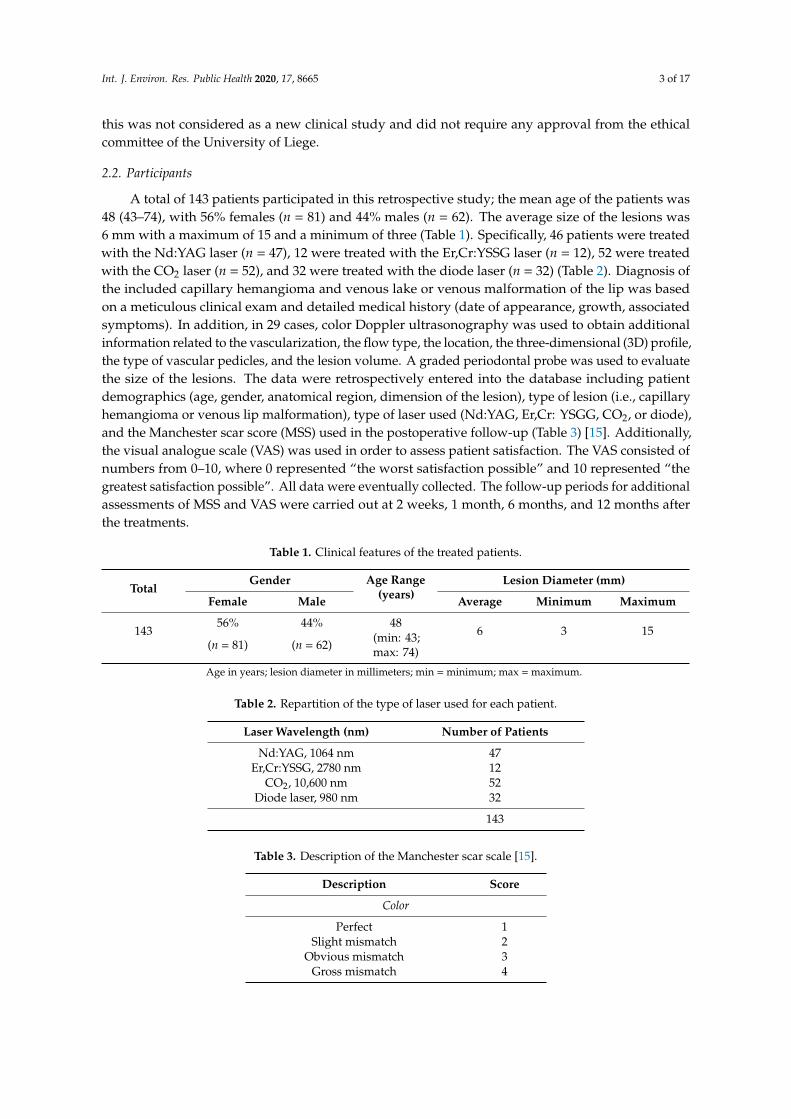
After 2 weeks, the highest values of satisfaction was obtained with the Er,Cr:YSSG laser (8.9 ± 0.23), followed by the diode laser (7.8 ± 1.58), CO2 laser (7.4 ± 0.7), and Nd:YAG laser (7.10 ± 0.86) (Table 6). The difference between the values of each group was statistically significant (p < 0.05). The values significantly increased with follow-up for all groups, attaining the highest satisfactory score (±10) at 6 months. Therefore, it can be considered that, regardless of the wavelength, patients showed satisfaction with regard to the aesthetic outcome within 6 months of follow-up (±10) (Table 6, Figure 10).
Table 6. Visual analogue scale of patient satisfaction at different follow-ups.
Laser Wavelength Visual Analogue at Different Time of Follow-Up 2 Weeks 1 Month 6 Months 12 Months
Er,Cr:YSSG 8.9 ± 0.23 A 9 ± 0.82 A 10 B 10 B CO2 7.4 ± 0.7 C 8.4 ± 0.6 D 10 B 10 B
Diode 7.8 ± 1.58 E 8.85 ± 0.85 A 10 B 10 B Nd:YAG 7.10 ± 0.86 F 8.30 ± 0.36 D 10 B 10 B
Zero represents the worst satisfaction possible, and 10 represents the highest satisfaction possible. Identical superscript letters indicate no statistically significant difference, and different superscript letters indicate a statistically significant difference (p < 0.05).
Figure 9. Global mean values of the Manchester scar scale for the different laser wavelengths and atdifferent follow-ups.
3.2. Recurrence Rate
It was necessary to have a second treatment session in groups treated with the diode and Nd:YAGlasers. Recurrence with obligation to perform a second treatment session was observed in 11% ± 1.4%of cases treated in the diode laser group and 8% ± 0.9% of cases treated in the Nd:YAG laser group. Onthe other hand, in the Er,Cr:YSSG and CO2 groups, no recurrence was noted after the first treatmentsessions (Table 5).
Table 5. Recurrence rate of different surgical procedures.
Laser Wavelength Er,Cr:YSSG(Excision)
CO2(Photo-Vaporization)
Diode(Transmucosal
Photo-Thermo-Coagulation)
Nd:YAG(Transmucosal
Photo-Thermo-Coagulation)
Recurrence rate (%) 0 A 0 A 11% ±1.4% B 8% ± 0.9% C
Identical superscript letters indicate no statistically significant difference and different superscript letters indicate astatistically significant difference (p < 0.05).
3.3. Patient Satisfaction
After 2 weeks, the highest values of satisfaction was obtained with the Er,Cr:YSSG laser (8.9 ± 0.23),followed by the diode laser (7.8 ± 1.58), CO2 laser (7.4 ± 0.7), and Nd:YAG laser (7.10 ± 0.86) (Table 6).The difference between the values of each group was statistically significant (p < 0.05). The valuessignificantly increased with follow-up for all groups, attaining the highest satisfactory score (±10)at 6 months. Therefore, it can be considered that, regardless of the wavelength, patients showedsatisfaction with regard to the aesthetic outcome within 6 months of follow-up (±10) (Table 6, Figure 10).
Table 6. Visual analogue scale of patient satisfaction at different follow-ups.
Laser Wavelength Visual Analogue at Different Time of Follow-Up
2 Weeks 1 Month 6 Months 12 Months
Er,Cr:YSSG 8.9 ± 0.23 A 9 ± 0.82 A 10 B 10 B
CO2 7.4 ± 0.7 C 8.4 ± 0.6 D 10 B 10 B
Diode 7.8 ± 1.58 E 8.85 ± 0.85 A 10 B 10 B
Nd:YAG 7.10 ± 0.86 F 8.30 ± 0.36 D 10 B 10 B
Zero represents the worst satisfaction possible, and 10 represents the highest satisfaction possible. Identicalsuperscript letters indicate no statistically significant difference, and different superscript letters indicate a statisticallysignificant difference (p < 0.05).
Int. J. Environ. Res. Public Health 2020, 17, 8665 11 of 17Int. J. Environ. Res. Public Health 2020, 17, x FOR PEER REVIEW 11 of 17
Visual analogue scale of the patient's satisfaction
2 Wee
ks
4 Wee
ks
6 months
12 m
onths0
2
4
6
8
10
Er,Cr:YSSGCO2Diode (980 nm)Nd:YAG
Time
VAS
(1-
10)
Figure 10. Visual analogue scale of patient satisfaction at different follow-ups. Zero represents the worst satisfaction possible and 10 represents the highest satisfaction possible.
The aesthetic appearance of the scarred treated areas varied depending on the surgery performed at each wavelength (Figures 11–15).
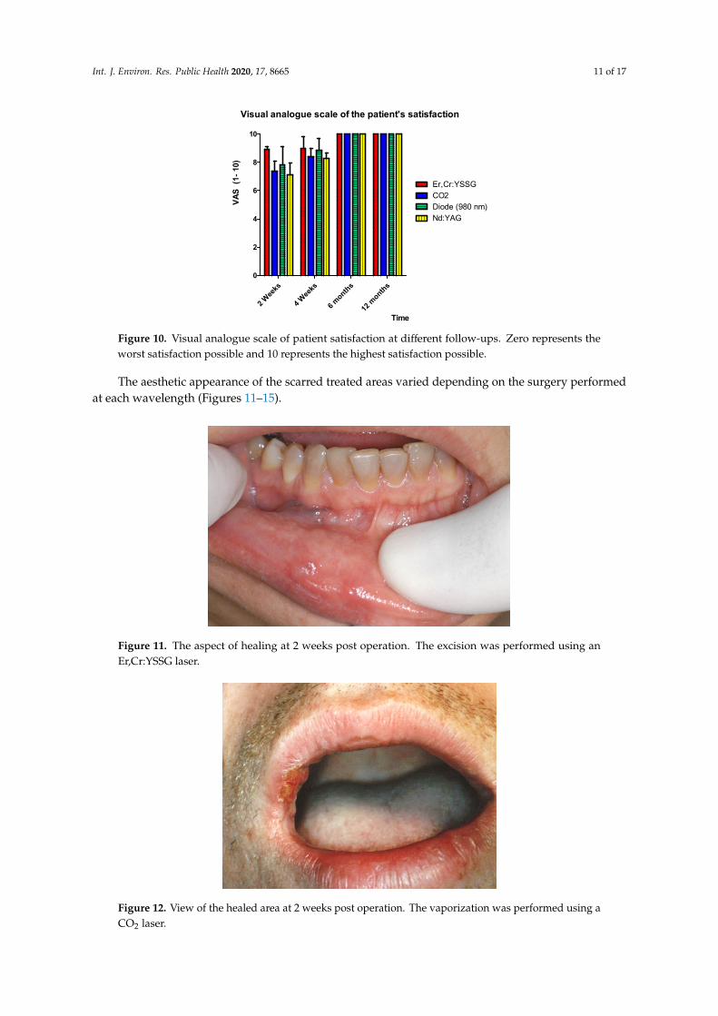
Figure 11. The aspect of healing at 2 weeks post operation. The excision was performed using an Er,Cr:YSSG laser.
Figure 10. Visual analogue scale of patient satisfaction at different follow-ups. Zero represents theworst satisfaction possible and 10 represents the highest satisfaction possible.
The aesthetic appearance of the scarred treated areas varied depending on the surgery performedat each wavelength (Figures 11–15).
Int. J. Environ. Res. Public Health 2020, 17, x FOR PEER REVIEW 11 of 17
Visual analogue scale of the patient's satisfaction
2 Wee
ks
4 Wee
ks
6 months
12 m
onths0
2
4
6
8
10
Er,Cr:YSSGCO2Diode (980 nm)Nd:YAG
Time
VAS
(1-
10)
Figure 10. Visual analogue scale of patient satisfaction at different follow-ups. Zero represents the worst satisfaction possible and 10 represents the highest satisfaction possible.
The aesthetic appearance of the scarred treated areas varied depending on the surgery performed at each wavelength (Figures 11–15).
Figure 11. The aspect of healing at 2 weeks post operation. The excision was performed using an Er,Cr:YSSG laser.
Figure 11. The aspect of healing at 2 weeks post operation. The excision was performed using anEr,Cr:YSSG laser.
Int. J. Environ. Res. Public Health 2020, 17, x FOR PEER REVIEW 11 of 17
Visual analogue scale of the patient's satisfaction
2 Wee
ks
4 Wee
ks
6 months
12 m
onths0
2
4
6
8
10
Er,Cr:YSSGCO2Diode (980 nm)Nd:YAG
Time
VAS
(1-
10)
Figure 10. Visual analogue scale of patient satisfaction at different follow-ups. Zero represents the worst satisfaction possible and 10 represents the highest satisfaction possible.
The aesthetic appearance of the scarred treated areas varied depending on the surgery performed at each wavelength (Figures 11–15).
Figure 11. The aspect of healing at 2 weeks post operation. The excision was performed using an Er,Cr:YSSG laser.
Figure 12. View of the healed area at 2 weeks post operation. The vaporization was performed using aCO2 laser.

Int. J. Environ. Res. Public Health 2020, 17, 8665 12 of 17
Int. J. Environ. Res. Public Health 2020, 17, x FOR PEER REVIEW 12 of 17
Figure 12. View of the healed area at 2 weeks post operation. The vaporization was performed using a CO2 laser.
Figure 13. Clinical aspect of the healed area at 4 weeks post operation. The vaporization was performed using a CO2 laser.
Figure 14. The aesthetic aspect at 6 months post operation. The vaporization was performed using a CO2 laser.
Figure 13. Clinical aspect of the healed area at 4 weeks post operation. The vaporization was performedusing a CO2 laser.
Int. J. Environ. Res. Public Health 2020, 17, x FOR PEER REVIEW 12 of 17
Figure 12. View of the healed area at 2 weeks post operation. The vaporization was performed using a CO2 laser.
Figure 13. Clinical aspect of the healed area at 4 weeks post operation. The vaporization was performed using a CO2 laser.
Figure 14. The aesthetic aspect at 6 months post operation. The vaporization was performed using a CO2 laser. Figure 14. The aesthetic aspect at 6 months post operation. The vaporization was performed using aCO2 laser.

Int. J. Environ. Res. Public Health 2020, 17, 8665 13 of 17
Int. J. Environ. Res. Public Health 2020, 17, x FOR PEER REVIEW 13 of 17
Figure 15. Clinical aspect of healing at 2 weeks post operation. Transmucosal photo-thermo-coagulation was performed using an Nd:>YAG laser.
4. Discussion
The use of laser beams for the management of vascular lesions dates back to the 1960s [16–18]. Back then, ruby and argon lasers were introduced to improve the color of hemangiomas and post-wine stains. However, their nonselective photothermolysis of the targeted tissue resulted in frequent negative postoperative scarring and pigmentation changes [19–22]. After the vast development of lasers and after improvements in the understanding of laser–tissue interactions and laser physics, the treatment of vascular lesions is considered as one of the most common indications of laser [23–27].
This multicentric retrospective study showed that the treatment outcome of capillary hemangioma, venous lake, and venous malformation of the lip with different wavelengths was reported to be successful, and both patients and operators reported satisfaction concerning the aesthetic outcome at 6 months and above of follow-up. In addition, it was revealed that the quality of the scar (the quality of the healing) after 4 weeks was higher in the group treated using the Er,Cr:YSSG and CO2 lasers showing an MSS of 1.16 ± 0.18 and 1.5 ± 0.27, respectively. The aesthetic outcome values obtained with the Er,Cr:YSSG (incision) and CO2 (total photo-vaporization of lesions) lasers were significantly better than those obtained with the diode and the Nd:YAG lasers (transmucosal photo-thermo-coagulation). Consequently, the Er,Cr:YSSG and CO2 lasers gave better scar quality at 2 weeks and 4 weeks after treatment. However, at 6 months and above, all surgical procedures and wavelengths showed similar satisfactory quality of the scar. Furthermore, the aesthetic quality of scars resulting from the diode and Nd:YAG lasers was low until 1 month of follow-up, but gave satisfactory results similar to other wavelengths after 6 months of follow-up.
In this retrospective study, the Manchester scar scale (MSS), proposed in 1998 by Beausang et al. was used because of its detailed and relevant description of the color, texture, size, and margin of the scar and its association with the surrounding tissue [15]. In addition to the MSS, a patient-based visual analogue scale was used to assess the patient’s perception of the healing aspect. Furthermore, it was noted that there is practically no standardized methodology and systematic approach for the assessment of scars and the quality of healing in post-operative surgical sites [28].
The nonsurgical procedure of transmucosal photo-thermo-coagulation applied using the diode and Nd:YAG lasers consists of targeting the chromophores inside the vascular lesions, essentially hemoglobin [29–31]. These chromophores absorb the laser’s energy and convert it into heat, which is transferred to the vessel wall, causing coagulation and vessel closure and, finally, thrombosis of the blood vessels [32]. Concerning photo-vaporization using the CO2 laser, the absorption of photons by the water content of the tumor provokes a sudden local increase in temperature resulting from vaporization of the blood cittern. At the end of the surgery, the mucosal layer covering the blood cittern is totally vaporized, exposing the bottom of the tumor cavity [33–35]. On the other hand, the
Figure 15. Clinical aspect of healing at 2 weeks post operation. Transmucosal photo-thermo-coagulationwas performed using an Nd:>YAG laser.
4. Discussion
The use of laser beams for the management of vascular lesions dates back to the 1960s [16–18].Back then, ruby and argon lasers were introduced to improve the color of hemangiomas and post-winestains. However, their nonselective photothermolysis of the targeted tissue resulted in frequentnegative postoperative scarring and pigmentation changes [19–22]. After the vast development oflasers and after improvements in the understanding of laser–tissue interactions and laser physics, thetreatment of vascular lesions is considered as one of the most common indications of laser [23–27].
This multicentric retrospective study showed that the treatment outcome of capillary hemangioma,venous lake, and venous malformation of the lip with different wavelengths was reported to besuccessful, and both patients and operators reported satisfaction concerning the aesthetic outcome at6 months and above of follow-up. In addition, it was revealed that the quality of the scar (the qualityof the healing) after 4 weeks was higher in the group treated using the Er,Cr:YSSG and CO2 lasersshowing an MSS of 1.16 ± 0.18 and 1.5 ± 0.27, respectively. The aesthetic outcome values obtained withthe Er,Cr:YSSG (incision) and CO2 (total photo-vaporization of lesions) lasers were significantly betterthan those obtained with the diode and the Nd:YAG lasers (transmucosal photo-thermo-coagulation).Consequently, the Er,Cr:YSSG and CO2 lasers gave better scar quality at 2 weeks and 4 weeks aftertreatment. However, at 6 months and above, all surgical procedures and wavelengths showed similarsatisfactory quality of the scar. Furthermore, the aesthetic quality of scars resulting from the diodeand Nd:YAG lasers was low until 1 month of follow-up, but gave satisfactory results similar to otherwavelengths after 6 months of follow-up.
In this retrospective study, the Manchester scar scale (MSS), proposed in 1998 by Beausang et al.was used because of its detailed and relevant description of the color, texture, size, and margin ofthe scar and its association with the surrounding tissue [15]. In addition to the MSS, a patient-basedvisual analogue scale was used to assess the patient’s perception of the healing aspect. Furthermore,it was noted that there is practically no standardized methodology and systematic approach for theassessment of scars and the quality of healing in post-operative surgical sites [28].
The nonsurgical procedure of transmucosal photo-thermo-coagulation applied using the diodeand Nd:YAG lasers consists of targeting the chromophores inside the vascular lesions, essentiallyhemoglobin [29–31]. These chromophores absorb the laser’s energy and convert it into heat, which istransferred to the vessel wall, causing coagulation and vessel closure and, finally, thrombosis of theblood vessels [32]. Concerning photo-vaporization using the CO2 laser, the absorption of photonsby the water content of the tumor provokes a sudden local increase in temperature resulting from

Int. J. Environ. Res. Public Health 2020, 17, 8665 14 of 17
vaporization of the blood cittern. At the end of the surgery, the mucosal layer covering the bloodcittern is totally vaporized, exposing the bottom of the tumor cavity [33–35]. On the other hand, theEr,Cr:YSSG laser procedure consists of complete excision of the vascular lesion, followed by coagulationof the bottom of the wound for primary hemostasis [36,37].
A large number of approaches are described in literature for the management of vascularlesions [8,38–40]. These methods principally depend on the type, severity, size, location, and possiblecomplications of the vascular lesion [14,38–43].
John et al. conducted a study on the treatment of venous lesions of the lips with a long-pulsedNd:YAG laser. The study included 31 patients with venous lesions of the lip [44]. The parametersused were spot size depending on the size of the lesion, variable energy density with an average of 80J/cm2, and pulse width of 20. They concluded that the treatment was effective with 87% of patientshaving no recurrence and one patient having a small, contracted scar [44]. In contrast to John et al., therecurrence rate in our study was lower (only ±8%) with the Nd:YAG laser and no contracted scar wasseen. Scherer et al. also studied the use of the Nd:YAG laser for venous malformations of the face,including 146 patients [45]. In their study, the Nd:YAG laser was used alone if the lesion was consideredrelatively small or followed by a surgical excision if the lesion was considered deep. In addition, aretrospective study by Mungnirandr et al. assessed the safety and effectiveness of the Nd:YAG laserfor venous malformation in the oral cavity [46]. The study included 10 children with inoperable VMand established that the Nd:YAG laser is a promising alternative treatment in pediatric patients withvenous malformations in the oral cavity, whereby oral complications mostly seen involved mild tomoderate scarring [46]. Interestingly, the treatment protocol used in their study included the use ofthe Nd:YAG in contact mode, in noncontact mode, and using interstitial techniques. The choice ofeach technique depended essentially on the size and depth of the lesions. Specifically, the contacttechnique involved placement of the fiber-optic end of the laser handpiece directly against the mucosalsurface during laser irradiation. The noncontact process, however, involved the laser beam beingemitted through a clear ice cube for purposes of minimizing epidermal injury [47]. The interstitialtechnique required the insertion of the fiber-optic end of the handpiece during laser irradiation insidethe lesion [46].
It can be found in the literature that most treatments for vascular lesions were performed withthe Nd:YAG laser, albeit using different wavelengths. Del Pozo et al. conducted a study with aCO2 laser on five cases of venous malformations with lip involvement [47]. The study revealed thatvaporization with the carbon dioxide laser was able to flatten the surface of the lip and, therefore,was considered an effective palliative treatment of lip venous malformations [47]. Compared to ourstudy, the treatment protocol with the CO2 laser was different. Del Pozo et al. irradiated the treatmentarea with several passages in contact mode to achieve a contraction and immediate flattering ofthe lesion surface. However, in our study, noncontact mode was used in order to generate heat tovaporize the water inside the vascular lesion; then, the external layer of the mucosa was vaporizedand removed in focused mode until complete exposure of the lesion bottom. Unlike our study, therewas no recurrence noted in Del Pozo’s study; however, their purpose was not to completely removethe venous malformation but to shrink its size. Bacci et al. performed a retrospective study on theuse of a diode laser to treat small oral vascular malformations. The study included 59 patients, whichshowed that the only complication related to the surgery was modest pain. This pain 30 days after thereduction of the lesions, however, was considered as excellent or good in 52 cases and fair or poor inseven cases, whereas six patients required a second diode laser application [48]. The study revealedthat the diode laser is considered acceptable for the treatment of small oral venous malformations andvenous lakes with shorter postoperative complications and shorter operating times when compared tothe conventional scalpel surgery [48]. Furthermore, Angiero et al. conducted a study on head and neckvascular lesions in pediatric patients treated with an endolesional 980 nm diode laser [48]. The studyincluded 160 patients with hemangiomas, 50 with vascular malformations, and 40 with lymphaticmalformations. The treatment results were analyzed by evaluating the decrease in lesion size and its

Int. J. Environ. Res. Public Health 2020, 17, 8665 15 of 17
complete clinical disappearance. All patients had a complete resolution of the vascular lesion exceptfor 38, for which an additional session was required [48].
Topical therapy such as timolol, high-potency topical corticosteroids, imiquimod, and becaplermingel are also recommended for superficial hemangiomas [40–42]. In fact, timolol has emerged as thepreferred topical treatment. On the other hand, Lee et al. concluded, in a study on 11 patients with liphemangioma, that the use of Dermabond after surgical excision prevents wound contamination andyields acceptable aesthetic results [43].
Since different operators used the Manchester scar scale (MSS) to assess the aesthetic outcome, apossible limitation of our study was the slight subjectivity obtained when different operators usedthe MSS.
Our retrospective study compared four laser wavelengths in the management of venousmalformation and capillary hemangioma of the lip with three different surgical approaches (excision,photo-vaporization, and transmucosal photo-thermo-coagulation). Further studies are necessary tocompare the aesthetic outcome, the recurrence rates, and the possible postoperative complications ofdifferent protocols.
The null hypothesis was rejected because no statistical difference in the aesthetic outcome atthe end of follow-up was obtained by the three different surgical procedures, i.e., transmucosalphoto-thermo-coagulation (Nd:YAG and diode lasers), photo-vaporization (CO2 laser), and surgicalexcision (Er,Cr:YSGG laser).
5. Conclusions
Our retrospective study showed that laser-assisted aesthetic treatment of vascular lesions of thelips can be considered effective regardless of the wavelength used (Er,Cr:YSGG, CO2, Nd:YAG, anddiode lasers) or the treatment procedure (transmucosal photo-thermo-coagulation, photo-vaporization,and surgical excision). There was no significant difference in patient and practitioner satisfactionwith aesthetic outcome at 6 months follow-up. Furthermore, the treatments of lip vascular lesionsperformed using Er,Cr:YSGG and CO2 lasers did not show any recurrence during the 12 months offollow-up, while recurrence rates of 11% ± 1.4% and 8% ± 0.9% were seen in the diode and Nd:YAGgroups, respectively.
Author Contributions: Conceptualization, S.N.; methodology, S.N., M.E.M., P.V., and A.V.; investigation, A.N.,M.N., P.V., K.G.-L., and J.A.-D.; writing—original draft preparation, S.N. and M.E.M.; writing—review andediting, S.N., M.N., and A.N.; supervision, S.N. and A.V. All authors read and agreed to the published version ofthe manuscript.
Funding: This research received no external funding.
Conflicts of Interest: Page: 15The authors declare no conflict of interest.
References
1. Jackson, I.T.; Carreño, R.; Potparic, Z.; Hussain, K. Hemangiomas, vascular malformations, and lymphovenousmalformations: Classification and methods of treatment. Plast. Reconstr. Surg. 1993, 91, 1216–1230. [CrossRef][PubMed]
2. Merrow, A.C.; Gupta, A.; Patel, M.N.; Adams, D.M. 2014 Revised classification of vascular lesions from theInternational Society for the Study of Vascular Anomalies: Radiologic-pathologic update. Radiographics 2016,36, 1494–1516. [CrossRef] [PubMed]
3. Cox, J.A.; Bartlett, E.; Lee, E.I. Vascular malformations: A review. Semin. Plast. Surg. 2014, 28, 58. [CrossRef][PubMed]
4. Fernandes, D.T.; Hebling, E.; Santos-Silva, A.R.; Lopes, M.A.A. Series of 33 older patients with lip venouslake treated by sclerotherapy. Int. J. Derm. 2019, 59, 42–46. [CrossRef]
5. Ernemann, U.; Kramer, U.; Miller, S.; Bisdas, S.; Rebmann, H.; Breuninger, H.; Zwick, C.; Hoffmann, J. Currentconcepts in the classification, diagnosis and treatment of vascular anomalies. Eur. J. Radiol. 2010, 75, 2–11.[CrossRef]

Int. J. Environ. Res. Public Health 2020, 17, 8665 16 of 17
6. Vesnaver, A.; Dovs, D.A. Treatment of vascular lesions in the head and neck using Nd: YAG laser.J. Cranio-Maxillofac. Surg. 2006, 34, 17–24. [CrossRef]
7. Maguiness, S.M.; Frieden, I.J. Current management of infantile hemangiomas. Inseminars Cutan. Med. Surg.2010, 30, 106–114. [CrossRef]
8. Nouri, K. Lasers in Dermatology and Medicine: Dermatologic Applications; Springer: Heidelberg, Germany, 2018.9. Almutairi, N.; Alshaiji, J. Lasers for Vascular Lesions. Pediatric chapter 17, Book Editors: Keyvan Nouri, Latanya
Benjamin, Jasem Alshaiji, Jan Izakovic, Pediatric Dermatologic Surgery; 2019 John Wiley & Sons Ltd.: Chichester,UK, 2019; pp. 189–196.
10. Voyatzi, M.; Grammatikopoulou, J.; Karlafti, E.; Mavroudi, A.; Lamprou, F.; Kypri, G.; Tsirmigka, M. Effcacyof lasers on vascular lesions and rejuvenation. SM Dermatol. J. 2017, 3, 1009. [CrossRef]
11. Bordin-Aykroyd, S.; Dias, R.B.; Lynch, E. Laser-tissue interaction. EC Dent. Sci. 2019, 18, 2303–2308.12. Ash, C.; Dubec, M.; Donne, K.; Bashford, T. Effect of wavelength and beam width on penetration in light-tissue
interaction using computational methods. Lasers Med. Sci. 2017, 32, 1909–1918. [CrossRef]13. Parker, S.; Cronshaw, M.; Anagnostaki, E.; Mylona, V.; Lynch, E.; Grootveld, M. Current Concepts of
Laser–Oral Tissue Interaction. Dent. J. 2020, 8, 61. [CrossRef] [PubMed]14. Dover, J.S.; Arndt, K.A. New approaches to the treatment of vascular lesions. Lasers Surg. Med. 2000, 26,
158–163. [CrossRef]15. Beausang, E.; Floyd, H.; Dunn, K.W.; Orton, C.I.; Ferguson, M.W. A new quantitative scale for clinical scar
assessment. Plast. Reconstr. Surg. 1998, 102, 1954–1961. [CrossRef] [PubMed]16. Goldman, L. Effects of new laser systems on the skin. Arch. Dermatol. 1973, 108, 385–390. [CrossRef]17. Goldman, L.; Dreffer, R.; Rockwell, R.J., Jr.; Perry, E. Treatment of portwine marks by an argon laser.
J. Dermatol. Surg. Oncol. 1976, 2, 385–388. [CrossRef] [PubMed]18. Goldman, L.; Wilson, R.G. Treatment of basal cell epithelioma by laser radiation. JAMA 1964, 189, 773–775.
[CrossRef] [PubMed]19. Apfelberg, D.B.; Maser, M.R.; Lash, H. Extended clinical use of the argon laser for cutaneous lesions.
Arch. Dermatol. 1979, 115, 719–721. [CrossRef] [PubMed]20. Apfelberg, D.B.; Kosek, J.; Maser, M.R.; Lash, H. Histology of port wine stains following argon laser treatment.
Br. J. Plast. Surg. 1979, 32, 232–237. [CrossRef]21. Cosman, B. Clinical experience in the laser therapy of port wine stains. Lasers Surg. Med. 1980, 1, 133–152.
[CrossRef]22. Riggle, G.C.; Hoye, R.C.; Ketcham, A.S. Laser effects on normal and tumor tissue. In Laser Applications in
Medicine and Biology; Springer: Heidelberg, Germany, 1971; pp. 35–65.23. Nelson, J.S.; Jia, W.; Phung, T.L.; Mihm, M.C., Jr. Observations on enhanced port wine stain blanching
induced by combined pulsed dye laser and rapamycin administration. Lasers Surg. Med. 2011, 43, 939.[CrossRef]
24. Bhatnagar, A.; Jindal, M.K. Excision of Capillary Hemangioma by Diode Laser. EC Dent. Sci. 2017, 13, 07–12.25. Azma, E.; Razaghi, M. Laser Treatment of Oral and Maxillofacial Hemangioma. J. Lasers Med. Sci. 2018,
9, 228. [CrossRef] [PubMed]26. Capodiferro, S.; Limongelli, L.; Tempesta, A.; Maiorano, E.; Favia, G. Diode laser treatment of venous lake of
the lip. Clin. Case Rep. 2018, 6, 1923–1924. [CrossRef] [PubMed]27. Hartmann, F.; Lockmann, A.; Himpel, O.; Kühnle, I.; Hensen, J.; Schön, M.P.; Thoms, K.M. Combination
therapy of oral propranolol and combined Nd: YAG/pulsed dye laser therapy in infantile hemangiomas: Aretrospective analysis of 48 treated hemangiomas in 30 children. JDDG J. Der Dtsch. Dermatol. Ges. 2020, 18,984–993.
28. Fearmonti, R.; Bond, J.; Erdmann, D.; Levinson, H. A review of scar scales and scar measuring devices.Eplasty 2010, 10, 354–362.
29. Glaessl, A.; Schreyer, A.G.; Wimmershoff, M.B.; Landthaler, M.; Feuerbach, S.; Hohenleutner, U. Laser surgicalplanning with magnetic resonance imaging–based 3-dimensional reconstructions for intralesional Nd: YAGlaser therapy of a venous malformation of the neck. Arch. Dermatol. 2001, 137, 1331–1335. [CrossRef]
30. Acland, K.M.; Barlow, R.J. Lasers for the dermatologist. Br. J. Dermatol. 2000, 143, 244–255. [CrossRef]31. Wanitphakdeedecha, R.; Thanomkitti, K.; Bunyaratavej, S.; Manuskiatti, W. Efficacy and safety of 1064-nm
Nd: YAG laser in treatment of onychomycosis. J. Dermatol. Treat. 2016, 27, 75–79. [CrossRef]

Int. J. Environ. Res. Public Health 2020, 17, 8665 17 of 17
32. Sumian, C.C.; Pitre, F.B.; Gauthier, B.E.; Bouclier, M.; Mordon, S.R. Laser skin resurfacing using a frequencydoubled Nd: YAG laser after topical application of an exogenous chromophore. Lasers Surg. Med. 1999, 25,43–50. [CrossRef]
33. Apfelberg, D.B.; Maser, M.R.; Lash, H.; White, D.N. Benefits of the CO2 laser in oral hemangioma excision.Plast. Reconstr. Surg. 1985, 75, 46–50. [CrossRef]
34. McCaffrey, T.V.; Cortese, D.A. Neodymium: YAG laser treatment of subglottic hemangioma. Otolaryngol. HeadNeck Surg. 1986, 94, 382–384. [CrossRef] [PubMed]
35. Niccoli Filho, W.D.; Americo, M.G.; Guimarães Filho, R.; Rodrigues, N.A.S. Lip hemangioma removed withCO2 laser: A case report. Braz. J. Oral Sci. 2002, 1, 89–91.
36. Suh, J.J.; Lee, J.; Park, J.C.; Lim, H.C. Lip Repositioning Surgery Using an Er, Cr: YSGG Laser: A Case Series.Int. J. Periodontics Restor. Dent. 2020, 40, 437–444. [CrossRef]
37. Cercadillo-Ibarguren, I.; España Tost, A.J.; Arnabat Domínguez, J.; Valmaseda Castellón, E.; Berini Aytés, L.;Gay Escoda, C. Histologic evaluation of thermal damage produced on soft tissues by CO2, Er, Cr: YSGG anddiode lasers. Med. Oral Patol. Oral Cir. Bucal. 2010, 15, 912–918. [CrossRef] [PubMed]
38. Vazquez, T.; Forouzandeh, M.; Gurnani, P.; Akhtar, S.; Nouri, K. Cutaneous vascular lesions in the pediatricpopulation: A review of laser surgery applications and lesion-specific device parameters. Lasers Med. Sci.2020, 35, 1681–1687. [CrossRef]
39. Buehler, D.; Billings, S.D. Cutaneous vascular lesions. In Soft Tissue Tumors of the Skin; Springer: New York,NY, USA, 2019; pp. 235–306.
40. Mannschreck, D.B.; Huang, A.H.; Lie, E.; Psoter, K.; Puttgen, K. Topical timolol as adjunct therapy to shortenoral propranolol therapy for infantile hemangiomas. Pediatric Dermatol. 2019, 36, 283–289. [CrossRef]
41. Polites, S.F.; Watanabe, M.; Crafton, T.; Jenkins, T.M.; Alvarez-Allende, C.R.; Hammill, A.M.; Dasgupta, R.Surgical resection of infantile hemangiomas following medical treatment with propranolol versuscorticosteroids. J. Pediatric Surg. 2019, 54, 740–743. [CrossRef]
42. Park, K.H.; Jang, Y.H.; Chung, H.Y.; Lee, W.J.; Kim, D.W.; Lee, S.J. Topical timolol maleate 0.5% for infantilehemangioma; it’s effectiveness and/or adjunctive pulsed dye laser—single center experience of 102 cases inKorea. J. Dermatol. Treat. 2015, 26, 389–391. [CrossRef]
43. Chang, J.W.; Cho, K.S.; Heo, W.; Lee, J.H. (CONSORT) Wound closure using Dermabond after excision ofhemangioma on the lip. Medicine 2019, 98, e15342. [CrossRef]
44. John, H.E.; Phen, H.S.; Mahaffey, P.J. Treatment of venous lesions of the lips and perioral area with along-pulsed Nd: YAG laser. Br. J. Oral Maxillofac. Surg. 2016, 54, 376–378. [CrossRef]
45. Scherer, K.; Waner, M. Nd:YAG lasers (1064 nm) in the treatment of venous malformations of the face andneck: Challenges and benefits. Lasers Med. Sci. 2007, 22, 119–126. [CrossRef] [PubMed]
46. Mungnirandr, A.; Nuntasunti, W.; Manuskiatti, W. Neodymium-doped yttrium aluminium garnet lasertreatment of pediatric venous malformation in the oral cavity. Dermatol. Surg. 2016, 42, 875–879. [CrossRef][PubMed]
47. Del Pozo, J.; Martínez-González, C.; Verea, M.M.; Fernández-Torres, R.; Fonseca, E. Venous malformationswith lip involvement: Palliative treatment with carbon dioxide laser vaporization in five cases. J. Cosmet.Laser Ther. 2009, 11, 14–18. [CrossRef] [PubMed]
48. A Bacci, C.; Sacchetto, L.; Zanette, G.; Sivolella, S. Diode laser to treat small oral vascular malformations: Aprospective case series study. Lasers Surg. Med. 2018, 50, 111–116. [CrossRef]
Publisher’s Note: MDPI stays neutral with regard to jurisdictional claims in published maps and institutionalaffiliations.
© 2020 by the authors. Licensee MDPI, Basel, Switzerland. This article is an open accessarticle distributed under the terms and conditions of the Creative Commons Attribution(CC BY) license (http://creativecommons.org/licenses/by/4.0/).